Embed Size (px)
Citation preview

Instructions for use
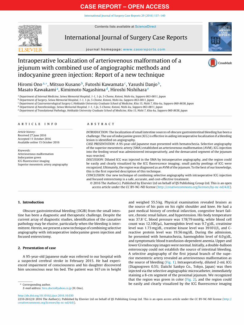
Title Intraoperative localization of arteriovenous malformation of a jejunum with combined use of angiographic methods andindocyanine green injection : Report of a new technique
Author(s) Ono, Hiromi; Kusano, Mitsuo; Kawamata, Futoshi; Danjo, Yasushi; Kawakami, Masato; Nagashima, Kimimoto;Nishihara, Hiroshi
Citation International journal of surgery case reports, 29, 137-140https://doi.org/10.1016/j.ijscr.2016.10.030
Issue Date 2016
Doc URL http://hdl.handle.net/2115/64566
Rights(URL) http://creativecommons.org/licenses/by-nc-nd/4.0/
Type article
File Information 1-s2.0-S2210261216304278-main.pdf
Hokkaido University Collection of Scholarly and Academic Papers : HUSCAP
CASE REPORT – OPEN ACCESSInternational Journal of Surgery Case Reports 29 (2016) 137–140
Contents lists available at ScienceDirect
International Journal of Surgery Case Reports
journa l h omepage: www.caserepor ts .com
Intraoperative localization of arteriovenous malformation of ajejunum with combined use of angiographic methods andindocyanine green injection: Report of a new technique
Hiromi Onoa,∗, Mitsuo Kusanob, Futoshi Kawamatac, Yasushi Danjob,Masato Kawakamia, Kimimoto Nagashimad, Hiroshi Nishiharae
a Department of Internal Medicine, Seiwa Memorial Hospital, 1-1, 1-Jo, 5-Chome, Kotoni, Nishi-ku, Sapporo 063-0811, Japanb Department of Surgery, Seiwa Memorial Hospital, 1-1, 1-Jo, 5-Chome, Kotoni, Nishi-ku, Sapporo 063-0811, Japanc Department of Gastroenterological Surgery I, Hokkaido University Graduate School of Medicine, Kita 15, Nishi 7, Kita-ku, Sapporo 060-8638, Japand Department of Anesthesiology, Seiwa Memorial Hospital, 1-1, 1-Jo, 5-Chome, Kotoni, Nishi-ku, Sapporo 063-0811, Japane Department of Translational Pathology, Hokkaido University Graduate School of Medicine, Kita 15, Nishi 7, Kita-ku, Sapporo 060-8638, Japan
a r t i c l e i n f o
Article history:Received 27 June 2016Accepted 11 October 2016Available online 15 October 2016
Keywords:Arteriovenous malformationIndocyanine greenICG fluorescence imagingSuperior mesenteric artery angiography
a b s t r a c t
INTRODUCTION: The localization of small intestine sources of obscure gastrointestinal bleeding has been achallenge. The use of indocyanine green (ICG) is effective in aiding intraoperative localization if a bleedinglesion is identified on angiography.CASE PRESENTATION: A 95-year-old Japanese man presented with hematochezia. Selective angiographyof the superior mesenteric artery (SMA) established an arteriovenous malformation (AVM). ICG injectioninto the feeding vessel was administered intraoperatively, and the demarcated segment of the jejunumwas resected.DISCUSSION: Diluted ICG was injected in the SMA by intraoperative angiography, and the region couldbe easily and clearly visualized by the ICG fluorescence imaging; small patchy poolings of ICG wererecognized. Ultimately, the region was diagnosed as an AVM of the jejunum. To the best of our knowledge,this is the first reported description of this technique.CONCLUSION: Our new technique of combining selective angiography with intraoperative ICG injectionand focused enterectomy is a safe, accurate, and cost-effective treatment.
© 2016 The Author(s). Published by Elsevier Ltd on behalf of IJS Publishing Group Ltd. This is an openaccess article under the CC BY-NC-ND license (http://creativecommons.org/licenses/by-nc-nd/4.0/).
1. Introduction
Obscure gastrointestinal bleeding (OGIB) from the small intes-tine has been a diagnostic and therapeutic challenge. Despite thecurrent array of diagnostic studies, identification of the causativepathology may be elusive, particularly when the bleeding is inter-mittent. Herein, we present a new technique of combining selectiveangiography with intraoperative indocyanine green injection andfocused enterectomy.
2. Presentation of case
A 95-year-old Japanese male was referred to our hospital witha suspected cerebral stroke in February 2015. He had experi-enced impairment of consciousness and his daughter discoveredhim unconscious near his bed. The patient was 167 cm in height
∗ Corresponding author.E-mail address: hiro [email protected] (H. Ono).
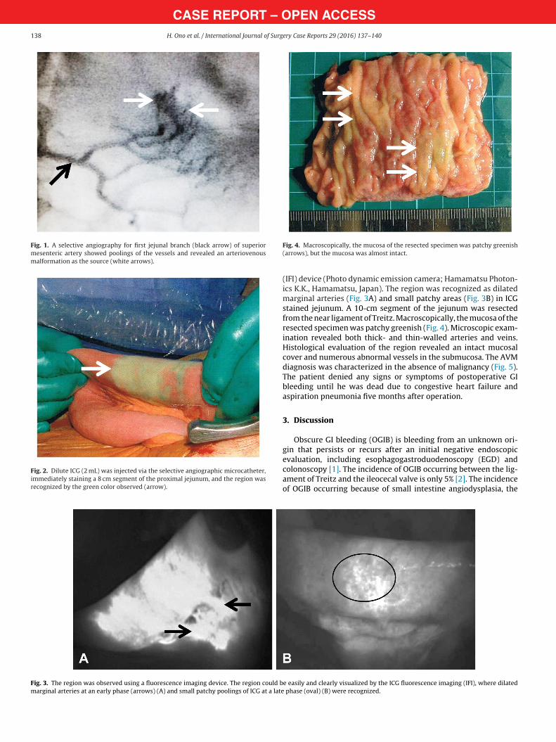
and weighed 55.5 kg. Physical examination revealed bruises asthe source of his pain on his right shoulder and knee. He had apast medical history of cerebral infarction, congestive heart fail-ure, chronic renal failure, and hypertension. His body temperaturewas 37.8 ◦C, blood pressure was 178/79 mmHg, white blood cellcount was 12,100/�L, haemoglobin level was 9.7 g/dL, creatininelevel was 1.73 mg/dL, creatine kinase level was 3910 U/L, and C-reactive protein level was 19.56 mg/dL. During the admission,he presented with hematochezia, haemoglobin level of 6.0 g/dL,and symptomatic blood transfusion-dependent anemia. Upper andlower GI endoscopy images were normal. Initially, a double-balloonenteroscopy could not establish the source of intestinal bleeding.A selective angiography of the first jejunal branch of the supe-rior mesenteric artery revealed an arteriovenous malformation asthe source of bleeding (Fig. 1). Intraoperatively, diluted 2-mL ICG(Diagnogreen 0.5%; Daiichi Sankyo Co., Tokyo, Japan) was theninjected via the selective angiographic microcatheter, immediatelystaining a 8-cm segment of the proximal jejunum. We recognizedthat the region was green in color (Fig. 2), and the region couldbe easily and clearly visualized by the ICG fluorescence imaging
http://dx.doi.org/10.1016/j.ijscr.2016.10.0302210-2612/© 2016 The Author(s). Published by Elsevier Ltd on behalf of IJS Publishing Group Ltd. This is an open access article under the CC BY-NC-ND license (http://creativecommons.org/licenses/by-nc-nd/4.0/).
CASE REPORT – OPEN ACCESS138 H. Ono et al. / International Journal of Surgery Case Reports 29 (2016) 137–140
Fig. 1. A selective angiography for first jejunal branch (black arrow) of superiormesenteric artery showed poolings of the vessels and revealed an arteriovenousmalformation as the source (white arrows).
Fig. 2. Dilute ICG (2 mL) was injected via the selective angiographic microcatheter,immediately staining a 8 cm segment of the proximal jejunum, and the region wasrecognized by the green color observed (arrow).
Fig. 4. Macroscopically, the mucosa of the resected specimen was patchy greenish(arrows), but the mucosa was almost intact.
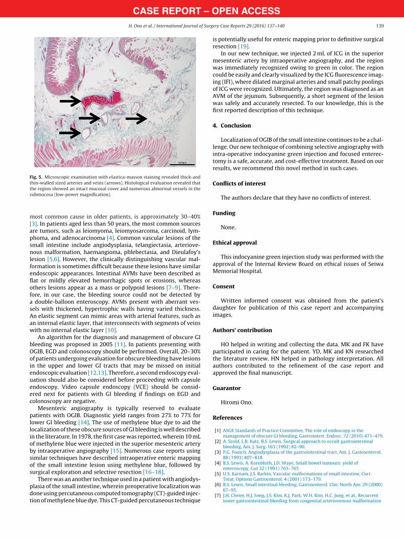
(IFI) device (Photo dynamic emission camera; Hamamatsu Photon-ics K.K., Hamamatsu, Japan). The region was recognized as dilatedmarginal arteries (Fig. 3A) and small patchy areas (Fig. 3B) in ICGstained jejunum. A 10-cm segment of the jejunum was resectedfrom the near ligament of Treitz. Macroscopically, the mucosa of theresected specimen was patchy greenish (Fig. 4). Microscopic exam-ination revealed both thick- and thin-walled arteries and veins.Histological evaluation of the region revealed an intact mucosalcover and numerous abnormal vessels in the submucosa. The AVMdiagnosis was characterized in the absence of malignancy (Fig. 5).The patient denied any signs or symptoms of postoperative GIbleeding until he was dead due to congestive heart failure andaspiration pneumonia five months after operation.
3. Discussion
Obscure GI bleeding (OGIB) is bleeding from an unknown ori-gin that persists or recurs after an initial negative endoscopicevaluation, including esophagogastroduodenoscopy (EGD) andcolonoscopy [1]. The incidence of OGIB occurring between the lig-ament of Treitz and the ileocecal valve is only 5% [2]. The incidenceof OGIB occurring because of small intestine angiodysplasia, the
Fig. 3. The region was observed using a fluorescence imaging device. The region could be easily and clearly visualized by the ICG fluorescence imaging (IFI), where dilatedmarginal arteries at an early phase (arrows) (A) and small patchy poolings of ICG at a late phase (oval) (B) were recognized.
CASE REPORT – OPEN ACCESSH. Ono et al. / International Journal of Surgery Case Reports 29 (2016) 137–140 139
Fig. 5. Microscopic examination with elastica-masson staining revealed thick-andthin-walled sized arteries and veins (arrows). Histological evaluation revealed thatthe region showed an intact mucosal cover and numerous abnormal vessels in thesubmucosa (low-power magnification).
most common cause in older patients, is approximately 30–40%[3]. In patients aged less than 50 years, the most common sourcesare tumors, such as leiomyoma, leiomyosarcoma, carcinoid, lym-phoma, and adenocarcinoma [4]. Common vascular lesions of thesmall intestine include angiodysplasia, telangiectasia, arteriove-nous malformation, haemangioma, phlebectasia, and Dieulafoy’slesion [5,6]. However, the clinically distinguishing vascular mal-formation is sometimes difficult because these lesions have similarendoscopic appearances. Intestinal AVMs have been described asflat or mildly elevated hemorrhagic spots or erosions, whereasothers lesions appear as a mass or polypoid lesions [7–9]. There-fore, in our case, the bleeding source could not be detected bya double-balloon enteroscopy. AVMs present with aberrant ves-sels with thickened, hypertrophic walls having varied thickness.An elastic segment can mimic areas with arterial features, such asan internal elastic layer, that interconnects with segments of veinswith no internal elastic layer [10].
An algorithm for the diagnosis and management of obscure GIbleeding was proposed in 2005 [11]. In patients presenting withOGIB, EGD and colonoscopy should be performed. Overall, 20–30%of patients undergoing evaluation for obscure bleeding have lesionsin the upper and lower GI tracts that may be missed on initialendoscopic evaluation [12,13]. Therefore, a second endoscopy eval-uation should also be considered before proceeding with capsuleendoscopy. Video capsule endoscopy (VCE) should be consid-ered next for patients with GI bleeding if findings on EGD andcolonoscopy are negative.
Mesenteric angiography is typically reserved to evaluatepatients with OGIB. Diagnostic yield ranges from 27% to 77% forlower GI bleeding [14]. The use of methylene blue dye to aid thelocalization of these obscure sources of GI bleeding is well describedin the literature. In 1978, the first case was reported, wherein 10 mLof methylene blue were injected in the superior mesenteric arteryby intraoperative angiography [15]. Numerous case reports usingsimilar techniques have described intraoperative enteric mappingof the small intestine lesion using methylene blue, followed bysurgical exploration and selective resection [16–18].
There was an another technique used in a patient with angiodys-plasia of the small intestine, wherein preoperative localization wasdone using percutaneous computed tomography (CT)-guided injec-tion of methylene blue dye. This CT-guided percutaneous technique
is potentially useful for enteric mapping prior to definitive surgicalresection [19].
In our new technique, we injected 2 mL of ICG in the superiormesenteric artery by intraoperative angiography, and the regionwas immediately recognized owing to green in color. The regioncould be easily and clearly visualized by the ICG fluorescence imag-ing (IFI), where dilated marginal arteries and small patchy poolingsof ICG were recognized. Ultimately, the region was diagnosed as anAVM of the jejunum. Subsequently, a short segment of the lesionwas safely and accurately resected. To our knowledge, this is thefirst reported description of this technique.
4. Conclusion
Localization of OGIB of the small intestine continues to be a chal-lenge. Our new technique of combining selective angiography withintra-operative indocyanine green injection and focused enterec-tomy is a safe, accurate, and cost-effective treatment. Based on ourresults, we recommend this novel method in such cases.
Conflicts of interest
The authors declare that they have no conflicts of interest.
Funding
None.
Ethical approval
This indocyanine green injection study was performed with theapproval of the Internal Review Board on ethical issues of SeiwaMemorial Hospital.
Consent
Written informed consent was obtained from the patient’sdaughter for publication of this case report and accompanyingimages.
Authors’ contribution
HO helped in writing and collecting the data. MK and FK haveparticipated in caring for the patient. YD, MK and KN researchedthe literature review. HN helped in pathology interpretation. Allauthors contributed to the refinement of the case report andapproved the final manuscript.
Guarantor
Hiromi Ono.
References
[1] ASGE Standards of Practice Committee, The role of endoscopy in themanagement of obscure GI bleeding, Gastrointest. Endosc. 72 (2010) 471–479.
[2] A. Szold, L.B. Katz, B.S. Lewis, Surgical approach to occult gastrointestinalbleeding, Am. J. Surg. 163 (1992) 82–90.
[3] P.G. Foutch, Angiodysplasia of the gastrointestinal tract, Am. J. Gastroenterol.88 (1993) 807–818.
[4] B.S. Lewis, A. Kornbluth, J.D. Waye, Small bowel tumours: yield ofenteroscopy, Gut 32 (1991) 763–765.
[5] U.S. Karnam, J.S. Barkin, Vascular malformations of small intestine, Curr.Treat. Options Gastroenterol. 4 (2001) 173–179.
[6] B.S. Lewis, Small intestinal bleeding, Gastroenterol. Clin. North Am. 29 (2000)67–95.
[7] J.H. Cheon, H.J. Song, J.S. Kim, K.J. Park, W.H. Kim, H.C. Jung, et al., Recurrentlower gastrointestinal bleeding from congenital arteriovenous malformation
CASE REPORT – OPEN ACCESS140 H. Ono et al. / International Journal of Surgery Case Reports 29 (2016) 137–140
in the terminal ileum mimicking intestinal varicosis: a case report, J. KoreanMed. Sci. 22 (2007) 746–749.
[8] L. Maeng, K.Y. Choi, A. Lee, C.S. Kang, K.M. Kim, Polypoid arteriovenousmalformation of colon mimicking inflammatory fibroid polyp, J.Gastroenterol. 39 (2004) 575–578.
[9] M.L. Krinsky, M.E. Robert, J.C. Garcia, J.R. Korzenik, M. Topazian, Polypoidvascular malformation of the small intestine, Gastrointest. Endosc. 48 (1998)530–533.
[10] J. Eastman, M. Nazek, D. Mangels, Localized arteriovenous malformation ofthe jejunum, Arch. Pathol. Lab. Med. 118 (1994) 181–183.
[11] M. Pennazio, G. Eisen, N. Goldfarb, ICCE consensus for obscure gastrointestinalbleeding, Endoscopy 37 (2005) 1046–1050.
[12] L.C. Fry, M. Bellutti, H. Neumann, P. Malfertheiner, K. Mönkemüller, Incidenceof bleeding lesions within reach of conventional upper and lower endoscopesin patients undergoing double-balloon enteroscopy for obscuregastrointestinal bleeding, Aliment. Pharmacol. Ther. 29 (2009) 342–349.
[13] C.A. Robinson, C. Jackson, D. Condon, L.B. Gerson, Impact of inpatient statusand gender on small-bowel capsule endoscopy findings, Gastrointest. Endosc.74 (2011) 1061–1066.
[14] G.R. Zuckerman, C. Prakash, Acute lower intestinal bleeding: part I: clinicalpresentation and diagnosis, Gastrointest. Endosc. 48 (1998) 606–617.
[15] R. Fogler, E. Golembe, Methylene blue injection: an intraoperative guide insmall bowel resection for arteriovenous malformation, Arch. Surg. 113 (1978)194–195.
[16] C.A. Athanasoulis, A.C. Moncure, A.J. Greenfield, J.A. Ryan, T.F. Dodson,Intraoperative localization of small bowel bleeding sites with combined useof angiographic methods and methylene blue injection, Surgery 87 (1980)77–84.
[17] S.M. Gifford, M.A. Peck, A.M. Reyes, J.B. Lundy, Methylene blue entericmapping for intraoperative localization in obscure small bowel hemorrhage:report of a new technique and literature review: combined intraoperativemethylene blue mapping and enterectomy, J. Gastrointest. Surg. 16 (2012)2177–2181.
[18] J. Frydman, H. Bahouth, M. Leiderman, A. Ofer, Y. Kluger, Methylene blueinjection via superior mesenteric artery microcatheter for focusedenterectomy in the treatment of a bleeding small intestinal arteriovenousmalformation, World J. Emerg. Surg. 9 (2014) 17.
[19] J.C. Martinez, J.L. Thomas, J.J. Lukaszczyk, Single incision laparoscopic surgeryapproach for obscure small intestine bleeding localized by CT guidedpercutaneous injection of methylene blue, Int. J. Surg. Case Rep. 5 (2014)1082–1085.
Open AccessThis article is published Open Access at sciencedirect.com. It is distributed under the IJSCR Supplemental terms and conditions, whichpermits unrestricted non commercial use, distribution, and reproduction in any medium, provided the original authors and source arecredited.