Embed Size (px)
Citation preview

Intracranial Complications of
Otitis MediaPrepared for: Prof. Shakova E.G.
Prepared by: Ahmed Kamarulzaman
Volgograd 2010

Factors Influencing Complications1. Age (newborn & elderly)2. Poor socio-economic group (poor health
education, poor personal hygiene, limited healthcare)
3. Virulence of organisms (resistant organisms)
4. Immune-compromised host (AIDS, diabetes, immunosuppressive drug user)
5. Preformed pathways 6. Cholesteatoma (destroy bones)

Pathways of Infection Spread
1.Direct bone erosion. ◦ In acute infections, it is the process of
hyperaemic decalcification. In chronic infection, it may be osteitis, erosion by cholesteatoma or granulation tissue.
2.Venous thrombophlebitis. ◦ Veins of Haversian canals are connected with
dural veins which in turn connect with dural venous sinuses and superficial veins of brain.
◦ Thus, infection from the mastoid bone can cause thrombophlebitis of venous sinuses and even cortical vein thrombosis.
◦ This mode of spread is common in acute infections.

3. Preformed pathways.◦ Congenital dehiscences, e.g. in bony facial canal, floor of
middle ear over the jugular bulb.◦ Patent sutures, e.g. petrosquamous suture.◦ Previous skull fractures. The fracture sites heal only by fibrous
scar which permits infection.◦ Surgical defects, e.g. stapedectomy, fenestration and
mastoidectomy with exposure of dura.◦ Oval and round windows.◦ Infection from labyrinth can travel along internal acoustic
meatus, aqueducts of the vestibule and that of the cochlea to the meninges.
4. General circulation (hematogenous metastases)
5. Osteomyelitis

Intracranial ComplicationsExtradural abscessSubdural abscessMeningitisOtogenic brain abscessLateral sinus thrombophlebitisOtitic hydrocephalus


Extradural Abscess
Pus collection between bone and duraOccurs in chronic & acute otitis media
AOM hyperaemic decalcificationCOM cholesteatoma
Both bone destruction abscessMay lie in:
◦Post cranial fossa◦Perisinus abscess
• Affected dura mater covered with granulations/unhealthy appearance/discoloured

Clinical picturesMost of time: asymptomatic & silentPersistent headache on side of OMSevere pain in earMalaise with low-grade feverPulsatile purulent ear dischargeDisappearance with free flow of pus
from ear(spontaneously)(+) signs of contrast-enhanced CT
or MRI

At right temporal bone area

TreatmentCortical/modified radical/radical
mastoidectomy◦Remove overlying bone till limits of
healthy dura reachedAbx
◦Minimum 5 days & patient closely observed for other complications

Subdural AbscessThrombophlebitic process
erosion of bone intact spread of pus in subdural space accumulation of pus in various places

Clinical PicturesMeningeal irritation
◦Headache, fever, malaise, ↑ drowsiness, neck rigidity, (+) Kernig’s sign
Cortical venous thrombophlebitis ◦Aphasia, hemiplegia, hemianopia,
Jacksonian type of epilepsy↑ intracranial tension
◦Papilloedema, ptosis, dilated pupil

In left parietal areaIn left temporal / parietal area
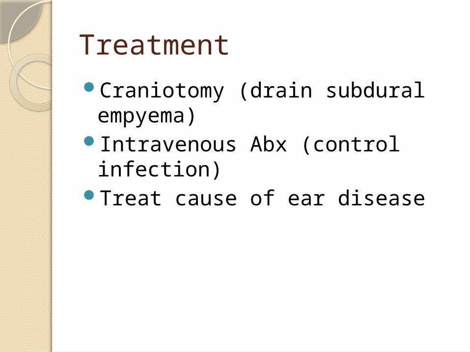
TreatmentCraniotomy (drain subdural
empyema)Intravenous Abx (control
infection)Treat cause of ear disease
MeningitisInflammation of pia mater &
arachnoid materUsually present with bacterial
invasion of CSF in subarachnoid space
In infants & children, spread by blood
In adults, spread by none erosion/retrograde thrombophlebitis
Clinical PicturesSymptoms due to :
◦Presence of infection◦↑ intracranial tension◦Meningeal & cerebral irritation
◦↑ temperature (with chills and rigors)◦Headache◦Neck rigidity◦Photophobia & mental irritability◦Nausea & vomiting◦Drowsiness◦Cranial nerve palsies & hemiplegia

Examination:◦Neck rigidity◦(+) Kernig’s sign (extension leg,
thigh flexed on abdomen pain)◦(+) Brudzinski’s sign (flexion neck
flexion hip & knee)◦Tendon reflex exaggerted, then
become absent◦Papilloedema (in late stage)

Diagnosis◦CT/MRI with contrast◦Lumbar puncture & CSF examination
Turbid Cell count ↑ ↑ protein level Decreased sugar & chlorides Culture reveal causative organisms

TreatmentAntimicrobial therapy
◦Corticosteroid + Abx (helps reduce neurological / audiological complication)
Myringotomy/cortical mastoidectomy (AOM)
Radical/modified radical mastoidectomy (COM with cholesteatoma)

Otogenic Sinus Thrombosis
◦Mastoiditis destruction of bone rupture into perisinus space abscess of perisinus periphlebitis sinus phlebitis

Symptoms:◦Parasinus abscess◦Periphlebitis◦Septicemia
Chills Spiking temperature ↑pulse rate Headache Vomit Somnolence Neck stiffness Dyspnoea (due to lung metastases)

Diagnosis:◦Skull x-ray◦CT scan◦Mastoid x-ray◦Lumbar puncture
↑ pressure ↑protein Normal glucose WBC < meningitis

Thrombus in lateral sinus

postcontrast enhancement of the sinus wall on the left side

venogram that shows nonfilling of the lateral sinus on the left side
TreatmentImmediate surgical excision of 1°
inflammation foci in mastoid & sigmoid sinus
Radical mastoidectomyThrombectomyInternal jugular vein ligated
Otitic Hydrocephalus↑ intracranial pressure + normal
CSF findingsMid ear infection lat sinus
thrombosis venous return obstruction
If thrombosis go to sup saggital sinus impedes f(x) of arachnoid villi (absorb CSF)
Clinical PicturesSymptoms:
◦Severe headcahe (maybe accompanied by nausea & vomiting)
◦Diplopia (bcoz paralysis of 6th cranial nerve)◦Blurring of vision (bcoz papilloedema / optic
atrophy)Signs:
◦Papilloedema 5-6 diopters (also with exudates & hemorrhages)
◦Nystagmus◦Lumbar puncture (CSF pressure >300mm)


TreatmentAim: decrease CSF & prevent
optic atrophy & blindnessAcetazolamide + corticosteroids
+ repeated lumbar puncture/lumbar drain
Abx therapy (in middle ear infection)
Mastoid exploration (in sinus thrombosis)