Embed Size (px)
Citation preview

Medical and Pediatric Oncology 26:397404 (1996)
lntracellular Retention of Cytosine Arabinoside Triphosphate in Blast Cells From Children With Acute Myelogenous and Lymphoblastic Leukemia
Joachim Boos, Barbara Hohenlochter, Petra Schulze-Westhoff, Meinhard Schiller, Martin Zimmermann, Ursula Creutzig,
Jorg Ritter, and Herbert jurgens
The importance of the cellular pharma- cokinetics of cytarabine triphosphate (ara- CTP) with regard to therapeutic efficacy i s well established. In vitro and in vivo moni- toring of pharmacokinetic parameters of leukemic blast cells were initiated in order to contribute to the pharmacological basis of optimal ara-C treatment strategies. Periph- eral or bone marrow blast cells from 66 leu- kemic patients [51 acute myelogenous leu- kemia (ALL), 15 acute lymphoblastic leukemia (AML) were separated and incu- bated with ara-C for 1 hour and in ara-C-free medium for another 3 hours, and the intra- cellular formation and retention of ara-CTP was measured. In eight children who re- ceived continuous ara-C infusion for induc- tion treatment, the ara-CTP concentration in circulating blast cells was monitored in vivo. The in vitro values observed in this assay corresponded to the cellular levels moni- tored in vivo. The ara-CTP retention differed clearly among the individual groups, as clas-
sified by immunphenotype at the time of the initial diagnosis: non-T-ALL 67 -C 25% (x 2 SD, n = 33), T-ALL 37 2 15% (n = 8), and AML 34 ? 18% (n = 14). The difference in ara-CTP retention between non-T-ALL and AML ( P < 0.05) as well as T-ALL (P < 0.05) was significant. There was a ten- dency toward lower ara-CTP retention in re- lapsed as compared with newly diagnosed ALL, but the difference was not significant. The maximal accumulation of ara-CTP (after 1 hour incubation) was comparable in AML, T-ALL, non-T-ALL, and blast cells from chil- dren in relapse. The observed similarity of cellular accumulation in all groups and the significantly more rapid decrease in T-ALL and AML provide the pharmacokinetic ratio- nale supporting the prolonged infusion du- ration for ara-C in these subgroups as an alternative to the intensification by high- dose ara-C schedules with short-term infu- sion. 0 1996 Wiley-Liss, Inc.
Key words: leukemia, cytarabine, cytarabine-triphosphate, cellular pharmacology, T-ALL
INTRODUCTION
Cytarabine (ara-C) is one of the most active cytotoxic drugs in the treatment of childhood acute leukemia. Dos- ing and scheduling vary widely and are still subject to intense discussion. During the last decade the treatment intensity was successfully increased by high-dose sched- ules (2-3 g/m2) in relapsed and refractory leukemias, and this strategy has since been included in the consolidation therapy of acute myeloid and lymphoblastic leukemias 11-31. The current therapy protocols of the German BFM group have incorporated a variety of ara-C schedules, namely, continuous infusion of 200 mg/m2/48 h conven- tional short-term infusion of 100 mg/m2/30 min every 12 hours, low-dose IV push of 75 mg/m2/day, and high-dose administration of 2,000-3,000 mg/m2 over 3 hours, ev- ery 12 hours.
The pharmacological basis for the different treatment modalities has been exhaustively investigated [4-81, but the optimal mode of application has remained controver- 0 1996 Wiley-Liss, Inc.
sial. Cellular uptake and intracellular phosphorylation to the nucleotide cytosine arabinoside triphosphate (ara- CTP) are preconditions and determinants of the cytostatic effect of cytarabine [9]. At conventional dosages (around 100 mg/m2), the cellular uptake of ara-C occurs by facil- itated diffusion and depends on the number of transmem- braneous nucleoside carrier sites [lo]. At higher plasma levels that can be obtained during the 2-3 hours of high- dose ara-C therapy (2-3 g/m2), the cellular uptake by passive diffusion occurs independent of transport capac- ity [ l l ] .
From the Department of Pediatric Hematology & Oncology, Univer- sity of Munster, Munster, Germany.
Received January 29, 1995; accepted August 10, 1995
Address reprint requests to Dr. Joachim Boos, Universitats-Kinder- klinik, Abteilung fur padiatrische Hamatologie/Onkologie, Albert- Schweitzer-Strape 33,48149 Munster, Germany.

398 Boos et al.
Several studies reported a correlation between the leu- kemic cells' ability to build and to retain the metabolite ara-CTP intracellularly and the clinical response to ther- apy in AML [ 12,131. Various strategies were proposed to custom tailor ara-C infusion regimens on the basis of intracellular ara-CTP pharmacokinetics [ 12,141. Such in vivo strategies, however, are restricted to therapeutic sit- uations with evaluable peripheral blast cell counts.
An in vitro assay introduced by Rustum and coworkers [ 131 showed a correlation between the ara-CTP retention of the leukemic cell and remission duration in patients with myeloid leukemia. Observations of cellular ara-CTP pharmacokinetics in lymphoblastic leukemias, especially in pediatric patients, are rare [ 15,161. Hence, the present study focused on in vitro intracellular ara-CTP formation and retention. The objectives were relevant differences in cellular ara-CTP pharmacokinetics in blast cells from children with acute T- lymphoblastic, acute non-T-lym- phoblastic, acute myeloid leukemia, and relapsed leuke- mias, which may provide a convincing background for the different dose schedules currently used in pediatric treatment protocols.
MATERIALS AND METHODS
Cell Culture
The myeloid cell lines K562 and HL60 and the pre- B-lymphoblastic cell line Blinl [ 171 were used to estab- lish and validate the incubation assay and the analytical procedures.
Separation of Blast Cells
Prior to any chemotherapy, bone marrow (2 ml) from the posterior iliac crest or peripheral blood (5 ml) was drawn into heparinized tubes as part of the routine diag- nostic sampling. The samples were immediately placed on ice, diluted with 15 ml RPMI medium (containing L-glutamine, Gibco-UK), and the blast cells were sepa- rated by Ficoll-Hypaque (Lymphoprep T Y , Nycomed-Nor- way) centrifugation. Cells at the interface were har- vested, washed three times with 50 ml medium, and centrifuged at 400 g (10 min/4"C). The cell count (Neu- bauer chamber) was determined, and pellets were then resuspended in RPMI- 1640 with 10% heat inactivated fetal calf serum (Boehringer-FRG) for the incubation ex- periments (37°C). Only samples with 290% blast cells were analyzed.
In Vitro Incubation Assay
The blast cell suspension was adjusted to 30-100 mil- lion cellsM0 ml medium (RPMI, 10% fetal calf serum), and ara-C was added to a final concentration of 1 pg/ml. A concentration of 3 p.g/ml was chosen if there were less than 30 X lo7 blast cells available in order to obtain
concentrations significantly above the limit of detection. In this ara- C-containing medium, the cells were incu- bated in a shaking water bath at 37°C for 1 hour (ara-CTP formation). The cells were then centrifuged and divided. One part was washed twice with RPMI medium and reincubated in ara-C free medium for another 3 hours (ara-CTP retention). The other part (detection of clh) and the pellet obtained after the reincubation (C, + 3h) were washed twice in ice-cold phosphate buffer (pH 7.2). The viability of the cells was tested to exceed 90% with trypan blue solution, and the cells were counted again immedi- ately prior to extraction. The ara-CTP retention at the end of the second incubation period was expressed as the percent of the 1 hour level (C, + &Ih X 100 = % re- tention; RT).
In order to determine the ara-CTP formation indepen- dent of facilitated diffusion, the inhibitor dipyridamole was added at a concentration of 10 pg/ml immediately before the addition of ara-C in a test series.
Reproducibility
The incubation assay was performed in triplicate on 9 subsequent days. Overall evaluation of these 27 experi- ments with HL60 cells showed a relative standard devia- tion (RSD) of 23% for Clh , 28% for C, + 3h, and 19% for the ara-CTP retention. The day-to-day RSDs based on the evaluation of only the first experiments each day were 22%, 26%, and 18%, respectively. With fourfold incuba- tion on a single day, the RSD of the ara-CTP retention was 14%.
Inhibition of Ara-CTP Formation by Nucleoside Transport Inhibitor
The cellular ara-C uptake by the transmembraneous carrier mechanism, and not by passive diffusion, was required in the vitro assay. Incubation of the three cell lines K562, HL60, and Blinl for 1 hour with increasing concentrations of ara-C was associated with an increase of cellular ara-CTP. Addition of 10 pg/ml dipyridamole, a well-known inhibitor of the nucleoside carrier system [22], suppressed the ara-CTP formation up to an extracel- lular ara-C concentration of 10 pg/ml. In K562 addition of 10 pg/ml dipyridamole resulted in only 20% ara-CTP formation following 1 hour incubation. In HL60 and Blinl cells, no ara-CTP was formed at all (n = 4).
lntracellular Ara-CTP
Extraction of the cells following 1 and I + 3 hours of incubation and quantification by high-performance liquid chromatography (HPLC) were performed as described elsewhere [ 181 [isocratic ion pair HPLC method with a reversed phase C,, column (NOVA-PAK'" , Waters- FRG) and 0.09 M phosphate buffer at pH 6 containing 0.35% tetrahydrofuran and 0.0 1 M tetrabutyl ammonium hydrogen sulphate]. UV detection at 270 nm shows the
Cellular Retention of Ara-CTP in Pediatric Leukemia 399
limit of detection at 25 ng/ml ara-CTP. Anthranilic acid is used as an internal standard in order to relate the mea- sured concentrations to the blast cell count in the lysed cell pellet [ 181.
PATIENTS
From January 1989 to December 1992 the in vitro investigation of ara-CTP retention was performed with blast cells from all children and adolescents with newly diagnosed acute leukemia or relapse. Informed consent was obtained prior to bone marrow puncture, and a pe- ripheral or bone marrow blast cell count high enough to exclude the necessity of additional sampling was re- quired. Unsuccessful sampling of blast cells was the only exclusion criteria. Mononuclear cells from normal bone marrow aspirates were obtained from seven children tested for leukemia or to confirm their remission status.
Acute Lymphoblastic leukemia (ALL)
Fifty-one patients aged between 3/12 and 17 years were investigated. Eight children at initial diagnosis and two in relapse were classified as T-lymphoblastic leukemia (T-ALL: TdT, CD3, and CD1 or CD7 positive). Thirty- three children at diagnosis and 1 1 in relapse presented with pre-pre-B-ALL (TdT and CD19 pos.), common- ALL (TdT, CD19, and CDlO pos.), orpre-B-ALL (cyto- plasmatic IgM pos.), comprised of non-T-ALL. Treat- ment was given according to the German therapy protocols of the Berlin-Frankfurt-Munster group ALL- BFM-86 and ALL-BFM-90 [19] and to the treatment protocol for relapsed ALL [20].
Acute Myeloid Leukemia (AML)
Fifteen patients aged between 9 months and 16 years were investigated. Fourteen times the assay was per- formed at the time of the initial diagnosis, once both initially and at relapse, and once at relapse only. The classification according to the French-American-British (FAB) criteria showed 1 X MO, 2 X MI, 1 X M3, 4 X M2, 3 X M4, and 3 X M5. One child developed the AML as a secondary malignancy. The patients were treated according to the German AML-BFM-87 treat- ment protocol [21].
lntracellular Ara-CTP Monitoring
The induction therapy according to the AML-BFM-87 protocol consists of a 48-hour continuous infusion of ara-C (100 mg/m2/day) with age-dependent intrathecal ara-C application on day 1 , followed by 12 infusions of 100 mg/m2 ara-C every 12 hours. On days 3,4, and 5 daunorubicin (30 mg/m2) is additionally applied every 12 hours, and on days 6, 7, and 8 etoposide (150 mg/m2) is added. Eight patients who received this induction proto- col had a peripheral blast cell count high enough for
1 80 !! 60
40
20 0
0
0 20 40 60 80 100 120 140 180
1 pglml 140 I
0 20 40 60 80 1DO 120 140
porlphonl Mast cells
Fig.1. Regression of the cellular ara-CTP retention in blast cells obtained from bone marrow vs. peripheral blast cells (bottom). Re- gression analysis of ara-CTP retention following incubation with 1 pg/rnl vs. 3 F g / d ara-C (top).
intracellular monitoring of ara-CTP levels during the con- tinuous infusion. Blood samples of 2-5 ml were then drawn into heparinized tubes, placed on ice without de- lay, and handled as described earlier. In two children peak and trough levels after the first ara-C short-term infusions were also monitored.
Statistics
To compare differences between the median among the immunological and morphological groups, the Kruskal-Wallis one-way analysis of variance on ranks was used. To isolate groups that differ from others, Dunn's method was used (both evaluations were done with Sigma StatTM statistical software, version 1.02). In addition, the Mann-Whitney U test on Statgraphics soft- ware, version 5.2, was used to compare different groups of individuals. A P value of G 0.1 was defined to indicate a tendency and P S 0.05 to indicate significance.
RESULTS
The comparability of the retention was tested with both the 1 pg/ml and the 3 pg/ml concentration on sam- ples from a suitable subgroup of 26 patients (AML and ALL). The 1 pg incubation was associated with an ara- CTP retention of 41 * 22%, and the 3 pg incubation with 48 k 26%, the regression analysis yielded in r = 0.9. As a consequence, the results obtained with the two modifications of the assay were not differentiated in the following evaluations (Fig. 1, top).
The comparison of bone marrow and peripheral blast cells was performed in 15 children (9 ALL, 6 AML). The mean ara-CTP retention in bone marrow blast cells was

400 Boos et al.
m.LD +. m.di.n
1000 0
26 9 8 12
167 233 225 186
169 141 144 97
120 191 162 159
i O Q
A 0 0
10 I I 1 1
non-T-ALL T-ALL AML relapse
Fig. 2. Capacity of ara-CTP formation. Ara-CTP concentration (pmol/107 cells) following 1 hour incubation with 1 pg/ml ara-C.
45 2 28% and in peripheral blast cells it was 48 2 3 1 %, and the correlation coefficient was r = 0.9. A distinction with regard to the source of the malignant cell clone was therefore deemed unnecessary (Fig. 1, bottom).
The maximum ara-CTP levels formed following 1 hour of incubation with 1 pg/ml ara-C (ara-CTP forma- tion) were associated with a wide range of cellular ara- CTP concentrations, but significant differences between T-ALL, non-T-ALL, relapsed ALL, or AML were not observed (Fig. 2; P = 0.23). The ara-CTP formation fol- lowing incubation with 3 pgirnl was significantly higher (not shown in Fig. 2). Linear regression analysis of the ara-CTP formation in 22 ALL samples (2 1 patients) incu- bated at 1 pg/ml (x) as well as at 3 pg/ml (y) yielded y = 2.4 X x - 39 (r = 0.94). Due to a cell count too low for incubation at 1 pg/ml, nonmalignant bone mar- row cells could not be included in the evaluation of ara- CTP formation.
Intracellular ara-CTP retention was comparable in common-ALL (67 k 25%, median 66%, n = 25 at ini- tial diagnosis) and the pre-B-ALL subtype (67 2 26%, median 59%, n = 8 at initial diagnosis) in the subgroups of non-T-ALL. However, the results in non-T-ALL were markedly different from T-ALL, AML, and normal mononuclear bone marrow cells (Fig. 3, Table I). Statis- tical differences were found between AML and non- T-ALL, and between T-ALL and non-T-ALL (Kruskall- Wallis: P < 0.05, U- test: P < 0.002). The difference between newly diagnosed non-T-ALL and relapsed non- T-ALL showed a tendency towards lower ara-CTP-reten- tion in the latter group (U-test, P < 0.07).
The ara-CTP retention was 34 5 18% in 14 children with newly diagnosed AML and 26% and 18% in two
TABLE I. Ara-CTP Retention in Leukemic Blast Cells and Mononuclear Cells From Normal Bone Marrow
X f S Ranee n Median
Normal BM 44 f 24% 37% 22-55% 7 Non-T- ALL 67 * 25% 64% 26130% 33
T-ALL 37 * 15% 31% 2040% 8 2 T-ALL relapse 29%/15%
AML 3 4 + 18% 29% 9 4 4 % 14
Non-T relapse 51 f 16% 52% 3240% 11
AML relapse 26%/18% 2
BM = bone marrow.
-
8 0
0
U n
I I I I normal bone non-T- non-T- TALL AML
ALL relapse
Fig. 3. Ara-CTP retention in normal bone marrow, initial non- T-ALL, relapsed non-T-ALL, T-ALL, and AML (relapses included, for detailed date, see Table 1).
children with relapsed AML. The distribution of these 16 observations was 615% in two, 15-29% in eight, 3 s 44% in two, 45-59% in two, and 260% in two. In eight children the ara-CTP concentration in the leukemic blast cells could be monitored in vivo during conventional continuous ara-C infusion (Fig. 4). The mean of all con- centrations depicted in Figure 4 was 70 2 69 pmol/107 cells.
No change in cellular ara-CTP retention was observed over the course of the continuous infusion: In four chil- dren the assay was repeated 36-44 hours after initiation of treatment. The RT%s (initiaU36-44 hr) were 20%/ 30%, 29%/20%, 65%/65%, and 29%/35%. In two chil- dren, blast cells could be separated following the ara-C short-term infusion. Thirty minutes after the end of the infusion, the cellular ara-CTP levels were 181 and 92 pmol/ lo7 cells. The corresponding trough levels dropped below the limit of detection.
DISCUSSION
Among all BFM treatment protocols, AML induction therapy is the only one that starts treatment with continu-
Cellular Retention of Ara-CTP in Pediatric Leukemia 401
1000
100
10
1
9
continuous infusion 200 mglm2I48h I
0 5 10 15 20 25 30 35 40 45 50 55 60
hwrs
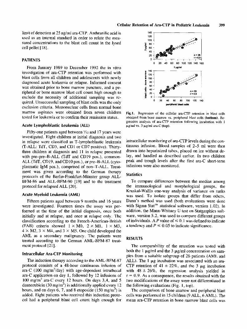
Fig. 4. Ara-CTP concentrations in myeloid blast cells during conven- tional continuous infusion therapy with ara-C.
ous ara-C infusion, a prerequisite for intracellular ara- CTP monitoring. The ara-CTP levels observed with this schedule varied widely (Fig. 4) and were slightly lower than those reported by Hiddemann et al. (median 26-96 ng/107 cells, q.e. 130 pmoY107 cells median) [23] or Rustum et al. (21-183 pmol/107 cells) [24]. The clinical relevance of interindividual variation of the cellular lev- els, however, is speculative. A relationship between clin- ical response and cellular ara-CTP levels under the condi- tions of conventional continuous infusions has not yet been systematically investigated. Target levels were al- ways defined on the basis of high-dose ara-C treatment and, therefore, were disproportionately high: Plunkett et al. proposed a minimal effective ara-CTP trough level of 75 pM ( ~ 2 0 0 pmoV107 cells) following high-dose (HD) ara-C short-term infusion [ 12,251. Estey et al. observed a median steady-state level of 122 pM in responding pa- tients and of 63 pM in nonresponding patients with HD continuous infusion [26]. These target steady-state levels were comparable with the peak values of short-term ara-C infusions with the conventional dosage. In two children we determined peak levels of 18 1 and 9 1 pmol/ lo7 cells.
These in vivo values were of the same order of magni- tude as the cellular concentrations following 1 hour of incubation in vitro (186 +_ 96 pm01/107 cells). Moreover, the relatively rapid disappearance within 12 hours in vivo is consistent with a low ara-CTP retention of 29% (me- dian) in AML and corresponds to published half-lives of 1.7-2.5 hours [23]. Our data thus support the assumption that the pharmacolunetic findings on blasts in vitro do reflect ara-CTP metabolism during conventional ara-C treatment.
One objection to the described in vitro model may be based on the relatively high ara-C concentrations used (1-3 pg/ml). With both concentrations, however, cellu- lar uptake was shown to be limited by the activity of the transmembraneous carrier system. The formation of ara- CTP was completely suppressed by coincubation with dipyridamole, an inhibitor of the nucleotide carrier sys- tem [22] (see earlier). The experimental conditions thus ensured carrier-mediated uptake and reliably reflected conventional ara-C therapy. In addition, ara-CTP reten- tion with 1 and 3 pg/ml ara-C was similar (Fig. 1). Single publications suggested a difference between cellular ara- CTP pharmacokinetics in bone marrow and peripheral blast cells [5,24]. Our findings do not indicate pharma- cokinetic differences of in vitro ara-CTP retention be- tween cells from these two sources (Fig. 1).
With a similar assay Rustum and coworkers reported a positive correlation between in vitro ara-CTP retention and remission duration following conventional ara-C therapy [ 131. The adverse prognostic significance of low ara-CTP retention, however, seemed to be reduced or even abrogated by the use of continuous ara-C infusion and HD regimens [28]. In the in vivo investigation of Plunkett and others, the elimination rates and correspond- ing trough levels were significantly higher in responding patients [12,25], while the ara-CTP peak levels were comparable. In another investigation, the cellular ara- CTP pharmacokinetics (AUC, Cmax, tYz) obtained with an initial high-dose ara-C infusion did not correlate with response duration in 147 AML patients on different ara-C therapy schedules. However, the steady-state concentra- tions on HD continuous infusion (6 g/m2/96 hr) were significantly lower in refractory disease [26]. The degree of ara-CTP formation in vitro appeared to be even higher in relapsed AML than in previously untreated patients [28]. Overall, data relating cellular parameters of ara- CTP pharmacokinetics and clinical outcome have been conflicting. Peak levels seem to be of minor relevance compared with the duration of exposure to the triphos- phate metabolite. During continuous infusion of ara-C, the ara-CTP steady-state levels in circulating blast cells increased proportionately to the ara-C application rate
Publications on cellular ara-CTP kinetics in ALL have been rare. In adults, the ara-CTP elimination in lympho- blasts (n = 18) appeared slower than in myeloblasts (n = 51) [ 181. Correspondingly, we found a higher ara- CTP retention in non-T-ALL (median 64%; see Fig. 2) than in AML (29%) for pediatric leukemias as well. In addition, our data clearly differentiate between non-T- ALL and T-ALL (3 1 % RT).
According to the majority of studies, the T-cell pheno- type has a poorer prognosis, compared with non-T-ALL, with cure rates much lower than 50% [29]. The list of biological features distinguishing between T- and B-lin-
1141.

402 Boos et al.
eage leukemia cells is long. Several groups attempted to specify pharmacological properties of T-lymphoblastic cells as a basis for treatment alterations and studied ara-C metabolism in T cells. Momparler et al. [30] reported increased formation and a longer intracellular half-life of ara-CTP in different cell lines and an associated increased sensitivity to ara-C of T-lymphoid as compared with B-lymphoid and myeloid cells. Tanaka and coworkers investigated ara-C uptake and ara-CTP retention in a sim- ilar assay system using blast cells from adults in vitro [ 3 I ] and described a 3.5 times greater intracellular accumula- tion and prolonged retention, which was 88% in T-ALL vs. 60% and 52% in AML and non-T-ALL. This finding correlated with a higher remission rate following treat- ment with mitoxantrone and continuous infusion of con- ventional ara-C.
Our own observations are inconsistent with these pub- lished findings. The cellular ara-CTP decrease was sig- nificantly more rapid in T-lymphoblastic than in non- T-lymphoblastic blast cells and was comparable with my- eloid blasts (see Fig. 2). The kinetics in normal bone marrow samples was on the same order of magnitude as in T-ALL and AML blast cells. The ara-CTP formation did not differ between different leukemias, including re- lapse (see Fig. 2). These observations correspond to a report from Ross and coworkers, indicating that blast cells obtained from patients with nonlymphoblastic leu- kemia, responders as well as nonresponders under con- ventional ara-C treatment, formed comparable amounts of ara-CTP in vitro [32], and the ratio relating ara-C incorporation into DNA with cellular ara-CTP formation decreased with increasing ara-C concentration in the assay.
What are the implications for the discussion of the optimal ara-C schedule? Treatment with ara-C aims at ara-CTP formation, ara-CTP incorporation into the DNA, and DNA strand breaks. Ara-CTP incorporation and inhibition of DNA polymerase depend on relevant amounts of intracellular ara-CTP and the relationship be- tween intracellular ara-CTP and dCTP [33]. An increase in cellular ara-CTP levels, however, will not automati- cally enhance cytotoxicity . The dose-response curve is apt to develop a plateau at a particular concentration, and any further increase will not necessarily result in in- creased cytotoxicity . In cell culture, cell kill correlates less significantly with cellular ara-CTP pools than with ara-C incorporation in DNA [ 341.
This observation might explain the fact that according to most publications ara-CTP peak levels failed to corre- late with clinical data, while the ara-CTP elimination kinetics were, in fact, associated with clinical parameters [12,13,25,26,28]. Extending the time of exposure to a certain ara-CTP concentration, therefore, should be fa- vored over an attempt to increase the maximum ara-CTP accumulation. Considering that even the ara-CTP forma-
tion with increasing ara-C dosages is a saturable process [5,27], the rationale of simply increasing the dose for ara-C treatment intensification is questionable. The pro- longed ara-CTP elimination in non-T-ALL compared with mononuclear bone marrow cells (Fig. 3 ) may prom- ise increased therapeutic gain and offers a rationale for short-term or push injection.
In T-ALL and AML there was no relevant difference between blast cells and bone marrow cells in our investi- gation. Effective treatment of the blast cell population, therefore, will be accompanied by corresponding bone marrow toxicity. This view is confirmed by the clinical observation that continuous infusion of conventional dose ara-C escalates bone marrow toxicity. The introduc- tion of the ADE induction (48 hr continuous ara-C infu- sion followed by short-term ara-C, Daunorubicin, and etoposide) into the German AML-BFM studies resulted in a significantly increased event-free interval and re- duced relapse rate [35,36]. In T-ALL the low ara-CTP retention offers arguments for the same strategy: Contin- uous infusion protects the cells from a decline in cellular ara-CTP, as we proved in patients receiving their first continuous infusion and then receiving the same dose as a short-term infusion (Fig. 4) according to the AML treat- ment strategy.
In comparison with our own results, Tanaka and Yoshida [3 11 found much higher ara-CTP retention in T-ALL. Still, their clinical observation of a 88% remis- sion rate was based on continuous infusion of ara-C. In addition, Lauer and coworkers reported a selective effect of continuous infusions of ara-C during maintenance therapy: The combination cyclophosphamide/ara-C in addition to 6-mercaptopurine/methotrexate significantly improved relapse-free survival in children with T-ALL (7118 = 36% vs. 0/8 = 0% after 30 months, P = 0.015) but not in non-T-ALL (35% vs. 48%) [37]. Over the last decade dose intensification has predominantly been real- ized by short-term infusion schedules with high doses of ara-C. High plasma levels of ara-C are expected to over- come cellular resistance by reduced expression of the nucleoside transport system via passive diffusion. As pointed earlier, high peak ara-CTP levels are not the main goal, but they do influence the AUC and the time above defined trough levels. Without a doubt, HD treatment resulted in enhanced toxicity and improved clinical out- come [ 1-31. This might be due, in part, to the normally longer infusion duration of 2 4 hours in HD regimens and the much longer maintenance of therapeutic plasma lev- els compared with push injection of 30-minute infusion of conventional dosages. Our data provide the pharmaco- logical basis for demonstrating that infusion duration is an essential variable in ara-C treatment schedules and that continuous infusion as an intensification strategy is a rational alternative or is even superior to HD treatment, especially in T-ALL and AML. Nevertheless, the dose of

Cellular Retention of Ara-CTP in Pediatric Leukemia 403
continuous ara-C schedules is the second important vari- able that has to be optimized and can perhaps be custom tailored [ 141 in the future.
ACKNOWLEDGMENTS
This work was supported by a research grant from the Deutsche Forschungsgemeinschaft and the Federal De- partment of Research and Technology (01 # 9401).
REFERENCES
I .
2.
3.
4.
5.
6.
7.
8.
9.
10.
11.
12.
13.
14.
15.
Peters WG. Colly LP, Willemze R: High-dose cytosine arabino- side: Pharmacological and clinical aspects. Blut 56: 1-1 1, 1988. Hiddemann W: Cytosine arabinoside in the treatment of acute myeloid leukemia: The role and place of high-dose regimens. Ann Hematol62:119-128, 1991. Lie SO, Slordahl S: High-dose cytosine arabinoside in the treat- ment of childhood malignancies. Semin Oncol 12(Suppl 3): 160- 165, 1985. van Prooijen HC, Dekker AW, Punt K: The use of intermediate dose cytosine arabinoside (ID ara-C) in the treatment of acute nonlymphocytic leukemia in relapse. Br J Haematol 57:291-299, 1984. Plunkett W, Liliemark JO, Adams TM, Nowak B, Estey E, Kan- tarjian H, Keating MJ: Saturation of 1 -P-D-arabinofuranosylcy- tosine 5’-triphosphate accumulation in leukemia cells during high- dose 1 -P-D- arabinofuranosylcytosine therapy. Cancer Res 47:
Slevin ML, Piall EM, Aheme GW, Johnston A, Lister TA: The pharmacokinetics of cytosine arabinoside in the plasma and cere- brospinal fluid during conventional and high-dose therapy. Med Pediatr Oncol (Suppl 1):157-168, 1982. Slevin ML, Piall EM, Aheme GW, Harvey VJ, Johnston A, Lister TA: Effect of dose and schedule on pharmacokinetics of high-dose cytosine arabinoside in plasma and cerebrospinal fluid. J Clin Oncol 1:54&551, 1983. Avramis Vl, Weinberg KI, Sat0 JK, Lenarsky C, Willoughby M, Coates TD, Ozkaynak MF, Parkman R: Pharmacology studies of 1 -P-D- arabinofuranosylcytosine in pediatric patients with leuke- mia and lymphoma after a biochemical optimal regimen of loading bolus plus continuous infusion of the drug. Cancer Res 49:241- 247, 1989. Kessel D, Hall TC, Wodinsky I: Transport and phosphorylation as factors in the antitumor action of cytosine arabinoside. Science 156: 124GI 24 1, 1967. Wiley JS, Jones SP, Sawyer WH, Paterson ARP: Cytosine arabi- noside influx and nucleoside transport sites in acute leukemia. J Clin Invest 69:479439, 1982. White JC, Rathmell JP, Capizzi RL: Membrane transport influ- ences the rate of accumulation of cytosine arabinoside in human leukemic cells. J Clin Invest 79:38&387, 1987. Plunkett W, Iacoboni S, Estey E, Danhauser L, Liliemark JO, Keating MJ: Pharmacologically directed ara-C therapy for refrac- tory leukemia. Semin Oncol 12(Suppl 1):20-30, 1985. Rustum YM, Preisler HD: Correlation between leukemic cell re- tention of 1 -P-D-arabinofuranosylcytosine 5‘-triphosphate and re- sponse to therapy. Cancer Res 39:4249, 1979. Heinemann V, Estey E, Keating MJ, Plunkett W: Patient-specific dose rate for continuous infusion high-dose cytarabine in relapsed acute myelogenous leukemia. J Clin Oncol7:622-8, 1989. Bekassy AN, Liliemark J, Garwicz S, Wiebe T, Gullikson H, Peterson C: Pharmacokinetics of cytosine arabinoside in cere-
3005-301 I , 1987.
16.
17.
18.
19.
20.
21.
22.
23.
24.
25.
26.
27.
28.
29.
30.
brospinal fluid and of its metabolite in leukemic cells. Med Pediatr Oncol 18:13&142, 1990. Avramis VI, Biener R, Krailo M, Finklestein J , Ettinger L, Willoughby M, Sieges SE, Holcenberg JS: Biochemical pharma- cology of high dose 1 -P-D-arabinofuranosylcytosine in childhood acute leukemia. Cancer Res 47:678&6792, 1987. Wormann B, Anderson JM, Liberty JA, Gajl-Peczalska K, Brun- ning RD, Silberman TL, Arthur DC, LeBien TW: Establishment of a leukemic cell model for studying human pre-B to B cell differentiation. J Immunol 142:11&117. 1989. Boos J: A simple isocratic ion-pair high-performance liquid chro- matographic determination of 1-P-D-arabinofuranosylcytosine 5’-triphosphate for intracellular drug-monitoring and in vitro incu- bation assays. Pharma Biom Anal 9:47-52, 1991. Riehm H, Gadner H, Henze G, Kornhuber B, Lampert F, Ni- ethammer D, Reiter A, Schellong G: Results and significance of six randomized trials in four consecutive ALL-BFM studies. In Buchner T, Schellong G, Hiddemann W, Ritter J (eds):“Haema- tology and Blood Transfusion, Acute Leukemias 11,” Berlin: Springer, 33 :43949 , 1990. Henze G, Fengler R, Hartmann R, for the BFM Relapse Study Group: Chemotherapy for relapsed childhood acute lymphoblastic leukemia: Results of the BFM Study Group. In Buchner T, Schel- long G, Hiddemann W, Ritter J (eds): “Haematology and Blood Transfusion, Acute Leukemias IV,” Berlin: Springer, 36:374- 383, 1994. Creutzig U, Ritter J, Zimmermann M, Schellong G: Does cranial inidiation reduce the risk for bone marrow relapse in acute myel- ogenous leukemia? Unexpected results of the childhood acute myelogenous leukemia study BFM-87. J Clin Oncol I1:279-286, 1993. Jarvis MS: Nitrobenzylthioinosine-sensitive nucleoside transport system: Mechanism of inhibition by dipyridamole. Mol Pharma- col30:659-665, 1986. Hiddemann W, Schleyer E, Unterhalt M, Zuhlsdorf M, Rolf C, Reuter C, Kewer U, Uhrmeister C, Wormann B, Buchner T: Differences in the intracellular pharmacokinetics of cytosine arab- inoside (AraC) between circulating leukemic blasts and normal mononuclear blood cells. Leukemia 6: 1273-1280, 1992. Rustum YM, Slocum HK, Wang G, Bakshi D, Kelly E. Buscaglia D, Wrzosek C, Early AP, Preisler HD: Relationship between plasma ara-C and intracellular ara-CTP pools under conditions of continuous infusion and high- dose ara-C treatment. Med Pediatr Oncol Suppl 1:33-43, 1982. Plunkett W, Iacoboni S, Keating M: Cellular pharmacology and optimal therapeutic concentrations of 1 -P-D-arabinofuranosylcy- tosine 5‘- triphosphate in leukemic blasts during treatment of refractory leukemia with high-dose 1 -P-D-arabinofuranosylcytko- sine. Scand J Haematol34(Suppl44):5 1-59, 1986. Estey EH, Keating MJ, McCredie KB, Freireich EJ, Plunkett W: Cellular ara-CTP pharmacokinetics, response, and karyotype in newly diagnosed acute myelogenous leukemia. Leukemia 4:95- 99, 1990. Jamieson GP, Snook MB, Wiley JS: Saturation of intracellular cytosine arabinoside triphosphate accumulation in human leuke- mic blast cells. Leuk Res 14:475479, 1990. Muus P, Drenthe-Schonk A, Haanen C, Wessels H, Linssen P: In- vitro studies on phosphorylation and dephosphorylation of cy- tosine arabinoside in human leukemic cells. Leuk Res I1:319- 325, 1987. Preisler HD, Rustum YM, Azamia N, Priore R: Abrogation of the prognostic significance of low leukemic cell retention of cytosine arabinoside triphosphate by intensification of therapy and by alter- ation in the dose and schedule of administration of cytosine arabi- noside. Cancer Chemother Pharmacol 19:69-74, 1987. Momparler RL, Onetto-Pothier N, Bouffard DY, Momparler LF:

404 Boos et ai.
Cellular pharmacology of 1-P-D-arabinofuranosylcytosine in hu- man myeloid, B- lyphoid and T-lymphoid leukemic cells. Cancer Chemother Pharmacol27: 141-146, 1990.
31. Tanaka M, Yoshida S: Efficient formation of cytosine arabino- side-5'-triphosphate in leukemic blasts of human T-cell acute lym- phoblastic leukemia. Leuk Res 13:93 1-936, 1989.
32. Ross D, Thompson B, Joneckis C, Akman S, Schiffer C: Metabo- lism of h a - C by blast cells from patients with ANLL. Blood 68:76-82, 1987.
33. Kawasaki H, Higashigawa M, Ohkubo T, Kamiya H, Sakurai M: Relationship between intracellular dCTP/Ara-CTP ratio and cyto- toxic effect of Ara-C. Adv Exp Med Biol253B:369-74, 1986.
34. Kufe D, Spriggs D, Munroe D: Relationships among Ara-CTP pools, formation of (Ara-C)DNA, and cytotoxicity of human leu- kemic cells. Blood 64:54-58, 1984.
35. Ritter J, Creutzig U, Reiter A, Riehm H, Schellong G: Childhood leukemia: Cooperative Berlin-Frankfurt-Munster trials in the Fed- eral Republic of Germany. J Cancer Res Clin Oncol 116:10&103, 1990.
36. Creutzig U, Ritter J, Schellong G for the AML-BFM Study Group. Identification of two risk groups in childhood acute myel- ogenous leukemia after therapy intensification in study AML- BFM-83 as compared with study AML- BFM-78. Blood 75:1932- 1940, 1990.
37. Lauer SJ, Pinkel D, Buchanan GR, Sartain P, Comet JM, Krance R, Borella LD, Casper JT, Kun LE, Hoffman RG, Camitta BM: Cytosine arabinosidekyclophosphamide pulses during continua- tion therapy for childhood acute lymphoblastic leukemia-poten- tial selective effect in T-cell leukemia. Cancer 60:2366-2371, 1987.


![[Ghiduri][Cancer]Chronic Myelogenous Leukemia](https://img.dokumen.tips/doc/110x75/577cc6ea1a28aba7119f80de/ghiduricancerchronic-myelogenous-leukemia.jpg)