Embed Size (px)
Citation preview
Intracellular Ca-carbonate biomineralization iswidespread in cyanobacteriaKarim Benzeraraa,1, Feriel Skouri-Paneta, Jinhua Lia, Céline Férarda, Muriel Guggerb, Thierry Laurentb,Estelle Couradeaua,c, Marie Ragona,c, Julie Cosmidisa, Nicolas Menguya, Isabel Margaret-Olivera, Rosaluz Taverad,Purificación López-Garcíac, and David Moreirac
aInstitut de Minéralogie, de Physique des Matériaux, et de Cosmochimie, Sorbonne Universités, Centre National de la Recherche Scientifique, Unité Mixtede Recherche 7590, Université Pierre et Marie Curie Paris 06, Muséum National d’Histoire Naturelle, Institut de Recherche pour le Développement Unité Mixtede Recherche 206, 75005 Paris, France; bCollection des Cyanobactéries, Institut Pasteur, 75724 Paris Cedex 15, France; cUnité d’Ecologie, Systématique etEvolution, Centre National de la Recherche Scientifique, Unité Mixte de Recherche 8079, Université Paris-Sud, 91405 Orsay Cedex, France; and dDepartamentode Ecología y Recursos Naturales, Universidad Nacional Autónoma de México, 04510 Mexico D.F., Mexico
Edited by Steven M. Stanley, University of Hawaii, Honolulu, HI, and approved June 11, 2014 (received for review March 3, 2014)
Cyanobacteriahaveplayedasignificantrole intheformationofpastandmodern carbonatedeposits at the surfaceof theEarthusingabiominer-alization process that has been almost systematically consideredinduced and extracellular. Recently, a deep-branching cyanobacterialspecies, Candidatus Gloeomargarita lithophora, was reported to formintracellular amorphous Ca-rich carbonates. However, the significanceand diversity of the cyanobacteria in which intracellular biomineraliza-tion occurs remain unknown. Here, we searched for intracellular Ca-carbonate inclusions in 68 cyanobacterial strainsdistributed throughoutthe phylogenetic tree of cyanobacteria.Wediscovered that diverse uni-cellular cyanobacterial taxa formintracellularamorphousCa-carbonateswith at least two different distribution patterns, suggesting the exis-tence of at least two distinct mechanisms of biomineralization: (i) onewithCa-carbonate inclusions scatteredwithin the cell cytoplasmsuchasinCa.G. lithophora,and(ii)anotheroneobservedinstrainsbelongingtothe Thermosynechococcus elongatus BP-1 lineage, in which Ca-carbon-ate inclusions lie at the cell poles. This pattern seems to be linked withthe nucleation of the inclusions at the septum of the cells, showing anintricate and original connection between cell division and biomineral-ization.Thesefindings indicatethat intracellularCa-carbonatebiominer-alization by cyanobacteria has been overlooked by past studies andopen new perspectives on the mechanisms and the evolutionary his-tory of intra- and extracellular Ca-carbonate biomineralizationby cyanobacteria.
calcification | amorphous calcium carbonate | polyphosphate
Cyanobacteria are a phylogenetically and ecologically diversephylum of Gram-negative bacteria that have impacted the
global cycle of carbon on the Earth for billions of years and in-duced the oxygenation of the atmosphere (1–3). By performingoxygenic photosynthesis—a unique capability that appeared onlyonce in evolution—in this particular group of bacteria, cyano-bacteria have contributed significantly to the primary productionon the past and present Earth (4). Moreover, cyanobacteria havereceived great attention from geologists as major players in theformation of carbonate sedimentary deposits such as stromato-lites (5, 6), the oldest ones formed by cyanobacteria possibly asold as 2.98 billion years (Ga) (7). Fossils of Ca-carbonate–encrusted cyanobacterial cells (calcimicrobes) have been lookedfor extensively. The temporal distribution of calcimicrobes in thegeological record dating back as far as the early Proterozoic(∼2.5–2.3 Ga) (8) has been interpreted as the result of paleo-environmental and/or evolutionary changes (9, 10). Despite theimportance of carbonate biomineralization by cyanobacteria inthe formation of calcimicrobes and sedimentary deposits, theinvolved mechanisms are still poorly understood (11, 12). Someauthors have proposed that cyanobacterial calcification mightbe promoted by CO2-concentrating mechanisms (CCMs) thatpossibly developed in the late Proterozoic to accommodatephotosynthetic carbon limitation under low CO2 fugacity (10).
This model states that bicarbonates are actively imported intothe cells, transformed to CO2 within carboxysomes for fixation byribulose-1,5-bisphosphate carboxylase/oxygenase. The resulting al-kalinity is transferred outside the cells, which raises the externalpH and thus induces CaCO3 precipitation (13). Other authorshave stressed the importance of cell-surface properties of somecyanobacteria for the nucleation of CaCO3 minerals and did notobserve notable effects of photosynthesis on CaCO3 preci-pitation (14). In any case, precipitation of CaCO3 by cyano-bacteria has been invariably considered a noncontrolled andextracellular process.This paradigm has been questioned recently by the discovery
of a new deep-branching cyanobacterial species, CandidatusGloeomargarita lithophora, enriched from the hyperalkalineLake Alchichica (Mexico) and forming amorphous carbonatesintracellularly (15). However, many studies characterized theultrastructure of cyanobacteria (16, 17) and explored their im-pact on calcification (11, 18, 19), but none of them reported thepresence of intracellular carbonates. Here, we investigated wh-ether intracellular carbonate biomineralization is restrictedto one particular species and/or specific environmental con-dition or whether it exists in other diverse cyanobacteria; thisis essential to assess the evolution of intracellular carbonate
Significance
Cyanobacteria are known to promote the precipitation ofCa-carbonate minerals by the photosynthetic uptake of in-organic carbon. This process has resulted in the formation ofcarbonate deposits and a fossil record of importance for deci-phering the evolution of cyanobacteria and their impact on theglobal carbon cycle. Though the mechanisms of cyanobacterialcalcification remain poorly understood, this process is invariablythought of as extracellular and the indirect by-product of met-abolic activity. Here, we show that contrary to common belief,several cyanobacterial species perform Ca-carbonate bio-mineralization intracellularly. We observed at least two phe-notypes for intracellular biomineralization, one of which showsan original connection with cell division. These findings opennew perspectives on the evolution of cyanobacterial calcification.
Author contributions: K.B., F.S.-P., M.G., P.L.-G., and D.M. designed research; K.B., F.S.-P.,J.L., C.F., T.L., E.C., M.R., J.C., N.M., I.M.-O., R.T., P.L.-G., and D.M. performed research; K.B.,J.L., N.M., P.L.-G., and D.M. analyzed data; M.G. provided cyanobacterial strains; and K.B.,F.S.-P., J.L., M.G., P.L.-G., and D.M. wrote the paper.
The authors declare no conflict of interest.
This article is a PNAS Direct Submission.
Data deposition: The sequence reported in this paper has been deposited in the GenBankdatabase (accession no. KJ566932).1To whom correspondence should be addressed. Email: [email protected].
This article contains supporting information online at www.pnas.org/lookup/suppl/doi:10.1073/pnas.1403510111/-/DCSupplemental.
www.pnas.org/cgi/doi/10.1073/pnas.1403510111 PNAS | July 29, 2014 | vol. 111 | no. 30 | 10933–10938
EART
H,A
TMOSP
HER
IC,
ANDPL
ANET
ARY
SCIENCE
SMICRO
BIOLO
GY
biomineralization and its significance at geological timescales.For this purpose, we screened 68 cyanobacterial strains scatteredthroughout the phylogenetic tree of cyanobacteria (Fig. 1) tosearch for intracellular carbonates.
Results and DiscussionSixty-eight strains of cyanobacteria were imaged by SEM usingan angle-selective backscattered electron (AsB) detector, whichprovides a chemical contrast; their elemental composition wasfurther analyzed by energy dispersive X-ray spectrometry(EDXS) coupled with SEM. Names of the strains, their geo-graphic origin, and the culture medium in which they were cul-tured are provided in Table S1. Most of the strains were obtainedfrom culture collections [notably, the Pasteur Culture Collection(PCC) of cyanobacteria], but we also studied a few other strains,in particular two strains that were isolated recently frommicrobialites from the alkaline Lake Alchichica in Mexico: Ca.G. lithophora (15) and a new species distantly related to Ther-mosynechococcus elongatus BP-1, which we have provisionallycalled Candidatus Synechococcus calcipolaris strain G9. Cyano-bacteria have been classically grouped by subsections, defined onthe basis of morphological and cell division features (20, 21). Wecovered all of the subsections, with 26 strains belonging to sub-section I, 5 to subsection II, 23 to subsection III, 10 to subsectionIV, and 4 to subsection V.All but eight of the tested strains showed intracellular inclu-
sions as detected by SEM with a size range from ∼100 to 800 nmin diameter (Table S1 and Fig. S1). Some strains contained nu-merous inclusions (e.g., PCC 73106, PCC 6308), whereas othershad only one (PCC 7376) or two (PCC 7421) inclusions per cell.In some cases, these inclusions were preferentially located at thepoles or showed alignments (PCC 7002, PCC 7429). These inclu-sions were in most cases composed of P as the major element withsome Mg and K and sometimes Ca (Fig. S2). According to theirsize, chemical composition, and distribution patterns, they wereinterpreted as polyphosphate (PolyP) granules. In the past, PolyPgranules have been identified by microscopy in many bacterialspecies, including some of the cyanobacterial strains analyzed in thepresent study (22, 23). Such granules have sometimes been namedmetachromatic granules or “volutin” (17) and constitute a stor-age form of P for the cells or as a source of energy underP-limited or stress conditions (24).The most striking result of this systematic survey was the dis-
covery of seven cyanobacterial strains forming spherical andpoorly crystalline intracellular carbonates (Figs. 2–4 and TableS2) in addition to Ca. G. lithophora, which was recently de-scribed (15). These strains were further analyzed by scanningtransmission electron microscopy (STEM) in the high-angularannular dark field (HAADF) mode, which provides a chemicalcontrast with a higher spatial resolution compared with SEM. Allthese strains formed Mg- and K-containing PolyP granules asdescribed above, but they also contained P-free, Ca-rich inclusionsmeasuring between 60 and 870 nm in diameter and appearing asamorphous by electron diffraction, that we interpreted as Ca-carbonates based also on X-ray absorption near-edge structure(XANES) spectroscopy analyses (Figs. S2 and S3). The presenceof Ca-carbonate inclusions was not dependent on the medium inwhich the strains were grown: first, several strains without Ca-carbonate inclusions were grown in BG11 similarly to the strainsforming intracellular Ca-carbonates. Moreover, Ca. G. lithophoraD10 and Ca. S. calcipolaris G9 showed intracellular Ca-carbonateinclusions when grown in the different culture media (e.g., BG11oor ASNIII) used for the other PCC strains screened in the presentstudy (Figs. S4 and S5).
Two Types of Spatial Distributions of the Ca-Carbonate InclusionsWere Observed. Ca. G. lithophora D10, Cyanothece sp. PCC7425, and Chroococcidiopsis thermalis PCC 7203 formed calcium
0.05
Gloeocapsa sp. PCC 7428
Cyanothece sp. PCC 7424
Synechococcus calcipolaris G9
Nostoc punctiforme PCC 73102
Pleurocapsa sp. PCC 7319
Arthrospira sp. PCC 8005
Chroococcidiopsis thermalis PCC 7203
Synechococcus elongatus PCC 6301
Spirulina major PCC 6313
Lyngbya sp. PCC 8106
Fischerella sp. PCC 9431
Synechococcus sp. PCC 7335
Synechococcus sp. PCC 6312
Gloeocapsa sp. PCC 73106
Cyanobacterium stanieri PCC 7202
Nostoc sp. PCC 7120
Oscillatoria acuminata PCC 6304
Nostoc sp. PCC 7107
Chamaesiphon minutus PCC 6605
Fischerella sp. PCC 9605
Cyanothece sp. PCC 7425
Geitlerinema sp. PCC 7407
Synechococcus sp. PCC 7002
Synechocystis sp. PCC 7509Calothrix sp. PCC 7103
Geitlerinema sp. PCC 7105
Coleofasciculus chthonoplastes PCC 7420
Mastigocladopsis repens PCC 10914
Calothrix sp. PCC 7507
Gloeomargarita lithophora D10
Prochlorothrix hollandica PCC 9006
Cyanobacterium aponinum PCC 10605
Microcystis aeruginosa PCC 7806
Oscillatoria sp. PCC 10802
Pseudanabaena sp. PCC 7367
Pseudanabaena sp. PCC 7429
Microcoleus sp. PCC 7113
Synechococcus sp. PCC 7502
Leptolyngbya sp. PCC 7376
Pleurocapsa sp. PCC 7327
Nostoc sp. PCC 7524
Nodosilinea nodulosa PCC 7104
Dactylococcopsis salina PCC 8305
Pseudanabaena sp. PCC 7408
Gloeobacter violaceus PCC 7421
Cyanobium gracile PCC 6307
Oscillatoria formosa PCC 6407
Pseudanabaena sp. PCC 6802
Rivularia sp. PCC 7116
Synechococcus sp. PCC 6716
Fischerella muscicola PCC 73103
Thermosynechococcus elongatus BP-1
Spirulina subsalsa PCC 9445
Synechococcus sp. JA-3-3Ab
Cylindrospermum stagnale PCC 7417
Stanieria cyanosphaera PCC 7437
Oscillatoria sp. PCC 6506
Synechocystis sp. PCC 6803
Tolypothrix sp. PCC 9009
Anabaena cylindrica PCC 7122
Synechococcus sp. PCC 6717
Leptolyngbya boryana PCC 6306
Pseudanabaena sp. PCC 6903
Leptolyngbya sp. PCC 6406
Cyanothece sp. PCC 8801
Leptolyngbya sp. PCC 7375
Chroococcidiopsis sp. PCC 6712
Geminocystis herdmanii PCC 6308
1
.94
1
1
1
1
1
1
1
1
1
.83
1
1
1
1
1
1
1
.94
1
1
1
1
1
1
1
1
1
.97
1
1
1
.93
1
1
1
.94
1
.95
1
1
1
1
1
1
1
1
1
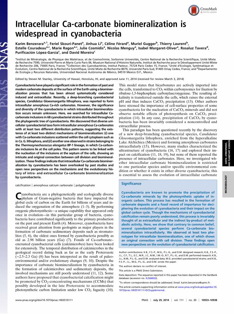
Fig. 1. Bayesian phylogenetic tree of 16S rRNA gene sequences of thecyanobacterial strains observed by electron microscopy. Strains forming in-tracellular Ca-carbonates are shown in color (green for those with Ca-car-bonate inclusions at the cell poles and red for those with inclusions scatteredin the cytoplasm). The tree is based on 1,292 conserved sites; numbers atbranches are posterior probabilities (only those >0.75 are shown).
10934 | www.pnas.org/cgi/doi/10.1073/pnas.1403510111 Benzerara et al.
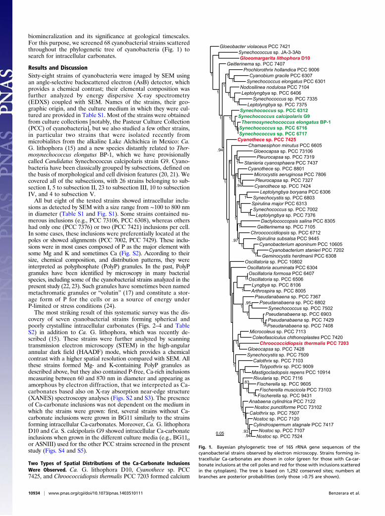
carbonate inclusions scattered throughout the cell cytoplasm(Fig. 2). In Cyanothece sp. PCC 7425, most of the cells containedbetween 2 and 20 Ca-carbonate inclusions measuring between130 and 700 nm. Few PolyP granules with no specific shape wereobserved, sometimes in very close spatial association with theCa-carbonate inclusions (Fig. 2D). Ca. G. lithophora D10 cellscontained between 3 and 19 Ca-carbonate inclusions measuringbetween 60 and 380 nm in diameter; they also contained PolyPgranules larger and in higher number than Cyanothece sp. PCC7425 cells (Fig. 2G). As a comparison, Ca. G. lithophora cellsobserved by Couradeau et al. (15) in laboratory aquaria thatcontained highly alkaline, Mg-rich solutions with Ca, Sr, and Bacontained no PolyP granule and a relatively higher number ofcarbonate inclusions enriched in Mg, Sr, and Ba. The absence orpresence of PolyP granules in Ca. G. lithophora cells can beexplained by the very low vs. high orthophosphate contents of thelaboratory aquaria vs. the BG11 medium, respectively, as shownpreviously for other strains (24). In contrast, Ca. G. lithophoraformed Ca-carbonate inclusions in laboratory aquaria as wellas BG11 medium with some variations in their chemical com-position (Mg, Sr, and Ba content), likely depending on theconcentration of these elements in the solutions. In Chroo-coccidiopsis thermalis PCC 7203, only few cells (baeocytes andvegetative cells) showed between 8 and 20 Ca-carbonateinclusions per cell, measuring between 350 and 870 nm indiameter (Fig. 2A).In contrast, Synechococcus sp. strains PCC6716, PCC6717, and
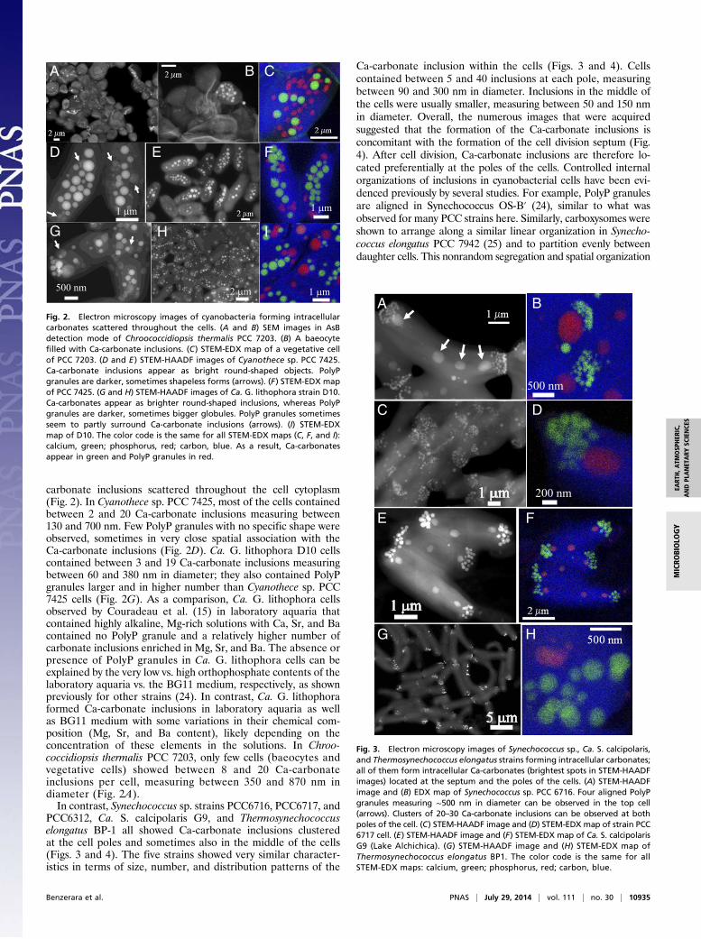
PCC6312, Ca. S. calcipolaris G9, and Thermosynechococcuselongatus BP-1 all showed Ca-carbonate inclusions clusteredat the cell poles and sometimes also in the middle of the cells(Figs. 3 and 4). The five strains showed very similar character-istics in terms of size, number, and distribution patterns of the
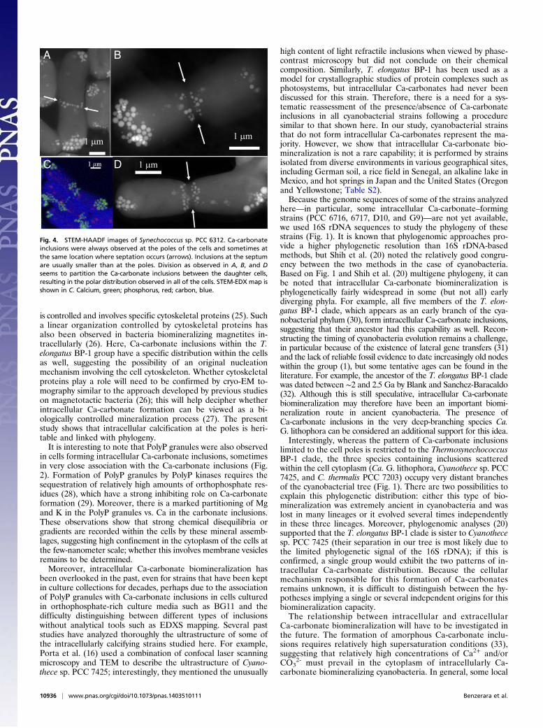
Ca-carbonate inclusion within the cells (Figs. 3 and 4). Cellscontained between 5 and 40 inclusions at each pole, measuringbetween 90 and 300 nm in diameter. Inclusions in the middle ofthe cells were usually smaller, measuring between 50 and 150 nmin diameter. Overall, the numerous images that were acquiredsuggested that the formation of the Ca-carbonate inclusions isconcomitant with the formation of the cell division septum (Fig.4). After cell division, Ca-carbonate inclusions are therefore lo-cated preferentially at the poles of the cells. Controlled internalorganizations of inclusions in cyanobacterial cells have been evi-denced previously by several studies. For example, PolyP granulesare aligned in Synechococcus OS-B′ (24), similar to what wasobserved for many PCC strains here. Similarly, carboxysomes wereshown to arrange along a similar linear organization in Synecho-coccus elongatus PCC 7942 (25) and to partition evenly betweendaughter cells. This nonrandom segregation and spatial organization
A B C
E
H
2 m
I
1 m
F
1 m1 m
D
500 nm
G
Fig. 2. Electron microscopy images of cyanobacteria forming intracellularcarbonates scattered throughout the cells. (A and B) SEM images in AsBdetection mode of Chroococcidiopsis thermalis PCC 7203. (B) A baeocytefilled with Ca-carbonate inclusions. (C) STEM-EDX map of a vegetative cellof PCC 7203. (D and E ) STEM-HAADF images of Cyanothece sp. PCC 7425.Ca-carbonate inclusions appear as bright round-shaped objects. PolyPgranules are darker, sometimes shapeless forms (arrows). (F) STEM-EDX mapof PCC 7425. (G and H) STEM-HAADF images of Ca. G. lithophora strain D10.Ca-carbonates appear as brighter round-shaped inclusions, whereas PolyPgranules are darker, sometimes bigger globules. PolyP granules sometimesseem to partly surround Ca-carbonate inclusions (arrows). (I) STEM-EDXmap of D10. The color code is the same for all STEM-EDX maps (C, F, and I):calcium, green; phosphorus, red; carbon, blue. As a result, Ca-carbonatesappear in green and PolyP granules in red.
C
500 nm
200 nm
A B
D
E F
HG
Fig. 3. Electron microscopy images of Synechococcus sp., Ca. S. calcipolaris,and Thermosynechococcus elongatus strains forming intracellular carbonates;all of them form intracellular Ca-carbonates (brightest spots in STEM-HAADFimages) located at the septum and the poles of the cells. (A) STEM-HAADFimage and (B) EDX map of Synechococcus sp. PCC 6716. Four aligned PolyPgranules measuring ∼500 nm in diameter can be observed in the top cell(arrows). Clusters of 20–30 Ca-carbonate inclusions can be observed at bothpoles of the cell. (C) STEM-HAADF image and (D) STEM-EDX map of strain PCC6717 cell. (E) STEM-HAADF image and (F) STEM-EDX map of Ca. S. calcipolarisG9 (Lake Alchichica). (G) STEM-HAADF image and (H) STEM-EDX map ofThermosynechococcus elongatus BP1. The color code is the same for allSTEM-EDX maps: calcium, green; phosphorus, red; carbon, blue.
Benzerara et al. PNAS | July 29, 2014 | vol. 111 | no. 30 | 10935
EART
H,A
TMOSP
HER
IC,
ANDPL
ANET
ARY
SCIENCE
SMICRO
BIOLO
GY
is controlled and involves specific cytoskeletal proteins (25). Sucha linear organization controlled by cytoskeletal proteins hasalso been observed in bacteria biomineralizing magnetites in-tracellularly (26). Here, Ca-carbonate inclusions within the T.elongatus BP-1 group have a specific distribution within the cellsas well, suggesting the possibility of an original nucleationmechanism involving the cell cytoskeleton. Whether cytoskeletalproteins play a role will need to be confirmed by cryo-EM to-mography similar to the approach developed by previous studieson magnetotactic bacteria (26); this will help decipher whetherintracellular Ca-carbonate formation can be viewed as a bi-ologically controlled mineralization process (27). The presentstudy shows that intracellular calcification at the poles is heri-table and linked with phylogeny.It is interesting to note that PolyP granules were also observed
in cells forming intracellular Ca-carbonate inclusions, sometimesin very close association with the Ca-carbonate inclusions (Fig.2). Formation of PolyP granules by PolyP kinases requires thesequestration of relatively high amounts of orthophosphate res-idues (28), which have a strong inhibiting role on Ca-carbonateformation (29). Moreover, there is a marked partitioning of Mgand K in the PolyP granules vs. Ca in the carbonate inclusions.These observations show that strong chemical disequilibria orgradients are recorded within the cells by these mineral assemb-lages, suggesting high confinement in the cytoplasm of the cells atthe few-nanometer scale; whether this involves membrane vesiclesremains to be determined.Moreover, intracellular Ca-carbonate biomineralization has
been overlooked in the past, even for strains that have been keptin culture collections for decades, perhaps due to the associationof PolyP granules with Ca-carbonate inclusions in cells culturedin orthophosphate-rich culture media such as BG11 and thedifficulty distinguishing between different types of inclusionswithout analytical tools such as EDXS mapping. Several paststudies have analyzed thoroughly the ultrastructure of some ofthe intracellularly calcifying strains studied here. For example,Porta et al. (16) used a combination of confocal laser scanningmicroscopy and TEM to describe the ultrastructure of Cyano-thece sp. PCC 7425; interestingly, they mentioned the unusually
high content of light refractile inclusions when viewed by phase-contrast microscopy but did not conclude on their chemicalcomposition. Similarly, T. elongatus BP-1 has been used as amodel for crystallographic studies of protein complexes such asphotosystems, but intracellular Ca-carbonates had never beendiscussed for this strain. Therefore, there is a need for a sys-tematic reassessment of the presence/absence of Ca-carbonateinclusions in all cyanobacterial strains following a proceduresimilar to that shown here. In our study, cyanobacterial strainsthat do not form intracellular Ca-carbonates represent the ma-jority. However, we show that intracellular Ca-carbonate bio-mineralization is not a rare capability; it is performed by strainsisolated from diverse environments in various geographical sites,including German soil, a rice field in Senegal, an alkaline lake inMexico, and hot springs in Japan and the United States (Oregonand Yellowstone; Table S2).Because the genome sequences of some of the strains analyzed
here—in particular, some intracellular Ca-carbonate–formingstrains (PCC 6716, 6717, D10, and G9)—are not yet available,we used 16S rDNA sequences to study the phylogeny of thesestrains (Fig. 1). It is known that phylogenomic approaches pro-vide a higher phylogenetic resolution than 16S rDNA-basedmethods, but Shih et al. (20) noted the relatively good congru-ency between the two methods in the case of cyanobacteria.Based on Fig. 1 and Shih et al. (20) multigene phylogeny, it canbe noted that intracellular Ca-carbonate biomineralization isphylogenetically fairly widespread in some (but not all) earlydiverging phyla. For example, all five members of the T. elon-gatus BP-1 clade, which appears as an early branch of the cya-nobacterial phylum (30), form intracellular Ca-carbonate inclusions,suggesting that their ancestor had this capability as well. Recon-structing the timing of cyanobacteria evolution remains a challenge,in particular because of the existence of lateral gene transfers (31)and the lack of reliable fossil evidence to date increasingly old nodeswithin the group (1), but some tentative ages can be found in theliterature. For example, the ancestor of the T. elongatus BP-1 cladewas dated between ∼2 and 2.5 Ga by Blank and Sanchez-Baracaldo(32). Although this is still speculative, intracellular Ca-carbonatebiomineralization may therefore have been an important biomi-neralization route in ancient cyanobacteria. The presence ofCa-carbonate inclusions in the very deep-branching species Ca.G. lithophora can be considered an additional support for this idea.Interestingly, whereas the pattern of Ca-carbonate inclusions
limited to the cell poles is restricted to the ThermosynechococcusBP-1 clade, the three species containing inclusions scatteredwithin the cell cytoplasm (Ca. G. lithophora, Cyanothece sp. PCC7425, and C. thermalis PCC 7203) occupy very distant branchesof the cyanobacterial tree (Fig. 1). There are two possibilities toexplain this phylogenetic distribution: either this type of bio-mineralization was extremely ancient in cyanobacteria and waslost in many lineages or it evolved several times independentlyin these three lineages. Moreover, phylogenomic analyses (20)supported that the T. elongatus BP-1 clade is sister to Cyanothecesp. PCC 7425 (their separation in our tree is most likely due tothe limited phylogenetic signal of the 16S rDNA); if this isconfirmed, a single group would exhibit the two patterns of in-tracellular Ca-carbonate distribution. Because the cellularmechanism responsible for this formation of Ca-carbonatesremains unknown, it is difficult to distinguish between the hy-potheses implying a single or several independent origins for thisbiomineralization capacity.The relationship between intracellular and extracellular
Ca-carbonate biomineralization will have to be investigated inthe future. The formation of amorphous Ca-carbonate inclu-sions requires relatively high supersaturation conditions (33),suggesting that relatively high concentrations of Ca2+ and/orCO3
2- must prevail in the cytoplasm of intracellularly Ca-carbonate biomineralizing cyanobacteria. In general, some local
1 m 1 m
1 m
A B
C D
Fig. 4. STEM-HAADF images of Synechococcus sp. PCC 6312. Ca-carbonateinclusions were always observed at the poles of the cells and sometimes atthe same location where septation occurs (arrows). Inclusions at the septumare usually smaller than at the poles. Division as observed in A, B, and Dseems to partition the Ca-carbonate inclusions between the daughter cells,resulting in the polar distribution observed in all of the cells. STEM-EDX map isshown in C. Calcium, green; phosphorus, red; carbon, blue.
10936 | www.pnas.org/cgi/doi/10.1073/pnas.1403510111 Benzerara et al.
increase in the concentration of CO32- may be expected close to
carboxysomes from which OH− are released to the cytoplasm bythe conversion of HCO3
− to CO2 by carboxysomal carbonicanhydrases (34). Intracellular calcification may occur in cyano-bacteria regulating less efficiently their intracellular pH. Alter-natively, regarding Ca2+, it has been shown that free Ca2+ ishighly regulated in the cytoplasm of living bacterial cells at a lowconcentration, including in some cyanobacteria (35); some var-iations can, however, occur (e.g., in response to stress) (35), andit has been noted that they may have a physiological role and thatcell division in particular appears very sensitive to the level ofintracellular Ca2+ in Escherichia coli (36). Interestingly, thepresent observations suggest that an increase of Ca2+ and/orCO3
2− concentrations may occur specifically during cell divisionand locally near the septum in the strains of the T. elongatus BP-1lineage as shown by the presence of Ca-carbonate inclusions.Whether some local Ca2+ influx related to cell division occurs inthese strains will have to be tested. Finally, the possible existenceof (specific or not) nucleation sites, and/or molecules stabilizingthe amorphous Ca-carbonates, and/or molecules inhibiting fur-ther Ca-carbonate growth after nucleation can be postulated toexplain (i) the discrete locations of Ca-carbonate inclusions, (ii)the persistence of these unstable amorphous phases, and (iii) thefact that the cytoplasm does not get eventually fully mineralized.In any case, such an intracellular biomineralization may lower atleast partly the export outside the cell of the alkalinity generatedby the photosynthetic activity and hence be detrimental to extra-cellular Ca-carbonate biomineralization. By sequestering at leasttransiently cytosolic inorganic carbon in solids, intracellularbiomineralization may also interfere with the physiology of thecells. Recent studies of Ca-carbonate precipitation in cyano-bacteria have been restricted to relatively few model strains—namely, Synechocystis sp. PCC 6803 (12, 37), Synechococcuselongatus PCC 7942 (38), Synechococcus sp. PCC 8806, andSynechococcus sp. PCC 8807 (18, 19). As shown here, Synecho-cystis sp. PCC 6803 does not form intracellular Ca-carbonateinclusions in BG11. Synechococcus elongatus PCC 7942 andSynechococcus sp. PCC 8806, previously observed using appro-priate techniques (19, 38), do not form intracellular Ca-car-bonate inclusions as well and can thus be considered as goodmodels to study extracellular biomineralization of Ca-carbonate.Here, alternatively, new models for the study of intracellularbiomineralization of Ca-carbonate are provided, offering a keyto better understand the mechanisms governing the interactionsbetween cyanobacteria and calcification by elucidating themolecular differences between these different patterns of bio-mineralization. Finally, many eukaryotic phyla have been shownto form intracellular amorphous minerals, some of which areinvolved, for example, in the production of the crystalline part ofexoskeletons (39). The possibility that at least part of the bio-chemical apparatus involved in this process has a unique evolu-tionary origin in cyanobacteria and eukaryotes will have to beexplored in the future.
Materials and MethodsStrains and Culture Conditions. Sixty-four axenic strains were available fromthe Pasteur Culture Collection of cyanobacteria (PCC strains) (20); they wereaxenic and have been described and studied by Rippka et al. (21) and Shihet al. (20). Culture media used for the different strains are reported in TableS1. Strains isolated from freshwater, soil, or thermal environments werecultured in medium BG-11 and its variants, and marine strains were culturedin the more saline medium ASN-III and its variants (Table S1) (21). Recipesof the culture media are available on http://cyanobacteria.web.pasteur.fr/.All PCC cultures were grown at 22 °C except PCC 6716, 6717, 9339, 9431, and9605, which were grown at 37 °C. An axenic Thermosynechococcus elon-gatus strain BP-1, isolated from a hot spring in Beppu (Japan), was providedby Alain Boussac (Commissariat à l’Énergie Atomique et aux Énergies Alter-natives Saclay, Gif-sur-Yvette, France) and was cultured at 37 °C in BG11. Onenonaxenic strain was an enrichment from Yellowstone. Two nonaxenic
strains were enriched from Lake Alchichica. Ca. G. lithophora D10 was pre-viously enriched and described in Couradeau et al. (15). A second strain wasenriched following the same protocol; because this strain was phylogeneti-cally relatively distant from Synechococcus sp. PCC 6312, PCC 6716, and PCC6717, and because it was enriched from a mesophilic environment on thecontrary to Thermosynechococcus elongatus BP-1, we propose the followingstatus for this new strain enriched from Lake Alchichica: order Chroo-coccales, Ca. S. calcipolaris sp. nov. Both cultures of Ca. G. lithophora D10and Ca. S. calcipolaris G9 were grown in BG11 at 22 °C.
Microscopy Sample Preparation. A total of 0.5 mL of cultures was centrifugedat 8,000 × g for 10 min. Pellets were rinsed three times in Milli-Q (mQ) water(Millipore). After the final centrifugation, pellets were resuspended in200 μL of mQ water. A drop of 5 μL was deposited on a carbon-coated 200-mesh copper grid and let dry at ambient temperature.
Electron Microscopy Analyses. SEM observations were performed on a ZeissSupra 55 SEM microscope at a 10-kV working voltage with a working distanceof 7.5 mm, an aperture of 60 μm, and at high current. SEM images werecollected with the AsB detector. Elemental maps of Ca, P, and C were retrievedfrom hyperspectral images (HyperMap) in an EDXS analysis for each pixel ofthe image. Some strains, especially those forming intracellular Ca-car-bonates, were further analyzed by TEM using a JEOL-2100F microscope op-erating at 200 kV, equipped with a field emission gun, a JEOL detector withan ultrathin window allowing detection of light elements, and a STEM device,which allows Z-contrast imaging in the HAADF mode. Compositional map-ping was acquired by performing EDXS analysis in the STEM HAADF mode.
Scanning Transmission X-ray Microscopy Analyses. STXM analyses were per-formed at the carbon K-edge and at the Ca L2,3-edges. Cultures wereresuspended in distilled water and deposited on silicon nitride windows.STXM and XANES analyses were performed on beamline 11.0.2.2 of theAdvanced Light Source (Lawrence Berkeley National Laboratory). Energycalibration was achieved using the well-resolved 3p Rydberg peak of gas-eous CO2 at 294.96 eV. A 25-nm zone plate was used. Cell proteins, poly-phosphate, and carbonate inclusions were discriminated based on theirdifferent spectral signatures at the C K-edge and the Ca L2,3-edges followingthe approach detailed by Cosmidis et al. (40).
Phylogenetic Analysis. Genomic DNA was extracted from an enriched cultureof Ca. S. calcipolaris with the PowerBiofilm DNA Isolation Kit (MoBio)and used for 16S rRNA gene PCR amplification with the specific cyano-bacterial primers CYA106F (CGGACGGGTGAGTAACGCGTGA) and 23S30R(CTTCGCCTCTGTGTGCCTAGGT). PCR reactions were carried out with 30cycles (each one including denaturation at 94 °C for 15 s, annealing at 55 °Cfor 30 s, and extension at 72 °C for 2 min), preceded by 2-min denaturationat 94 °C, and followed by 7-min extension at 72 °C. PCR products were di-rectly sequenced by Beckman Coulter Genomics using the Cya106F forwardprimer and the 1492R (GGTTACCTTGTTACGACTT) as reverse universal primerfor bacteria. The other cyanobacterial 16S rRNA gene sequences were re-trieved directly from GenBank (http://ncbi.nlm.nih.gov/). Sequences werealigned using MAFFT (41), and the conserved sites were identified usinggBlocks (42). Bayesian phylogenetic analysis was done using MrBayes 3.2.2(43) with the general time-reversible model of sequence evolution, andtaking among-site rate variation into account by using an eight-categorydiscrete approximation of a Γ distribution and a proportion of invariantsites. Two parallel MCMC runs with four chains each (three hot and one cold)were performed for 10 million generations and sampled every 10,000 gen-erations, discarding a burn-in of 25% before constructing a consensus tree.The 16S rRNA gene sequence of Ca. S. calcipolaris has been submitted toGenBank under accession no. KJ566932.
ACKNOWLEDGMENTS. Funding for this work was provided by the EuropeanResearch Council under European Community’s Seventh Framework Pro-gramme FP7/2007-2013 Grant 307110, ERC CALCYAN; the Pasteur CultureCollection of cyanobacteria supported by the Institut Pasteur (M.G. andT.L.); the SEM facility at L’Institut de Minéralogie, de Physique des Matériauxet de Cosmochimie (IMPMC) purchased by Région Ile de France GrantSESAME 2006 I-07-593/R; the transmission electron microscopy facility atIMPMC purchased by Region Ile de France Grant SESAME 2000 E 1435; andAdvanced Light Source Molecular Environmental Science beamline 11.0.2 sup-ported by the Office of Science, Office of Basic Energy Sciences, Division ofChemical Sciences, Geosciences, and Biosciences and Materials Sciences Divi-sion, US Department of Energy at Lawrence Berkeley National Laboratory.
Benzerara et al. PNAS | July 29, 2014 | vol. 111 | no. 30 | 10937
EART
H,A
TMOSP
HER
IC,
ANDPL
ANET
ARY
SCIENCE
SMICRO
BIOLO
GY
1. Tomitani A, Knoll AH, Cavanaugh CM, Ohno T (2006) The evolutionary diversificationof cyanobacteria: Molecular-phylogenetic and paleontological perspectives. Proc NatlAcad Sci USA 103(14):5442–5447.
2. Rosing MT, Bird DK, Sleep NH, Glassley W, Albarede F (2006) The rise of continents—Anessay on the geologic consequences of photosynthesis. Palaeogeogr PalaeoclimatolPalaeoecol 232(2-4):99–113.
3. Buick R (2008) When did oxygenic photosynthesis evolve? Philos Trans R Soc Lond BBiol Sci 363(1504):2731–2743.
4. Jansson C, Northen T (2010) Calcifying cyanobacteria—the potential of biomineraliza-tion for carbon capture and storage. Curr Opin Biotechnol 21(3):365–371.
5. Golubic S, Lee SJ (1999) Early cyanobacterial fossil record: Preservation, palae-oenvironments and identification. Eur J Phycol 34(4):339–348.
6. Altermann W, Kazmierczak J, Oren A, Wright DT (2006) Cyanobacterial calcificationand its rock-building potential during 3.5 billion years of Earth history. Geobiology4(3):147–166.
7. Bosak T, Knoll AH, Petroff AP (2013) The meaning of stromatolites. Annu Rev EarthPlanet Sci 41(5):21–44.
8. Klein C, Beukes NJ, Schopf JW (1987) Filamentous microfossils in the early ProterozoicTransvaal Supergroup: Their morphology, significance, and paleoenvironmental set-ting. Precambrian Res 36(1):81–94.
9. Arp G, Reimer A, Reitner J (2001) Photosynthesis-induced biofilm calcification andcalcium concentrations in Phanerozoic oceans. Science 292(5522):1701–1704.
10. Riding R (2006) Cyanobacterial calcification, carbon dioxide concentrating mecha-nisms, and Proterozoic–Cambrian changes in atmospheric composition. Geobiology4(4):299–316.
11. Kamennaya NA, Ajo-Franklin CM, Northen T, Jansson C (2012) Cyanobacteria as bi-ocatalysts for carbonate mineralization. Minerals 2(2):338–364.
12. Jiang HB, Cheng HM, Gao KS, Qiu BS (2013) Inactivation of Ca(2+)/H(+) exchanger inSynechocystis sp. strain PCC 6803 promotes cyanobacterial calcification by upregu-lating CO(2)-concentrating mechanisms. Appl Environ Microbiol 79(13):4048–4055.
13. Merz M (1992) The biology of carbonate precipitation by cyanobacteria. Facies 26(1):81–101.
14. Obst M, Wehrli B, Dittrich M (2009) CaCO3 nucleation by cyanobacteria: Laboratoryevidence for a passive, surface-induced mechanism. Geobiology 7(3):324–347.
15. Couradeau E, et al. (2012) An early-branching microbialite cyanobacterium formsintracellular carbonates. Science 336(6080):459–462.
16. Porta D, Rippka R, Hernández-Mariné M (2000) Unusual ultrastructural features inthree strains of Cyanothece (cyanobacteria). Arch Microbiol 173(2):154–163.
17. Allen MM (1984) Cyanobacterial cell inclusions. Annu Rev Microbiol 38:1–25.18. Lee BD, Apel WA, Walton MR (2006) Calcium carbonate formation by Synechococcus
sp. strain PCC 8806 and Synechococcus sp. strain PCC 8807. Bioresour Technol 97(18):2427–2434.
19. Liang A, Paulo C, Zhu Y, Dittrich M (2013) CaCO3 biomineralization on cyanobacterialsurfaces: Insights from experiments with three Synechococcus strains. Colloids Surf BBiointerfaces 111C(11):600–608.
20. Shih PM, et al. (2013) Improving the coverage of the cyanobacterial phylum usingdiversity-driven genome sequencing. Proc Natl Acad Sci USA 110(3):1053–1058.
21. Rippka R, Deruelles J, Waterbury JB, Herdman M, Stanier RY (1979) Generic assign-ments, strain histories and properties of pure cultures of cyanobacteria. J Gen Mi-crobiol 111(3):1–61.
22. Morohoshi T, et al. (2002) Accumulation of inorganic polyphosphate in phoU mutantsof Escherichia coli and Synechocystis sp. strain PCC6803. Appl Environ Microbiol 68(8):4107–4110.
23. Jacobson L, Halmann M (1982) Polyphosphate metabolism in the blue-green algaMicrocystis aeruginosa. J Plankton Res 4(3):481–488.
24. Gomez-Garcia MR, Fazeli F, Grote A, Grossman AR, Bhaya D (2013) Role of poly-phosphate in thermophilic Synechococcus sp. from microbial mats. J Bacteriol 195(15):3309–3319.
25. Savage DF, Afonso B, Chen AH, Silver PA (2010) Spatially ordered dynamics of thebacterial carbon fixation machinery. Science 327(5970):1258–1261.
26. Scheffel A, et al. (2006) An acidic protein aligns magnetosomes along a filamentousstructure in magnetotactic bacteria. Nature 440(7080):110–114.
27. Weiner S, Dove PM (2003) An overview of biomineralization processes and theproblem of the vital effect. Rev Mineral Geochem 54:1–29.
28. Omelon SJ, Grynpas MD (2008) Relationships between polyphosphate chemistry,biochemistry and apatite biomineralization. Chem Rev 108(11):4694–4715.
29. Dove PM, Hochella F, Jr (1993) Calcite precipitation mechanisms and inhibition byorthophosphate: In situ observations by scanning force microscopy. Geochim Cos-mochim Acta 57(3):705–714.
30. Robertson BR, Tezuka N, Watanabe MM (2001) Phylogenetic analyses of Synecho-coccus strains (cyanobacteria) using sequences of 16S rDNA and part of the phyco-cyanin operon reveal multiple evolutionary lines and reflect phycobilin content. Int JSyst Evol Microbiol 51(Pt 3):861–871.
31. Szöllosi GJ, Boussau B, Abby SS, Tannier E, Daubin V (2012) Phylogenetic modeling oflateral gene transfer reconstructs the pattern and relative timing of speciations. ProcNatl Acad Sci USA 109(43):17513–17518.
32. Blank CE, Sánchez-Baracaldo P (2010) Timing of morphological and ecological in-novations in the cyanobacteria—a key to understanding the rise in atmospheric ox-ygen. Geobiology 8(1):1–23.
33. Wang D, et al. (2012) Revisiting geochemical controls on patterns of carbonate de-position through the lens of multiple pathways to mineralization. Faraday Discuss159:371–386.
34. Rae BD, Long BM, Badger MR, Price GD (2013) Functions, compositions, and evolutionof the two types of carboxysomes: Polyhedral microcompartments that facilitate CO2fixation in cyanobacteria and some proteobacteria. Microbiol Mol Biol Rev 77(3):357–379.
35. Barrán-Berdón AL, Rodea-Palomares I, Leganés F, Fernández-Piñas F (2011) Free Ca2+
as an early intracellular biomarker of exposure of cyanobacteria to environmentalpollution. Anal Bioanal Chem 400(4):1015–1029.
36. Holland IB, Jones HE, Campbell AK, Jacq A (1999) An assessment of the role of in-tracellular free Ca2+ in E. coli. Biochimie 81(8-9):901–907.
37. Han Z, et al. (2013) Precipitation of calcite induced by Synechocystis sp. PCC6803.World J Microbiol Biotechnol 29(10):1801–1811.
38. Obst M, et al. (2009) Precipitation of amorphous CaCO3 (aragonite-like) by cyano-bacteria: A STXM study of the influence of EPS on the nucleation process. GeochimCosmochim Acta 73(14):4180–4198.
39. Addadi L, Raz S, Weiner S (2003) Taking advantage of disorder: Amorphous calciumcarbonate and its roles in biomineralization. Adv Mater 15:959–970.
40. Cosmidis J, Benzerara K (2014) Soft X-ray scanning transmission micro-spectroscopy.Handbook of Biomineralization, eds Gower L, DiMasi E (Taylor & Francis, London).
41. Katoh K, Misawa K, Kuma K, Miyata T (2002) MAFFT: A novel method for rapidmultiple sequence alignment based on fast Fourier transform. Nucleic Acids Res30(14):3059–3066.
42. Castresana J (2000) Selection of conserved blocks from multiple alignments for theiruse in phylogenetic analysis. Mol Biol Evol 17(4):540–552.
43. Ronquist F, et al. (2012) MrBayes 3.2: Efficient Bayesian phylogenetic inference andmodel choice across a large model space. Syst Biol 61(3):539–542.
10938 | www.pnas.org/cgi/doi/10.1073/pnas.1403510111 Benzerara et al.