Embed Size (px)
Citation preview
Veterinary Surgery, 19,5380-388, 1990
Intra-Articular Replacement of Bovine Cranial Cruciate Ligaments with an Autogenous Fascia1 Graft
Transected cranial cruciate ligaments in nine Holstein heifers were replaced with an autograft harvested from fascia on the medial surface of the gluteobiceps muscle and a connected segment of the lateral patellar ligament. There was minimal lameness at month 1, and all animals were sound by month 2. There was no evidence of degenerative joint disease in any stifles examined at necropsy on days 90, 120, or 365. Augmented healing of the severed cranial cruciate ligaments occurred in several animals. The mean failure strength of the cranial cruciate ligament replacements was 29.7% of the failure strength of the contralateral normal cranial cruciate ligament in four heifers. The technique was successful in eight of 13 bulls and cows with ruptured cranial cruciate ligaments. Animals treated successfully had significantly lower body weights than those with unsuccessful outcomes.
R A N I A L CRUCIATE LIGAMENT (CCI,) injuries in dairy C and beef cattle result in significant economic losses from inability to endure pasture conditions, loss of con- dition, reduced milk production, conception failures, and inability of bulls to mount for semen collection.',* Acute injury occurs from falls associated with slippery footing, ataxia from metabolic disease, or estrous behav- ior. In chronic injuries, the CCL may be stretched or par- tially ruptured. At the University of Wisconsin, chronic injuries are usually seen in breeding bulls. It has been suggested that degenerative joint disease may precede CCL injury in bulls.' This form of injury is thought to be related to the forces generated when the stifle is extended during mounting and copulation. The pathogenesis of the injury in breeding bulls may be similar to acute CCL injuries associated with stifle hyperextension in dog^.^-^
Nine of 27 cattle (33%) examined for stifle injuries in a 23-month period had acute CCL injuries. This inci- dence is slightly higher than that reported (2 1.4%) in a retrospective study of stifle disease in cattle.? There are no published reports ofthe morbidity of the condition in the dairy and beef cattle populations.
Conservative management of CCL injuries in cattle by box stall rest, with or without some form ofexternal limb immobilization and anti-inflammatory medication, may
be satisfactory when complete rupture has not occurred, but this form of treatment offers a poor prognosis for maintaining the animal in useful production.' Surgical repair of bovine CCL injuries has been described.'.*,'-'' Stabilizing the joint by plication of the dorsolateral and dorsomedial retinacular tissues has been reported.* It was concluded from a study of intra-articular replace- ment of the CCL with nonvascularized autografts from various tissue sources alone and in combination with synthetic implants that skin was a suitable prosthetic ma- terial.' A strip of fascia lata was used for intra-articular replacement of a ruptured CCL in a Jersey bull.' A seg- ment of the lateral patellar ligament and fascia from the gluteobiceps muscle were used in an evaluation of intra- articular repair of CCL injury.'.''
The purpose of this paper is to describe a technique for replacing the CCL in cattle with autogenous tissue hav- ing sufficient strength to maintain joint stability. and to report the results obtained with this technique in 13 clini- cal cases.
Experimental Trial Anafomy
The superficial gluteal and biceps femoris muscles are continuous in cattle, forming the gluteobiceps muscle
From the Department of Surgical Science, School of Veterinary Medicine, University of Wisconsin-Madison, Madison, Wisconsin. This study was conducted at the School of Veterinary Medicine, University of Wisconsin-Madison. Funding for the project was by the Graduate
Excerpts from this paper were presented at the Annual Meeting of the Veterinary Orthopedic Society, Breckenridge, Colorado, February,
Reprint requests: W. H. Crawford, DVM, School of Veterinary Medicine, University of Wisconsin-Madison, 201 5 Linden Drive West, Madison,
School, University of Wisconsin-Madison.
1988, and the Annual Meeting of the American Association of Bovine Practitioners, Calgary, Alberta, Canada, September, 1988.
WI 53706.
380

CRAWFORD 38 1
(Fig. l) ." The medial fascia ofthe gluteobiceps muscle is 5 to 7 mm thick and approximately 7 cm wide i n a ma- ture Holstein cow. Distally, the fibers of this fascia are continuous with the fibers ofthe lateral patellar ligament. A bursa over the lateral epicondyle of the femur is deep to the aponeurosis of the gluteobiceps fascia and the lat- eral patellar ligament.
A strip of fascia from the medial surface of the gluteo- biceps muscle was incised so that it was continuous with the lateral patellar ligament. This patellar ligament-glu- teobiceps strip was used to form an intra-articular re- placement for the CCL.
Mar criul\ und Method\
Nine Holstein heifers weighing 320 to 365 kg without radiographic cvidence of preexisting stifle joint disease were used in the study. The right stifle of each animal was used as a nonsurgical control, while the left CCL was surgically transected but not removed. and a replace-
E -
G
/I
D
- F
. c
. A
Fig 1 Autogenousgraft formed from the fascia of the right gluteobi- ceps muscle and the lateral patellar ligament A cranial portion of the gluteobiceps muscle (E) has been removed to show the underlying gluteobiceps fascia (G) A-proximal extent of the tibial crest, B- lateral patellar ligament, C-middle patellar ligament, D-vastus later- alis muscle, F-lateral epicondyle of the femur
Fig 2 Graft passer manufactured by the University of Wtsconsin- Madison, Clinical Science Center machine shop The overall length is 30 cm and the blade width is 12 mm The radius of curvature in the instruments varies from 6 5 to 10 0 cm, depending upon the size of the patient The instrument enters between the middle and lateral pa- tellar ligaments and exits through the muscle fibers of the lateral head of the gastrocnemius muscle at the lateral epicondyle
ment was provided by using a modification of the over- the-top technique. '
Before surgery, food was withheld for 48 hours and water was withheld for 24 hours. Anesthesia was induced with thiamylal* and guaifenesin,? and maintained with halothane.$ Each heifer was placed in right lateral re- cumbency. After aseptic preparation and draping, a skin incision was made from the major trochanter to the tibial crest. The fascia lata was incised over and parallel to the plane between the vastus lateralis and gluteobiceps mus- cles. A strip 2.5 cm wide was dissected from the cranial margin of the medial fascia of the gluteobiceps muscle, parallel to its fibers. The dissection was continued through the fibrocartilaginous thickening over the lateral epicondylar bursa and extended distally to divide the lat- eral patellar ligament in a frontal plane parallel to its fi- bers. A 30 cm continuous tissue strip was thereby formed, attached distally to the tibial crest and contain- ing the craniomedial half of the lateral patellar ligament, a portion of the supra-bursa1 fibrocartilage, and approxi- mately 20 cm ofthe gluteobiceps fascia (Fig. 1). The fem-
* Bio-tal. Bio-Ceutic Laboratories Inc. St. Joseph. MO. t Gecolate. Summit Hill Labs. Akalon, NJ. $ Fluothane. Ayerst Labs. New York. NY.

382 BOVINE CRANIAL CRUCIATE LIGAMENT REPLACEMENT
Fig. 3. A bone staple used to attach the graft to the lateral epicondy- lar surface of the femur. The legs of this cobalt-chromium alloy staple spread as it is driven into the bone. The triangular barbs on the legs reduce the potential for the staple to back out.
orotibial joint was approached between the middle and lateral patellar ligaments. The patellar fat pad was re- flected with self-retaining retractors, and the CCL was identified and transected. The stifle was flexed and ex- tended to permit palpation of the free ends of the CCL. A custom-manufactured curved graft passerg (Fig. 2), fashioned after a previously described instrument,'' was placed through the intercondylar space in a cranial to caudal direction, exiting through the fibers of the lateral head of the gastrocnemius muscle at the lateral epicon- dyle. A 60 cm length of 12 mm umbilical tape was threaded into the eye of the graft passer, which was then retracted from caudal to cranial, leaving the umbilical tape in the route to be taken by the CCL replacement. The fascioligamentous strip was sutured to the umbilical tape and trimmed to a width of I .75 to 2.5 cm to allow it to pass through the intercondylar space. The strip was drawn from cranial to caudal through the intercondylar space, exiting through the muscle fibers at the origin of the lateral head of the gastrocnemius muscle. Periosteum was elevated from the epicondyle. The fascioligamen- tous strip, after being pulled tightly through the intercon- dylar space with the stifle held in approximately 140" of flexion, was stapled to the prepared area of the epicon- dyle with a 1.75 cm bone staple.7 (Figs. 3 ,4) The free end
$ Bill Messer. Machine shop, Clinical Science Center. University of
7 Techmedica Inc. Camarillo. CA. Wisconsin-Madison. Madison. WI.
E -
f
Fig. 4. The autograft (G) in place before application of tension and stapling. A-tibia1 crest; B-lateral patellar ligament; C-middle pa- tellar ligament; D-vastus lateralis muscle; E-gluteobiceps muscle.
of the strip was folded caudad, over the stapled attach- ment and a second staple was placed to include a double thickness of the strip. A short piece of 18-gauge stainless steel wire was placed as a marker in the fascial strip and its location relative to the most caudal staple was noted. The fascial planes, lateral retinaculum of the patella, and joint capsule were closed with size I polyglactin 910." A suction drain was placed in the wound and the skin was closed with size 2 nylon.# A stent bandage was placed on the skin.
The heifers were returned to their stalls for recovery from anesthesia. Suction drains were removed when they no longer produced fluid, usually within 12 to 24 hours. Procaine penicillin (10,000 IU/kg IM) was administered 2 hours before surgery and twice daily until 24 hours af- ter the suction drain was removed. A lateral radiograph was made 1 day after removal of the suction drain.
To simulate clinical conditions, each heifer was kept in a box stall for approximately 5 days, in group box stall housing for 60 days, and then in stanchion housing. Phenylbutazone (4 mg/lb) was to be administered orally to any animal not bearing weight on the treated limb at hour 24. At days 30 and 60, each animal was examined
' I Vicryl. Ethicon. Inc, Somerville, NJ. t Ethilon, Ethicon, Inc, Somerville. NJ.

CRAWFORD 383
and graded for the degree of lameness. Three heifers were randomly selected for euthanasia on each of days 90, 120. and 365. Both stifles of each animal were examined immediately after euthanasia.
Biomechanical tests were conducted on the stifles from four heifers. After removing all soft tissue except the CCL or CCL replacement, the femoral diaphysis was transected approximately 5 cm proximal to the proximal extent of the trochlea. The tibia was transected approxi- mately 15 cm distal to the tibial articular surface. The joints were stored at -20°C until testing was performed. The treated joints from three heifers (E3, E5, E9) were excluded from the strength analysis because of artifacts created during preparation of the specimens. The four pairs of stifles tested biomechanically were from animals euthanatized at days 90 (E2). 120 (E4 and E6). and 365 (E8).
In order to apply the distracting forces ofthe materials testing machine** through a line corresponding to the origin and insertion of the CCL, the hole for a Yx inch transverse pin was drilled from the lateral surface of the femoral condyle approximately 2 cm distal to thc junc- tion of the caudal and middle thirds of a line marking the position of the physis. The hole was thereby located in the epiphysis at the distal extent of the linea aspera femoris (Fig. 5 ) .
Custom-manufactured clamps.tt which held the tib- ial diaphysis at 20" from a vertical plane and allowed the femoral component to rotate about the axis of the trans- verse femoral pin, were fastened to the femur and tibia (Fig. 6). The joint was distracted at a velocity of 10 cm per second until the CCL or CCL replacement failed. The force at failure was recorded by interfacing$$ the materials testing machine with a microcomputer.33 The failure force of the CCL replacement was calculated as a percentage of the failure force of the normal CCL.
K c \ 1 /It 5
All heifers were reluctant to bear weight on the trcated limb for 3 to 5 days. but none was considered painful enough. by the protocol criteria, for administration of phenylbutazone. At month 1 . two animals had slightly shortened cranial phases to the stride ofthe treated limb. and at month 2. all the heifers appeared to have normal gaits.
In several treated stifles (E3. E6, E8. and E9) there were fibrous tissue attachments between the tibial remnant of thc CCL and the CCL replacement. The CCL in the left
** lnstron model 1350. Canton. MA. i f Bill Maser. Machine shop. Clinical Science Center. Uiii\ersit) o f
i t Sensorhus modulc SB2003 Transcnsorl Devices Inc. F-rcciiiont.
$4 Maclntosti. 4pplc C'omputcr Inc. Cupertino. CA.
Wisconsin-Madison. Madison. WI.
C.4.
Fig. 5. Position of hole for the transverse pin in the femur to test the strength of bovine CCLs. When the femoral and tibia1 components are distracted, the attachments of the CCL and the pin are aligned, with the forces transmitted in a plane parallel to the fibers of the CCL.
stifle of heifer E3 had healed with the only evidence of transection being a discolored area at the previous inci- sion site. A vascularized, membranous tissue surrounded the CCL replacements. The staples at the proximal end of the replacement strip were buried under a layer of fi- brous tissue. Dense fibrous adhesions had formed be- tween the CCL replacement strip and the caudal aspect of the joint capsule where the graft passed through the intercondylar space, adjacent to the meniscofemoral lig- ament and the supracondylar area. There was no evi- dence of injury to the menisci or the articular cartilages. No marginal osteophytes were present in any of the joints with replacement CCLs (Fig. 7).
No differences could be detected in soundness at the time of euthanasia or in gross necropsy findings between the animals at each termination period.
The mean failure strength of four CCL replacements, expressed as a percentage of the strength of the contralat- era1 CCL. was 29.7 t 1.67 (SD). All CCL replacements failed by the replacement tissue being elevated off the caudal aspect of the femur and pulling out from under the staples. One of the normal CCLs failed at its femoral origin, but the other normal ligaments ruptured in the middle segment of their length.

384 BOVINE CRANIAL CRUCIATE LIGAMENT REPLACEMENT
Fig. 6. Clamps used to hold the femoral (top) and tibial (bottom) com- ponents to the materials testing machine. The femoral component ro- tates about the transverse pin, while the tibial diaphysis is held at 20" from the vertical plane.
Clinical Application
,!4atcrials mid Mctiiod,s
Over 25 months. 13 animals (9 bulls and 4 cows) un- derwent autogenous replacement of a ruptured CCL. The bulls were from artificial insemination centers. Six had been in semen collection programs. and three youn- ger bulls were awaiting the results of progeny evaluation. The affected stifles were radiographed before surgery. Preanesthetic management and antibiotic administra- tion were as described in the experimental protocol. Phenylbutazone was administered as required to pa- lients that appeared depressed as a result of postoperative pain.
Bulls used for artificial insemination were maintained in box stalls and semen was not collected for 3 months. Bulls awaiting the results of progeny evaluation tests were maintained in individual paddocks. Owners were asked to maintain thc cows in box stalls for 6 weeks be-
fore returning them to their normal pasture or housing environment.
Follow-up information was gained from clinical or necropsy examinations by the author (7 animals) or the referring veterinarian (4 animals), or by conversation with the owner (2 animals). A comparison of the mean body weights of successful and unsuccessful patients was made with a one-tail t test.
Rcwlts
In addition to obvious lameness, the most consistent signs of CCL rupture in cattle were visible instability of the stifle when weight-bearing animals were walked (1 1 cases), an elicited cranial drawer sign (8 cases). and an increase in elicited internal tibial rotation ( 5 cases) (Ta- ble I ) . Pronounced femorotibial joint space effusion was present in five animals, while in most of the others a slight increase in effusion was palpable between the me- dial patellar and medial collateral ligaments.
Cranial subluxation of the tibia was evident on radio- graphs of 12 animals. Degenerative joint disease was rec- ognized radiographically in I I animals, and avulsion fractures ofthe intercondylar eminence were observed in five animals.
Replacement of the CCL was successful in eight ani- mals (C I , C2, C3. C7, C8, C 10, C 1 1, C 13). Three of the six bulls in artificial insemination collection centers (C1, C2. C3) were able to resume service for 12 to 15 months. The treated stifles from all six bulls were recovered when they were taken out of service and sent to slaughter. Bull C3 became quite lame at month 18; there were advanced degenerative changes in the joint. and the CCL replace- ment was no longer present. Bulls CI and C2 had ad- vanced degenerative joint disease but the graft was still present.
Two bulls awaiting the results of progeny tests re- turned to soundness, although subsequent injuries re- sulted in their demise. One femur was fractured in bull C7 in month 18, and a coxofemoral luxation occurred in bull C8 in month 5 . At necropsy. both CCL replace- ments were intact and there was only minimal evidence of degenerative joint disease.
The surgery was considered to be successful in three cows. Cows C I I and C I3 are still alive at months I9 and 12. while cow C10 was culled from the milking herd at month 13 because of chronic mastitis. The stifle of cow C 10 was unavailable for postmortem examination.
In the bulls in which surgical repair failed (C4, C5. C6. C9), dehiscence ofthe lateral fascia of the joint appeared to be the most significant problem. In one large bull (C4). there was dehiscence of the skin incision and septic ar- thritis developed. However. at necropsy the replacement ligament remained intact. In bull C9, severe osteochon- dritis dissecans lesions were noted at the time of surgery

CRAWFORD 385
Fig. 7. Postmortem specimen Of
a cranial cruciate ligament replace- ment graft. Heifer E8, year 1
on the lateral trochlear ridge. intertrochlear groove, and patella. In this animal. the suture line in the lateral fascia of the joint and the lateral femoropatellar ligament su- tures failed, resulting in medial displacement of the pa- tella. The lameness resulting from these complications caused the bull to fall and fracture a large metatarsal bone. At necropsy. the graft was intact.
The graft failed in two bulls. Bull C6, with advanced degenerative joint disease at the time of surgery, never returned to serviceable soundness after surgery and in- stability of the joint recurred in month 8. At necropsy in month 9, the graft was no longer present within the joint but as in bull C3, fibrous tissue was still in place under the staples. In bull C5, instability of the stifle was noted in week 2. This bull was taken home by the owner and has been unavailable for follow-up. but the clinical signs indicated failure of the graft.
Cow C 12 had radial nerve paralysis after recovery from anesthesia and was euthanatized at week I . The CCL replacement was intact.
The mean body weight (778 kg) of the animals in which surgery was successful was significantly less than the mean body weight (918 kg) of those in which it was unsuccessful (p < 0.05 j .
Discussion
I n dogs, the over-the-top CCL replacement proccdure places the prosthesis in approximately the path of the normal CCL. and eliminates potential inaccuracies when a graft is passed through bone tunnels.' '' Although corresponding anatomic studies in cattle have not been
published, it seemed reasonable to use a similar ap- proach.
After transection of the lateral femoropatellar liga- ment, the patella can be luxated medially if the leg is placed in extension.'" This maneuver was not conducted in the experimental series. but in some of the clinical cases it provided a means for better assessment of the femoropatellar joint space and associated structures. It has been stated that the relatively large medial trochlear ridge prevents postoperative medial patellar luxation in cattle." However, in this study, two bulls had medial lux- ation of the patella at necropsy. In one bull, the osteo- chondritis dissecans lesions were so severe that after cu- rettage of the lesions, abnormal congruency between the articulating surfaces of the patella and femur may have resulted in a tendency for the patella to luxate medially.
The segment of fascia harvested from the medial sur- face of the gluteobiceps muscle is approximately 4 mm thick in a 300 kg heifer and approximately 7 mm thick in a 1200 kg bull. In animals the size ofthe heifers, calcu- lated values for cross-sectional area requirements indi- cate the CCL would have a cross-sectional area of ap- proximately 0.6 1 crn2.l4 The tensile strength of the liga- mentous fascia1 strip has not been determined. A strip of material 4 mrn thick and 2.5 cm wide provided approxi- mately 1 .0 cm' of cross-sectional area for the prosthesis. Pulling a strip this size through the intercondylar space is difficult and requires that the umbilical tape leader be securely fastened to the graft tissue. The graft passer de- signed for use in this procedure provided a more direct method than that previously described for pulling the leader through the tissues and inter-condylar joint space of cattle.'."'
386 BOVINE CRANIAL CRUCIATE LIGAMENT REPLACEMENT
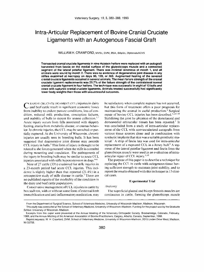
TABLE 1. Summary of Clinical Cases of Ruptured Cranial Cruciate Ligaments in Cattle Repaired with an Autogenous Graff
FOIIOW-UP Case Breed/ Age Weight Radiographic No. Sex (yr) (kg) Location Time* Findings Duration Result
c 1
c 2
c 3
c 4
c 5
C6
c7
C8
c 9
c10
c11
c12
C13
Holstein Male
Polled Hereford Male
Holstein Male
Holstein Male
Holstein Male
Holstein Male
Holstein Male
Holstein Male
Holstein Male
Holstein
Angus Female
Female
Holstein Female
Holstein Female
4
7
5
8
8
6
1
3
2
7
7
7
5
91 0
1050
1136
1275
1135
955
400
81 0
523
700
600
700
620
Left
Right
Left
eft
eft
eft
Right
Left
Left
Left
Right
Right
Right
1
8
3
3
8
40
2
1
8
1
12
1
3
Moderate DJD,t OCD,S cranial subluxation tibia
Moderate DJD
Osteophytes, fracture ICES cranial subluxation tibia, moderate DJD
Osteophytes, fracture ICE, cranial subluxation tibia, DJD
cranial subluxation tibia
Osteophytes, lateral collateral ligament injury, fracture ICE, DJD, cranial subluxation tibia
Fracture ICE, DJD,
Joint effusion
Osteophytes, cranial subluxation tibia
OCD lesions of trochlea, and patella, DJD
Moderate DJD, cranial subluxation tibia
Minimal osteophytes, cranial subluxation tibia
Cranial subluxation tibia
Osteophytes, fracture ICE, DJD, joint distension
17 months
15 months
18 months
3 months
2 months
9 months
18 months
5 months
2 months
13 months
19 months
1 week
12 months
Success. Remained in A.I. service, culled for genetic reasons, advanced DJD
Success. A.I. service, culled for genetic reasons, patellar luxation, DJD
Success. A.I. service, moderate bilateral lameness, CCL replacement gone, advanced DJD
Failure. Wound dehiscence, septic arthritis, CCL replacement intact
Failure. Progressed well for 2 weeks then sudden joint instability, lost to follow-up
to return to A.I. use, CCL replacement gone
Failure. Chronic lameness, unable
Success. Fracture femoral neck,
Success. Coxofemoral luxation,
Failure. Fracture metatarsus in
CCL replacement intact, no DJD
CCL replacement intact
barn accident, patellar luxation, CCL replacement intact
culled for mastitis, no necropsy
lameness
Success. Moderate lameness,
Success. Remains in herd, no
Failure. Radial nerve paralysis, euthanatized, CCL replacement intact
herd, minimal lameness Success. Remains in breeding
Time in weeks from first sign of CCL injury until surgical repair t Degenerative joint disease. # Osteochondritis dissecans 9 Intercondylar eminence
In a previous study in cattle. bone plates used to secure CCL grafts to the femur were believed to have caused pain.’ The staples used in the current study appeared to perform their intended function with no associated com- plications. There was no evidence of slippage of the re- placement tissues in the postoperative radiographs or at necropsy examination. N o lameness was noted that could be associated with the staples.
Dehiscence of the lateral fascia1 incision was a problem in the larger bulls. This occurred with flexion of the stifle and has been a recognized complication after lateral stifle arthrotomy in horse^.'^.'^ Mild pain that prevents the an-
imal from fully flexing the stifle in the early postoperative period may be advantageous. Therefore, analgesia should be used only if there is significant pain. Early post- operative pain should be distinguished from pain that continues due to an unstable joint.
The size of the patient is an important prognostic fac- tor with this procedure. Other important considerations are the degree and duration of degenerative joint disease, the presence of other joint diseases such as osteochondri- tis dissecans or intra-articular fractures, and the de- meanor of the animal.
Degenerative joint disease was present at the initial

CRAWFORD 387
Fig. 8. The importance of the lo- cation of distracting forces. a. When the loading pin is the posi- tion used for testing the strength of a normal CCL, an eccentric loading is created by the distance between the loading pin and the staple. b. When the load is applied, the femo- ral component rotates about the axis of the pin and causes the ad- hesions between the graft and the caudal surface of the distal femur to be torn. c. A testing system that applies the distracting load parallel to the fibers of the graft will take advantage of the strength devel- oped by the adhesions.
a b c
preoperative examination in 11 of the 13 clinical pa- tients. Eight of these animals were examined within 3 weeks of the onset of signs associated with the CCL in- jury. This supports the view that degenerative joint dis- ease precedes CCL rupture or that degenerative joint dis- ease develops more rapidly after instability than is gener- ally accepted.’
In a trial in which six steers were observed for up to 5 months after replacement of transected CCLs with glu- teobiceps-patellar ligament grafts, the authors suggested that a longer-term study was required and, based on the biomechanical strength assessments of the replacement ligaments, proposed the technique would not be useful in breeding bulls at 4 or 5 months following repair.“’ In the study reported here, three heifers observed for 6 months and three heifers observed for I year were still clinically normal. Three of six bulls in this study that weighed over 900 kg were returned to artificial insemina- tion service for longer than 1 year after CCL replace- ment.
The clinical results obtained are inconsistent with the values obtained for the failure strength ratios in this and a previous study.’ In The “over-the-top” technique has become a clinically successful method to replace the CCL in dogs.I4 However, biomechanical testing of the CCL replacements placed by this technique in the canine stifle showed the ultimate strength of the grafts to be 14% to 28% of the strength of the normal CCL.” I t has been suggested that the discrepancy between the clinical and biomechanical results was due to the fact that biological tissues are subjected to forces ranging from one-tenth to not more than one-fourth of their breaking loads under normal conditions.”
In this study, the equipment and testing procedure were designed to determine the rupture strength of nor- mal CCLs. In order to cause the normal CCL to undergo tension failure rather than shear failure at its origin or insertion, the distracting force was directed in a plane
through, and parallel to, the fibers of the normal CCL. The load-carrying pin was placed distal to the femoral physis to avoid distracting the metaphysis from the epiphysis (Fig. 5). With the pin in this location, the sta- pled attachment of the CCL replacement was approxi- mately 1.5 cm caudal to the center of the pin, thereby creating eccentric loading (Fig. 8a). This eccentricity (moment) created rotational displacement of the joint, causing the CCL replacement to be torn perpendicularly (“peeled”) away from the caudal aspect of the femur (Fig. 8b). At the conclusion of the rotation the staple and the transverse pin were aligned vertically, with the distract- ing force acting directly on the staple and allowing the replacement strip to be pulled out from under the staples. All CCL replacements failed in this manner. It appears this method of testing the prosthesis does not take into account the full strength of adhesions between the re- placement tissues and the caudal aspect of the femur and joint capsule. The importance of applying the tensile load along the axis of the ligament being tested has been demonstrated.I8 When the load is applied through the axis of the ligament or its replacement, a maximum number of collagen bundles resist the deformation; therefore, higher strength properties are recorded. It is possible that had the replacement ligaments been dis- tracted in the manner suggested in Figure 8c, the ulti- mate tensile strength might have been higher and more consistent with the clinical assessment of the procedure. A more physiologic method of testing the system might be to distract the joint with the CCL or its replacement and the caudal cruciate ligaments intact. The caudal cru- ciate ligament would prevent rotation of the femur about the transverse pin, thereby preventing the prosthesis from being torn off the femur in a perpendicular direc- tion.
In conclusion, based on the results from the heifers, it appears that the autogenous graft formed from a contin- uous strip of gluteobiceps fascia and the lateral patellar
388 BOVINE CRANIAL CRUCIATE LIGAMENT REPLACEMENT
ligament can provide a CCL replacement that will main- tain joint stability for at least 1 year in a joint free of preexisting disease. Furthermore. it appears that the re- placement graft will provide enough strength to support a mature cow with a naturally occurring CCL rupture. If the limited number of large breeding bulls operated on in this trial can be used as an indicator. about 50% can be salvaged for at least an additional year of breeding ser- vice.
References
1 . Hamilton GF. Adams OR. Anterior cruciate repair in cattle. J Am Vet Med Assoc 1971:158:378-183.
2. Nelson DR. Koch DB. Surgical stabilization of the stifle i n cranial cruciate ligament injury i n cattle. Vet Rec 1982: 1 I I :256)-162.
3 . Huhn JC. Kneller SK. Nelson DR. Radiographic assessment of cranial cruciate ligament rupture in the dairy cow: A retrospec- tive study. Vet Radio1 19 86;27: 184- 186.
4. Rudy RL. Stifle joint. In: Archibald J. ed. ( h r i i r z c Sirrgrvy Santa Barbara: American Veterinary Publications, Inc. I965:86 I .
5. Strande A. Replacement of the anterior cruciate ligament in the dog. J Sm Anini Pract 1966:7:35 1-359.
6. Arnoczky SP. Marshall JL. The cruciate ligaments of the canine stifle: An anatomical and functional analysis. Am J Vet Res 1977:38: 1807-1 8 14.
7. Ducharme NG. Stanton ME. Ducharme GR. Stifle lameness in cattle at two veterinary teaching hospitals: A retrospective study of42 cases. Can Vet J 1985:26:212-~17.
8. Hofmeyr CFB. Reconstruction of the ruptured anterior cruciate ligament in the stifle o f a bull. The Veterinarian 1968;5:89-92.
9. Moss EW. The cranial cruciate ligament in the bovine: Its tensile strength and surgical repair using a patellar ligament graft. 1980. M.S. thesis. Iowa State University. Ames. Iowa.
10. Moss EW. McCurnin DM. Ferguson TH. Experimental cranial cruciate replacement in cattle using a patellar ligament graft. Can Vet J 1988:29:157-162.
I I . Gctty R. Si.c.\or? mid Gro.s,srnan '.s .4iiufoinj' c ! / ' t / i c DomcJstic' .In;- rnols. 5th ed. Philadelphia: W. B. Saunders. 1975:849-850.
11. Arnoczky SP. Tarvin GB. Marshall JL. Saltzman B. The over-the- top procedure: A technique for anterior cruciate ligament sub- stitution in the dog. J Am Anim Hosp Assoc 1979: 15:283-290.
13. van Oosterom RA. Intra-articular graftpasser. Vet Surg 1982: 1 I : 131-133.
14. Moss EM, Ferguson MS. Tensile strength of the cranial cruciate ligament in cattle. Am J Vet Res 1980:41:1408-141 I .
15. Trotter GW. Mcllwraith CW. Norrdin RW. A comparison of two surgical approaches to the equine femoropatellar joint for the treatment of osteochondritis dissecans. Vet Surg 1983; I2:33- 40.
16. Pascoe JR. Pool RR. Wheat JD. O'Brien TR. Osteochondral de- fects of the lateral trochlear ridge of the distal femur ofthe horse: Clinical. radiographic. and pathological examination and results ofsurgical treatment. Vet Surg 1984: 13:99-1 10.
17. Butler DL. Hulse DA. Kay MD. et al. Biomechanics of cranial cruciate ligament reconstruction in the dog: 11. Mechanical properties. Vet Surg 1983: 12: I I 3- I 18.
18. Woo SL-Y. Hollis JM. Roux RD. et al. Effects of knee flexion on the structural properties of the rabbit femur-anterior cruciate ligament-tibia complex (FATC). J Biomech 1987:20:557-563.
ACVS COLLEGE CALENDAR
Veterinary Surgical Forum October 28-30,1990 Chicago I European Surgical Forum
ACVS Veterinary Symposium and Annual Meeting
European Surgical Forum
April 12-14, 1991
October 1 1-1 8,1991
May 8-1 0,1992
Nice, France
San Francisco
Wurtzburg, West Germany
ACVS Veterinary Symposium October 30-November 6,1992 Miami
ACVS Veterinary Symposium October 22-29,1993 San Francisco
and Annual Meeting
and Annual Meeting