Embed Size (px)
Citation preview

Intra-Aortic Clusters Undergo Endothelial toHematopoietic Phenotypic Transition during EarlyEmbryogenesisChiyo Mizuochi1, Stuart T. Fraser2, Katia Biasch3, Yuka Horio1, Yoshikane Kikushige4, Kenzaburo Tani5,
Koichi Akashi4, Manuela Tavian3, Daisuke Sugiyama1*
1 Department of Hematopoietic Stem Cells,SSP Stem Cell Unit, Kyushu University Faculty of Medical Sciences, Fukuoka, Japan, 2 Laboratory of Blood Cell Development,
Disciplines of Physiology, Anatomy and Histology, School of Medical Sciences, University of Sydney, Camperdown, New South Wales, Australia, 3 Unite 682 INSERM,
Strasbourg, France, 4 Department of Medicine and Biosystemic Science, Kyushu University Graduate School of Medical Sciences, Fukuoka, Japan, 5 Department of
Molecular Genetics, Medical Institute of Bioregulation, Kyushu University, Fukuoka, Japan
Abstract
Intra-aortic clusters (IACs) attach to floor of large arteries and are considered to have recently acquired hematopoietic stemcell (HSC)-potential in vertebrate early mid-gestation embryos. The formation and function of IACs is poorly understood. Toaddress this issue, IACs were characterized by immunohistochemistry and flow cytometry in mouse embryos.Immunohistochemical analysis revealed that IACs simultaneously express the surface antigens CD31, CD34 and c-Kit. Asembryos developed from 9.5 to 10.5 dpc, IACs up-regulate the hematopoietic markers CD41 and CD45 while down-regulating the endothelial surface antigen VE-cadherin/CD144, suggesting that IACs lose endothelial phenotype after9.5 dpc. Analysis of the hematopoietic potential of IACs revealed a significant change in macrophage CFC activity from 9.5to 10.5 dpc. To further characterize IACs, we isolated IACs based on CD45 expression. Correspondingly, the expression ofhematopoietic transcription factors in the CD45(neg) fraction of IACs was significantly up-regulated. These results suggestthat the transition from endothelial to hematopoietic phenotype of IACs occurs after 9.5 dpc.
Citation: Mizuochi C, Fraser ST, Biasch K, Horio Y, Kikushige Y, et al. (2012) Intra-Aortic Clusters Undergo Endothelial to Hematopoietic Phenotypic Transitionduring Early Embryogenesis. PLoS ONE 7(4): e35763. doi:10.1371/journal.pone.0035763
Editor: Alfons Navarro, University of Barcelona, Spain
Received March 3, 2011; Accepted March 22, 2012; Published April 27, 2012
Copyright: � 2012 Mizuochi et al. This is an open-access article distributed under the terms of the Creative Commons Attribution License, which permitsunrestricted use, distribution, and reproduction in any medium, provided the original author and source are credited.
Funding: This research was supported in part by the Project for Realization of Regenerative Medicine, Special Coordination Funds for Promoting Science andTechnology of the Ministry of Education, Science, Sports and Culture (www.mext.go.jp/english); and SAKURA program of the Japan Society for the Promotion ofScience (www.jsps.go.jp/english/index.html). The funders had no role in study design, data collection and analysis, decision to publish, or preparation of themanuscript.
Competing Interests: The authors have declared that no competing interests exist.
* E-mail: [email protected]
Introduction
During mouse embryogenesis, hematopoiesis begins at the
extra-embryonic yolk sac (YS) at 7.5 days post-coitum (dpc) and
shifts to fetal liver after mid-gestation, then to spleen and finally to
bone marrow shortly before birth. There are two distinct waves of
hematopoietic emergence: a transient wave, primarily restricted to
erythropoiesis in YS blood islands prior to the connection of the
circulation from the YS to the embryo; and a definitive wave
originating in both the YS and embryo proper. The embryonic site
has been identified in the aortic region, in the para-aortic
splanchnopleura (p-Sp)/aorta-gonad-mesonephros (AGM) region
[1–6]. Functional hematopoietic stem cells (HSCs) that can
reconstitute adult recipients are first identified in the AGM region
at 10.5 dpc after ex vivo organ culture [7]. The cells at 10.5 dpc
that were not cultured ex vivo rarely reconstitute adult recipients,
whereas those at 11.5 dpc can regardless [7–9]. Therefore, the
cells that acquire HSC activity after culture step, have been
termed ‘‘pre-HSC’’s. Although several reports characterize the
surface marker expression on both pre-HSCs at 10.5 dpc and
HSCs at 11.5 dpc, the developmental process of HSC generation
still remains unclear [8–11]. Cell populations capable of
reconstituting neonatal recipients are detected in the p-Sp/AGM
region at 9.5 dpc [12–13]. These observations suggest that
ancestor cells of HSC from the p-Sp/AGM region at 9.5 dpc
require special microenvironments to acquire HSC activity and
that HSCs undergo phenotypic changes from 9.5 to 10.5 dpc. In
the AGM region, intra-aortic/arterial clusters (IACs) are observed
attached to floors of large arteries in several species including
chicken, mouse and humans [3]. Mouse IACs have been
characterized morphologically and are primarily located in three
large arteries, namely, the dorsal aorta (DA), the omphalomesen-
teric (vitelline) artery (OMA; VA) and the umbilical artery (UA)
[3,14–15]. IACs express both hematopoietic (CD41 and CD45)
and endothelial (CD31, CD34 and VE-cadherin) surface markers
[3,15–16] suggesting that IACs are likely equivalent to ancestor
cells of HSC and/or pre-HSCs and are derived from endothelial
cells (ECs) at aortic/arterial regions. Although recent genetic
approaches and novel tracing methods demonstrate that IACs are
derived from ECs in zebrafish and mice, it is unclear how IACs
form and acquire HSC activity [17–25].
To address how IACs form and function in HSC generation, we
first visualized IACs by immunohistochemistry and confocal
imaging and were found to simultaneously express CD31, CD34
and c-Kit. This approach enabled us to investigate the phenotypic
PLoS ONE | www.plosone.org 1 April 2012 | Volume 7 | Issue 4 | e35763

characterization of IACs by flow cytometry and hematopoiesis
assays. Here, we demonstrate a significant transition from
endothelial to hematopoietic cell phenotype of IAC cells after
9.5 dpc.
Results
Visualization of IACs in mouse embryosPrevious studies identified intra-aortic/arterial clusters (IACs)
primarily by immunocytochemistry and microscopy [3,14–15].
Recently, we successfully visualized hematopoietic cell clusters in
mouse placenta using thick (20 mm) cryo-sections and antibodies
recognizing the embryonic HSC markers c-Kit, CD31 and CD34
and applied this method to quantifying IACs [26]. Cell aggregates
consisting of more than three c-Kit-positive cells were defined as
an IAC. Here, we used confocal microscopy to expand upon our
previous study and characterize the cell types found within IACs
according to c-Kit, CD31 and CD34 expression (Figure 1). The
first IACs were observed as spherical structures in the omphalo-
mesentric artery (OMA) at 9.0 dpc (12–14 somite pairs [SP])
(Figure 1A, left). Between 9.5 dpc (18–22 SP) to 10.5 dpc (30–34
SP), large arteries such as the dorsal aorta (DA), OMA and
umbilical artery (UA) form [14]. IACs were observed in DA,
OMA and UA at 10.5 dpc, and the size of IACs in the OMA and
UA was significantly larger than those seen in the DA (Figure 1A,
right). Localization of IACs in DA was not restricted to the ventral
wall of DA, but rather some IACs were observed at dorsal and
lateral sides of the wall (data not shown). All IACs in the DA,
OMA and UA at 10.5 dpc simultaneously expressed c-Kit, CD31
and CD34 (Figure 1B-D). IACs expressing c-Kit in the different
arteries analyzed were also positive for Ki-67, a marker of cell
proliferation, regardless of location, suggesting that cells within
IACs are highly proliferative (Figure 1E).
Characterization of IACs by flow cytometry andhematopoietic progenitor assays
To further characterize IACs, the caudal portion of embryos
containing the p-Sp/AGM region was dissociated and analyzed by
flow cytometry. At 10.5 dpc, c-Kit+/CD31+/CD34+ cells, which
are equivalent to IACs, were assessed for expression of the cell
surface markers VE-cadherin/CD144 (an endothelial cell marker),
CD41 (the earliest hematopoietic cell marker), CD45 (a pan-
leukocyte marker), Sca-1 (a late fetal and adult HSC marker) and
CD150 and EPCR (adult HSC markers) (Figure 2A-H). c-Kit+/
CD31+/CD34+ cells represented 0.06960.01% in whole caudal
portion of embryos. Among c-Kit+/CD31+/CD34+ cells, VE-
cadherin surface antigen expression decreased significantly within
24 hours from 9.5 to 10.5 dpc. Concomitantly, expression of the
hematopoietic markers CD41 and CD45 increased from negative
or low levels of expression on IAC cells at 9.5 dpc to abundant
Figure 1. Confocal images of IACs expressing CD31/CD34/c-Kitin the AGM region. Transverse sections of AGM region from ICRmouse embryos at 9.0 and 10.5 dpc were stained with antibodies andobserved by confocal microscopy. (A) IACs were observed in the
omphalomesenteric artery (OMA) at 9.0 dpc (left; magnified view ofIACs in upper right panel) and in the OMA, dorsal aorta (DA) andumbilical artery (UA) at 10.5 dpc (right). CD31 (red), c-Kit (green), andTOTO-3 (blue). Arrows indicate IACs. Original magnification is 20x. (B-D)IACs were observed in the DA (B), OMA (C) and UA (D) at 10.5 dpc. Leftpanel shows staining for CD31 (red), c-Kit (green), and TOTO-3 (blue),and right panel shows staining for CD34 (red), c-Kit (green), and TOTO-3(blue) staining. Images were taken at 40x and zoom was used to show adetail at right lower panel. Another IAC in the DA is shown in Figure S1.(E) IACs expressing Ki-67, a marker of proliferation, were observed in theDA (left), OMA (middle) and UA (right). Ki-67 (red), c-Kit (green), andTOTO-3 (blue). Images were taken at 40x and zoom was used to show adetail.doi:10.1371/journal.pone.0035763.g001
Characterization of Intra-Aortic Clusters
PLoS ONE | www.plosone.org 2 April 2012 | Volume 7 | Issue 4 | e35763

Figure 2. Flow cytometric analysis of CD31+/CD34+/c-Kit+ AGM cells using surface expression of hematopoietic and endothelial cellmarkers. Single cell suspensions of the caudal portion of embryos containing the p-Sp/AGM region at 9.5 and 10.5 dpc were prepared and analyzedby flow cytometry. (A) Cells expressing CD31, CD34 and c-Kit markers of IACs were gated first. Isotype control of flow cytometric analysis is shown in
Characterization of Intra-Aortic Clusters
PLoS ONE | www.plosone.org 3 April 2012 | Volume 7 | Issue 4 | e35763

levels at 10.5 dpc. Sca-1 expression also increased from 9.5 to
10.5 dpc.
We next separated c-Kit+/CD31+/CD34+ cells based on CD45
expression by flow cytometry and performed colony assays and
transplantation assays. As shown in Figure 2I (left), the number of
CFU-M generated from CD45-positive c-Kit+/CD31+/CD34+
cells (27.3) was significantly higher than CFU-M from CD45-
negative c-Kit+/CD31+/CD34+ cells (8.0) (p,0.05). However, the
total number of hematopoietic colonies did not differ between
CD45-negative and CD45-positive c-Kit+/CD31+/CD34+ cells
(p.0.05). When 50–100 c-Kit+/CD31+/CD34+ cells were
transplanted into neonate recipients, there was no significant
difference in reconstitution ability (CD45-negative, 3.55%; CD45-
positive 3.07%) (p.0.05) (Figure 2J). c-Kit+/CD31+/CD34+ cells
at 9.5 dpc were able to reconstitute recipients and chimerism to
9.89% was achieved. Presumptive ancestor cells of HSC can
reportedly reconstitute neonate recipients but not adult recipients
[13]. In addition, pre-HSCs at 10.5 dpc rarely reconstitute adult
recipients without culture step [7–9,11]. When 100 c-Kit+/
CD31+/CD34+ cells were transplanted into adult recipients, no
reconstitution was observed (data not shown).
Expression of CD45 in mouse and human intra-aortic/arterial clusters
CD45-negative and CD45-positive c-Kit+/CD31+/CD34+ cells
showed no difference in hematopoietic potential except within the
macrophage lineage. To further investigate a role of CD45
expression on c-Kit+/CD31+/CD34+ cells, we used flow cytom-
etry to segregate c-Kit+/CD31+/CD34+ cells into three fractions.
Three distinct populations became apparent; CD45negative cells,
CD45low cells, and CD45high cells (Figure 3A). The proportion of
CD45-negative and CD45-low positive c-Kit+/CD31+/CD34+
cells was higher at 9.5 dpc than at 10.5 dpc, whereas the
percentage of CD45-high positive c-Kit+/CD31+/CD34+ cells
increased by 5-fold at 10.5 dpc (31.0%) compared to 9.5 dpc
(6.3%) (Figure 3B). These data suggest that CD45-negative c-Kit+/
CD31+/CD34+ cells are precursors of CD45-high positive c-Kit+/
CD31+/CD34+ cells and that CD45 is a marker of IAC
maturation. To address this issue, we examined expression levels
of the gene encoding CD45 (Ptprc; protein tyrosine phosphatase, receptor
type, C) and of various hematopoietic transcription factors (Runx1,
c-Myb, Evi-1, SCL and Gata2) (Figure 3C-H). CD45-negative c-
Kit+/CD31+/CD34+ cells expressed low levels of CD45 mRNA.
Ptprc transcript levels increased significantly as CD45 surface
protein expression was up-regulated in the c-Kit+/CD31+/CD34+
population. Expression levels of all hematopoietic transcription
factor genes assayed except Evi-1 was highest in CD45-low positive
c-Kit+/CD31+/CD34+ cells. In agreement with flow cytometric
analysis, evaluation of CD45 protein expression by immunohis-
tochemistry indicated that IACs in the OMA at 9.5 dpc were
CD45-negative while some IACs in the DA, OMA and UA were
CD45-positive by 10.5 dpc (Figure 4A-D).
IAC formation in the developing human embryo is poorly
defined. Having defined the developmental progression of IAC in
the mouse above, we next examined IAC morphology and
phenotype in a 32 day-old human embryo. Immunohistochemis-
try of embryonic human cryosections was performed using anti-
human CD34 and CD45 antibodies. As shown in Figure 4E, IACs
can be detected in ventral wall of the dorsal aorta. CD34 was
expressed by a wide range of vascular endothelial cells throughout
the embryo. CD45 was restricted to round and in many cases
clearly circulating cells. However, within the IAC observable on
the ventral wall of the dorsal aorta, cells expressing both CD34
and CD45 can be seen. This reflects the expression pattern we
have identified in embryonic mouse IACs.
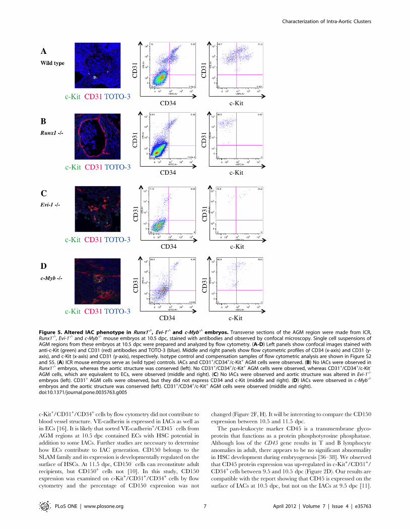
Transcription factor hierarchy in IAC developmentWe next observed IAC formation by immunohistochemistry
and flow cytometry in mouse embryos harboring mutations
associated with aberrant embryonic hematopoiesis [27–32].
Immunohistochemical analysis of Runx1-/- embryos lacked IACs
in the DA, OMA and UA. Flow cytometric analyses confirmed the
absence of c-Kit+/CD31+/CD34+ cells in Runx1-/- embryos
compared to wild type embryos (Figure 5A-B). Evi-1-/- embryos
also lacked IACs in the DA, OMA and UA by immunohisto-
chemistry. However, a small frequency of c-Kit+/CD31+/CD34+
cells could be detected by flow cytometry (Figure 5C). In c-Myb-/-
embryos, IACs were observed at the DA, OMA and UA, and c-
Kit+/CD31+/CD34+ cells were also observed by flow cytometry
(Figure 5D). Collectively, these results demonstrate that Runx1 is
essential for IAC formation while Evi-1 appears to be playing a
function downstream of Runx1 in this process.
Discussion
During embryogenesis, a unique cell biological shift takes places
in which endothelial cells with adherens junctions detach from
each other, alter gene expression and become hematopoietic cells.
This process is limited both anatomically and temporally. We here
demonstrated that the transition from endothelial to hematopoi-
etic phenotype of IACs occurs from 9.5 dpc in the mouse embryo,
earlier than previously described. Furthermore, we show that IACs
are identifiable in the human embryo based on CD45 expression,
implying that this process in mice is applicable to human.
Previously, we reported an immunohistochemistry visualization
technique revealing hematopoietic cell clusters in placenta using
thick (20 mm) cryo-sections and antibodies recognizing embryonic
HSC markers [26]. Here, we applied this technique to obtain high
quality confocal images of intra-aortic/arterial clusters (IACs) in the
AGM region. We defined IACs as c-Kit+/CD31+/CD34+ cells.
Recently, c-Kit+/CD31+/SSEA-1– cells were also identified in the
AGM region [11]. As CD31 is expressed on both IACs and
primordial germ cells (PGCs), it was necessary to exclude PGCs
according to SSEA-1 expression. As shown in Figure 2 and 5, we
Figure S2. (B-G) Expression of hematopoietic and endothelial cell markers was analyzed on CD31+/CD34+/c-Kit+ cells at 9.5 dpc (left) and 10.5 dpc(right) with the following antibodies: (B) VE-cadherin/CD144 (an endothelial cell marker), (C) CD41 (the earliest hematopoietic cell marker), (D) CD45(a pan-leukocyte marker), (E) Sca-1 (a late fetal and adult HSC marker), (F) CD150 and (G) EPCR (adult HSC markers). At least 1,000 cells were assessedfor each surface antigen. Representative profiles are shown. (H) Percentage of expression was summarized. At least 3 independent experiments wereperformed. Mean 6 2SD was calculated and shown at the top of bars. (I) One thousand sorted CD45-negative or CD45-positive CD31+/CD34+/c-Kit+
cells were cultured in semisolid medium containing the hematopoietic cytokines, SCF (Stem Cell Factor), IL (Interleukin)-3, IL-6 and EPO(Erythropoietin). Left and right panels show each fraction and the total number of colonies, respectively. GEMM (colony-forming units of granulocyteerythrocyte monocyte macrophages); GM (of granulocyte macrophages); M (of macrophages); G (of granulocytes); BFU (burst forming units oferythroid cells). (J) 50–100 sorted CD31+/CD34+/c-Kit+ cells at 9.5 dpc, as well as CD45-negative and CD45-positive CD31+/CD34+/c-Kit+ cells weretransplanted into busulfan-treated Ly5.1 mouse neonates. Approximately one year after transplantation, blood samples were collected and analyzedfor CD45.2 expression by flow cytometry. Representative profile of flow cytometric analysis and its negative and positive controls are shown in FigureS3 and S6, respectively.doi:10.1371/journal.pone.0035763.g002
Characterization of Intra-Aortic Clusters
PLoS ONE | www.plosone.org 4 April 2012 | Volume 7 | Issue 4 | e35763

could observe a small number of CD31+/CD34– cells, which are
likely to be PGCs. Since PGCs do not express CD34 at this stage, we
could positively select the IAC fraction based on our definition by
flow cytometry [33]. Our observation of IACs is compatible with the
result showing large IACs were primarily observed in omphalome-
sentric artery (OMA) and umbilical artery (UA) at 10.5 dpc [11]. In
the mouse, IACs protruding into the lumen of arteries were
previously reported at 9.5 dpc in studies using microscopy and Tie-
2 immunohistochemistry [14,34]. Prior to 9.5 dpc, we identified the
first IACs, which formed a spherical structure, in the OMA at
9.0 dpc (Figure 1A). The OMA appears at 8.0 dpc and directly
connects with the dorsal aorta (DA). The OMA anastomoses with
the DA after 9.5 dpc and loses its connection with the UA by
10.5 dpc [14,35]. Our data (Figure 1E) indicate that IACs are
proliferative, based on Ki-67 staining. Taken together, it is likely
that the first IACs in the OMA proliferate and are distributed into
Figure 3. Gene expression analysis in CD31+/CD34+/c-Kit+ AGM cells separated by CD45 expression. (A) Single cell suspensions of thecaudal portion of embryos containing the AGM region at 10.5 dpc were prepared and analyzed by flow cytometry. Cells expressing CD31 and CD34,IAC markers, were first gated. The profile shows expression of c-Kit (x-axis) and CD45 (y-axis) in CD31+/CD34+ AGM cells (left). Based on intensity ofCD45 expression, CD31+/CD34+/c-Kit+ AGM cells were separated into three fractions, CD45-negative (under 102 of CD45-fluorescence, same asnegative control), -low positive (from 102.5 to 103.5 of CD45-fluorescence), and -high positive (approximately over 104 of CD45-fluorescence). Isotypecontrol and compensation samples of flow cytometric analysis are shown in Figure S4 and S5. (B) The percentage of CD45-negative, -low positive,and -high positive c-Kit+/CD31+/CD34+ AGM cells was calculated both at 9.5 dpc (white bars) and 10.5 dpc (black bars). (C-H) Gene expression ofCD45 (C), Runx1 (D), c-Myb (E), Evi-1 (F), SCL (G) and Gata2 (H) was analyzed in sorted CD45-negative, -low positive and -high positive c-Kit+/CD31+/CD34+ AGM cells. Expression levels of CD45 mRNA are up-regulated as c-Kit+/CD31+/CD34+ cells express CD45 surface protein. Expression levels ofRunx1, c-Myb, Evi-1, SCL and Gata2 were highest in CD45-low positive c-Kit+/CD31+/CD34+ cells, whereas that of Evi-1 was highest in CD45-negative c-Kit+/CD31+/CD34+ cells. RQ represents relative quantity of template in the original sample.doi:10.1371/journal.pone.0035763.g003
Characterization of Intra-Aortic Clusters
PLoS ONE | www.plosone.org 5 April 2012 | Volume 7 | Issue 4 | e35763

large arteries, such as the DA and UA, as the arterial system
develops. Although several reports provide direct evidence that
endothelial cells (ECs) generate IACs, we cannot rule out the
possibility that either mesodermal cells, the ancestors of hemato-
poietic cells, or so-called hemangioblasts, which give rise both to
ECs and hematopoietic cells, generate IACs by another pathway
[17–25]. When VE-cadherin+/CD45– cells were sorted out from
AGM regions at 10.5 dpc, and co-aggregated with OP9 stromal
cells, these cells acquired HSC activity [8]. As embryos develop,
VE-cadherin+/CD45+ cells from AGM regions at 11.5 dpc can
reconstitute adult recipients without culture step, whereas both VE-
cadherin+/CD45+/– cells can after aggregation culture with OP9
stromal cells. It suggests that the transition from endothelial to
hematopoietic phenotype in pre-HSCs occurs between 10.5 and
11.5 dpc. According to our flow cytometric analysis of IACs, the
transition from endothelial to hematopoietic phenotype occurs after
9.5 dpc (Figure 2). Although we found that 33% of c-Kit+/CD31+/
CD34+ cells at 9.5 dpc express VE-cadherin, most IACs defined as
Figure 4. Expression of CD45 by mouse and human IACs. Transverse sections of AGM region were made from ICR mouse embryos at 9.5 and10.5 dpc and from human embryos at 32 day-old, according to the Carnegie classification, stained with antibodies and observed by confocalmicroscopy. Arrowheads indicate IACs. (A) Mouse IACs in the omphalomesenteric artery (OMA) at 9.5 dpc expressed c-Kit, but not CD45. CD45(green) and c-Kit (red). Magnified view of IACs is shown at right upper panel in Merge panel. Original magnification is 40x. (B-D) Mouse IACs in thedorsal aorta (DA) (B), OMA (C) and umbilical artery (UA) (D) at 10.5 dpc expressed c-Kit, and some expressed CD45. CD45 (green) and c-Kit (red).Original magnification is 40x. (E) All human IACs in the DA expressed CD34, and some expressed CD45. CD34 (green), CD45 (red) and TOTO-3 (blue).NT (Neural Tube); Ao (Aorta); Mn (Mesonephros). Original magnification is 20x.doi:10.1371/journal.pone.0035763.g004
Characterization of Intra-Aortic Clusters
PLoS ONE | www.plosone.org 6 April 2012 | Volume 7 | Issue 4 | e35763
c-Kit+/CD31+/CD34+ cells by flow cytometry did not contribute to
blood vessel structure. VE-cadherin is expressed in IACs as well as
in ECs [16]. It is likely that sorted VE-cadherin+/CD45– cells from
AGM regions at 10.5 dpc contained ECs with HSC potential in
addition to some IACs. Further studies are necessary to determine
how ECs contribute to IAC generation. CD150 belongs to the
SLAM family and its expression is developmentally regulated on the
surface of HSCs. At 11.5 dpc, CD150– cells can reconstitute adult
recipients, but CD150+ cells not [10]. In this study, CD150
expression was examined on c-Kit+/CD31+/CD34+ cells by flow
cytometry and the percentage of CD150 expression was not
changed (Figure 2F, H). It will be interesting to compare the CD150
expression between 10.5 and 11.5 dpc.
The pan-leukocyte marker CD45 is a transmembrane glyco-
protein that functions as a protein phosphotyrosine phosphatase.
Although loss of the CD45 gene results in T and B lymphocyte
anomalies in adult, there appears to be no significant abnormality
in HSC development during embryogenesis [36–38]. We observed
that CD45 protein expression was up-regulated in c-Kit+/CD31+/
CD34+ cells between 9.5 and 10.5 dpc (Figure 2D). Our results are
compatible with the report showing that CD45 is expressed on the
surface of IACs at 10.5 dpc, but not on the IACs at 9.5 dpc [11].
Figure 5. Altered IAC phenotype in Runx1-/-, Evi-1-/- and c-Myb-/- embryos. Transverse sections of the AGM region were made from ICR,Runx1-/-, Evi-1-/- and c-Myb-/- mouse embryos at 10.5 dpc, stained with antibodies and observed by confocal microscopy. Single cell suspensions ofAGM regions from these embryos at 10.5 dpc were prepared and analyzed by flow cytometry. (A-D) Left panels show confocal images stained withanti-c-Kit (green) and CD31 (red) antibodies and TOTO-3 (blue). Middle and right panels show flow cytometric profiles of CD34 (x-axis) and CD31 (y-axis), and c-Kit (x-axis) and CD31 (y-axis), respectively. Isotype control and compensation samples of flow cytometric analysis are shown in Figure S2and S5. (A) ICR mouse embryos serve as (wild type) controls. IACs and CD31+/CD34+/c-Kit+ AGM cells were observed. (B) No IACs were observed inRunx1-/- embryos, whereas the aortic structure was conserved (left). No CD31+/CD34+/c-Kit+ AGM cells were observed, whereas CD31+/CD34+/c-Kit-
AGM cells, which are equivalent to ECs, were observed (middle and right). (C) No IACs were observed and aortic structure was altered in Evi-1-/-
embryos (left). CD31+ AGM cells were observed, but they did not express CD34 and c-Kit (middle and right). (D) IACs were observed in c-Myb-/-
embryos and the aortic structure was conserved (left). CD31+/CD34+/c-Kit+ AGM cells were observed (middle and right).doi:10.1371/journal.pone.0035763.g005
Characterization of Intra-Aortic Clusters
PLoS ONE | www.plosone.org 7 April 2012 | Volume 7 | Issue 4 | e35763

In agreement with previous reports, we observed no significant
differences in HSC activity based on neonatal transplantation,
whereas myeloid potential differs based on colony formation assay
between CD45-negative and CD45-positive c-Kit+/CD31+/
CD34+ cells, suggesting that CD45 expression is not required for
hematopoietic cell identity (Figure 2I, J) [39–40]. However, pre-
HSCs that can reconstitute both adult and neonatal recipients
appear at 10.5 dpc, whereas presumptive ancestor cells of HSC
that can reconstitute only neonatal but not adult recipients appear
at 9.5 dpc [7,12–13]. In accordance with flow cytometric data,
some IACs expressed CD45 while others did not in both 10.5 dpc
mouse embryos and 32 day-old human embryos (Figure 4B-E).
Taken together, although CD45 does not function in HSC
development, its expression on the cell surface might serve as a
marker of pre-HSC maturation from ancestor cells of HSC. With
regard to myeloid potential, only macrophage development differs
(Figure 2I). At 10.5 dpc, macrophages are reportedly c-Kit–/
CD31–/CD45+ cells, and we could observe some c-Kit–/CD45+
cells in the AGM regions (Figure 4) [11]. CD45 expression on c-
Kit+/CD31+/CD34+ cells might be the diverging point of myeloid
potential. Furthermore, we identified CD45 gene expression in
CD45-negative c-Kit+/CD31+/CD34+ cells, suggesting that these
cells are primed to differentiate into CD45-positive c-Kit+/
CD31+/CD34+ cells. Expression levels of Runx1, c-Myb, SCL and
Gata2 were highest in CD45-low positive c-Kit+/CD31+/CD34+
cells, implying that the transition from endothelial to hematopoi-
etic phenotype of IACs occurs in CD45-low positive c-Kit+/
CD31+/CD34+ cells, as these transcription factors are reportedly
important for the switch to hematopoietic cells [22]. Evi-1 is
involved in vasculo-angiogenesis in addition to HSC development
[31]. Therefore, high expression level of Evi-1 gene in CD45-
negative c-Kit+/CD31+/CD34+ cells implies that this population
still preserves some endothelial identity.
We also investigated IACs from Runx1-/-, Evi-1-/- or c-Myb-/-
mouse embryos. Runx1 is essential for definitive hematopoiesis,
and its expression marks the site of de novo generation of definitive
hematopoietic cells [28–30]. In agreement with previous reports,
we observed an absence of IACs in Runx1-/- mouse embryos. Evi-
1-/- mouse embryos displayed abnormalities in vascular and
hematopoietic development [31–32]. As shown in Figure 5C, Evi-
1-/- mouse embryos comprised a few c-Kit+/CD31+/CD34+ cells
based on flow cytometric analysis. High expression of Evi-1 in
CD45-negative c-Kit+/CD31+/CD34+ cells may correlate with
vascular development and impairment of IAC formation. c-Myb is
essential for HSC maturation and proliferation, and c-Myb-/-
mouse embryos die at 15.5 dpc from impaired definitive
hematopoiesis in fetal liver, although primitive hematopoiesis
appears normal [27]. In contrast to Runx1-/- or Evi-1-/- mouse
embryos, c-Myb-/- mouse embryos exhibited IACs.
Several evidences reveal that HSCs are generated from ECs
[17–21]. Taken together, our results corroborate HSC-generation
from ECs and imply that IACs gradually acquire hematopoietic
phenotype after 9.5 dpc. Understanding how IACs are generated
could lead to an understanding of how to manipulate HSC
generation from ES/iPS cells and thus be applicable to future
clinical applications.
Materials and Methods
MiceLy5.1 (Sankyo Labo Service, Tokyo, Japan) mice, Ly5.2 adult
C57/BL6 mice (Kyudo, Tosu, Japan), ICR mice (SLC, Hama-
matsu, Japan), Runx1+/- mice (provided by Dr. Speck at University
of Pennsylvania), Evi-1+/- mice (JAX mice and Services, Bar
Harbor, ME) and c-Myb+/- mice (JAX mice and Services) were
used in these studies. To analyze cells, pregnant mice were
sacrificed at 9.0–10.5 dpc and somite pair number was counted.
Embryos at 9.0 dpc with 12–14 somite pairs (SP), 9.5 dpc with
18–22 SP and 10.5 dpc with 30–34 SP were dissected out,
respectively. Animals were handled according to the Guidelines
for the Care and Use of Laboratory Animals of Kyushu
University. This study was approved by Animal Care and Use
Committee, Kyushu University (Approval ID: A21-068-0).
Mouse immunohistochemistryEmbryos were dissected out and fixed in 2% paraformaldehyde
in PBS, followed by equilibration in 30% sucrose in PBS. Embryos
were embedded in OCT compound (SAKURA, Tokyo, Japan)
and frozen in liquid nitrogen. Tissues were sliced at 20 mm on a
Leica CM1900 UV cryostat, transferred to glass slides (Matsu-
nami, Osaka, Japan) and dried thoroughly. Sections were blocked
in 1% BSA in PBS and incubated in PBS containing 1% BSA with
appropriate dilutions of the following primary antibodies: goat
anti-mouse c-Kit (R&D Systems, Minneapolis, MN), rat anti-
mouse CD31 (BD Biosciences, San Diego, CA), rat anti-mouse
CD34 (BD Biosciences), rat anti-mouse CD45 (Biolegend) and rat
anti-mouse Ki-67 antigen (Dako Corporation, Carpinteria, CA) at
4C overnight. After washing in PBS three times, sections were
incubated with appropriate dilutions of the following secondary
antibodies: Alexa Fluor 488 donkey anti-rat IgG (Invitrogen,
Carlsbad, CA), Alexa Fluor 488 donkey anti-goat IgG (Invitrogen),
Alexa Fluor 546 donkey anti-goat IgG (Invitrogen) and Alexa
Fluor 568 donkey anti-goat IgG (Invitrogen), as well as TOTO-3
(Invitrogen) to stain nuclei, at room temperature for 30 minutes.
Samples were mounted on coverslips using fluorescent mounting
medium (Dako Corporation) and assessed using a FluoView 1000
confocal microscope (Olympus, Tokyo, Japan).
Human tissuesHuman embryos were obtained from voluntary abortions
performed according to guidelines and with the approval of the
French National Ethics Committee. In all cases, written consent
allowing use of the embryo for research was obtained from the
patient. Developmental age was estimated based on anatomical
criteria and the Carnegie classification as previously described
[41–42].
Human immunohistochemistryEmbryos were fixed overnight at 4uC in PBS plus 4%
paraformaldehyde (Sigma-Aldrich), rinsed twice in PBS, then in
PBS/15% sucrose (Sigma-Aldrich) for at least 24 hours. Tissues
were then embedded in PBS with 15% sucrose and 7.5% gelatin
(Sigma-Aldrich), frozen and stored at -80uC. Frozen sections
(5 mm) were stored at –20uC until use, and then thawed and
hydrated in PBS [37]. For double-staining, the TSA Plus
Fluorescence amplification system was used, according to the
manufacturer’s instructions (NEN-Perkin Elmer). Endogenous
peroxidases were inhibited for 20 minutes in PBS containing
0.2% hydrogen peroxide (Sigma-Aldrich). Sections were washed in
PBS and non-specific binding sites were blocked with PBS/5%
goat serum (Vector Laboratories) for 1 hour. Sections were then
incubated with uncoupled antibody to CD45 (overnight at room
temperature). After rinsing, sections were incubated with biotiny-
lated goat anti-mouse IgG antibody (Immunotech) for 1 hour and
then with peroxidase-labeled streptavidin (Immunotech) for
1 hour. Staining was revealed using fluorescent tyramide (TMR,
Tetramethylrhodamine). Residual peroxidase activity was inhibit-
ed in PBS/0.2% hydrogen peroxide for 10 min at RT. After 3
Characterization of Intra-Aortic Clusters
PLoS ONE | www.plosone.org 8 April 2012 | Volume 7 | Issue 4 | e35763
washings in PBS, slides were treated with an Avidin/Biotin
blocking kit according to the manufacturer’s instructions (Vector
Laboratories). Sections were washed and incubated with anti-
CD34 antibody at room temperature for 2 hours, then with
biotinylated goat anti-mouse IgG antibody (Immunotech) for
1 hour at RT, and with Alexa 488-labeled streptavidin for 1 hour.
Slides were mounted in Vectashield medium (Vector Laborato-
ries). Monoclonal antibodies to CD34 (IgG1, clone Qbend-10) and
CD45 (IgG1, clone Hle-1) were purchased from Immunotech and
Becton-Dickinson Biosciences, respectively.
Cell preparationThe caudal portion of embryos containing the p-Sp/AGM
region was used to obtain a single cell suspension. Tissues were
incubated with 1 mg/ml collagenase in medium supplemented
with 10% fetal bovine serum for 30 minutes at 37C and filtered
through 40-mm nylon cell strainers (BD Biosciences).
Flow cytometry and cell sortingAntibodies used for analysis were: FITC-conjugated anti-mouse
CD41 (eBioscience, San Diego, CA), FITC-conjugated anti-mouse
Sca-1 (eBioscience), FITC-conjugated anti-mouse EPCR (Endo-
thelial Protein C Receptor) known as CD201 (Stem Cell
Technologies inc, Vancouver, BC), PE-conjugated anti-mouse
CD31 (BD Biosciences), PE-Cy7-conjugated anti-mouse CD45
(BioLegend), APC and APC-Cy7-conjugated anti-mouse c-Kit
(BD Biosciences), Aexa Fluor488-conjugated anti-mouse CD150
(BioLegend), APC-conjugated anti-mouse VE-cadherin (clone
name; VECD-1, provided by Dr. Ogawa at Kumamoto
University), and FITC and Pacific Blue-conjugated anti-mouse
CD34 (eBioscience). Flow cytometric analysis and cell sorting were
carried out using a FACSAria SORP cell sorter (BDIS, San Jose,
CA). Data files were analyzed using FlowJo software (Tree Star,
Inc., San Carlos, CA).
RNA extraction and real-time PCR analysisTotal RNA was isolated using the RNAqueous 4PCR kit
(Ambion Inc., Austin, Texas). mRNA was reverse transcribed
using a High-Capacity RNA-to-cDNA kit (Life Technologies,
Carlsbad, CA). The quality of cDNA synthesis was evaluated by
amplifying mouse ß-actin using PCR. Thirty thermal cycles were
used as follows: denaturation at 95uC for 10 sec, annealing at
60uC for 20 sec, followed by extension at 72uC for 20 seconds.
Gene expression levels were measured by real time PCR with
TaqManH Gene Expression Master Mix and StepOnePlusTM real
time PCR (Life Technologies). All probes were from TaqManHGene Expression Assays (Life Technologies). All analyses were
performed in triplicate wells; mRNA levels were normalized to ß-
actin and the relative quantity (RQ) of expression was compared
with a reference sample.
Colony formation assaySorted cells were suspended in 3 ml of MethoCultH GF M3434
(Stemcell Technologies) distributed into three 35 mm dishes and
then incubated in 5% CO2 at 37uC. Colonies were counted up
14 days later using an inverted phase contrast microscope CKX41
(Olympus, Tokyo, Japan).
Transplantation assayTo examine neonatal repopulating HSCs, sorted cells were
transplanted into busulfan-treated Ly5.1 mouse neonates as
described previously [9,15]. Briefly, time-pregnant mice were
injected on days 17 and 18 after conception with 15 mg of
busulfan/gram body weight of the mother (Sigma-Aldrich,
St.Louis MO). Isolated cells were suspended in 25 ml PBS and
transplanted into neonates at the time of delivery using a 100 ml
Hamilton syringe (Hamilton, Reno, NV). Approximately one year
after transplantation, blood samples were collected, lysed in BD
Pharm Lyse (BD Biosciences) and analyzed for CD45.2 expression
by flow cytometry.
Supporting Information
Figure S1 Additional confocal images of IAC expressingCD31/CD34/c-Kit in the dorsal aorta of AGM region at10.5 dpc. Staining for CD34 (red), c-Kit (green), and TOTO-3
(blue) is shown. Original magnification is 40x.
(TIFF)
Figure S2 Single cell suspensions of the caudal portionof embryos containing the p-Sp/AGM region at 9.5 and10.5 dpc were prepared and analyzed by flow cytometry.Upper panels show isotype control of analysis corresponding to
Figure 2A. Lower panels show isotype control of analysis
corresponding to Figure 5.
(TIFF)
Figure S3 50–100 sorted CD312/CD34+/c-Kit+ cells at9.5 dpc, as well as CD45-negative and CD45-positiveCD31+/CD34+/c-Kit+ cells were transplanted into bu-sulfan-treated Ly5.1 mouse neonates. Approximately one
year after transplantation, blood samples were collected, lysed in
lysing solution and analyzed for CD45.2 expression by flow
cytometry. Representative profile of flow cytometric analysis is
shown.
(TIFF)
Figure S4 Single cell suspensions of the caudal portionof embryos containing the AGM region at 10.5 dpc wereprepared and analyzed by flow cytometry. The profile
shows isotype control of analysis corresponding to Figure 3A.
Based on the isotype control, sorting gates are set into three
fractions, CD45-negative (under 102 of CD45-fluorescence, same
as negative control), -low positive (from 102.5 to 103.5 of CD45-
fluorescence), and -high positive (approximately over 104 of
CD45-fluorescence).
(TIFF)
Figure S5 Single cell suspensions of the caudal portionof embryos containing the p-Sp/AGM region at 9.5 and10.5 dpc were prepared and analyzed by flow cytometry.Compensation samples of analysis corresponding to Figure 3A and
5 were shown.
(TIFF)
Figure S6 Negative and positive controls to transplan-tation analysis are shown corresponding to Figure S3.Peripheral blood samples were obtained from Ly5.1 adult mouse
for negative control and Ly5.2 adult C57/BL6 mice for positive
control, respectively.
(TIFF)
Acknowledgments
We thank the Research Support Center, the Graduate School of Medical
Sciences, Kyushu University for technical support, Drs. K. Nakao and K.
Kulkeaw for technical support, and Dr. Elise Lamar for critical reading of
our manuscript.
Characterization of Intra-Aortic Clusters
PLoS ONE | www.plosone.org 9 April 2012 | Volume 7 | Issue 4 | e35763

Author Contributions
Conceived and designed the experiments: DS. Performed the experiments:
CM KB YH YK MT DS. Analyzed the data: CM SF KB MT DS.
Contributed reagents/materials/analysis tools: CM KB MT KT KA DS.
Wrote the paper: CM SF DS.
References
1. Dzierzak E, Speck NA (2008) Of lineage and legacy: the development of
mammalian hematopoietic stem cells. Nat Immunol 9: 129–136.2. Mikkola HK, Orkin SH (2006) The journey of developing hematopoietic stem
cells. Development 133: 3733–3744.3. Godin I, Cumano A (2002) The hare and the tortoise: an embryonic
haematopoietic race. Nat Rev Immunol 2: 593–604.
4. Dieterlen-Lievre F, Pouget C, Bollerot K, Jaffredo T (2006) Are intra-aortichemopoietic cells derived from endothelial cells during ontogeny? Trends
Cardiovasc Med 16: 128–139.5. Jaffredo T, Bollerot K, Sugiyama D, Gautier R, Drevon C (2005) Tracing the
hemangioblast during embryogenesis: developmental relationships between
endothelial and hematopoietic cells. Int J Dev Biol 49: 269–277.6. Sugiyama D, Tsuji K (2006) Definitive hematopoiesis from endothelial cells in
the mouse embryo; a simple guide. Trends Cardiovasc Med 16: 45–49.7. Medvinsky A, Dzierzak E (1996) Definitive hematopoiesis is autonomously
initiated by the AGM region. Cell 86: 897–906.8. Taoudi S, Gonneau C, Moore K, Sheridan JM, Blackburn CC, et al. (2008)
Extensive hematopoietic stem cell generation in the AGM region via maturation
of VE-cadherin+CD45+ pre-definitive HSCs. Cell Stem Cell 3: 99–108.9. Rybtsov S, Sobiesiak M, Taoudi S, Souilhol C, Senserrich J, et al. (2011)
Hierarchical organization and early hematopoietic specification of thedeveloping HSC lineage in the AGM region. J Exp Med 208: 1305–1315.
10. McKinney-Freeman SL, Naveiras O, Yates F, Loewer S, Philitas M, et al. (2009)
Surface antigen phenotypes of hematopoietic stem cells from embryos andmurine embryonic stem cells. Blood 114: 268–278.
11. Yokomizo T, Dzierzak E (2010) Three-dimensional cartography of hematopoi-etic clusters in the vasculature of whole mouse embryos. Development 137:
3651–3661.
12. Kumano K, Chiba S, Kunisato A, Sata M, Saito T, et al. (2003) Notch1 but notNotch2 is essential for generating hematopoietic stem cells from endothelial cells.
Immunity 18: 699–711.13. Yoder MC, Hiatt K, Dutt P, Mukherjee P, Bodine DM, et al. (1997)
Characterization of definitive lymphohematopoietic stem cells in the day 9murine yolk sac. Immunity 7: 335–344.
14. Garcia-Porrero JA, Godin IE, Dieterlen-Lievre F (1995) Potential intraembry-
onic hemogenic sites at pre-liver stages in the mouse. Anat Embryol (Berl) 192:425–435.
15. Garcia-Porrero JA, Manaia A, Jimeno J, Lasky LL, Dieterlen-Lievre F, et al.(1998) Antigenic profiles of endothelial and hemopoietic lineages in murine
intraembryonic hemogenic sites. Dev Comp Immunol 22: 303–319.
16. Fraser ST, Ogawa M, Yokomizo T, Ito Y, Nishikawa S (2003) Putativeintermediate precursor between hematogenic endothelial cells and blood cells in
the developing embryo. Dev Growth Differ 45: 63–75.17. Jaffredo T, Gautier R, Eichmann A, Dieterlen-Lievre F (1998) Intraaortic
hemopoietic cells are derived from endothelial cells during ontogeny.Development 125: 4575–4583.
18. Sugiyama D, Ogawa M, Hirose I, Jaffredo T, Arai K, et al. (2003)
Erythropoiesis from acetyl LDL incorporating endothelial cells at the preliverstage. Blood 101: 4733–4738.
19. Sugiyama D, Arai K, Tsuji K (2005) Definitive hematopoiesis from acetyl LDLincorporating endothelial cells in the mouse embryo. Stem Cells Dev 14:
687–696.
20. Bertrand JY, Giroux S, Golub R, Klaine M, Jalil A, et al. (2005)Characterization of purified intraembryonic hematopoietic stem cells as a tool
to define their site of origin. Proc Natl Acad Sci U S A 102: 134–139.21. Zovein AC, Hofmann JJ, Lynch M, French WJ, Turlo KA, et al. (2008) Fate
tracing reveals the endothelial origin of hematopoietic stem cells. Cell Stem Cell3: 625–636.
22. Chen MJ, Yokomizo T, Zeigler BM, Dzierzak E, Speck NA (2009) Runx1 is
required for the endothelial to haematopoietic cell transition but not thereafter.Nature 457: 887–891.
23. Bertrand JY, Chi NC, Santoso B, Teng S, Stainier DY, et al. (2010)
Haematopoietic stem cells derive directly from aortic endothelium during
development. Nature 464: 108–111.
24. Kissa K, Herbomel P (2010) Blood stem cells emerge from aortic endothelium
by a novel type of cell transition. Nature 464: 112–115.
25. Boisset JC, van Cappellen W, Andrieu-Soler C, Galjart N, Dzierzak E, et al.
(2010) In vivo imaging of haematopoietic cells emerging from the mouse aortic
endothelium. Nature 464: 116–120.
26. Sasaki T, Mizuochi C, Horio Y, Nakao K, Akashi K, et al. (2010) Regulation of
hematopoietic cell clusters in the placental niche through SCF/Kit signaling in
embryonic mouse. Development 137: 3941–3952.
27. Mucenski ML, McLain K, Kier AB, Swerdlow SH, Schreiner CM, et al. (1991)
A functional c-myb gene is required for normal murine fetal hepatic
hematopoiesis. Cell 65: 677–689.
28. Okuda T, van Deursen J, Hiebert SW, Grosveld G, Downing JR (1996) AML1,
the target of multiple chromosomal translocations in human leukemia, is
essential for normal fetal liver hematopoiesis. Cell 84: 321–330.
29. Wang Q, Stacy T, Binder M, Marin-Padilla M, Sharpe AH, et al. (1996)
Disruption of the Cbfa2 gene causes necrosis and hemorrhaging in the central
nervous system and blocks definitive hematopoiesis. Proc Natl Acad Sci U S A
93: 3444–3449.
30. North T, Gu TL, Stacy T, Wang Q, Howard L, et al. (1999) Cbfa2 is required
for the formation of intra-aortic hematopoietic clusters. Development 126:
2563–2575.
31. Yuasa H, Oike Y, Iwama A, Nishikata I, Sugiyama D, et al. (2005) Oncogenic
transcription factor Evi1 regulates hematopoietic stem cell proliferation through
GATA-2 expression. EMBO J 24: 1976–1987.
32. Goyama S, Yamamoto G, Shimabe M, Sato T, Ichikawa M, et al. (2008) Evi-1
is a critical regulator for hematopoietic stem cells and transformed leukemic
cells. Cell Stem Cell 3: 207–220.
33. Wood HB, May G, Healy L, Enver T, Morris-Kay GM (1997) CD34 expression
patterns during early mouse development are related to modes of blood vessel
formation and reveal additional sites of hematopoiesis. Blood 90: 2300–2311.
34. Takakura N, Huang XL, Naruse T, Hamaguchi I, Dumont DJ, et al. (1998)
Critical role of the TIE2 endothelial cell receptor in the development of
definitive hematopoiesis. Immunity 9: 677–686.
35. Theiler K (1972) The house mouse: development and normal stages from
fertilization to 4 weeks of age. Springer, Berlin Heidelberg New York.
36. Kishihara K, Penninger J, Wallace VA, Kundig TM, Kawai K, et al. (1993)
Normal B lymphocyte development but impaired T cell maturation in CD45-
exon6 protein tyrosine phosphatase-deficient mice. Cell 74: 143–156.
37. Byth KF, Conroy LA, Howlett S, Smith AJ, May J, et al. (1996) CD45-null
transgenic mice reveal a positive regulatory role for CD45 in early thymocyte
development, in the selection of CD4+CD8+ thymocytes, and B cell maturation.
J Exp Med 183: 1707–1718.
38. Mee PJ, Turner M, Basson MA, Costello PS, Zamoyska R, et al. (1999) Greatly
reduced efficiency of both positive and negative selection of thymocytes in CD45
tyrosine phosphatase-deficient mice. Eur J Immunol 29: 2923–2933.
39. North TE, de Bruijn MF, Stacy T, Talebian L, Lind E, et al. (2002) Runx1
expression marks long-term repopulating hematopoietic stem cells in the
midgestation mouse embryo. Immunity 16: 661–672.
40. Matsubara A, Iwama A, Yamazaki S, Furuta C, Hirasawa R, et al. (2005)
Endomucin, a CD34-like sialomucin, marks hematopoietic stem cells through-
out development. J Exp Med 202: 1483–1492.
41. O’Rahilly R, Muller F (1987) Development Stages in Human Embryos.
Washington: Carnegie Institution of Washington.
42. Tavian M, Peault B (2005) The changing cellular environments of hematopoiesis
in human development in utero. Exp Hematol 33: 1062–1069.
Characterization of Intra-Aortic Clusters
PLoS ONE | www.plosone.org 10 April 2012 | Volume 7 | Issue 4 | e35763