Embed Size (px)
Citation preview

Intestinal Obstruction in NewbornsMaria Georgiades, MD
August 4, 2011Morbidity and Mortality Conference
www.downstatesurgery.org

Case Presentation
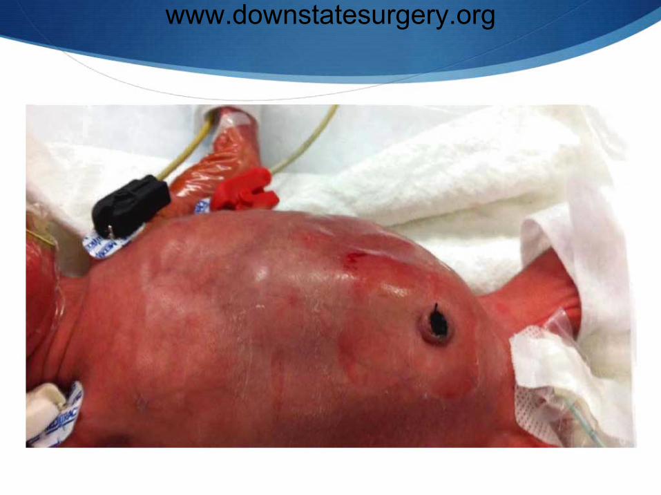
SG -10 day old baby girl, born at 30 weeks gestation
Associated with an in utero twin fetal demise
Apgar score 2, 7 (1, 5 min); Birth weight 1.3 kg
PMH: patent PDA
Increasing abdominal distention and obstipation for first 4 days of life
www.downstatesurgery.org

Physical Exam
• VS: T 97.8 F BP 68/34 HR 164 RR 16 O2 sat 91%
• CV: RRR, S1S2 normal
• Pulm: CTA bilaterally ; intubated
• Abdomen: very distended with visible bowel loops, soft, nontender
• Rectal: unable to go beyond 1.5 cm
Labs:
CBC : 3.1/12/36/261
BMP: 138/3.8 108/24 8.4/0.7 <158
PT/INR/PTT: 13/1.33/29.8
www.downstatesurgery.org

Radiologic Studies
AXR-dilated small bowel loops with no air in pelvis
Barium enema: microcolon with only left colon visualized
Upper GI: no progression of contrast beyond the stomach consistent with marked ileus
www.downstatesurgery.org
Abdominal Examwww.downstatesurgery.org
www.downstatesurgery.org

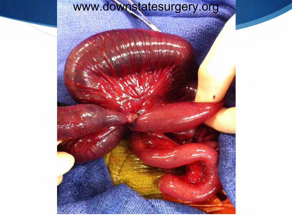
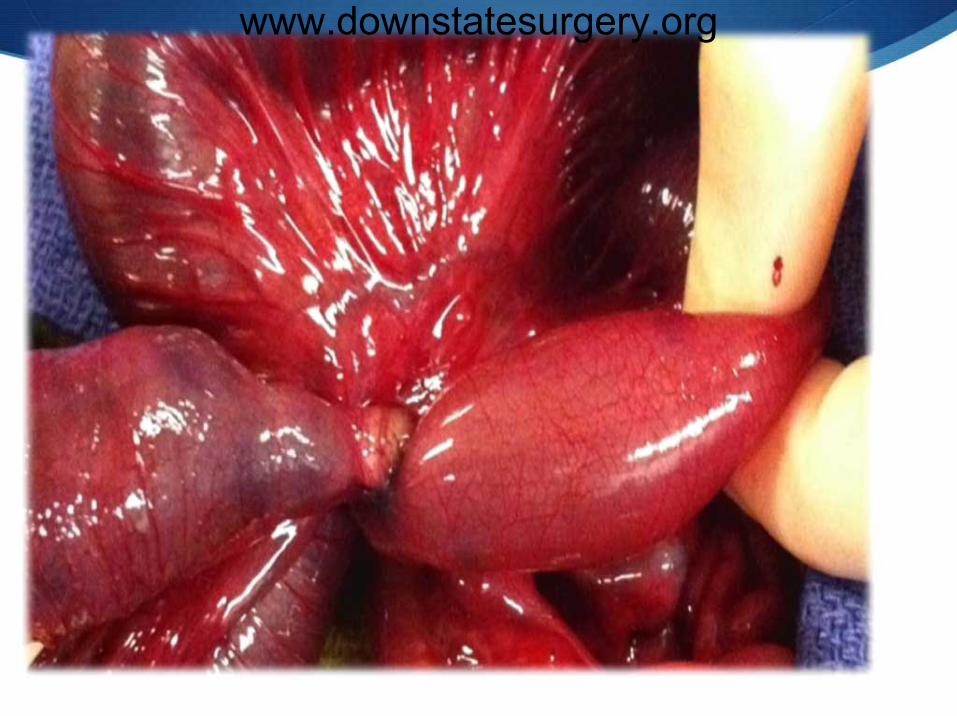
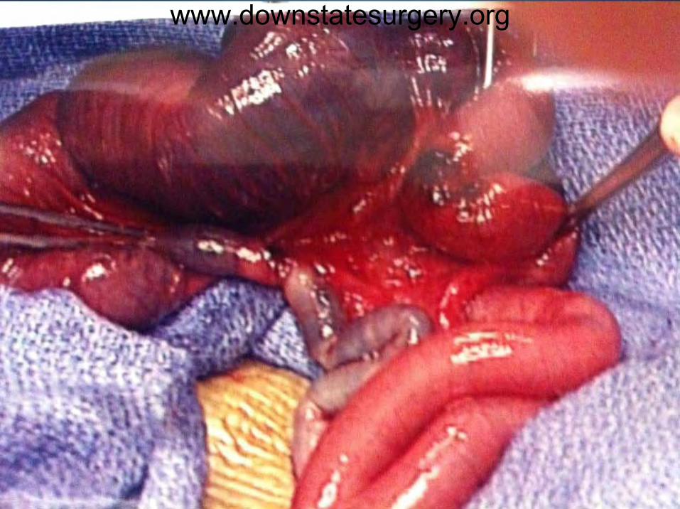
Intraoperatively
6/9: Exploratory laparotomy Reduction of jejunal volvulus Intestinal resection of atretic segments Tapering enteroplasty of the dilated reduced
bowel with primary anastamosis
www.downstatesurgery.org
www.downstatesurgery.org
www.downstatesurgery.org

www.downstatesurgery.org
www.downstatesurgery.org
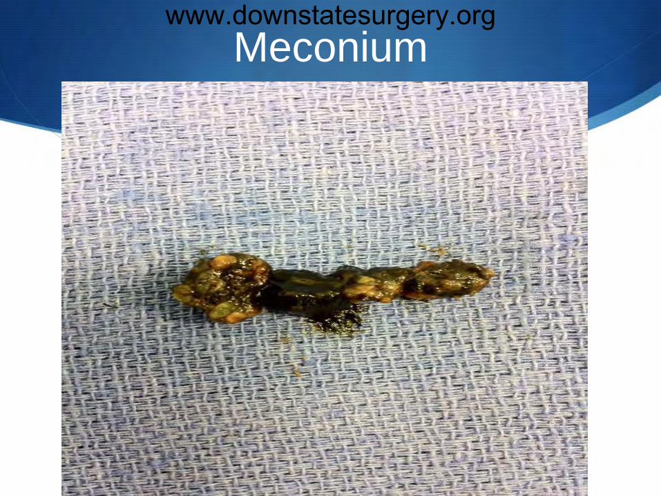
Meconiumwww.downstatesurgery.org
Hospital course
Abdomen remained nondistended and soft
No bowel movement for 3 ½ weeks except for mucus plugs
Upper GI series: dilated proximal loops of bowel
7/5: Exploratory laparotomy, adhesiolysis, small bowel resection, enterorrhaphy (from staple line) and intestinal decompression with removal of meconium plugs
www.downstatesurgery.org
Hospital Course
Post operatively no bowel movement for 2 weeks
7/19: exploratory laparotomy, adhesiolysis, intestinal decompression through enterotomy, jejunal resection of previously volvulated segment
Meconium passed on POD #3.
Extubated currently with normal bowel function.
www.downstatesurgery.org

Intestinal Obstruction in the Newborn
Think of… Atresia Malrotation with volvulus Meconium ileus Hirschsprung’s disease
www.downstatesurgery.org

Differential Diagnosis in neonate with abdominal
distention and obstipation
Meconium Ileus
Distal jejuno-ileal atresia
Hirschsprung’s Disease
www.downstatesurgery.org

Intestinal Atresia
Congenital obstruction caused by complete occlusion of intestinal lumen
Mesenteric vascular accidents in utero
Multifactorial
Incidence: Jejunoileal: 1 in 330 (US)- 1 in 1500
Increased in maternal use of pseudoephedrine and ergotamine + caffeine
www.downstatesurgery.org

Clinical Presentation
Symptoms: Bilious emesis- proximal Abdominal distention- distal Maternal polyhydramnios (24%) Failure to pass meconium
Prenatal Ultrasound More detectable in duodenal atresia Multiple distended loops of bowel with vigorous peristalsis Echogenic bowel <1/3 cases recognized
www.downstatesurgery.org

Radiographic findings
High JejunalAtresia Few air fluid
levels
www.downstatesurgery.org

Ileal Atresiawww.downstatesurgery.org

Intestinal Atresia Classifications
Type I: mucosal atresia with intact bowel wall and mesentary
Type 2: 2 atretic blind ends joined by fibrous cord + intact mesentary
www.downstatesurgery.org
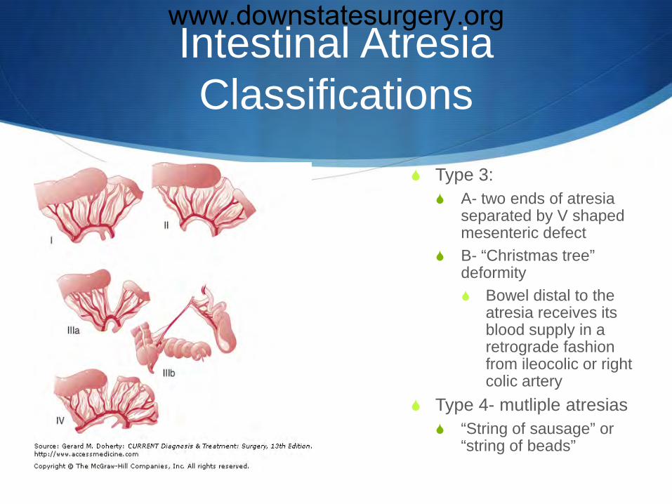
Intestinal Atresia Classifications
Type 3: A- two ends of atresia
separated by V shaped mesenteric defect
B- “Christmas tree” deformity Bowel distal to the
atresia receives its blood supply in a retrograde fashion from ileocolic or right colic artery
Type 4- mutliple atresias “String of sausage” or
“string of beads”
www.downstatesurgery.org

Type 3 Intestinal Atresia
3A 3B
www.downstatesurgery.org

Operative Interventions
In proximal jejunal atresia- resection at ligament of Treitzfollowed by end – to- oblique anastamosis
If limited length- tapering enteroplasty Retention of dilated blind proximal segment- functional
obstruction Smooth muscle hypertrophy and enlargement of bowel diameter Ineffective peristalsis
www.downstatesurgery.org

www.downstatesurgery.org

Attempting to preserve bowel length
Multiple Atresiaswww.downstatesurgery.org
Morbidity and Mortality
Most common cause of early death: infection related to pneumonia, peritonitis or sepsis
Postoperative complications: Functional intestinal obstuction at anastamosis Anastamotic leak (15%)
De Lorimier and associates concluded that resection improved survival in jejunal atresia from 39% to 66%. Little effect on overall survival in ileal atresia
www.downstatesurgery.org

Hirschsprung’s Disease
Incidence: 1 in 5000 live births
M:F ratio 4:1
Among families of children with HD incidence to 6%
Associated with trisomy 21 ( 4.5-16%) atresias
www.downstatesurgery.org

HD- Clinical Presentation
Any child with constipation dating back to newborn period 90% diagnosed as newborns
History of delayed passage of meconium w/in 48 hours of life
Other si/sx: Abdominal distention absent at birth and tight anus Poor feeding Emesis
www.downstatesurgery.org
Radiographic Studies
AXR- distended loops of intestine with paucity of air in rectum
Contrast enema (76-92% accuracy) Narrow spastic distal intestinal segement with a dilated
proximal segment **the point of caliber change is the key radiographic finding Most commonly the transition point at rectosigmoid
www.downstatesurgery.org

Hirschsprung’s Disease
Hirschsprung’s Disease Constipation
www.downstatesurgery.org

Anorectal Manometry
Measures absence of a relaxation reflex after a distending bolus is created in the rectal lumen
Elevated resting anal sphincter pressure
www.downstatesurgery.org

Rectal Biopsy
GOLD STANDARD for diagnosis of Hirschsrung’s disease
Diagnostic accuracy 99.7%
Alternate method: full thickness posterior rectal wall biopsy
www.downstatesurgery.org

Cause of Hirschsprung’sDisease
Sporadic occurrence accounts for 80-90% of cases
RET gene mutations in 35% of sporadic cases and 49% of familial cases
5-10% show mutations in other genes
www.downstatesurgery.org

Embryology
3 phases in development of enteric nervous system: Induction phase Neural crest cell migration Differentiation of the neural crest cell precursos
5 wks gestation- NC cells in esophagus
7 weeks- midgut
12 weeks- distal colon
www.downstatesurgery.org

Theories of embyologic defect:
“Failure of migration”
“Hostile environment”
www.downstatesurgery.org
Pathology
In infancy the intestine may appear fairly normal
As infant ages- proximal ganglionic bowel hypertrophies
Rectosigmoid – 80%
Histology: Absence of ganglion cells in the distal intestine
www.downstatesurgery.org

Endorectal Pull-Through
Single stage operation; Initially described by Soave in 1964
LLQ incision
At least 5 cm proximal to the first area of normal ganglion cells
Endorectal dissection approximatly 2 cm below peritoneal reflection by incising the seromuscular layer Carried down 0.5cm of the dentate line in newborns
www.downstatesurgery.org

Endorectal Pull through -2
Evert mucosal-submucosaltube and incise on anterior half
Normal ganglionic intestine brought down to this point
Anastamosis with absorbable sutures
www.downstatesurgery.org

Different surgical techniques
Laparoscopic
Transanal
www.downstatesurgery.org
Complications
Early Post-Pull-Through Intestinal obstruction (8-13%) Early anorectal stenosis (10-20%)
Late Incontinence (3-8%) Constipation (6-30%)
www.downstatesurgery.org

Meconium Plug Syndrome
Confirmed by contrast enema radiograph to find “plugs” in sigmoid or descending colon
Spontaneously pass after withdrawal of the enema catheter
Pathogenesis may relate to bowel hypomotility
Associated with: prematurity, hypotonia, hypermagnesemia, sepsis, hypothyroidism, and Hirshsprung’s disease in 5%
Sweat test and rectal biopsy
www.downstatesurgery.org

Meconium Ileus
1905- Landsteiner
Seen in 20-35% patients with cystic fibrosis Mutation in CF transmembrane regulator (CFTR) gene
Patients with meconium ileus represent a distinct phenotype Earlier presentation and worse pulmonary function
Survival 95-100% with aggressive management, nutrition and close monitoring of pulmonary function
www.downstatesurgery.org

Pathophysiology of Meconium Ileus
Intestinal obstruction secondary to intraluminal accumulation of inspissated and dessicated meconium
EARLIEST clinical manifestation of CF- 20.8%
CFTR- chromosome 7, band q31 ( F508 mutation) Cyclic adenosine monophosphate-induced chloride channel
Reduced clearance of secretions Respiratory, GI, biliary, pancreatic and reproductive systems
www.downstatesurgery.org

Pathogenesis of Meconium Ileus
Intestinal glandular disease plays a dominant role Pancreatic disease plays a secondary role
Meconium in CF patients is twice as high in concentrations of sodium, potassium and magnesium; protein nitrogen
Protein + mucopolysaccharides highly viscid rubbery meconium
Complications : volvulus of heavy loop with perforation, peritonitis, atresia microcolon
− Degradation enzymes
www.downstatesurgery.org
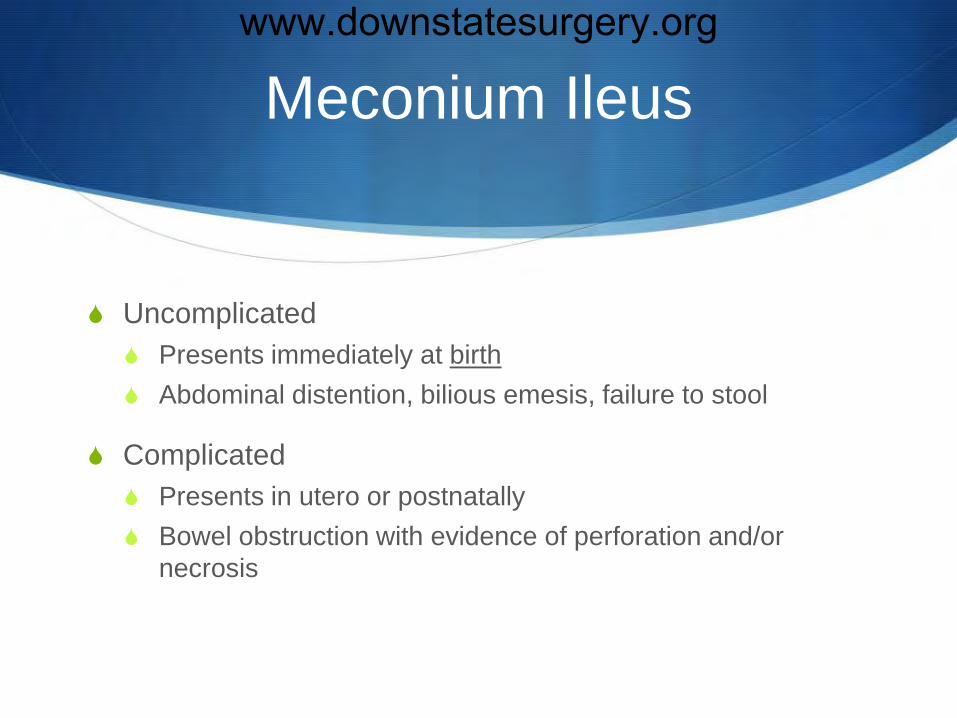
Meconium Ileus
Uncomplicated Presents immediately at birth Abdominal distention, bilious emesis, failure to stool
Complicated Presents in utero or postnatally Bowel obstruction with evidence of perforation and/or
necrosis
www.downstatesurgery.org
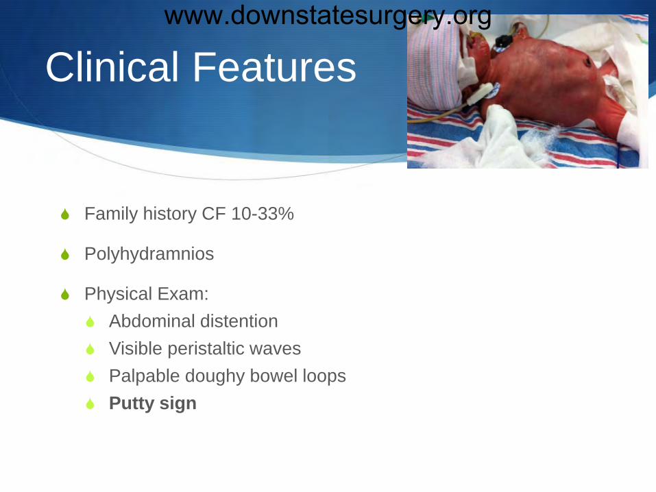
Clinical Features
Family history CF 10-33%
Polyhydramnios
Physical Exam: Abdominal distention Visible peristaltic waves Palpable doughy bowel loops Putty sign
www.downstatesurgery.org

Radiologic Studies
In utero: echogenic bowel in 3rd trimester; distended bowel
Abdominal XR: Great disparity in size of bowel loops NO or few air fluid levels “soap bubble” or “ground glass” in meconium peritonitis
Contrast Enema- microcolon
www.downstatesurgery.org
Lab studies
Labs: Sweat test- 100mg Sweat Cl >60 mEq/L
Stool Studies Meconium albumin>80mg/g Stool trypsin >80 mg/g
www.downstatesurgery.org
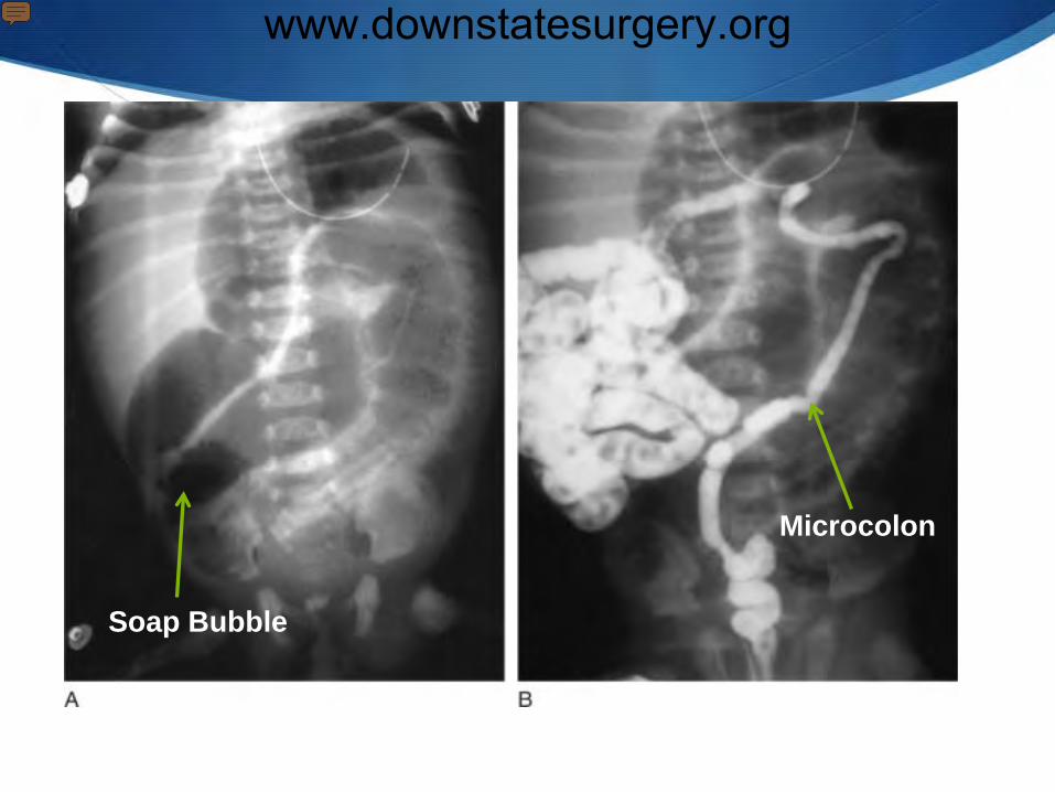
Soap Bubble
Microcolon
www.downstatesurgery.org

Meconium Ileus
Meconium Ileus
www.downstatesurgery.org

Non-operative Management
Gastrograffin enema Hyperosmolar, water-soluble, radioopaque solution 0.1% polysorbate 80 (solubilizing agent) and 37%
organically bound iodine Transient osmotic diarrhea and a putative osmotic diuresis
Advantages: reduction in pulmonary morbidity, decreased hospital length of stay
Disadvantages: delay, intestinal injury, hypovolemia
www.downstatesurgery.org

Operative Management
Enterotomies with irrigation Warmed saline, 50% diatrizoate solution, H2O2, Mucomyst Meconium milked distally into the colon or enterotomy Enterostomy + T- tube Continued irrigation until POD #14 where catheter removed
Mikulicz double-barreled enterostomy
Santulli – proximal chimney enterostomy
www.downstatesurgery.org

Operative Management
Bishop-Koop procedure Limit intraoperative bowel trauma in neonatal period Resect disparately enlarged ileal loop filled with meconium Create an approximately sized end of prox to side of distal
ileum Access to the insertion of a catheter into the distal bowel
containing the meconium pellets Permit eventual enterostomy closure by bedside ligation of
“chimney” stoma
www.downstatesurgery.org

Various Techniqueswww.downstatesurgery.org

Postoperative Management
Instillation of 2%-4% acetylcysteine delivered through a nasogastric tube to solubilize the residual meconium
When gut patency verified – elemental formula
Supplemental pancreatic enzymes also begun with formula
Short term post operative antibiotics
Total parenteral nutrition
www.downstatesurgery.org

Outcomes Survival rates
approaching 100%
No overall significant differences in outcome was present with regard to patient gender, complication of meconium ileus or type of operation performed
Outcomeswww.downstatesurgery.org
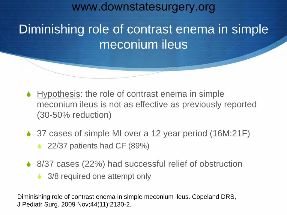
Diminishing role of contrast enema in simple meconium ileus
Hypothesis: the role of contrast enema in simple meconium ileus is not as effective as previously reported (30-50% reduction)
37 cases of simple MI over a 12 year period (16M:21F) 22/37 patients had CF (89%)
8/37 cases (22%) had successful relief of obstruction 3/8 required one attempt only
Diminishing role of contrast enema in simple meconium ileus. Copeland DRS, J Pediatr Surg. 2009 Nov;44(11):2130-2.
www.downstatesurgery.org
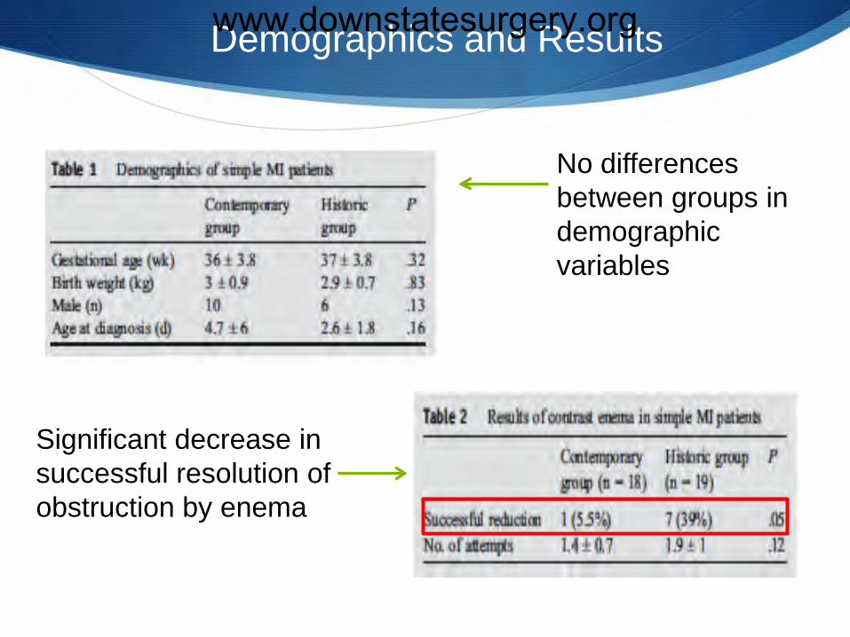
Demographics and Results
No differences between groups in demographic variables
Significant decrease in successful resolution of obstruction by enema
www.downstatesurgery.org

Discussion
Low perforation rate with a high failure rate of enema
Enema substrate used in this study (cysto-conray II- 400 mOsm/kg water) less osmotically active than Gastrografin(1940 mOsm/kg water).
Small sample size- need a prospective multicenter evaluation
www.downstatesurgery.org

References
Diminishing role of contrast enema in simple meconiumileus.Copeland DR, St Peter SD, Sharp SW, Islam S, Cuenca A, Tolleson JS, Dassinger MS, Little DC, Jackson RJ, Kokoska ER, Smith SD. J Pediatr Surg. 2009 Nov;44(11):2130-2.
Grosfeld. Pediatric Surgery 6th edition
Schwartz's Principles of Surgery . Chapter 39: PEDIATRIC SURGERY
Dalla V, Grosfeld JL, West KWl. Intestinal atresia and stenosis: a 25 year experience with 277 cases. Arch Surg. 1998 May; 133 (5) 490-6.
Current Diagnosis and treatment, Surgery. 13th edition. Pediatric Surgery
www.downstatesurgery.org
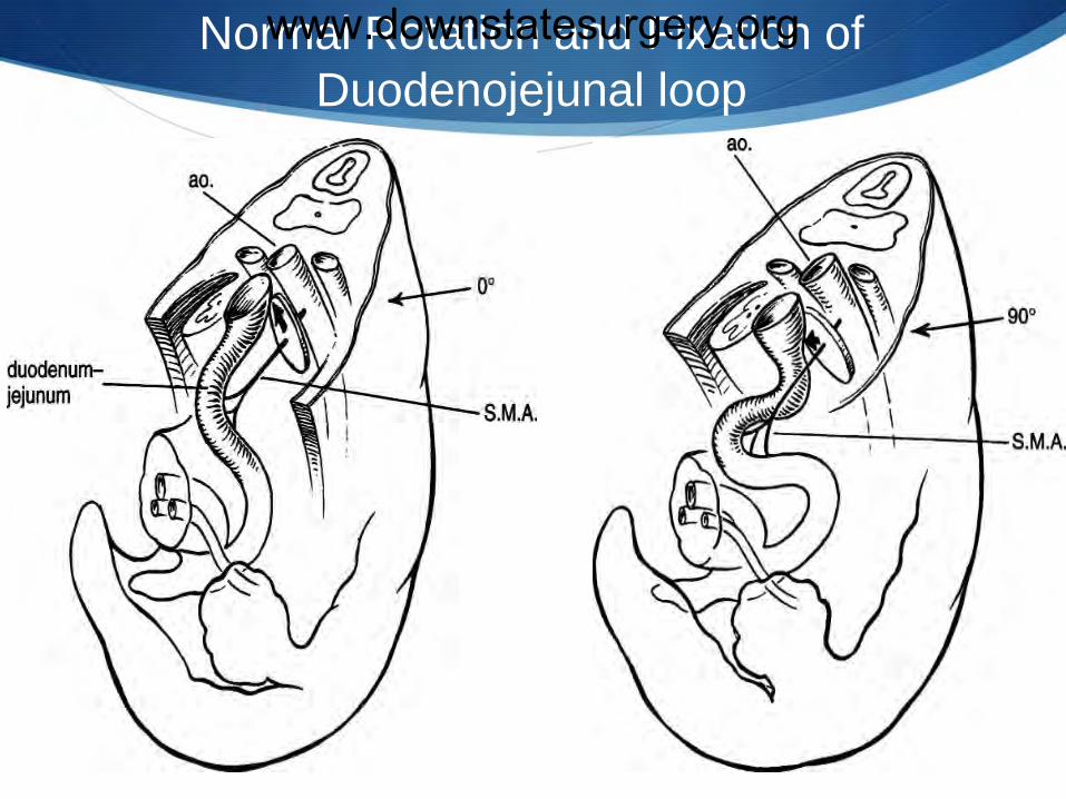
Normal Rotation and Fixation of Duodenojejunal loop
www.downstatesurgery.org
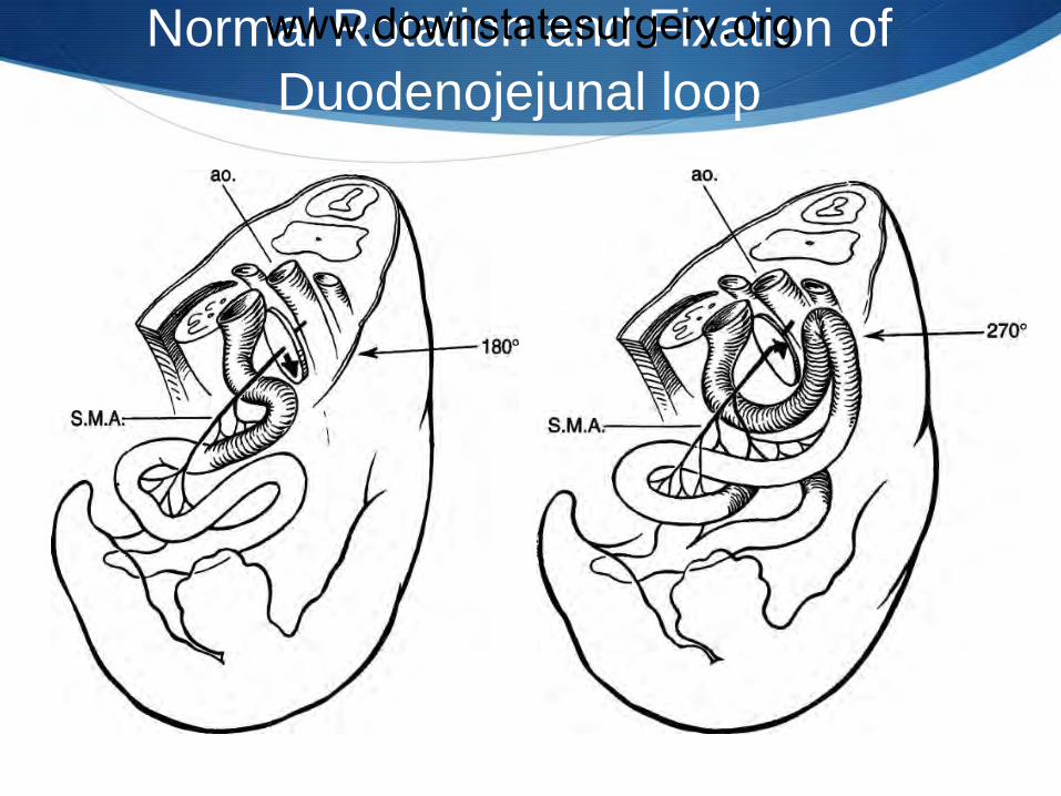
Normal Rotation and Fixation of Duodenojejunal loopwww.downstatesurgery.org

Normal Rotation and Fixation of Cecocolic loopwww.downstatesurgery.org

Normal Rotation and Fixation of Cecocolic loopwww.downstatesurgery.org

4th week of fetal life the embryo is at 5 mm stage Intestinal tract is a straight
tube with slight anterior bulge in central portion
8th week Duodenojejunal rotates
during extracoelomicphase to 180 degress
10th week Intestines return to
abdomen
Intestinal Rotationwww.downstatesurgery.org

Malrotation
All abnormalities of intestinal position and attachment Atypical
malrotation-ligament of Treitz is to the left of the midline
Nonrotation
Incomplete rotation
NonrotationIncompleterotation
Normal
www.downstatesurgery.org

Acute Midgut Volvulus
Narrow pedicle formed by the base of the mesentery in malrotation predisposes midgut to clockwise twisting from duodenum to transverse colon
Various causes: Unusual movement of torso Abnormal intestinal
peristalsis Segmental bowel distention
www.downstatesurgery.org
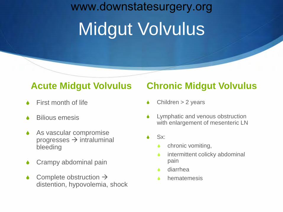
Midgut Volvulus
Acute Midgut Volvulus First month of life
Bilious emesis
As vascular compromise progresses intraluminal bleeding
Crampy abdominal pain
Complete obstruction distention, hypovolemia, shock
Chronic Midgut Volvulus Children > 2 years
Lymphatic and venous obstruction with enlargement of mesenteric LN
Sx: chronic vomiting, intermittent colicky abdominal
pain diarrhea hematemesis
www.downstatesurgery.org
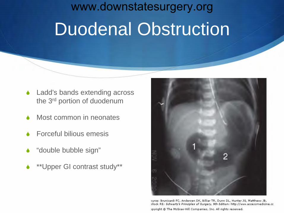
Duodenal Obstruction
Ladd’s bands extending across the 3rd portion of duodenum
Most common in neonates
Forceful bilious emesis
“double bubble sign”
**Upper GI contrast study**
www.downstatesurgery.org

Radiologic Diagnosis
Contrast radiography
Normal
MIDGUTVOLVULUS
www.downstatesurgery.org
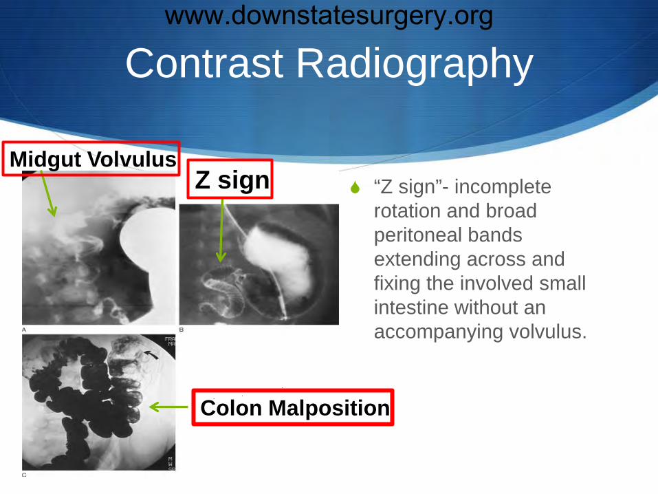
Contrast Radiography
“Z sign”- incomplete rotation and broad peritoneal bands extending across and fixing the involved small intestine without an accompanying volvulus.
Z signMidgut Volvulus
Colon Malposition
www.downstatesurgery.org
Ladd Procedure
(1) Evisceration of the bowel and inspection of mesenteric root
(2) Counterclockwise derotation of midgut volvulus
(3) Lysis of Ladd’s peritoneal bands with straightening of the duodenum along the right abdominal gutter
(4) Appendectomy
(5) Placement of the cecum in the left lower quadrant
www.downstatesurgery.org

Ladd Procedure
Supraumbilical right transverse incision
2 constant anatomic points: Pylorus and splenic flexure
www.downstatesurgery.org

LADD Procedurewww.downstatesurgery.org

Intestinal Resection and 2nd
Look
At 12-24 hours recovery of questionable bowel or demarcation is obvious
3 principles should be considered: Preserving minimum length of intestine required for survival
is of highest priority Avoid anastamoses between end f intestine of ? viability Resection of entire midgut will necessitate lifelong
parenteral nutrition and small intestinal transplantation
www.downstatesurgery.org

Postoperative Management
In uncomplicated duodenal obstruction- peristalsis in 1-5d
Complications: Postoperative intussusception (3.1%) Abdominal distention and bilious emesis on POD # 5-8
Recurrent volvulus (0.5-1.25%) Bowel obstruction (4%) Death ( associated with peritonitis)
www.downstatesurgery.org