Embed Size (px)
Citation preview

Hindawi Publishing CorporationAdvances in OrthopedicsVolume 2013, Article ID 437570, 8 pageshttp://dx.doi.org/10.1155/2013/437570
Research ArticleIntervertebral Disc Rehydration after Lumbar DynamicStabilization: Magnetic Resonance Image Evaluation witha Mean Followup of Four Years
Li-Yu Fay,1,2,3 Jau-Ching Wu,1,2,3 Tzu-Yun Tsai,4,5 Tsung-Hsi Tu,1,2 Ching-Lan Wu,6
Wen-Cheng Huang,1,2 and Henrich Cheng1,2,3
1 Department of Neurosurgery, Neurological Institute, Taipei Veterans General Hospital,Taipei 11217, Taiwan
2 School of Medicine, National Yang-Ming University, Taipei 11221, Taiwan3 Institute of Pharmacology, National Yang-Ming University, Taipei 11221, Taiwan4Department of Ophthalmology, National Taiwan University Hospital, College of Medicine, National Taiwan University,Taipei 10002, Taiwan
5Department of Ophthalmology, New Taipei City Hospital, New Taipei City 241, Taiwan6Department of Radiology, Taipei Veterans General Hospital, Taipei 11217, Taiwan
Correspondence should be addressed to Jau-Ching Wu; [email protected]
Received 7 July 2012; Accepted 27 March 2013
Academic Editor: Mehdi Sasani
Copyright © 2013 Li-Yu Fay et al.This is an open access article distributed under theCreative CommonsAttribution License, whichpermits unrestricted use, distribution, and reproduction in any medium, provided the original work is properly cited.
Objective.To compare the clinical and radiographic outcomes in patients of different ages who underwent theDynesys stabilization.Methods. This retrospective study included 72 patients (mean age 61.4 years) with one- or two-level lumbar spinal stenosis whounderwent laminectomy and the Dynesys (Zimmer Spine, Minneapolis) dynamic stabilization system. Thirty-seven patients wereyounger than 65-year old while the other 35 were older. Mean followup was 46.7 months. Pre- and postoperative radiographic andclinical evaluations were analyzed.Results.Themean calibrated disc signal (CDS) at the index level was significantly improved from60.2 ± 25.2 preoperatively to 66.9 ± 26.0 postoperatively (𝑃 > 0.001). Screw loosening occurred in 22.2% of patients and 5.1% ofscrews. The improvement in CDS at index level was seen to be significant in younger patients but not in older patients. Overall,the mean visual analogue scale (VAS) of back pain, VAS of leg pain, and the Oswestry disability index (ODI) scores improvedsignificantly after operation. There were no significant differences in pre- and postoperative VAS and ODI and screw looseningrates between the younger and older patients.Conclusions.There is significant clinical improvement after laminectomy and dynamicstabilization for symptomatic lumbar spinal stenosis. Intervertebral disc rehydration was seen in younger patients.
1. Introduction
Instrumented spinal fusion is the treatment of choice fordegenerative spondylosis with instability refractory to con-servative treatment [1, 2]. Spine surgeons have also usedmodern biologics such as recombinant human bone mor-phogenetic protein-2 to increase the rate of spinal fusion inselected patients [3–7]. However, using biologics to enhancespinal fusion has been sometimes reported with complica-tions postoperatively and during followup. Moreover, evenautograft has been repeatedly reported with adverse events,such as donor site morbidity. Not to mention that loss
of segmental motion and subsequent adjacent segmentaldegeneration have also been concerned for the spinal fusionsurgery [8–10].
In the recent years, there is the emerging option ofdynamic stabilization to spare spinal fusion and still yieldsatisfactory outcomes in the surgical management of lum-bar spondylosis and back pain. Fischgrund and colleaguesreported application of the Dynesys (Zimmer Spine, Min-neapolis, USA), a pedicle-based lumbar dynamic stabilizationsystem, as an effective alternative to treat lumbar spondylosisin 1994 [11–16]. Theoretically Dynesys can unload the inter-vertebral disc while providing a restricted range of motion

2 Advances in Orthopedics
and thus alleviates symptoms in the indexed level of spine.Although its long-term effect on the adjacent segment is stilluncertain, there are reports demonstrating improved clinicaloutcomes with acceptably low rate of screw loosening whenDynesys is used in degenerative disc diseases (DDD), lumbarstenosis, and some spondylolisthesis [13, 17]. Despite thecommon nature of spondylosis in the diseases treated, quitesome disparity exists among these patients, who have variableage and bone quality that could affect the screw anchoringin the nonfused constructs. However, there is a paucity ofdata addressing the differences of age and clinical outcomes inthese patients treated by dynamic stabilization with Dynesys.
This study aims to compare the clinical and radiographicoutcomes in patients of different ages who underwent surgi-cal decompression and Dynesys stabilization. This is the firststudy focusing on the discrepancies between the elderly andthe younger patients. The pre- and postoperative magneticresonance imaging (MRI) especially of each patient werecompared to evaluate the condition of the discs at the indexsegments. All clinical data was prospectively collected andoutcomes were measured by standardized parameters withmore than two years of followup.
2. Methods
2.1. Patient Enrollment. From September 2007 to August2009, the study enrolled 88 consecutive patients who under-went surgical decompression and dynamic stabilization withDynesys (Zimmer Spine, Minneapolis, MN) in the authors’service. The clinical and radiological data were collectedprospectively and then analyzed retrospectively. Three expe-rienced spine surgeons performed all operations in a verysimilar fashion by the same technique.
The inclusion criteria were the patients who had lum-bar DDD with persistent symptoms such as intermittentclaudication, low back pain, buttock pain, leg pain, orany combination of the above. Each patient underwentpreoperative MRI to confirm the diagnosis and had failedmedical treatment for longer than 12 weeks. The exclusioncriteria were degenerative scoliosis, prior lumbar surgery,disc diseases requiring discectomies, and spondylolisthesisworse than grade II. Patients with psychiatric disorders,cerebral vascular accidents, coexisting cervical or thoracicmyelopathy, or neoplasms were also excluded. Among the88 patients enrolled in this cohort, 72 completed the followup for more than 24 months and were thus analyzed. Thesepatients were divided into two groups according to the age atoperation: young age (<65 years) and old age (≧65 years).
2.2. Operative Technique. Patients were placed in proneposition under general anesthesia. Fluoroscopy was usedroutinely before sterilization to confirm the treated leveland keep the lumbar spine in a neutral lordotic curve.Prophylactic antibiotics were infused thirty minutes beforeincision. After midline incision by subperiosteal dissection,the laminae and spinous process were exposed. The facetjoints capsules were preserved intact. Standard total laminec-tomies and foraminotomies with resection of hypertrophic
spur and ligamentum flavum were performed carefully topreserve the complex structure of the facet joints.The exitingand traversing nerve roots were probed through to confirmthe adequate decompression without any bony fragment.Bilateral fascial incisions were made by subdermal dissectionthrough the same wound. Along the Wiltse and Spencerintermuscular plane, the titanium alloy screws were placedtranspedicularly without destruction of the facet joints [18].The modular spacers were cut appropriately and the tensioncords were assembled to reach 300N for dynamic stabiliza-tion. Fluoroscope was used to assure the correct position ofeach screw. Drainage catheters were left and the wound wasclosed layer by layer.
2.3. Clinical Assessment. TheOswestry disability index (ODI)scale was used for functional assessment. The 0–10 visualanalogue scale (VAS) was used for back and leg pain eval-uation. The patients themselves completed the ODI andVAS questionnaire preoperatively and at 1.5-, 3-, 6-, 12-,and 24-month followup regularly. Preoperative scores werecompared to postoperative scores.
2.4. Radiographic Assessment. Standard anterior/posteriorand lateral radiographs, computed tomography (CT), andmagnetic resonance imaging (MRI) studies were routinelyperformed in preoperative assessment. Radiographs weretaken regularly at 1.5-, 3-, 6-, 12-, and 24-month followup.
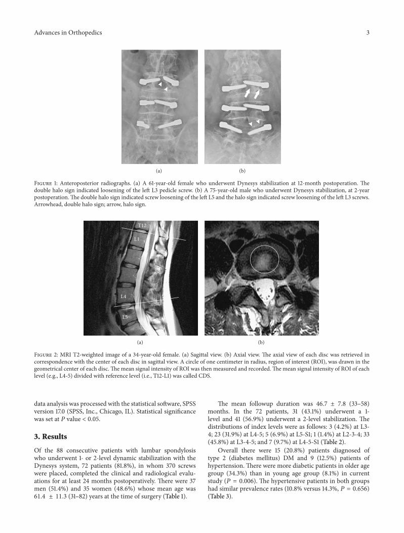
The presence of “halo zone sign” or “double halo sign” onanteroposterior radiographs was defined as screw looseningduring followup (Figure 1). CT was used to determine ques-tionable screw loosening. The halo zone sign was defined asa radiolucent zone alone the screws with 1mm in width andin any length. The double halo sign was further defined as aradiolucent zone with a radiopaque rim alone the screws.
Pre- and post-operative MRI images were comparedfocusing on the condition of the discs, including the indexedand adjacent segments. Due to the lack of an absolute unitof measuring the signal intensities on MRI, for example, theHounsfield unit used in computed tomography to describeradiodensity, these signals of the same patient are difficultto compare. Ideally the pre- and post-operative MRI studiesneed to be taken by the very same MRI machine to unifythe signal intensity references. We tried to allocate the sameMRI machine to each patient; however, not every patientachieved the goal. To address this issue, the digital signal incentral intervertebral disc of the levels T12-L1 in T2-weightedimage (T2WI) was designated as a reference (Figure 2). Thesignal intensity in the center of the discs in the lumbarspine of each patient was recorded and compared to eachpatient’s own signal intensity recorded at the levels of T12-L1in T2-weighted MRI (T2WI). The ration differences of signalintensity were calculated and defined as the calibrated discsignal (CDS). The pre- and post-operative CDS of bridgedlevels were thus all compared and analyzed.
2.5. Statistical Analysis. Clinical and radiographic assess-ments were compared and analyzed using Student’s 𝑡-test,chi-square, andMann-WhitneyU test, where appropriate. All
Advances in Orthopedics 3
(a) (b)
Figure 1: Anteroposterior radiographs. (a) A 61-year-old female who underwent Dynesys stabilization at 12-month postoperation. Thedouble halo sign indicated loosening of the left L3 pedicle screw. (b) A 75-year-old male who underwent Dynesys stabilization, at 2-yearpostoperation.The double halo sign indicated screw loosening of the left L5 and the halo sign indicated screw loosening of the left L3 screws.Arrowhead, double halo sign; arrow, halo sign.
T12
L1
L4
L5
(a) (b)
Figure 2: MRI T2-weighted image of a 34-year-old female. (a) Sagittal view. (b) Axial view. The axial view of each disc was retrieved incorrespondence with the center of each disc in sagittal view. A circle of one centimeter in radius, region of interest (ROI), was drawn in thegeometrical center of each disc. The mean signal intensity of ROI was then measured and recorded.The mean signal intensity of ROI of eachlevel (e.g., L4-5) divided with reference level (i.e., T12-L1) was called CDS.
data analysis was processed with the statistical software, SPSSversion 17.0 (SPSS, Inc., Chicago, IL). Statistical significancewas set at 𝑃 value < 0.05.
3. Results
Of the 88 consecutive patients with lumbar spondylosiswho underwent 1- or 2-level dynamic stabilization with theDynesys system, 72 patients (81.8%), in whom 370 screwswere placed, completed the clinical and radiological evalu-ations for at least 24 months postoperatively. There were 37men (51.4%) and 35 women (48.6%) whose mean age was61.4 ± 11.3 (31–82) years at the time of surgery (Table 1).
The mean followup duration was 46.7 ± 7.8 (33–58)months. In the 72 patients, 31 (43.1%) underwent a 1-level and 41 (56.9%) underwent a 2-level stabilization. Thedistributions of index levels were as follows: 3 (4.2%) at L3-4; 23 (31.9%) at L4-5; 5 (6.9%) at L5-S1; 1 (1.4%) at L2-3-4; 33(45.8%) at L3-4-5; and 7 (9.7%) at L4-5-S1 (Table 2).
Overall there were 15 (20.8%) patients diagnosed oftype 2 (diabetes mellitus) DM and 9 (12.5%) patients ofhypertension. There were more diabetic patients in older agegroup (34.3%) than in young age group (8.1%) in currentstudy (𝑃 = 0.006). The hypertensive patients in both groupshad similar prevalence rates (10.8% versus 14.3%, 𝑃 = 0.656)(Table 3).

4 Advances in Orthopedics
Table 1: Clinical and demographic characteristics (𝑛 = 72).
Characteristic ValueGenderMale 37 (51.4%)Female 35 (48.6%)
Age (years) mean 61.4 ± 11.3 (31–82)Follow-up (months) mean 46.7 ± 7.8 (33–58)Number of instrumented levels1-level 31 (43.1%)2-levels 41 56.9%)
Table 2: Distribution of treated levels.
Instrumentation Level Number of patients
1-level (𝑛 = 31)L3-4 3 (4.2%)L4-5 23 (31.9%)L5-S1 5 (6.9%)
2-levels (𝑛 = 41)L2-3-4 1 (1.4%)L3-4-5 33 (45.8%)L4-5-S1 7 (9.7%)
3.1. Clinical Outcomes. The overall mean VAS for backpain improved with statistical significance, from 6.3 ± 3.3preoperatively to 2.8 ± 2.9 postoperatively (𝑃 < 0.001). Therewas significant improvement in both the young age group(6.1 ± 3.4 preoperatively to 2.9 ± 2.9 postoperatively; 𝑃 <0.001) and the old age group (6.4 ± 3.1 preoperatively to2.7 ± 2.9 postoperatively; 𝑃 < 0.001) (Table 3).There were nosignificant differences in pre- and postoperative VAS betweenthe young and old age groups (𝑃 = 0.811 and𝑃 = 0.754, resp.)(Figure 1).
The overall mean VAS for leg pain improved with statis-tical significance, from 7.0 ± 2.7 preoperatively to 2.6 ± 3.2postoperatively (𝑃 < 0.001). There was significant improve-ment in both the young age group (6.7 ± 3.0 preoperativelyto 2.7 ± 3.1 postoperatively; 𝑃 < 0.001) and the old agegroup (7.4 ± 2.3 preoperatively to 2.4 ± 3.2 postoperatively;𝑃 < 0.001) (Table 3). There were no significant differences inpre- and postoperative VAS between the young and old agegroups (𝑃 = 0.497 and 𝑃 = 0.474, resp.) (Figure 1).
The overall ODI functional scores significantly improvedfrom 51.1 ± 19.6 preoperatively to 23.4 ± 21.4 postoperatively(𝑃 < 0.001).The young age group specifically improved from50.4 ± 21.3preoperatively to 21.8 ± 22.0postoperatively (𝑃 <0.001) and the old group from 51.7 ± 17.8 preoperativelyto 25.2 ± 20.8 postoperatively (𝑃 < 0.001) (Table 3). Therewere no significant differences in pre- and postoperative ODIscores between the young and old groups (𝑃 = 0.612 and𝑃 = 0.291, resp.) (Figure 1).
3.2. Radiographic Outcome of Screw Loosening. There wasscrew loosening in 16 out of 72 patients (22.2% per patient)and 19 out of 370 screws (5.1% per screw). Specifically, therewere 8 of 37 patients (21.6%) and 9 of 188 screws (4.8%)found loosened in the young age group. In contrast, therewere 8 of 35 patients (22.9%) and 10 of 182 screws (5.5%)
Table 3: Comparison between young age and old age.
Variables Total Age P values<65-year
old≧65-year
oldNumber of patients 72 37 35Age (years) 61.4 ± 11.3 53.0 ± 9.0 70.3 ± 4.8 <0.001∗
GenderMale 37 23 (62.2%) 14 (40.0%) 0.060Female 35 14 (37.8%) 21 (60.0%)
Number ofinstrumented levelsOne 31 17 (45.9%) 14 (40.0%) 0.611Two 41 20 (54.1%) 21 (60.0%)
Mean pre-op scoresVAS back pain 6.3 ± 3.3 6.1 ± 3.4 6.4 ± 3.1 0.811VAS leg pain 7.0 ± 2.7 6.7 ± 3.0 7.4 ± 2.3 0.497ODI (%) 51.1 ± 19.6 50.4 ± 21.3 51.7 ± 17.8 0.612
Mean 24-monthpost-op scoresVAS back pain 2.8 ± 2.9 2.9 ± 2.9 2.7 ± 2.9 0.754VAS leg pain 2.6 ± 3.2 2.7 ± 3.1 2.4 ± 3.2 0.474ODI (%) 23.4 ± 21.4 21.8 ± 22.0 25.2 ± 20.8 0.291
Serum glucose statusType 2 DM 15 3 (8.1%) 12 (34.3%) 0.006∗
Euglycemia 57 34 (91.9%) 23 (65.7%)Blood pressureHypertensive 9 4 (10.8%) 5 (14.3%) 0.656Normotensive 63 33 (89.2%) 30 (85.7%)
Mean values are presented ± SD.∗P < 0.05, statistically significant.DM: diabetes mellitus.VAS: visual analogue scale.ODI: Oswestry disability index.
found loosened in the old age group.Therewas no statisticallysignificant difference found between the two groups in therates of screw loosening per patient or per screw (𝑃 = 0.900,𝑃 = 0.739, resp.) (Table 4).
3.3. Radiographic Outcome of Bridged Disc Signal. Themeanoverall CDS at bridged level improved (i.e., increased) withstatistical significance, from 60.2 ±25.2 preoperatively to 66.9± 26.0 postoperatively (𝑃 = 0.014). There was significantincrease in the young age group (58.9 ± 24.7 preoperativelyto 67.6 ± 28.7 postoperatively; 𝑃 = 0.013) (Figure 3).However, the change in the old age group was not significant(62.1 ± 26.1 preoperatively to 65.9 ± 22.2 postoperatively;𝑃 = 0.366). There were no significant differences in pre- andpost-operative bridged level CDS between the young and oldage groups (𝑃 = 0.732 and 𝑃 = 0.800, resp.) (Table 4).

Advances in Orthopedics 5
Table 4: Comparison of disc signal change between young age andold age.
Variables Total Age P values<65-year
old≧65-year
oldNumber ofpatients 72 37 35
Screwlooseningper patientYes 16 8 (21.6%) 8 (22.9%) 0.900No 56 29 (78.4%) 27 (77.1%)
Screwlooseningper screwYes 19 9 (4.8%) 10 (5.5%) 0.739
No 351 179 (95.2%) 172(94.5%)
CDS of bridgeddiscPre-op 60.2 ± 25.2 58.9 ± 24.7 62.1 ± 26.1 0.732Post-op 66.9 ± 26.0∗∗ 67.6 ± 28.7∗∗ 65.9 ± 22.2 0.800
Mean values are presented ± SD.∗∗P < 0.05, the post-op value compared with the pre-op value demonstratedsignificant difference.CDS: calibrated disc signal.
4. Discussion
The current study collected total 72 patients with lumbarspondylosis who underwent decompression and dynamicstabilization. A total 370 screws of Dynesys were placedduring the operations. In a mean follow-up period of 46.7months, the results of both the young age (<65 years old,𝑛 = 37) and the old age (≧65 years old, 𝑛 = 35) groupsdemonstrated satisfactory improvement in clinical outcomes.The overall screw loosening rate was 22.2% per patient (16 in72 patients) and 5.1% per screw (19 in 370 screws). The rateof screw loosening was slightly higher in the old age patients.Regarding disc signal, in the overall CDS of bridged discs, thechange significantly improved from 60.2±25.2 preoperativelyto 66.9 ± 26.0 postoperatively (𝑃 = 0.014). However, thechange in the young age group (58.9 ± 24.7 to 67.6 ± 28.7,𝑃 = 0.013) appeared to be more obvious than in the old agegroup (62.1 ± 26.1 to 65.9 ± 22.2, 𝑃 = 0.366).
Using the 10% difference as minimal clinically importantdifference (MCID) of the ODI defined by Hagg et al., and the1.2 unit improvement of VAS reported by Copay et al., overallthere were 68.1% (49/72) of patients had MCID of VAS backpain, 84.7% (61/72) had MCID of VAS leg pain, and 81.9%(59/72) had MCID of ODI [19, 20]. Therefore, a significantnumber of patients had clinically significant improvement inthe present series at a mean followup of 47 months.
According to the population projections of the UnitedNations, the number of elderly patients (older than 65years) in the world will increase from 8 to 14 percentand the percentage will increase far more to 25 percent
in more developed nations between 2010 and 2040. Lowback pain and lumbar spondylosis is a common problemin the elderly patients which may greatly affect their qualityof life. Pain, numbness, claudication, and risk to fall arethe frequent symptoms of the patients. The prevalence oflumbar spondylosis will increase as the population ages [21,22]. Conservative treatment such as medication, physicaltherapy, or manual therapy may be the first-line treatmentfor most patients and surgical intervention can still achievesatisfactory improvement in selected patients refractory toconservative treatment [23]. Weinstein et al. conducted acohort study, spine patient outcome research trial (SPORT),to analyze the pain improvement and functional outcomes.They enrolled a randomized cohort of 304 patients andan observational cohort of 303 patients for analysis. Theyconcluded that patients with degenerative spondylolisthesisand spinal stenosis treated surgically, decompression withor without fusion, would maintain greater pain relief andfunctional improvement [24].
Regarding the influence of age, Deyo et al. reportedthat complications associatedwith procedures were primarilyrelated to patients’ age [25]. The mortality and morbidity,length of hospitalization, and hospital charge all increasedwith the ages of the patients. Wang et al. conducted aretrospective study to analyze the clinical outcomes andcomplications associated with lumbar stenosis surgery inelderly patients (>75 years) in 2003. Eighty-eight patientsolder than 75 years of agewho underwent lumbar spondylosissurgery were collected and fifty-two (59.1%) patients receivedspinal fusion. Among them, 76% experienced complete orpartial improvement of back pain, 85% experienced completeor partial relief of leg pain, and 61% of 33 patients withpreoperative gait disturbance experienced at least one pointon the ambulatory scale. They concluded that lumbar spinalsurgery could be conducted safely and with satisfactoryoutcomes as in young age patients [26]. Formore complicatedspinal degeneration, Daubs et al. surveyed forty-six patientsolder than 60 years of age in 2007.These elderly patients withmean age of 67 years underwent spinal deformity surgery toperform thoracic or lumbar arthrodesis procedures of 5 levelsor more.The overall complication rate is 37%, including duratear in 4, iliac vein injury in 4, misplaced pedicle screw in1, and nerve root injury in 1 patient. The overall mean ODIimprovement is 24 points which is statistically significant(𝑃 < 0.001). They concluded that increasing age was apredicting factor for complication. However, the presence ofcomplications had no association with final clinical outcomes[27].
Dynamic stabilization, Dynesys, is an alternative sur-gical treatment for lumbar spondylosis. It aims to changethe mechanical loading of lumbar spinal segments. Severalstudies have proved the safety and efficacy of dynamicstabilization but there were only few series involving theDynesys in the elderly patients [12, 13, 15, 16, 28–30]. DiSilvestre et al. reported that to use this pedicle screw-basedsystem in elderly patients with degenerative scoliosis wasable to achieve significant improvement of clinical outcomeat last followup. In twenty-nine elderly patients, with meanage of 68.5 years, 51.6% of them had improvement in

6 Advances in Orthopedics
L5
L4
T12
L1
Pre-op
(a)
L5
L4
T12
L1
Post-op
(c)
Pre-op
(b)
Post-op
(d)
Figure 3:MRI T2-weighted image of a 56-year-old female. ((a) and (c)) Sagittal view. ((b) and (d)) Axial view.The significant increase in CDSwas seen in L4-5 level (preoperative 0.44 to postoperative 0.92) at 24-month followup.Meanwhile, the signal intensity of T12-L1 demonstratedto be similar brightness. Arrow, reduced bulging disc after stabilization; arrowhead, significant increase in CDS.
ODI and 58.2% had improvement in Roland Morris score.The mean improvement was 51.7% and 57.8% in VAS forback and leg pain, respectively. The scoliosis and associatedspondylolisthesis remained stable at the last followup [31].Wu et al. conducted a study to investigate risk factor andoutcomes associated with screw loosening of Dynesys. Theycollected 126 patients with more than 24-month followup.Besides diabetes mellitus, they concluded that old age wasidentified as major risk factors for screw loosening. Screwloosening can be asymptomatic to clinical outcomes [17].
However, there was no comparative study to discuss theoutcomes between young and elderly patients. To date, thereis a paucity of long-term data for the application of Dynesysin the elderly patients.The current retrospective study specif-ically aimed to compare the cohort of young to old agepatients with lumbar spondylosis who underwent dynamicstabilization. Regarding the low back pain, the prevalencemay be quite high and it may be often underestimated due toinconsistent study design and definition. Cho et al. reportedthe lifetime prevalence of low back pain was as high as
53.8% in the Asian population [22]. In our study, betweenthe young and elderly patients, the preoperative VAS for backpain was the same (6.1 ± 3.4 versus 6.4 ± 3.1, 𝑃 = 0.811).After decompression and Dynesys implant, they both hadsignificant improvement. Regarding the improvement of VASback pain, 64.9% (24/37) of patients in young age grouphad MCID and 71.4% (25/35) of patients in old age grouphad MCID. The treatment of back pain in elderly seems tobe as effective as in young patients. The results of VAS legpain were similar to back pain. Between the two groups, thepreoperative VAS for leg pain was the same (6.7 ± 3.0 versus7.4 ± 2.3, 𝑃 = 0.497). After surgery, 78.4% (29/37) of patientsin young age group had MCID and 91.4% (32/35) of patientsin old age group hadMCID.The elderly group seemed to havemuch better result than the young age group.
The preoperative ODI scores were similar between thetwo groups (50.4 ± 21.3 versus 51.7 ± 17.8, 𝑃 = 0.612). Thirtyin 37 (81.1%) young patients and 29 in 35 (82.9%) old patientshadMCID inODI. Both groups had significant improvementafter surgery.
Advances in Orthopedics 7
Ko et al. reported the screw loosening rate was 19.7%per patient and 4.6% per screw at last followup in 2010.They collected 71 patients with a mean age of 59.2 yearsand concluded that screw loosening was not associated withclinical outcomes. Most patients with screw loosening weremore than 55-year old [13]. Later Wu et al. conducted a studyof 126 patients with 658 screws in 2011 and they reported thescrew loosening rate as 19.8% per patient and 4.7% per screw.The patients with screw loosening may be asymptomaticat last followup as 37 months long. They concluded thathyperglycemia and old age may be the risk factors of screwloosening [17]. In current study the overall screw looseningrate was 22.2% per patient and 5.1% per screw which weresimilar to previous studies.The screw loosening rate in the oldage group was higher than the young age group in per patient(22.9% versus 21.6%) and per screw (5.5% versus 4.8%).Even the difference did not reach statistically significant butthe result was consistent with previous reports. The smallerpatient numbers and uncertain cut value of age both couldaffect the 𝑃 value. Further study with larger number ofpatients will be required for this question.
In concern of the bridged disc degeneration, Kumar etal. collected 32 patients with lumbar spondylosis who under-went Dynesys stabilization and completed 2-year follow-upMRI in 2008. Till now, there were only few in vivo reportsto discuss disc change. By using Woodend scores to definethe degeneration classification, they found that there was asignificant increase in the scores from preoperative 1.95 topostoperative 2.52 (𝑃 < 0.001) at the treated levels. The datademonstrated progressive disc degeneration at bridged levelafter dynamic stabilization. The authors concluded that thedegeneration may be due to natural disease progression [32].Regarding this topic, Vaga et al. collected ten patients withlow back pain who underwent Dynesys and analyzed thequantification of glycosaminoglycan (GAG) concentration byMRI at followup. The GAG was increased in 61% of bridgedlevel discs and the result suggested that dynamic stabiliza-tion was able to stop or partially reverse the degeneration.Interestingly they thought Pfirrmann grading, even achievedexcellent intra- and interobserver agreement, is not sensitiveenough to detect the disc change in their study [33]. In2001, Pfirrmann et al. conducted a study for classification oflumbar disc degeneration. They collected 60 patients with300 intervertebral discs for analysis. They have establisheda reliable classification on routine T2-weighted MRI [34].However, the same protocol for MRI evaluation was notpracticable in routine clinical followup. Our study evaluatedthe disc signal on T2WI series in pre- and post-operativeimaging study. Regarding the different MRI machines ateach study, we used the CDS which referred to compare thetarget disc signal with the least mobile level. The significantincrease in bridged level CDS, which was consistent to Vaga’sreport, represented the Dynesys to stop and reverse the discdegeneration. In detail, the young patients had much moreimprovement than the old patients had.
There were limitations to this study. The cohort com-position was not strictly uniform. The old age patients hadmuch higher prevalence of type 2 diabetes which may havesome influence on the outcomes. Smoking habit, osteoporosis
profile, and body mass index may also have some adverseeffect on the outcomes. There were also two iatrogenic duratear in the old age group (5.7%), but no related symptom wasrecorded. No other complications (i.e. wound infection) orreoperations have happened to date in the current series.
5. Conclusions
There was significant clinical improvement after laminec-tomy and dynamic stabilization with the Dynesys for symp-tomatic lumbar spinal stenosis in both the young and oldage patients. The screw loosening rate was slightly higher inthe old age patients. Disc degeneration may stop or reversein the young age patients but not for the elderly patients.Further studies are needed to evaluate the regenerative effectof dynamic stabilization on the intervertebral discs.
Disclosure
No funds were received in support of this work and nobenefits in any form have been or will be received from acommercial party related directly or indirectly to the subjectof this paper.
References
[1] S. D. Glassman, L. Y. Carreon, M. Djurasovic et al., “Lumbarfusion outcomes stratified by specific diagnostic indication,”Spine Journal, vol. 9, no. 1, pp. 13–21, 2009.
[2] A. R. Vaccaro and S. R. Garfin, “Internal fixation (pedicle screwfixation) for fusions of the lumbar spine,” Spine, vol. 20, no. 24,pp. 157S–165S, 1995.
[3] V. Arlet, L. Jiang, T. Steffen, J. Ouellet, R. Reindl, and M. Aebi,“Harvesting local cylinder autograft from adjacent vertebralbody for anterior lumbar interbody fusion: surgical technique,operative feasibility and preliminary clinical results,”The Euro-pean Spine Journal, vol. 15, no. 9, pp. 1352–1359, 2006.
[4] K. H. Bridwell, T. A. Sedgewick, M. F. O’Brien, L. G. Lenke,and C. Baldus, “The role of fusion and instrumentation inthe treatment of degenerative spondylolisthesis with spinalstenosis,” Journal of Spinal Disorders, vol. 6, no. 6, pp. 461–472,1993.
[5] Z.Ghogawala, E. C. Benzel, S. Amin-Hanjani et al., “Prospectiveoutcomes evaluation after decompression with or withoutinstrumented fusion for lumbar stenosis and degenerativeGrade I spondylolisthesis,” Journal of Neurosurgery, vol. 1, no.3, pp. 267–272, 2004.
[6] H. N. Herkowitz and L. T. Kurz, “Degenerative lumbar spondy-lolisthesis with spinal stenosis: a prospective study comparingdecompression with decompression and intertransverse pro-cess arthrodesis,” Journal of Bone and Joint Surgery A, vol. 73,no. 6, pp. 802–808, 1991.
[7] A. R. Vaccaro, D. G. Anderson, T. Patel et al., “Comparison ofOP-1 putty (rhBMP-7) to iliac crest autograft for posterolaterallumbar arthrodesis: a minimum 2-year follow-up pilot study,”Spine, vol. 30, no. 24, pp. 2709–2716, 2005.
[8] J. C. Banwart, M. A. Asher, and R. S. Hassanein, “Iliac crestbone graft harvest donor sitemorbidity: a statistical evaluation,”Spine, vol. 20, no. 9, pp. 1055–1060, 1995.

8 Advances in Orthopedics
[9] A. Cakmak, A. Gyedu, I. Kepenekci, C. Ozcan, and A. E.Unal, “Colon perforation caused by migration of a bone graftfollowing a posterior lumbosacral interbody fusion operation:case report,” Spine, vol. 35, no. 3, pp. E84–E85, 2010.
[10] M. D. Rahm and B. B. Hall, “Adjacent-segment degenerationafter lumbar fusionwith instrumentation: a retrospective study,”Journal of Spinal Disorders, vol. 9, no. 5, pp. 392–400, 1996.
[11] J. S. Fischgrund, M. Mackay, H. N. Herkowitz, R. Brower, D.M.Montgomery, and L. T. Kurz, “Degenerative lumbar spondy-lolisthesis with spinal stenosis: a prospective, randomized studycomparing decompressive laminectomy and arthrodesis withand without spinal instrumentation,” Spine, vol. 22, no. 24, pp.2807–2812, 1997.
[12] D. Grob, A. Benini, A. Junge, and A. F. Mannion, “Clinicalexperience with the dynesys semirigid fixation system for thelumbar spine: surgical and patient-oriented outcome in 50 casesafter an average of 2 years,” Spine, vol. 30, no. 3, pp. 324–331,2005.
[13] C. C. Ko, H.W. Tsai, W. C. Huang et al., “Screw loosening in theDynesys stabilization system: radiographic evidence and effecton outcomes,” Neurosurgical Focus, vol. 28, no. 6, article E10,2010.
[14] S. Schaeren, I. Broger, and B. Jeanneret, “Minimum four-yearfollow-up of spinal stenosis with degenerative spondylolisthesistreated with decompression and dynamic stabilization,” Spine,vol. 33, no. 18, pp. E636–E642, 2008.
[15] K. J. Schnake, S. Schaeren, and B. Jeanneret, “Dynamic stabi-lization in addition to decompression for lumbar spinal stenosiswith degenerative spondylolisthesis,” Spine, vol. 31, no. 4, pp.442–449, 2006.
[16] T. M. Stoll, G. Dubois, and O. Schwarzenbach, “The dynamicneutralization system for the spine: a multi-center study of anovel non-fusion system,” The European Spine Journal, vol. 11,no. 2, pp. S170–S178, 2002.
[17] J. C. Wu, W. C. Huang, H. W. Tsai, C. C. Ko, C. L. Wu, T.H. Tu et al., “Pedicle screw loosening in dynamic stabilization:incidence, risk, and outcome in 126 patients,” NeurosurgicalFocus, vol. 31, no. 4, article E9, 2011.
[18] L. L. Wiltse and C. W. Spencer, “New uses and refinements ofthe paraspinal approach to the lumbar spine,” Spine, vol. 13, no.6, pp. 696–706, 1988.
[19] A. G. Copay, S. D. Glassman, B. R. Subach, S. Berven, T. C.Schuler, and L. Y. Carreon, “Minimum clinically importantdifference in lumbar spine surgery patients: a choice ofmethodsusing the Oswestry Disability Index, Medical Outcomes Studyquestionnaire Short Form 36, and Pain Scales,” Spine Journal,vol. 8, no. 6, pp. 968–974, 2008.
[20] O. Hagg, P. Fritzell, and A. Nordwall, “The clinical importanceof changes in outcome scores after treatment for chronic lowback pain,”The European Spine Journal, vol. 12, no. 1, pp. 12–20,2003.
[21] H. B. Bressler, W. J. Keyes, P. A. Rochon, and E. Badley, “Theprevalence of low back pain in the elderly: a systematic reviewof the literature,” Spine, vol. 24, no. 17, pp. 1813–1819, 1999.
[22] N. H. Cho, Y. O. Jung, S. H. Lim, C. K. Chung, and H. A.Kim, “The prevalence and risk factors of low back pain in ruralcommunity residents of Korea,” Spine, vol. 37, no. 24, pp. 2001–2010, 2012.
[23] K. M. Backstrom, J. M. Whitman, and T. W. Flynn, “Lumbarspinal stenosis-diagnosis and management of the aging spine,”Manual Therapy, vol. 16, no. 4, pp. 308–317, 2011.
[24] J. N. Weinstein, J. D. Lurie, T. D. Tosteson et al., “Surgicalcomparedwith nonoperative treatment for lumbar degenerativespondylolisthesis: four-year results in the Spine Patient Out-comes Research Trial (SPORT) randomized and observationalcohorts,” Journal of Bone and Joint Surgery A, vol. 91, no. 6, pp.1295–1304, 2009.
[25] R. A. Deyo, D. C. Cherkin, J. D. Loeser, S. J. Bigos, and M. A.Ciol, “Morbidity and mortality in association with operationson the lumbar spine. The influence of age, diagnosis, andprocedure,” Journal of Bone and Joint Surgery A, vol. 74, no. 4,pp. 536–543, 1992.
[26] M. Y. Wang, B. A. Green, S. Shah, S. Vanni, and A. D. Levi,“Complications associated with lumbar stenosis surgery inpatients older than 75 years of age,”Neurosurgical Focus, vol. 14,no. 2, article e7, 2003.
[27] M. D. Daubs, L. G. Lenke, G. Cheh, G. Stobbs, and K. H.Bridwell, “Adult spinal deformity surgery: complications andoutcomes in patients over age 60,” Spine, vol. 32, no. 20, pp.2238–2244, 2007.
[28] M. Putzier, S. V. Schneider, J. F. Funk, S. W. Tohtz, and C.Perka, “The surgical treatment of the lumbar disc prolapse:nucleotomy with additional transpedicular dynamic stabiliza-tion versus nucleotomy alone,” Spine, vol. 30, no. 5, pp. E109–E114, 2005.
[29] W. C. Welch, B. C. Cheng, T. E. Awad et al., “Clinical outcomesof the Dynesys dynamic neutralization system: 1-year prelimi-nary results,”Neurosurgical Focus, vol. 22, no. 1, article E8, 2007.
[30] C. C. Wurgler-Hauri, A. Kalbarczyk, M. Wiesli, H. Landolt,and J. Fandino, “Dynamic neutralization of the lumbar spineafter microsurgical decompression in acquired lumbar spinalstenosis and segmental instability,” Spine, vol. 33, no. 3, pp. E66–E72, 2008.
[31] M. di Silvestre, F. Lolli, G. Bakaloudis, and P. Parisini,“Dynamic stabilization for degenerative lumbar scoliosis inelderly patients,” Spine, vol. 35, no. 2, pp. 227–234, 2010.
[32] A. Kumar, J. Beastall, J. Hughes et al., “Disc changes in thebridged and adjacent segments afterDynesys dynamic stabiliza-tion system after two years,” Spine, vol. 33, no. 26, pp. 2909–2914,2008.
[33] S. Vaga, M. Brayda-Bruno, F. Perona et al., “Molecular MRimaging for the evaluation of the effect of dynamic stabilizationon lumbar intervertebral discs,”TheEuropean Spine Journal, vol.18, no. 1, supplement, pp. S40–S48, 2009.
[34] C. W. A. Pfirrmann, A. Metzdorf, M. Zanetti, J. Hodler,and N. Boos, “Magnetic resonance classification of lumbarintervertebral disc degeneration,” Spine, vol. 26, no. 17, pp. 1873–1878, 2001.

Submit your manuscripts athttp://www.hindawi.com
Hindawi Publishing Corporationhttp://www.hindawi.com Volume 2013
Oxidative Medicine and Cellular Longevity
Hindawi Publishing Corporation http://www.hindawi.com Volume 2013Hindawi Publishing Corporation http://www.hindawi.com Volume 2013
The Scientific World Journal
International Journal of
EndocrinologyHindawi Publishing Corporationhttp://www.hindawi.com
Volume 2013
ISRN Anesthesiology
Hindawi Publishing Corporationhttp://www.hindawi.com Volume 2013
OncologyJournal of
Hindawi Publishing Corporationhttp://www.hindawi.com Volume 2013
PPARRe sea rch
Hindawi Publishing Corporationhttp://www.hindawi.com Volume 2013
OphthalmologyJournal of
Hindawi Publishing Corporationhttp://www.hindawi.com Volume 2013
ISRN Allergy
Hindawi Publishing Corporationhttp://www.hindawi.com Volume 2013
BioMed Research International
Hindawi Publishing Corporationhttp://www.hindawi.com Volume 2013
ObesityJournal of
Hindawi Publishing Corporationhttp://www.hindawi.com Volume 2013
ISRN Addiction
Hindawi Publishing Corporationhttp://www.hindawi.com Volume 2013
Hindawi Publishing Corporationhttp://www.hindawi.com Volume 2013
Computational and Mathematical Methods in Medicine
ISRN AIDS
Hindawi Publishing Corporationhttp://www.hindawi.com Volume 2013
Clinical &DevelopmentalImmunology
Hindawi Publishing Corporationhttp://www.hindawi.com
Volume 2013
Diabetes ResearchJournal of
Hindawi Publishing Corporationhttp://www.hindawi.com Volume 2013
Evidence-Based Complementary and Alternative Medicine
Volume 2013Hindawi Publishing Corporationhttp://www.hindawi.com
Hindawi Publishing Corporationhttp://www.hindawi.com Volume 2013
Gastroenterology Research and Practice
Hindawi Publishing Corporationhttp://www.hindawi.com Volume 2013
ISRN Biomarkers
Hindawi Publishing Corporationhttp://www.hindawi.com Volume 2013
MEDIATORSINFLAMMATION
of