Embed Size (px)
Citation preview

28 Korean J Radiol 7(1), March 2006
Interventional Procedures in SuperficialLesions: The Value of 2D with AdditionalCoronal Reformatted 4D UltrasonographyGuidance
Objective: We wanted to assess the usefulness of four-dimensional (4D) ultra-sonography (US), i.e., real-time three-dimensional US, as an adjunct for perform-ing various US-guided interventional procedures in superficial lesions.
Materials and Methods: Thirty-three patients were referred for US-guidedinterventional procedures for superficial lesions, including core biopsy in 19, fine-needle aspiration in eight, therapeutic drug injection in four and needle puncturein two. The procedures were performed under 4D US guidance. We reviewed thepathologic/cytologic results of the core biopsies or needle aspirations, and alsothe outcomes of drug injection or needle puncture.
Results: For all the patients who underwent 4D US-guided core biopsy, thespecimens were adequate for making the pathological diagnosis, and specimenswere successfully obtained for those patients who underwent 4D US-guided aspi-ration. The patients treated with 4D US-guided therapeutic drug injection or nee-dle puncture had a good response. No major procedure-related complicationsoccurred. The procedural times were similar to those procedural times with usingtwo-dimensional US.
Conclusion: Combining the two dimensional and 4D US techniques aids thephysician when performing US-guided interventional procedures for the superfi-cial lesions.
ltrasonography (US)-guided interventional procedures are widely usedfor both diagnostic and therapeutic purposes to treat a variety of diseaseentities (1, 2). Examples of such procedures include core biopsy, fine-
needle aspiration and other various treatments. Moreover, the refinement ofultrasound machines and the interventional techniques has expanded their clinicalapplications. For example, the evolution of high-resolution US has made US-guidedinterventional procedures more suitable for superficial lesions than they werepreviously. By using a conventional ultrasound scanner, these procedures areperformed by using two-dimensional (2D) US with real-time visualization of theinvestigated structures. The location of the biopsy needle and the anatomic orpathologic structures are judged on the real-time single-planar images that areobtained along the axis of the US transducer.
Three-dimensional (3D) US is an advanced US technique in which a large number ofparallel or nearly parallel 2D US images are acquired with a high frame rate and thenthey are assembled into a single volume dataset. The information in the volumedataset undergoes instant image reconstruction and this is displayed by using variousrendering techniques, including surface rendering, volume rendering and multiplanarreformatting (3, 4). The rapid data acquisition and display of the imaging by the
Cheng-Yen Chang, MD1
Hsin-Kai Wang, MD1
Hong-Jen Chiou, MD1
Yi-Hong Chou, MD1
Tain-Hsiung Chen, MD2
See-Ying Chiou, MD1
Index terms:Ultrasound (US)Interventional procedure3D ultrasonography4D ultrasonography
Korean J Radiol 2006;7:28-34Received April 14, 2005; accepted after revision October 14, 2005.
1Department of Radiology, TaipeiVeterans General Hospital and School ofMedicine, National Yang-Ming University,Taipei 11217, Taiwan; 2Department ofOrthopedics, Taipei Veterans GeneralHospital and School of Medicine, NationalYang-Ming University, Taipei 11217,Taiwan
Address reprint requests to:Cheng-Yen Chang, MD, Department ofRadiology, Taipei Veterans GeneralHospital, 201, Sec 2, Shih-Pai Road,Peitou district, Taipei 11217, TaiwanTel. +886-2-28757005, Fax. +886-2-28757348e-mail: [email protected]
U

contemporary 3D US scanners makes it possible to usefour-dimensional (4D) US imaging, i.e., real-time 3D USdisplay, in clinical applications (3). 4D US allows thephysician to monitor the position of the needle in a real-time 3D display during the interventional procedure. Therehave been several articles that have described the clinicalapplication of static 3D US in various interventionalprocedures (5 11), but the reports regarding the applica-tion of 4D US-guided procedures are rather limited (12).Therefore, the objective of this study was to evaluate theapplication of 4D US as an adjunct to 2D US for perform-ing guided interventional procedures in superficial lesions.
PATIENTS AND METHODS
PatientsWe enrolled 33 consecutive patients who underwent 4D
US-guided interventional procedures for superficial lesionduring a 2-year period (from September 2002 to October2004). They included 17 men and 16 women with an agerange of 13 82 years (mean age: 48 years). Nineteenpatients with initial clinical impression of neoplasm hadbeen referred for US-guided core biopsy (range of tumorsize: 2.2 to 9.2 cm, mean size: 4.7 cm). Eight patients withprevious imaging diagnosis of a small nodule (range ofnodule size: 0.5 to 2.9 cm in long axis, mean size: 1.3 cm)had been referred for needle aspiration (in four of theseeight patients, the nodule is impalpable). Three patientshad been referred for needle aspiration to prove uratecrystal deposition. One patient had been referred forneedle aspiration of a suspicious subdeltoid ganglion. Fourpatients had been referred for therapeutic drug injection(range of targeted lesion size: 2 to 7.3 cm, mean size: 3.8cm), including corticosteroid injection to treat bursitis orfasciitis in three patients and LN immune therapy in onepatient. Two patients had been referred for needlepuncture for management of rotator cuff calcific tendonitis(size of calcified plaque: 12 mm). All the patients provideda written informed consent before their procedures.
An automated biopsy needle (Temno, Allegiance,McGraw Park, IL) was used for US-guided core biopsy. Asyringe with a 22-gauge needle was used for the fineneedle aspirations, the therapeutic drug injections orneedle punctures.
TechniquesThe procedures were performed with using a 3D US
machine (Voluson 730 Expert; GE MedicalSystems/Kretztechnik, Zipf, Austria), that was equippedwith a 6-MHz to 12-MHz mechanically driven curved-array transducer. The machine provided a dual-screen
format that simultaneously displayed the real-time 2Daxial images and the 4D volume-rendered images. Thelatter images displayed the coronal reformatting plane inthe selected volume box with a 50%/50% mix of thesurface mode and the transparent maximal mode. Oneradiologist (H-J.C.) performed the procedures by using afreehand technique with one hand holding the transducerand the other hand handling the needle. A technicianadjusted the parameters on the panel of the US machine toachieve the best imaging quality during the procedures. Atthe beginning of the procedure, the 2D axial images wereused for guidance, starting from the needle penetration ofthe skin to the tip of the needle approached the margin ofthe targeted lesion. This stage of the procedure was thesame as was done in our previous study (13). We thenactivated the 4D US volume acquisition before we contin-ued with the procedure. The location and volume acquisi-tion angle of the volume box for the 4D volume-renderedimage were optimally selected according to the locationand the field of the needle path for each targeted lesion.4D US volume acquisition was used to guide the needle tipto penetrate the lesions. The relationships of the needletips within the lesions were monitored by using the real-time 2D axial and 4D volume-rendered images, whichwere simultaneously displayed on a dual-screen format.For the biopsy cases, three to six specimens were takenbased on the continuity of the specimens. Each specimenwas put in small formalin bottles and then they were sentto the department of pathology. In the aspiration cases, weused a 10 cc syringe connected with a 22# needle that wasrepeatedly passed through the nodule three to five times.During aspiration, the syringe was kept at 5 cc negativepressure and the needle was focused on the lymph nodecortex or the center of the tiny nodule. For drug injection,10 mg kenakort mixed with 1 cc distilled water and 1 cc2% xylocaine was applied to manage the inflammatoryprocess. For performing immune therapy with dendriticcell injection (a culture from the patient’s immune system),the drug was injected into the cortex of the lymph node.
Postprocedural EvaluationWe reviewed the pathologic and cytologic results of the
specimens that were obtained during core biopsy or needleaspiration. The therapeutic outcomes were evaluated forthe patients who received therapeutic drug injection orneedle puncture. After biopsy, the biopsy site wascompressed by a sand bag for at least two hours. For thepatients who received aspiration or injection, handcompression for about half to one hour was adequate.Every patient was followed up each day for at least threedays after the procedure by an appointment if the patient
2D with Additional Coronal Reformatted 4D US Guidance in Superficial Lesion Intervention
Korean J Radiol 7(1), March 2006 29
was in the ward and a phone call was used if the patientwas referred to the out-patient department.
RESULTS
Table 1 summarizes the results of the 4D US-guidedinterventional procedures for the 33 patients.
Nineteen patients received 4D US-guided core biopsy ofsuperficial lesions (Fig. 1). All the procedures wereperformed successfully. The entire procedure from needleinsertion to the finish of the procedure took about 20minutes for the core biopsies, 10 minutes for the aspira-
tions and five minutes for the drug injections and repeatedpunctures were usually necessary. There was no significantprolonged time as compared with our previous study (13).The pathologists were able to make the pathologicaldiagnosis on the basis of the specimen obtained during 4DUS-guided core biopsy in 18 (95%) patients. Sevenpatients had a pathological diagnosis of a nontumorallesion (either an inflammatory process or fibrosis of themusculoskeletal system). Five of the seven patients had noevidence of tumor growth at the 1-year clinical follow-upafter the procedure. The remaining two patients, who hadlimited clinical follow-up after biopsy, were identified as
Chang et al.
30 Korean J Radiol 7(1), March 2006
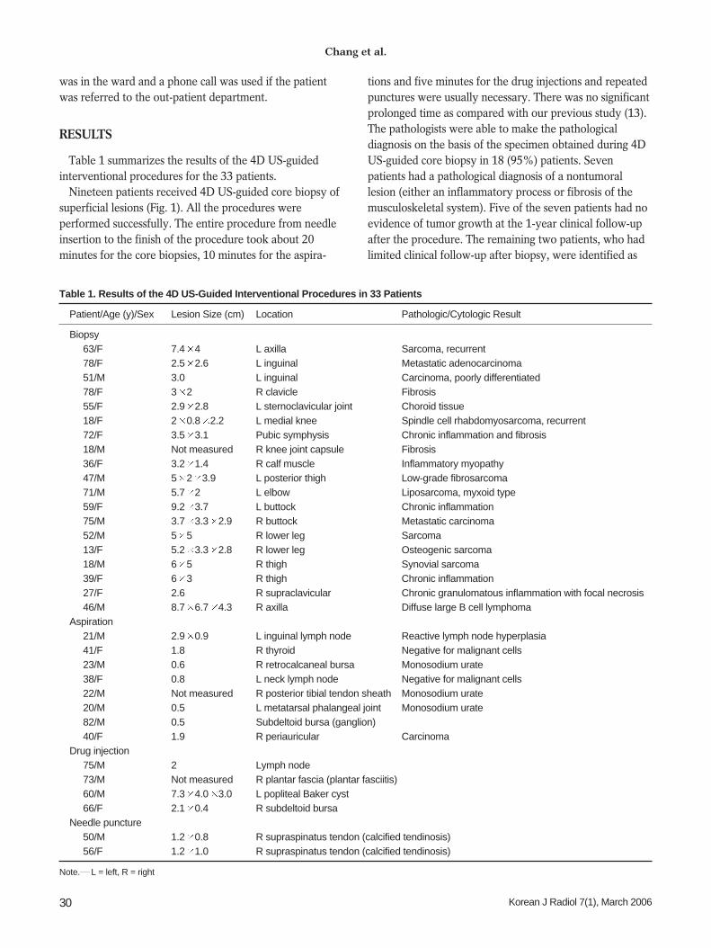
Table 1. Results of the 4D US-Guided Interventional Procedures in 33 Patients
Patient/Age (y)/Sex Lesion Size (cm) Location Pathologic/Cytologic Result
Biopsy63/F 7.4 4 L axilla Sarcoma, recurrent78/F 2.5 2.6 L inguinal Metastatic adenocarcinoma51/M 3.0 L inguinal Carcinoma, poorly differentiated78/F 3 2 R clavicle Fibrosis55/F 2.9 2.8 L sternoclavicular joint Choroid tissue18/F 2 0.8 2.2 L medial knee Spindle cell rhabdomyosarcoma, recurrent72/F 3.5 3.1 Pubic symphysis Chronic inflammation and fibrosis18/M Not measured R knee joint capsule Fibrosis36/F 3.2 1.4 R calf muscle Inflammatory myopathy47/M 5 2 3.9 L posterior thigh Low-grade fibrosarcoma71/M 5.7 2 L elbow Liposarcoma, myxoid type59/F 9.2 3.7 L buttock Chronic inflammation75/M 3.7 3.3 2.9 R buttock Metastatic carcinoma52/M 5 5 R lower leg Sarcoma13/F 5.2 3.3 2.8 R lower leg Osteogenic sarcoma18/M 6 5 R thigh Synovial sarcoma39/F 6 3 R thigh Chronic inflammation27/F 2.6 R supraclavicular Chronic granulomatous inflammation with focal necrosis46/M 8.7 6.7 4.3 R axilla Diffuse large B cell lymphoma
Aspiration21/M 2.9 0.9 L inguinal lymph node Reactive lymph node hyperplasia41/F 1.8 R thyroid Negative for malignant cells23/M 0.6 R retrocalcaneal bursa Monosodium urate38/F 0.8 L neck lymph node Negative for malignant cells22/M Not measured R posterior tibial tendon sheath Monosodium urate20/M 0.5 L metatarsal phalangeal joint Monosodium urate82/M 0.5 Subdeltoid bursa (ganglion)40/F 1.9 R periauricular Carcinoma
Drug injection75/M 2 Lymph node73/M Not measured R plantar fascia (plantar fasciitis)60/M 7.3 4.0 3.0 L popliteal Baker cyst66/F 2.1 0.4 R subdeltoid bursa
Needle puncture50/M 1.2 0.8 R supraspinatus tendon (calcified tendinosis)56/F 1.2 1.0 R supraspinatus tendon (calcified tendinosis)
Note. L = left, R = right

having osteomyelitis (one patient) and tuberculosis (onepatient). Eleven patients who received 4D US-guided corebiopsy had evidence of tumor growth on the pathologicexaminations. The last patient, who presented with ahistory of polyarthralgia, developed a mass lesion in theleft sternoclavicular joint. This patient received 4D US-guided biopsy of the lesion and the pathologic specimenshowed nonspecific chondroid tissue. Repeat biopsy under2D US guidance was performed two months later becauseof an inconclusive pathologic result, and the pathologicalfindings from the second biopsy were similar to the firstbiopsy with no evidence of neoplasm. The lesion did notprogress during 18-month follow-up; it was thought to bearthropathy that involved the left sternoclavicular joint.
Eight patients received 4D US-guided needle aspiration oftheir superficial lesions (Figs. 2, 3). All the procedures wereperformed successfully. Three of the patients had a clinicalpresentation of a gout attack. Monosodium urate crystalswere identified in the aspirated specimens upon microscopic
examination, which confirmed the clinical suspicion of gout.One patient with nasopharyngeal carcinoma presented withan enlarged lymph node (1.9 cm) in the right periauricularregion, and this patient was diagnosed as having metastaticnasopharyngeal carcinoma after 4D US-guided aspiration.Two patients with enlarged lymph nodes in the neck (0.8cm) or the inguinal region (2.9 cm) underwent 4D US-guided needle aspiration, and this showed no evidence ofmalignancy. These two patients had no evidence of progres-sive change in their nodal status at the 1-year clinicalfollow-up. One patient with a hypoechoic nodule in theright thyroid (1.8 cm in size), underwent 4D US-guideaspiration and there was no evidence of malignancy. Onepatient suffered from a small subdeltoid ganglion (0.5 cm).After aspiration of about 0.5 cc of jelly-like material, thepatient’s symptoms were dramatically relieved. There is norecurrence at six months follow up.
Four patients received 4D US-guided injection of atherapeutic drug. All the procedures were performed
2D with Additional Coronal Reformatted 4D US Guidance in Superficial Lesion Intervention
Korean J Radiol 7(1), March 2006 31
Fig. 1. A 78-year-old woman with ahistory of surgery for colon cancerdeveloped an enlarged, left inguinallymph node. During the four-dimensionalUS-guided biopsy, the needle did notshow up on the two-dimensional axialimage (left), indicating that it haddeviated out of plane. The real-timevolume-rendered image in the coronalreformatted plane (right) clearly showedthat the needle (arrowheads) wasbasically in the center of the lesion.
Fig. 2. A 21-year-old man suffering with osteogenic sarcoma in the left distal femur received neoadjuvant chemotherapy and surgicalresection of the tumor. An enlarged, left inguinal lymph node was palpated. A. Color Doppler US showed the enlarged node with a prominent cortex of increased vascularity. The proposed optimal site for theneedle aspiration was in the cortical portion of the node (arrowheads). B. Four-dimensional US-guided fine-needle aspiration was performed. Axial 2D image (left) showed that the needle (arrowheads) hadpenetrated into the cortex of the node. Real-time volume-rendered imaging in the coronal reformatted plane (right) showed the needle tip(arrow) in the central cortical portion of the node. By combining information from both images, we confirmed that the needle tip was in thecentral cortical portion of the node in all three orthogonal planes.
A B

successfully. One patient with metastatic lymphadenopathydue to a transitional cell carcinoma of the urinary bladderreceived a direct injection of a chemotherapeutic drug intothe diseased lymph node (node size: 2 cm). Anotherpatient received a local injection of corticosteroid into apopliteal cyst (Baker’s cyst). The remaining two patientsalso received corticosteroid injections: one was done in theplantar fascia to treat plantar fasciitis and the other onewas done in the subdeltoid bursa to treat subdeltoidbursitis.
Two patients with calcified tendinosis of the supraspina-tus tendon of the right shoulder underwent repeat 4D US-guided needle punctures of the calcified plaque. Thecalcified deposits were resorbed, as was observed on thefollow-up US performed two weeks after the procedure.
On the postprocedure follow-up, no major complicationsor significant minor complications that needed furthersurgical or invasive procedure management were noted inthis study.
DISCUSSION
Two-dimensional US is commonly used for imagingguidance in interventional radiology because the modern2D US provides images of adequate quality for mostclinical procedures. This modality offers the advantages ofreal-time capability and wide availability in clinicalpractice. An experienced radiologist or physician canusually apply this imaging tool without great difficulty.However, real-time 2D US provides only 2D imagesduring image-guided procedures, and it does not offer 3Dinformation about the lesion or the region of interest.
During a 2D US-guided procedure, the operator mustsweep the transducer to delineate the spatial relationshipbetween the needle and the target lesion, as is estimatedon the serial 2D images. While the needed is beingadvanced toward or within the lesion, the operator shouldkeep the US transducer steady to enable visualization ofthe moving needle. Any deviation of the needle out of theplane of the ultrasound beam or any slight movement ofthe transducer results in the loss of needle visualization.Hence, during the procedure, the operator alwaysadvances the needle step by step. 4D US enables real-time,simultaneous monitoring of the advancing needle and ofthe spatial relationship between the needle and thetargeted lesion.
Static 3D US-guided interventional procedures havebeen applied in several studies and beneficial results wereobtained 5 11 for any of the investigations that involvedusing static 3D US for interventional procedures in deeplesions, such as the ablation of hepatic tumors (8),transjugular intrahepatic portosystemic shunt procedures(5), drainage of intra-abdominal fluid6, and treatment ofprostate tumors (9, 11). A few articles refer to the applica-tion of 4D US-guided procedure (12). Real-time coronalreformatted images (described as 4D coronal reformattedimages) provide an additional image plane to demonstratelesions, which is not possible with using the 2D US system.With visualization from both the 2D axial images and the4D coronal reformatted images in the dual-screen format,we are able to obtain information on the needle position inboth the axial and coronal planes, and so we were able todirect the needle more precisely during the procedures.
Image-guided interventional procedures in superficial
Chang et al.
32 Korean J Radiol 7(1), March 2006
Fig. 3. A 41-year-old woman presented with a nodule on the right side of her neck. A. The image showed the nodule with central cystic change in the right lobe of the thyroid. The solid parts were mainly at the periphery(arrowheads). B. Four-dimensional US-guided fine-needle aspiration of the nodule was performed. The axial two-dimensional image (left) did not showthe needle because it had deviated out of the central plane. The real-time volume-rendered image in the coronal reformatted plane (right)showed the needle tip (arrows) in the periphery of the nodule, which was the desired location for tissue sampling.
A B

lesions (e.g., those in the breast, the musculoskeletalsystem and in small areas) rely heavily on US. With theadvent of high-resolution US, US-guided procedures forperipheral lesions have become more commonly applied inclinical practice, though a few reports refer to the applica-tion of static 3D US in breast biopsy 7, 10. One maypropose that a superficial lesion is easier to approach thana deep lesion; hence, the application of 3D US in superfi-cial lesions may not be necessary (12). However, as in ourcases, some superficial lesions are small for approachingwith a needle, and the target site for the needle in eachlesion is even smaller. For instance, for the biopsy of a softtissue tumor, the optimal site for specimen acquisition isthe solid part of the lesion. In the case of lymph nodeaspiration or biopsy, the optimal site is the cortex of thelymph node. Likewise, with urate deposits in soft tissue,the echogenic crystals may be localized in rather smallareas. Therefore, precise needle positioning during aspira-tion may decrease the false-negative rate for identifyingurate crystals. Similarly, during therapeutic drug injection,precise needle positioning for drug injection yieldsfavorable therapeutic effects. In our experience, 4D USaids the physician in directing the biopsy needle as it isadvanced toward the desired portion of the target lesions,and this increases the operator’s confidence of the needleposition during the procedure.
At the initial stage of the procedures, we selected the 2Dmode to guide the needles from the skin penetration untilthe needle tips approached the margin of the lesion (thepre-lesional tract). This was due to the similarity of theechogenicity between the needles and thesubcutaneous/muscle layers, which would hinder thevisualization of the needles during 3D reconstruction. Werecommend 4D US guidance for visualizing the pre-lesional tract only if the echogenicity of the surroundingtissue is low enough to differentiate between the echogenicneedle and the tissue within the tract.
In this study, 11 patients who received 4D US-guidedcore biopsy were found to have soft tissue tumors (sevenmalignant tumors, two recurrent soft tissue sarcomas andtwo metastatic carcinomas). The pathologists were able tomake the diagnoses on the basis of the representativespecimens that were obtained from 4D US-guided biopsy.Furthermore, the diagnoses were identical to the finalpathological diagnoses in 10 (90.9%) of the patients. Theeight patients who received 4D US-guided core biopsywere found to have non-tumoral lesions. Among them, thepathologic results of core biopsy were identical to the finaldiagnosis in seven patients (87.5%). As compared with ourprevious experience, the concordance of the pathologicaldiagnoses between the conventional US-guided core
biopsy and the open biopsy for the soft tissue tumors wasaround 90%, whereas identical pathological resultsbetween conventional US-guided core biopsy and openbiopsy were observed in 55% (13). Both studies wereperformed by the same radiologist (H-J.C.); the majordifference of the interventional method between bothstudies was only the use of a different US machine in thecurrent study, i.e., 4D with coronal sections. The operationtimes were similar between both studies, but differentneedle sizes were used and there was a different number ofpatients. Therefore, the significance of the discrepancybetween both studies needs further study in the future.
In summary, the patients of this study received 4D US-guided core biopsy and the results had a very high concor-dance with pathological results and they were identical tothe final diagnosis of the surgical specimens. Becauseaccurate pathological diagnosis relies on a sufficientrepresentative specimen obtained from core biopsy, wepropose that the additional imaging information from thecoronal reformatted 4D US aids the physician in selectingthe optimal site for needle placement. This information ledto the adequate acquisition of representative specimens forthe pathologic examination in our cases.
Conventional 2D US guidance does have severaladvantages. For example, operators are familiar with thetraditional 2D US-guided procedures. In addition, the lighttransducer used in 2D US is easier to handle during theprocedures than the other transduces. Last, adjustment ofthe angle of the transducer is easier because it is less bulkythan the 4D US transducer.
Although our study had a limited number of cases andalthough we did not compare the results of 2D US- and 4DUS-guided procedures, our preliminary results showed thepotential of performing 4D US guidance in clinical applica-tions that involved superficial lesions. Further randomizedstudies for comparing 2D- and 4D US-guided proceduresare needed to objectively determine the role of 4D US.
Involuntary motions of patient, such as cardiac motion,arterial pulsation or respiratory motion can result inimaging artifacts when performing 4D US of deep lesions.In our cases, this drawback was minimized because theimaging of superficial lesions is less affected by theseinvoluntary motions. Constant transducer vibration during4D US acquisition is another major source of imageartifacts during procedures (4, 12). However, this did notdisturb our procedures nor did it affect the manipulation ofthe needle. Last, the mechanically driven transducer maybe problematic if the operating physician is not comfort-able with using such a bulky device, and the fluency of theprocedure could potentially be affected. However, wehave found that this problem was minimized once the
2D with Additional Coronal Reformatted 4D US Guidance in Superficial Lesion Intervention
Korean J Radiol 7(1), March 2006 33

Chang et al.
34 Korean J Radiol 7(1), March 2006
operator adapts to the device.In conclusion, recent advances in US technique have
improved 4D US imaging for its use in clinical practice.Combining the 4D US and 2D US-guided techniques isfeasible for performing interventional procedures forsuperficial lesions.
References1. Cho N, Moon WK, Cha JH. Sonographically guided core biopsy
of the breast: comparison of 14-gauge automated gun and 11-gauge directional vacuum-assisted biopsy methods. Korean JRadiol 2005;6:102-109
2. Lim HK. Radiofrequency thermal ablation of hepatocellularcarcinomas. Korean J Radiol 2000;1:175-184
3. Lees W. Ultrasound imaging in three and four dimensions.Semin Ultrasound CT MR 2001;22:85-105
4. Downey DB, Fenster A, Williams JC. Clinical utility of three-dimensional US. RadioGraphics 2000;20:559-571
5. Rose SC, Pretorius DH, Nelson TR, Kinney TB, Huynh TV,Roberts AC, et al. Adjunctive 3D US for achieving portal veinaccess during transjugular intrahepatic portosystemic shuntprocedures. J Vasc Interv Radiol 2000;11:611-621
6. Rose SC, Roberts AC, Kinney TB, Pretorius DH, Nelson TR.Three-dimensional ultrasonography for planning percutaneousdrainage of complex abdominal fluid collections. J Vasc Interv
Radiol 2003;14:451-4597. Smith WL, Surry KJ, Mills GR, Downey DB, Fenster A. Three-
dimensional ultrasound-guided core needle breast biopsy.Ultrasound Med Biol 2001;27:1025-1034
8. Rose SC, Hassanein TI, Easter DW, Gamagami RA, Bouvet M,Pretorius DH, et al. Value of three-dimensional US for optimiz-ing guidance for ablating focal liver tumors. J Vasc Interv Radiol2001;12:507-515
9. Strasser H, Janetschek G, Horninger W, Bartsch G. Three-dimensional sonographic guidance for interstitial laser therapyin benign prostatic hyperplasia. J Endourol 1995;9:497-501
10. Weismann CF, Forstner R, Prokop E, Rettenbacher T. Three-dimensional targeting: a new three-dimensional ultrasoundtechnique to evaluate needle position during breast biopsy.Ultrasound Obstet Gynecol 2000;16:359-364
11. Chin JL, Downey DB, Mulligan M, Fenster A. Three-dimensional transrectal ultrasound guided cryoablation forlocalized prostate cancer in nonsurgical candidates: a feasibilitystudy and report of early results. J Urol 1998;159:910-914
12. Won HJ, Han JK, Do KH, Lee KH, Kim KW, Kim SH, et al.Value of four-dimensional ultrasonography in ultrasonographi-cally guided biopsy of hepatic masses. J Ultrasound Med 2003;22:215-220
13. Liu JC, Chiou HJ, Chen WM, Chou YH, Chen TH, Chen W, etal. Sonographically guided core needle biopsy of soft tissueneoplasms. J Clin Ultrasound 2004;32:294-298