Embed Size (px)
Citation preview
Deepak AggarwalDept of Pulmonary Medicine
Government Medical College and Hospital, Chandigarh
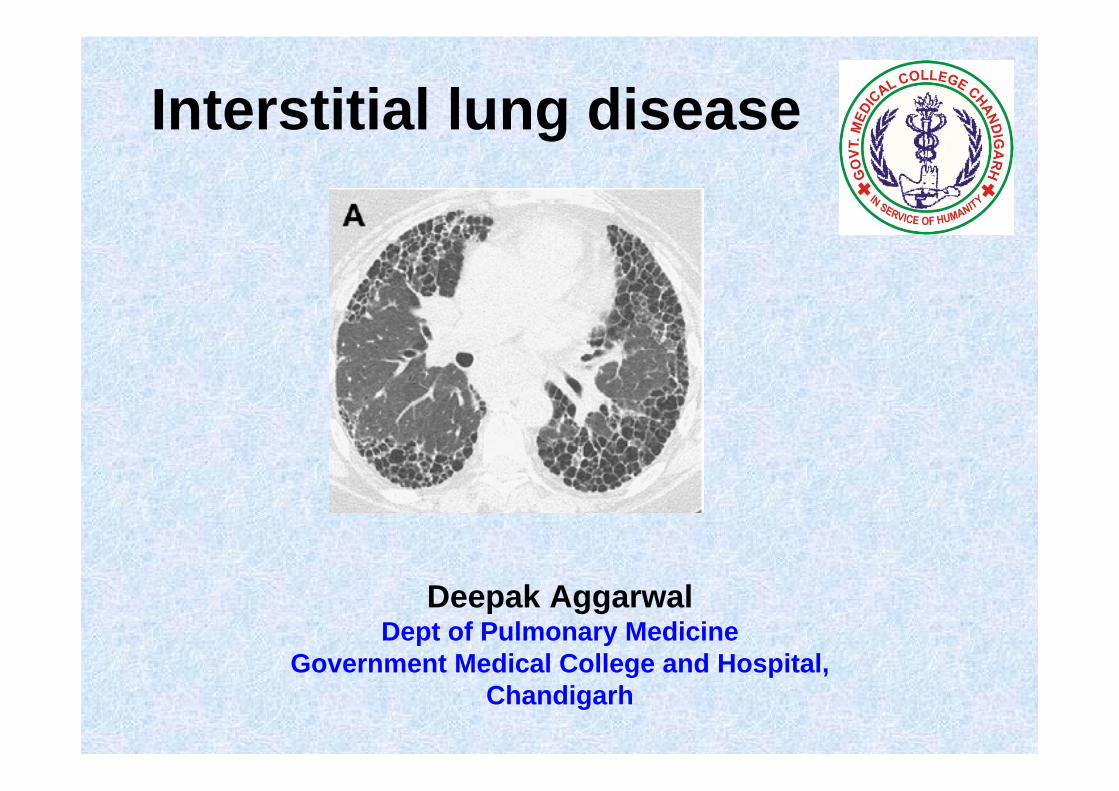
Interstitial lung disease
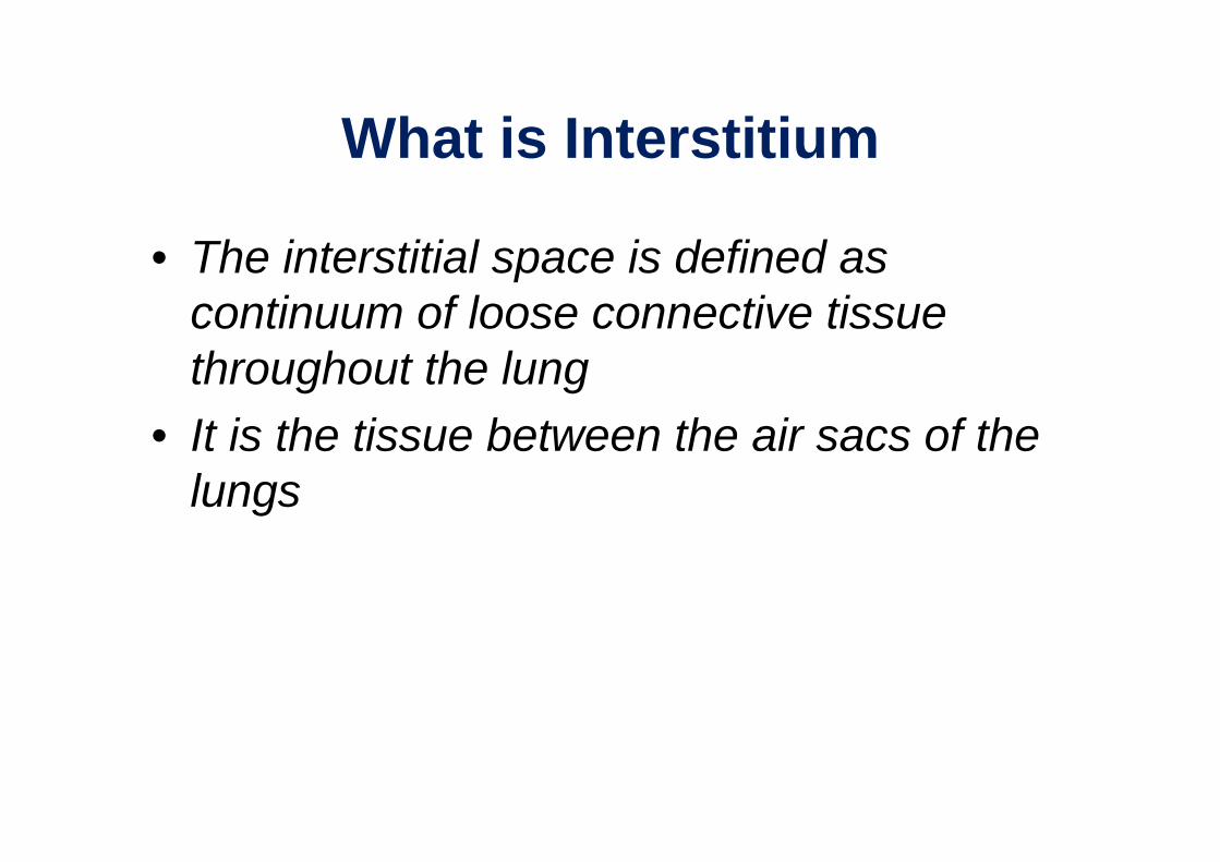
What is Interstitium
• The interstitial space is defined as continuum of loose connective tissue throughout the lung
• It is the tissue between the air sacs of the lungs

Subdivisions of lung interstitium
Axial interstitium(peribronchovascular)
Surrounding the bronchi, arteries, and veins from the lung root to the level of the
respiratory bronchiole;
Centrilobular interstitiumsituated between the alveolar
and capillary basement membranes
peripheral interstitiumcontains the pulmonary venules, lymphatics and
interlobular septae
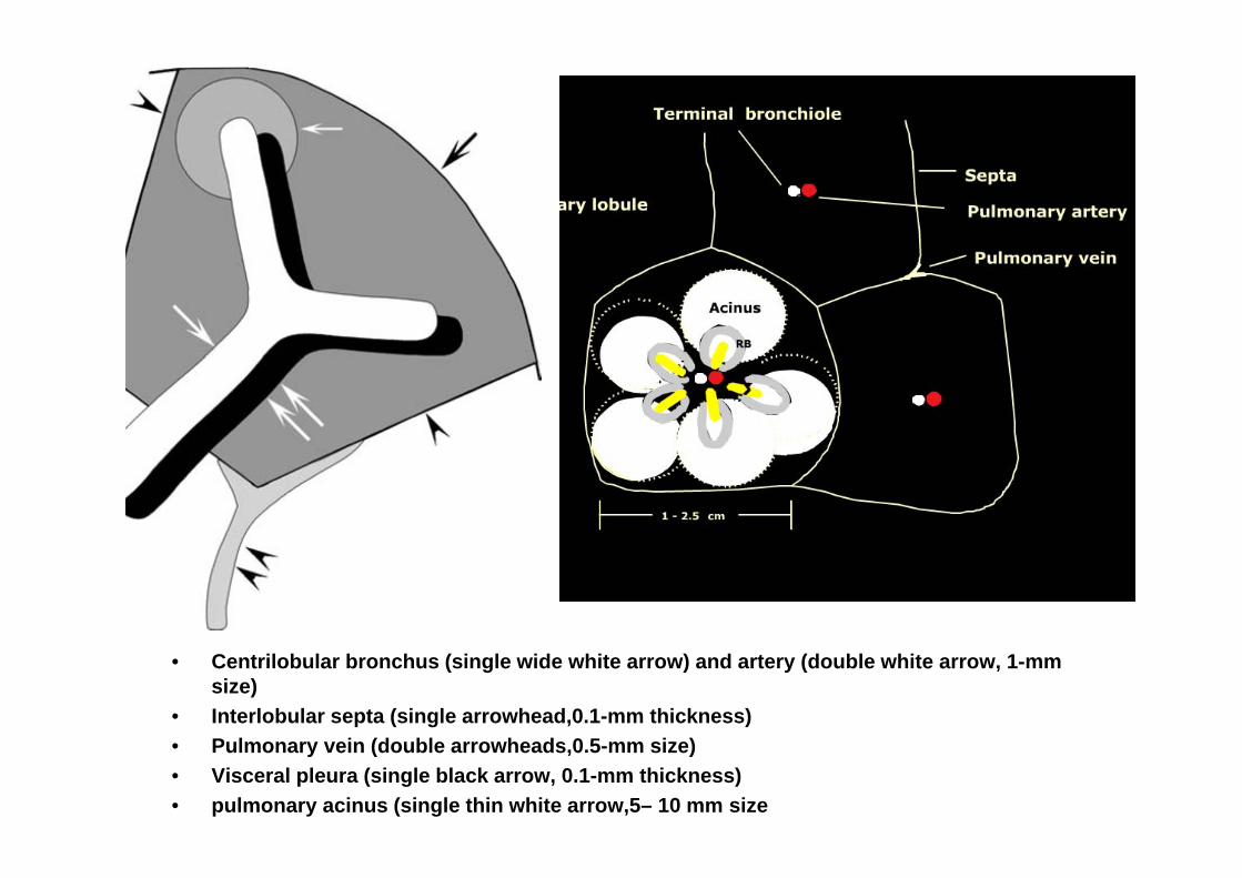
• Centrilobular bronchus (single wide white arrow) and artery (double white arrow, 1-mm size)
• Interlobular septa (single arrowhead,0.1-mm thickness)• Pulmonary vein (double arrowheads,0.5-mm size)• Visceral pleura (single black arrow, 0.1-mm thickness)• pulmonary acinus (single thin white arrow,5– 10 mm size

Introduction
• There are >200 disorders• Can classified into
–known vs unknown with subcategory of granulomatous and nongranulomatous. OR
–occupational vs nonoccupational–Acute vs Chronic

ILD Classification
Approach to ILD
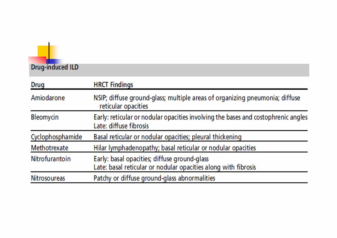
ILD of knownCause
Drugs Exposure
HypersensitivityPneumonitis Pneumoconiosis
Toxic Inhalation Radiation
CVD
Idiopathic Interstitial
Pneumonias
IPF IIP other than IPF
DesquamativeInterstitialPneumonia
Respiratory Bronchiolitis-
Interstitial Lung disease
Acute InterstitialPneumonia
Cryptogenic Organizing Pneumonia
Lymphocytic Interstitial
Pneumonia
Non Specific Interstitial
Pneumonia
GranulomatousLung Diseases(Sarcoidosis)
Others
LAMHistiocytosis X
Malignancy
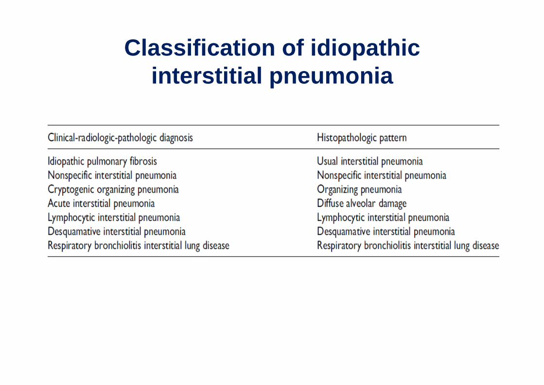
Classification of idiopathic interstitial pneumonia
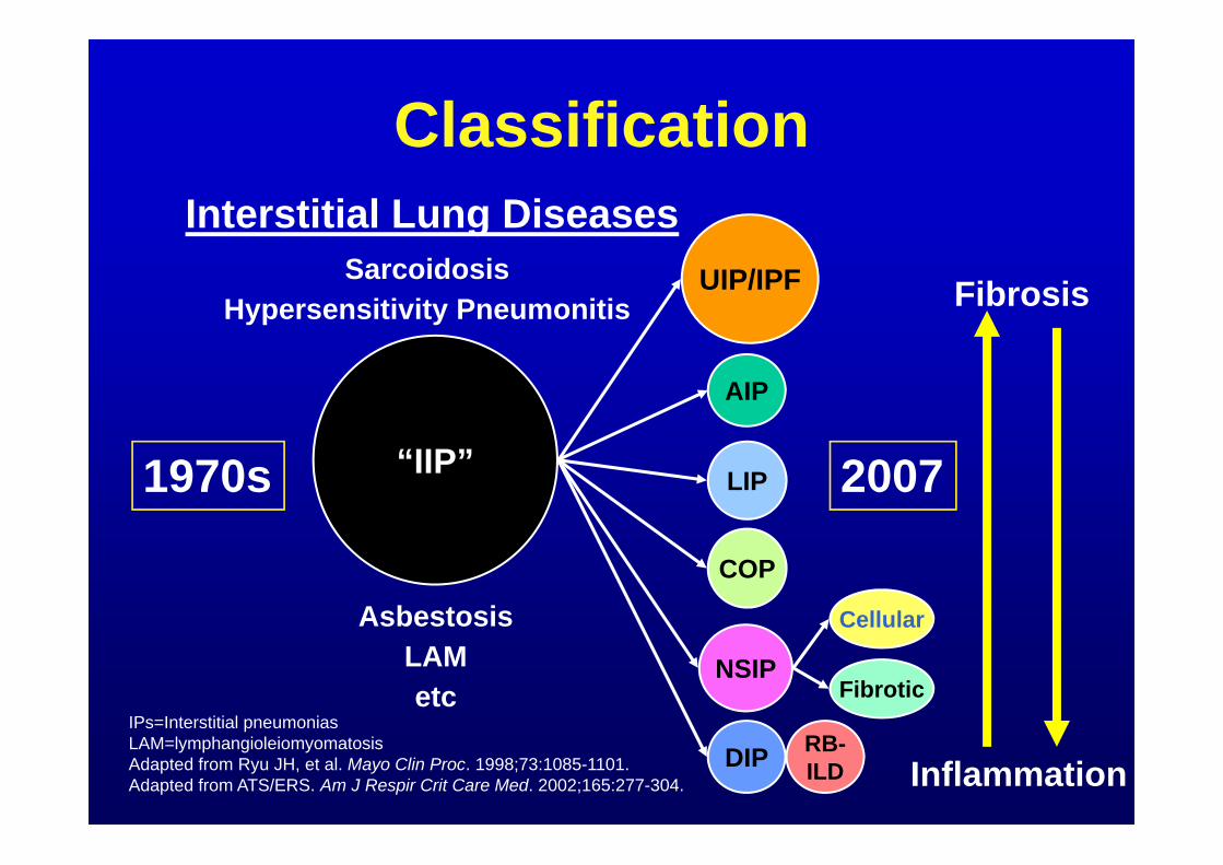
Classification
IPs=Interstitial pneumonias LAM=lymphangioleiomyomatosisAdapted from Ryu JH, et al. Mayo Clin Proc. 1998;73:1085-1101.Adapted from ATS/ERS. Am J Respir Crit Care Med. 2002;165:277-304.
1970s 2007“IIP”
UIP/IPF
NSIP
DIP RB-ILD
AIP
Cellular
Fibrotic
Interstitial Lung DiseasesSarcoidosis
Hypersensitivity Pneumonitis
AsbestosisLAMetc
COP
LIP
Fibrosis
Inflammation
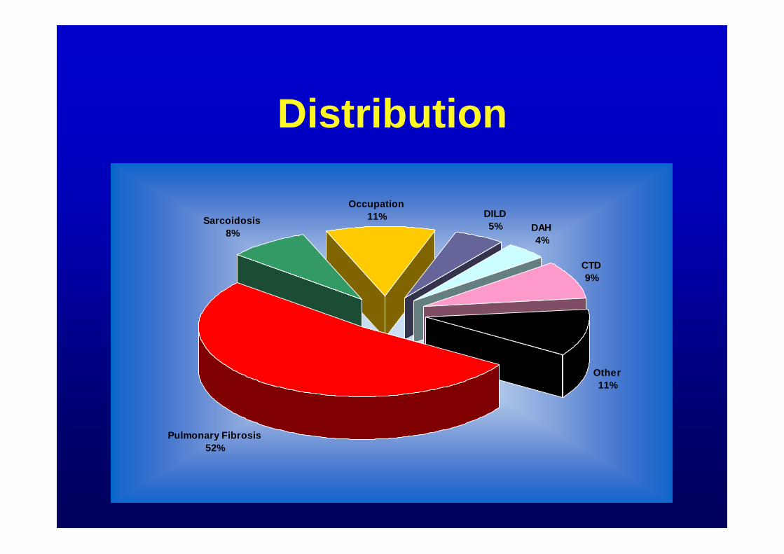
Distribution
Sarcoidosis8%
Occupation11% DILD
5% DAH4%
CTD9%
Other11%
Pulmonary Fibrosis52%
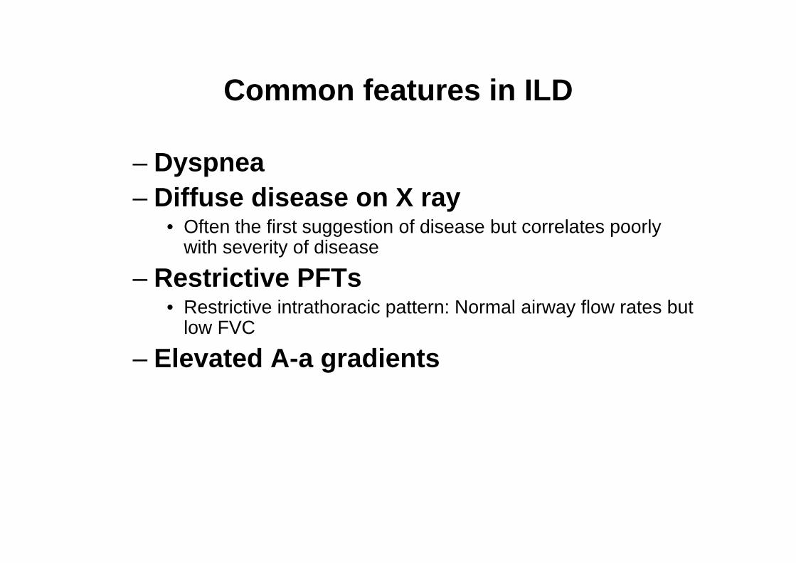
Common features in ILD
– Dyspnea– Diffuse disease on X ray
• Often the first suggestion of disease but correlates poorly with severity of disease
– Restrictive PFTs• Restrictive intrathoracic pattern: Normal airway flow rates but
low FVC
– Elevated A-a gradients
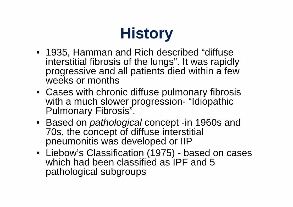
History• 1935, Hamman and Rich described “diffuse
interstitial fibrosis of the lungs”. It was rapidly progressive and all patients died within a few weeks or months
• Cases with chronic diffuse pulmonary fibrosis with a much slower progression- “Idiopathic Pulmonary Fibrosis”.
• Based on pathological concept -in 1960s and 70s, the concept of diffuse interstitial pneumonitis was developed or IIP
• Liebow’s Classification (1975) - based on cases which had been classified as IPF and 5 pathological subgroups

Idiopathic Pulmonary Fibrosis

Katzenstein ALA et al. Am J Respir Crit Care Med. 1998;157:1301.
PATHOGENESIS AND COURSE OF UIP / IPF
UIP / IPFMultiple microscopic foci of injury leading to inflamation
( stage of alveolitis)
Focal fibroblast proliferation (fibroblastic foci)
Collagen deposition ( Stage of fibrosis)
Progressive clinical course
Death
Recurrent microscopic injury

Clinical context
• Idiopathic Pulmonary Fibrosis (IPF) affects predominantly middle-aged and older subjects
• Subacute to chronic dry cough, progressive breathlessness
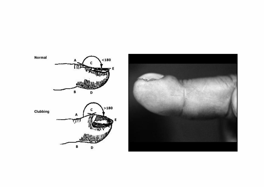
• The “Velcro” crackles is nearly universal with IPF
• digital clubbing - in up to two thirds of patients with IPF but is rare in patients with sarcoidosis

Clinical Evaluation: History, PE, CXR, PFTs, 6MWT
Not IIP Potential IIP
HRCT
Diagnostic of diffuse lung disease
Diagnosis uncertain
Surgical lung biopsy
Transbronchial Bx or BAL
Diagnostic Nondiagnostic
IPFNot IPF
Approach to Diagnosing diffuse lung disease
Adapted from ATS/ERS Consensus Statement. Am J Respir Crit Care Med. 2002;165:277-304.
Approach to the diagnosis• Thorough medical history is a must
o ageo duration o symptoms o smoking history o occupational and environmental exposureso medications and drugs o family medical historyo signs and symptoms of connective tissue disease
should be carefully elicited

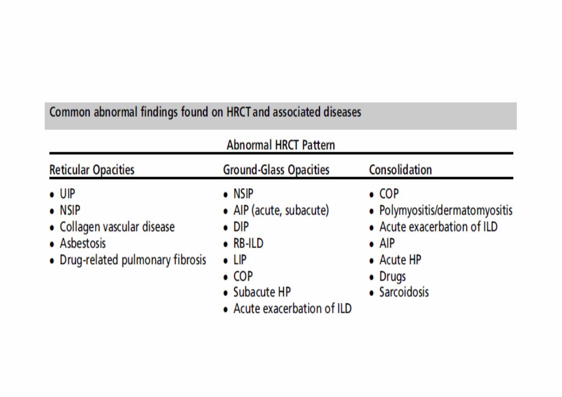
Patterns of ILD on X-ray

Normal CXR Abnormal CXR
Typical Features of IPF on Chest X-Ray

Chest Radiograph
• Abnormal >90%• peripheral reticular opacities • more profuse at lung bases• bilateral• asymmetrical• commonly associated with
decreased lung volumes• confluent alveolar opacities,
lymphadenopathy and pleural involvement uncommon

HRCT Chest
• More sensitive and specific
• Better diagnostic accuracy
• Assessment of disease activity
• May obviate need of biopsy
• Site for biopsy
• Follow up
• Prognosis
INITIAL DIAGNOSTIC MODALITY OF CHOICE
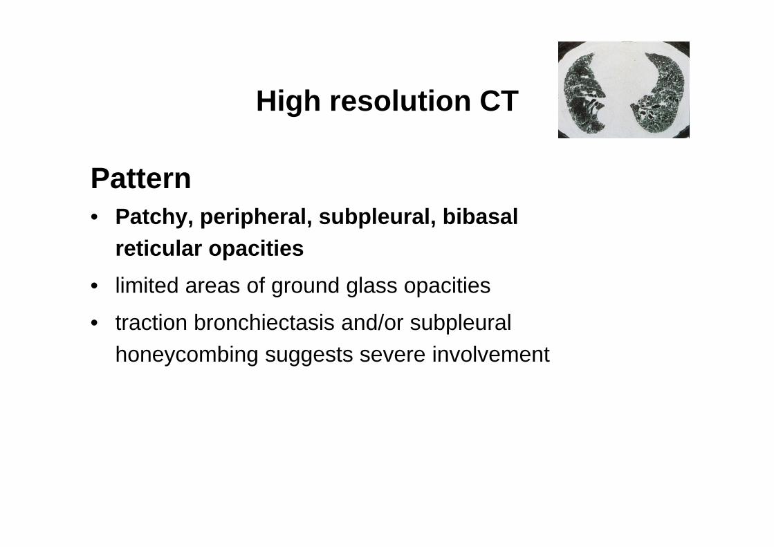
High resolution CT
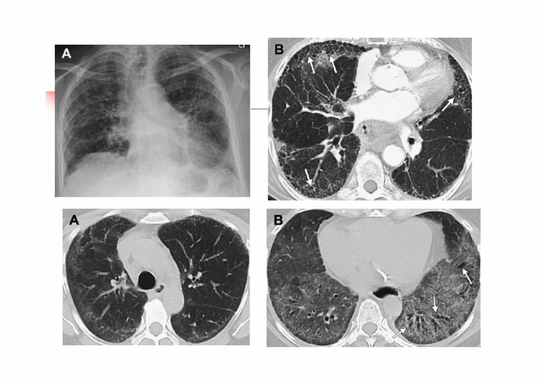
Pattern• Patchy, peripheral, subpleural, bibasal
reticular opacities• limited areas of ground glass opacities
• traction bronchiectasis and/or subpleuralhoneycombing suggests severe involvement
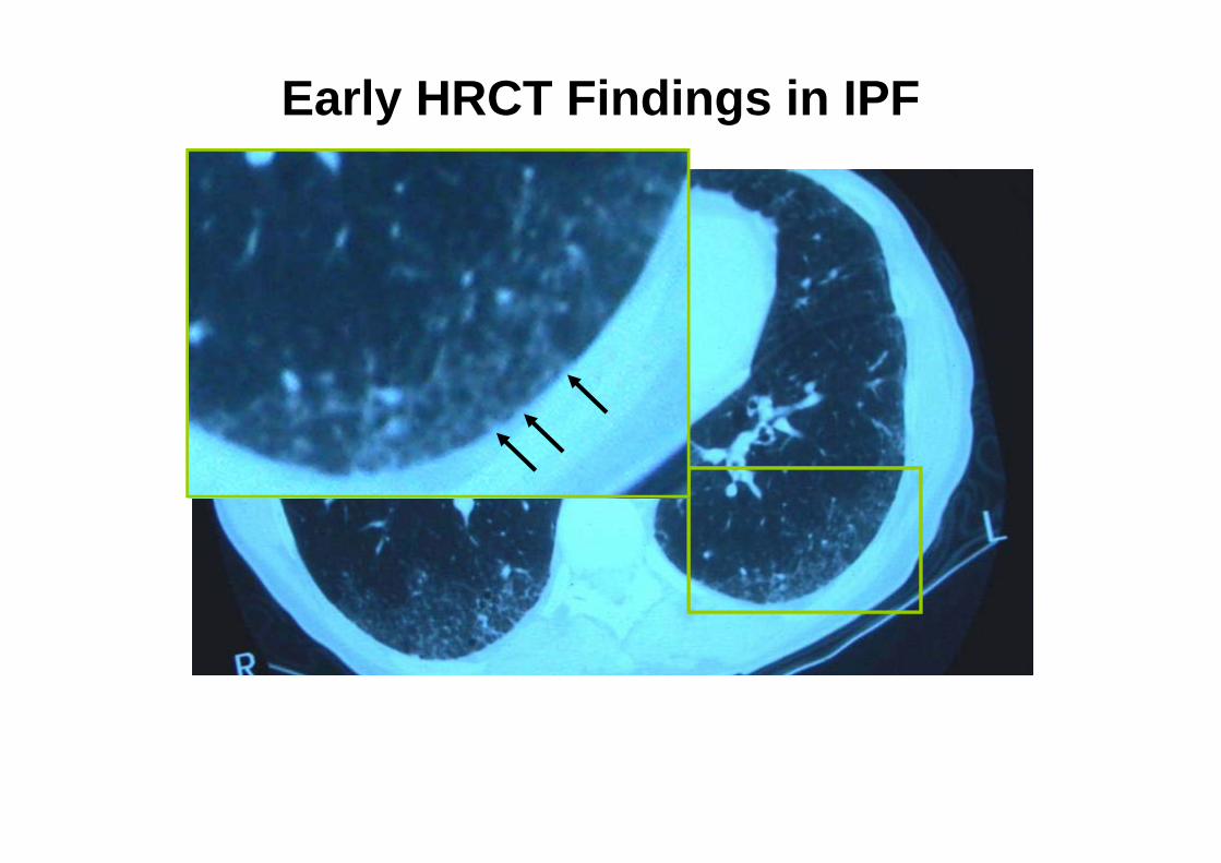
Early HRCT Findings in IPF

Subpleural and Basal Predominance
Slide courtesy of W. Richard Webb, MD.
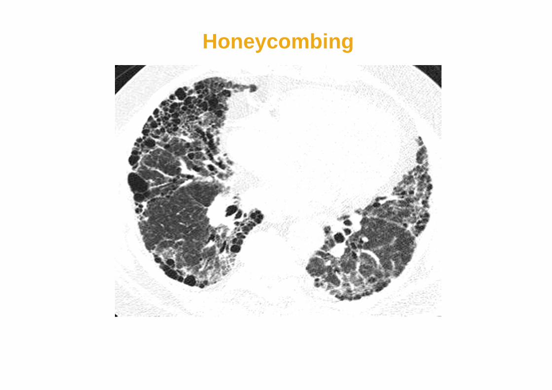
Honeycombing

“Traction” Bronchiectasis

High resolution CT
• Differential diagnosis of HRCT patternConnective tissue diseasesAsbestosisHypersensitivity pneumonitisSarcoidosis
• Extensive GROUND GLASS OPACITIES on CT of lung should prompt alternative diagnosis such as DIP, RBILD, NSIP, idiopathic BOOP, hypersensitivity pneumonitisetc.

Spirometry

Approach to the diagnosis of IPFPulmonary function testing
• LUNG VOLUMESTLC , FRC, RV are decreased
• AIRWAY MECHANICSFVC, FEV1, PEFR are often decreasedFEV1/FVC is maintained or increased
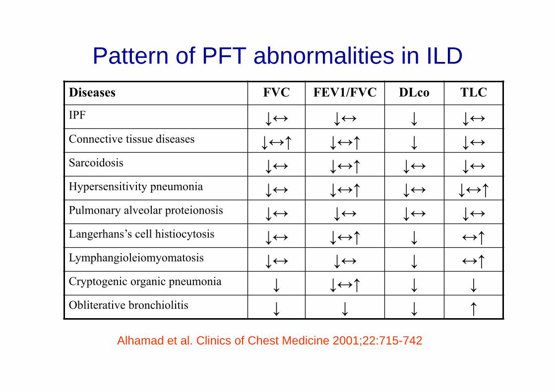
Pattern of PFT abnormalities in ILDDiseases FVC FEV1/FVC DLco TLC
IPF ↓↔ ↓↔ ↓ ↓↔Connective tissue diseases ↓↔↑ ↓↔↑ ↓ ↓↔Sarcoidosis ↓↔ ↓↔↑ ↓↔ ↓↔Hypersensitivity pneumonia ↓↔ ↓↔↑ ↓↔ ↓↔↑Pulmonary alveolar proteionosis ↓↔ ↓↔ ↓↔ ↓↔Langerhans’s cell histiocytosis ↓↔ ↓↔↑ ↓ ↔↑Lymphangioleiomyomatosis ↓↔ ↓↔ ↓ ↔↑Cryptogenic organic pneumonia ↓ ↓↔↑ ↓ ↓Obliterative bronchiolitis ↓ ↓ ↓ ↑
Alhamad et al. Clinics of Chest Medicine 2001;22:715-742

Role of BAL vs Biopsy in IPF
Procedure Role
Brochoalveolar Lavage (BAL)• May rule out alternate diagnoses but
not diagnostic of IPF
Transbronchial Biopsy (TBLB)• May rule out alternate diagnoses but
not diagnostic of IPF• Often abnormal in IPF but does not
confirm diagnosis
Video-assisted Thoracoscopic Biopsy (VATS)• Preferred technique• Provides best tissue samples• Excludes other processes that mimic IPF• Biopsies should be obtained from more
than one lobe of the lung
ATS/ERS Consensus Statement. Am J Respir Crit Care Med. 2000;161:646-664; 2002;165:277-304.

Katzenstein ALA et al. Am J Respir Crit Care Med. 1998;157:1301.
Feature UIP DIP/RBILD AIP NSIP
Temporal appearance Variegated Uniform Uniform Uniform
Interstitial inflammation Scant Scant Scant Usually prominent
Collagen fibrosis Patchy Variable, diffuse No Variable, diffuse
in DIP; focal, mild in RBILD
Fibroblast proliferation Fibroblastic foci No Diffuse Occasional, diffuse, or rare fibroblastic foci
Organizing pneumonia No No No Occasional, focal
Honeycomb changes Yes No No Rare
Intraalveolar macrophage Occasional, focal Diffuse in DIP; No Occasional, patchyaccumulation peribronchiolar in
RBILD
Hyaline membranes No No Occasional, Nofocal
CONTRASTING PATHOLOGIC FEATURES OF IIP

ATS/ERS diagnostic criteria for IPF
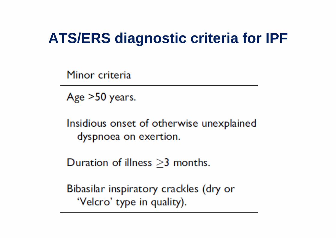
ATS/ERS diagnostic criteria for IPF
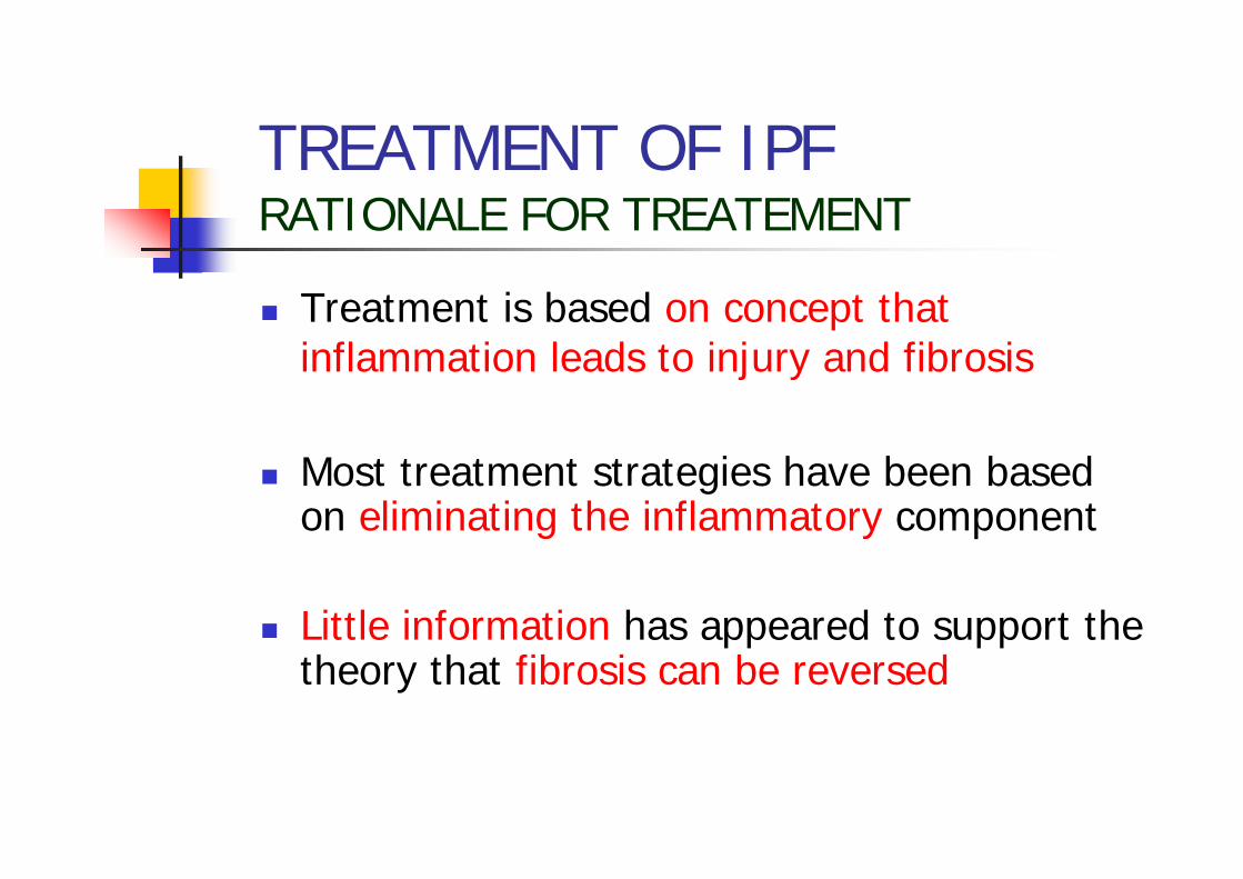
TREATMENT OF IPFRATIONALE FOR TREATEMENT
Treatment is based on concept that inflammation leads to injury and fibrosis
Most treatment strategies have been based on eliminating the inflammatory component
Little information has appeared to support the theory that fibrosis can be reversed

TREATMENT OF IPFCONVENTIONAL TREATMENT OPTIONS
CORTICOSTEROIDS IMMUNOSUPPRESSIVE / CYTOTOXIC AGENTS
AZATHIOPRIMCYCLOPHOSPHAMIDE
ANTIFIBROTIC AGENTCOLCHICINEPIRFENIDONED-PENICILLAMINE
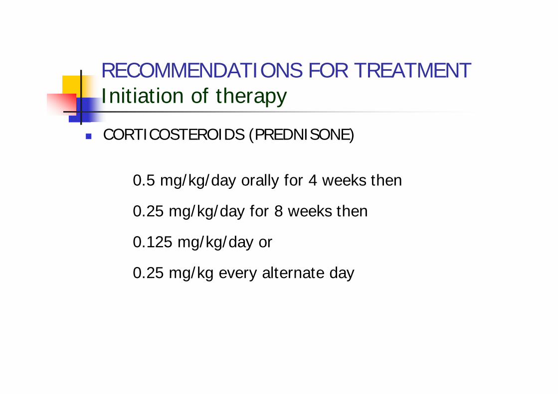
RECOMMENDATIONS FOR TREATMENTInitiation of therapy
CORTICOSTEROIDS (PREDNISONE)
0.5 mg/kg/day orally for 4 weeks then
0.25 mg/kg/day for 8 weeks then
0.125 mg/kg/day or
0.25 mg/kg every alternate day
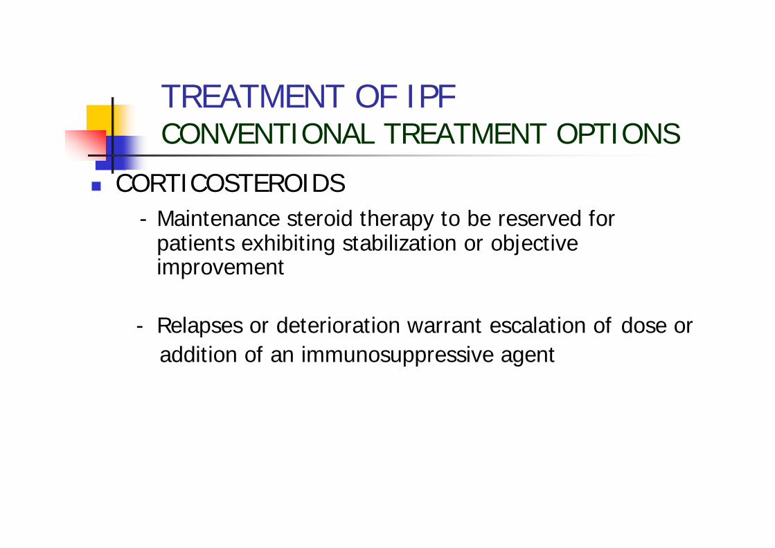
TREATMENT OF IPFCONVENTIONAL TREATMENT OPTIONS
CORTICOSTEROIDS- Maintenance steroid therapy to be reserved for
patients exhibiting stabilization or objective improvement
- Relapses or deterioration warrant escalation of dose oraddition of an immunosuppressive agent
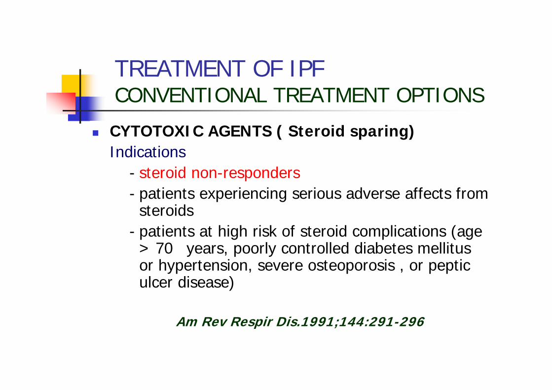
TREATMENT OF IPFCONVENTIONAL TREATMENT OPTIONS
CYTOTOXIC AGENTS ( Steroid sparing)Indications
- steroid non-responders- patients experiencing serious adverse affects from steroids
- patients at high risk of steroid complications (age > 70 years, poorly controlled diabetes mellitus or hypertension, severe osteoporosis , or peptic ulcer disease)
Am Rev Respir Dis.1991;144:291-296
TREATMENT OF IPFCONVENTIONAL TREATMENT OPTIONS
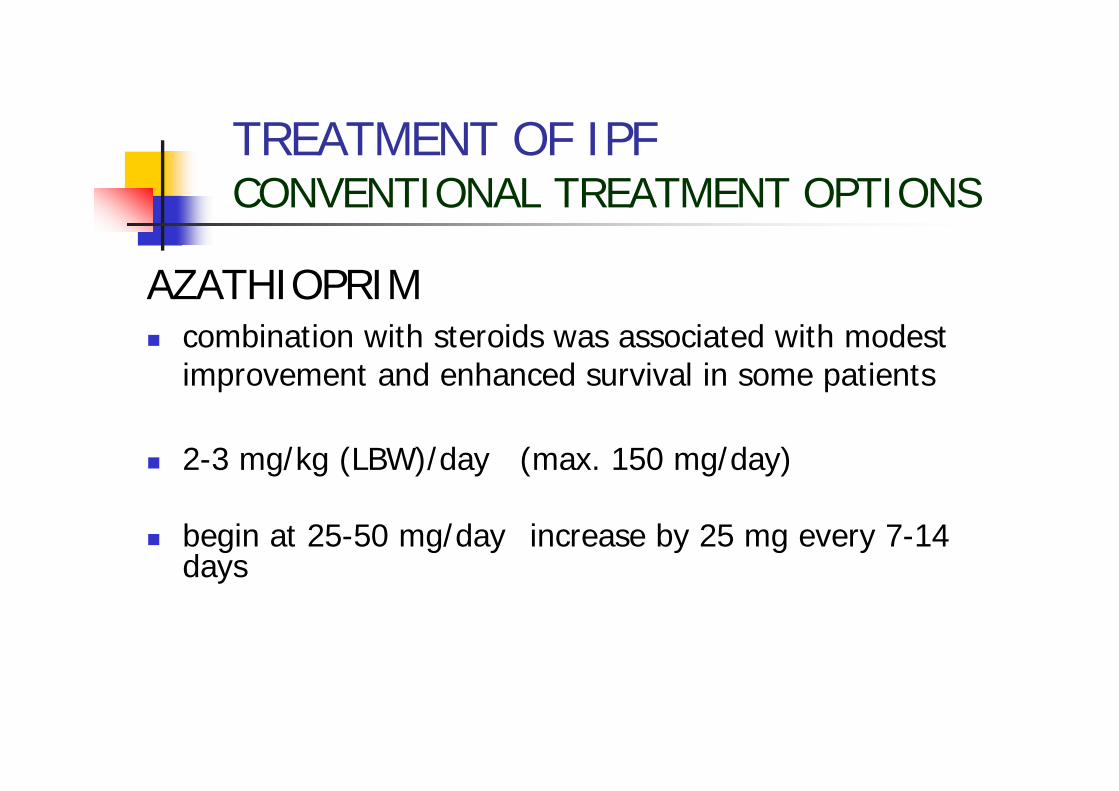
AZATHIOPRIM combination with steroids was associated with modest
improvement and enhanced survival in some patients
2-3 mg/kg (LBW)/day (max. 150 mg/day)
begin at 25-50 mg/day increase by 25 mg every 7-14 days

TREATMENT OF IPFPOTENTIAL ALTERNATIVE TREATMENTS
Possible future therapeutic strategies include
- agents that inhibit cytokines, proteases, `oxidants, or fibroblast growth factors
- antifibrotic agents- dietary modifications- diphosphonates- antioxidants- gene therapy
TREATMENT OF IPFPOTENTIAL ALTERNATIVE TREATMENTS
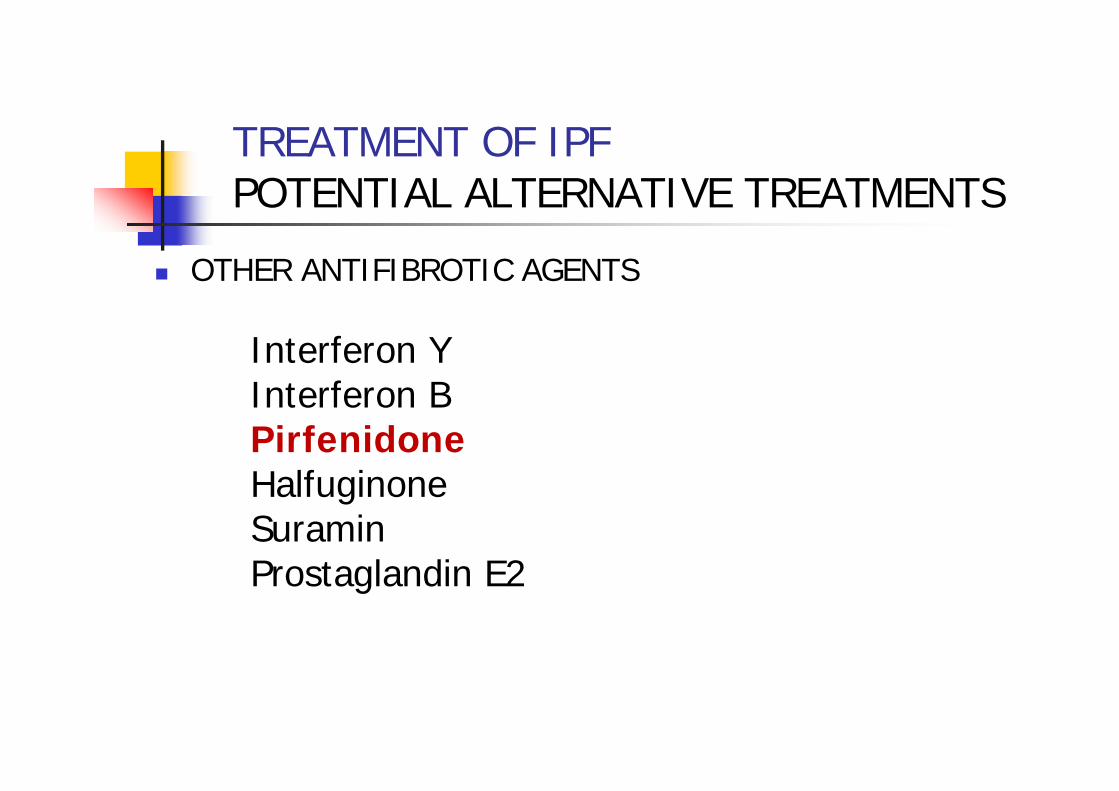
OTHER ANTIFIBROTIC AGENTS
Interferon YInterferon BPirfenidoneHalfuginoneSuraminProstaglandin E2
TREATMENT OF IPFPOTENTIAL ALTERNATIVE TREATMENTS
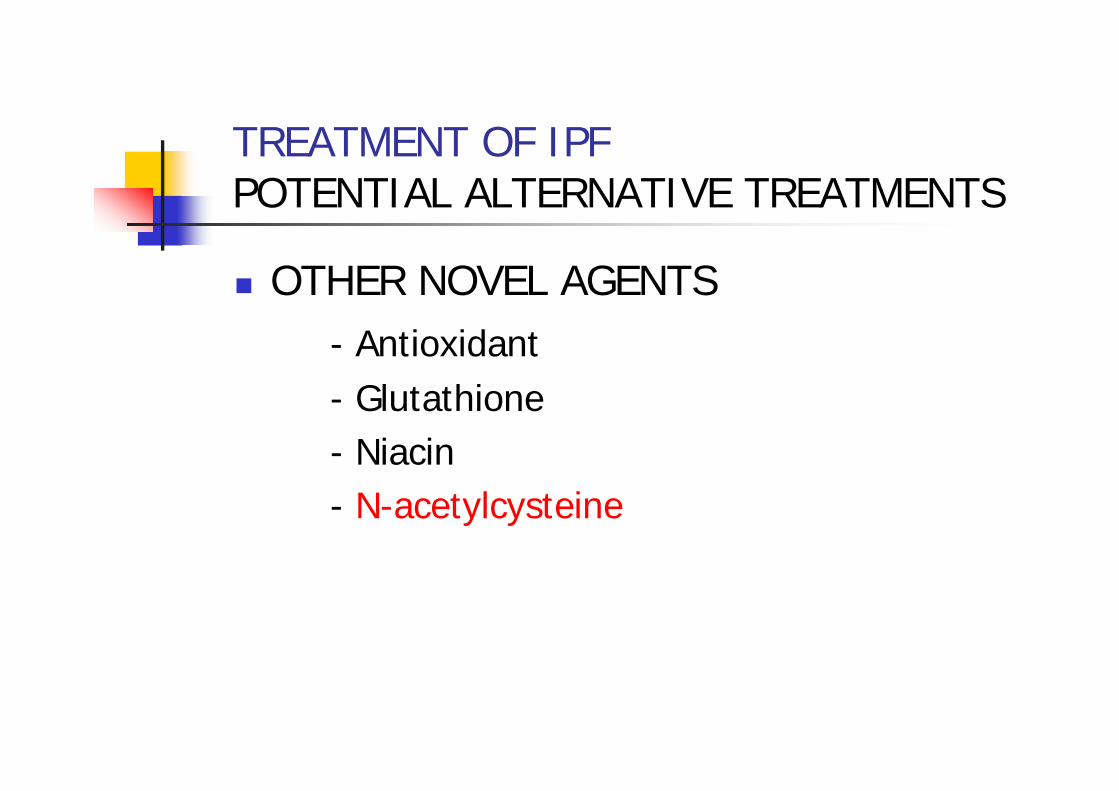
OTHER NOVEL AGENTS- Antioxidant- Glutathione- Niacin- N-acetylcysteine
STAGING AND PROGNOSIS
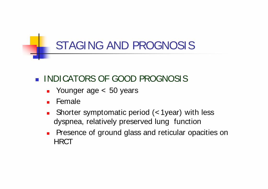
INDICATORS OF GOOD PROGNOSIS Younger age < 50 years Female Shorter symptomatic period (<1year) with less
dyspnea, relatively preserved lung function Presence of ground glass and reticular opacities on
HRCT
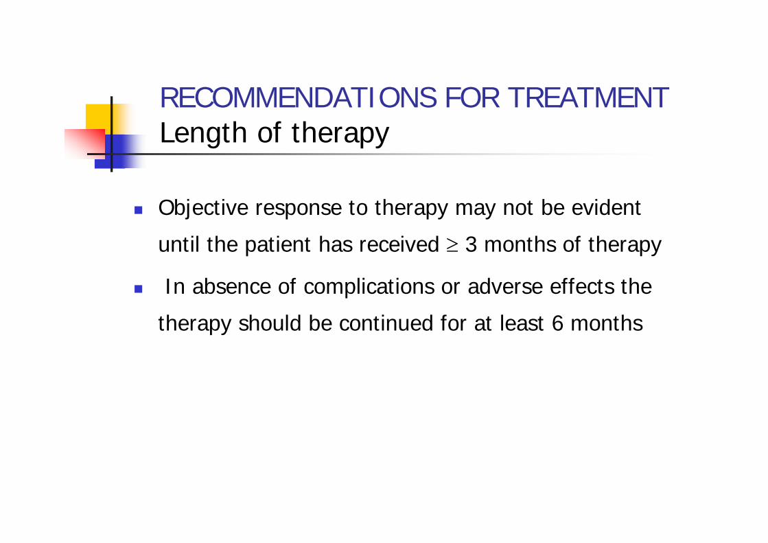
RECOMMENDATIONS FOR TREATMENTLength of therapy
Objective response to therapy may not be evident
until the patient has received 3 months of therapy
In absence of complications or adverse effects the
therapy should be continued for at least 6 months
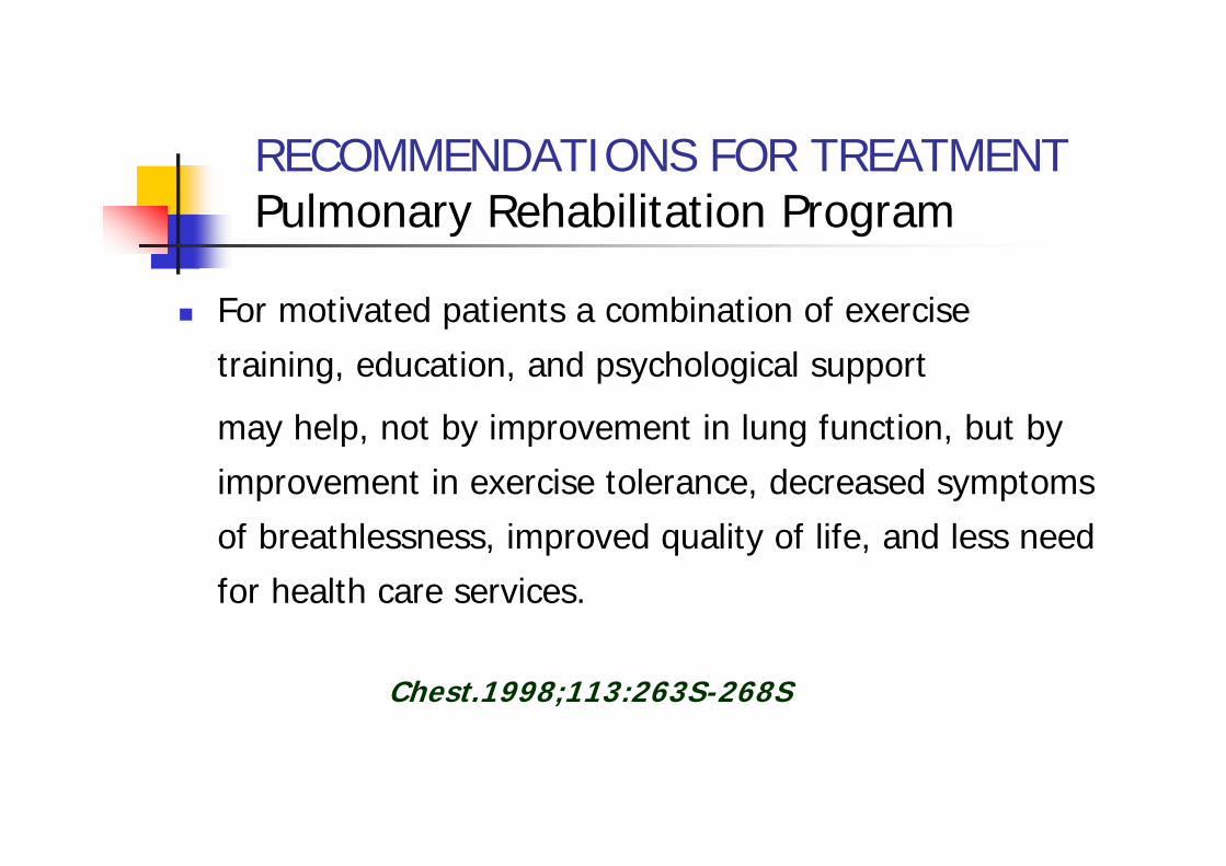
RECOMMENDATIONS FOR TREATMENTPulmonary Rehabilitation Program
For motivated patients a combination of exercise training, education, and psychological support
may help, not by improvement in lung function, but by improvement in exercise tolerance, decreased symptoms of breathlessness, improved quality of life, and less need for health care services.
Chest.1998;113:263S-268S
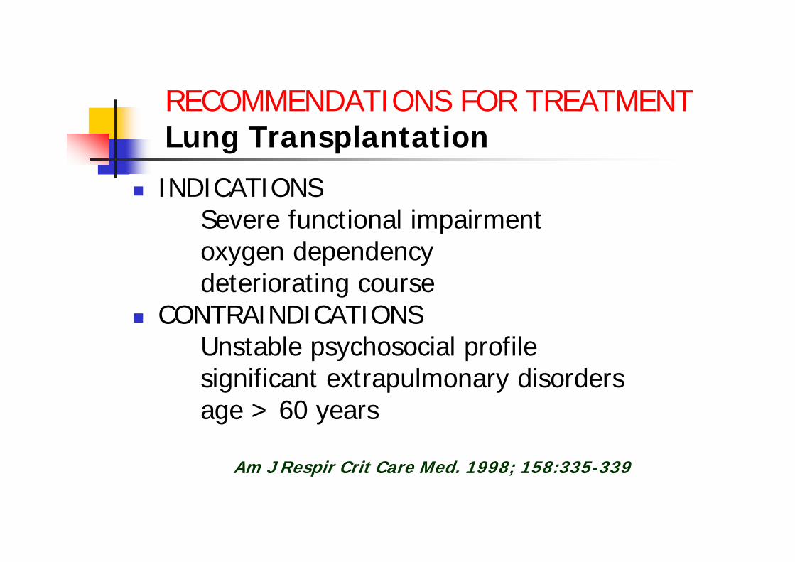
RECOMMENDATIONS FOR TREATMENTLung Transplantation
INDICATIONSSevere functional impairmentoxygen dependencydeteriorating course
CONTRAINDICATIONSUnstable psychosocial profile significant extrapulmonary disordersage > 60 years
Am J Respir Crit Care Med. 1998; 158:335-339
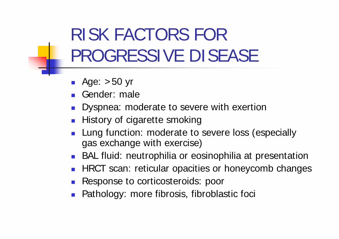
RISK FACTORS FOR PROGRESSIVE DISEASE Age: >50 yr Gender: male Dyspnea: moderate to severe with exertion History of cigarette smoking Lung function: moderate to severe loss (especially
gas exchange with exercise) BAL fluid: neutrophilia or eosinophilia at presentation HRCT scan: reticular opacities or honeycomb changes Response to corticosteroids: poor Pathology: more fibrosis, fibroblastic foci

CONCLUSSION(Treatment of IPF)
Till date insufficient clinical evidence to conclude that any treatment improves survival or quality of life for patients with IPF

ILD secondary to Connective tissue diseases Rheumatoid arthritis Systemic sclerosis Sjogren syndrome Systemic lupus erythematosus Idiopathic inflammatory myopathies
General approach ILD is often asymptomatic at presentation Rarely , it may be the first sign of
presentation of disease Systemic disease so disease usually has
features in joints, skin, etc Difficulty in swallowing, raynaud’s
phenomena, proximal muscle weakness. Restrictive pattern on PFT

Serological tests

Treatment principles Treatment of underlying disease Steroids and immunosuppressive agents Multi-disciplinary approach Long duration of treatment Supportive treatment and pulmonary
rehabilitation Treat co-morbidities and limit treatment
related side effects Lung transplantation

Thank You


![Alveolitis Seca[1]](https://img.dokumen.tips/doc/110x75/55cf9d9b550346d033ae5e07/alveolitis-seca1.jpg)