Embed Size (px)
Citation preview
General rights Copyright and moral rights for the publications made accessible in the public portal are retained by the authors and/or other copyright owners and it is a condition of accessing publications that users recognise and abide by the legal requirements associated with these rights.
Users may download and print one copy of any publication from the public portal for the purpose of private study or research.
You may not further distribute the material or use it for any profit-making activity or commercial gain
You may freely distribute the URL identifying the publication in the public portal If you believe that this document breaches copyright please contact us providing details, and we will remove access to the work immediately and investigate your claim.
Downloaded from orbit.dtu.dk on: Nov 18, 2021
Interrogating T cells specific to mutated and shared tumor epitopes in mouse and man
Petersen, Nadia Viborg
Publication date:2019
Document VersionPublisher's PDF, also known as Version of record
Link back to DTU Orbit
Citation (APA):Petersen, N. V. (2019). Interrogating T cells specific to mutated and shared tumor epitopes in mouse and man.DTU Health Technology.

Interrogating T cells specific to mutated and shared tumor epitopes in mouse and man
PhD thesis
Nadia Viborg Petersen December 2019

Department of Health Technology
Technical University of Denmark
Kemitorvet
Building 202
2800 Kgs. Lyngby
Interrogating T cells specific to mutated and shared tumor epitopes in mouse and man i
PREFACE
The following PhD thesis has been submitted to the Technical University of Denmark,
Department of Health Technology, as part of the requirements for obtaining a PhD degree.
The presented research was carried out from January 2017 to December 2019 as a
collaboration between the Technical University of Denmark (first the Veterinary Institute,
then Department of Micro- and Nanotechnology, and lastly at the Department of Health
Technology) and Evaxion Biotech. The project was supervised by Sine Reker Hadrup
(Professor, Section for Experimental and Translational Immunology, Department of Health
Technology, Technical University of Denmark) and Birgitte Rønø (PhD, Senior Director of
Cancer Immunotherapy, Evaxion Biotech). The research was in part funded by the Danish
Innovation Fund via their Industrial PhD programme.
The thesis is comprised of a common introduction followed by relevant research papers,
each introduced by a short preface, and finally an epilogue discussing the major findings and
perspectives of the work.
Copenhagen, December 2019
Nadia Viborg Petersen

Interrogating T cells specific to mutated and shared tumor epitopes in mouse and man ii
Interrogating T cells specific to mutated and shared tumor epitopes in mouse and man iii
ABSTRACT
It is by now acknowledged that immunological recognition of cancer is a many-sided
interplay, able to both promote and reject tumor growth. With cancer immunotherapy, the
ambition is to harness immunity towards long lasting tumor elimination, for which T cells
are important mediators. Tumor cells differ genetically from healthy tissue, which will be
visible to surveilling T cells via aberrant peptide-MHC presentation. Hence, a large effort is
being put into understanding T cells and their epitope targets in cancer, via techniques to
monitor and augment tumor specific T cells. Little is known about the characteristics that
govern immunogenicity of T cell epitopes, and current strategies depend on in silico prediction
of peptide-MHC binding affinity. The outlined thesis contains three research papers that
investigate T cells and their associated epitopes in mice and humans, with emphasis on
cancer.
In the first study, we developed novel conditional ligands for murine H-2 alleles H-2Dd and
H-2Kd, that enable rapid generation of multiple pH-2 multimers via UV-mediated peptide
exchange, without the need for individual peptide-H-2 refolding. pH-2d multimers were
successfully used for fluorescent tetramer staining and large-scale DNA barcode labelled
multimer libraries. This provides the first description of H-2Dd and H-2Kd conditional
ligands, and proof-of-concept of DNA barcode labelled multimers for murine H-2d
haplotype screening. This technology will enable epitope mapping in a wide variety of disease
models on e.g. BALB/c background, including syngeneic models of cancer.
The second study investigated neo-epitope vaccination in the murine CT26 syngeneic tumor
model. We explored DNA and peptide-based delivery of neo-epitopes and found only DNA
vaccination to induce protective tumor immunity. Correspondingly, DNA based vaccination
preferentially induced neo-epitope specific CD8+ T cells, whereas peptide vaccination
induced neo-epitope specific CD4+ T cells. Our DNA based vaccination strategy thus
represents an interesting framework for future neo-epitope discovery, from which putative
epitope libraries are often large and will benefit from the technology outlined in the first
study.
Interrogating T cells specific to mutated and shared tumor epitopes in mouse and man iv
In the third study we show T cell recognition of epitopes from previously undescribed shared
tumor associated antigens in breast cancer patients. Similar to the first study, this study
employs high-throughput DNA barcode labelled pHLA multimers, and the investigated T
cell epitopes do not give rise to recognition in HLA-matched healthy donors. Breast cancer
remains a major cause of female mortality worldwide and is not thoroughly researched in the
field of immunotherapy. Breast cancer harbors low mutational burden compared to other
cancer types, thus investigations on shared tumor epitopes are of importance compared to
mutation-derived neo-epitopes. As such, our findings represent novel T cell targets in breast
cancer, that might be of relevance in patient immune monitoring.
Collectively, these studies represent tools for, and investigations of, T cell epitopes in cancer.
With an increased understanding of what the T cells “see” and how to enhance them, we can
better steer them towards tumor eradication via immunotherapy.
Interrogating T cells specific to mutated and shared tumor epitopes in mouse and man v
DANSK RESUMÉ
Det er anerkendt, at immunologisk genkendelse af cancer udgør et mangesidet sammenspil,
der kan både fremme og hæmme tumorvækst. Ambitionen med cancer immunterapi er at
dirigere immunsystemet for at opnå langvarig tumor eliminering, og her er T celler
betydningsfulde. Tumorceller er genetisk forskellige fra raskt væv, hvilket vil være synligt for
overvågende T celler via afvigende peptid-MHC-præsentation. Der bliver derfor ydet en stor
indsats for at forstå T celler og deres epitoper i kræft, via teknikker til at monitorere og forøge
tumor-specifikke T celler. Der findes begrænset viden om de karakteristika der afgør
immunogenicitet af T celle epitoper, og nuværende strategier afhænger af in silico prædiktion
af peptid-MHC bindingsaffinitet. Den her beskrevne afhandling indeholder tre studier, der
undersøger T celler og deres associerede epitoper i mus og mennesker, med fokus på cancer.
I det første studie udviklede vi nye betingede ligander for murine H-2 alleler; H-2Dd og H-
2Kd, der muliggør sideløbende generering af adskillige pH-2 multimerer via UV-medieret
peptidudveksling, uden behov for individuel peptid-H-2 refoldning. pH-2d multimerer blev
med succes anvendt til fluorescerende tetramer-farvninger og stor-skala DNA mærkede
multimer biblioteker. Dette udgør den første beskrivelse af betingede ligander for H-2Dd og
H-2Kd, samt belæg for at kunne benytte DNA mærkede multimer til murin H-2d vævstype
screening. Denne teknologi vil facilitetere kortlægning af epitoper i mange sygdomsmodeller
på BALB/c baggrund, inklusive syngene cancer modeller.
Det andet studie undersøgte neo-epitoper in den murine syngene CT26 tumor model. Vi
udforskede DNA og peptid-baseret levering af neo-epitoper, og fandt, at kun DNA-
vaccinering inducerede beskyttende tumorimmunitet. Tilsvarende inducerede DNA-baseret
vaccinering præferentielt neo-epitop specifikke CD8+ T celler, hvorimod peptid-baseret
vaccinering inducerede neo-epitop specifikke CD4+ T celler. Vores DNA-baserede
vaccinationsstrategi repræsenterer derfor en interessant ramme til fremtidig kortlægning af
neo-epitoper, hvor de undersøgte epitop-biblioteker ofte er omfattende og vil drage fordel
af teknologien beskrevet i det første studie.
I det tredje studie observerer vi T celle genkendelse af nogle hidtil ubeskrevne epitoper fra
tumor associerede antigener in brystkræftpatienter. Ligesom i det første studie, benytter vi
Interrogating T cells specific to mutated and shared tumor epitopes in mouse and man vi
her DNA mærkede pHLA multimerer, og de undersøgte T celle epitoper giver ikke anledning
til genkendelse i vævstype-matchede raske donorer. Brystkræft er fortsat en væsentlig
dødsårsag på verdensplan, og er ikke videre undersøgt indenfor cancer immunterapi.
Brystkræft har generelt en lav mutationsbyrde sammenlignet med andre typer af cancer, og
derfor er undersøgelser af ikke-muterede epitoper af høj relevans, sammenlignet med
mutationsderiverede neo-eptioper. Samlet set præsenterer vores forskning nye T celle
epitoper i brystkræft, som kan blive relevante indenfor immunmonitorering af patienter.
Tilsammen repræsenterer disse studier værktøjer til, og undersøgelser af, T celle epitoper i
cancer. Med en øget forståelse for hvad det er, T cellerne ”ser”, og hvordan vi kan styrke
dem, kan vi i højere grad benytte dem til tumor destruktion via immunterapi.
Interrogating T cells specific to mutated and shared tumor epitopes in mouse and man vii
ACKNOWLEDGEMENTS
After more than eight years at DTU as a BSc and MSc student, an employee and latest an aspiring PhD graduate I am now handing in my PhD thesis. This is an emotional and grand milestone for me. First, the biggest of thank you goes to the power-women who supervised me during my PhD project: Sine Reker Hadrup and Birgitte Rønø. Sine for including me in your wonderful research group in early 2014, which led me to BSc, MSc and now a PhD project under your supervision. I am grateful for everything you have taught me, and before my projects with you, I did not even consider pursuing a PhD. You continuously challenge and inspire me in my scientific ventures, which is why I have become quite the MHC multimer nerd. Birgitte for your patience, trust, inclusion and your never-ending encouragement of my work. For going with me to the lab on a Christmas holiday, staining cells and sharing pizza in the FACS room. Both of you, for hours of sparring, optimism and confidence boosts. Thank you. Many appreciations go out to everyone in the SRH research group at DTU for great collaborations, countless hours of science, laughter, FACS sorting, dancing, travels, coffees and group meetings. My years spent at DTU will be truly unforgettable thanks to all of you. To Amalie, Natasja and Sofie, for following me since my early steps as a scientist (in the lab and on the dancefloor…). Sofie, O.G. grandma, for feedback on my first thesis draft. Trine and Sara for fruitful and fun collaborations in the mouse team and long hours spent processing mouse organs. Trine, for many long talks and your last-minute comments and corrections that I am immensely grateful for. To Agnes, Ulla, Jeppe, Simone, Rasmus, Keith, Matilda, and Emma, for sharing the ups and downs of the PhD journey with me. To everyone in the Evaxi-family for inclusion and encouragement. For listening to all my talk about multimers, T cells and whatnot. You all contributed to making me feel right at home, especially in the Friday bar. Brilliant PioNerds for sharing my neo-epitope enthusiasm. Marina for numerous hours spent immunizing mice and a growing friendship, always there to help me get my mind off things when I need it (and when I don’t know that I need it). To Stine, Bettina and the IO EXP team for making experiments feasible and fun. I look forward to getting to know you all even better in the time to come. Lastly, to my family and friends for your understanding, enthusiasm and curiosity of my work, always believing in me even when I do not. Nadia Viborg Petersen (Doctor of Philosophy in spe)

Interrogating T cells specific to mutated and shared tumor epitopes in mouse and man viii
Interrogating T cells specific to mutated and shared tumor epitopes in mouse and man ix
PUBLICATIONS
The following research is included in this thesis:
Manuscript I:
Nadia Viborg, Trine Sundebo Meldgaard, Tripti Tamhane, Sara Suarez Hernandez,
Christian Garde, Thomas Trolle, Dario Vazquez Albacete, Sine Reker Hadrup. ”H-2
multimers for high-throughput T cell interrogation and description of novel conditional
ligands for H-2Dd and H-2Kd”
(manuscript formatted for submission to Journal of Immunology)
Manuscript II:
Nadia Viborg, Marina Barrio Calvo, Stine Friis, Thomas Trolle, Christian Skjødt Hansen,
Jens Kringelum, Sine Reker Hadrup and Birgitte Rønø. “DNA based neo-epitope
vaccination induces tumor control in the CT26 syngeneic mouse model”
(manuscript in preparation)
Paper III:
Nadia Viborg, Sofie Ramskov, Rikke Sick Andersen, Theo Sturm, Tim Fugmann, Amalie
Kai Bentzen, Vibeke Mindahl Rafa, Per thor Straten, Inge Marie Svane, O ̈zcan Met, Sine
Reker Hadrup (2019). “T cell recognition of novel shared breast cancer antigens is
frequently observed in peripheral blood of breast cancer patients”
Oncoimmunology, 8:12, DOI: 10.1080/2162402X.2019.1663107

Interrogating T cells specific to mutated and shared tumor epitopes in mouse and man x
Interrogating T cells specific to mutated and shared tumor epitopes in mouse and man xi
CONTENTS
1. Introduction ................................................................................................................. 1
From innate to adaptive immunity ............................................................................ 1
T cells ........................................................................................................................... 3
Immunological recognition by pMHC-TCR interactions ................................... 3
Tumor immunology ..................................................................................................... 5
Immunoediting ........................................................................................................... 6
T cells and their targets in cancer ............................................................................ 7
Monitoring of antigen specific T cells ..................................................................... 12
Cancer immunotherapy ............................................................................................. 14
Clinical advances and considerations .................................................................... 14
Lessons from preclinical mouse models .............................................................. 18
2. Manuscript I .............................................................................................................. 21
3. Manuscript II ............................................................................................................ 47
4. Paper III ..................................................................................................................... 79
5. Epilogue ................................................................................................................... 109
6. Bibliography ............................................................................................................ 115

Interrogating T cells specific to mutated and shared tumor epitopes in mouse and man xii

1
1. INTRODUCTION
From innate to adaptive immunity The human immune system is complex and comprises numerous components able to sense,
respond and defend us against threats of different nature, such as pathogens or malignantly
transformed cells. Immediate sensing is facilitated by components of the innate immune
system which discriminates non-self from self via interactions between host pattern
recognition receptors (PRRs) and conserved pathogen associated molecular patterns
(PAMPs) and cellular stress related damage associated molecular patterns (DAMPs). Several
families of PRRs have been described, e.g. Toll-like receptors (TLRs) that have different
cellular distribution, enabling recognition of distinct invariant molecular patterns or threats
in intra- and extracellular compartments. Downstream of PRR-PAMP interactions innate
immune responses recruit relevant cells and produces effector molecules in the form of
cytokines and chemokines [1]. Innate immune responses are rapid and generic and do not
induce memory for future re-encounters. However, the type of innate immune response that
is mounted fundamentally shapes the adaptive immune response that will follow, as this
depends profoundly on the signature of cytokines produced by the initial innate phase [2],
an attribute utilized actively in vaccination, schematically illustrated in Figure 1.
Antigen presenting cells (APCs), particularly dendritic cells (DCs), are essential in bridging
innate and adaptive immunity. DCs recognize, take up and degrade pathogens and their
antigens at the site of recognition. Antigen loaded DCs move via lymphatics to lymph nodes,
where they present antigens on the major histocompatibility complex (MHC) to naïve
lymphocytes; the key effectors of adaptive immunity [1], [3]. Upon antigen presentation and
co-stimulatory signals from DCs (“priming”), lymphocytes will mature and proliferate, giving
rise to antigen specific immune responses. The cytokine milieu surrounding DCs during
antigen uptake shapes the induction of adaptive immunity that follows [4], [5]. Adaptive
immunity is developed throughout life and responds slower than innate immunity. Due to
selective induction of specific and persisting memory cells, responses occur rapidly and
effectively upon re-exposure [3]. B lymphocytes (B cells) and T lymphocytes (T cells) embody

2
adaptive humoral and cellular immune responses respectively via their antigen specific
receptors. T cells are key components of adaptive immunity responsible for the direct killing
of infected or transformed cells (i.e. CD8+ T cells) and orchestrating the immune response
in several ways (CD4+ T cells) and will be the focus for the remainder of this thesis.
Figure 1 - From innate to adaptive immunity via vaccination. The immune response obtained via vaccination starts
already at the injection site where PAMPs and DAMPs are sensed by local cells and APCs via PRRs. Antigen loaded DCs
travel to lymph nodes, capable of antigen presentation and priming of lymphocytes. Here, adaptive immunity starts, which
is composed of various types of effector B and T cells. The type of lymphocytes that are activated depends on cytokines
present in the milieu and signals from the DC. Proper co-stimulation from DC to T cells is also driven by innate sensing
via PRRs. Abbreviations: PAMP; pathogen-associated molecular pattern, DAMP; damage-associated molecular pattern,
PRR; pathogen recognition receptor, pDC; plasmacytoid dendritic cell, DC; dendritic cell, TFH; T follicular helper cell, IL;
interleukin, IFN; interferon, IgG; immunoglobulin. Figure adapted from [6]
Consider for discussion
Importance of innate signals to get the adaptive response you want and need
From Nucleic acid sensing at the interface between innate and adaptive immunity in vaccination (Christophe J. Desmet1 and Ken J. Ishii)
INNATE
SENSIN
GANDACTIVATIO
NADAPTIVE
EFFECTORRESPO
NSES
3
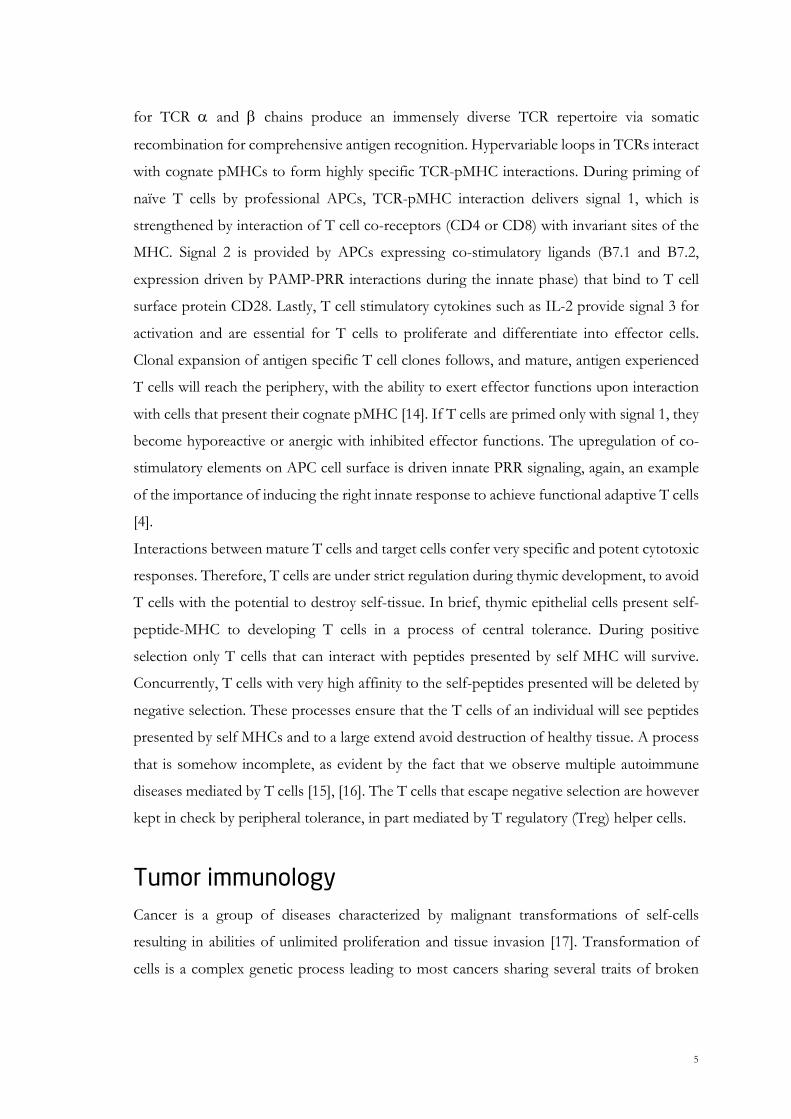
T cells T cells originate from hematopoietic progenitors in the bone marrow and mature in the
thymus, where they undergo extensive selection processes. T cells express a T cell receptor
(TCR) for antigen recognition associated with co-receptor CD3 [7]. Two subtypes of T cells
are relevant for this thesis, distinguished by expression of surface co-receptors CD4 or CD8.
CD4+ T cells or T helper cells (TH) instruct and influence adaptive immunity and comprise
multiple functionally distinct subtypes (Figure 1) under the control of separate transcription
factors, induced as a consequence of signaling patterns from APCs. Most important for the
scope of this thesis are TH1 cells, which are characterized by interleukin-2 (IL-2) and
interferon-g (IFNg) cytokine production, controlled by transcription factor T-bet, and
support responses to intracellular threats such as viral infections and malignant
transformations. CD8+ T cells or cytotoxic T lymphocytes (CTLs) are supported by TH1
cytokines (as visible on Figure 1) and respond to changes of intracellular origin. The CD8+
T cell respond by selectively killing target cells via release of cytolytic effector molecules upon
recognition of aberrant cells.
Immunological recognition by pMHC-TCR interactions MHC presentation
T cells depend on self MHC display of peptides to exert effector functions via TCR
interactions. The MHC locus is polygenic and constitutes the most polymorphic region
within the human genome, of which each allele has distinct amino acid binding preferences.
This makes for a comprehensive repertoire of peptide-MHC (pMHC) presentations to the
immune system, a benefit in the continuous battle against pathogens. In humans the MHC
is referred to as human leukocyte antigen (HLA) and in mice H-2, and across species two
MHC regions facilitate antigen presentation to T cells: MHC class I and class II molecules.
MHC class I is expressed on all nucleated cells and presents peptides of intracellular origin
to CD8+ T cells, as outlined in detail in Figure 2A. MHC class I typically binds and presents
peptides of 9-11 amino acid length [8]. MHC class II is restrictively expressed by APCs and
presents peptides of exogenous (vesicular and extracellular) origin for interaction with CD4+
T cells, as outlined in detail in Figure 2B. MHC class II has an open peptide binding groove,
allowing longer peptides to be presented, typically 13-25 amino acid length [9]. Also
noteworthy is cross presentation, in which peptides of extracellular origin are presented by

4
MHC class I on a subset of DCs, which gives rise to effective CD8+ T cell responses [10].
Cross presentation is mainly facilitated by immunoproteasomal degradation of peptides and
driven by the presence of interferons in the milieu as an effect of PRR signaling in the innate
immune phase [11], [12].
Figure 2 – peptide processing and presentation by MHC molecules. (A) Proteins of intracellular origin are degraded
and presented by MHC class I in the form of peptides by the following steps: 1) proteasomal degradation, 2) transport to
endoplasmic reticulum (ER) by transporter associated with antigen processing (TAP), 3) ER trimming by aminopeptidases
to a length of 9-11 amino acids, 4) loading onto newly synthetized MHC class I molecules in the ER, and 5) the stable
pMHC complex departs for cell surface presentation to cognate CD8+ T cells. (B) Proteins of exogenous origin are taken
up, degraded and presented by MHC class II molecules and comprises: 1) uptake and endolysosomal degradation, 2) fusing
with MHC class II containing endosomes, produced in ER loaded with invariant chain (Ii) and later class II-associated
invariant chain peptide (CLIP), 3) displacement CLIP from class II binding groove with exogenous peptide, 4) the stable
pMHC complex departs for cell surface presentation to cognate CD4+ T cells. Abbreviations: TCR; T cell receptor, MHC;
major histocompatibility complex, HLA; human leukocyte antigen, b2m; beta-2-microglobulin. Figure adapted from [13]
TCR recognition
The majority of T cells in humans and mice bear a heterodimeric ab-TCR with a constant
and a variable region, of which the variable region is the antigen-binding site. Genetic loci
A B
1)
2)
3 + 4)
5)
Adapted from Koichi S. Kobayashi and Peter J. van den NLRC5: a key regulator of MHC class
1)2)3)
4)
5
for TCR a and b chains produce an immensely diverse TCR repertoire via somatic
recombination for comprehensive antigen recognition. Hypervariable loops in TCRs interact
with cognate pMHCs to form highly specific TCR-pMHC interactions. During priming of
naïve T cells by professional APCs, TCR-pMHC interaction delivers signal 1, which is
strengthened by interaction of T cell co-receptors (CD4 or CD8) with invariant sites of the
MHC. Signal 2 is provided by APCs expressing co-stimulatory ligands (B7.1 and B7.2,
expression driven by PAMP-PRR interactions during the innate phase) that bind to T cell
surface protein CD28. Lastly, T cell stimulatory cytokines such as IL-2 provide signal 3 for
activation and are essential for T cells to proliferate and differentiate into effector cells.
Clonal expansion of antigen specific T cell clones follows, and mature, antigen experienced
T cells will reach the periphery, with the ability to exert effector functions upon interaction
with cells that present their cognate pMHC [14]. If T cells are primed only with signal 1, they
become hyporeactive or anergic with inhibited effector functions. The upregulation of co-
stimulatory elements on APC cell surface is driven innate PRR signaling, again, an example
of the importance of inducing the right innate response to achieve functional adaptive T cells
[4].
Interactions between mature T cells and target cells confer very specific and potent cytotoxic
responses. Therefore, T cells are under strict regulation during thymic development, to avoid
T cells with the potential to destroy self-tissue. In brief, thymic epithelial cells present self-
peptide-MHC to developing T cells in a process of central tolerance. During positive
selection only T cells that can interact with peptides presented by self MHC will survive.
Concurrently, T cells with very high affinity to the self-peptides presented will be deleted by
negative selection. These processes ensure that the T cells of an individual will see peptides
presented by self MHCs and to a large extend avoid destruction of healthy tissue. A process
that is somehow incomplete, as evident by the fact that we observe multiple autoimmune
diseases mediated by T cells [15], [16]. The T cells that escape negative selection are however
kept in check by peripheral tolerance, in part mediated by T regulatory (Treg) helper cells.
Tumor immunology Cancer is a group of diseases characterized by malignant transformations of self-cells
resulting in abilities of unlimited proliferation and tissue invasion [17]. Transformation of
cells is a complex genetic process leading to most cancers sharing several traits of broken
6
homeostasis, commonly referred to as Hallmarks of Cancer [18]. Despite major medical
advancements, cancer remains a leading cause of mortality worldwide. Following decades of
research and debate, it is now well established and described that the immune system plays
an important, yet dichotomous, role in tumor establishment, progression and eradication.
Acknowledged through descriptions such as the cancer immunity cycle [19] and the addition
of immunological enabling in the updated Hallmarks of Cancer [20], the concept of tumor
immunology is today textbook material that describe both the pro- and anti-tumor effects of
the immune system. Additionally, the Immunoscore concept described by Galon et al. [21],
[22] provide a clinical correlate between intratumoral presence of T cells and disease
improved prognosis, and could be used to stratify patients to a suitable therapy [23].
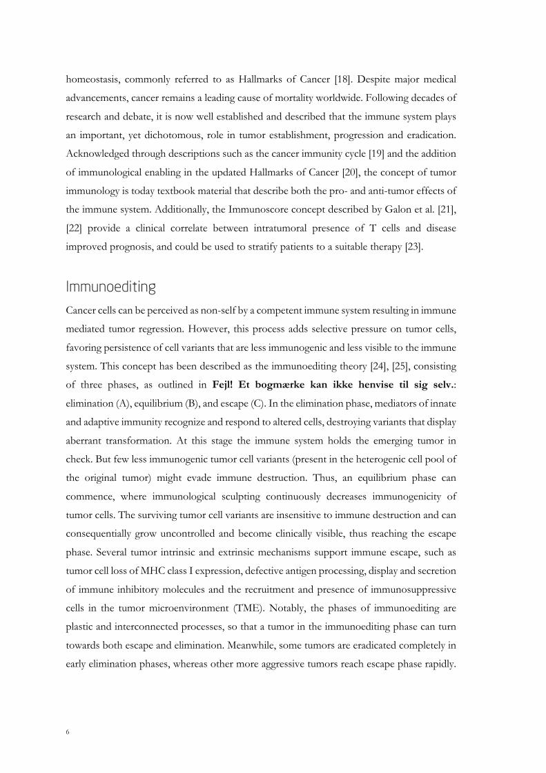
Immunoediting Cancer cells can be perceived as non-self by a competent immune system resulting in immune
mediated tumor regression. However, this process adds selective pressure on tumor cells,
favoring persistence of cell variants that are less immunogenic and less visible to the immune
system. This concept has been described as the immunoediting theory [24], [25], consisting
of three phases, as outlined in Fejl! Et bogmærke kan ikke henvise til sig selv.:
elimination (A), equilibrium (B), and escape (C). In the elimination phase, mediators of innate
and adaptive immunity recognize and respond to altered cells, destroying variants that display
aberrant transformation. At this stage the immune system holds the emerging tumor in
check. But few less immunogenic tumor cell variants (present in the heterogenic cell pool of
the original tumor) might evade immune destruction. Thus, an equilibrium phase can
commence, where immunological sculpting continuously decreases immunogenicity of
tumor cells. The surviving tumor cell variants are insensitive to immune destruction and can
consequentially grow uncontrolled and become clinically visible, thus reaching the escape
phase. Several tumor intrinsic and extrinsic mechanisms support immune escape, such as
tumor cell loss of MHC class I expression, defective antigen processing, display and secretion
of immune inhibitory molecules and the recruitment and presence of immunosuppressive
cells in the tumor microenvironment (TME). Notably, the phases of immunoediting are
plastic and interconnected processes, so that a tumor in the immunoediting phase can turn
towards both escape and elimination. Meanwhile, some tumors are eradicated completely in
early elimination phases, whereas other more aggressive tumors reach escape phase rapidly.
7
With cancer immunotherapy the aim is to harness immunity in favor of elimination
processes.
Figure 3 - The process of immunoediting. (A) Elimination phase: due to foreignness of tumor cells they are recognized
and attacked by cells of the immune system. (B) Equilibrium phase: constant pressure from immune system favors the
survival of less immunogenic tumor cell variants, the immune system is thus less capable of ‘seeing’ tumor cells. (C) Escape:
outgrowth of tumor cell variants with very low immunogenicity. Tumor cells evade immune destruction and are able to
grow more or less uncontrollably. Abbreviations: NK; natural killer, NKT; natural killer T cell. Figure adapted from [25]
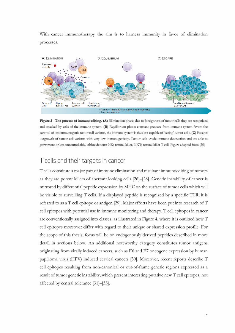
T cells and their targets in cancer T cells constitute a major part of immune elimination and resultant immunoediting of tumors
as they are potent killers of aberrant looking cells [26]–[28]. Genetic instability of cancer is
mirrored by differential peptide expression by MHC on the surface of tumor cells which will
be visible to surveilling T cells. If a displayed peptide is recognized by a specific TCR, it is
referred to as a T cell epitope or antigen [29]. Major efforts have been put into research of T
cell epitopes with potential use in immune monitoring and therapy. T cell epitopes in cancer
are conventionally assigned into classes, as illustrated in Figure 4, where it is outlined how T
cell epitopes moreover differ with regard to their unique or shared expression profile. For
the scope of this thesis, focus will be on endogenously derived peptides described in more
detail in sections below. An additional noteworthy category constitutes tumor antigens
originating from virally induced cancers, such as E6 and E7 oncogene expression by human
papilloma virus (HPV) induced cervical cancers [30]. Moreover, recent reports describe T
cell epitopes resulting from non-canonical or out-of-frame genetic regions expressed as a
result of tumor genetic instability, which present interesting putative new T cell epitopes, not
affected by central tolerance [31]–[33].
A: ELIMINATION B: EQUILIBRIUM C: ESCAPE
8
Figure 4 – categories of endogenously derived T cell antigens. Aberrant expression of antigens by MHC class I on
the surface of tumor cells (top), due to genetic alterations (middle). Antigen expression on normal/healthy tissue (bottom).
From left to right, increasing degree of tumor restricted expression. Figure adapted from [29]
Shared T cell epitopes
T cell epitopes expressed by both tumor and healthy tissue are commonly referred to as
shared, self- or tumor associated antigens and include overexpression, differentiation and
cancer testis antigens. Overexpression antigens are profoundly expressed by tumor cells
compared to corresponding healthy tissue, but expression in healthy tissue is often below a
threshold for T cells to recognize. An example is the expression of survivin in several cancers
and HER2 in certain breast cancers [34], [35]. Expression of differentiation antigens is
normally limited to distinct differentiated cell types, such as melanocytes of the skin, and
tumor cells originating from that tissue. An example is tyrosinase in melanomas, where
specific T cells are observed in circulation of cancer patients and healthy donors alike,
suggestive of incomplete thymic negative selection [36]. Cancer testis antigens constitute a
group of proteins that are normally only expressed in immune privileged sites of the body,
such as placenta and male germ cells that lack HLA expression. Tumor epigenetic changes
lead to expression of these proteins in some tumors, resulting in tumors appearing non-self
to T cells, such as New York esophageal squamous cell carcinoma 1 (NY-ESO-1) in multiple
cancers [37]. Shared T cell epitopes have been explored for therapeutic applicability in
different cancers, but past attempts have not been clinically successful [38], [39]. In part, this
Altered peptide sequences due to mutations. Mutation in MHC binding or TCR facing site of
peptide
Altered expression due to epigenetic changes. In healthy
tissue expressed only in immune privileged sites
Tissue specific, derive from proteins that characterize
differentiation of a certain cell type
Proteins expressed by normal tissue but significantly
overexpressed by malignant cells
Spermatocyte Other cellsMelanocyte Other cells
MUTATION-DERIVED(NEO) ANTIGENSCANCER TESTIS ANTIGENSDIFFERENTIATION ANTIGENSOVEREXPRESSION ANTIGENS
9
is attributed by the fact that stimulating T cells to target self-epitopes confers a risk of
autoimmune destruction of healthy tissues [40], [41]. Furthermore, T cells to these self-
targets are likely hampered by mechanisms central and peripheral tolerance.
However, shared T cell antigens are still of clinical relevance due to their advantageous
potentially wide applicability across cohorts of patients and cancers. As such, cancer
immunotherapy company BioNTech are currently exploring RNA vaccines containing
overexpression, cancer testis, and differentiation T cell antigens in melanoma and prostate
cancer in clinical phase I/II trials with promising preliminary results, as presented by Dr.
Özlem Tureci at the 5th CRI-CIMT-EATI-AACR International Cancer Immunotherapy
Conference in September 2019. The clinical potential of shared tumor antigens is therefore
not exhausted, and novel delivery modalities and adjuvant systems might facilitate exploiting
said antigens in off-the-shelf therapeutics to benefit many patients.
Neo-epitopes
In contrast to shared T cell epitopes stand T cell neo-epitopes; the products of genetic
alterations such as somatic mutations or in tumor cells. From the overall limited successes
of exploiting shared T cell epitopes for cancer therapy, there is now an increasing interest in
neo-epitopes, whose expression is restricted to tumor cells. In theory, if we are able to
harness immune cells to target neo-epitopes, we can achieve tumor elimination while
avoiding destruction of healthy tissue. Neo-epitopes have been described as the “Achille’s
heel” of cancer, owing to the fact that mutations are what defines a tumor and enables
malignant behavior, but at the same time mutations flag tumors as aberrant and non-self to
cells of the immune system. There are several reports on patient tumor mutational burden
(TMB) and putative neo-epitope load correlating with intratumoral T cell activity and disease
prognosis [42]–[44]. Combined with observations that beneficial clinical responses from
checkpoint inhibitor (CPI) therapy [24]-[25] and adoptive cell transfer (ACT) [47], [48] have
been associated with neo-epitope reactive T cells, tumor neo-epitopes are now a major focus
in the field of cancer immunotherapy. Interestingly, harnessing the immune system to target
neo-epitopes has been suggested to make up a “final common pathway of cancer” [49], based
on the positive associations described above. Ironically, this would mean that mutations, the
very enablers of malignant transformation, will also provide the ideal targets for
immunotherapy in a variety of cancers.
10
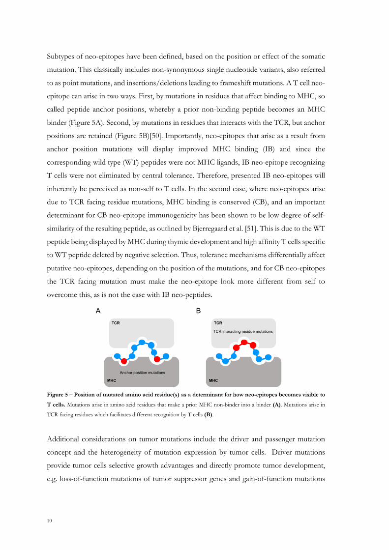
Subtypes of neo-epitopes have been defined, based on the position or effect of the somatic
mutation. This classically includes non-synonymous single nucleotide variants, also referred
to as point mutations, and insertions/deletions leading to frameshift mutations. A T cell neo-
epitope can arise in two ways. First, by mutations in residues that affect binding to MHC, so
called peptide anchor positions, whereby a prior non-binding peptide becomes an MHC
binder (Figure 5A). Second, by mutations in residues that interacts with the TCR, but anchor
positions are retained (Figure 5B)[50]. Importantly, neo-epitopes that arise as a result from
anchor position mutations will display improved MHC binding (IB) and since the
corresponding wild type (WT) peptides were not MHC ligands, IB neo-epitope recognizing
T cells were not eliminated by central tolerance. Therefore, presented IB neo-epitopes will
inherently be perceived as non-self to T cells. In the second case, where neo-epitopes arise
due to TCR facing residue mutations, MHC binding is conserved (CB), and an important
determinant for CB neo-epitope immunogenicity has been shown to be low degree of self-
similarity of the resulting peptide, as outlined by Bjerregaard et al. [51]. This is due to the WT
peptide being displayed by MHC during thymic development and high affinity T cells specific
to WT peptide deleted by negative selection. Thus, tolerance mechanisms differentially affect
putative neo-epitopes, depending on the position of the mutations, and for CB neo-epitopes
the TCR facing mutation must make the neo-epitope look more different from self to
overcome this, as is not the case with IB neo-peptides.
Figure 5 – Position of mutated amino acid residue(s) as a determinant for how neo-epitopes becomes visible to
T cells. Mutations arise in amino acid residues that make a prior MHC non-binder into a binder (A). Mutations arise in
TCR facing residues which facilitates different recognition by T cells (B).
Additional considerations on tumor mutations include the driver and passenger mutation
concept and the heterogeneity of mutation expression by tumor cells. Driver mutations
provide tumor cells selective growth advantages and directly promote tumor development,
e.g. loss-of-function mutations of tumor suppressor genes and gain-of-function mutations
TCR
MHC
Anchor position mutations
TCR interacting residue mutations
TCR
MHC
A B
11
of oncogenes [52]. Passenger mutations have seemingly no role in tumorigenesis but are
products of tumor genetic instability that are carried along by driver mutations. Some
mutations are proposedly categorized in-between, as “mini-drivers” [53] or essential
passengers [54], and harboring multiple of these might provide tumorigenic effects as a driver
mutation. Mutations in tumors happen at random, why the resulting neo-epitopes are strictly
patient unique, with very few exceptions of described “public” or recurrent neo-epitopes
[55]. The concept of tumor heterogeneity can be analyzed bioinformatically and exists on
different levels: from patient to patient, between two different tumor lesions (metastases) in
the same patient (intra-patient), and within a single tumor lesion of one patient (intra-
tumoral). The intra-patient/intra-tumor framework encompasses mutations taking place at
different stages of tumor evolution, and mutations are described as clonal when present in
all tumor cells or subclonal when only present in a subset of cells [56]. Our understanding of
tumor heterogeneity is still developing, and reports suggest that targeting clonal mutations
over subclonal confers clinical benefit [57].
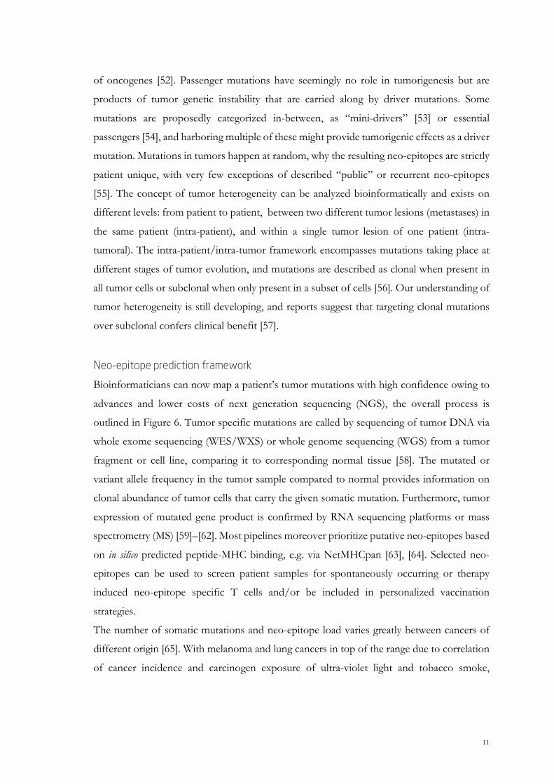
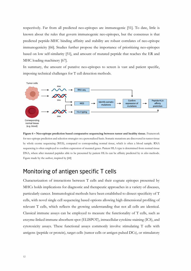
Neo-epitope prediction framework
Bioinformaticians can now map a patient’s tumor mutations with high confidence owing to
advances and lower costs of next generation sequencing (NGS), the overall process is
outlined in Figure 6. Tumor specific mutations are called by sequencing of tumor DNA via
whole exome sequencing (WES/WXS) or whole genome sequencing (WGS) from a tumor
fragment or cell line, comparing it to corresponding normal tissue [58]. The mutated or
variant allele frequency in the tumor sample compared to normal provides information on
clonal abundance of tumor cells that carry the given somatic mutation. Furthermore, tumor
expression of mutated gene product is confirmed by RNA sequencing platforms or mass
spectrometry (MS) [59]–[62]. Most pipelines moreover prioritize putative neo-epitopes based
on in silico predicted peptide-MHC binding, e.g. via NetMHCpan [63], [64]. Selected neo-
epitopes can be used to screen patient samples for spontaneously occurring or therapy
induced neo-epitope specific T cells and/or be included in personalized vaccination
strategies.
The number of somatic mutations and neo-epitope load varies greatly between cancers of
different origin [65]. With melanoma and lung cancers in top of the range due to correlation
of cancer incidence and carcinogen exposure of ultra-violet light and tobacco smoke,
12
respectively. Far from all predicted neo-epitopes are immunogenic [51]. To date, little is
known about the rules that govern immunogenic neo-epitopes, but the consensus is that
predicted peptide-MHC binding affinity and stability are robust correlates of neo-epitope
immunogenicity [66]. Studies further propose the importance of prioritizing neo-epitopes
based on low self-similarity [51], and amount of mutated peptide that reaches the ER and
MHC loading machinery [67].
In summary, the amount of putative neo-epitopes to screen is vast and patient specific,
imposing technical challenges for T cell detection methods.
Figure 6 – Neo-epitope prediction based comparative sequencing between tumor and healthy tissue. Framework
for neo-epitope prediction and selection strategies on a personalized basis. Somatic mutations are discovered in tumor tissue
by whole exome sequencing (WES), compared to corresponding normal tissue, which is often a blood sample. RNA
sequencing is often employed to confirm expression of mutated genes. Patient HLA type is determined from normal tissue
DNA, where after mutated peptides able to be presented by patient HLAs can be affinity predicted by in silico methods.
Figure made by the author, inspired by [68]
Monitoring of antigen specific T cells Characterization of interactions between T cells and their cognate epitopes presented by
MHCs holds implications for diagnostic and therapeutic approaches in a variety of diseases,
particularly cancer. Immunological methods have been established to dissect specificity of T
cells, with novel single cell sequencing based options allowing high dimensional profiling of
relevant T cells, which reflects the growing understanding that not all cells are identical.
Classical immune assays can be employed to measure the functionality of T cells, such as
enzyme-linked immune absorbent spot (ELISPOT), intracellular cytokine staining (ICS), and
cytotoxicity assays. These functional assays commonly involve stimulating T cells with
antigens (peptide or protein), target cells (tumor cells or antigen pulsed DCs), or stimulatory

13
molecules (IL-2, anti-CD3/anti-CD28 beads) before measuring cytokine production in bulk
culture, sorted cell fractions, or individual cells. Advantageous to functional assays is that
peptide antigens of varying amino acid length can be used in the T cell stimulation,
accommodating both MHC class I and II presentation to CD4+ and CD8+ T cells
respectively. As an alternative or add-on to further dissect the specific peptide recognition
of T cells, MHC multimer analyses can be applied. The affinity of a single TCR-pMHC
interaction is very low, which originally hindered direct ex vivo detection of antigen specific
T cells via pMHC molecules [69]. Altman and colleagues uncovered a solution by
multimerization of recombinant biotinylated pMHCs with streptavidin, to form tetramer
pMHC complexes that hold sufficient avidity to monitor specific T cells via fluorescent
labeling and flow cytometry analysis [70]. A breakthrough that has significantly advanced
epitope discovery in multiple disease settings. Later, the introduction of effective peptide-
MHC exchange technologies, particularly UV-mediated peptide exchange by Toebes and
colleagues, allowed production of larger pMHC multimer libraries in parallel, thus
circumventing the prior tedious individual refolding of MHC with each putative T cell
epitope [71]. There are now multiple descriptions of HLA and H-2 allele restricted ligands
(i.e. conditional ligands) for UV-mediated peptide exchange, why this technology is broadly
used for pMHC multimer screening purposes [71]–[75].
Newer developments in the MHC multimer field include: 1) applying backbones for higher
order multimerizations, potentially increasing sensitivity to detect lower affinity pMHC-TCR
interactions [76], 2) fluorescent combinatorial encoding or metal tags for higher complexity
screening of multiple T cell specificities via flow or mass cytometry [77], [78], 3) DNA
barcodes as multimer tags allowing high throughput screening for >1000 pMHC specific T
cells in parallel in a single sample [79], 4) applying stable empty loadable MHC molecules to
form multimers [80]. MHC multimer based T cell detection methods are, for now, mostly
applicable for CD8+ T cells. This is in part due to the fact that MHC class II multimers have
proved difficult to produce, requiring mammalian or insect cell expression systems [81].
Furthermore, in silico MHC class II affinity predictions are less developed than the MHC class
I counterparts and CD4+ T cells often display low TCR-pMHC affinity, as reviewed by
Hadrup and Newell [82]. Monitoring of antigen specific CD4+ T cells therefore often relies
on functional readouts such as ELISPOT and ICS, where the measured cytokine production
will uncover presence, frequency, type and functionality of CD4+ T cells in a given sample.
14
The number of potential T cell epitopes in human diseases is immense and will to a large
extent be patient specific due to HLA diversity and, for cancer, unique neo-epitope
landscapes resulting from somatic mutations. As such, the described advances in T cell
detection technologies are imperative to explore the large candidate neo-epitope outputs
from NGS pipelines.
The above described methods are often combined with further T cell characterization via
cell surface markers and transcription factors, to determine e.g. exhaustion, tissue residency
and memory status of T cells. As outlined, several T cell detection assays are flow cytometry
based and the advent of ever developing polychromatic flow cytometry with novel
fluorescent markers and increased capacities of flow cytometers has paved the way for in-
depth T cell interrogation. In cancer, these advances have proved valuable for diagnosis and
immune monitoring and potential stratification of patients to certain immunotherapies based
on their T cell signatures.
Cancer immunotherapy
Clinical advances and considerations The ability of the immune system to specifically recognize and eliminate cancer cells is
exploited through cancer immunotherapy. Though intrinsic interactions between a
developing tumor and the immune system are, as outlined previously in this text, multifaceted
and often lead to tumor outgrowth, immunotherapy offers ways to shift that balance towards
tumor elimination. As such, advances in immunotherapy have revolutionized cancer
treatment through the last decades, and immune mediated interventions have become
mainstream clinical therapy for several cancer indications [83]. Cancer immunotherapy has
thus joined surgery, radiation, and chemotherapy as a pillar of cancer therapy.
Successful immunotherapies harbor approaches of ‘off-the-shelf’ or more personalized
character. The advent of monoclonal antibodies for immune checkpoint inhibition such as
anti-cytotoxic T lymphocyte associated protein 4 (CTLA-4) and anti-programmed death 1 or
anti-programmed death ligand 1 (PD-1/PD-L1) have provided remarkable results in the
clinic and now benefit patients with multiple cancer types [84]. Discoveries that were awarded
with the Nobel Prize in Physiology and Medicine in 2018. Checkpoint inhibitor (CPI) therapy
releases the brakes imposed on T cells in the often suppressive TME, thus reinvigorating
their effector functions from an otherwise dysfunctional state. The mechanisms behind

15
beneficial CPI therapy are not completely understood, but for anti-CTLA-4 the mechanism
is thought to be in part by depletion of regulatory T cells from the TME [85], [86]. Anti-PD-
1 is recognized to induce expansion of exhausted-like, tumor recognizing PD-1+ CD8+ T
cells [87]–[89] and response to therapy has recently been associated with a clonal replacement
of intratumoral CD8+ T cells that were not present in the tumor prior to treatment [90].
Response to anti-PD-1 therapy and overall improved prognosis for cancer patients has been
associated with TIL presence of CD8+ T cells that express CD39 and CD103 [91], [92].
CD8+ T cells in tumors with these markers are considered tumor specific or tumor reactive,
distinguishing them from bystander T cells that are also present in the tumor infiltrate. CPI
therapy is termed as somewhat unspecific, since it confers potential unleashing of all systemic
T cells, which can lead to severe autoimmune adverse events [93].
Adoptive cell transfer (ACT) is another and more personalized approach that relies on ex vivo
expansion of autologous TILs, genetically engineered Chimeric Antigen Receptor (CAR) T
cells or otherwise TCR transduced T cells for cancer therapy [94]. These cellular therapies
have been successful across multiple cancer types, with particularly CAR T cells making a
recent breakthrough in treatments of hematological B cell malignancies [95].
However, for reasons that are not fully understood, only a fraction of patients respond to
current cancer immunotherapy, and some even relapse after initially responding [96]. There
is a growing interest in trying to define and stratify which patients will respond, and which
will not. The field is therefore investigating new approaches in the form of combination
therapies and novel treatment modalities.
Therapeutic cancer vaccines
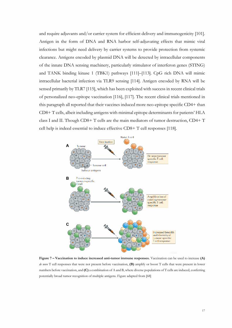
Cancer vaccines are considered to work synergistically with CPI therapy in cancer, since
vaccination can ideally both boost existing T cell responses and induce them de novo, as
outlined in Figure 7. Several considerations go into rational cancer vaccine design, including
choice of antigen(s), antigen format, and adjuvants [97]. Regarding antigen selection, there
are options of more or less personalized character, of which neo-epitopes are proposedly
ideal tumor targets since they are truly tumor specific [49], [98]. With neo-epitopes as targets,
vaccines are selected and produced individually for each patient, leading to higher cost and
time consumption than off-the-shelf therapeutics. Epitopes of shared origin are in this sense
advantageous since they are broadly expressed between different cancer types and patients,
however they bring risks of autoimmunity if targeted and are highly affected by central
16
tolerance. Furthermore, since not all cancers are equally burdened by somatic mutations,
different cancer types call for different antigen selection strategies. Leading companies in the
field of personalized vaccinations BioNTech and Neon Therapeutics are both currently
pursuing therapeutic combinations of neo- and off-the-shelf targeted antigens [97]. Whatever
the approach, it is sensible to include multiple tumor epitopes in a cancer vaccine to increase
the breadth of the anti-tumor response and overcome challenges of tumor heterogeneity and
loss of tumor cell variants as an escape mechanism.
Fundamental to inducing potent anti-tumor responses is the activation and priming of T cells
by APCs. As outlined previously, this process starts with innate sensing of danger signals,
which shapes the adaptive immune responses that follows. A process, that can be controlled
by both the format of the antigen, administration route and choice of adjuvant. A plethora
of adjuvants have been developed, and these can overall be divided in two categories: depot-
effect delivery systems and immunostimulants, or combinations thereof [99], [100]. In
general, adjuvants contribute to upregulate co-stimulatory molecules, MHC presentation and
cytokines by DCs. A frequently used depot-effect delivery is aluminum salts (mostly
aluminum hydroxide/alum), used in common pathogen vaccines, but a robust inducer of
TH2-like immunity when delivered alone and thus not considered suitable for cancer vaccines
[101]. Another option is water-in-oil emulsions such as Montanide, that ensures slow release
of e.g. peptide antigens which induce prolonged immune responses and has been investigated
in clinical cancer vaccine trials without observation of toxicities [102]. Polymer and liposome-
based delivery technologies are also being investigated for capability to deliver cancer
antigens and cytotoxic drugs, offering biocompatible cargo protection and possibilities of
specific delivery to e.g. DCs via coupling to antibodies [103], [104]. Cationic liposomes have
been reported to improve cross-presentation by DCs in murine models, allowing priming of
CD8+ T cells [105], [106].
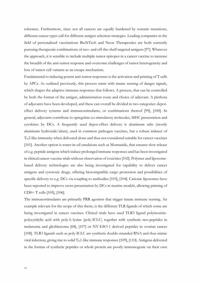
The immunostimulants are primarily PRR agonists that trigger innate immune sensing. An
example relevant for the scope of this thesis, is the different TLR ligands of which some are
being investigated in cancer vaccines. Clinical trials have used TLR3 ligand polyinosinic-
polycytidylic acid with poly-L-lysine (poly-ICLC) together with synthetic neo-peptides in
melanoma and glioblastoma [68], [107] or NY-ESO-1 derived peptides in ovarian cancer
[108]. TLR3 ligands such as poly-ICLC are synthetic double-stranded RNA and thus mimic
viral infection, giving rise to solid TH1-like immune responses [109], [110]. Antigens delivered
in the format of synthetic peptides or whole protein are poorly immunogenic on their own
17
and require adjuvants and/or carrier system for efficient delivery and immunogenicity [101].
Antigen in the form of DNA and RNA harbor self-adjuvating effects that mimic viral
infections but might need delivery by carrier systems to provide protection from systemic
clearance. Antigens encoded by plasmid DNA will be detected by intracellular components
of the innate DNA sensing machinery, particularly stimulator of interferon genes (STING)
and TANK binding kinase 1 (TBK1) pathways [111]–[113]. CpG rich DNA will mimic
intracellular bacterial infection via TLR9 sensing [114]. Antigen encoded by RNA will be
sensed primarily by TLR7 [115], which has been exploited with success in recent clinical trials
of personalized neo-epitope vaccination [116], [117]. The recent clinical trials mentioned in
this paragraph all reported that their vaccines induced more neo-epitope specific CD4+ than
CD8+ T cells, albeit including antigens with minimal epitope determinants for patients’ HLA
class I and II. Though CD8+ T cells are the main mediators of tumor destruction, CD4+ T
cell help is indeed essential to induce effective CD8+ T cell responses [118].
Figure 7 – Vaccination to induce increased anti-tumor immune responses. Vaccination can be used to increase (A)
de novo T cell responses that were not present before vaccination, (B) amplify or boost T cells that were present in lower
numbers before vaccination, and (C) a combination of A and B, where diverse populations of T cells are induced, conferring
potentially broad tumor recognition of multiple antigens. Figure adapted from [68]
A
B
C
18
In conclusion, after years of limited clinical success [38], [119] cancer vaccines are revisited
and show promising preliminary results particularly in the neo-epitope space. Vaccine
combinations with CPI therapy confers the potential to unleash the brakes imposed on
immune cells and specifically steer the immune system towards relevant tumor targets. The
field is increasingly moving towards tailored, personalized vaccination strategies, which
imposes increased demands for neo-epitope prediction and prioritization based on
bioinformatic approaches as well as individual vaccine manufacturing [120].
Lessons from preclinical mouse models Mice have for decades been the most commonly used animal model for immunology and
cancer research. Importantly, mouse models have provided important insights into the
dynamic interplay between cancer and the immune system, reviewed in detail by Dunn et al.
[121]. Initially, mouse models demonstrated the role of immune surveillance and
immunologic recognition of tumors by showing how mice that lack central components of
innate and adaptive immunity have enhanced susceptibility to chemically induced and
spontaneously developed tumors. Next, mouse models provided the basis for the
immunoediting theory, showing how tumor immunogenicity is shaped by the immune
system. This was apparent by several studies, where tumors grown on immunocompromised
mice were immunogenic and rapidly rejected when transplanted to immunocompetent mice.
Conversely, tumors grown on immunocompetent mice were not immunogenic and were not
rejected when transplanted to other immunocompetent mice.
The most commonly used mouse models in cancer are the syngeneic models. Here,
immunocompetent host mice, often of inbred C57BL/6 and BALB/c background, are
transplanted with histocompatible tumor cell lines. Many syngeneic models have been
developed, representing diverse tissue origins with differential levels of immunogenicity,
TME compositions, and varying sensitivity to immunotherapy [122]. These inbred mice offer
reproducible tumor growth experiments, in which tumor cells are often inoculated
subcutaneously and easily measurable. However, inbred mice inherently have a limited MHC
class I and II repertoire and syngeneic models neither recapitulate interpatient or intratumor
heterogeneity. Alternatively, patient-derived xenograft (PDX) models can be used. PDX
models entail immunodeficient mice that allow inoculation of patient derived tumor cell lines
for evaluation of personalized, clinically relevant therapies. However, in conventional PDX
19
models that lack a competent immune system, the tumor is not subjected to immune
surveillance and immunoediting. To mitigate this, humanized mice have been developed,
such as the human IL-2 expressing PDX model that facilitates assessment of a patient’s
autologous ex vivo expanded TILs towards patient’s tumor cell line [123] and also mice that
have further humanized immune systems after transfer of hematopoietic stem cells [124], for
testing of immunotherapies.
For several well characterized syngeneic tumor models there are descriptions of
therapeutically relevant neo-epitopes, such as: CT26 [125], [126], MC38 [127], [128], and
B16-F10 [129]. Murine neo-epitope vaccination studies, either alone or in combination with
CPI therapy, have contributed to our understanding of tumor neo-epitopes and the
framework we use to predict them [129], [130]. These models have uncovered that CPI
therapy-mediated rejection of established tumors is mainly driven by neo-epitope specific T
cell responses [131]. Further, that CPI therapy modulates neo-epitope specific T cells and
myeloid cells inside the tumor and not to the same extent those that are present in periphery
[132], [133]. Moreover, we have recently seen evidence from syngeneic models that
vaccination and CPI therapy can induce CD4+ and CD8+ neo-epitope specific T cells
important for tumor rejection [126], [134], even when tumor does not express MHC class II
[135]. Thus, both clinical and preclinical observations underline an important role of
cytotoxic and helper T cells in the anti-tumor response [136].
Neo-epitope discovery studies in murine models will, together with clinical studies,
contribute to interrogate the rules that define ideal neo-epitopes. However, the
aforementioned narrow MHC repertoire of these mouse models naturally limits the epitopes
that can be displayed, compared to humans. Similarly, majority of tumor antigens, let alone
neo-epitopes, will not be shared between mice and humans, why therapies to target them are
difficult to translate. Furthermore, many common syngeneic tumors grow rapidly upon
transplantation on the mice, why there is a narrow therapeutic window and a short time to
mount anti-tumor immunity via e.g. vaccination [122], [137]. Since the syngeneic tumor is
transplanted, there is no extensive phase of co-development between mouse and tumor as
there is in patients, where a tumor can develop over several years. Though some lessons from
mouse models might be ‘lost in translation’ [138], they remain applicable for examining the
effects of different vaccination approaches such as dosing, vaccination schedules, antigen
formats and adjuvants [97]. Mouse models provide mechanistic insights of relevance to the
clinical realm.

20
21
2. MANUSCRIPT I
H-2 multimers for high-throughput T cell interrogation and description of novel conditional ligands for H-2Dd and H-2Kd
MHC multimer technologies have for long been instrumental in the monitoring of antigen
specific T cells and epitope discovery in a wide range of diseases. Here, we designed and
validated murine H-2Dd and H-2Kd conditional ligands for UV-mediated peptide exchange.
Furthermore, we used H-2Dd, H-2Kd and H-2Ld molecules for H-2 tetramer stainings and
provided proof-of-concept for their use in DNA barcode-labeled H-2 multimers for large-
scale CD8+ T cell interrogation. We expect these new conditional ligands and the high
throughput screening methodology to improve epitope mapping in various diseases in H-2d
models, including cancer and autoimmunity.
This manuscript has been written and formatted for submission to Journal of Immunology,
specifically their subsection on ‘Novel Immunological Methods’.

22

Manuscript I 1
H-2 multimers for high-throughput T cell interrogation and
description of novel conditional ligands for H-2Dd and H-2Kd
Authors: Nadia Viborg1*, Trine Sundebo Meldgaard1*, Tripti Tamhane1, Sara Suarez Hernandez1,
Christian Garde2, Thomas Trolle2, Dario Vazquez Albacete1#, and Sine Reker Hadrup1
Affiliations: 1Department of Health Technology, Technical University of Denmark, Lyngby,
Denmark, 2Evaxion Biotech, Copenhagen, Denmark, *These authors contributed equally, #Current
affiliation: Department of Biotechnology and Biomedicine, DTU Bioengineering, Technical
University of Denmark, Lyngby, Denmark
Corresponding author: Sine Reker Hadrup, Section for Experimental and Translational
Immunology, Department of Health Technology, Technical University of Denmark, Kemitorvet
Building 204, 2800 Kgs. Lyngby, Denmark. Phone: +45 35886290. E-mail: [email protected].
Financial support: This research was funded in part The Danish Innovation Fund (project XVAC
and NeoPepVac) and The European Research Council, ERC StG nextDART.
Number of figures: 4
Number of tables: 1
Number of supplementary figures: 2
Number of supplementary tables: 1
Manuscript I 2
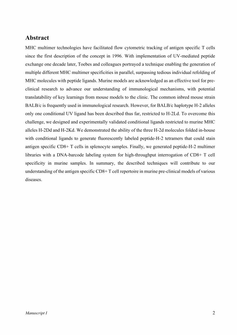
Abstract
MHC multimer technologies have facilitated flow cytometric tracking of antigen specific T cells
since the first description of the concept in 1996. With implementation of UV-mediated peptide
exchange one decade later, Toebes and colleagues portrayed a technique enabling the generation of
multiple different MHC multimer specificities in parallel, surpassing tedious individual refolding of
MHC molecules with peptide ligands. Murine models are acknowledged as an effective tool for pre-
clinical research to advance our understanding of immunological mechanisms, with potential
translatability of key learnings from mouse models to the clinic. The common inbred mouse strain
BALB/c is frequently used in immunological research. However, for BALB/c haplotype H-2 alleles
only one conditional UV ligand has been described thus far, restricted to H-2Ld. To overcome this
challenge, we designed and experimentally validated conditional ligands restricted to murine MHC
alleles H-2Dd and H-2Kd. We demonstrated the ability of the three H-2d molecules folded in-house
with conditional ligands to generate fluorescently labeled peptide-H-2 tetramers that could stain
antigen specific CD8+ T cells in splenocyte samples. Finally, we generated peptide-H-2 multimer
libraries with a DNA-barcode labeling system for high-throughput interrogation of CD8+ T cell
specificity in murine samples. In summary, the described techniques will contribute to our
understanding of the antigen specific CD8+ T cell repertoire in murine pre-clinical models of various
diseases.

Manuscript I 3
Introduction
Major histocompatibility complex (MHC) class I molecules display endogenous peptide products
of proteasomal degradation on the surface of all nucleated cells. T cell based immune surveillance
relies on this presentation and the specific interaction between peptide-MHC (pMHC) and the T cell
receptor (TCR), enabling cytotoxic T cells to identify and eliminate aberrant cells. Two decades of
research have provided tools to identify, characterize and isolate antigen specific T cells. In
particular, multimerization and flurochrome labeling of MHC I molecules have accelerated the
understanding of pMHC-TCR interactions within infections, autoimmune diseases and cancer,
where CD8+ T cells play a pivotal role [1]. pMHC tetramers are commonly used in the field and can
facilitate the detection of multiple different antigen specific CD8+ T cells in parallel, when e.g.
combinatorial fluorescent labeling is used [2].
An innovative contribution to the pMHC multimer space occurred when MHC protein was produced
and refolded with a so-called conditional ligand. A technique first described by Toebes et al. [3],
where an amino acid moiety in the T cell receptor-exposed site of a known MHC ligand is replaced
with a non-natural amino acid (2‐nitrophenylglycine or 3‐amino‐3‐(2‐nitrophenyl)‐propionic acid)
that is cleavable upon exposure to 366 nm UV light (denoted “J”). Upon UV light mediated cleavage
of the conditional ligand, the MHC binding groove is left empty and receptive of another ligand of
choice. This UV-mediated exchange allows for the rapid and high-throughput generation of large
panels of distinct pMHC specificities.
Since the development of pMHC tetramers and the introduction of the UV exchange technology,
higher order pMHC multimerizations and high-throughput labeling systems have been developed.
Recently, DNA barcode labeled pMHC multimers were proven to allow large-scale detection of
antigen specific CD8+ T cells, with the possibility to screen samples for recognition of >1000
different pMHC multimers simultaneously [4]. A technology that can contribute to uncover new T
cell epitopes and understand pMHC-TCR interactions in a wide variety of diseases.
Preclinical models have proven important for acquiring mechanistic understanding of
immunological diseases and supporting the development and evaluation of new therapeutic
interventions. Mouse models are frequently used in cancer research, where especially the tumor
neoepitope field is being extensively studied [5]–[8]. A number syngeneic murine tumors hase been
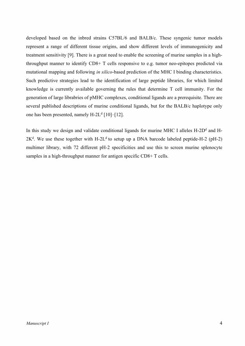
Manuscript I 4
developed based on the inbred strains C57BL/6 and BALB/c. These syngenic tumor models
represent a range of different tissue origins, and show different levels of immunogenicity and
treatment sensitivity [9]. There is a great need to enable the screening of murine samples in a high-
throughput manner to identify CD8+ T cells responsive to e.g. tumor neo-epitopes predicted via
mutational mapping and following in silico-based prediction of the MHC I binding characteristics.
Such predictive strategies lead to the identification of large peptide libraries, for which limited
knowledge is currently available governing the rules that determine T cell immunity. For the
generation of large librabries of pMHC complexes, conditional ligands are a prerequisite. There are
several published descriptions of murine conditional ligands, but for the BALB/c haplotype only
one has been presented, namely H-2Ld [10]–[12].
In this study we design and validate conditional ligands for murine MHC I alleles H-2Dd and H-
2Kd. We use these together with H-2Ld to setup up a DNA barcode labeled peptide-H-2 (pH-2)
multimer library, with 72 different pH-2 specificities and use this to screen murine splenocyte
samples in a high-throughput manner for antigen specific CD8+ T cells.
Manuscript I 5
Results
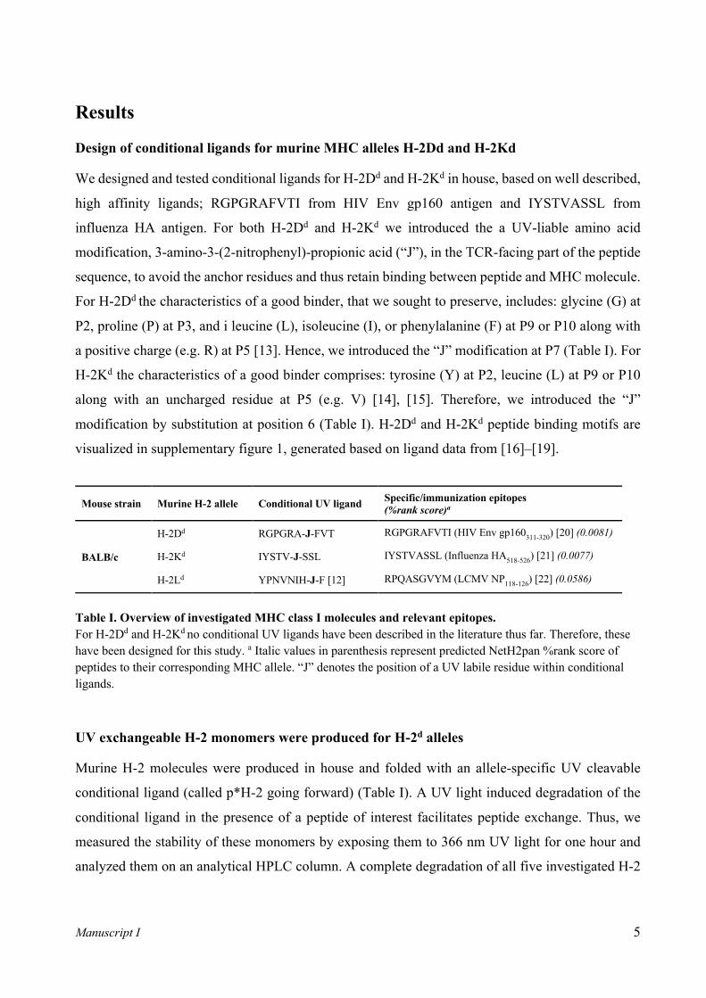
Design of conditional ligands for murine MHC alleles H-2Dd and H-2Kd
We designed and tested conditional ligands for H-2Dd and H-2Kd in house, based on well described,
high affinity ligands; RGPGRAFVTI from HIV Env gp160 antigen and IYSTVASSL from
influenza HA antigen. For both H-2Dd and H-2Kd we introduced the a UV-liable amino acid
modification, 3‐amino‐3‐(2‐nitrophenyl)‐propionic acid (“J”), in the TCR-facing part of the peptide
sequence, to avoid the anchor residues and thus retain binding between peptide and MHC molecule.
For H-2Dd the characteristics of a good binder, that we sought to preserve, includes: glycine (G) at
P2, proline (P) at P3, and i leucine (L), isoleucine (I), or phenylalanine (F) at P9 or P10 along with
a positive charge (e.g. R) at P5 [13]. Hence, we introduced the “J” modification at P7 (Table I). For
H-2Kd the characteristics of a good binder comprises: tyrosine (Y) at P2, leucine (L) at P9 or P10
along with an uncharged residue at P5 (e.g. V) [14], [15]. Therefore, we introduced the “J”
modification by substitution at position 6 (Table I). H-2Dd and H-2Kd peptide binding motifs are
visualized in supplementary figure 1, generated based on ligand data from [16]–[19].
Mouse strain Murine H-2 allele Conditional UV ligand Specific/immunization epitopes (%rank score)a
BALB/c
H-2Dd RGPGRA-J-FVT RGPGRAFVTI (HIV Env gp160311-320) [20] (0.0081)
H-2Kd IYSTV-J-SSL IYSTVASSL (Influenza HA518-526) [21] (0.0077)
H-2Ld YPNVNIH-J-F [12] RPQASGVYM (LCMV NP118-126) [22] (0.0586)
Table I. Overview of investigated MHC class I molecules and relevant epitopes. For H-2Dd and H-2Kd no conditional UV ligands have been described in the literature thus far. Therefore, these have been designed for this study. a Italic values in parenthesis represent predicted NetH2pan %rank score of peptides to their corresponding MHC allele. “J” denotes the position of a UV labile residue within conditional ligands.
UV exchangeable H-2 monomers were produced for H-2d alleles
Murine H-2 molecules were produced in house and folded with an allele-specific UV cleavable
conditional ligand (called p*H-2 going forward) (Table I). A UV light induced degradation of the
conditional ligand in the presence of a peptide of interest facilitates peptide exchange. Thus, we
measured the stability of these monomers by exposing them to 366 nm UV light for one hour and
analyzed them on an analytical HPLC column. A complete degradation of all five investigated H-2
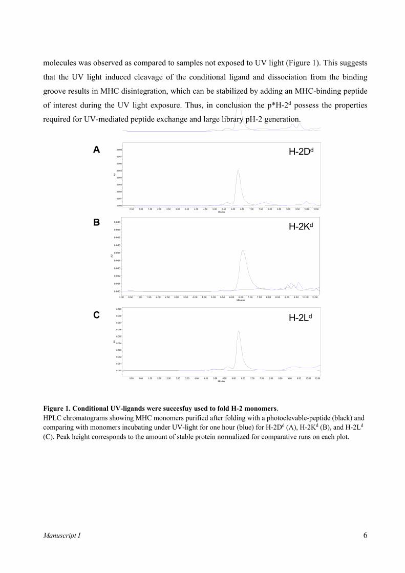
Manuscript I 6
molecules was observed as compared to samples not exposed to UV light (Figure 1). This suggests
that the UV light induced cleavage of the conditional ligand and dissociation from the binding
groove results in MHC disintegration, which can be stabilized by adding an MHC-binding peptide
of interest during the UV light exposure. Thus, in conclusion the p*H-2d possess the properties
required for UV-mediated peptide exchange and large library pH-2 generation.
Figure 1. Conditional UV-ligands were succesfuy used to fold H-2 monomers. HPLC chromatograms showing MHC monomers purified after folding with a photoclevable-peptide (black) and comparing with monomers incubating under UV-light for one hour (blue) for H-2Dd (A), H-2Kd (B), and H-2Ld (C). Peak height corresponds to the amount of stable protein normalized for comparative runs on each plot.
E
AU
0.000
0.001
0.002
0.003
0.004
0.005
0.006
0.007
0.008
0.009
Minutes0.50 1.00 1.50 2.00 2.50 3.00 3.50 4.00 4.50 5.00 5.50 6.00 6.50 7.00 7.50 8.00 8.50 9.00 9.50 10.00 10.50
H-2Ld
H-2DbA
AU
0.000
0.001
0.002
0.003
0.004
0.005
0.006
0.007
0.008
0.009
Minutes0.50 1.00 1.50 2.00 2.50 3.00 3.50 4.00 4.50 5.00 5.50 6.00 6.50 7.00 7.50 8.00 8.50 9.00 9.50 10.00 10.50
H-2KbB
AU
0.000
0.001
0.002
0.003
0.004
0.005
0.006
0.007
0.008
Minutes0.50 1.00 1.50 2.00 2.50 3.00 3.50 4.00 4.50 5.00 5.50 6.00 6.50 7.00 7.50 8.00 8.50 9.00 9.50 10.00 10.50
H-2DdC
AU
0.000
0.001
0.002
0.003
0.004
0.005
0.006
0.007
0.008
0.009
Minutes0.00 0.50 1.00 1.50 2.00 2.50 3.00 3.50 4.00 4.50 5.00 5.50 6.00 6.50 7.00 7.50 8.00 8.50 9.00 9.50 10.00 10.50 11.00 11.50 12.00 12.50 13.00
H-2KdD
H-2Ld
A
B
C
H-2Dd
H-2Kd
Manuscript I 7
Detection of antigen specific CD8+ T cells in murine splenocyte samples via pH-2 tetramer
staining
To confirm the presence of antigen specific CD8+ T cells in our murine splenocyte samples and
validate the potential use of the newly designed UV ligand p*H-2 monomers, we generated pH-2
tetramers by UV exchange and coupling of exchanged biotinylated pH-2 products to PE-conjugated
streptavidin. Splenocyte samples from immunized or transgenic mice (from here on: antigen specific
mice) were stained with either 1) pH-2 tetramers exchanged with the specific peptides corresponding
to specificity induced by immunization or transgenic TCR clone specificity (Table I), and 2) pH-2
tetramers exchanged with an irrelevant peptide (predicted H-2d ligand neo-peptides from murine
syngenic tumor cell line CT26 – supplementary table 1). As controls, splenocyte samples from naïve
BALB/c mice were stained with the same specific peptide tetramers as the antigen specific mice.
By flow cytometry, we confirmed pH-2 tetramer specific CD8+ T cells present in all antigen specific
mouse samples (Figure 2). Observed frequencies of antigen specific CD8+ T cells varied, with the
TCR transgenic mouse sample (IYSTVASSL/H-2Kd) having >90% tetramer specific cells, and the
immunized mice ranging from 2.7% (RGPGRAFVTI/H-2Dd) to 10% (RPQASGVYM/H-2Ld). The
different antigen specific CD8+ T cell populations also displayed varying binding for their cognate
epitope as apparent by the difference in separation of the tetramer positive from the tetramer negative
CD8+ T cells. Control staining of antigen specific samples with irrelevant pH-2 tetramers and
staining of naïve splenocyte samples with specific pH-2 tetramers revealed that the H-2 tetramers
generate limited background, and display a pH-2-TCR restricted T cell interaction. FACS gating
strategy for pH-2 tetramer stainings is shown in supplementary figure 2A. Thus, we have
successfully generated pH-2d tetramers after UV mediated peptide–exchange, and demonstrated
successful staining of antigen specific CD8+ T cells in relevant mouse spleenocytes.

Manuscript I 8
Figure 2. Detection of antigen specific CD8+ T cells via PE-labeled pH-2 tetramers Successful UV-mediated peptide exchange and pH-2 tetramer assembly was validated for all H-2 alleles by tetramer staining (PE fluorophore label). Antigen specific splenocytes were stained with specific peptide tetramers and tetramers exchanged with irrelevant peptides to examine unspecific background staining from the tetramer reagents. Naïve control splenocytes from BALB/c mice were stained with specific peptide tetramers as a negative control. The frequency of pH-2 multimer specific T cells out of total CD8+ T cells is noted on each plot.
Large-scale interrogation of CD8+ T cell specificity using DNA barcode-labeled pH-2
multimer libraries
To evaluate the use of pH-2d for large-scale interrogation of CD8+ T cell speificity, we generated a
DNA barcode-labeled pH-2 multimer panel as previously described [4]. In brief, we performed UV
mediated peptide exchange, followed by multimerization of the peptide-exchanged products onto a
PE-labeled polysaccharide backbone individually coupled to DNA barcodes, forming a unique label
Antigen specific sample Naive control sampleC
D8-
BV4
80
Tetramer-PE
H-2Dd
H-2Kd
H-2Ld
2.7% 0.5% 0.8%
96% 0.2% 0.1%
10% 0.2% 0.2%
RGPGRAFVTI RGPGRAFVTI
IYSTVASSL IYSTVASSL
RPQASGVYM RPQASGVYM
KADCLFTHM
IYQRKSDGI
IPRDLFEGEL
+ specific pH-2 tetramer + irrelevant pH-2 tetramer + specific pH-2 tetramer
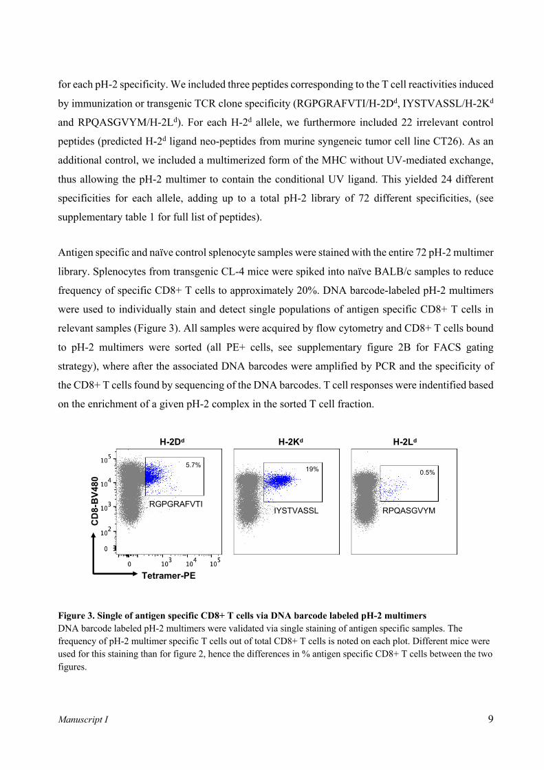
Manuscript I 9
for each pH-2 specificity. We included three peptides corresponding to the T cell reactivities induced
by immunization or transgenic TCR clone specificity (RGPGRAFVTI/H-2Dd, IYSTVASSL/H-2Kd
and RPQASGVYM/H-2Ld). For each H-2d allele, we furthermore included 22 irrelevant control
peptides (predicted H-2d ligand neo-peptides from murine syngeneic tumor cell line CT26). As an
additional control, we included a multimerized form of the MHC without UV-mediated exchange,
thus allowing the pH-2 multimer to contain the conditional UV ligand. This yielded 24 different
specificities for each allele, adding up to a total pH-2 library of 72 different specificities, (see
supplementary table 1 for full list of peptides).
Antigen specific and naïve control splenocyte samples were stained with the entire 72 pH-2 multimer
library. Splenocytes from transgenic CL-4 mice were spiked into naïve BALB/c samples to reduce
frequency of specific CD8+ T cells to approximately 20%. DNA barcode-labeled pH-2 multimers
were used to individually stain and detect single populations of antigen specific CD8+ T cells in
relevant samples (Figure 3). All samples were acquired by flow cytometry and CD8+ T cells bound
to pH-2 multimers were sorted (all PE+ cells, see supplementary figure 2B for FACS gating
strategy), where after the associated DNA barcodes were amplified by PCR and the specificity of
the CD8+ T cells found by sequencing of the DNA barcodes. T cell responses were indentified based
on the enrichment of a given pH-2 complex in the sorted T cell fraction.
Figure 3. Single of antigen specific CD8+ T cells via DNA barcode labeled pH-2 multimers DNA barcode labeled pH-2 multimers were validated via single staining of antigen specific samples. The frequency of pH-2 multimer specific T cells out of total CD8+ T cells is noted on each plot. Different mice were used for this staining than for figure 2, hence the differences in % antigen specific CD8+ T cells between the two figures.
CD8-BV480
Tetramer-PE
H-2Dd H-2Kd H-2Ld
IYSTVASSL RPQASGVYMRGPGRAFVTI
5.7% 19% 0.5%

Manuscript I 10
With this large-scale technology, we were able to detect the specific CD8+ T cells in the antigen
specific samples, corresponding to the observations with single pH-2 multimer stainings, i.e. in the
RGPGRAFVTI /H-2Dd immunized mouse, a RGPGRAFVTI /H-2Dd CD8+ T cell response was
detected, out of the total pool of 72 specificities, and so forth (Figure 4). In naïve control splenocytes
(BALB/c) no antigen specific CD8+ T cell responses were detected. These findings were consistent
across two replicate experiments.
Figure 4. Detection of H-2 antigen specific CD8+ T cell responses via DNA barcode labeled pH-2 multimer screening Screening for T cell recognition by DNA barcode-labeled pH-2 multimers in splenocytes from antigen specific mice (A) and naïve control mice (B), with a library of 72 different pH-2 specificities (24 specificities per allele). Filled symbols represent CD8+ T cell responses detected in H-2Dd RGPGRAFVTI specific sample (blue), H-2Kd IYSTVASSL specific sample (green), or H-2Ld RPQASGVYM specific sample (dark red) samples. Data has been plotted on the y-axis as -Log10(p) of the relevant pH-2 associated DNA barcode. The values represent replicates from two different screenings (1, circles) and (2, squares).
A B
0
5
10
153540
- Log
10(p
)
H-2Dd
H-2Dd_RGPGRAFVTI
H-2Kd_IYSTVASSL
H-2Ld_RPQASGVYM
H-2Kd_IYSTVASSL
H-2Dd_RGPGRAFVTI H-2Ld_RPQASGVYM
H-2Kd H-2Ld0
5
10
15
- Log
10(p
)
H-2Dd H-2Kd H-2Ld
H-2Kd IYSTVASSL specific sample (2)
H-2Ld RPQASGVYM specific sample (2)
H-2Dd RGPGRAFVTI specific sample (2)
H-2Kd IYSTVASSL specific sample (1)
Naive BALB/c (1)Naive BALB/c (2)
H-2Ld RPQASGVYM specific sample (1)
H-2Dd RGPGRAFVTI specific sample (1)
Manuscript I 11
Discussion
The data presented here display, for the first time, the design and use of conditional ligands for
murine MHC class I alleles H-2Dd and H-2Kd. These novel conditional ligands facilitated the
generation of specific pH-2 multimers via UV-mediated peptide exchange, alongside p*H-2Ld for
which a conditional ligands has previously been described. UV-mediated peptide exchange
technology enabled the production of several pH-2 multimers in parallel, overcoming the challenges
and need for tedious individual folding of H-2 molecule with specific peptides. First, we were able
to generate fluorescently labeled H-2 tetramers to stain murine pH-2d haplotype samples for single
antigen specific CD8+ T cell populations, obtained via peptide immunization or from transgenic T
cells. Flow cytometric analysis confirmed presence of specific CD8+ T cells in relevant splenocyte
samples and limited background signal in irrelevant samples. Second, we used the UV-mediated
peptide exchange to generate a DNA barcode-labeled pH-2 multimer library containing 72 different
pH-2 specificities. Flow cytometric sorting of multimer positive populations and recovery of DNA
barcodes by sequencing corresponded to our findings from the tetramer stainings, i.e. antigen
specific CD8+ T cells were observed in specific splenocyte samples.
The high-throughput DNA barcode labeled pMHC multimer methodology has recently been used to
describe human autoreactive CD8+ T cells in narcolepsy [23] and CD8+ T cells specific to novel
overexpression antigens in breast cancer [24]. This technology enables the interrogation of samples
for hundreds of pMHC specificities in parallel, which is particularly advantageous when sample
material is sparse, as described previously [4]. Importantly, platforms like the one described here
allow extensive screening for pMHC-TCR interactions that will contribute to characterize what
makes for a CD8+ T cell epitope, and not merely a good MHC binder, as is the output from current
in silico prediction platforms. As an additional layer to these analyses, a recent publication reported
the use of barcode labeled pMHC multimers to interrogate the motifs or “fingerprints” that govern
TCR recognition of pMHC [25].
In cancer research preclinical mouse models have provided lessons that were translatable to clinical
settings. Syngeneic tumor models based on inbred strains C57BL/6 and BALB/c are being
extensively studied in immunotherapeutic settings, where particularly tumor neo-epitopes are
considered therapeutically relevant. The in silico platforms that map tumor mutations and predict
MHC binding peptides from the tumor peptidome generate large output libraries of putative neo-

Manuscript I 12
epitope. Use of p*H-2 molecules with high-throughput multimer screening via the methodology
described here, provides a tool to address the neo-epitope specific T cell repertoire across tumor
models and therapies. It has been established, that different syngeneic tumor models are not equally
sensitive to immunotherapies [9], [26]. Large-scale multimer analyses of the T cell repertoire will
contribute to investigate the signatures of neo-epitope reactive T cells that favor immunotherapy
responses.
We therefore argue that our description of novel conditional ligands for p*H-2Dd and p*H-2Kd adds
to the value of previously described ligands and the proof of concept of pH-2d multimers for large-
scale detection of antigen specific CD8+ T cells in murine samples will be of useful in the field of
epitope discovery and immune monitoring going forward.
Manuscript I 13
Materials and Methods
UV ligand design
To produce stable murine H-2 class I monomers, the molecules were loaded with a conditional
photocleavable peptide for UV exchange. For three of the conditional UV ligands the amino acid
sequence was described in the literature, the other two were designed for the purpose of this study
(Table 1). The conditional UV ligands were designed with anchor residues in mind that differ
between different MHC alleles, based on their structure and different binding motifs. The
photolabile “J” amino acid was placed in the TCR-facing part of the peptide sequence, and was
either replacing one of the amino acids of the original peptide, or was positioned between two amino
acids in the sequence. The predicted rank score for each UV ligand was predicted using NetH2pan
[27] and reported in this study as %rank score (Table 1). As per recommendations, strong H-2
binders are generally considered to have a %rank score<0.5, weak MHC binders have a %rank score
<2. Peptides to be used as conditional UV ligands were purchased and synthethized from Leiden
University Medical Center peptide synthesis unit (LUMC).
Expression and purification of murine MHC class I heavy chains and β2 microglobulin
Bacterial expression system was used to produce murine H-2 class I heavy chains (H-2Dd, H-2Kd,
and H-2Ld), and human β2m macroglobulin as described previously [28]. Briefly, proteins were
produced in Escherichia coli Bl21(DE3) pLysS strain using pET series plasmids. Protein expression
was induced by 0.5 mM isopropyl-beta-D-thiogalactopyranoside for 4 h. Inclusion bodies containing
expressed proteins were lysed in lysis buffer (50 mM Tris·HCl (pH 8.0), 25% sucrose, 1 mM EDTA,
and Lysozyme). Inclusion bodies were harvested by washing in detergent buffer (20 mM Tris-HCl,
(pH 7.5), 200 mM NaCl, 1% NP40, and 1% Deoxycholic acid) followed by wash buffer (1 mM
EDTA, 5% Triton X-100, 1 mM DTT). Next, inclusion bodies were dissolved in 8M Urea buffer
(8M Urea, 50 mM K·HEPES pH 6.5 and 100 µM β-mercaptoethanol) and insoluble impurities were
removed by centrifugation at 40,000 x g for 20 minutes. The soluble fraction containing proteins
was stored at -80 °C until used for in vitro folding.
In vitro folding and purification of murine MHC class I monomers
The in vitro folding of murine H-2 class I molecules were performed using photocleavable ligand
as described above and previous [3]. To setup the refolding reaction, heavy chains (1 µM) and β2m
Manuscript I 14
(2 µM) were diluted in a folding buffer composed of 0.1M Tris pH 8.0, 500 mM L-Arginine-HCl, 2
mM EDTA, 0.5 mM oxidized glutathione and 5 mM reduced glutathione with 60 µM respective
photocleavable peptide (p*H-2Dd: RGPGRA-J-FVT, p*H-2Kd: IYSTV-J-SSL, p*H-2Ld:
YPNVNIH-J-F) (Table 1). After folding for 3-5 days at 4 °C, folded protein was upconcentrated
with a 10 kDa cut off membrane filters (Vivaflow-200; Sartorius) and biotinylated using BirA biotin-
protein ligase standard reaction kit (Avidity, LLC- Aurora, Colorado). Finally, folded biotinylated
monomer complexes were purified with size exclusion chromatography using HPLC (Waters
Corporation, USA), and aliquots were stored at -80 °C until further use.
In vivo studies and splenocyte preparation
BALB/cJRj mice were acquired from Janvier Labs and housed at the Department of Health
Technology, Technical University of Denmark. CL-4-TCR transgenic mice were a kind gift from
Dr. Ana Misslitz, Hannover Medical School and Torsten Joeris from Lund University, respectively.
To asses the binding of murine H-2 class I monomers, the following in vivo studies were performed
to induce reactive CD8+ T cells:
H-2Dd antigen specific T cells: BALB/cJRj mice were injected with 80µg HIV Env gp160 peptide
(RGPGRAFVTI) adjuvanted by 30µg poly-ICLC (Hiltonol®, from Oncovir). The mice were
immunized four times i.p. with a weekly interval, spleens were harvested 7 days post last
immunization. H-2Kd antigen specific T cells: splenocytes from transgenic CL-4 mice were
harvested and 1-10% specific cells spiked into splenocytes from naïve BALB/cJRj mice. H-2Ld
antigen specific T cells: BALB/cJRj mice were injected with 80µg LCMV NP (RPQASGVYM)
adjuvanted by 30µg poly-ICLC. The mice were immunized four times i.p. with a weekly interval,
spleens were harvested 7 days post last immunization.
Mice were euthanized by cervical dislocation and spleens were harvested and kept on 4°C in cRPMI
(RPMI (Gibco RPMI 1640) and 10% FCS (Gibco)). Spleens were placed in a GentleMACS C-tube
(Miltenyi, #130-093-237) with 3 mL cRPMI. The tubes were loaded onto GentleMACS dissociator
and run with the appointed program. The dissociated splenocytes were placed on top of a 70 µm cell
strainers (Corning, # 43175) and 50mL Falcon tube and filtered through. Cells were washed twice
by resuspension in cRPMI and centrifugation at 1500 rpm for 5 minutes at 4°C. The cells were
finally dissolved in pure FCS and 10% DMSO (dimethyl sulfoxide, Sigma-Aldrich, Cat#C6164) and
Manuscript I 15
1 mL was aliquoted into cryo tubes and stored at (-80 in a Mr. Frosty for 24 hours before moved to)
a -180 tank for precevation.
Generation of specific pH-2 multimers
All peptides for pH-2 multimer libraries were purchased from Pepscan (Pepscan Presto BV,
Lelystad, Netherlands) or TAG Copenhagen (Frederiksberg, Denmark) and dissolved to 10 mM in
100% DMSO. A full list of peptides used in the study can be found in Supplementary Table 1.
Specific peptide binding to H-2 class I monomers was in silico predicted via NetH2pan [1], as
described in above section on UV ligand design. The five different murine H-2 class I monomers
and their corresponding peptides were diluted in PBS. Monomers (50-100 µg/mL final
concentration) were added to the corresponding peptide (100-200 µM final concentration) and
placed under a 366 nm UV light for 1 hour at room temperature, to remove the UV ligand and replace
with peptide of interest. A corresponding UV ligand control for each H-2 allele was in parallel
incubated at RT on the bench.
Generation of PE-conjugated pH-2 tetramers and detection of pH-2-specific T cells
Tetramers were used to screen for T cell recognition against the specific peptide or irrelevant
peptides in immunized or naïve mice. PE labeled streptavidin (phycoerythrin, Biolegend,
Cat#405203, 0.2 mg/mL/100 µl pH-2) was loaded with exchanged pH-2 (100 µg/mL monomer and
200 µM peptide) for 30 minutes on ice. 500 µM D-biotin was added 0.05x and incubated 20 minutes
on ice, and the final product was stabilized using a 10x freezing media (0.5% BSA and 5% glycerol)
and stored at -20°C.
Splenocytes from immunized and naïve mice were thawed and washed twice in cRPMI. 3-4x10⁶
cells were plated into individual wells in a V-bottom 96 well plate and washed with FACS buffer
(PBS and 2% FCS). Cells were treated with 0.5 µl Fc receptor block (Biolegend #101301) for 15
mintues to block unspecific binding. Tetramers were centrigufed prior to use. 1 µl of each pH-2
tetramer specificity was added to the corresponding sample with 49 µl BV buffer (BD) and 0.5nM
Dasatinib and incubated for 15 mintues at 37°C. Cells were stained with an antibody mix (CD3-
FITC: Biolegend #100306, CD8-BV480: BD ##566096 and the dead cell marker LIVE/DEAD
Fixable Near-IR: ThermoFischer L10119) for 30 minutes at 4°C. After two washes in FACS buffer
Manuscript I 16
the cells were either filtered using blue cap FACS tubes (Falcon # 352235) and acquired directly on
the flow cytometer or fixed using 1% filtered paraformaldehyde for 1 – 24 hours previous acquision.
Generation of large pH-2 multimer library and detection of pH-2-specific T cells
A DNA barcode-labeled pH-2 library was used to screen for T cell recognition against the five
specific pH-2 or 115 irrelevant pH-2 specificities in immunized or naïve mice. The technique is as
decribed earlier [4]. Briefly, 72 different pH-2 complexes (50 µg/mL monomer and 100 µM peptide)
were prepared (Supplementary Table 1) and coupled to PE-labeled dextran backbones loaded with
a unique DNA barcode (2.17*10^-6 M). Splenocytes from immunized and naïve mice were thawed
and washed twice in cRPMI. 3-4x10⁶ cells were plated into individual wells in a 96 well plate and
washed with BCB. 1.5 µl of each specificity was pooled and filtred through a 10 kDa cut off
membrane filters (Vivaflow-200; Sartorius). Cells were stained with the pool of the 72 pH-2-
multimers and with an antibody mix (CD3-FITC, CD8-BV480 and the dead cell marker
LIVE/DEAD Fixable Near-IR). pH-2 multimer-specific T cells were sorted based on single, live
CD3+ CD8+ PE+ cells. The sorted cells with DNA barcodes were centrifuged and pellet stored at -
20°C. The DNA barcodes that were present in the samples at sorting were amplified along with
triplicate full library baseline samples for comparison (aliquot of pH-2 multimer reagent pool). The
amplified product was purified using QIAquick PCR Purification kit (Qiagen, Cat#28104),
sequenced (IonTorrent, Primbio) using a 314 or 316 chip (Life Technologies) and data was
processed by the online publically assessible software Barracoda, developed at DTU
(http://www.cbs.dtu.dk/services/barracoda).Baracoda calculates the total reads and clonally-reduced
reads for each DNA barcode (relating to its coupled pH-2 specificity). Log2 fold changes in read
counts linked to a given sample, related to the mean read counts, is compared to the baseline samples
and estimated with normalization factors determined by the trimmed mean of M-values method.
False-discovery rates (FDRs) were estimated using the Benjamini–Hochberg method. A p-value was
calculated based on the Log2 fold change distribution, determining the strength of the signal
compared to the input, and p < 0.001, corresponding to FDR < 0.1%, is established as the
significance level determining a T cell response.
Flow cytometry
All flow cytometry experiments were carried out on Fortessa and Melody instruments (BD
Biosciences). Data were analyzed in FlowJo version 10.6.1 (TreeStar, Inc.).

Manuscript I 17
Statistical analyses
GraphPad Prism 8 for Mac OS X was used for graphing, statistical analyses and tools.
Acknowledgements
The authors would like to thank Bente Rotbøl and Anna Gyllenberg Burkal for excellent technical
assistance, as well as all current and former members of the SRH group for scientific discussions.
Conflict of interest disclosure
The authors declare no potential conflicts of interest.
Manuscript I 18
References
[1] J. D. Altman et al., “Phenotypic Analysis of Antigen-Specific T Lymphocytes,” vol. 274, no. 5284, pp. 94–96, 1996.
[2] R. S. Andersen et al., “Parallel detection of antigen-specific T cell responses by combinatorial encoding of MHC multimers.,” Nat. Protoc., vol. 7, no. 5, pp. 891–902, May 2012.
[3] B. Rodenko et al., “Generation of peptide-MHC class I complexes through UV-mediated ligand exchange,” Nat. Protoc., vol. 1, no. 3, pp. 1120–1132, 2006.
[4] A. K. Bentzen et al., “Large-scale detection of antigen-specific T cells using peptide-MHC-I multimers labeled with DNA barcodes,” Nat. Biotechnol., vol. 34, no. 10, pp. 1037–1045, 2016.
[5] J. C. Castle et al., “Exploiting the mutanome for tumor vaccination,” Cancer Res., vol. 72, no. 5, pp. 1081–1091, 2012.
[6] S. Kreiter et al., “Mutant MHC class II epitopes drive therapeutic immune responses to cancer,” Nature, vol. 520, no. 7549, pp. 692–696, 2015.
[7] B. J. Hos et al., “Identification of a neo-epitope dominating endogenous CD8 T cell responses to MC-38 colorectal cancer,” Oncoimmunology, vol. 00, no. 00, p. 1673125, Oct. 2019.
[8] L. Aurisicchio et al., “Poly-specific neoantigen-targeted cancer vaccines delay patient derived tumor growth,” J. Exp. Clin. Cancer Res., vol. 38, no. 1, pp. 1–13, 2019.
[9] S. I. S. Mosely et al., “Rational selection of syngeneic preclinical tumor models for immunotherapeutic drug discovery,” Cancer Immunol. Res., vol. 5, no. 1, pp. 29–41, 2017.
[10] M. Toebes et al., “Design and use of conditional MHC class I ligands,” Nat. Med., vol. 12, no. 2, pp. 246–251, Feb. 2006.
[11] G. M. Grotenbreg et al., “Discovery of CD8+ T cell epitopes in Chlamydia trachomatis infection through use of caged class I MHC tetramers,” Proc. Natl. Acad. Sci. U. S. A., vol. 105, no. 10, pp. 3831–3836, 2008.
[12] E. Frickel et al., “Parasite Stage–Specific Recognition of Endogenous Toxoplasma gondii –Derived CD8 + T Cell Epitopes,” J. Infect. Dis., vol. 198, no. 11, pp. 1625–1633, Dec. 2008.
[13] M. Corr, L. F. Boyd, E. A. Padlan, and D. H. Margulies, “H-2Dd exploits a four residue peptide binding motif,” J. Exp. Med., vol. 178, no. 6, pp. 1877–1892, Dec. 1993.
[14] V. Mitaksov and D. H. Fremont, “Structural definition of the H-2Kd peptide-binding motif.,” J. Biol. Chem., vol. 281, no. 15, pp. 10618–10625, Apr. 2006.
[15] G. Eberl, A. Sabbatini, C. Servis, P. Romero, J. L. Maryanski, and G. Corradin, “MHC class I H-2Kd-restricted antigenic peptides: additional constraints for the binding motif,” Int. Immunol., vol. 5, no. 11, pp. 1489–1492, Nov. 1993.
[16] R. Vita et al., “The Immune Epitope Database (IEDB): 2018 update.,” Nucleic Acids Res., vol. 47, no. D1, pp. D339–D343, Jan. 2019.
[17] D. de Verteuil et al., “Deletion of immunoproteasome subunits imprints on the transcriptome and has a broad impact on peptides presented by major histocompatibility complex I molecules.,” Mol. Cell. Proteomics, vol. 9, no. 9, pp. 2034–2047, Sep. 2010.
Manuscript I 19
[18] N. A. Nagarajan et al., “ERAAP Shapes the Peptidome Associated with Classical and Nonclassical MHC Class I Molecules.,” J. Immunol., vol. 197, no. 4, pp. 1035–1043, Aug. 2016.
[19] N. L. Dudek, C. T. Tan, D. G. Gorasia, N. P. Croft, P. T. Illing, and A. W. Purcell, “Constitutive and inflammatory immunopeptidome of pancreatic beta-cells.,” Diabetes, vol. 61, no. 11, pp. 3018–3025, Nov. 2012.
[20] C. Bergmann, S. A. Stohlmann, and M. McMillan, “An endogenously synthesized decamer peptide efficiently primes cytotoxic T cells specific for the HIV-1 envelope glycoprotein,” Eur. J. Immunol., vol. 23, no. 11, pp. 2777–2781, Nov. 1993.
[21] K. Kuwano, T. J. Braciale, and F. A. Ennis, “Cytotoxic T lymphocytes recognize a cross-reactive epitope on the transmembrane region of influenza H1 and H2 hemagglutinins.,” Viral Immunol., vol. 2, no. 3, pp. 163–173, 1989.
[22] J. L. Whitton et al., “Molecular analyses of a five-amino-acid cytotoxic T-lymphocyte (CTL) epitope: an immunodominant region which induces nonreciprocal CTL cross-reactivity.,” J. Virol., vol. 63, no. 10, pp. 4303–4310, Oct. 1989.
[23] N. W. Pedersen et al., “CD8+ T cells from patients with narcolepsy and healthy controls recognize hypocretin neuron-specific antigens,” Nat. Commun., vol. 10, no. 1, p. 837, Dec. 2019.
[24] N. Viborg et al., “T cell recognition of novel shared breast cancer antigens is frequently observed in peripheral blood of breast cancer patients,” Oncoimmunology, vol. 8, no. 12, p. e1663107, Dec. 2019.
[25] A. K. Bentzen et al., “T cell receptor fingerprinting enables in-depth characterization of the interactions governing recognition of peptide–MHC complexes,” Nat. Publ. Gr., vol. 36, no. 12, 2018.
[26] M. J. Selby et al., “Preclinical development of ipilimumab and nivolumab combination immunotherapy: Mouse tumor models, In vitro functional studies, and cynomolgus macaque toxicology,” PLoS One, vol. 11, no. 9, pp. 1–19, 2016.
[27] C. I. DeVette et al., “NetH2pan: A computational tool to guide MHC peptide prediction on murine tumors,” Cancer Immunol. Res., vol. 6, no. 6, pp. 636–644, 2018.
[28] D. N. Garboczi, D. T. Hung, and D. C. Wiley, “HLA-A2-peptide complexes: refolding and crystallization of molecules expressed in Escherichia coli and complexed with single antigenic peptides.,” Proc. Natl. Acad. Sci., vol. 89, no. 8, pp. 3429 LP – 3433, Apr. 1992.
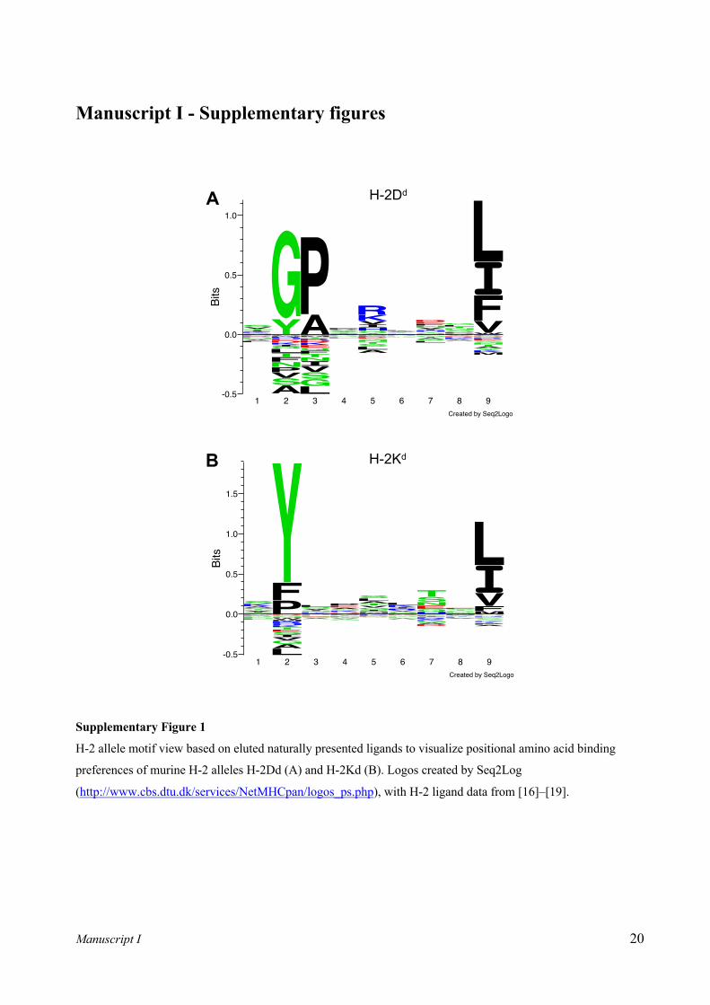
Manuscript I 20
Manuscript I - Supplementary figures
Supplementary Figure 1
H-2 allele motif view based on eluted naturally presented ligands to visualize positional amino acid binding
preferences of murine H-2 alleles H-2Dd (A) and H-2Kd (B). Logos created by Seq2Log
(http://www.cbs.dtu.dk/services/NetMHCpan/logos_ps.php), with H-2 ligand data from [16]–[19].
Created by Seq2Logo
0.0
0.5
1.0
1.5
-0.5
Bits
1
H
YIVTGAKS
CWP
M
D
E
RQFNL
2
PFYC
DQNWMRHKTESIVGAL
3
M
H
KNIVQL
WCPT
D
FEGSA
4
H
DQKSAEP
WCYFMRILNTVG
5
C
MNIGVTALS
YRFHKPDQ
6
HQGVRIKL
WCDME
P
Y
T
SFN
A
7
HQENST
WP
YLIFRVKGAD
8
H
YNDQTSE
WPCIMR
F
V
K
GLA
9
MFVILHC
D
NRGESYQKTPA
A
Created by Seq2Logo
0.0
0.5
1.0
-0.5
Bits
1
YQKITVS
WCPMH
F
DREN
GAL
2
YGW
CMRHEDKIQLTFNPVSA
3
APWC
H
MYRDKEQFTNIVSGL
4
Q
ITASVPE
WMHFN
K
R
G
D
L
5
YQHIVKRCWM
PFDEGNTSLA
6
HF
Q
K
TVIL
WMRNDS
E
G
A
7
W
QYHIVNFED
CMP
K
GTASL
8
YHFQSNTLG
CMWPI
VDRKEA
9
WVFILH
CDQRPESGNTAYKM
B
H-2Dd
H-2Kd

Manuscript I 21
Supplementary Figure 2
FACS gating strategy, representative pH-2 multimer stainings with tetramers (A), or DNA barcode labeled
multimers (B).
SSC-A
CD3-FITC
SSC-A
CD8-BV480
CD8-BV480
Multimer-PE
SSC-A
FSC-A
FSC-H
FSC-A
SSC-A
Live/dead-APC-CY7
A
B
SSC-A
CD3-FITC
SSC-A
CD8-BV480
CD8-BV480
Multimer-PE
SSC-A
FSC-A
FSC-H
FSC-A
SSC-A
Live/dead-APC-CY7
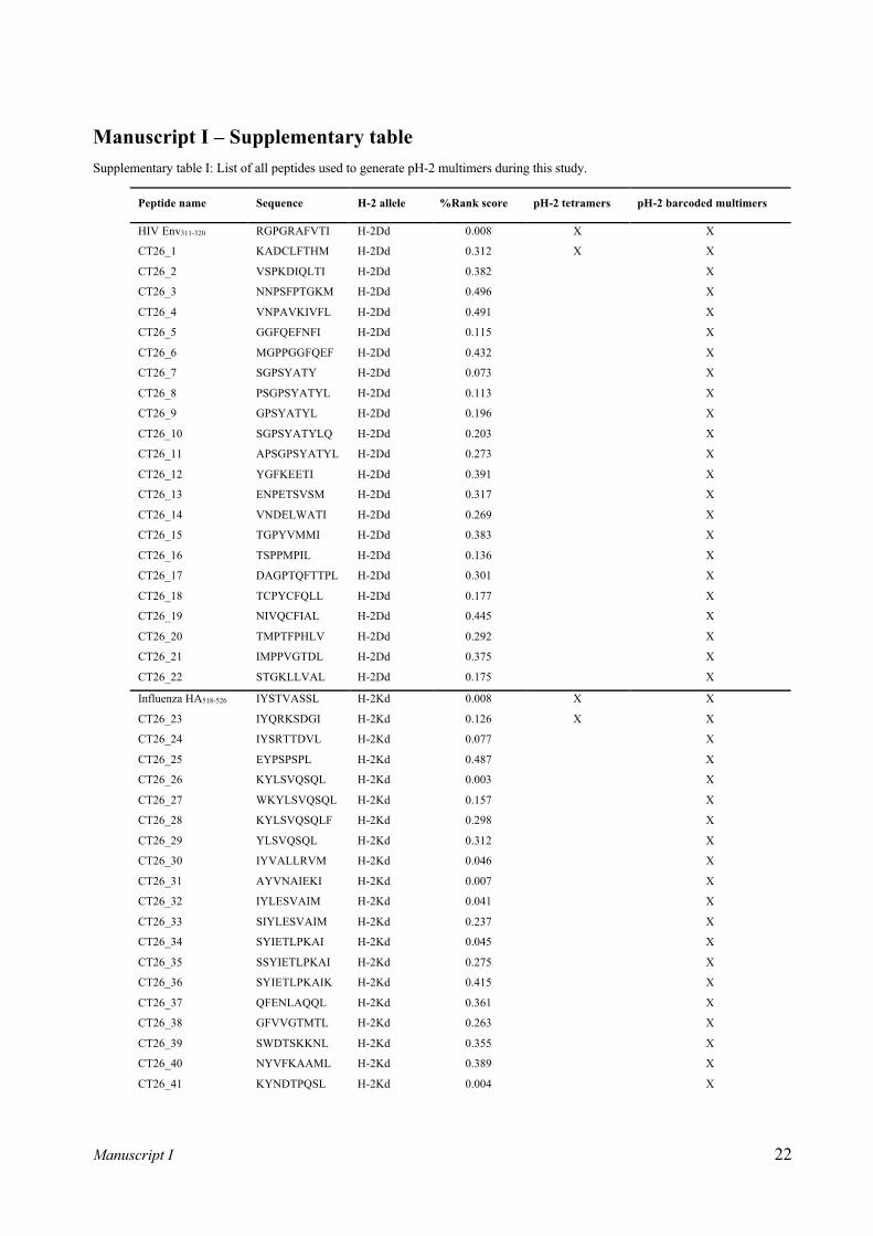
Manuscript I 22
Manuscript I – Supplementary table Supplementary table I: List of all peptides used to generate pH-2 multimers during this study.
Peptide name Sequence H-2 allele %Rank score pH-2 tetramers pH-2 barcoded multimers
HIV Env311-320 RGPGRAFVTI H-2Dd 0.008 X X
CT26_1 KADCLFTHM H-2Dd 0.312 X X
CT26_2 VSPKDIQLTI H-2Dd 0.382
X
CT26_3 NNPSFPTGKM H-2Dd 0.496
X
CT26_4 VNPAVKIVFL H-2Dd 0.491
X
CT26_5 GGFQEFNFI H-2Dd 0.115
X
CT26_6 MGPPGGFQEF H-2Dd 0.432
X
CT26_7 SGPSYATY H-2Dd 0.073
X
CT26_8 PSGPSYATYL H-2Dd 0.113
X
CT26_9 GPSYATYL H-2Dd 0.196
X
CT26_10 SGPSYATYLQ H-2Dd 0.203
X
CT26_11 APSGPSYATYL H-2Dd 0.273
X
CT26_12 YGFKEETI H-2Dd 0.391
X
CT26_13 ENPETSVSM H-2Dd 0.317
X
CT26_14 VNDELWATI H-2Dd 0.269
X
CT26_15 TGPYVMMI H-2Dd 0.383
X
CT26_16 TSPPMPIL H-2Dd 0.136
X
CT26_17 DAGPTQFTTPL H-2Dd 0.301
X
CT26_18 TCPYCFQLL H-2Dd 0.177
X
CT26_19 NIVQCFIAL H-2Dd 0.445
X
CT26_20 TMPTFPHLV H-2Dd 0.292
X
CT26_21 IMPPVGTDL H-2Dd 0.375
X
CT26_22 STGKLLVAL H-2Dd 0.175
X
Influenza HA518-526 IYSTVASSL H-2Kd 0.008 X X
CT26_23 IYQRKSDGI H-2Kd 0.126 X X
CT26_24 IYSRTTDVL H-2Kd 0.077
X
CT26_25 EYPSPSPL H-2Kd 0.487
X
CT26_26 KYLSVQSQL H-2Kd 0.003
X
CT26_27 WKYLSVQSQL H-2Kd 0.157
X
CT26_28 KYLSVQSQLF H-2Kd 0.298
X
CT26_29 YLSVQSQL H-2Kd 0.312
X
CT26_30 IYVALLRVM H-2Kd 0.046
X
CT26_31 AYVNAIEKI H-2Kd 0.007
X
CT26_32 IYLESVAIM H-2Kd 0.041
X
CT26_33 SIYLESVAIM H-2Kd 0.237
X
CT26_34 SYIETLPKAI H-2Kd 0.045
X
CT26_35 SSYIETLPKAI H-2Kd 0.275
X
CT26_36 SYIETLPKAIK H-2Kd 0.415
X
CT26_37 QFENLAQQL H-2Kd 0.361
X
CT26_38 GFVVGTMTL H-2Kd 0.263
X
CT26_39 SWDTSKKNL H-2Kd 0.355
X
CT26_40 NYVFKAAML H-2Kd 0.389
X
CT26_41 KYNDTPQSL H-2Kd 0.004
X
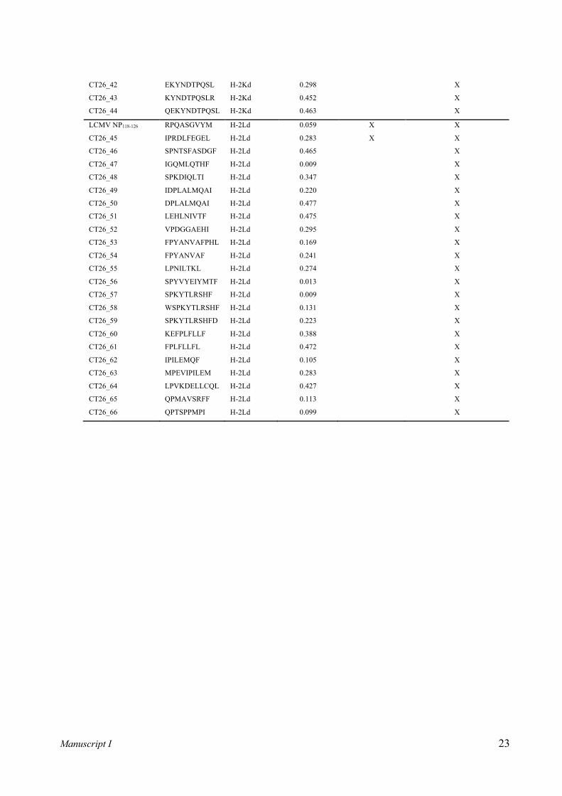
Manuscript I 23
CT26_42 EKYNDTPQSL H-2Kd 0.298
X
CT26_43 KYNDTPQSLR H-2Kd 0.452
X
CT26_44 QEKYNDTPQSL H-2Kd 0.463
X
LCMV NP118-126 RPQASGVYM H-2Ld 0.059 X X
CT26_45 IPRDLFEGEL H-2Ld 0.283 X X
CT26_46 SPNTSFASDGF H-2Ld 0.465
X
CT26_47 IGQMLQTHF H-2Ld 0.009
X
CT26_48 SPKDIQLTI H-2Ld 0.347
X
CT26_49 IDPLALMQAI H-2Ld 0.220
X
CT26_50 DPLALMQAI H-2Ld 0.477
X
CT26_51 LEHLNIVTF H-2Ld 0.475
X
CT26_52 VPDGGAEHI H-2Ld 0.295
X
CT26_53 FPYANVAFPHL H-2Ld 0.169
X
CT26_54 FPYANVAF H-2Ld 0.241
X
CT26_55 LPNILTKL H-2Ld 0.274
X
CT26_56 SPYVYEIYMTF H-2Ld 0.013
X
CT26_57 SPKYTLRSHF H-2Ld 0.009
X
CT26_58 WSPKYTLRSHF H-2Ld 0.131
X
CT26_59 SPKYTLRSHFD H-2Ld 0.223
X
CT26_60 KEFPLFLLF H-2Ld 0.388
X
CT26_61 FPLFLLFL H-2Ld 0.472
X
CT26_62 IPILEMQF H-2Ld 0.105
X
CT26_63 MPEVIPILEM H-2Ld 0.283
X
CT26_64 LPVKDELLCQL H-2Ld 0.427
X
CT26_65 QPMAVSRFF H-2Ld 0.113
X
CT26_66 QPTSPPMPI H-2Ld 0.099
X

46
47
3. MANUSCRIPT II
DNA based neo-epitope vaccination induces tumor control in the CT26 syngeneic mouse model
Preclinical and clinical evidence support the notion of mutation-derived neo-epitopes as
crucial targets for immunotherapy in cancer. In manuscript II, we established a vaccination-
based platform in the syngeneic CT26 model, which is suitable for neo-epitope screening
and detection. Here, multi-neo-epitope vaccination with DNA plasmid was found to be more
anti-tumor effective than neo-peptides + TLR3 ligand poly IC. This was mirrored by a strong
induction of CD8+ T cells by neo-epitope DNA vaccination. The framework presented here,
will provide valuable feedback to neo-epitope prediction and selection algorithms.
The research presented in manuscript II represent current results from an ongoing study.
We intend to extend the manuscript with further analyses to dissect immunogenicity of neo-
epitopes in the CT26 syngeneic mouse model. Specifically, future analyses will address which
of the neo-epitopes are driving anti-tumor effects and whether the therapeutic effect is
mediated by CD4+ or CD8+ T cells or both via depletion studies.

48

Manuscript II 1
DNA based neo-epitope vaccination induces tumor control in the CT26 syngeneic mouse model Authors: Nadia Viborg1,2, Marina Barrio Calvo2, Stine Friis2, Thomas Trolle2, Christian Skjødt Hansen2, Jens Kringelum2, Sine Reker Hadrup1 and Birgitte Rønø2
Affiliations: 1 Department of Health Technology, Technical University of Denmark, Lyngby, Denmark 2 Evaxion Biotech, Copenhagen, Denmark Corresponding author: Birgitte Rønø, PhD, Senior Director, Cancer Immunotherapy, Evaxion Biotech, Bredgade 34E, 1260 Copenhagen K, Phone: +45 31207309, Email: [email protected] Financial support: This research was funded in part by the Industrial Researcher programme, Innovation Fund Denmark Number of figures: 4 Number of tables: 1 Number of supplementary figures: 2
Manuscript II 2
Abstract
Cancer vaccines can be emplyed to engage T cell mediated immune recognition and elimination of
tumors. Despite extensive past attempts, it has proven challenging to mediate substantial anti-tumor
activity in the clinic with cancer vaccines. Recently, several clinical and preclinical discoveries
position mutation derived neo-epitopes as important targets for effective immune responses against
tumors. As such, personalized cancer vaccines with neo-epitopes delivered as synthetic peptides or
RNA have reached the clinic with promising preliminary results and induction of neo-antigen
specific T cells. However, the majority of in silico predicted neo-epitopes are not immunogenic, and
we are yet to uncover the rules that govern neo-epitope immunogenicity. Research in murine
preclinical models continuously increased our understanding of neo-epitopes and their potential
therapeutic applicability. In this project, we set out to test DNA and peptide delivery platforms for
their ability to induce neo-epitopes immunogenicity and anti-tumor effects in the syngeneic CT26
model. Our data demonstrate that prophylactic neo-epitope DNA vaccination confers anti-tumor
immunity in the CT26 tumor model, whereas neo-peptide vaccination adjuvanted by poly IC does
not. This is mirrored by a preferential induction of neo-epitope specific CD8+ T cells by DNA
vaccination, which is skewed towards CD4+ T cells with neo-peptide vaccination. DNA vaccination
confers a versatile platform that allows encoding of multiple neo-epitopes in a single formulation, a
feasible strategy for robust neo-epitope vaccination, and thus a pipeline to validate neo-epitopes for
immunogenicity and therapeutic applicability.
Manuscript II 3
Introduction
T cells are acknowledged to play an essential role in immunological recognition and rejection of
tumors [1]. Mutation derived T cell epitopes, also known as neo-epitopes, are being thoroughly
explored as targets in cancer as they differentiate aberrant tumor tissue from the healthy self. Neo-
epitope specific T cells have been reported in peripheral blood and tumor infiltrating lymphocytes
(TILs) in patient with various cancers [2]–[6]. Furthermore, durable response to checkpoint
inhibitor (CPI) therapy and good cancer prognosis have been related to tumor mutational burden or
neo-epitope load and intratumoral presence of T cells [7]–[12]. This underlines the relevance of
understanding and further investigating neo-epitope expression and subsequent T cell recognition in
cancer therapy and immune monitoring.
With tumor-restricted expression, neo-epitopes are theorized as ideal therapeutic cancer targets
minimally affected by immune tolerance and with limited risk of autoimmune adverse events.
Therapeutic cancer vaccination with patient specific neo-epitopes offers a promising strategy to
harness immune responses against a tumor. Such personalized strategies have recently been pursued
in clinical trials with encouraging results when delivering the patient specific neo-epitopes loaded
on autologous dendritic cells [13], encoded by RNA [14], [15] or neo-peptide pools adjuvanted by
polyinosinic-polycytidylic acid, and poly-L-lysine (poly-ICLC) [16], [17]. In each clinical trial,
several neo-epitopes were able to enhance existing or induce de novo T cells responses, confirming
immunogenicity of neo-epitopes in humans. Interestingly, the majority of the T cell responses
observed in these clinical trial with RNA and peptide delivery were MHC class II restricted and thus
recognized by CD4+ T cells.
Preclinical research in murine models have contributed to our understanding of the dynamic
interplay between T cells and neo-epitopes. There are several neo-epitope descriptions of relevance
in therapeutic syngeneic tumor models based on common inbred strains C57BL/6 and BALB/c such
as CT26 [18], [19], MC38 [20], [21], and B16-F10 [22]. These syngeneic tumor models represent
different tissue origins and harbor diverse levels of immunogenicity and sensitivity to
immunotherapy [23]. Various vaccination strategies have been employed to induce neo-epitope
specific immunity in these syngeneic platforms of which some provide tumor growth control. One
approach is synthetic neo-peptides adjuvanted by the Toll-like receptor (TLR) 3 agonist
polyinosinic-polycytidylic acid (poly IC) or CpG, shown to induce anti-tumor immunity and growth
delay in MC38 and B16-F10 models [20]–[22]. Another is delivery of neo-epitope encoding RNA,
Manuscript II 4
with inherent self-adjuvating properties via TLR7 stimulation tested in CT26, B16-F10, and 4T1
models [19], [24]. Recent reports further position DNA based neo-epitope immunization as a
relevant approach with induced immunity and anti-tumor effect in CT26, B16-F10, MC38, and
Lewis lung carcinoma models [25]–[28]. In these studies, neo-epitopes were encoded by DNA
plasmids or an adenoviral vector, delivered either alone or in combination with CPIs.
DNA vaccination harbors self-adjuvating effects via the innate DNA sensing machinery of
mammalian cells. This directs the immune response towards Th1-like immunity which is preferable
in therapies for cancer and viral infections [29]–[31]. Antigens delivered in DNA format have direct
access to the MHC class I processing and presentation pathway in transfected cells, which is
essential for induction of cytotoxic CD8+ T cells. Exogenous antigen delivery via peptides or whole
protein will be presented by MHC class II on APCs and require cross presentation to reach MHC
class I and CD8+ T cell priming [32]. Cross presentation can be promoted by interferons (IFN) in
the surrounding milieu during antigen uptake by antigen presenting cells, and has been shown to be
more efficient for synthetic long peptides than whole proteins [33], [34]. Delivery systems and
adjuvants are commonly employed to facilitate efficient vaccination with various antigen formats.
For DNA vaccination, delivery systems can protect against potential degradation of the DNA
plasmid after injection and increase transfection efficiency [35]. Nonionic block co-polymers form
micelle-like structures with DNA and enhance gene delivery to several tissues [36], [37]. Block co-
polymers potentially augment immunogenicity of DNA via directing immune responses on the
Th1/Th2 axis based on their chemical components/structure [38]. Synthetic peptides have low
inherent immunogenicity on their own, why there is a need to co-formulate with adjuvants or
carriers. A well described adjuvant for peptide delivery is poly IC, a double stranded RNA analogue
and a facilitator of Th1-like adaptive immunity [39], [40] which has been described to induce anti-
tumor immune responses [41], [42].
In this study, we set out to investigate immunogenicity and anti-tumor efficacy of in silico predicted
neo-epitopes in the CT26 tumor model. Our aim is to obtain a drug development-like preclinical
platform suitable for screening the large putative neo-epitope libraries resulting from mutational
mapping and in-silico based prediction algorithms. Via this approach, we wish to expand our
understanding of what characterizes a good neo-epitope with therapeutic applicability, which will
be fed back to the neo-epitope prediction and selection algorithm. Here, we have compared two
Manuscript II 5
delivery modalities of a set of CT26 neo-epitopes; neo-plasmid DNA vaccination with and without
block co-polymer facilitated delivery, and neo-peptide vaccination adjuvanted by poly IC.
Manuscript II 6
Results
Prophylactic vaccination with CT26 neo-epitope DNA and block co-polymer inhibits tumor
growth and shows higher immunogenicity than neo-peptides + poly IC
We decided on five neo-epitopes to investigate in immunization studies based on mapping of
nonsynonymous somatic mutations in the CT26 tumor cell line. The neo-epitopes selected all span
a somatic mutation and encode all together the five peptides with the best in silico predicted affinity
to MHC class II (H-2-IAd). The expression of the five peptides was also confirmed by RNA
sequencing. Each neo-epitope sequence contains ³ 1 predicted strong binder for H-2d class I and
class II alleles, thus is potentially able to induce both CD4+ and CD8+ T cell responses as it is
reported to be important in murine tumor models [19], [43].
To find the superior delivery platform for our CT26 neo-epitopes, we initially compared delivery as
DNA encoding neo-epitopes or neo-peptides. Plasmid DNA encoding the five CT26 neo-epitope
sequences (from here on: neo-pentatope DNA) was formulated with nonionic block co-polymer
Lutrol to possibly enhance longevity of the plasmid DNA after injection and thereby increase antigen
expression and exposure. This approach was benchmarked to delivery of CT26 neo-peptides
formulated with poly IC. To overcome the inherent challenge of inducing a potent CD8+ T cell
response with peptide-based vaccination, we set out to mimic the course of viral infections by
exposing the mice to high-dose neo-peptides + poly IC. This set up is inspired by previous reports
of successful cross presentation and generation of antigen specific CD8+ T cells by cluster
vaccination with peptide antigens [44]. We have previously demonstrated that this cluster-priming
approach of high dose neo-peptide delivery is more immunogenic and induce more functional T
cells compared to conventional prime-boost and lower peptide doses (supplementary figure S1).
Additionally, we have across several previous studies not observed anti-tumor effects in the CT26
tumor model of poly IC adjuvant delivered alone (data not shown).
In a small prophylactic setup we compared the induced T cell responses and anti-tumor effect of
intramuscularly (i.m) immunized BALB/c mice with 100 µg of neo-pentatope DNA + Lutrol (n =
5) or Lutrol only (n = 4), with the responses obtained in mice immunized intraperitoneally (i.p.) with
50 µg of each neo-peptide + 40 µg of poly IC (n = 5).
Manuscript II 7
The mice in the neo-peptide group underwent cluster priming vaccination schedule consisting of
immunizations on four consecutive days followed by weekly boosts. The mice in the neo-pentatope
DNA group received weekly immunizations over the course of the experiment. Three to four weeks
after priming, mice were inoculated with 5 x 10⁵ CT26 tumor cells s.c. in the right flank (Fig. 1A).
The vaccination approaches were validated for anti-tumor effect by monitoring tumor growth until
majority of tumors in the control group reached the maximum allowed size (18 days after tumor cell
inoculation). Mice immunized with neo-pentatope DNA + Lutrol developed considerably smaller or
no tumors compared to mice immunized with Lutrol (Fig. 1B). Interestingly, immunization with
neo-peptides + poly IC conferred no anti-tumor effect, and these mice developed tumor to the same
extent as the control group. To explore if the differences in anti-tumor efficacy between the two neo-
epitope based deliveries could be explained by their ability to induce CT26 epitope recognizing
CD8+ T cells, we exploited the MHC tetramer staining methodology. PE and APC fluorochrome
conjugated MHC class I tetramers were loaded with a confirmed H-2Kd restricted minimal epitope
(KFKASRASI) derived from the C22 neo-epitope (present both in the DNA and peptide based CT26
vaccines) and added to tail vein whole blood in conjunction with anti-CD3 and anti-CD8 antibodies.
In samples collected two weeks after priming we observed high frequent tetramer-specific CD8+ T
cells in mice immunized with neo-pentatope DNA + Lutrol compared to neo-peptides + poly IC and
Lutrol only groups (Fig. 1D). Epitope recognition is an important step towards anti-tumor immunity,
however, to induce cancer cell killing a functional response is required. To compare the functional
T cell responses induced by the two delivery methodologies, splenocytes from all groups were re-
stimulated with immunization-relevant neo-peptide pool (five 27-mers) for IFNg enzyme linked
immunospot (ELISPOT) assay. By ELISPOT we observed significantly more IFNg spot forming
units (SFUs) in mice immunized with neo-pentatope DNA + Lutrol than neo-peptides + poly IC
(Fig. 1C). To further interrogate T cell immunogenicity, splenocytes were re-stimulated with the
immunization-relevant neo-peptide pool for intracellular cytokine staining (ICS) assay to identify
IFNg and tumor necrosis factor (TNF)a producing CD4+ and CD8+ T cells. Neo-pentaope DNA +
Lutrol immunization resulted in high frequencies of single- and double-cytokine producing CD8+
T cells and in lower frequencies of cytokine producing CD4+ T cells (Fig. 1E). Neo-peptides + poly
IC immunization led to convincing frequencies of IFNg cytokine producing CD4+ T cells but less
double-cytokine producing CD4+ T cells. With neo-peptide immunization, cytokine producing
CD8+ T cells could not be measured after peptide stimulation. Based on these results, neo-pentatope
DNA + Lutrol offers a more immunogenic and specific CD8+ T cell inducing platform for neo-
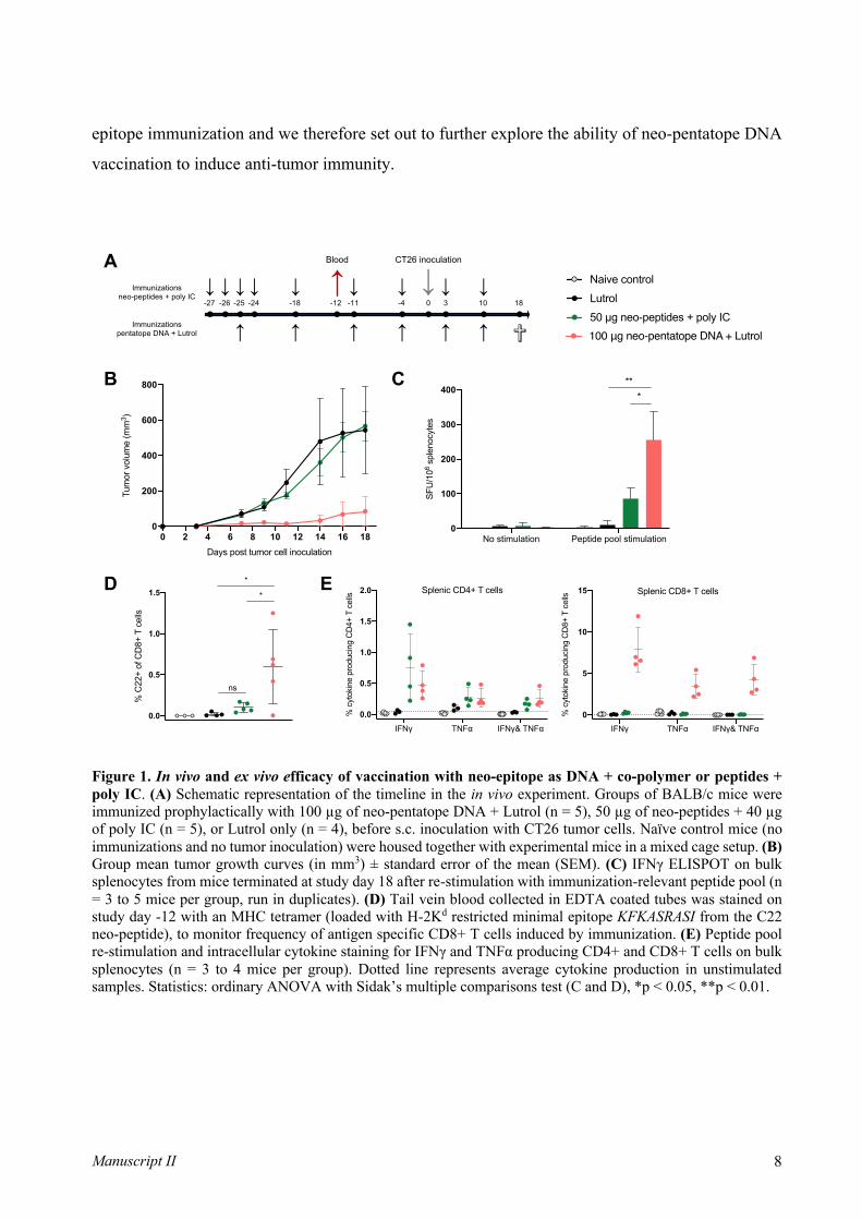
Manuscript II 8
epitope immunization and we therefore set out to further explore the ability of neo-pentatope DNA
vaccination to induce anti-tumor immunity.
Figure 1. In vivo and ex vivo efficacy of vaccination with neo-epitope as DNA + co-polymer or peptides + poly IC. (A) Schematic representation of the timeline in the in vivo experiment. Groups of BALB/c mice were immunized prophylactically with 100 µg of neo-pentatope DNA + Lutrol (n = 5), 50 µg of neo-peptides + 40 µg of poly IC (n = 5), or Lutrol only (n = 4), before s.c. inoculation with CT26 tumor cells. Naïve control mice (no immunizations and no tumor inoculation) were housed together with experimental mice in a mixed cage setup. (B) Group mean tumor growth curves (in mm3) ± standard error of the mean (SEM). (C) IFNγ ELISPOT on bulk splenocytes from mice terminated at study day 18 after re-stimulation with immunization-relevant peptide pool (n = 3 to 5 mice per group, run in duplicates). (D) Tail vein blood collected in EDTA coated tubes was stained on study day -12 with an MHC tetramer (loaded with H-2Kd restricted minimal epitope KFKASRASI from the C22 neo-peptide), to monitor frequency of antigen specific CD8+ T cells induced by immunization. (E) Peptide pool re-stimulation and intracellular cytokine staining for IFNγ and TNFα producing CD4+ and CD8+ T cells on bulk splenocytes (n = 3 to 4 mice per group). Dotted line represents average cytokine production in unstimulated samples. Statistics: ordinary ANOVA with Sidak’s multiple comparisons test (C and D), *p < 0.05, **p < 0.01.
A
B C
ED
-27 -24 181030-4-11-12-18-26 -25
CT26 inoculationBlood
Immunizationsneo-peptides + poly IC
Immunizationspentatope DNA + Lutrol
↓↓ ↓
✞● ●●●●●●● ● ● ● ●
↓ ↓ ↓ ↓↓↓
↓ ↓ ↓ ↓ ↓ ↓
↓↓
100 μg neo-pentatope DNA + Lutrol
Lutrol
50 µg neo-peptides + poly IC
Naive control
IFNγ TNFα IFNγ& TNFα
0
5
10
15
% c
ytok
ine
prod
ucin
g C
D8+
T c
ells Splenic CD8+ T cells
IFNγ TNFα IFNγ& TNFα
0.0
0.5
1.0
1.5
2.0
% c
ytok
ine
prod
ucin
g C
D4+
T c
ells Splenic CD4+ T cells
No stimulation Peptide pool stimulation0
100
200
300
400
SFU
/106
sple
nocy
tes
***
0 2 4 6 8 10 12 14 16 180
200
400
600
800
Days post tumor cell inoculation
Tum
or v
olum
e (m
m3 )
100 μg neo-pentatope DNA + Lutrol
Lutrol
50 µg neo-peptides + poly IC
Naive control
0.0
0.5
1.0
1.5
% C
22+
of C
D8+
T c
ells
*
ns
*
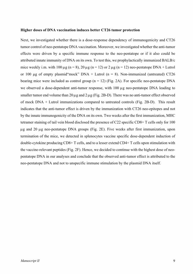
Manuscript II 9
Higher doses of DNA vaccination induces better CT26 tumor protection
Next, we investigated whether there is a dose-response dependency of immunogenicity and CT26
tumor control of neo-pentatope DNA vaccination. Moreover, we investigated whether the anti-tumor
effects were driven by a specific immune response to the neo-pentatope or if it also could be
attributed innate immunity of DNA on its own. To test this, we prophylactically immunized BALB/c
mice weekly i.m. with 100 µg (n = 8), 20 µg (n = 12) or 2 µg (n = 12) neo-pentatope DNA + Lutrol
or 100 µg of empty plasmid“mock” DNA + Lutrol (n = 8). Non-immunized (untreated) CT26
bearing mice were included as control group (n = 12) (Fig. 2A). For specific neo-pentatope DNA
we observed a dose-dependent anti-tumor response, with 100 µg neo-pentatope DNA leading to
smaller tumor end volume than 20 µg and 2 µg (Fig. 2B-D). There was no anti-tumor effect observed
of mock DNA + Lutrol immunizations compared to untreated controls (Fig. 2B-D). This result
indicates that the anti-tumor effect is driven by the immunization with CT26 neo-epitopes and not
by the innate immunogenicity of the DNA on its own. Two weeks after the first immunization, MHC
tetramer staining of tail vein blood disclosed the presence of C22 specific CD8+ T cells only for 100
µg and 20 µg neo-pentatope DNA groups (Fig. 2E). Five weeks after first immunization, upon
termination of the mice, we detected in splenocytes vaccine specific dose-dependent induction of
double-cytokine producing CD8+ T cells, and to a lesser extend CD4+ T cells upon stimulation with
the vaccine-relevant peptides (Fig. 2F). Hence, we decided to continue with the highest dose of neo-
pentatope DNA in our analyses and conclude that the observed anti-tumor effect is attributed to the
neo-pentatope DNA and not to unspecific immune stimulation by the plasmid DNA itself.
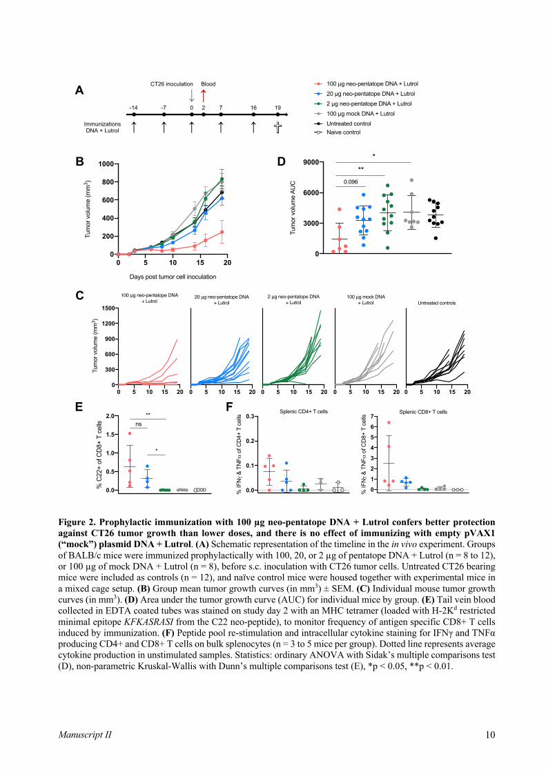
Manuscript II 10
Figure 2. Prophylactic immunization with 100 µg neo-pentatope DNA + Lutrol confers better protection against CT26 tumor growth than lower doses, and there is no effect of immunizing with empty pVAX1 (“mock”) plasmid DNA + Lutrol. (A) Schematic representation of the timeline in the in vivo experiment. Groups of BALB/c mice were immunized prophylactically with 100, 20, or 2 µg of pentatope DNA + Lutrol (n = 8 to 12), or 100 µg of mock DNA + Lutrol (n = 8), before s.c. inoculation with CT26 tumor cells. Untreated CT26 bearing mice were included as controls (n = 12), and naïve control mice were housed together with experimental mice in a mixed cage setup. (B) Group mean tumor growth curves (in mm3) ± SEM. (C) Individual mouse tumor growth curves (in mm3). (D) Area under the tumor growth curve (AUC) for individual mice by group. (E) Tail vein blood collected in EDTA coated tubes was stained on study day 2 with an MHC tetramer (loaded with H-2Kd restricted minimal epitope KFKASRASI from the C22 neo-peptide), to monitor frequency of antigen specific CD8+ T cells induced by immunization. (F) Peptide pool re-stimulation and intracellular cytokine staining for IFNγ and TNFα producing CD4+ and CD8+ T cells on bulk splenocytes (n = 3 to 5 mice per group). Dotted line represents average cytokine production in unstimulated samples. Statistics: ordinary ANOVA with Sidak’s multiple comparisons test (D), non-parametric Kruskal-Wallis with Dunn’s multiple comparisons test (E), *p < 0.05, **p < 0.01.
A
B D
C
E
1916720-7-14
CT26 inoculation Blood
ImmunizationsDNA + Lutrol
● ● ● ●
✞● ●●↓ ↓ ↓ ↓
↓ ↓
↓
2 µg neo-pentatope DNA + Lutrol
100 µg neo-pentatope DNA + Lutrol
20 µg neo-pentatope DNA + Lutrol
100 µg mock DNA + Lutrol
Untreated control Naive control
0.0
0.1
0.2
0.3
% IF
Nγ
& T
NFα
of C
D4+
T c
ells
Splenic CD4+ T cells
0
1
2
3
4
5
6
7
% IF
Nγ
& T
NFα
of C
D8+
T c
ells
Splenic CD8+ T cells
0.0
0.5
1.0
1.5
2.0
% C
22+
of C
D8+
T c
ells ns
**
*
F
0 5 10 15 200
200
400
600
800
1000
Days post tumor cell inoculation
Tum
or v
olum
e (m
m3 )
0
3000
6000
9000
Tum
or v
olum
e A
UC
*
0.096
**
0 5 10 15 200
300
600
900
1200
1500
Tum
or v
olum
e (m
m3 )
100 μg neo-pentatope DNA + Lutrol
0 5 10 15 20
20 μg neo-pentatope DNA + Lutrol
0 5 10 15 20
2 μg neo-pentatope DNA + Lutrol
0 5 10 15 20
100 μg mock DNA + Lutrol
0 5 10 15 20
Untreated controls

Manuscript II 11
Clinical grade co-polymer Kolliphor performs similarly to Lutrol and increases early onset
immunogenicity compared to naked DNA
To increase potential clinical translatability of the observed anti-tumor effect of neo-pentatope DNA
formulated with a co-polymer, we tested the clinical grade block co-polymer Kolliphor. Kolliphor´s
ability to facilitate neo-pentatope DNA delivery in the CT26 tumor model was compared to the anti-
tumor effect induced by the polymer Lutrol and by naked neo-pentatope on its own. We
prophylactically immunized BALB/c mice weekly i.m. with 100 µg neo-pentatope DNA + Lutrol
or Kolliphor, 100 µg of naked neo-pentatope DNA, 100 µg of mock DNA + Kolliphor, or Kolliphor
only (n = 13 per group) (Fig. 3A). The majority of mice immunized with naked neo-pentatope DNA
and neo-pentatope DNA + Lutrol or Kolliphor showed similar substantial tumor growth control,
compared to mock DNA and Kolliphor immunized mice (Fig. 3B-D). Two weeks after the first
immunization, a tail vein blood MHC tetramer staining revealed presence of C22 specific CD8+ T
cells in the groups immunized with naked neo-pentatope DNA and neo-pentatope DNA + Lutrol or
Kolliphor, albeit the level was significantly lower for naked neo-pentatope DNA at this timepoint
(Fig. 3E). This is indicative of polymer facilitated increase of plasmid immunogenicity by adjuvating
effects or prevention of degradation. Upon endpoint (21 days after tumor cell inoculation) we
stimulated splenocytes with peptide pool, and performed ICS assay to investigate differences in T
cell functionality between the groups. We detected similar specific induction of double-cytokine
producing CD8+ and CD4+ T cells by neo-pentatope DNA with and without co-polymers (Fig. 3F).
At endpoint high dose neo-pentatope DNA were thus equally immunogenic and anti-tumor efficient
with and without co-polymer facilitated delivery. Clinical grade co-polymer Kolliphor performed
similarly to Lutrol, and is therefore used for future experiments.

Manuscript II 12
Figure 3. Clinical-grade co-polymer Kolliphor performs similarly to Lutrol in tumor growth control. (A) Schematic representation of the timeline in the in vivo experiment. Groups of BALB/c mice were immunized prophylactically with 100 µg of pentatope DNA + Lutrol or Kolliphor (n = 13 per group), 100 µg of naked pentatope DNA (n = 13), 100 µg of mock DNA + Lutrol (n = 13), or Kolliphor only (n = 13), before s.c. inoculation with CT26 tumor cells. Naïve control mice were housed together with experimental mice in a mixed cage setup. (B) Group mean tumor growth curves (in mm3) ± SEM. (C) Individual mouse tumor growth curves (in mm3). (D) Area under the tumor growth curve (AUC) for individual mice by group. (E) Tail vein blood collected in EDTA coated tubes was stained on study day 1 with an MHC tetramer (loaded with H-2Kd restricted minimal epitope KFKASRASI from the C22 neo-peptide), to monitor frequency of antigen specific CD8+ T cells induced by immunization. (F) Peptide pool re-stimulation and intracellular cytokine staining for IFNγ and TNFα producing CD4+ and CD8+ T cells on bulk splenocytes (n = 4 to 6 mice per group). Dotted line represents average cytokine production in unstimulated samples. Statistics: non-parametric Kruskal-Wallis with Dunn’s multiple comparisons test (D), ordinary ANOVA with Sidak’s multiple comparisons test (E), *p < 0.05, **p < 0.01, ***p < 0.001, ****p < 0.0001.
A
B
C
E F
2114710-6-13
CT26 inoculation Blood
Immunizations
↓ ●● ● ● ● ● ●
↓ ↓
✞↓ ↓↓ ↓
0 2 4 6 8 10 12 14 16 18 20 220
100
200
300
400
500
600
100 µg naked neo-pentatope DNA
100 µg neo-pentatope DNA + Lutrol
100 µg neo-pentatope DNA + Kolliphor
100 µg mock DNA + Kolliphor
Kolliphor
Group mean ± SEM
Naive mice
D
0
1
2
3
4
% C
22+
of C
D8+
T c
ells
***
*
ns
0.0
0.1
0.2
0.3
% IF
Nγ
& T
NFα
of C
D4+
T c
ells
Splenic CD4+ T cells
0
1
2
3
% IF
Nγ
& T
NFα
of C
D8+
T c
ells
Splenic CD8+ T cells
0 7 14 210
200
400
600
800
1000
Tum
or v
olum
e (m
m3 )
100 μg neo-pentatope DNA + Lutrol
0 7 14 21
100 μg neo-pentatope DNA+ Kolliphor
0 7 14 21
100 μg mock DNA + Kolliphor
0 7 14 21
100 μg naked neo-pentatope DNA
0 7 14 21
Kolliphor
0 7 14 210
200
400
600
Days post tumor cell inoculation
Tum
or v
olum
e (m
m3 )
100 µg naked neo-pentatope DNA
100 µg neo-pentatope DNA + Lutrol
100 µg neo-pentatope DNA + Kolliphor
100 µg mock DNA + Kolliphor
KolliphorNaive mice
0
2000
4000
6000
8000
Tum
or v
olum
e A
UC
****
****

Manuscript II 13
Neo-pentatope DNA vaccinated mice retain long-term tumor growth control
For the study described in the previous paragraph a subset of mice, immunized with neo-pentatope
DNA with and without co-polymers, with small or no tumors were kept alive until 44 days after
CT26 cell inoculation. Majority of the mice remained tumor free until day 44 (data not shown). Tail
vein blood from these mice was MHC tetramer stained on study day 42. Here, we observed presence
of neo-peptide C22 specific CD8+ T cells at levels comparable to what observed in the early blood
staining (Fig. 3E and data not shown). This is indicative of long lasting anti-tumor effects of neo-
pentatope DNA immunization possibly via immunological memory.
Prophylactic vaccination outperforms therapeutic vaccination and two epitopes drive the
observed neo-pentaope immunogenicity
We next investigated the ability of the neo-pentatope DNA vaccine to induce tumor control in a
therapeutic setting, and compare it with the robust anti-tumor effect of the same vaccine
administered prophylactically. We immunized BALB/c mice weekly i.m. with 100 µg neo-pentatope
DNA + Kolliphor or 100 µg of naked neo-pentatope DNA either prophylactically or therapeutically
(n = 13 per group) relative to CT26 tumor cell inoculation (Fig. 4A). The prophylactic treatment
started two weeks prior tumor cell inoculation while the therapeutic treatment was initiated after
detection of palpable tumors. Mice injected with Kolliphor only, following the prophylactic
immunization schedule were included as a negative control (n = 13). Similarly to Fig. 3B
prophylactic immunization with neo-pentatope DNA ± Kolliphor resulted in tumor growth control
for the majority of mice in both groups (Fig. 4B-D). Therapeutic immunization, however, offered
no anti-tumor effect compared to Kolliphor only controls, apparent by tumor growth curves (Fig.
4B-D) and tumor volume AUC (Fig. 4C). Immune monitoring via tail vein blood MHC tetramer
staining was carried out three weeks after the first prophylactic immunization and 5 days after the
first therapeutic immunization. Tetramer staining revealed presence of C22 specific CD8+ T cells
after prophylactic immunization, again the level was significantly lower for naked neo-pentatope
DNA than neo-pentatope DNA + Kolliphor group (Fig. 4E). To dissect the contribution of CD4+
and CD8+ T cells to neo-neo-pentatope immunogenicity, splenocytes were subjected to peptide pool
re-stimulation and ICS. At endpoint, we detected similar specific induction of double-cytokine
producing CD4+ T cells across prophylactic and therapeutic groups (Fig. 4F), albeit at a slightly
higher frequency for prophylactic immunization with neo-pentatope DNA + Kolliphor. The same
tendency was observed for CD8+ T cells, where prophylactic immunization with naked neo-

Manuscript II 14
pentatope DNA and neo-pentatope DNA + Kolliphor induced more double-cytokine producers than
therapeutic immunization. Lastly, splenocytes from prophylactic immunized groups were re-
stimulated with neo-pentatope corresponding peptide pool or individual neo-peptides (C22, C23,
C25, 30, C38) for IFNg ELISPOT to deconvolute immunogenicity. C22 and C23 neo-epitopes from
the neo-pentatope were the main drivers of observed immunogenicity (Fig. 4G), with a smaller
contribution from peptide C25. IFNg spot counts were similar for naked neo-pentatope DNA and
neo-pentatope DNA + Kolliphor groups.
A
B C
D
E
G
21144 90-7-14
CT26 inoculation Blood
Prophylacticimmunizations
7 1710
Therapeuticimmunizations
● ●●
↓ ↓↓ ↓● ●●● ● ● ●
✞↓
↓ ↓ ↓↓ ↓
↓↓
0 7 14 210
100
200
300
400
500
Days post tumor cell inoculation
Tum
or v
olum
e (m
m3 )
F
No stim
ulatio
n
C22 pe
ptide
C23 pe
ptide
C25 pe
ptide
C30 pe
ptide
C38 pe
ptide
Pentat
ope p
eptid
e poo
l0
100
200
300
400
500
600
SFU
/106
sple
nocy
tes
Naive control
Kolliphor + S16A Neovector (100 ug), prophylactic
Naked S16A Neovector (100 ug), prophylactic
Kolliphor only, prophylactic
0.0
0.1
0.2
0.3
0.4
% IF
Nγ
& T
NFα
of C
D4+
T c
ells
Splenic CD4+ T cells
0
1
2
3
4
5
% IF
Nγ
& T
NFα
of C
D8+
T c
ells
Splenic CD8+ T cells
Kolliphor + S16A Neovector (100 ug), therapeutic
Naked S16A Neovector (100 ug), therapeutic
Kolliphor + S16A Neovector (100 ug), prophylactic
Naked S16A Neovector (100 ug), prophylactic
Kolliphor only, prophylactic
Naive mice
0 7 14 210
200
400
600
800
1000
Tum
or v
olum
e (m
m3 )
100 μg neo-pentatope DNA + Kolliphor, therapeutic
0 7 14 21
100 μg naked neo-pentatope DNAtherapeutic
0 7 14 21
100 μg neo-pentatope DNA + Kolliphor, prophylactic
0 7 14 21
100 μg naked neo-pentatope DNAprophylactic
0 7 14 21
Kolliphorprophylactic
100 µg neo-pentatope DNA + Kolliphor
100 µg naked neo-pentatope DNA
100 µg neo-pentatope DNA + Kolliphor
100 µg naked neo-pentatope DNA
Kolliphor
Naive control
Therapeutic
Prophylactic
0
1500
3000
4500
Tum
or v
olum
e A
UC
nsns
**
**
0.0
0.5
1.0
1.5
2.0
2.5
3.0
% C
22+
of C
D8+
T c
ells
Manuscript II 15
Figure 4. CT26 anti-tumor effect is obtained only by prophylactic and not therapeutic immunization with neo-pentatope DNA. (A) Schematic representation of the timeline in the in vivo experiment. Groups of BALB/c mice were immunized prophylactically or therapeutically with 100 µg of neo-pentatope DNA ± Kolliphor (n = 13 per group), before s.c. inoculation with CT26 tumor cells. Naïve control mice (no immunizations and no tumor inoculation) were housed together with experimental mice in a mixed cage setup. (B) Group mean tumor growth curves (in mm3) ± SEM. (C) Area under the tumor growth curve (AUC) for individual mice by group. (D) Individual mouse tumor growth curves (in mm3). (E) Tail vein blood collected in EDTA coated tubes was stained on study day 9 with an MHC tetramer (loaded with H-2Kd restricted minimal epitope KFKASRASI from the C22 neo-peptide), to monitor frequency of antigen specific CD8+ T cells induced by immunization. (F) Peptide pool re-stimulation and intracellular cytokine staining for IFNγ and TNFα producing CD4+ and CD8+ T cells on bulk splenocytes (n = 4 to 6 mice per group). Dotted line represents average cytokine production in unstimulated samples. (G) IFNγ ELISPOT on bulk splenocytes from prophylactically immunized mice after re-stimulation with immunization-relevant peptide pool or individual peptides (n = 3 to 6 mice per group, run in duplicates). Statistics: non-parametric Kruskal-Wallis with Dunn’s multiple comparisons test (C), *p < 0.05, **p < 0.01.
Manuscript II 16
Discussion
In this study we demonstrate a comparision between vaccination with five CT26 tumor neo-epitopes
in the form of neo-plasmid DNA and neo-peptides. Our first observation was that prophylactic
vaccination with neo-epitope DNA and co-polymer Lutrol provided protection against CT26 tumor
challenge, whereas prophylactic neo-peptide + poly IC vaccination did not. This was flanked by
higher immunogenicity of neo-pentatope DNA vaccination as apparent by MHC tetramer staining
of peripheral blood and two ex vivo functional assays for cytokine production upon re-stimulation.
Interestingly, we observed by ICS that neo-pentatope DNA preferrentially induced cytokine
producing CD8+ T cells, whereas neo-peptides + poly IC induced cytokine producing CD4+ T cells
upon antigen re-exposure. We further found that prophylactic protection against CT26 tumor from
neo-pentatope DNA was dose-deependent, and similar high dose of mock DNA did not confer this
protection. Immunogenicity of the DNA delivered neo-epitopes was also dose-dependent, apparent
by MHC tetramer staining and ex vivo functional assays. Prophylactic neo-pentatope DNA
vaccination with and without clinical grade co-polymer Kolliphor obtained similar prophylactic
protection in the CT26 tumor model. Thus, co-polymer facilitated DNA delivery conferred similar
protection as obtained naked pentatope DNA, albeit MHC tetramer staining of blood samples
showed that Kolliphor increased early onset immunogenicity compared to naked DNA. Lastly, we
found that CT26 anti-tumor effects of neo-pentatope DNA vaccination were only obtained with a
prophylactic vaccination regimen, and not effective when vaccinations were initiated after tumor
onset. Ex vivo re-stimulation and assays for cytokine production showed that prophylactic and
therapeutic groups reached reached similar levels of vaccine induced immunogenicity, though
prophylactic groups at a slightly higher level of cytokine producing CD8+ T cells. We deconvoluted
the vaccine induced response via individual neo-peptide re-stimulation and ELISPOT, where we
observed that two of the five neo-epitopes drive the pentatope immunogenicity.
Including multiple neo-epitopes in a cancer vaccine is investigated in both mice and humans [14],
[25]–[27], [45]. The multi neo-epitope approach potentially induces several clones of tumor
recognizing T cells, and a broad recognition of tumor antigens is expected to have a greater
protective potential in cancer. An explanation for this, is that the breadth of the anti-tumor response
provides better protection in situations where tumor variants that express a certain mutation are
eliminated as a result of immune pressure. Additionally, since studies have shown that far from all
predicted neo-epitopes are immunogenic [46], including multiple possible hits improves chances of
Manuscript II 17
raising a T cell response at all. In our investigated pentatope vaccine, only two out of five neo-
epitopes were immunogenic. To investigate this further, our future studies will entail single neo-
epitope immunzations to examine immunogenic and therapeutic effects of single neo-epitope
delivery compared to the pentatope. With the current data, we can only speculate wheter the neo-
pentatope confers epitope competition, resulting in loss of immunogenicity of less dominant
epitopes.
The assembly of multiple epitopes in a vaccine is somewhat easier for DNA delivery compared to
synthetic peptide delivery. This is because DNA offers a versalite platform that allows encoding of
multiple neo-epitopes in a single formulation, whereas synthetic neo-peptides require indivual
production and purification of each putative epitope. Some synthetic neo-peptides will inherently
possess solubility issues, making them difficult or impossible to formulate in a vaccine without
precipitation.
Reports show, that the induction of both CD4+ and CD8+ T cell responses is important for cancer
immunotherapy [19], [43]. The 27-mer length neo-epitopes included in this study encompass
multiple predicted MHC class I and II ligands, potentially priming both CD4+ and CD8+ T cells.
As suggested by our results, neo-epitope DNA delivery offers enhanced CD8+ T cell reactivity than
neo-peptide + poly IC. When DNA is injected intramuscularly, local resident cells (e.g. muscle and
stroma) and APCs will be transfected (reference). DNA encoded antigens will then be produced
inside the cells and gain access to the MHC class I pathway for presentation to CD8+ T cells.
Peptides will naturally enter the exogenous pathway and MHC class II presentation to CD4+ T cells,
and require cross presentation to reach the MHC class I pathway. Intracellular components of the
innate DNA sensing machinery, e.g. stimulator of interferon genes (STING) and TANK binding
kinase 1 (TBK1) pathways, detect plasmid DNA [29], [30], [47]. This detection is crucial for
directing the adaptive immune response towards a Th1-like pathway, where CD4+ T cells support
the development of cytotoxic CD8+ T cells capable of tumor destruction.
Indeed, we are able to induce both poly-functional [48] CD4+ and CD8+ T cells with neo-pentatope
DNA vaccination, as apparent by the IFNg and TNFa double producers detected via ICS. To dissect
the importance of each subset of T cells for anti-tumor immunity, future neo-pentatope vaccination
studies will include antibody-mediated depletion of CD4+ and CD8+ T cells.
Manuscript II 18
In thus study, we were not able to obtain elimination of early established CT26 tumors with
therapeutic neo-pentatope DNA vaccination. Prophylactic immunization was however very
efficient. We hypothethize, that the therapeutic window in the model is too short, and that the neo-
pentatope DNA vaccination needs more time to establish T cell immunity [49]. Future analyses will
include multiple blood samplings for immune monitoring to uncover when specific adaptive
immunity kicks in. To overcome this, one could incorporate immune stimulatory sequences in the
plasmid vector, encoding chemokines, cytokines, or CpG elements. A potential synergistic effect
could also be obtained by combination of neo-pentatope DNA vaccination with CPI therapy [25],
which could mitigate the effects of an immunosuppressive TME. Another possible approach is to
use mechanical injection devices for increased DNA transfection, such as microneedles, genegun,
and electroporation [50], [51]. Moreover, we only tested one combination of five neo-epitopes in
the presented studies, and it is possible that other combinations of neo-epitopes would fare better in
the therapeutic vaccination setting.
Preclinical models somewhat lack direct translatability to the clinic, but nonetheless provide
important mechanistic lessons about the dynamic interplay between tumors and immunological
recognition. DNA based vaccination has not yet been a clinical success, despite promising data from
animal models [52]. However, multiple clinical trials involving personalized neo-epitope DNA
vaccination are currently recruiting or ongoing [53]. Most of these entail DNA delivery with
electroporation or combined with CPI therapy, and it will be interesting to follow these
developments.
Herein, we present a framework to screen libraries of neo-epitopes for therapeutic relevance in
syngeneic tumor models. This can be applied for neo-epitope discovery, and results can contiously
feed back back to improve neo-epitope prediction algorithms.
Manuscript II 19
Materials and Methods
Mice
6 to 8 weeks old BALB/c JrJ females were acquired from Janvier Labs (France). The mice were
acclimated for one week before initiation of experiments. The experiment was conducted under
license 2017-15-0201-01209 from the Danish Animal Experimentation Inspectorate in accordance
with the Danish Animal Experimentation Act (BEK nr. 12 of 7/01/2016), which is compliant with
the European directive (2010/63/EU).
Cell lines
BALB/c syngeneic colon cancer cell line CT26 (#CRL2638) was purchased from ATCC and
cultured in R10 medium prepared from RPMI (Gibco #72400-021) supplemented with 10% heat
inactivated fetal calf serum (FCS, Gibco # 10500-064)) at 37°C and 5% CO2 as per supplier’s
instructions.
Neo-epitope prediction and selection
Somatic mutations in the CT26 cell line were retrieved from previous work [18] and filtered to
only include nonsynonymous mutations. Reference source protein sequences were downloaded
using the Biopython Entrez module. For each somatic mutation, a 29-mer neoepitope sequence
was extracted from the corresponding source protein, with the mutated amino acid (AA) at
position 15. If the mutated AA was located within 15 AA of the ends of the source protein, a
shorter neoepitope sequence was extracted instead. If the extracted neo-epitope sequence was
shorter than 15 AAs it was discarded. Furthermore, if the annotated wildtype AA was not found at
the mutated position in the reference source protein, the neoepitope was also discarded.
For each neo-epitope sequence, NetMHCIIpan-3.1[54] was used to generate MHC class II binding
predictions for all overlapping 15mers towards the mouse MHC class II molecule H-2-IAd. The
15mer with the best NetMHCIIpan %rank score was selected as representative of the neo-epitope.
Neoepitopes with a gene expression >20 RPKM were ranked based on their best NetMHCIIpan
%rank score and the top 5 were selected. Finally, the N- and C-terminal AAs of the selected neo-
epitopes were truncated to convert the 29-mers to the final 27-mer neo-epitope sequences.
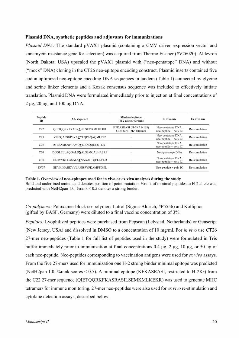
Manuscript II 20
Plasmid DNA, synthetic peptides and adjuvants for immunizations
Plasmid DNA: The standard pVAX1 plasmid (containing a CMV driven expression vector and
kanamycin resistance gene for selection) was acquired from Thermo Fischer (#V26020). Aldevron
(North Dakota, USA) upscaled the pVAX1 plasmid with (“neo-pentatope” DNA) and without
(“mock” DNA) cloning in the CT26 neo-epitope encoding construct. Plasmid inserts contained five
codon optimized neo-epitope encoding DNA sequences in tandem (Table 1) connected by glycine
and serine linker elements and a Kozak consensus sequence was included to effectively initiate
translation. Plasmid DNA were formulated immediately prior to injection at final concentrations of
2 μg, 20 μg, and 100 μg DNA.
Peptide ID AA sequence Minimal epitope
(H-2 allele, %rank) In vivo use Ex vivo use
C22 QIETQQRKFKASRASILSEMKMLKEKR KFKASRASI (H-2Kd, 0.168)
Used for H-2Kd tetramer Neo-pentatope DNA, neo-peptide + poly IC
Re-stimulation
C23 VILPQAPSGPSYATYLQPAQAQMLTPP - Neo-pentatope DNA, neo-peptide + poly IC
Re-stimulation
C25 DTLSAMSNPRAMQVLLQIQQGLQTLAT - Neo-pentatope DNA, neo-peptide + poly IC
Re-stimulation
C30 DGQLELLAQGALDNALSSMGALHALRP - Neo-pentatope DNA Re-stimulation
C38 RLHVVKLLASALSTNAAALTQELLVLD - Neo-pentatope DNA, neo-peptide + poly IC
Re-stimulation
EV07 GDVKIHAHKVVLANISPYFKAMFTGNL - Neo-peptide + poly IC Re-stimulation
Table 1. Overview of neo-epitopes used for in vivo or ex vivo analyses during the study Bold and underlined amino acid denotes position of point mutation. %rank of minimal peptides to H-2 allele was predicted with NetH2pan 1.0, %rank < 0.5 denotes a strong binder.
Co-polymers: Poloxamer block co-polymers Lutrol (Sigma-Aldrich, #P5556) and Kolliphor (gifted by BASF, Germany) were diluted to a final vaccine concentration of 3%.
Peptides: Lyophilized peptides were purchased from Pepscan (Lelystad, Netherlands) or Genscript
(New Jersey, USA) and dissolved in DMSO to a concentration of 10 mg/ml. For in vivo use CT26
27-mer neo-peptides (Table 1 for full list of peptides used in the study) were formulated in Tris
buffer immediately prior to immunization at final concentrations 0.4 μg, 2 μg, 10 μg, or 50 μg of
each neo-peptide. Neo-peptides corresponding to vaccination antigens were used for ex vivo assays.
From the five 27-mers used for immunization one H-2 strong binder minimal epitope was predicted
(NetH2pan 1.0, %rank scores < 0.5). A minimal epitope (KFKASRASI, restricted to H-2Kd) from
the C22 27-mer sequence (QIETQQRKFKASRASILSEMKMLKEKR) was used to generate MHC
tetramers for immune monitoring. 27-mer neo-peptides were also used for ex vivo re-stimulation and
cytokine detection assays, described below.
Manuscript II 21
Poly IC: liposome based TLR3 ligand (gifted by Dennis Christensen, Statens Serum Institut,
Denmark) was mixed with peptides to a content of 40 μg poly IC per injection.
Neo-epitope immunizations
Mice were prophylactically or therapeutically immunized relative to tumor cell inoculation, i.e.
therapeutic immunizations were initiated upon establishment of palpable tumors. Immunization
schedules are outlined for individual studies in results section and figures. Mice were either
immunized intraperitoneally (i.p.) with CT26 neo-peptides adjuvanted by poly IC in isotonic Tris
buffer (9% sucrose, pH 7.4) in a total volume of 200 μl per injection, or were immunized in left and
right tibialis anterior muscles (i.m.) with 2 x 50 μl CT26 neo-epitope encoding (“neo-pentatope”) or
empty (“mock”) plasmid formulated with and without block co-polymers (3% final polymer
concentration). Plasmid DNA and co-polymers were formulated with isotonic buffer (149 nM NaCl,
6mM KCl, 3mM CaCl2, 2mM MgCl2, 10mM Hepes pH 7.4, 10 mM glucose). Peptide based
immunizations were performed by cluster priming (4 sequential daily immunizations) followed by
up to five weekly boosts. DNA based immunizations were performed by weekly immunizations (5
to 6 in total).
Tumor challenge experiments
At the day of tumor cell inoculation (defined as study day 0), exponentially growing CT26 cells
were trypsinized and washed twice in RPMI medium. 5 x 10⁵ CT26 cells, in a volume of 100 μl
RPMI medium, were inoculated subcutaneously in the right flank of BALB/c mice. Once
established, the tumor diameters were measured three times per week with a digital caliper. The
tumor volumes were calculated using the following formula: tumor volume = !" ∗ (d& ∗ d'))/' , where
d1 and d2 are orthogonal diameters of the tumor. The mice were euthanized by cervical dislocation
when the majority of tumors in the control groups reached the maximum allowed size of 12 mm
diameter in either direction or upon observed of humane endpoints. At termination, spleens were
collected in cold RPMI, before processing to single cells suspensions via GentleMACS processing
(Miltenyi, C-tubes #130-096-334 and Dissociater #130-093-235) and passage through a 70 µm filter
(Falcon, #) before cryopreservation. Approximately two weeks after the first immunization 50-80
µl tail vein blood was drawn into EDTA coated tubes (BD #367525) from a representative number
of mice from each group for ex vivo analyses.

Manuscript II 22
IFNg enzyme-linked immune absorbent spot (ELISPOT) assay
MultiScreen IP Filter Plates (Merck Milipore #MAIPNTR10) were coated with anti-IFNg capture
antibody (BD #551881) and blocked with R10. Approximately 5 x 10⁵ splenocytes were added to
each well and stimulated with synthetic 27-mer neo-peptides in duplicates (peptide pools or single
peptides individually at 2 µg/ml final concentration of each peptide) corresponding to the neo-
epitope content of the vaccines. Cells were incubated with stimulants or R10 media overnight (12-
18 hours) before addition of anti-IFNg detection antibody (BD #551881), and signal intensification
with streptavidin-HRP enzyme (BD #557630) and AEC chromogen substrate (BD #551951).
ELISPOT plates were air dried and read on ELISPOT analyzer (CTL ImmunoSpot) to count spot
forming units (SFU) in each well. All counts were normalized to SFU per 106 splenocytes.
Peptide re-stimulation and intracellular cytokine staining (ICS)
2 x 10⁶ splenocytes were stimulated in U-bottom 96-well culture plates (Costar #3799) with R10
media or synthetic 27-mer neo-peptides (peptide pools at 5 µg/ml final concentration of each
peptide) corresponding to the neo-epitope content of the vaccines. Two hours after initiation of
stimulation, protein transport was inhibited by Brefeldin A (GolgiPlug, BD #555029) and monensin
(GolgiStop, BD #554724), and cells were incubated overnight (6-18 hours) at 37°C and 5% CO2.
Cells were washed, Fc receptor blocked (Biolegend #101301) 10 minutes at 4°C and surface stained
for anti-CD3 (FITC, Biolegend #100305), -CD4 (PE, BD #100443), -CD8 (PerCP-CY5.5, BD
#551162), and dead cells (Near IR-viability dye, Invitrogen #L10119) for 30 minutes at 4°C.
Hereafter cells underwent fixation and permeabilization (eBioscience #88-8824-00) and were
stained intracellularly for IFNγ (APC, BD #554413) and TNFα (BV421, BD #563387) for 30
minutes at 4°C. Samples were washed and analyzed by flow cytometry and frequencies of cytokine
producing CD4+ and CD8+ T cells were determined.
MHC tetramer staining for neo-peptide specific CD8+ T cells
50 µl tail vein blood or 3-4 x 10⁶ splenocytes were placed in deep 96-well plates (Sigma #575653)
or U-bottom 96-well plates, respectively. Cells were Fc receptor blocked with an anti-mouse
CD16/32 antibody (Biolegend #101301) 10 minutes at 4°C while MHC tetramer reagents
(phycoerythrin; PE, and allophycocyanin; APC streptavidin conjugated H-2Kd(KFKASRASI) at
100 µg/ml pMHC concentration, produced in house as previously described [55]) were centrifuged
Manuscript II 23
to sediment potential MHC aggregates (10.000 rpm, 2 minutes). 1 µl of individual MHC tetramers
were added to cells in a total volume of 80 µl PBS + 2% FCS and incubated for 15 minutes at 37°C.
Cells were surface stained for 30 minutes at 4°C with anti-CD3 (FITC, Biolegend #100305), -CD4
(PE, BD #100443), -CD8 (PerCP-CY5.5, BD #551162), and dead cells (Near IR-viability dye,
Invitrogen #L10119). Blood samples were 1-step erythrocyte-lysed and fixed (eBioscience #00-
5333-57). Blood and splenocyte samples were washed in PBS + 2% FCS. Samples were moved to
round bottom FACS tubes (Corning #352052) before analysis by flow cytometry and frequencies of
MHC tetramer+ CD8+ T cells were determined.
Flow cytometry
All flow cytometry experiments were carried out on BD Fortessa instruments. Data were analyzed
in FlowJo version 10.6.1 (BD).
Statistical analyses
GraphPad Prism 8 for Mac OS X was used for graphing, statistical analyses and tools. Data was
subjected to Kolmogorov-Smirnov test for normality (alpha = 0.05). Parametric data was analyzed
by ordinary ANOVA with Sidak’s multiple comparison correction. Non-parametric data was
analyzed by Kruskal-Wallis test with Dunn’s multiple comparison correction. For all results, the
following levels of statistical significance are applied: *p < 0.05, **p < 0.01, ***p < 0.001, ****p
< 0.0001.
Acknowledgements
We would like to thank Bettina Eide Holm, Anne Lund, and Henriette Løwe Press for excellent
technical assistance.
Disclosure of conflict of interest
NVP, MBC, SF, TT, CSH, JK, BR are employed at Evaxion Biotech.
Manuscript II 24
References [1] P. G. Coulie, B. J. Van Den Eynde, P. Van Der Bruggen, and T. Boon, “Tumour antigens recognized by T
lymphocytes: At the core of cancer immunotherapy,” Nat. Rev. Cancer, vol. 14, no. 2, pp. 135–146, 2014.
[2] Y. C. Lu et al., “Efficient identification of mutated cancer antigens recognized by T cells associated with
durable tumor regressions,” Clin. Cancer Res., vol. 20, no. 13, pp. 3401–3410, 2014.
[3] A. Gros et al., “Prospective identification of neoantigen-specific lymphocytes in the peripheral blood of
melanoma patients,” Nat. Med., vol. 22, no. 4, pp. 433–438, 2016.
[4] N. McGranahan et al., “Clonal neoantigens elicit T cell immunoreactivity and sensitivity to immune
checkpoint blockade.,” Science, vol. 351, no. 6280, pp. 1463–9, Mar. 2016.
[5] S. Bobisse et al., “Sensitive and frequent identification of high avidity neo-epitope specific CD8 + T cells in
immunotherapy-naive ovarian cancer,” Nat. Commun., vol. 9, no. 1, p. 1092, Dec. 2018.
[6] N. Zacharakis et al., “Immune recognition of somatic mutations leading to complete durable regression in
metastatic breast cancer,” Nat. Med., vol. 24, no. 6, pp. 724–730, 2018.
[7] J. Galon, “Type, Density, and Location of Immune Cells Within Human Colorectal Tumors Predict Clinical
Outcome,” Science (80-. )., vol. 313, no. 5795, pp. 1960–1964, Sep. 2006.
[8] L. B. Alexandrov et al., “Signatures of mutational processes in human cancer,” Nature, vol. 500, no. 7463, pp.
415–421, Aug. 2013.
[9] F. Duan et al., “Genomic and bioinformatic profiling of mutational neoepitopes reveals new rules to predict
anticancer immunogenicity,” J. Exp. Med., vol. 211, no. 11, pp. 2231–2248, Oct. 2014.
[10] M. M. Gubin et al., “Checkpoint blockade cancer immunotherapy targets tumour-specific mutant antigens,”
Nature, vol. 515, no. 7528, pp. 577–581, Nov. 2014.
[11] M. S. Rooney, S. A. Shukla, C. J. Wu, G. Getz, and N. Hacohen, “Molecular and Genetic Properties of
Tumors Associated with Local Immune Cytolytic Activity,” Cell, vol. 160, no. 1–2, pp. 48–61, Jan. 2015.
[12] N. A. Rizvi et al., “Cancer immunology. Mutational landscape determines sensitivity to PD-1 blockade in
non-small cell lung cancer.,” Science, vol. 348, no. 6230, pp. 124–8, Apr. 2015.
[13] B. M. Carreno et al., “A dendritic cell vaccine increases the breadth and diversity of melanoma neoantigen-
specific T cells,” Science (80-. )., vol. 348, no. 6236, pp. 803–808, May 2015.
[14] U. Sahin et al., “Personalized RNA mutanome vaccines mobilize poly-specific therapeutic immunity against
cancer.,” Nature, vol. 547, no. 7662, pp. 222–226, 2017.
[15] N. Hilf et al., “Actively personalized vaccination trial for newly diagnosed glioblastoma.,” Nature, vol. 565,
no. 7738, pp. 240–245, Jan. 2019.
[16] P. A. Ott et al., “An immunogenic personal neoantigen vaccine for patients with melanoma,” Nature, vol. 547,
no. 7662, pp. 217–221, 2017.
[17] D. B. Keskin et al., “Neoantigen vaccine generates intratumoral T cell responses in phase Ib glioblastoma
trial,” Nature, vol. 565, no. 7738, pp. 234–239, Jan. 2019.
[18] J. C. Castle et al., “Immunomic, genomic and transcriptomic characterization of CT26 colorectal carcinoma,”
BMC Genomics, vol. 15, no. 1, 2014.
[19] S. Kreiter et al., “Mutant MHC class II epitopes drive therapeutic immune responses to cancer,” Nature, vol.
Manuscript II 25
520, no. 7549, pp. 692–696, 2015.
[20] M. Yadav et al., “Predicting immunogenic tumour mutations by combining mass spectrometry and exome
sequencing,” Nature, vol. 515, no. 7528, pp. 572–576, 2014.
[21] B. J. Hos et al., “Identification of a neo-epitope dominating endogenous CD8 T cell responses to MC-38
colorectal cancer,” Oncoimmunology, vol. 00, no. 00, p. 1673125, Oct. 2019.
[22] J. C. Castle et al., “Exploiting the mutanome for tumor vaccination,” Cancer Res., vol. 72, no. 5, pp. 1081–
1091, 2012.
[23] S. I. S. Mosely et al., “Rational selection of syngeneic preclinical tumor models for immunotherapeutic drug
discovery,” Cancer Immunol. Res., vol. 5, no. 1, pp. 29–41, 2017.
[24] L. M. Kranz et al., “Systemic RNA delivery to dendritic cells exploits antiviral defence for cancer
immunotherapy,” Nature, vol. 534, no. 7607, pp. 396–401, 2016.
[25] E. Tondini et al., “A poly-neoantigen DNA vaccine synergizes with PD-1 blockade to induce T cell-mediated
tumor control,” Oncoimmunology, vol. 8, no. 11, p. 1652539, 2019.
[26] E. K. Duperret et al., “A Synthetic DNA, Multi-Neoantigen Vaccine Drives Predominately MHC Class I CD8
+ T-cell Responses, Impacting Tumor Challenge,” Cancer Immunol. Res., vol. 7, no. 2, pp. 174–182, Feb.
2019.
[27] L. Aurisicchio et al., “Poly-specific neoantigen-targeted cancer vaccines delay patient derived tumor growth,”
J. Exp. Clin. Cancer Res., vol. 38, no. 1, pp. 1–13, 2019.
[28] A. M. D’Alise et al., “Adenoviral vaccine targeting multiple neoantigens as strategy to eradicate large tumors
combined with checkpoint blockade,” Nat. Commun., vol. 10, no. 1, pp. 1–12, 2019.
[29] K. J. Ishii et al., “TANK-binding kinase-1 delineates innate and adaptive immune responses to DNA
vaccines,” Nature, vol. 451, no. 7179, pp. 725–729, 2008.
[30] H. Ishikawa, Z. Ma, and G. N. Barber, “STING regulates intracellular DNA-mediated, type i interferon-
dependent innate immunity,” Nature, vol. 461, no. 7265, pp. 788–792, 2009.
[31] E. Raz et al., “Preferential induction of a Th1 immune response and inhibition of specific IgE antibody
formation by plasmid DNA immunization,” Proc. Natl. Acad. Sci., vol. 93, no. 10, pp. 5141 LP – 5145, May
1996.
[32] A. L. Ackerman and P. Cresswell, “Cellular mechanisms governing cross-presentation of exogenous
antigens.,” Nat. Immunol., vol. 5, no. 7, pp. 678–684, Jul. 2004.
[33] H. Zhang et al., “Comparing pooled peptides with intact protein for accessing cross-presentation pathways for
protective CD8+ and CD4+ T cells.,” J. Biol. Chem., vol. 284, no. 14, pp. 9184–9191, Apr. 2009.
[34] R. A. Rosalia et al., “Dendritic cells process synthetic long peptides better than whole protein, improving
antigen presentation and T-cell activation.,” Eur. J. Immunol., vol. 43, no. 10, pp. 2554–2565, Oct. 2013.
[35] M. Bello-Roufai, O. Lambert, and B. Pitard, “Relationships between the physicochemical properties of an
amphiphilic triblock copolymers/DNA complexes and their intramuscular transfection efficiency,” Nucleic
Acids Res., vol. 35, no. 3, pp. 728–739, Jan. 2007.
[36] A. Kabanov, J. Zhu, and V. Alakhov, “Pluronic block copolymers for gene delivery.,” Adv. Genet., vol. 53, pp.
231–261, 2005.
[37] L. Desigaux et al., “Nonionic amphiphilic block copolymers promote gene transfer to the lung.,” Hum. Gene
Manuscript II 26
Ther., vol. 16, no. 7, pp. 821–829, Jul. 2005.
[38] D. McIlroy et al., “DNA/amphiphilic block copolymer nanospheres promote low-dose DNA vaccination.,”
Mol. Ther., vol. 17, no. 8, pp. 1473–1481, Aug. 2009.
[39] M. P. Longhi et al., “Dendritic cells require a systemic type I interferon response to mature and induce CD4+
Th1 immunity with poly IC as adjuvant.,” J. Exp. Med., vol. 206, no. 7, pp. 1589–1602, Jul. 2009.
[40] A. J. Currie, R. G. van der Most, S. A. Broomfield, A. C. Prosser, M. G. Tovey, and B. W. S. Robinson,
“Targeting the effector site with IFN-alphabeta-inducing TLR ligands reactivates tumor-resident CD8 T cell
responses to eradicate established solid tumors.,” J. Immunol., vol. 180, no. 3, pp. 1535–1544, Feb. 2008.
[41] H. Il Cho, K. Barrios, Y. R. Lee, A. K. Linowski, and E. Celis, “BiVax: A peptide/poly-IC subunit vaccine
that mimics an acute infection elicits vast and effective anti-tumor CD8 T-cell responses,” Cancer Immunol.
Immunother., vol. 62, no. 4, pp. 787–799, 2013.
[42] H. Sultan et al., “Designing therapeutic cancer vaccines by mimicking viral infections,” Cancer Immunol.
Immunother., vol. 66, no. 2, pp. 203–213, 2017.
[43] E. Alspach et al., “MHC-II neoantigens shape tumour immunity and response to immunotherapy.,” Nature,
no. February, 2019.
[44] D. A. Wick, S. D. Martin, B. H. Nelson, and J. R. Webb, “Profound CD8+ T cell immunity elicited by
sequential daily immunization with exogenous antigen plus the TLR3 agonist poly(I:C),” Vaccine, vol. 29, no.
5, pp. 984–993, 2011.
[45] P. A. Ott et al., “An Immunogenic Personal Neoantigen Vaccine for Melanoma Patients,” Nature, vol. 547,
no. 7662, pp. 217–221, Jul. 2017.
[46] A. M. Bjerregaard et al., “An analysis of natural T cell responses to predicted tumor neoepitopes,” Front.
Immunol., vol. 8, no. NOV, pp. 1–9, 2017.
[47] M. Motwani, S. Pesiridis, and K. A. Fitzgerald, “DNA sensing by the cGAS–STING pathway in health and
disease,” Nat. Rev. Genet., vol. 20, no. November, 2019.
[48] W. N. Haining, “Travels in time: Assessing the functional complexity of T cells,” Proc. Natl. Acad. Sci., vol.
109, no. 5, pp. 1359–1360, Jan. 2012.
[49] M. A. Taylor et al., “Longitudinal immune characterization of syngeneic tumor models to enable model
selection for immune oncology drug discovery,” J. Immunother. Cancer, vol. 7, no. 1, p. 328, 2019.
[50] L. Lambricht, A. Lopes, S. Kos, G. Sersa, V. Preat, and G. Vandermeulen, “Clinical potential of
electroporation for gene therapy and DNA vaccine delivery.,” Expert Opin. Drug Deliv., vol. 13, no. 2, pp.
295–310, 2016.
[51] S. H. T. Jorritsma, E. J. Gowans, B. Grubor-Bauk, and D. K. Wijesundara, “Delivery methods to increase
cellular uptake and immunogenicity of DNA vaccines.,” Vaccine, vol. 34, no. 46, pp. 5488–5494, Nov. 2016.
[52] M. A. Kutzler and D. B. Weiner, “DNA vaccines: Ready for prime time?,” Nat. Rev. Genet., vol. 9, no. 10, pp.
776–788, 2008.
[53] A. Lopes, G. Vandermeulen, and V. Préat, “Cancer DNA vaccines: current preclinical and clinical
developments and future perspectives,” J. Exp. Clin. Cancer Res., vol. 38, no. 1, pp. 1–24, 2019.
[54] M. Andreatta, E. Karosiene, M. Rasmussen, A. Stryhn, S. Buus, and M. Nielsen, “Accurate pan-specific
prediction of peptide-MHC class II binding affinity with improved binding core identification.,”

Manuscript II 27
Immunogenetics, vol. 67, no. 11–12, pp. 641–650, Nov. 2015.
[55] N. Viborg, T. S. Meldgaard, T. Tamhane, S. S. Hernandez, D. V. Albacete, and S. R. Hadrup, “H-2 multimers
for high-throughput T cell interrogation and description of novel conditional ligands for H-2Dd and H-2Kd,”
no. (Manuscript in preparation), 2019.
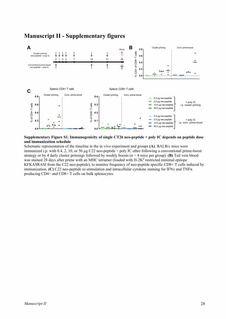
Manuscript II 28
Manuscript II - Supplementary figures
Supplementary Figure S1. Immunogenicity of single CT26 neo-peptide + poly IC depends on peptide dose and immunization schedule Schematic representation of the timeline in the in vivo experiment and groups (A). BALB/c mice were immunized i.p. with 0.4, 2, 10, or 50 µg C22 neo-peptide + poly IC ether following a conventional prime-boost strategy or by 4 daily cluster primings followed by weekly boosts (n = 4 mice per group). (B) Tail vein blood was stained 28 days after prime with an MHC tetramer (loaded with H-2Kd restricted minimal epitope KFKASRASI from the C22 neo-peptide), to monitor frequency of neo-peptide specific CD8+ T cells induced by immunization. (C) C22 neo-peptide re-stimulation and intracellular cytokine staining for IFNγ and TNFα producing CD4+ and CD8+ T cells on bulk splenocytes.
A B
C
+ poly ICi.p. cluster priming
+ poly ICi.p. conv. prime-boost
0 3 28211471 2
Blood
Cluster primingneo-peptide + poly IC
Conventional prime-boostneo-peptide + poly IC
● ●● ● ● ● ●
↓ ↓ ↓↓●
↓
↓
↓↓↓
↓ ↓ ✞
2.0 µg neo-peptide10.0 µg neo-peptide
0.4 µg neo-peptide
50.0 µg neo-peptide
0.4 µg neo-peptide2.0 µg neo-peptide10.0 µg neo-peptide50.0 µg neo-peptide
2.0 µg neo-peptide10.0 µg neo-peptide
0.4 µg neo-peptide
50.0 µg neo-peptide
0.4 µg neo-peptide2.0 µg neo-peptide10.0 µg neo-peptide50.0 µg neo-peptide
0.0
0.1
0.2
0.3
0.4
% o
f CD
8+ T
cel
lsSplenic CD8+ T cells
Cluster priming Conv. prime-boost
0.0
0.2
0.4
0.6
0.8
% o
f CD
4+ T
cel
ls
Splenic CD4+ T cells
Cluster priming Conv. prime-boost
0.0
0.2
0.4
0.6
0.8
% C
22+
of C
D8+
T c
ells
Cluster priming Conv. prime-boost 0.4 µg neo-peptide
2 µg neo-peptide
10 µg neo-peptide
50 µg neo-peptide
0.4 µg neo-peptide
2 µg neo-peptide
10 µg neo-peptide
50 µg neo-peptide

Manuscript II 29
Supplementary Figure S2. FACS gating strategy FACS gating strategy used for samples after peptide re-stimulation and intracellular cytokine staining (A). FACS gating strategy used for samples after H-2 tetramer staining. All FACS gating was performed in FlowJo (B).
Lymphocytes71,4%
0 50K 100K 150K 200K 250K
FSC-A
0
50K
100K
150K
200K
250K
SSC
-A
Single Cells98,8%
0 50K 100K 150K 200K 250K
FSC-A
0
50K
100K
150K
200K
250K
FSC
-H Live cells16,3%
0-10 3 10 3 10 4 10 5
Comp-APC-Cy7-A :: Live_dead
0
5.0K
10K
15K
20K
Cou
nt
CD3+52,7%
0 10 3 10 4 10 5
Comp-FITC-A :: CD3
0
50K
100K
150K
200K
250K
SSC
-A
CD4+74,2%
CD8+22,2%
0 10 3 10 4 10 5
Comp-PE-A :: CD4
0
-10 3
10 3
10 4
10 5
Com
p-Pe
rCP-
Cy5
-5-A
:: C
D8
CD4 IFN-gamma0,26%
0 10 3 10 4 10 5
Comp-APC-A :: IFN-gamma
0
50K
100K
150K
200K
250K
SSC
-A
CD4 TNF-alpha0,18%
0 10 3 10 4 10 5
Comp-BV421-A :: TNF-alpha
0
50K
100K
150K
200K
250K
SSC
-A
CD4 IFN+ & TNF+0,19%
0 10 3 10 4 10 5
Comp-BV421-A :: TNF-alpha
0
-10 2
10 2
10 3
10 4
10 5
Com
p-AP
C-A
:: IF
N-g
amm
a
CD8 IFN-gamma6,96%
0 10 3 10 4 10 5
Comp-APC-A :: IFN-gamma
0
50K
100K
150K
200K
250K
SSC
-A
CD8 TNF-alpha3,43%
0 10 3 10 4 10 5
Comp-BV421-A :: TNF-alpha
0
50K
100K
150K
200K
250K
SSC
-A
CD8 IFN+ & TNF+4,31%
0 10 3 10 4 10 5
Comp-BV421-A :: TNF-alpha
0
-10 2
10 2
10 3
10 4
10 5
Com
p-AP
C-A
:: IF
N-g
amm
a
Subset Name Count Freq. of ParentCD4 IFN+ & TNF+ 61,0 0,19% CD4 IFN-gamma 83,0 0,26% CD4 TNF-alpha 56,0 0,18% CD8 IFN+ & TNF+ 407 4,31% CD8 IFN-gamma 657 6,96% CD8 TNF-alpha 324 3,43%
Lymphocytes14,8%
0 50K 100K 150K 200K 250K
FSC-A
0
50K
100K
150K
200K
250K
SSC
-A
blood day 9_ID 3 C22_008.fcsUngated7,43E5
Single Cells99,5%
0 50K 100K 150K 200K 250K
FSC-A
0
50K
100K
150K
200K
250K
FSC
-H
blood day 9_ID 3 C22_008.fcsLymphocytes110176
Live100,0%
0-10 3 10 3 10 4 10 5
Comp-APC-Cy7-A
0
10K
20K
30K
40K
Cou
nt
blood day 9_ID 3 C22_008.fcsSingle Cells109672
CD3+37,3%
0 10 3 10 4 10 5
Comp-FITC-A
0
50K
100K
150K
200K
250K
SSC
-A
blood day 9_ID 3 C22_008.fcsLive109668
CD8+23,6%
0 10 3 10 4 10 5
Comp-BV421-A
0
-10 3
10 3
10 4
10 5
Com
p-Pe
rCP-
Cy5
-5-A
blood day 9_ID 3 C22_008.fcsCD3+40852
C22 tetramer+2,48%
0 10 3 10 4 10 5
Comp-APC-A
0
10 3
10 4
10 5
Com
p-PE
-A
blood day 9_ID 3 C22_008.fcsCD8+9622
Subset Name Count Freq. of Parent TUBE NAMELymphocytes 110176 14,8% ID 3 C22 Single Cells 109672 99,5% ID 3 C22 Live 109668 100,0% ID 3 C22 CD3+ 40852 37,3% ID 3 C22 CD8+ 9622 23,6% ID 3 C22 C22 tetramer+ 239 2,48% ID 3 C22
A
B

78
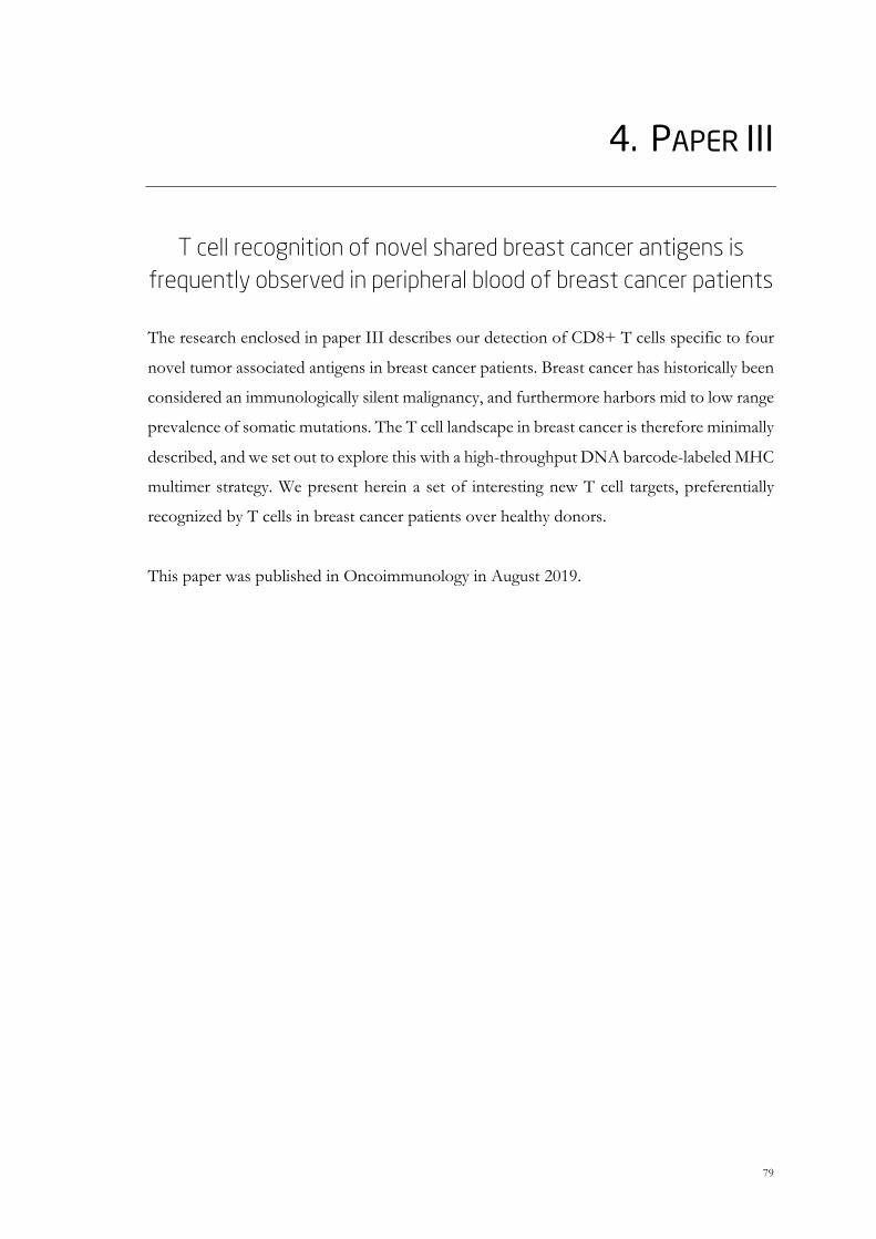
79
4. PAPER III
T cell recognition of novel shared breast cancer antigens is frequently observed in peripheral blood of breast cancer patients
The research enclosed in paper III describes our detection of CD8+ T cells specific to four
novel tumor associated antigens in breast cancer patients. Breast cancer has historically been
considered an immunologically silent malignancy, and furthermore harbors mid to low range
prevalence of somatic mutations. The T cell landscape in breast cancer is therefore minimally
described, and we set out to explore this with a high-throughput DNA barcode-labeled MHC
multimer strategy. We present herein a set of interesting new T cell targets, preferentially
recognized by T cells in breast cancer patients over healthy donors.
This paper was published in Oncoimmunology in August 2019.

80
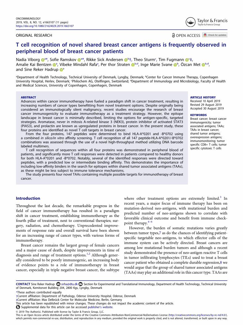
ORIGINAL RESEARCH
T cell recognition of novel shared breast cancer antigens is frequently observed inperipheral blood of breast cancer patientsNadia Viborg a*, Sofie Ramskov a*, Rikke Sick Andersen b†, Theo Sturmc, Tim Fugmann c‡,Amalie Kai Bentzen a, Vibeke Mindahl Rafaa, Per thor Straten b,d, Inge Marie Svane b, Özcan Met b,d,and Sine Reker Hadrup a
aDepartment of Health Technology, Technical University of Denmark, Lyngby, Denmark; bCenter for Cancer Immune Therapy, CopenhagenUniversity Hospital, Herlev, Denmark; cPhilochem AG, Otelfingen, Switzerland; dDepartment of Immunology and Microbiology, Faculty of Healthand Medical Sciences, University of Copenhagen, Copenhagen, Denmark
ABSTRACTAdvances within cancer immunotherapy have fueled a paradigm shift in cancer treatment, resulting inincreasing numbers of cancer types benefitting from novel treatment options. Despite originally beingconsidered an immunologically silent malignancy, recent studies encourage the research of breastcancer immunogenicity to evaluate immunotherapy as a treatment strategy. However, the epitopelandscape in breast cancer is minimally described, limiting the options for antigen-specific, targetedstrategies. Aromatase, never in mitosis A-related kinase 3 (NEK3), protein inhibitor of activated STAT3(PIAS3), and prolactin are known as upregulated proteins in breast cancer. In the present study, thesefour proteins are identified as novel T cell targets in breast cancer.
From the four proteins, 147 peptides were determined to bind HLA-A*0201 and -B*0702 usinga combined in silico/in vitro affinity screening. T cell recognition of all 147 peptide-HLA-A*0201/-B*0702combinations was assessed through the use of a novel high-throughput method utilizing DNA barcodelabeled multimers.
T cell recognition of sequences within all four proteins was demonstrated in peripheral blood ofpatients, and significantly more T cell responses were detected in patients compared to healthy donorsfor both HLA-A*0201 and -B*0702. Notably, several of the identified responses were directed towardpeptides, with a predicted low or intermediate binding affinity. This demonstrates the importance ofincluding low-affinity binders in the search for epitopes within shared tumor associated antigens (TAAs),as these might be less subject to immune tolerance mechanisms.
The study presents four novel TAAs containing multiple possible targets for immunotherapy of breastcancer.
ARTICLE HISTORYReceived 10 April 2019Revised 29 August 2019Accepted 30 August 2019
KEYWORDSBreast cancer; breast cancerimmunogenicity; tumorassociated antigens; TAAs;TAAs in breast cancer;shared tumor antigens;overexpression antigens;immunomonitoring; tumorspecific CD8+ T cells; tumorspecific cytotoxic T cells
Introduction
Throughout the last decade, the remarkable progress in thefield of cancer immunotherapy has resulted in a paradigmshift in cancer treatment, establishing immunotherapy as thefourth pillar of treatment, next to conventional therapies; sur-gery, radiation, and chemotherapy. Unprecedented improve-ments of response rate and overall survival have been shownfor an increasing range of cancer types, with various forms ofimmunotherapy.
Breast cancer remains the largest group of female cancersand a major cause of death, despite improvements in time ofdiagnosis and range of treatment options.1,2 Although gener-ally considered to be poorly immunogenic, an increasing bodyof evidence points to a role of immunotherapy in breastcancer, especially in triple negative breast cancer, the subtype
where other treatment options are extremely limited.3 Inrecent years, a major focus of immune therapy has been onmutation-derived neo-antigens, with mutational burden andpredicted number of neo-antigens shown to correlate withfavorable clinical outcome and benefit from immune check-point therapy.4–6
However, the burden of somatic mutations varies greatlybetween tumor types,7 as do the chances of identifying patient-specific targetable neo-antigens, to which effector cells of theimmune system can be actively directed. Breast cancers areamong low mutational burden tumors and although a recentstudy demonstrated the presence of neo-antigen reactive T cellsin tumor infiltrating lymphocytes (TILs) used to treat a breastcancer patient who obtained a complete durable regression,8 wewould argue that the group of shared tumor associated antigens(TAAs) may play an additional role in this cancer type. TAAs are
CONTACT Sine Reker Hadrup [email protected] Section for Experimental and Translational Immunology, Department of Health Technology, Technical Universityof Denmark, Kemitorvet Building 204, 2800 Kgs, Lyngby, Denmark*These authors contributed equally†Current affiliation: Department of Pathology, Odense University Hospital, Odense, Denmark‡Current affiliation: Max Delbrück Center for Molecular Medicine, Berlin, GermanyThis article has been republished with minor changes. These changes do not impact the academic content of the article.
Supplemental data for this article can be accessed publisher’s website.
ONCOIMMUNOLOGY2019, VOL. 8, NO. 12, e1663107 (11 pages)https://doi.org/10.1080/2162402X.2019.1663107
© 2019 The Author(s). Published with license by Taylor & Francis Group, LLC.This is an Open Access article distributed under the terms of the Creative Commons Attribution-NonCommercial-NoDerivatives License (http://creativecommons.org/licenses/by-nc-nd/4.0/),which permits non-commercial re-use, distribution, and reproduction in any medium, provided the original work is properly cited, and is not altered, transformed, or built upon in any way.

of value both in the antigen-defined treatment setting, wherethey can act directly as targets of therapy, as well as in theantigen-undefined setting, where they can be used for immuno-monitoring and potentially be able to predict treatmentoutcome.9 Few breast cancer-specific TAAs are known; thosethat have already been described (e.g. HER210 and MUC111) areonly expressed in subgroups of patients; and when used invaccines, they have shown limited clinical activity, albeit modestimmunological activity.2 Therefore, there is an unmet need foridentification of TAAs in breast cancer.
In this study we investigate aromatase, prolactin, never inmitosis A-related kinase 3 (NEK3) and protein inhibitor ofactivated signal transducer and activator of transcription 3(PIAS3) as possible TAAs in breast cancer. Aromatase is thekey enzyme in estrogen synthesis and is a heterodimer composedof the ubiquitously expressed flavoprotein NADPH-cytochromeP450 reductase and aromatase cytochrome P450.12 Aromataseinhibitors are already successful in treating breast and otherfemale cancers, underlining the relevance of investigating aro-matase as a TAA.13 Prolactin and two of its downstream intra-cellular signaling proteins, NEK3 and PIAS3, are involved inbreast lobulo-alveolar expansion and differentiation prior tomilk synthesis and secretion.14,15 All four proteins have beendescribed to have an increased expression in breast cancer com-pared to healthy breast tissue.12–16
Results
Peptides were selected based on a combined in silico andin vitro selection pipeline
The four proteins aromatase, NEK3, PIAS3, and prolactin wereselected for investigation based on a literature search, prioritiz-ing proteins that fulfilled criteria of tissue restriction andincreased expression in malignant compared to healthy breasttissue (Table 1). Aromatase mRNA levels have been shown to besignificantly increased in breast cancer compared to healthybreast tissue, e.g. in a study by Harada, where mRNA levelswere significantly elevated (p < 0.01).16 Likewise, significantlyincreased expression of prolactin, and the two intracellular sig-naling proteins, NEK3 and PIAS3, have been demonstratedin breast tumor tissue using immunohistochemistry staining.This revealed a significant difference in expression between
normal/hyperplastic epithelium and ductal carcinoma in situ/invasive carcinoma (t-tests, all p values < 0.02).14
Expression of all four proteins was validated in two breastductal carcinoma cell lines (BT-549 and HCC1937), as well asin two breast adenocarcinoma cell lines (EFM-192A andMDA-MB-231) (Supplementary Figure S1). MHC classI presentation of peptides from the four proteins was investi-gated by mass spectrometry analysis and demonstrated pre-sentation of peptides from PIAS3 in three of three tested celllines (Supplementary Figure S1, Supplementary Table S3).
A library of peptides was generated based on a two-step insilico + in vitro selection process (Table 2, Supplementary TableS2). In the initial in silico prediction step, two available computa-tional pipelines, SYFPEITHI and NetMHC 3.0, were used forpredicting the MHC class I binding affinity of the individualpeptides in the selected proteins. We identified potential HLA-A*0201 and -B*0702 binding 9- and 10-mer peptides within thesequences of the four proteins. The number of predicted peptidesout of total possible peptides from each protein sequence corre-lated with the size of the protein, as would be expected. 415 of thetotal 3644 peptides were predicted with selection cut offs set toaccommodate inclusion of both high and low affinity HLA-A*0201 and -B*0702 ligands (SYFPEITHI: ≥ 19, NetMHC 3.0:≤ 1000 nM), given that the proteins of interest are shared anti-gens. Previous analyses have demonstrated very low affinityepitopes of relevance in shared antigens.17, 18 As such, the bindingof the predicted peptides was experimentally tested in vitro withanMHC ELISA.19 147 of the 415 in silico predicted peptides wereselected for further analysis, based on 50% (A*0201) and 70%(B*0702) binding affinity, compared to a reference of high affinityvirus-derived ligands (Table 2, Supplementary Table S2).
Table 1. Overview of protein targets selected for investigation as tumor associated antigens.
Protein Full name Normal function Role in breast cancer Normal expressionIncreasedexpression Reference
Aromatase Aromatase Key enzyme in estrogen synthesis.Heterodimer composed of aromatasecytochrome P450 and NADPH-cytochromeP450 reductasea
Enhanced tumor cellgrowth
Breast, ovary, testes,adipose tissue, skin,hypothalamus,placenta
Breast,endometrial &ovarian cancer
12,13,16
NEK3 Never in mitosisA-related kinase 3
Prolactin receptor-associated protein,positive regulator
Enhanced survival,motility, and invasion oftumor cells
Ubiquitous Breast, colorectal,carcinoid &testicular cancer
14
PIAS3 Protein inhibitor ofactivated STAT3
Prolactin receptor-associated protein,negative regulator
Induction of malignanttransformation
Ubiquitous Breast cancer,glioma
14
Prolactin Prolactin Growth and differentiation hormone Enhanced breast epithelialsurvival and motility,inhibition of tumor cellapoptosis
Breast, brain(pituitary gland),placenta,endometrium
Breast cancer 14,15
aOnly aromatase cytochrome P450 is investigated in this study.
Table 2. Peptide selection from the four proteins through a combined in silicoprediction and in vitro binding assay.
Protein
Size(aminoacids)
Total number ofpeptides
(9- and 10-mers)
In silico predictedpeptidesa
(9- and 10-mers)
In vitro selectedpeptidesb
(9- and 10-mers)
Aromatase 503 989 141 49NEK3 506 995 85 36PIAS3 628 1239 148 50Prolactin 227 437 41 12Total 3644 415 147
aIn silico prediction cut offs: SYFPEITHI score ≥ 19 and/or NetMHC 3.0 IC50 ≤ 1000 nM.bIn vitro binding cut offs: A*0201: 50% of CMV control, B*0702: 70% of CMV control.
e1663107-2 N. VIBORG ET AL.
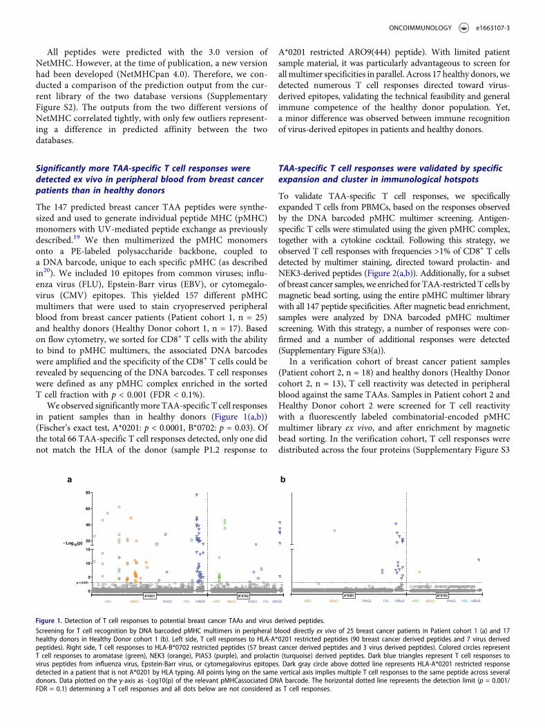
All peptides were predicted with the 3.0 version ofNetMHC. However, at the time of publication, a new versionhad been developed (NetMHCpan 4.0). Therefore, we con-ducted a comparison of the prediction output from the cur-rent library of the two database versions (SupplementaryFigure S2). The outputs from the two different versions ofNetMHC correlated tightly, with only few outliers represent-ing a difference in predicted affinity between the twodatabases.
Significantly more TAA-specific T cell responses weredetected ex vivo in peripheral blood from breast cancerpatients than in healthy donors
The 147 predicted breast cancer TAA peptides were synthe-sized and used to generate individual peptide MHC (pMHC)monomers with UV-mediated peptide exchange as previouslydescribed.19 We then multimerized the pMHC monomersonto a PE-labeled polysaccharide backbone, coupled toa DNA barcode, unique to each specific pMHC (as describedin20). We included 10 epitopes from common viruses; influ-enza virus (FLU), Epstein-Barr virus (EBV), or cytomegalo-virus (CMV) epitopes. This yielded 157 different pMHCmultimers that were used to stain cryopreserved peripheralblood from breast cancer patients (Patient cohort 1, n = 25)and healthy donors (Healthy Donor cohort 1, n = 17). Basedon flow cytometry, we sorted for CD8+ T cells with the abilityto bind to pMHC multimers, the associated DNA barcodeswere amplified and the specificity of the CD8+ T cells could berevealed by sequencing of the DNA barcodes. T cell responseswere defined as any pMHC complex enriched in the sortedT cell fraction with p < 0.001 (FDR < 0.1%).
We observed significantlymore TAA-specific T cell responsesin patient samples than in healthy donors (Figure 1(a,b))(Fischer’s exact test, A*0201: p < 0.0001, B*0702: p = 0.03). Ofthe total 66 TAA-specific T cell responses detected, only one didnot match the HLA of the donor (sample P1.2 response to
A*0201 restricted ARO9(444) peptide). With limited patientsample material, it was particularly advantageous to screen forallmultimer specificities in parallel. Across 17 healthy donors, wedetected numerous T cell responses directed toward virus-derived epitopes, validating the technical feasibility and generalimmune competence of the healthy donor population. Yet,a minor difference was observed between immune recognitionof virus-derived epitopes in patients and healthy donors.
TAA-specific T cell responses were validated by specificexpansion and cluster in immunological hotspots
To validate TAA-specific T cell responses, we specificallyexpanded T cells from PBMCs, based on the responses observedby the DNA barcoded pMHC multimer screening. Antigen-specific T cells were stimulated using the given pMHC complex,together with a cytokine cocktail. Following this strategy, weobserved T cell responses with frequencies >1% of CD8+ T cellsdetected by multimer staining, directed toward prolactin- andNEK3-derived peptides (Figure 2(a,b)). Additionally, for a subsetof breast cancer samples, we enriched for TAA-restrictedT cells bymagnetic bead sorting, using the entire pMHC multimer librarywith all 147 peptide specificities. After magnetic bead enrichment,samples were analyzed by DNA barcoded pMHC multimerscreening. With this strategy, a number of responses were con-firmed and a number of additional responses were detected(Supplementary Figure S3(a)).
In a verification cohort of breast cancer patient samples(Patient cohort 2, n = 18) and healthy donors (Healthy Donorcohort 2, n = 13), T cell reactivity was detected in peripheralblood against the same TAAs. Samples in Patient cohort 2 andHealthy Donor cohort 2 were screened for T cell reactivitywith a fluorescently labeled combinatorial-encoded pMHCmultimer library ex vivo, and after enrichment by magneticbead sorting. In the verification cohort, T cell responses weredistributed across the four proteins (Supplementary Figure S3
a b
0
5
10
15
p = 0.001
20
40
60
80
- Log10(p)
B*0702A*0201ARO NEK3 PIAS3 PRL VIRUS ARO NEK3 PIAS3 PRL VIRUS
B*0702A*0201ARO NEK3 PIAS3 PRL VIRUS ARO NEK3 PIAS3 PRL VIRUS
Figure 1. Detection of T cell responses to potential breast cancer TAAs and virus derived peptides.Screening for T cell recognition by DNA barcoded pMHC multimers in peripheral blood directly ex vivo of 25 breast cancer patients in Patient cohort 1 (a) and 17healthy donors in Healthy Donor cohort 1 (b). Left side, T cell responses to HLA-A*0201 restricted peptides (90 breast cancer derived peptides and 7 virus derivedpeptides). Right side, T cell responses to HLA-B*0702 restricted peptides (57 breast cancer derived peptides and 3 virus derived peptides). Colored circles representT cell responses to aromatase (green), NEK3 (orange), PIAS3 (purple), and prolactin (turquoise) derived peptides. Dark blue triangles represent T cell responses tovirus peptides from influenza virus, Epstein-Barr virus, or cytomegalovirus epitopes. Dark gray circle above dotted line represents HLA-A*0201 restricted responsedetected in a patient that is not A*0201 by HLA typing. All points lying on the same vertical axis implies multiple T cell responses to the same peptide across severaldonors. Data plotted on the y-axis as -Log10(p) of the relevant pMHCassociated DNA barcode. The horizontal dotted line represents the detection limit (p = 0.001/FDR = 0.1) determining a T cell responses and all dots below are not considered as T cell responses.
ONCOIMMUNOLOGY e1663107-3
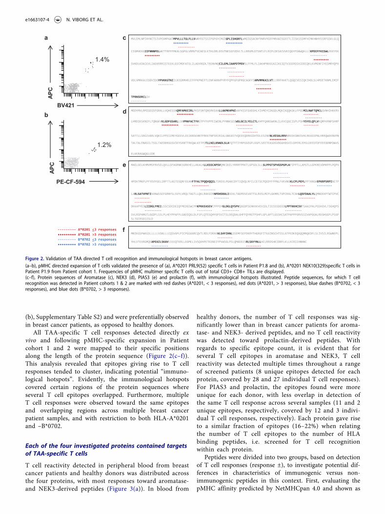
(b), Supplementary Table S2) and were preferentially observedin breast cancer patients, as opposed to healthy donors.
All TAA-specific T cell responses detected directly exvivo and following pMHC-specific expansion in Patientcohort 1 and 2 were mapped to their specific positionsalong the length of the protein sequence (Figure 2(c–f)).This analysis revealed that epitopes giving rise to T cellresponses tended to cluster, indicating potential “immuno-logical hotspots”. Evidently, the immunological hotspotscovered certain regions of the protein sequences whereseveral T cell epitopes overlapped. Furthermore, multipleT cell responses were observed toward the same epitopesand overlapping regions across multiple breast cancerpatient samples, and with restriction to both HLA-A*0201and –B*0702.
Each of the four investigated proteins contained targetsof TAA-specific T cells
T cell reactivity detected in peripheral blood from breastcancer patients and healthy donors was distributed acrossthe four proteins, with most responses toward aromatase-and NEK3-derived peptides (Figure 3(a)). In blood from
healthy donors, the number of T cell responses was sig-nificantly lower than in breast cancer patients for aroma-tase- and NEK3- derived peptides, and no T cell reactivitywas detected toward prolactin-derived peptides. Withregards to specific epitope count, it is evident that forseveral T cell epitopes in aromatase and NEK3, T cellreactivity was detected multiple times throughout a rangeof screened patients (8 unique epitopes detected for eachprotein, covered by 28 and 27 individual T cell responses).For PIAS3 and prolactin, the epitopes found were moreunique for each donor, with less overlap in detection ofthe same T cell response across several samples (11 and 2unique epitopes, respectively, covered by 12 and 3 indivi-dual T cell responses, respectively). Each protein gave riseto a similar fraction of epitopes (16–22%) when relatingthe number of T cell epitopes to the number of HLAbinding peptides, i.e. screened for T cell recognitionwithin each protein.
Peptides were divided into two groups, based on detectionof T cell responses (response ±), to investigate potential dif-ferences in characteristics of immunogenic versus non-immunogenic peptides in this context. First, evaluating thepMHC affinity predicted by NetMHCpan 4.0 and shown as
c
d
e
f
bBV421
APC
a
1.4%
1.2%
--------- A*0201 <3 responses ••••••••• A*0201 >3 responses--------- B*0702 <3 responses ••••••••• B*0702 >3 responses
PE-CF-594
APC
MVLEMLNPIHYNITSIVPEAMPAATMPVLLLTGLFLLVWNYEGTSSIPGPGYCMGIGPLISHGRFLWMGIGSACNYYNRVYGEFMRVWISGEETLIISKSSSMFHIMKHNHYSSRFGSKLGLQYNITSIVPEAMPAAXXXXXXXXXXT•••••••••FLLVWNYEGTSSIPGPGYCMGI••••••••••
VELPHIT----------WNYEGTSSIPGPGYCMGIGPLISHGRFLWMGIGSACNYYNRVYGEFMRVWISGEETLIISKSSSMFHIMKHNHYSCIGMHEKGIIFNNNPELWKTTRPFFMKALSGPGLVRMVTVCAESLKTHLDRLEEVTNESGYVDVLTLLRRVMLDTSNTLFLRIPLDESAIVVKIQGYFDAWQALLIKPDIFFKISWLYKKYEKXXXXXXXX•••••••••XXXXXXXXXXXXXXXXXXXXXXXXXXXXXXXXXXXXXXXXXXXXXXXXXXXXXXXXXXXXXXXXXXXXXXXXXXXXXXXXXXXXXXXXX----------XXXXXXXXXXXXXXXXXXXXXXXXXXXXXXXXXXXXXXXXXXXXXXXXXXXXXXXXXXXXXXXXXXXXXXXXXXXXXXXXXXXXXXXXXXXXXXXXXXXXXXXXXXXX•••••••••YSVKDLKDAIEVLIAEKRRRISTEEKLEECMDFATELILAEKRGDLTRENVNQCILEMLIAAPDTMSVSLFFMLFLIAKHPNVEEAIIKEIQTVIGERDIKIDDIQKLKVMENFIYESMRYQPVXXXXXXXXXXXXXXXXXXXXXXXXXXXXXXXXXXXXXXXXXXXXXXXXXXXX----------D
----------XXXXXXXXXXXXXXXXXXXXXXXXXXXXXXXXXXXXXXXXXXXXXXXXXXXXXXXXXX---------SVDLVMRKALEDDVIDGYPVKKGTNIILNIGRMHRLEFFPKPNEFTLENFAKNVPYRYFQPFGFGPRGCAGKYIAMVMMKAILVTLLRRFHVKTLQGQCVESIQKIHDLSLHPDETKNMLEMIFXXXXXXXXXXXXXXXX---------XXXXXXXXXXXXXXXXXXXXXXXXXXXXXXXXXXXXXXXXXXXXXXXX----------XXXXXXXXXXXXXXXXXXXXXXXXXXXXXXXXXXXXXXXXXXXXXXXXXXXXXXXXXXXXXXXXXXXXXXXXXX•••••••••
---------TPRNSDRCLEH---------
MDDYMVLRMIGEGSFGRALLVQHESSNQMFAMKEIRLPKSFSNTQNSRKEAVLLAKMKHPNIVAFKESFEAEGHLYIVMEYCDGGDLMQKIKQQKGKLFPEDMILNWFTQMCLGVNHIHKKRVXXXXXXXXXXXXXXXXXXXXXXXXXXX•••••••••• ---------- ---------
----------LHRDIKSKNIFLTQNGKVKLGDFGSARLLSNPMAFACTYVGTPYYVPPEIWENLPYNNKSDIWSLGCILYELCTLKHPFQANSWKNLILKVCQGCISPLPSHYSYELQFLVKQMFKRNPSHRP
•••••••••• --------- ---------- ------------------
---------SATTLLSRGIVARLVQKCLPPEIIMEYGEEVLEEIKNSKHNTPRKKTNPSRIRIALGNEASTVQEEEQDRKGSHTDLESINENLVESALRRVNREEKGNKSVHLRKASSPNLHRRQWEKNVPN
••••••••••TALTALENASILTSSLTAEDDRGGSVIKYSKNTTRKQWLKETPDTLLNILKNADLSLAFQTYTIYRPGSEGFLKGPLSEETEASDSVDGGHDSVILDPERLEPGLDEEDTDFEEEDDNPDWVS
-------------------
ELKKRAGWQGLCDR
MAELGELKHMVMSFRVSELQVLLGFAGRNKSGRKHELLAKALHLLKSSCAPSVQMKIKELYRRRFPRKTLGPSDLSLLSLPPGTSPVGSPGPLAPIPPTLLAPGTLLGPKREVDMHPPLPQPV---------- ---------
-------------------
HPDVTMKPLPFYEVYGELIRPTTLASTSSQRFEEAHFTFALTPQQVQQILTSREVLPGAKCDYTIQVQLRFCLCETSCPQEDYFPPNLFVKVNGKLCPLPGYLPPTKNGAEPKRPSRPINITP---------- --------- ---------
---------LARLSATVPNTIVVNWSSEFGRNYSLSVYLVRQLTAGTLLQKLRAKGIRNPDHSRALIKEKLTADPDSEVATTSLRVSLMCPLGKMRLTVPCRALTCAHLQSFDAALYLQMNEKKPTWTCPVC
--------- --------- --------------------
DKKAPYESLIIDGLFMEILSSCSDCDEIQFMEDGSWCPMKPKKEASEVCPPPGYGLDGLQYSPVQGGDPSENKKKVEVIDLTIESSSDEEDLPPTKKHCSVTSAAIPALPGSKGVLTSGHQPS---------- --------- ---------- ----------
SVLRSPAMGTLGGDFLSSLPLHEYPPAFPLGADIQGLDLFSFLQTESQHYGPSVITSLDEQDALGHFFQYRGTPSHFLGPLAPTLGSSHCSATPAPPPGRVSSIVAPGGALREGHGGPLPSGPSLTGCRSDIISLD
MNIKGSPWKGSLLLLLVSNLLLCQSVAPLPICPGGAARCQVTLRDLFDRAVVLSHYIHNLSSEMFSEFDKRYTHGRGFITKAINSCHTSSLATPEDKEQAQQMNQKDFLSLIVSILRSWNEPL---------
YHLVTEVRGMQEAPEAILSKAVEIEEQTKRLLEGMELIVSQVHPETKENEIYPVWSGLPSLQMADEESRLSAYYNLLHCLRRDSHKIDNYLKLLKCRIIHNNNC---------- ---------
Figure 2. Validation of TAA directed T cell recognition and immunological hotspots in breast cancer antigens.(a–b), pMHC directed expansion of T cells validated the presence of (a), A*0201 PRL9(52) specific T cells in Patient P1.8 and (b), A*0201 NEK10(329)specific T cells inPatient P1.9 from Patient cohort 1. Frequencies of pMHC multimer specific T cells out of total CD3+ CD8+ TILs are displayed.(c–f), Protein sequences of Aromatase (c), NEK3 (d), PIAS3 (e) and prolactin (f), with immunological hotspots illustrated. Peptide sequences, for which T cellrecognition was detected in Patient cohorts 1 & 2 are marked with red dashes (A*0201, < 3 responses), red dots (A*0201, > 3 responses), blue dashes (B*0702, < 3responses), and blue dots (B*0702, > 3 responses).
e1663107-4 N. VIBORG ET AL.

nM binding affinity and % eluted ligand Rank(Supplementary Figure S4(a–b)). Second, stability was pre-dicted by NetMHCStabpan 1.0 and shown as %Rank(Supplementary Figure S4(c)). There were no significant dif-ferences in mean predicted affinity or stability between pep-tides, yielding a T cell response (+) or not (-).
T cell reactivity toward breast cancer TAAs occursindependently of cancer subtype and disease stage
Breast cancer patients in cohort 1 were stratified into differentmolecular subtypes based on pathological examination oftumors (Supplementary Table S1). Four major subtypes of breastcancer are commonly described based on tumor cell expressionof hormone receptors (estrogen receptor; ER, and progesteronereceptor; PR) and growth factor HER2 as follows: luminal A (ER+ and/or PR+, HER2-), luminal B (ER+ and/or PR+, HER2+),HER2 enriched (ER- and PR-, HER2+), and triple negative (ER-,PR-, HER2-). Breast cancer patients with TAA-specific T cellresponses were observed for all subtypes and there seemed to beno preferential distribution for any particular subtype in theinvestigated patient cohort (Figure 3(b)). However, for certainsubtypes, i.e. HER2 enriched and triple negative, the number ofpatients included is <5, and hence too few to firmly evaluate forpotential differences.
For 20 out of 25 breast cancer patients from cohort 1 it waspossible to determine disease stage based on UICC TNM
guidelines (Supplementary Table S1). Interestingly, TAA-specific T cell responses were observed in blood from patientsat disease stage II and III, and none in blood from patients atstage I, with significantly more responses in disease stage IIIthan disease stage I (Figure 3(c)). This interesting observationfrom a relatively small cohort of patients at each stage (I: 4patients, II: 12 patients, and III: 4 patients), could indicateenhanced T cell reaction with higher tumor burden or meta-static disease, but such findings should be validated in a largercohort.
Breast cancer TAA-specific T cells have functionalcapacity upon stimulation
To assess the functional capacity of TAA-responsive T cells inpatients, we combined the DNA barcoded pMHC multimerscreening with measuring cytokine production by intracellularstaining, as previously described.20 A selection of PBMCs fromPatient cohort 1 were stimulated with 1) a pool of TAA peptidesspecific for each patient sample, selected based on T cellresponses found by multimer screening of ex vivo or enrichedmaterial, or 2) a mixture of HLA matching breast cancer celllines (BT-549, HCC1937, EFM-192A and MDA-MB-231). Afterstimulation, cells were stained and analyzed simultaneously forfunctional reactivity and T cell specificity by sorting T cells solelybased on their cytokine secretion profile, IFNγ and/or TNFαsecretion (ICSpos) or no cytokine secretion (ICSneg), after which
a b
Aromatase NEK3 PIAS3 Prolactin
0
2
4
6
8
Num
bero
fTce
llre
spon
ses
BC HD
* * ns ns
BC HD BC HD BC HD
Responders
T cell responses
Unique epitopes
T cell epitopes ofscreened HLA binders
10 (40%)
27
1 (6%)
1
8
16.3%
8 (32%)
26
1 (6%)
1
8
22.2%
4 (16%)
10
2 (12%)
2
11
22%
3 (12%)
3
0 (0%)
0
2
16.7%
Luminal A Luminal B HER2 enrich. Triple neg.0
3
6
9
12
Num
bero
fTce
llre
spon
ses
15 (60%)
6 (40%)
34
5 (20%)
2 (40%)
18
3 (12%)
2 (66.7%)
9
2 (8%)
1 (50%)
5
Patients
Responders
T cell responses
c
I II III
0
3
6
9
Primary breast cancer stage
Num
bero
fTce
llre
spon
ses *
ns (p=0.06)
Figure 3. Distribution of detected T cell responses by protein and breast cancer subtype or stage.Plots display distribution of T cell responses detected in breast cancer Patient cohort 1 and Healthy Donor cohort 1 ex vivo. (a), T cell response distribution across thefour potential TAAs; aromatase (green), NEK3 (orange), PIAS3 (purple), and prolactin (turquoise). (b), Distribution of TAA specific T cell responses observed in breastcancer patients separated by breast cancer subtypes. (c), Distribution of TAA specific T cell responses observed in breast cancer patients by staging of primary tumor.Asterisks indicate significance levels (*, p < 0.05), nonparametric Mann-Whitney test.
ONCOIMMUNOLOGY e1663107-5
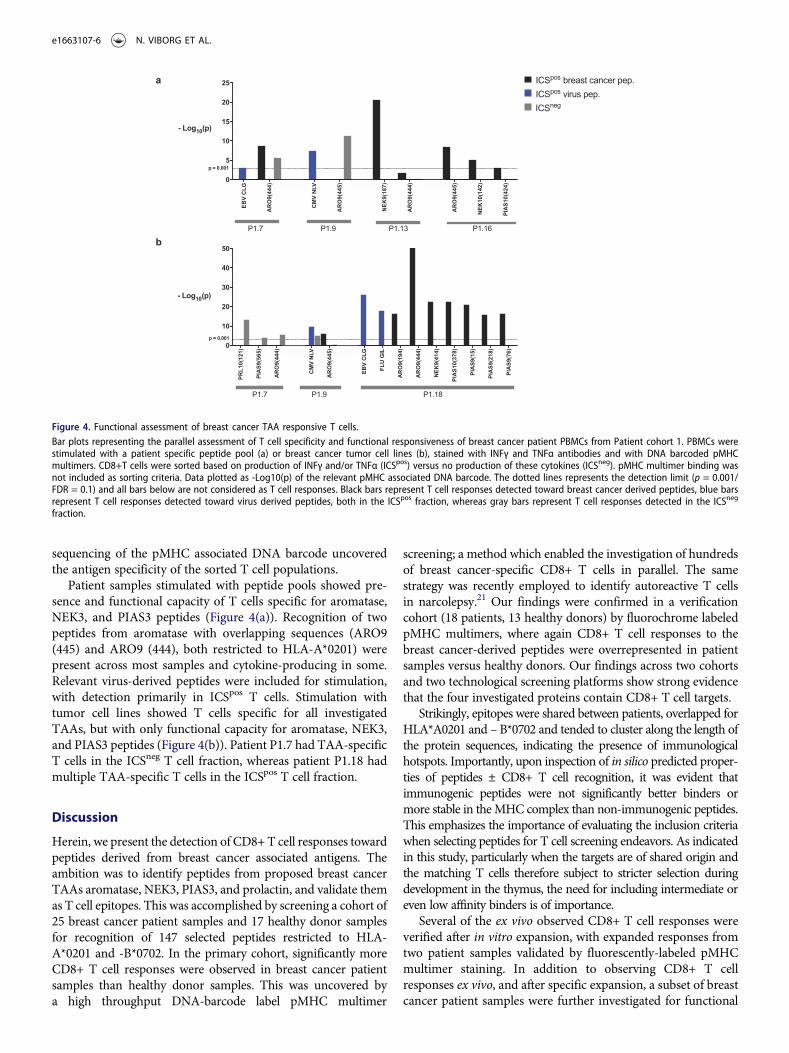
sequencing of the pMHC associated DNA barcode uncoveredthe antigen specificity of the sorted T cell populations.
Patient samples stimulated with peptide pools showed pre-sence and functional capacity of T cells specific for aromatase,NEK3, and PIAS3 peptides (Figure 4(a)). Recognition of twopeptides from aromatase with overlapping sequences (ARO9(445) and ARO9 (444), both restricted to HLA-A*0201) werepresent across most samples and cytokine-producing in some.Relevant virus-derived peptides were included for stimulation,with detection primarily in ICSpos T cells. Stimulation withtumor cell lines showed T cells specific for all investigatedTAAs, but with only functional capacity for aromatase, NEK3,and PIAS3 peptides (Figure 4(b)). Patient P1.7 had TAA-specificT cells in the ICSneg T cell fraction, whereas patient P1.18 hadmultiple TAA-specific T cells in the ICSpos T cell fraction.
Discussion
Herein, we present the detection of CD8+T cell responses towardpeptides derived from breast cancer associated antigens. Theambition was to identify peptides from proposed breast cancerTAAs aromatase, NEK3, PIAS3, and prolactin, and validate themas T cell epitopes. This was accomplished by screening a cohort of25 breast cancer patient samples and 17 healthy donor samplesfor recognition of 147 selected peptides restricted to HLA-A*0201 and -B*0702. In the primary cohort, significantly moreCD8+ T cell responses were observed in breast cancer patientsamples than healthy donor samples. This was uncovered bya high throughput DNA-barcode label pMHC multimer
screening; a method which enabled the investigation of hundredsof breast cancer-specific CD8+ T cells in parallel. The samestrategy was recently employed to identify autoreactive T cellsin narcolepsy.21 Our findings were confirmed in a verificationcohort (18 patients, 13 healthy donors) by fluorochrome labeledpMHC multimers, where again CD8+ T cell responses to thebreast cancer-derived peptides were overrepresented in patientsamples versus healthy donors. Our findings across two cohortsand two technological screening platforms show strong evidencethat the four investigated proteins contain CD8+ T cell targets.
Strikingly, epitopes were shared between patients, overlapped forHLA*A0201 and – B*0702 and tended to cluster along the length ofthe protein sequences, indicating the presence of immunologicalhotspots. Importantly, upon inspection of in silico predicted proper-ties of peptides ± CD8+ T cell recognition, it was evident thatimmunogenic peptides were not significantly better binders ormore stable in theMHC complex than non-immunogenic peptides.This emphasizes the importance of evaluating the inclusion criteriawhen selecting peptides for T cell screening endeavors. As indicatedin this study, particularly when the targets are of shared origin andthe matching T cells therefore subject to stricter selection duringdevelopment in the thymus, the need for including intermediate oreven low affinity binders is of importance.
Several of the ex vivo observed CD8+ T cell responses wereverified after in vitro expansion, with expanded responses fromtwo patient samples validated by fluorescently-labeled pMHCmultimer staining. In addition to observing CD8+ T cellresponses ex vivo, and after specific expansion, a subset of breastcancer patient samples were further investigated for functional
a
bEB
VCL
G
ARO
9(44
4)
CMV
NLV
ARO
9(44
5)
NEK9
(187
)
ARO
9(44
4)
ARO
9(44
5)
NEK1
0(14
2)
PIAS
10(4
24)0
5
10
15
20
25
p = 0.001
- Log10(p)
P1.7 P1.9 P1.13 P1.16PR
L10(
121)
PIAS
9(56
5)
ARO
9(44
4)
CMV
NLV
ARO
9(44
5)
EBV
CLG
FLU
GIL
ARO
9(19
4)
ARO
9(44
4)
NEK9
(414
)
PIAS
10(3
78)
PIAS
9(15
)
PIAS
9(21
8)
PIAS
9(76
)0
10
20
30
40
50
- Log10(p)
p = 0.001
P1.7 P1.9 P1.18
ICSpos breast cancer pep.ICSpos virus pep.ICSneg
Figure 4. Functional assessment of breast cancer TAA responsive T cells.Bar plots representing the parallel assessment of T cell specificity and functional responsiveness of breast cancer patient PBMCs from Patient cohort 1. PBMCs werestimulated with a patient specific peptide pool (a) or breast cancer tumor cell lines (b), stained with INFγ and TNFα antibodies and with DNA barcoded pMHCmultimers. CD8+T cells were sorted based on production of INFγ and/or TNFα (ICSpos) versus no production of these cytokines (ICSneg). pMHC multimer binding wasnot included as sorting criteria. Data plotted as -Log10(p) of the relevant pMHC associated DNA barcode. The dotted lines represents the detection limit (p = 0.001/FDR = 0.1) and all bars below are not considered as T cell responses. Black bars represent T cell responses detected toward breast cancer derived peptides, blue barsrepresent T cell responses detected toward virus derived peptides, both in the ICSpos fraction, whereas gray bars represent T cell responses detected in the ICSneg
fraction.
e1663107-6 N. VIBORG ET AL.
capacity upon stimulation and ICS. Interestingly, the functionalanalysis revealed breast cancer patient CD8+ T cell reactivity tosome of the TAA-derived peptides that were observed in ex vivoscreenings. A limitation of the study is the lack of patient auto-logous tumor cells. Although we document T cell recognition ofallogeneic, commercially available cancer cell lines, the pairing ofpatient T cells and autologous tumor cell lines would enableverification of autologous recognition. The same limitationapplies to the mass spectrometry analysis, where MHC classI expression of peptides from all four proteins was investigatedin three commercially-available breast cancer cell lines. Here oneputative HLA-A*02:01 ligand (PIAS3; SIVAPGGAL) identifiedby mass spectrometry was overlapping with the T cell screeningpeptide library. This peptide did not result in T cell responses.Despite the limited overlap, the detection of four MHC classI embedded PIAS3 peptides does document expression andantigen-processing of PIAS3 in breast cancer.
Though breast cancer was originally considered an immuno-logically silent malignancy, the data reported herein and recentpublications from others encourage further research of immuno-genicity of breast cancer.22–24 In the current era of neo-epitopesand a strong focus on personalized strategies, the breast cancerTAAs investigated here provide a broader approachwith potentialapplicability across many patients. Of note, the present studyfocused on HLA-A*0201 and – B*0702, where there was alreadysome sequence-overlap between epitopes observed. Expandingthe analysis to additional HLA alleles would enable coverage ofa greater part of the population and possibly strengthen thetendency of immunological hotspots. For therapeutic use, theexistence of immunological hotspots suggests a compellingapproach, where patients could be vaccinated with a long peptidespanning a hotspot, regardless of HLA status. Such an “off-the-shelf” antigen could possibly provide an add-on to more perso-nalized approaches, targeting e.g., neo-epitopes. Notably, wefound TAA-specific T cell responses in all breast cancer subtypessuggesting that a therapy based on targeting these antigens couldbenefit many patients. Furthermore, TAA-specific T cellresponses seem to be enhanced at more advanced stages of dis-ease, indicating that at an early stage of breast cancer develop-ment, where tumor size is small and there is no spread to lymphnodes, these tumors are less immunogenic. Also this findingargues for enhancement of T cell reactivity by boosting thetumor specific T cells in both frequency and functionality, e.g.through checkpoint inhibition. Identification and understandingof T cell epitopes can contribute to diagnostics, immunomonitor-ing, and immunotherapy in cancer patients. This study providesan important contribution of four novel TAAs containing multi-ple epitopes in a poorly described landscape of breast cancerantigens and encourages further investigation of aromatase,NEK3, PIAS3 and prolactin in a preclinical setting, e.g. assessmentof expression and immunogenicity in mouse models.
Materials and methods
Patient and healthy donor samples
Breast cancer patient samples were kindly provided by theDepartment of Oncology and the Center for Cancer ImmuneTherapy, Herlev Hospital, Denmark, with approval by the
regional ethics committee for the Capital Region of Denmark.Breast cancer patient samples came from two different cohorts,as listed in Supplementary Table S1. All patient blood samples incohort 1 were drawn at the time of primary diagnosis, before anytreatment was initiated. Patients in cohort 2 were untreated, ortreated with one or more standard therapies (chemo-, radiationand endocrine), but did not receive immunotherapy prior toblood sampling. Healthy donor samples were collected byapproval of the local Scientific Ethics Committee, with donorwritten informed consent obtained according to the Declarationof Helsinki. Healthy donor blood samples were obtained fromthe blood bank at Rigshospitalet, Copenhagen, Denmark. Allsamples were obtained anonymously.
PBMC isolation from whole blood
Peripheral bloodmononuclear cells (PBMCs) from breast cancerpatients and healthy donors were obtained from whole bloodwith density centrifugation on Lymphoprep (Axis-Shield PoC,Cat#1114544) in Leucosep tubes (Greiner Bio-One, Cat#227288)and cryopreserved at -150°C in FCS (fetal calf serum, Gibco,Cat#10500064) + 10% DMSO (dimethyl sulfoxide, Sigma-Aldrich, Cat#C6164).
HLA tissue typing
HLA class I tissue typing was determined by either flowcytometry with anti-HLA-A*02 (Abcam, Cat#ab27728) andanti-HLA-B*07 antibodies (Abcam, Cat#ab33331), PCR, or byhigh-resolution next generation sequencing (IMGM,Martinsried, Germany).
Tumor cell lines
Breast cancer cell lines were kindly provided by the Center forCancer ImmuneTherapy,HerlevHospital, Denmark (BT-549 andMDA-MB-231) or purchased from ATCC (HCC1937, Cat#CRL-2336) and DSMZ (EFM192A, Cat#ACC258) and grown in R10(RPMI+GlutaMAXTM,Gibco,Cat#61870010+10%FCS) includ-ing 1% penicillin-streptomycin (P/S, Sigma-Aldrich, Cat#P0781).
Expression of TAAs by tumor cell lines
Expression of proteins aromatase, NEK3, PIAS3, and prolactinwas investigated in breast cancer cell lines BT-549, EFM192A,HCC1937, and MDA-MB-231 by intracellular staining withanti-aromatase (Bioss, Cat#bs-1292R), anti-NEK3 (Abcam,Cat#83221), anti-PIAS3 (Abcam, Cat#ab77231) and anti-prolactin (Lifespan Biosciences, Cat#LS-C209024) antibodies,with analysis by flow cytometry. Tumor cells were harvested andwashed twice in R10, once in PBS + 2% FCS. Thereafter, cells werepermeabilized and fixatedwith the Transcription Factor Buffer Set(BD, Cat#562574) and stained for 30 min at 4°C with specificantibodies or isotype/stain control antibodies. Followingly, cellswere washed twice in PBS + 2% FCS and stained with secondaryantibodies when relevant, washed twice in PBS + 2% FCS andfinally fixated in 1% paraformaldehyde (PFA, Santa CruzBiotechnology, Cat#sc-281692) until analysis.
ONCOIMMUNOLOGY e1663107-7
Endogenous peptide presentation on tumor cell lines
The endogenous MHC class I immunopeptidomes of thebreast cancer cell lines BT-549, EFM-192A and MDA-MB-231 were analyzed by MHC immunoaffinity chromatography(MHC-IAC) followed by liquid chromatography-mass spec-trometry (LC-MS) as detailed in Supplementary Methods.Briefly, each cell line was subjected to two parallel MHC-IACs using 108 cells per analysis and employing the W6/32antibody. Peptides were measured at a Q Exactive (ThermoScientific) using a top 10 data dependent acquisition strategy,and data were processed with Proteome Discoverer version1.4 employing Sequest HT and Percolator to achieve a falsediscovery rate of 5%.
In silico HLA-A*0201 and -B*0702 binding affinity andstability prediction of peptides
The binding affinity and stability to HLA-A*0201 and HLA-B*0702 of 9- and 10- amino acid peptides from the fourinvestigated proteins were predicted by SYFPEITHI,25
NetMHC 3.0,26 NetMHCpan 4.027 and NetMHCstabpan1.0.28 Peptides with a binding score ≥ 19 in SYFPEITHIand/or ≤1000 nM in NetMHC 3.0 were analyzed.
Peptides
All breast cancer associated and virus-derived peptides were pur-chased from Pepscan (Pepscan Presto BV, Lelystad, Netherlands)and dissolved to 10 mM in DMSO. A full list of breast cancerassociated peptides used in the study can be found inSupplementary Table S2. The following virus-derived peptideswere included in the study; HLA-A*0201 restriction: CMV pp65(NLVPMVATV), CMV IE1 (VLEETSVML), EBV BMF1(GLCTLVAML), EBV BRLF1 (YVLDHLIVV), EBV LMP2(CLGGLLTMV), EBV LMP2 (FLYALALLL), FLU MP(GILGFVFTL), HIV pol (ILKEPVHGV), HLA-B*0702 restric-tion: CMV pp65 (RPHERNGFTV), CMV pp65(TPRVTGGGAM), EBV EBNA (RPPIFIRLL).
In vitro affinity testing and selection of peptides
The experimental binding of in silico predicted peptides wasassessed by MHC ELISA, as previously described byRodenko et al.19 Briefly, biotinylated peptide-HLA (pHLA)-A*0201/B*0702 complexes were generated by UV-mediatedpeptide exchange and incubated on streptavidin (Invitrogen,Cat#S888)-coated Maxisorp plates (NUNC, Cat#44-2404-21)for 1 h at 37°C. The pHLA complexes were then incubatedwith horseradish peroxidase-conjugated β2 microglobulin(β2m) antibody (Acris GmbH, Cat# 604HRP) for an addi-tional 1 h at 37°C, binding only to correctly folded peptide-MHC (pMHC) molecules. Plates were washed anda colorimetric reaction was initiated by the addition of tetra-methylbenzidine peroxidase substrate (KPL, Cat#506606).Finally, the optical density (OD) of each well was measuredat 405 nm by an ELISA reader (Epoch microplate spectro-photometer, Bio-Tek) and values were normalized to the OD
of a CMV-derived virus peptide with a known high bindingaffinity by the formula,
Index value ¼ODpeptide " ODPBS
ODCMVcontrol peptide
HLA-binding peptides were selected based on their ability torescue the A*0201/B*0702 molecule from degradation afterUV-mediated cleavage of the conditional ligand and com-pared to that of a CMV virus-derived control with knownhigh binding affinity. The threshold for selection was set at50% (A*0201) and 70% (B*0702) of the binding affinity of therespective virus-derived peptides.
MHC monomer production and generation of specificpMHC multimers
The production of MHC monomers was performed as pre-viously described by Hadrup et al.29 In brief, HLA-A*0201and -B*0702 heavy chains and human β2m light chain wereexpressed in bacterial BL21 (DE3) pLysS strain (Novagen,Cat# 69451) and purified as inclusion bodies. After solubili-zation, A*0201/B*0702 inclusion bodies were refolded withβ2m light chain and a UV-sensitive ligand, 30,31 and thefolded monomers were biotinylated with BirA biotin-protein ligase standard reaction kit (Avidity, 318 LLC-Aurora, Colorado) and purified using a size-exclusion col-umn (Waters, BioSuite125, 13µm SEC 21.5 × 300 mm) andHPLC (Waters 2489). Specific pMHC monomers were gen-erated by UV-induced peptide exchange30 and multimerizedwith fluorochrome-conjugated streptavidin or coupled todextramer according to specific protocols.
Enrichment of pMHC-specific T cells by magnetic beadsorting
Cryopreserved PBMCs were thawed and washed in R10 mediacontaining DNase (40 U/ml, Stem Cell Technologies,Cat#07900) before incubation with PE (phycoerythrin)-coupled pMHC multimers (0.1 mg of each specificity) for1 h at 4°C. Hereafter, cells were washed twice in R10 andincubated 15 min at 4°C with anti-PE microbeads (MiltenyiBiotec, Cat#130-048-801). Cells were washed twice after incu-bation, resuspended in 0.5 mL RPMI with DNase and appliedto magnetic separation columns (MS; Miltenyi Biotec;Cat#130-042-201) placed in a magnetic field of a magnetic-activated cell sorting (MACS) separator through a 30 µm pre-separation filter. After washing, cells were flushed out in 2mLX-vivo (Lonza, Cat#Be04-418Q), 5% human serum, 100 U/mLIL-2 (Proleukin; Novartis, Cat#200-02), 15 ng/mL IL-15(Peprotech, Cat#200-15), centrifuged and resuspended in200 µL of the same media containing 5 × 10⁴ feeder cells,prepared and irradiated from the negative fraction duringseparation and 5 × 10³ anti-CD3/CD28-coated Dynabeads(Invitrogen, Cat#111.31.D). Enriched cells were cultured in96-well plates for 2–3 weeks, with bi-weekly change of med-ium and analyzed by pMHC multimer-specific T cell detec-tion methods.
e1663107-8 N. VIBORG ET AL.
pMHC-specific expansion of T cells
Cryopreserved PBMCs were thawed and washed in R10media, resuspended and cultured in X-vivo + 5% humanserum. Cells were stimulated twice a week with the givenpMHC complex, IL-2 and IL-21, all co-complexed ona dextran molecule for expansion of specific T cell popula-tions and analyzed after two weeks by combinatorial fluores-cently-encoded pMHC multimers.
Detection of pMHC-specific T cells by combinatorialfluorescently-encoded MHC multimers
The combinatorial encoding method and gating strategy isdescribed in detail by Andersen et al.32 Briefly, a UV exchangereaction was carried out for each selected HLA-A*0201/B*0702peptide ligand, followed by multimerization on two differentstreptavidin-conjugated fluorochromes. Thus, each of the peptidespecificities was assigned a unique two-color combination. Fivedifferent streptavidin-conjugated fluorochromes were used fordetection of specific T cells in Patient cohort 1: PE (phycoerythrin,Biolegend, Cat#405203), APC (allophycocyanin, Biolegend,Cat#405207), PE-Cy7 (phycoerythrin-cyanin 7, Biolegend,Cat#405206), PE-CF594 (BD, Cat#562284), and BV421 (brilliantviolet, Biolegend, Cat#405226). Nine streptavidin-conjugatedfluorochromes were used for detection of specific T cells inPatient cohort 2 and Healthy Donor cohort 2: PE, APC, PE-Cy7, BV421, Q-dot 585 (quantum dot, Invitrogen: Q10111MP),Q-dot 605 (Invitrogen, Cat#Q10101MP), Q-dot 625 (Invitrogen,Cat#A10196), Q-dot655 (Invitrogen: Q10121MP) and Q-dot705(Invitrogen, Cat#Q10161MP). Breast cancer and healthy donorPBMCs were stained with a panel of up to 36 combinatoriallyencoded pMHC-multimers at a time, followed by staining with anantibody mix consisting of either CD8-BV480 (BD, Cat#566121(clone RPA-T8)) in Patient cohort 1 or CD8-PerCP (Invitrogen,Cat#MHCD0831) in Patient cohort 2 and Healthy Donor cohort2, dump channel antibodies CD4-FITC (CD4- fluorescein iso-thiocyanate, BD, Cat#345768), CD14-FITC (BD, Cat#345784)CD19-FITC (BD, Cat#345776), CD16-FITC (BD, Cat#335035)and CD40-FITC (Bio-rad, Cat#MCA1590F), and the dead cellmarker LIVE/DEAD Fixable Near-IR (Invitrogen, Cat#L10119).Multimer positive T cell responses were gated as single, live,CD8+, FITC− (dump channel), multimer color 1+, multimercolor 2+, and negative for the remaining multimer colors, anddefined by a minimum of 10 dual color positive events.
Detection of pMHC-specific T cells by barcoding
DNA-barcoded pMHCmultimers were used to screen for T cellrecognition against 157 pMHC specificities in a single sample.Themethod is described in detail in Bentzen et al.20 Briefly, a UVexchange reactionwas carried out for each selectedHLA-A*0201/B*0702 peptide ligand, as described above. Each generatedpMHC complex was coupled to DNA barcode- and PE-labeleddextran backbones, so that each specific peptide was encoded bya uniqueDNA barcode. Breast cancer and healthy donor PBMCswere stained with a pool of all barcodedMHC-multimers and anantibody mix of CD8-BV480, dump channel antibodies CD4-FITC, CD14-FITC, CD19-FITC, CD16-FITC, and CD40-FITC,
and the dead cell marker LIVE/DEAD Fixable Near-IR.Multimer-specific T cells were then sorted as single, live, CD8+,FITC− (dump channel), PE+ fraction of cells, pelleted by centri-fugation and cryopreserved at -80ºC. DNA barcodes were ampli-fied from the cell pellet and from a stored aliquot of the pMHCmultimer reagent pool (used as baseline for comparison) byPCR, purified with a QIAquick PCR Purification kit (Qiagen,Cat#28104) and sequenced (Sequetech) using an Ion TorrentPGM 316 or 318 chip (Life Technologies). Sequencing data wereprocessed by the software package Barracoda, available online atwww.cbs.dtu.dk/services/barracoda. This tool identifies bar-codes used in a given experiment, assigns sample ID andpMHC specificity to each barcode, and counts the total numberof reads and clonally-reduced reads for each pMHC-associatedDNA barcode. Log2 fold changes in read counts mapped toa given sample relative to the mean read counts mapped totriplicate baseline samples are estimated with normalizationfactors determined by the trimmed mean of M-values method.False-discovery rates (FDRs) were estimated using theBenjamini–Hochberg method. A p-value is calculated based onthe Log2 fold change distribution, determining the strength ofthe signal compared to the input. Also, p < 0.001, correspondingto FDR < 0.1%, is established as the significance level determin-ing a T cell response.
Functional assessment of pMHC-specific T cells bybarcoding and intracellular flow cytometry
The functionality of pMHC-specific T cells was tested by com-bining intracellular cytokine staining (ICS) with a DNA bar-code-based pMHCmultimer staining, as previously described inBentzen et al.20 Briefly, patient-derived PBMCs were stimulatedwith either pre-stimulated IFNγ (PreproTech, Cat#300-02)breast cancer cell lines at a 10:1 ratio (PBMCs:cell lines), a poolof peptides with known reactivity in the specific patient ora leucocyte activation cocktail (LAC, BD, Cat#550583) positivecontrol for 4 h at 37°C. After stimulation, cells were stained withbarcoded MHC multimers, followed by staining with extracel-lular surface antibodies: CD3-FITC (BD, Cat#345763), CD8-BV480, and dead cell marker LIVE/DEAD Fixable Near-IR.Hereafter cells were permeabilized with the Intracellular Fixand Perm kit (eBioscience, Cat#88-8824-00), stained with intra-cellular antibodies TNFα-PE-Cy7 (BioLegend, Cat#502930) andIFNγ-APC (BD, Cat#341117), and fixated in 1% PFA untilanalysis.
Flow cytometry
All flow cytometry experiments were carried out on LSRII,Fortessa, and AriaFusion instruments (BD Biosciences). Datawere analyzed in FACSDiva Software version 8.0.2 (BDBiosciences) and FlowJo version 10.4.2 (TreeStar, Inc.)
Statistical analyses
GraphPad Prism 7 for Mac OS X was used for graphing, statis-tical analyses and tools. This study included the D’agostino-Pearson omnibus normality test, unpaired parametric T-test
ONCOIMMUNOLOGY e1663107-9

(following log-transformation to reach Gaussian distribution asneeded), nonparametric Mann-Whitney test, Fischer’s exact test,and Pearson’s correlation coefficient.
Acknowledgments
We would like to thank all breast cancer patients and healthy donors forcontributing blood samples to this study; Tina Seremet, Bente Rotbøl,Tripti Tamhane, Anna Gyllenberg Burkal, and Julien Candrian for excel-lent technical assistance, as well as all current and former members of theSRH group for scientific discussions.
Disclosure of interest
The authors declare no potential conflicts of interest.
Funding
This research was funded in part through the Independent Research fundDanmarks Frie Forskningsfond (DK) (Grant no. DFF – 4004-00422) andthe European Research Council, StG 677268 NextDART.
ORCID
Nadia Viborg http://orcid.org/0000-0001-9471-0895Sofie Ramskov http://orcid.org/0000-0001-9413-8673Rikke Sick Andersen http://orcid.org/0000-0001-7237-2215Tim Fugmann http://orcid.org/0000-0002-7447-7480Amalie Kai Bentzen http://orcid.org/0000-0002-5184-3054Per thor Straten http://orcid.org/0000-0002-4731-4969Inge Marie Svane http://orcid.org/0000-0002-9451-6037Özcan Met http://orcid.org/0000-0002-3256-7592Sine Reker Hadrup http://orcid.org/0000-0002-5937-4344
References
1. Bray F, Ferlay J, Soerjomataram I, Siegel RL, Torre LA, Jemal A.Global cancer statistics 2018: GLOBOCAN estimates of incidenceand mortality worldwide for 36 cancers in 185 countries. CACancer J Clin. 2018;68:394–424. doi:10.3322/caac.v68.6.
2. Emens LA. Breast cancer immunotherapy: facts and hopes. ClinCancer Res. 2018;24:511–520. doi:10.1158/1078-0432.CCR-16-3001.
3. Nathan MR, Schmid P. The emerging world of breast cancerimmunotherapy. Breast. 2017;37:200–206. doi:10.1016/j.breast.2017.05.013.
4. Rooney MS, Shukla SA, Wu CJ, Getz G, Hacohen N. Molecularand genetic properties of tumors associated with local immunecytolytic activity. Cell. 2015;160:48–61. doi:10.1016/j.cell.2014.12.033.
5. Brown SD, Warren RL, Gibb EA, Martin SD, Spinelli JJ,Nelson BH, Holt RA. Neo-antigens predicted by tumor genomemeta-analysis correlate with increased patient survival. GenomeRes. 2014;24:743–750. doi:10.1101/gr.165985.113.
6. Mcgranahan N, Furness AJS, Rosenthal R, Ramskov S, Lyngaa R,Saini SK, Jamal-Hanjani M, Wilson GA, Birkbak NJ, Hiley CT,et al. Clonal neoantigens elicit T cell immunoreactivity and sensi-tivity to immune checkpoint blockade. Science.2016;351:1463–1469. doi:10.1126/science.aaf1490.
7. Alexandrov LB, Nik-Zainal S, Wedge DC, Aparicio SAJR,Behjati S, Biankin AV, Bignell GR, Bolli N, Borg A, Børresen-Dale A-L, et al. Signatures of mutational processes in humancancer. Nature. 2013;500:415–421. doi:10.1038/nature12477.
8. Zacharakis N, Chinnasamy H, Black M, Xu H, Lu Y-C, Zheng Z,Pasetto A, Langhan M, Shelton T, Prickett T, et al. Immunerecognition of somatic mutations leading to complete durable
regression in metastatic breast cancer. Nat Med.2018;24:724–730. doi:10.1038/s41591-018-0040-8.
9. Bjoern J, Lyngaa R, Andersen R, Hölmich LR, Hadrup SR,Donia M, Svane IM. Influence of ipilimumab on expandedtumour derived T cells from patients with metastatic melanoma.Oncotarget. 2017;8:27062–27074. doi:10.18632/oncotarget.v8i16.
10. Krishnamurti U, Silverman J. HER2 in breast cancer: a review andupdate. Adv Anat Pathol. 2014;21:100–107. doi:10.1097/PAP.0000000000000015.
11. Nath S, Mukherjee P. MUC1: A multifaceted oncoprotein witha key role in cancer progression. Trends Mol Med.2014;20:332–342. doi:10.1016/j.molmed.2014.02.007.
12. Bulun SE, Chen D, Lu M, Zhao H, Cheng Y, Demura M, Yilmaz B,Martin R, Utsunomiya H, Thung S, et al. Aromatase excess incancers of breast, endometrium and ovary. J Steroid Biochem MolBiol. 2007;106:81–96. doi:10.1016/j.jsbmb.2007.05.027.
13. Bulun SE, Lin Z, Imir G, Amin S, Demura M, Yilmaz B, Martin R,Utsunomiya H, Thung S, Gurates B, et al. Regulation of aromatase expres-sion in estrogen- responsive breast and uterine disease: from bench totreatment. Pharmacol Rev. 2005;57:359–383. doi:10.1124/pr.57.3.6.
14. McHale K, Tomaszewski JE, Puthiyaveettil R, Livolsi VA,Clevenger CV. Altered expression of prolactin receptor-associatedsignaling proteins in human breast carcinoma. Mod Pathol.2008;21:565–571. doi:10.1038/modpathol.2008.7.
15. Gill S, Peston D, Vonderhaar BK, Shousha S. Expression ofprolactin receptors in normal, benign, and malignant breast tis-sue: an immunohistological study. J Clin Pathol. 2001;54:956–960.doi:10.1136/jcp.54.12.956.
16. Harada N. Aberrant expression of aromatase in breast cancertissues. J Steroid Biochem Mol Biol. 1997;61:175–184.doi:10.1016/S0960-0760(97)80010-6.
17. Speiser DE, Baumgaertner P, Voelter V, Devevre E, Barbey C,Rufer N, Romero P. Unmodified self antigen triggers human CD8T cells with stronger tumor reactivity than altered antigen. PNAS.2008;10:3849–3854. doi:10.1073/pnas.0800080105.
18. Lesterhuis WJ, Schreibelt G, Scharenborg NM, Brouwer HM-LH,Gerritsen M-JP, Croockewit S, Coulie PG, Torensma R,Adema GJ, Figdor CG, et al. Wild-type and modified gp100peptide-pulsed dendritic cell vaccination of advanced melanomapatients can lead to long-term clinical responses independent ofthe peptide used. Cancer Immunol Immunother.2011;60:249–260. doi:10.1007/s00262-010-0942-x.
19. Rodenko B, Toebes M, Hadrup SR, van Esch WJE, Molenaar AM,Schumacher TNM, Ovaa H. Generation of peptide-MHC classI complexes through UV-mediated ligand exchange. Nat Protoc.2006;1:1120–1132. doi:10.1038/nprot.2006.121.
20. Bentzen AK, Marquard AM, Lyngaa R, Saini SK, Ramskov S,Donia M, Such L, Furness AJS, McGranahan N, Rosenthal R,et al. Large-scale detection of antigen-specific T cells usingpeptide-MHC-I multimers labeled with DNA barcodes. NatBiotechnol. 2016;34:1037–1045. doi:10.1038/nbt.3662.
21. Pedersen NW,HolmA, Kristensen NP, Bjerregaard A-M, Bentzen AK,Marquard AM, Tamhane T, Burgdorf KS, Ullum H, Jennum P, et al.CD8+ T cells from patients with narcolepsy and healthy controlsrecognize hypocretin neuron-specific antigens. Nat Commun.2019;10:1–12. doi:10.1038/s41467-019-08774-1.
22. Mahmoud SMA, Paish EC, Powe DG, Macmillan RD,Grainge MJ, Lee AHS, Ellis IO, Green AR. Tumor-infiltratingCD8+ lymphocytes predict clinical outcome in breast cancer.J Clin Oncol. 2011;29:1949–1955. doi:10.1200/JCO.2010.30.5037.
23. Savas P, Virassamy B, Ye C, Salim A, Mintoff CP, Caramia F,Salgado R, Byrne DJ, Teo ZL, Dushyanthen S, et al. Single-cellprofiling of breast cancer T cells reveals a tissue-resident memorysubset associated with improved prognosis. Nat Med.2018;24:986–993. doi:10.1038/s41591-018-0078-7.
24. Schmid P, Adams S, Rugo HS, Schneeweiss A, Barrios CH,Iwata H, Diéras V, Hegg R, Im S-A, Shaw Wright G, et al.Atezolizumab and nab-paclitaxel in advanced triple-negativebreast cancer. N Engl J Med. 2018;379:2108–2121. doi:10.1056/NEJMoa1809615.
e1663107-10 N. VIBORG ET AL.

25. Rammensee H-G, Bachmann J, Emmerich NPN, Bachor OA,Stevanovic S. SYFPEITHI: database for MHC ligands and pep-tide motifs. Immunogenetics. 1999;50:213–219. doi:10.1007/s002510050595.
26. Lundegaard C, Lamberth K, Harndahl M, Buus S, Lund O,Nielsen M. NetMHC-3.0: accurate web accessible predictions ofhuman, mouse and monkey MHC class I affinities for peptides oflength 8–11. Nucleic Acids Res. 2008;36:509–512. doi:10.1093/nar/gkn202.
27. Jurtz V, Paul S, Andreatta M, Marcatili P, Peters B, Nielsen M.NetMHCpan-4.0: improved peptide–MHC class I interaction pre-dictions integrating eluted ligand and peptide binding affinitydata. J Immunol. 2017;199:3360–3368. doi:10.4049/jimmunol.1700893.
28. Nielsen M, Lundegaard C, Blicher T, Lamberth K, Harndahl M,Justesen S, Røder G, Peters B, Sette A, Lund O, et al. NetMHCpan,a method for quantitative predictions of peptide binding to anyHLA-A and -B locus protein of known sequence. PLoS One.2007;2:e796. doi:10.1371/journal.pone.0000796.
29. Hadrup SR, Toebes M, Rodenko B, Bakker AH, Egan DA,Ovaa H, Schumacher TN. High-throughput T-cell epitope discov-ery through MHC peptide exchange. Methods Mol Biol.2009;524:383–405.
30. Toebes M, Coccoris M, Bins A, Rodenko B, Gomez R,Nieuwkoop NJ, van de Kasteele W, Rimmelzwaan GF,Haanen JBAG, Ovaa H, et al. Design and use of conditional MHCclass I ligands. Nat Med. 2006;12:246–251. doi:10.1038/nm1360.
31. Bakker AH, Hoppes R, Linnemann C, Toebes M, Rodenko B,Berkers CR, Hadrup SR, van Esch WJE, Heemskerk MHM,Ovaa H, et al. Conditional MHC class I ligands and peptideexchange technology for the human MHC gene productsHLA-A1, -A3, -A11, and -B7. PNAS. 2008;105:3825–3830.doi:10.1073/pnas.0709717105.
32. Andersen RS, Kvistborg P, Frøsig TM, Pedersen NW, Lyngaa R,Bakker AH, Shu CJ, Straten PT, Schumacher TN, Hadrup SR.Parallel detection of antigen-specific T cell responses by combi-natorial encoding of MHC multimers. Nat Protoc.2012;7:891–902. doi:10.1038/nprot.2012.037.
ONCOIMMUNOLOGY e1663107-11

BT549 EFM192A MDA-MB-231HCC1937
Aromatase
Isotype control
Negative control
FITC
NEK3
Isotype control
Negative control
PIAS3
Isotype control
Negative control
Prolactin
Stain control
Negative control
APC
Supplementary Figure S1.Aromatase, NEK3, PIAS3 and prolactin protein and endogenousMHC class I peptide expression in breast cancer cell lines.A, Characterization of aromatase (1st panel), NEK3 (2nd panel), PIAS3 (3rd panel) and prolactin (4th panel) protein expression in breast cancer cell lines BT549, EFM192A, HCC1937 and MDA-MB-231 by flow cytometry. B, MS2 spectra of SIVAPGGAL, a putative HLA-A*02:01 ligand. The upper panel shows the MS2 spectrum obtained from the BT-549 cell line, whereas the lower MS2 spectrum depicts the MS2 spectrum of synthetic SIVAPGGAL. C, MS2 spectra of endogenous PIAS3 peptides putatively derived from MHC allotypesother than HLA-A*02:01 and HLA-B*07:02.The upper panel shows the MS2 spectrum of ASEVCPPPGY containing a cysteinylated cysteine and was obtained from the BT-549 cell line, the middle panel displays the MS2 spectrum of RFEEAHFTF obtained from the EFM192A cell line, and the lower panel depicts the MS2 spectrum of GELIRPTTL derived from MDA-MB-231.
A
b₈⁺653.35754
b₆²⁺263.15509
b₇⁺-H₂O564.31543
y₆⁺485.27328
b₇⁺582.32269
b₄⁺371.22760
b₃⁺300.19186
b₂⁺201.12325
y₅⁺414.23431
a₂⁺173.12846
y₁⁺132.10196
I86.09702
100 200 300 400 500 600
m/z
0
5
10
15
20
Inte
nsity
[cou
nts]
(10^
6)
Extracted from: E:\data\20180821_Sine_TS_HLAI_set_082018\20180906_Sine_TS_DDA_Synpep_HLA-A2-plate1_L5-2.raw #19525 RT: 60.26 FTMS, [email protected], z=+2, Mono m/z=392.73126 Da, MH+=784.45525 Da, Match Tol.=0.02 Da
b₈⁺653.36053
b₆²⁺-H₂O254.15073
b₇⁺-H₂O564.31317
y₆⁺485.27322
b₇⁺582.32355
b₄⁺371.22861
b₃⁺300.19247
y₅⁺414.23541
b₂⁺201.12366
a₂⁺173.12874
y₁⁺132.10220
I86.09717
100 200 300 400 500 600
m/z
0
100
200
300
400
500
600
700
800
Inte
nsity
[cou
nts]
(10^
3)
Extracted from: E:\data\20180821_Sine_TS_HLAI_set_082018\20180831_Sine_DDA_7ul_BT549_2_L5-2.raw #15336 RT: 53.19 FTMS, [email protected], z=+2, Mono m/z=392.73367 Da, MH+=784.46007 Da, Match Tol.=0.02 Da b₉⁺
957.38110y₇⁺
851.34698b₆⁺
706.26019
b₇²⁺402.15759 y₆⁺
752.27423
b₃⁺288.11978
y₃⁺336.15604
y₁⁺182.08160
y₄⁺433.20923
y₅⁺530.26227
100 200 300 400 500 600 700 800 900 1000
m/z
0
50
100
150
200
250
Inte
nsity
[cou
nts]
(10^
3)
Extracted from: E:\data\20180821_Sine_TS_HLAI_set_082018\20180831_Sine_DDA_7ul_BT549_2_L5-2.raw #11881 RT: 41.22 FTMS, [email protected], z=+2, Mono m/z=569.73175 Da, MH+=1138.45622 Da, Match Tol.=0.02 Da
y₃⁺-H₂O396.19180
b₇⁺917.41821
a₇⁺889.42822
a₆⁺742.36047
b₈²⁺-H₂O500.73563y₁⁺
166.08632b₄⁺
562.26184
b₈²⁺509.74091
y₄²⁺-H₂O, y₂⁺267.13385
y₂⁺-H₂O249.12328
b₇²⁺459.21655
a₆²⁺371.68503
b₆⁺770.35699
b₅⁺633.29907
H110.07166
b₆²⁺385.68250
100 200 300 400 500 600 700 800 900 1000
m/z
0
100
200
300
400
500
Inte
nsity
[cou
nts]
(10^
3)
Extracted from: E:\data\20180821_Sine_TS_HLAI_set_082018\20180822_Sine_DDA_7ul_EFM192A_t1_L5-2.raw #16163 RT: 55.62 FTMS, [email protected], z=+2, Mono m/z=592.28137 Da, MH+=1183.55547 Da, Match Tol.=0.02 Da
y₄⁺-H₂O, b₄⁺413.23862
b₇⁺767.43140
b₈⁺868.48413
y₆⁺-H₂O682.42377
y₄⁺431.24753
y₂⁺233.14774
b₃⁺300.15424
y₅⁺-H₂O, b₅⁺569.33795
y₅⁺587.34888
a₂⁺159.07567
b₂⁺187.07040
y₇⁺813.51532
y₆⁺700.43195
I, L86.09664
100 200 300 400 500 600 700 800 900
m/z
0.0
0.2
0.4
0.6
0.8
1.0
Inte
nsity
[cou
nts]
(10^
6)
Extracted from: E:\data\20180821_Sine_TS_HLAI_set_082018\20180828_Sine_DDA_7ul_MDAMB231_r1_L5-2.raw #14117 RT: 47.81 FTMS, [email protected], z=+2, Mono m/z=500.29556 Da, MH+=999.58385 Da, Match Tol.=0.02 Da
SIVAPGGALendogenous
SIVAPGGALsynthetic
ASEVCPPPGY(cysteinylated)
RFEEAHFTF
GELIRPTTL
B
C
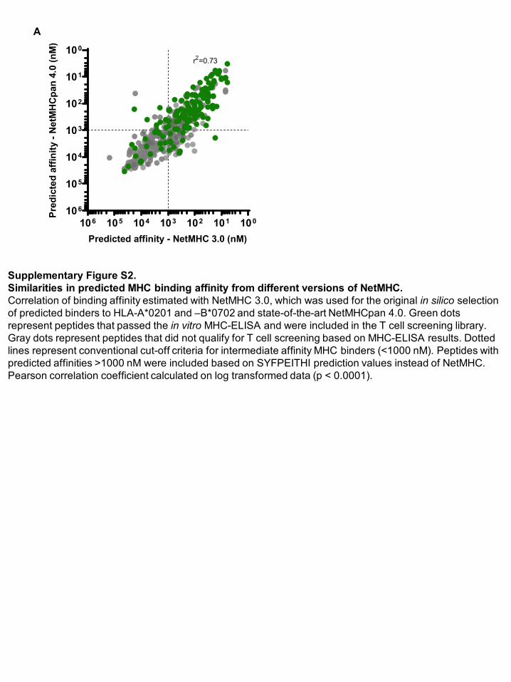
Supplementary Figure S2.Similarities in predicted MHC binding affinity from different versions of NetMHC.Correlation of binding affinity estimated with NetMHC 3.0, which was used for the original in silico selection of predicted binders to HLA-A*0201 and –B*0702 and state-of-the-art NetMHCpan 4.0. Green dots represent peptides that passed the in vitro MHC-ELISA and were included in the T cell screening library. Gray dots represent peptides that did not qualify for T cell screening based on MHC-ELISA results. Dotted lines represent conventional cut-off criteria for intermediate affinity MHC binders (<1000 nM). Peptides with predicted affinities >1000 nM were included based on SYFPEITHI prediction values instead of NetMHC. Pearson correlation coefficient calculated on log transformed data (p < 0.0001).
A
100101102103104105106
100
101
102
103
104
105
106
Predicted affinity - NetMHC 3.0 (nM)
Pre
dict
ed a
ffin
ity -
Net
MH
Cpa
n 4.
0 (n
M)
r2=0.73
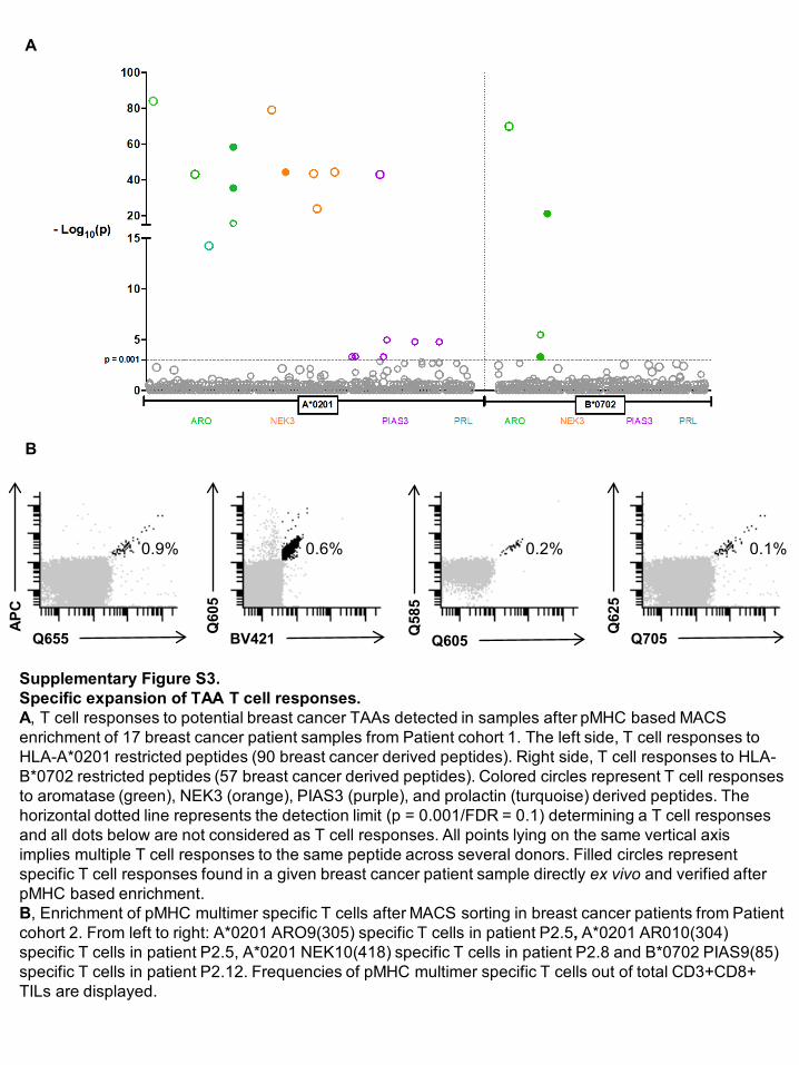
Supplementary Figure S3.Specific expansion of TAA T cell responses.A, T cell responses to potential breast cancer TAAs detected in samples after pMHC based MACS enrichment of 17 breast cancer patient samples from Patient cohort 1. The left side, T cell responses to HLA-A*0201 restricted peptides (90 breast cancer derived peptides). Right side, T cell responses to HLA-B*0702 restricted peptides (57 breast cancer derived peptides). Colored circles represent T cell responses to aromatase (green), NEK3 (orange), PIAS3 (purple), and prolactin (turquoise) derived peptides. The horizontal dotted line represents the detection limit (p = 0.001/FDR = 0.1) determining a T cell responses and all dots below are not considered as T cell responses. All points lying on the same vertical axis implies multiple T cell responses to the same peptide across several donors. Filled circles representspecific T cell responses found in a given breast cancer patient sample directly ex vivo and verified afterpMHC based enrichment. B, Enrichment of pMHC multimer specific T cells after MACS sorting in breast cancer patients from Patient cohort 2. From left to right: A*0201 ARO9(305) specific T cells in patient P2.5, A*0201 AR010(304) specific T cells in patient P2.5, A*0201 NEK10(418) specific T cells in patient P2.8 and B*0702 PIAS9(85)specific T cells in patient P2.12. Frequencies of pMHC multimer specific T cells out of total CD3+CD8+ TILs are displayed.
A
B
Q655
APC
BV421Q60
5
Q605Q58
5
Q705Q62
5
0.9% 0.6% 0.2% 0.1%
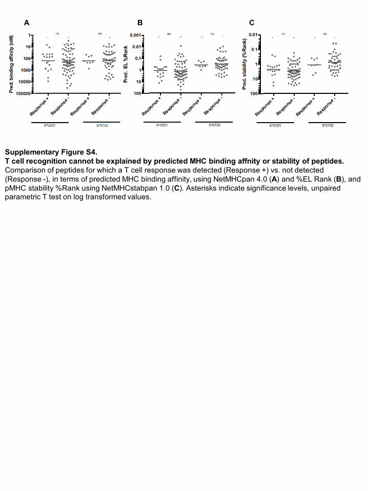
Supplementary Figure S4.T cell recognition cannot be explained by predicted MHC binding affnity or stability of peptides.Comparison of peptides for which a T cell response was detected (Response +) vs. not detected (Response -), in terms of predicted MHC binding affinity, using NetMHCpan 4.0 (A) and %EL Rank (B), and pMHC stability %Rank using NetMHCstabpan 1.0 (C). Asterisks indicate significance levels, unpaired parametric T test on log transformed values.
A B C

1
Supplementary Materials and Methods 1
2 Endogenous peptide presentation on tumor cell lines 3 4 Cells 5 The breast cancer cell lines, BT-549, MDA-MB-231 and EF192A, used for mass 6 spectometry analyses, expressed the following HLA class I alleles; BT-549: A*01:01, 7 A*02:01, B*15:17, B*56, C*03:03 and C*07:01; EFM-192A: A*01:01, A*24:02, 8 B*07:02, B*39:01, C*07:02 and C*12:03; MDA-MB-231: A*02:01, A*02:17, B*40:02, 9 B*41:01, C*0202, C*17:01 (1). All cells were cultured in R10 medium + 1% P/S. For 10 harvesting, cells were washed in PBS for 5 min, loosened with 0.25% trypsin-EDTA 11 (Gibco, Cat#25200056) for maximum 15 min at 37°C, resuspended in R10 and 12 washed three times in PBS. They were then incubated for 1 h on ice in lysis buffer 13 (0.5% IGEPAL CA-630 (Sigma-Aldrich, Cat#I8896), 0.25% sodium deoxycholate 14 (Sigma-Aldrich, Cat#D6750), 1 mM EDTA (ethylenediaminetetraacetic acid, Sigma-15 Adrich, Cat#798681) 0.2 mM iodoacetamide (Sigma-Aldrich, Cat#I6125), 1 mM 16 PMSF (phenylmethanesulfonyl fluoride, Sigma-Aldrich, Cat#P7626), Roche 17 Complete Protease Inhibitor Cocktail (Sigma-Aldrich, Cat# 11697498001) in PBS). 18 Lysates were cleared by centrifugation at 21,000 g for 30 min at 4°C and stored at -19 80°C. 20 21 MHC-IAC and desalting 22 We used the W6/32 monoclonal antibody (2) to purify peptide-HLA-A, -B, and –C 23 complexes. W6/32 was purified from HB-95 hybridoma supernatant using Protein-A 24 sepharose, and it was coupled to AminoLink Plus Coupling Resin (Thermo Scientific) 25 following the manufacturer’s instructions. The supernatant of the cell lysate was 26 slowly rotated with the W6/32 resin for 2 h at 4 °C. The resin was washed at 4 °C 27 with lysis buffer, followed by buffer A (150 mM NaCl, 20 mM Tris, pH 7.5), buffer B 28 (400 mM NaCl, 20 mM Tris, pH 7.5), a second buffer A treatment and was finally 29 washed with buffer C (20 mM Tris, pH 8.0). Peptide-MHC complexes were eluted 30 with 0.1 M acetic acid. 31 32 Peptides were desalted and separated from MHC proteins using C18 Macro 33 SpinColumns (Harvard Apparatus) prewashed with 50% acetonitrile. We equilibrated 34 and washed the C18 columns with 0.1% TFA and eluted using 30% acetonitrile / 35 0.1% TFA followed by vacuum-centrifugation. 36 37 LC-MS measurements 38 The fully dried samples were solved in 11 µl 3% CH3CN / 0.1% HCOOH, and 7 µl 39 thereof were injected into the LC-MS system. The Q Exactive was coupled online to 40 an EASY-nLC 1000 (both Thermo Scientific) via a nanoelectrospray ion source 41 (Nanospray Flex, Proxeon). Peptides were loaded at 800 bar and then separated at 42 a flow rate of 300 nl/min on a 15 cm x 50 µm column with C18 beads of 2 µm 43
2
diameter (Acclaim PepMap RSLC, Thermo Scientific). The linear gradient of 120 min 44 ranged from 0% to 30% CH3CN in 0.1% HCOOH. MS1 scans were acquired at a 45 resolution of 70,000 at 200 Th. The measured MS1 m/z range was 250 Th - 2000 46 Th, and only 2+ and 3+ charged precursors were allowed for fragmentation. The ten 47 most abundant precursors from every MS1 scan were selected for higher-energy 48 collisional dissociation (HCD) using an isolation width of 2 Th, and they were then 49 dynamically excluded from repeated fragmentation for 15 s. We applied a normalized 50 collision energy of 27%, and we recorded MS2 spectra at a resolution of 17,500 at 51 200 Th. Automatic Gain Control targets were 3,000,000 ions to be reached in 52 maximal 80 ms for MS1 and 100,000 ions to be reached in maximal 240 ms for MS2. 53 54
LC-MS data analysis 55
MS data were processed with Proteome Discoverer 1.4.1.14 from Thermo Fisher. 56 We utilized Sequest (3) HT for searching the LC-MS data allowing an MS1 mass 57 error of 10 ppm and an MS2 mass error of 0.02 Da. Oxidation of methionine and 58 cysteinylation of cysteine were set as variable modifications. The target protein 59 database contained the human reference proteome downloaded from 60 www.uniprot.org on 7 July 2017 (93,563 proteins). Our peptide spectral match false 61 discovery rate (PSM FDR) cut-off of ≤5% was calculated by Percolator (4) using the 62 reversed target protein sequences as a decoy database. The MS2 spectra of the 63 four identified peptides from PIAS3 were manually checked taking alternative peptide 64 suggestions from Proteome Discoverer into account. The putative HLA-A*02:01 65 ligand SIVAPGGAL was additionally validated by acquiring the MS2 spectrum of its 66 synthetic form (see Supplementary Figure S1). Peptides differing only in the 67 modification status of methionine or cysteine were considered as equal for peptide 68 counting. 69 70 1. Boegel S, Löwer M, Bukur T, Sahin U, Castle JC. A catalog of HLA type, HLA 71
expression, and neoepitope candidates in human cancer cell lines. Oncoimmunology. 72 2014;3:1–12. 73
2. Barnstable CJ, Bodmer WF, Brown G, Galfre G, Milstein C, Williams AF, et al. 74 Production of monoclonal antibodies to group A erythrocytes, HLA and other human 75 cell surface antigens-new tools for genetic analysis. Cell. 1978;14:1–20. 76
3. Eng JK, McCormack AL, Yates JR. An Approach to Correlate Tandem Mass Spectral 77 Data of Peptides with Amino Acid Sequences in a Protein Database. Am Soc Mass 78 Spectometry. 1994;5:976–89. 79
4. Käll L, Canterbury JD, Weston J, Noble WS, MacCoss MJ. Semi-supervised learning 80 for peptide identification from shotgun proteomics datasets. Nat Methods. 81 2007;4:923–5. 82
83
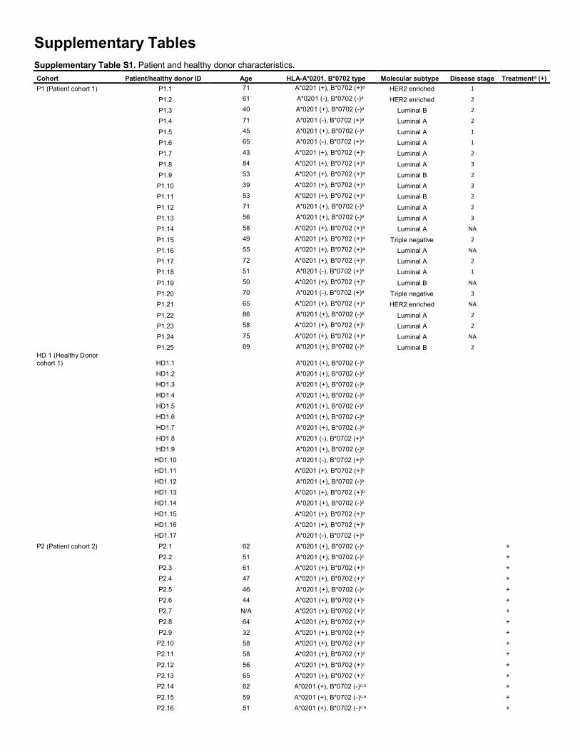
Supplementary Tables Supplementary Table S1. Patient and healthy donor characteristics. Cohort Patient/healthy donor ID Age HLA-A*0201, B*0702 type Molecular subtype Disease stage Treatmentd (+) P1 (Patient cohort 1) P1.1 71 A*0201 (+), B*0702 (+)a HER2 enriched 1
P1.2 61 A*0201 (-), B*0702 (-)a HER2 enriched 2
P1.3 40 A*0201 (+), B*0702 (-)a Luminal B 2
P1.4 71 A*0201 (-), B*0702 (+)a Luminal A 2
P1.5 45 A*0201 (+), B*0702 (-)a Luminal A 1
P1.6 65 A*0201 (-), B*0702 (+)a Luminal A 1
P1.7 43 A*0201 (+), B*0702 (+)b Luminal A 2
P1.8 84 A*0201 (+), B*0702 (+)a Luminal A 3
P1.9 53 A*0201 (+), B*0702 (+)a Luminal B 2
P1.10 39 A*0201 (+), B*0702 (+)a Luminal A 3
P1.11 53 A*0201 (+), B*0702 (+)a Luminal B 2
P1.12 71 A*0201 (+), B*0702 (-)b Luminal A 2
P1.13 56 A*0201 (+), B*0702 (-)a Luminal A 3
P1.14 58 A*0201 (+), B*0702 (+)a Luminal A NA
P1.15 49 A*0201 (+), B*0702 (+)a Triple negative 2
P1.16 55 A*0201 (+), B*0702 (+)a Luminal A NA
P1.17 72 A*0201 (+), B*0702 (+)a Luminal A 2
P1.18 51 A*0201 (-), B*0702 (+)b Luminal A 1
P1.19 50 A*0201 (+), B*0702 (+)b Luminal B NA
P1.20 70 A*0201 (-), B*0702 (+)a Triple negative 3
P1.21 65 A*0201 (+), B*0702 (+)a HER2 enriched NA
P1.22 86 A*0201 (+), B*0702 (-)b Luminal A 2
P1.23 58 A*0201 (+), B*0702 (+)b Luminal A 2
P1.24 75 A*0201 (+), B*0702 (+)a Luminal A NA
P1.25 69 A*0201 (+), B*0702 (-)b Luminal B 2
HD 1 (Healthy Donor cohort 1) HD1.1
A*0201 (+), B*0702 (-)b
HD1.2
A*0201 (+), B*0702 (-)b
HD1.3
A*0201 (+), B*0702 (-)b
HD1.4
A*0201 (+), B*0702 (-)b
HD1.5
A*0201 (+), B*0702 (-)b
HD1.6
A*0201 (+), B*0702 (-)b
HD1.7
A*0201 (+), B*0702 (-)b
HD1.8
A*0201 (-), B*0702 (+)b
HD1.9
A*0201 (+), B*0702 (-)b
HD1.10
A*0201 (-), B*0702 (+)b
HD1.11
A*0201 (+), B*0702 (+)b
HD1.12
A*0201 (+), B*0702 (-)b
HD1.13
A*0201 (+), B*0702 (+)b
HD1.14
A*0201 (+), B*0702 (-)b
HD1.15
A*0201 (+), B*0702 (+)b
HD1.16
A*0201 (+), B*0702 (+)b
HD1.17
A*0201 (-), B*0702 (+)b
P2 (Patient cohort 2) P2.1 62 A*0201 (+), B*0702 (-)c +
P2.2 51 A*0201 (+), B*0702 (-)c +
P2.3 61 A*0201 (+), B*0702 (+)c +
P2.4 47 A*0201 (+), B*0702 (+)c +
P2.5 46 A*0201 (+), B*0702 (-)c +
P2.6 44 A*0201 (+), B*0702 (+)c +
P2.7 N/A A*0201 (+), B*0702 (+)c +
P2.8 64 A*0201 (+), B*0702 (+)c +
P2.9 32 A*0201 (+), B*0702 (+)c +
P2.10 58 A*0201 (+), B*0702 (+)c +
P2.11 58 A*0201 (+), B*0702 (+)c +
P2.12 56 A*0201 (+), B*0702 (+)c +
P2.13 65 A*0201 (+), B*0702 (+)c +
P2.14 62 A*0201 (+), B*0702 (-)c,a +
P2.15 59 A*0201 (+), B*0702 (-)c,a +
P2.16 51 A*0201 (+), B*0702 (-)c,a +

P2.17 65 A*0201 (+), B*0702 (-)c,a
P2.18 56 A*0201 (+), B*0702 (-)c,a HD2 (Healthy Donor cohort
2) HD2.1
A*0201 (-), B*0702 (+)c
HD2.2
A*0201 (+), B*0702 (+)c
HD2.3
A*0201 (+), B*0702 (+)c
HD2.4
A*0201 (-), B*0702 (+)c
HD2.5
A*0201 (-), B*0702 (+)c
HD2.6
A*0201 (-), B*0702 (+)c
HD2.7
A*0201 (+), B*0702 (+)c
HD2.8
A*0201 (+), B*0702 (+)c
HD2.9
A*0201 (+), B*0702 (-)c
HD2.10
A*0201 (+), B*0702 (-)c
HD2.11
A*0201 (+), B*0702 (-)c
HD2.12
A*0201 (+), B*0702 (-)c HD2.13 A*0201 (+), B*0702 (-)c
aHLA type determined by flow cytometry. bHLA type determined by sequencing. cHLA type determined by PCR. dPrior treatments included chemo-, radiation and endocrine therapy. No patients were treated with immunotherapy. NA: disease stage classification not possible due to lack of information.
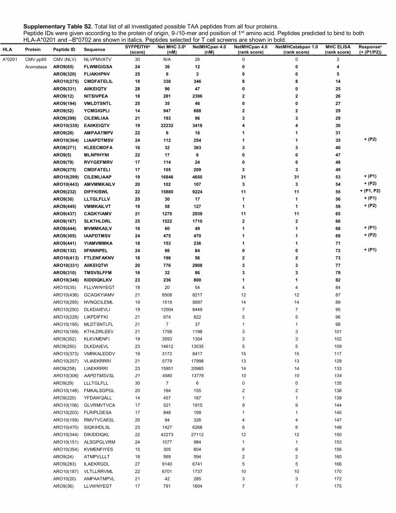
Supplementary Table S2. Total list of all investigated possible TAA peptides from all four proteins. Peptide IDs were given according to the protein of origin, 9-/10-mer and position of 1st amino acid. Peptides predicted to bind to both HLA-A*0201 and –B*0702 are shown in italics. Peptides selected for T cell screens are shown in bold.
HLA Protein Peptide ID Sequence SYFPEITHIa (score)
Net MHC 3.0b (nM)
NetMHCpan 4.0 (nM)
NetMHCpan 4.0 (rank score)
NetMHCstabpan 1.0 (rank score)
MHC ELISA (rank score)
Responsec (+ (P1/P2))
A*0201 CMV pp65 CMV (NLV) NLVPMVATV 30 N/A 26 0 0 3
Aromatase ARO9(65) FLWMGIGSA 24 36 12 0 0 4
ARO9(320) FLIAKHPNV 25 6 3 0 0 5
ARO10(275) CMDFATELIL 18 338 346 8 8 14
ARO9(331) AIIKEIQTV 28 96 47 0 0 25
ARO9(12) NITSIVPEA 18 281 2396 2 2 26
ARO9(194) VMLDTSNTL 25 35 46 0 0 27
ARO9(52) YCMGIGPLI 14 947 688 2 2 29
ARO9(299) CILEMLIAA 21 193 96 3 3 29
ARO10(330) EAIIKEIQTV 19 22232 3418 4 4 30
ARO9(20) AMPAATMPV 22 6 16 1 1 31
ARO10(304) LIAAPDTMSV 24 112 254 1 1 35 + (P2)
ARO9(271) KLEECMDFA 16 32 363 3 3 40
ARO9(5) MLNPIHYNI 22 17 6 0 0 47
ARO9(79) RVYGEFMRV 17 114 24 0 0 48
ARO9(275) CMDFATELI 17 105 209 3 3 49
ARO10(299) CILEMLIAAP 19 16846 4650 31 31 53 + (P1)
ARO10(443) AMVMMKAILV 20 102 107 3 3 54 + (P2)
ARO9(232) DIFFKISWL 22 15880 9224 11 11 55 + (P1, P2)
ARO9(30) LLTGLFLLV 25 30 17 1 1 56 + (P1)
ARO9(445) VMMKAILVT 19 58 127 1 1 59 + (P2)
ARO9(437) CAGKYIAMV 21 1270 2939 11 11 65
ARO9(167) SLKTHLDRL 25 1522 1710 2 2 66
ARO9(444) MVMMKAILV 16 60 49 1 1 68 + (P1)
ARO9(305) IAAPDTMSV 24 475 470 1 1 69 + (P2)
ARO9(441) YIAMVMMKA 18 153 236 1 1 71
ARO9(132) IIFNNNPEL 24 86 84 0 0 72 + (P1)
ARO10(413) FTLENFAKNV 18 196 56 2 2 73
ARO10(331) AIIKEIQTVI 20 776 2908 3 3 77
ARO9(310) TMSVSLFFM 16 32 86 3 3 79
ARO10(346) KIDDIQKLKV 23 236 800 1 1 82
ARO10(35) FLLVWNYEGT 18 20 54 4 4 84
ARO10(436) GCAGKYIAMV 21 6508 8217 12 12 87
ARO10(295) NVNQCILEML 19 1519 5697 14 14 89
ARO10(250) DLKDAIEVLI 19 12004 8449 7 7 95
ARO10(228) LIKPDIFFKI 21 674 822 5 5 96
ARO10(195) MLDTSNTLFL 21 7 37 1 1 98
ARO10(169) KTHLDRLEEV 21 1758 1198 3 3 101
ARO9(352) KLKVMENFI 19 3593 1354 3 3 102
ARO9(250) DLKDAIEVL 23 14612 13035 5 5 109
ARO10(373) VMRKALEDDV 19 3172 8417 15 15 117
ARO10(257) VLIAEKRRRI 21 5779 17998 13 13 129
ARO9(258) LIAEKRRRI 23 15951 20985 14 14 133
ARO10(306) AAPDTMSVSL 21 4580 13776 10 10 134
ARO9(29) LLLTGLFLL 30 7 6 0 0 135
ARO10(148) FMKALSGPGL 20 164 155 2 2 138
ARO9(220) YFDAWQALL 14 457 187 1 1 139
ARO10(156) GLVRMVTVCA 17 521 1915 9 9 144
ARO10(203) FLRIPLDESA 17 848 199 1 1 145
ARO10(159) RMVTVCAESL 20 94 326 4 4 147
ARO10(470) SIQKIHDLSL 23 1427 6268 6 6 148
ARO10(344) DIKIDDIQKL 22 42273 27112 12 12 150
ARO10(151) ALSGPGLVRM 24 1077 984 1 1 153
ARO10(354) KVMENFIYES 15 305 604 6 6 156
ARO9(24) ATMPVLLLT 18 569 594 2 2 160
ARO9(283) ILAEKRGDL 27 9140 6741 5 5 166
ARO10(187) VLTLLRRVML 22 6701 1737 10 10 170
ARO10(20) AMPAATMPVL 21 42 285 3 3 172
ARO9(36) LLVWNYEGT 17 791 1604 7 7 175

ARO9(150) KALSGPGLV 19 3616 4206 5 5 177
ARO10(355) VMENFIYESM 14 418 941 6 6 182
ARO10(442) IAMVMMKAIL 19 6257 3091 19 19 184
ARO9(28) VLLLTGLFL 24 285 155 1 1 188
ARO9(443) AMVMMKAIL 22 1166 1245 4 4 198
ARO10(25) TMPVLLLTGL 23 261 474 7 7 201
ARO9(303) MLIAAPDTM 18 182 209 1 1 203
ARO10(283) ILAEKRGDLT 20 7703 11960 11 11 208
ARO10(205) RIPLDESAIV 20 3859 1006 2 2 209
ARO9(58) PLISHGRFL 19 17500 12429 8 8 211
ARO9(332) IIKEIQTVI 19 6366 5685 3 3 214
ARO9(23) AATMPVLLL 21 10302 8692 7 7 218
ARO9(446) MMKAILVTL 25 52 36 1 1 219
ARO10(446) MMKAILVTLL 21 72 78 4 4 223
ARO9(188) LTLLRRVML 20 12623 10599 14 14 224
ARO9(186) DVLTLLRRV 20 14321 11734 9 9 226
ARO9(182) SGYVDVLTL 20 10396 11249 6 6 230
ARO9(35) FLLVWNYEG 16 320 83 3 3 231
ARO9(170) THLDRLEEV 19 11002 15724 11 11 232
ARO10(462) TLQGQCVESI 26 172 856 4 4 234
ARO9(414) TLENFAKNV 19 5794 2336 2 2 235
ARO9(339) VIGERDIKI 21 2562 5586 3 3 236
ARO10(441) YIAMVMMKAI 20 359 440 7 7 237
ARO10(395) ILNIGRMHRL 24 932 787 2 2 238
ARO9(345) IKIDDIQKL 21 16405 24301 8 8 239
ARO10(445) VMMKAILVTL 28 7 32 1 1 242
ARO9(207) PLDESAIVV 19 3812 1715 1 1 243
ARO10(363) SMRYQPVVDL 25 3481 1328 2 2 245
ARO9(95) LIISKSSSM 19 3537 7434 8 8 246
ARO10(29) LLLTGLFLLV 25 8 11 1 1 247
ARO9(153) SGPGLVRMV 19 28324 20957 14 14 248
ARO10(239) WLYKKYEKSV 22 129 304 4 4 249
ARO9(313) VSLFFMLFL 14 937 2632 12 12 250
ARO10(88) WISGEETLII 19 2070 4923 11 11 251
ARO10(282) LILAEKRGDL 22 17787 19054 24 24 252
ARO10(454) LLRRFHVKTL 27 5358 4168 15 15 254
ARO9(311) MSVSLFFML 11 926 352 6 6 255
ARO10(131) GIIFNNNPEL 23 752 2044 2 2 256
ARO9(454) LLRRFHVKT 21 16441 10164 13 13 257
ARO10(453) TLLRRFHVKT 20 3324 5895 14 14 258
ARO10(178) VTNESGYVDV 20 4496 10738 11 11 259
ARO9(317) FMLFLIAKH 19 8354 12450 20 20 260
ARO10(4) EMLNPIHYNI 16 906 177 3 3 261
ARO9(314) SLFFMLFLI 24 17 18 1 1 261
ARO9(300) ILEMLIAAP 19 25105 5320 9 9 262
ARO10(271) KLEECMDFAT 17 372 1293 7 7 263
ARO10(5) MLNPIHYNIT 18 906 418 4 4 264
ARO10(28) VLLLTGLFLL 27 75 25 1 1 265
ARO10(310) TMSVSLFFML 18 32 63 5 5 267
ARO10(312) SVSLFFMLFL 17 656 702 9 9 268
ARO10(314) SLFFMLFLIA 17 116 97 5 5 269
NEK3 NEK9(226) YSYELQFLV 16 14 8 0 0 1 + (P1)
NEK10(104) ILNWFTQMCL 23 55 46 3 3 2 + (P1)
NEK9(151) LLSNPMAFA 19 121 47 1 1 7
NEK10(407) WLKETPDTLL 21 2625 1003 2 2 8
NEK9(97) KLFPEDMIL 24 39 27 0 0 8
NEK9(425) SLAFQTYTI 26 63 16 0 0 9
NEK9(414) TLLNILKNA 19 424 198 1 1 10 + (P1)
NEK10(160) CTYVGTPYYV 16 97 373 7 7 12
NEK9(187) SLGCILYEL 29 22 8 0 0 15 + (P1)
NEK9(301) ALGNEASTV 24 469 288 0 0 17
NEK9(269) IIMEYGEEV 26 12 6 0 0 19
NEK10(142) KLGDFGSARL 24 55 258 1 1 20 + (P1)

NEK10(150) RLLSNPMAFA 18 58 140 1 1 23
NEK9(90) KIKQQKGKL 20 41685 34481 25 25 36
NEK10(186) WSLGCILYEL 21 555 72 6 6 37 + (P1)
NEK10(53) LLAKMKHPNI 23 274 1217 7 7 39 + (P1)
NEK10(213) KVCQGCISPL 19 2155 1274 12 12 43
NEK10(28) QMFAMKEIRL 19 149 194 3 3 44 + (P1)
NEK9(103) MILNWFTQM 14 100 163 3 3 44 + (P2)
NEK9(155) PMAFACTYV 16 517 278 6 6 57 + (P1)
NEK10(329) NLVESALRRV 26 450 884 2 2 58 + (P1)
NEK10(418) ILKNADLSLA 19 3055 1205 4 4 63 + (P2)
NEK10(221) PLPSHYSYEL 20 4539 7824 15 15 67
NEK10(371) ALTALENASI 23 225 679 3 3 70
NEK10(268) EIIMEYGEEV 19 5960 400 3 3 76
NEK9(190) CILYELCTL 25 1165 798 10 10 81 + (P1,P2)
NEK10(366) NVPNTALTAL 19 9006 13190 13 13 93
NEK9(110) QMCLGVNHI 23 8659 1346 4 4 94
NEK9(270) IMEYGEEVL 20 2713 847 2 2 100
NEK10(134) FLTQNGKVKL 24 309 601 1 1 107
NEK10(269) IIMEYGEEVL 23 111 462 1 1 108
NEK10(325) SINENLVESA 23 465 2824 3 3 111
NEK10(260) LVQKCLPPEI 17 576 2304 16 16 114
NEK10(69) FEAEGHLYIV 20 1596 2386 6 6 114
NEK9(418) ILKNADLSL 24 669 430 1 1 116
NEK10(189) GCILYELCTL 19 17640 1698 11 11 120
NEK9(250) TLLSRGIVA 16 619 1185 1 1 122
NEK10(300) IALGNEASTV 22 4313 1467 2 2 124
NEK9(211) ILKVCQGCI 19 10329 3904 2 2 125
NEK9(379) SILTSSLTA 19 740 1972 4 4 136
NEK9(377) NASILTSSL 19 11707 16784 17 17 142
NEK10(168) YVPPEIWENL 18 300 456 1 1 143
NEK9(407) WLKETPDTL 23 3069 318 0 0 145
NEK9(455) SVDGGHDSV 22 2473 1595 1 1 152
NEK10(417) NILKNADLSL 23 1468 4278 6 6 154
NEK10(415) LLNILKNADL 25 2379 1653 9 9 159
NEK9(135) LTQNGKVKL 22 19777 21714 12 12 164
NEK10(132) NIFLTQNGKV 21 3874 1975 8 8 165
NEK9(372) LTALENASI 20 3270 5993 7 7 169
NEK9(206) SWKNLILKV 19 28235 22990 13 13 176
NEK9(325) SINENLVES 21 7128 7182 5 5 179
NEK10(250) TLLSRGIVAR 19 23113 15255 13 13 180
NEK10(251) LLSRGIVARL 28 92 160 1 1 186
NEK10(210) LILKVCQGCI 20 1951 3366 27 27 187
NEK9(252) LSRGIVARL 20 19628 19707 14 14 191
NEK10(349) HLRKASSPNL 21 4663 8660 9 9 196
NEK9(330) LVESALRRV 19 7626 12072 10 10 199
NEK10(179) YNNKSDIWSL 19 10130 17082 20 20 201
NEK9(251) LLSRGIVAR 21 24993 16822 13 13 204
NEK9(184) DIWSLGCIL 19 14064 8795 14 14 207
NEK10(322) DLESINENLV 19 15053 17498 13 13 227
NEK9(441) FLKGPLSEE 19 27751 8179 7 7 231
NEK10(205) NSWKNLILKV 18 570 1596 7 7 240
NEK9(469) RLEPGLDEE 19 32961 25608 16 16 244
PIAS3 PIAS9(15) RVSELQVLL 19 456 203 0 0 13
PIAS10(106) LLGPKREVDM 20 9971 14031 12 12 16 + (P1)
PIAS9(345) HLQSFDAAL 24 177 128 0 0 18
PIAS9(79) SLPPGTSPV 28 23 24 0 0 22 + (P1, P2)
PIAS10(160) FTFALTPQQV 16 33 77 2 2 28 + (P1)
PIAS9(569) FLGPLAPTL 27 15 7 0 0 32
PIAS9(249) RLSATVPNT 19 426 937 2 2 33
PIAS9(347) QSFDAALYL 13 820 1315 2 2 34
PIAS10(494) VLRSPAMGTL 23 6956 2095 4 4 41
PIAS10(44) LLKSSCAPSV 24 123 84 4 4 42 + (P1)
PIAS10(346) LQSFDAALYL 12 862 174 2 2 44 +(P2)
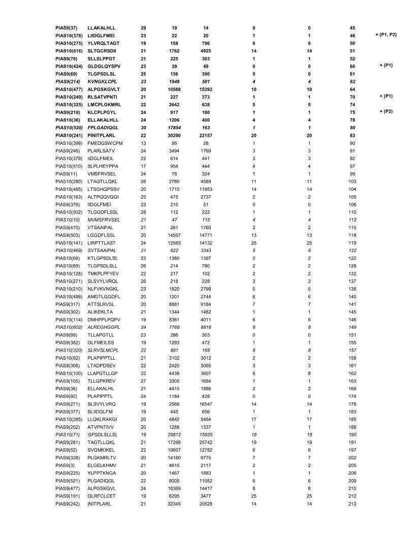
PIAS9(37) LLAKALHLL 29 19 14 0 0 45
PIAS10(378) LIIDGLFMEI 23 22 20 1 1 46 + (P1, P2)
PIAS10(275) YLVRQLTAGT 19 158 796 6 6 50
PIAS10(616) SLTGCRSDII 21 1762 4925 14 14 51
PIAS9(76) SLLSLPPGT 21 225 303 1 1 52
PIAS10(424) GLDGLQYSPV 23 39 49 0 0 60 + (P1)
PIAS9(69) TLGPSDLSL 25 156 390 0 0 61
PIAS9(214) KVNGKLCPL 23 1948 501 4 4 62
PIAS10(477) ALPGSKGVLT 20 10588 15292 10 10 64
PIAS10(249) RLSATVPNTI 21 227 373 1 1 70 + (P1)
PIAS10(325) LMCPLGKMRL 22 2642 638 5 5 74
PIAS9(218) KLCPLPGYL 24 917 180 1 1 75 + (P2)
PIAS10(36) ELLAKALHLL 24 1206 400 4 4 78
PIAS10(520) FPLGADIQGL 20 17854 163 1 1 80
PIAS10(241) PINITPLARL 22 30290 22157 20 20 83
PIAS10(399) FMEDGSWCPM 13 95 28 1 1 90
PIAS9(246) PLARLSATV 24 3494 1769 3 3 91
PIAS10(379) IIDGLFMEIL 22 614 441 3 3 92
PIAS10(510) SLPLHEYPPA 17 954 444 4 4 97
PIAS9(11) VMSFRVSEL 24 76 324 1 1 99
PIAS10(280) LTAGTLLQKL 26 2760 4589 11 11 103
PIAS10(485) LTSGHQPSSV 20 1715 11953 14 14 104
PIAS10(163) ALTPQQVQQI 25 475 2737 2 2 105
PIAS9(379) IIDGLFMEI 23 215 51 0 0 106
PIAS10(502) TLGGDFLSSL 28 112 222 1 1 110
PIAS10(10) MVMSFRVSEL 21 47 115 4 4 112
PIAS9(470) VTSAAIPAL 21 261 1760 2 2 115
PIAS9(503) LGGDFLSSL 20 14557 14771 13 13 118
PIAS10(141) LIRPTTLAST 24 12565 14132 25 25 119
PIAS10(469) SVTSAAIPAL 21 622 3343 5 5 122
PIAS10(68) KTLGPSDLSL 22 1380 1397 2 2 122
PIAS10(69) TLGPSDLSLL 26 214 780 2 2 128
PIAS10(128) TMKPLPFYEV 22 217 102 2 2 132
PIAS10(271) SLSVYLVRQL 26 218 228 3 3 137
PIAS10(210) NLFVKVNGKL 23 1820 2799 5 5 138
PIAS10(499) AMGTLGGDFL 20 1201 2744 6 6 140
PIAS9(317) ATTSLRVSL 20 8881 9184 7 7 141
PIAS9(302) ALIKEKLTA 21 1344 1482 1 1 145
PIAS10(114) DMHPPLPQPV 19 8361 4011 6 6 146
PIAS10(602) ALREGHGGPL 24 7768 8818 8 8 149
PIAS9(99) TLLAPGTLL 23 286 303 0 0 151
PIAS9(382) GLFMEILSS 19 1293 472 1 1 155
PIAS10(320) SLRVSLMCPL 22 881 168 8 8 157
PIAS10(92) PLAPIPPTLL 21 3102 3012 2 2 158
PIAS9(308) LTADPDSEV 22 2420 3065 3 3 161
PIAS10(100) LLAPGTLLGP 22 4438 3607 8 8 162
PIAS9(105) TLLGPKREV 27 3305 1684 1 1 163
PIAS9(36) ELLAKALHL 21 4415 1886 2 2 168
PIAS9(92) PLAPIPPTL 24 1184 428 0 0 174
PIAS9(271) SLSVYLVRQ 19 2568 16547 14 14 178
PIAS9(377) SLIIDGLFM 19 445 656 1 1 183
PIAS10(285) LLQKLRAKGI 20 4842 5464 17 17 185
PIAS9(252) ATVPNTIVV 20 1288 1337 1 1 189
PIAS10(71) GPSDLSLLSL 19 29812 15935 18 18 190
PIAS9(281) TAGTLLQKL 21 17299 25742 19 19 191
PIAS9(52) SVQMKIKEL 22 10607 12782 6 6 197
PIAS9(328) PLGKMRLTV 20 14160 9775 7 7 202
PIAS9(3) ELGELKHMV 21 4615 2117 2 2 205
PIAS9(225) YLPPTKNGA 20 1467 1883 1 1 206
PIAS9(521) PLGADIQGL 22 8005 11052 6 6 209
PIAS9(477) ALPGSKGVL 24 16369 14417 8 8 210
PIAS9(191) QLRFCLCET 19 6295 3477 25 25 212
PIAS9(242) INITPLARL 21 32345 20528 14 14 213
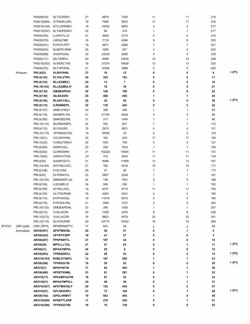
PIAS9(616) SLTGCRSDI 21 6679 7393 11 11 215
PIAS10(564) GTPSHFLGPL 19 7685 5872 17 17 216
PIAS10(104) GTLLGPKREV 19 14432 6853 3 3 217
PIAS10(307) KLTADPDSEV 22 95 316 1 1 217
PIAS9(100) LLAPGTLLG 21 3849 3770 4 4 219
PIAS9(378) LIIDGLFME 19 7116 6386 8 8 220
PIAS9(221) PLPGYLPPT 19 4671 2946 7 7 221
PIAS9(324) SLMCPLGKM 24 1955 327 2 2 222
PIAS9(595) SIVAPGGAL 22 22029 6582 4 4 225
PIAS9(171) QILTSREVL 20 9599 14816 12 12 228
PIAS10(302) ALIKEKLTAD 19 27274 19428 13 13 233
PIAS9(333) RLTVPCRAL 21 12349 2988 5 5 253
Prolactin PRL9(52) VLSHYIHNL 27 10 12 0 0 6 + (P1)
PRL9(122) PLYHLVTEV 25 325 163 0 0 11
PRL9(153) RLLEGMELI 24 13 7 0 0 16
PRL10(153) RLLEGMELIV 25 19 16 0 0 21
PRL9(132) GMQEAPEAI 20 146 160 0 0 22
PRL9(139) AILSKAVEI 25 408 455 1 1 24
PRL9(192) RLSAYYNLL 22 32 45 0 0 38 + (P1)
PRL9(115) ILRSWNEPL 22 139 224 2 2 42
PRL9(157) GMELIVSQV 22 308 456 1 1 85
PRL9(118) SWNEPLYHL 21 27144 6428 3 3 86
PRL9(185) QMADEESRL 21 317 1040 1 1 88
PRL10(114) SILRSWNEPL 22 555 627 7 7 113
PRL9(110) SLIVSILRS 19 2815 3621 4 4 121
PRL10(175) YPVWSGLPSL 19 16548 42 0 0 123
PRL10(51) VVLSHYIHNL 22 162 343 17 17 126
PRL10(42) TLRDLFDRAV 22 1361 745 3 3 127
PRL9(209) KIDNYLKLL 23 209 1033 1 1 130
PRL9(202) CLRRDSHKI 21 152223 10544 11 11 131
PRL10(82) AINSCHTSSL 23 515 4932 11 11 134
PRL9(35) GAARCQVTL 21 8548 11959 13 13 167
PRL10(194) SAYYNLLHCL 21 782 5518 15 15 171
PRL9(108) FLSLIVSIL 25 21 26 1 1 173
PRL9(45) DLFDRAVVL 23 3857 2248 1 1 181
PRL10(102) QMNQKDFLSL 22 136 1501 5 5 188
PRL9(154) LLEGMELIV 24 509 236 1 1 192
PRL9(195) AYYNLLHCL 19 6747 9773 11 11 193
PRL9(125) HLVTEVRGM 19 4653 5341 3 3 194
PRL9(173) EIYPVWSGL 19 11018 5615 5 5 195
PRL9(176) PVWSGLPSL 21 1656 1572 2 2 200
PRL10(132) GMQEAPEAIL 23 266 1030 1 1 207
PRL9(213) YLKLLKCRI 20 7459 2576 8 8 229
PRL10(213) YLKLLKCRII 19 9803 4079 23 23 241
PRL10(110) SLIVSILRSW 20 23774 15402 16 16 266
B*0702 CMV pp65 CMV (RPH) RPHERNGFTV 17 N/A 34 0 0 26
Aromatase ARO9(307) APDTMSVSL 25 30 51 0 1 2
ARO9(422) VPYRYFQPF 18 41 27 0 0 5
ARO9(407) FPKPNEFTL 21 187 44 0 0 10
ARO9(26) MPVLLLTGL 21 47 63 0 1 11 + (P1)
ARO9(21) MPAATMPVL 24 25 9 0 0 12
ARO9(493) TPRNSDRCL 22 28 53 0 0 13 + (P2)
ARO10(193) RVMLDTSNTL 12 187 500 1 1 23
ARO9(386) YPVKKGTNI 19 20 52 0 0 29 + (P1)
ARO10(7) NPIHYNITSI 17 85 592 1 1 30
ARO9(480) HPDETKNML 21 81 281 0 1 32
ARO10(17) VPEAMPAATM 18 87 53 0 1 33
ARO10(21) MPAATMPVLL 24 46 36 0 1 37
ARO10(307) APDTMSVSLF 20 152 439 0 2 47
ARO10(57) GPLISHGRFL 21 73 184 0 1 48 + (P1)
ARO9(154) GPGLVRMVT 19 903 455 1 6 49
ARO10(409) KPNEFTLENF 17 279 259 0 1 51
ARO10(386) YPVKKGTNII 18 15 129 0 0 53
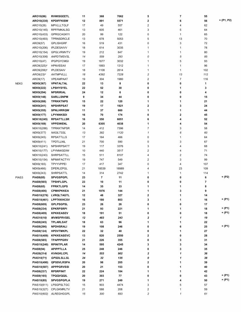
ARO10(86) RVWISGEETL 11 368 7062 5 7 55
ARO10(230) KPDIFFKISW 12 891 5571 2 6 56 + (P1, P2)
ARO10(26) MPVLLLTGLF 17 49 557 2 4 62
ARO10(145) RPFFMKALSG 13 605 461 3 5 64
ARO10(433) GPRGCAGKYI 20 99 122 0 1 65
ARO10(493) TPRNSDRCLE 13 678 5053 5 13 70
ARO9(57) GPLISHGRF 16 519 431 0 3 72
ARO10(206) IPLDESAIVV 18 614 3035 1 1 78
ARO10(154) GPGLVRMVTV 19 212 647 1 1 80
ARO10(306) AAPDTMSVSL 14 559 293 0 5 85
ARO10(47) IPGPGYCMGI 19 1677 3032 1 5 93
ARO9(325)d HPNVEEAII 17 1893 1312 1 1 96
ARO9(206)d IPLDESAIV 18 1108 2614 1 1 102
ARO9(23)d AATMPVLLL 18 4392 7228 2 13 112
ARO9(17) VPEAMPAAT 19 304 1980 2 7 116
NEK3 NEK9(367) VPNTALTAL 22 13 8 0 0 1
NEK9(222) LPSHYSYEL 22 52 30 0 1 3
NEK9(294) NPSRIRIAL 24 12 6 0 0 4
NEK9(148) SARLLSNPM 9 34 44 0 0 15
NEK9(288) TPRKKTNPS 15 22 120 1 1 21
NEK9(241) NPSHRPSAT 19 17 1921 2 3 24
NEK9(355) SPNLHRRQW 11 37 660 1 1 44
NEK9(177) LPYNNKSDI 16 70 174 0 2 45
NEK10(245) RPSATTLLSR 16 350 6051 6 4 52
NEK9(169) VPPEIWENL 20 6305 4038 1 4 54
NEK10(288) TPRKKTNPSR 14 412 7396 7 3 58
NEK9(377) NASILTSSL 13 382 1120 1 6 60
NEK9(245) RPSATTLLS 17 164 466 0 1 64
NEK9(411) TPDTLLNIL 21 700 590 0 3 67
NEK10(241) NPSHRPSATT 19 117 3376 3 4 68
NEK10(177) LPYNNKSDIW 10 440 3517 2 4 71
NEK10(243) SHRPSATTLL 15 511 8107 4 2 74
NEK10(154) NPMAFACTYV 19 747 549 2 3 99
NEK9(165) TPYYVPPEI 17 417 347 0 4 107
NEK9(466) DPERLEPGL 21 18539 18989 4 23 109
NEK9(243) SHRPSATTL 14 314 2742 1 1 114
PIAS3 PIAS9(85) SPVGSPGPL 23 7 11 0 0 6 + (P2)
PIAS9(565) TPSHFLGPL 23 10 11 0 0 7
PIAS9(65) FPRKTLGPS 14 35 33 1 1 8
PIAS9(406) CPMKPKKEA 21 1976 144 1 3 9
PIAS10(276) LVRQLTAGTL 12 48 327 2 1 14
PIAS10(461) LPPTKKHCSV 16 180 803 3 1 16 + (P1)
PIAS9(609) GPLPSGPSL 23 26 26 0 0 17
PIAS9(234) EPKRPSRPI 21 93 221 1 1 18 + (P1)
PIAS9(409) KPKKEASEV 18 191 51 0 0 18 + (P1)
PIAS10(10) MVMSFRVSEL 12 483 243 2 1 19
PIAS9(245) TPLARLSAT 18 63 96 1 1 22
PIAS9(296) NPDHSRALI 19 108 246 0 6 25 + (P1)
PIAS9(124) HPDVTMKPL 22 32 40 0 1 27
PIAS10(409) KPKKEASEVC 13 820 2550 2 0 28
PIAS9(585) TPAPPPGRV 21 226 355 0 3 31
PIAS10(240) RPINITPLAR 14 595 4245 3 3 34
PIAS9(94) APIPPTLLA 25 248 246 0 3 35
PIAS9(214) KVNGKLCPL 14 553 902 2 1 36
PIAS10(71) GPSDLSLLSL 24 32 135 0 1 38
PIAS10(490) QPSSVLRSPA 20 98 205 2 1 39
PIAS10(587) APPPGRVSSI 20 21 153 0 1 40
PIAS9(237) RPSRPINIT 22 224 184 1 1 42
PIAS9(165) TPQQVQQIL 20 303 77 0 0 43 + (P1)
PIAS10(85) SPVGSPGPLA 19 271 249 1 4 50 + (P1)
PIAS10(611) LPSGPSLTGC 15 903 4474 3 5 57
PIAS10(327) CPLGKMRLTV 21 588 208 2 1 59
PIAS10(602) ALREGHGGPL 15 300 693 2 1 61
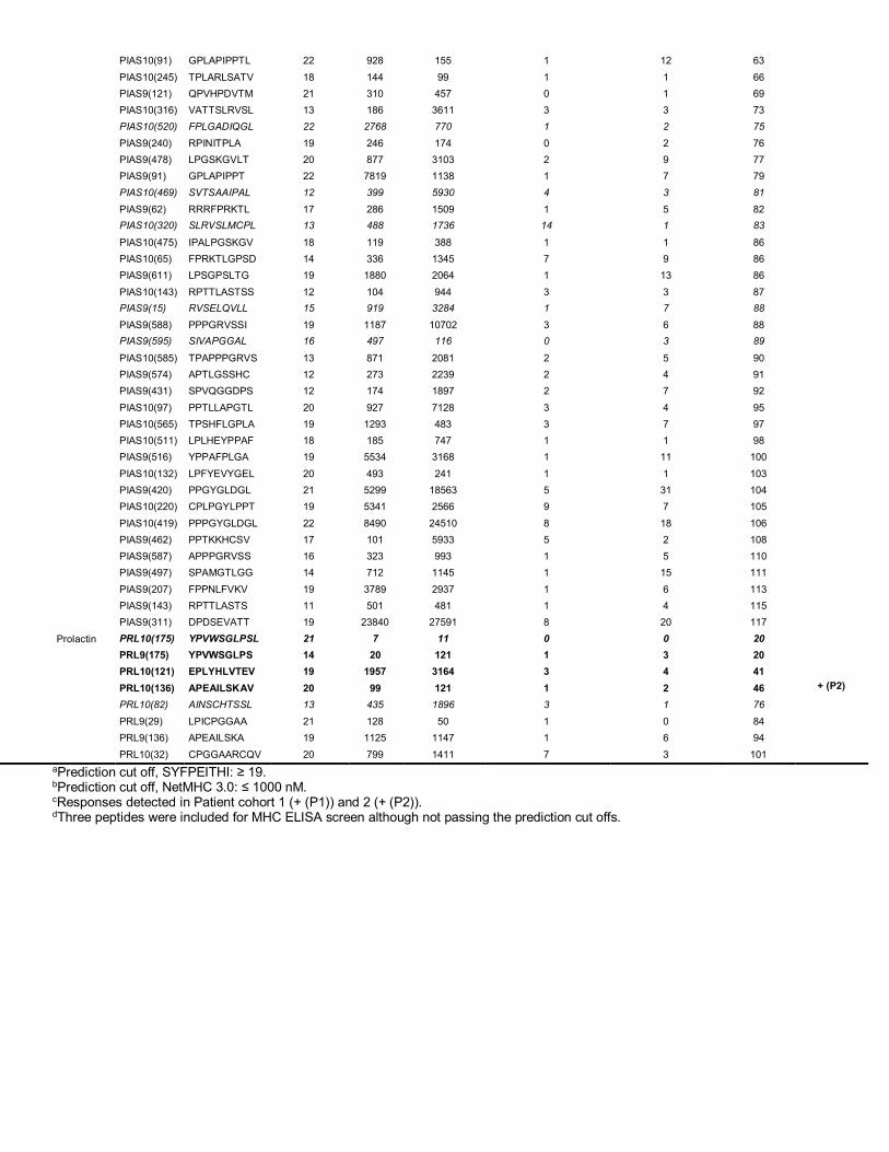
PIAS10(91) GPLAPIPPTL 22 928 155 1 12 63
PIAS10(245) TPLARLSATV 18 144 99 1 1 66
PIAS9(121) QPVHPDVTM 21 310 457 0 1 69
PIAS10(316) VATTSLRVSL 13 186 3611 3 3 73
PIAS10(520) FPLGADIQGL 22 2768 770 1 2 75
PIAS9(240) RPINITPLA 19 246 174 0 2 76
PIAS9(478) LPGSKGVLT 20 877 3103 2 9 77
PIAS9(91) GPLAPIPPT 22 7819 1138 1 7 79
PIAS10(469) SVTSAAIPAL 12 399 5930 4 3 81
PIAS9(62) RRRFPRKTL 17 286 1509 1 5 82
PIAS10(320) SLRVSLMCPL 13 488 1736 14 1 83
PIAS10(475) IPALPGSKGV 18 119 388 1 1 86
PIAS10(65) FPRKTLGPSD 14 336 1345 7 9 86
PIAS9(611) LPSGPSLTG 19 1880 2064 1 13 86
PIAS10(143) RPTTLASTSS 12 104 944 3 3 87
PIAS9(15) RVSELQVLL 15 919 3284 1 7 88
PIAS9(588) PPPGRVSSI 19 1187 10702 3 6 88
PIAS9(595) SIVAPGGAL 16 497 116 0 3 89
PIAS10(585) TPAPPPGRVS 13 871 2081 2 5 90
PIAS9(574) APTLGSSHC 12 273 2239 2 4 91
PIAS9(431) SPVQGGDPS 12 174 1897 2 7 92
PIAS10(97) PPTLLAPGTL 20 927 7128 3 4 95
PIAS10(565) TPSHFLGPLA 19 1293 483 3 7 97
PIAS10(511) LPLHEYPPAF 18 185 747 1 1 98
PIAS9(516) YPPAFPLGA 19 5534 3168 1 11 100
PIAS10(132) LPFYEVYGEL 20 493 241 1 1 103
PIAS9(420) PPGYGLDGL 21 5299 18563 5 31 104
PIAS10(220) CPLPGYLPPT 19 5341 2566 9 7 105
PIAS10(419) PPPGYGLDGL 22 8490 24510 8 18 106
PIAS9(462) PPTKKHCSV 17 101 5933 5 2 108
PIAS9(587) APPPGRVSS 16 323 993 1 5 110
PIAS9(497) SPAMGTLGG 14 712 1145 1 15 111
PIAS9(207) FPPNLFVKV 19 3789 2937 1 6 113
PIAS9(143) RPTTLASTS 11 501 481 1 4 115
PIAS9(311) DPDSEVATT 19 23840 27591 8 20 117
Prolactin PRL10(175) YPVWSGLPSL 21 7 11 0 0 20
PRL9(175) YPVWSGLPS 14 20 121 1 3 20
PRL10(121) EPLYHLVTEV 19 1957 3164 3 4 41
PRL10(136) APEAILSKAV 20 99 121 1 2 46 + (P2)
PRL10(82) AINSCHTSSL 13 435 1896 3 1 76
PRL9(29) LPICPGGAA 21 128 50 1 0 84
PRL9(136) APEAILSKA 19 1125 1147 1 6 94
PRL10(32) CPGGAARCQV 20 799 1411 7 3 101 aPrediction cut off, SYFPEITHI: ≥ 19. bPrediction cut off, NetMHC 3.0: ≤ 1000 nM. cResponses detected in Patient cohort 1 (+ (P1)) and 2 (+ (P2)). dThree peptides were included for MHC ELISA screen although not passing the prediction cut offs.
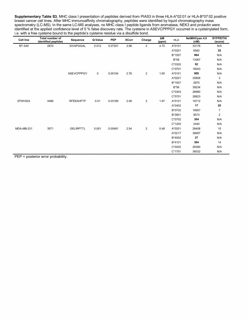
Supplementary Table S3. MHC class I presentation of peptides derived from PIAS3 in three HLA-A*02:01 or HLA-B*07:02 positive breast cancer cell lines. After MHC immunoaffinity chromatography, peptides were identified by liquid chromatography mass spectrometry (LC-MS). In the same LC-MS analyses, no MHC class I peptide ligands from aromatase, NEK3 and prolactin were identified at the applied confidence level of 5 % false discovery rate. The cysteine in ASEVCPPPGY occurred in a cysteinylated form, i.e. with a free cysteine bound to the peptide’s cysteine residue via a disulfide bond.
PEP = posterior error probability.
Cell line Total number of identified peptides Sequence Q-Value PEP XCorr Charge ΔM
(ppm) HLA NetMHCpan 4.0 (nM)
SYFPEITHI (score)
BT-549 2870 SIVAPGGAL 0.012 0.07301 2.96 2 4.70 A*0101 33178 N/A A*0201 6582 22 B*1507 964 N/A B*56 13367 N/A C*0303 92 N/A C*0701 18043 N/A ASEVCPPPGY 0 0.00104 2.76 2 1.60 A*0101 909 N/A A*0201 35808 3 B*1507 2870 N/A B*56 39234 N/A C*0303 28980 N/A C*0701 28823 N/A
EFM192A 4486 RFEEAHFTF 0.01 0.03169 2.06 2 1.97 A*0101 16712 N/A A*2402 17 20 B*0702 16957 7 B*3901 9073 2 C*0702 304 N/A C*1203 2440 N/A
MDA-MB-231 3571 GELIRPTTL 0.001 0.00481 2.54 2 0.48 A*0201 28408 15 A*0217 36657 N/A B*4002 27 N/A B*4101 364 14 C*0202 26390 N/A C*1701 36032 N/A

108
109
5. EPILOGUE
The research presented in this thesis is tied together by the search for antigen specific T cells
in the scheme of epitope discovery, immune monitoring and therapy. Each research project
utilizes MHC multimers to identify relevant CD8+ T cells, which are the mediators of
aberrant cell destruction in cancer and infections of viral and intracellular bacterial origin.
During the PhD project, research has been conducted in preclinical mouse models
(manuscript I-II) and a clinical cohort of breast cancer patients (paper III), all with the
perspective to characterize T cell epitopes in small or large scale. As such, manuscript I
describes a technology to screen for antigen specific T cells in mice, whereas manuscript II
and paper III concern T cell epitope analysis in cancer settings.
For MHC multimer-based research, efficient peptide exchange technologies, such as UV-
mediated peptide exchange, have facilitated rapid and high-throughput generation of larger
panels of pMHC multimers in parallel. In manuscript I we design and validate novel
conditional ligands for UV-mediated peptide exchange for two murine alleles; H-2Dd and H-
2Kd. Despite there being multiple descriptions of such conditional ligands for murine and
human MHC class I alleles, thus far only one allele was covered for the H-2d haplotype (i.e.
inbred BALB/c background) namely H-2Ld. This imposes technical difficulties for T cell
epitope mapping in BALB/c models, because of the need for tedious individual refolding of
each peptide with the H-2 allele, an obstruction for investigating large pH-2 libraries. We
successfully used H-2Dd, H-2Kd, and H-2Ld to generate DNA barcode labelled pH-2
multimer panels with 72 different specificities via UV-mediated exchange. With these large
pH-2 panels we screened several murine splenocyte samples for presence of specific CD8+
T cells. Thus, we have provided proof-of-concept of using a DNA barcode labelling system
for murine pH-2 multimers, which was confirmed by multimer staining with fluorescent
labels.
The novel conditional ligands described for H-2Dd and H-2Kd in manuscript I, were
applicable to the neo-epitope immunogenicity investigations in manuscript II. Here, we
110
employ DNA- and peptide-based vaccination strategies with selected neo-epitopes in the
syngeneic CT26 tumor model on BALB/c mice. Our data implies, that the choice of vaccine
formulation and adjuvant properties is crucial, both for immunogenicity and therapeutic
effects of the neo-epitopes investigated. As such, protective anti-tumor immunity was
obtained by prophylactic neo-epitope DNA vaccination and not neo-peptides + poly IC.
This was paralleled by neo-epitope DNA vaccination preferentially inducing cytokine
reactive CD8+ T cells, whereas neo-peptide + poly IC vaccination induced more CD4+ T
cells. In common for both delivery strategies, pH-2 tetramer staining disclosed presence of
neo-epitope specific CD8+ T cells in the periphery, albeit at a significantly higher level for
DNA based vaccination. Though neo-epitope specific CD8+ T cells were indeed present in
peptide immunized mice, they were less reactive measured by bulk IFNg ELISPOT, and ICS
showed almost no IFNg and TNFa producing CD8+ T cells upon re-stimulation.
Conversely, DNA based neo-epitope vaccination induced significantly more IFNg SFUs
measured by ELISPOT, and impressive numbers of IFNg and TNFa producing CD8+ T
cells via ICS. We expect that the anti-tumor response is primarily driven by these functional
CD8+ T cells induced by neo-epitope DNA vaccination. The question arises, whether the
anti-tumor effect is driven by sheer functionality and to what extent it is a “numbers game”.
It is possible, that the neo-peptide + poly IC vaccination strategy employed here induces
somewhat anergic or otherwise unresponsive CD8+ T cells. We propose that this is in part
due to innate signals present already at APC antigen uptake and priming of T cells.
Exogenous peptide vaccination inherently leads to presentation by MHC class II on APCs
and relies on cross presentation to reach the MHC class I pathway, as discussed in manuscript
II. The addition of TLR3 agonist poly IC to the neo-peptide vaccination could be an inducer
of cross presentation signals to DCs [139], [140], but in our studies this strategy primarily
primed CD4+ T cells. Interestingly, previous reports in a different setting with the syngeneic
MC38 model on C57BL/6 background suggests neo-peptide vaccination adjuvanted by poly
IC and anti-CD40 antibody induces strong CD8+ T cell responses and anti-tumor immunity
[127], [128]. Other studies have established anti-tumor potential of poly IC alone, when
injected intratumorally [141]. On the other side, DNA based delivery confers direct access
to the MHC class I processing and presentation pathway, since antigens are produced inside
APCs, and innate DNA sensing leads to induction of Th1 like immunity. DNA delivery of
neo-epitopes has in recent preclinical studies induced anti-tumor effects in other syngeneic
111
models with and without combination with anti-PD-1 antibody [134], [142]–[144]. We
therefore think, that DNA based neo-epitope vaccination is worth pursuing further.
Specifically, we will expand these studies with CD4+ and CD8+ T cell depletion to
interrogate which T cell subsets are crucial for retaining anti-tumor effects observed by
vaccination. We further hypothesize that we will be able to achieve anti-tumor effects with
therapeutic neo-epitope DNA vaccination in combination with anti-PD-1 therapy. This
synergistic combination therapy could abrogate some immune suppressive effects of the
established CT26 TME and boost vaccine induced T cells. Furthermore, the CT26 tumor
model has a rapid tumor onset and thus offers a short therapeutic window before mice must
be euthanized for reaching humane endpoints [122], [137]. This in an important factor to
consider, when talking about translatability from mice to humans, as human tumor
development takes months to years.
The neo-epitopes included in our vaccination study were of 27-mer amino acid length and
contain multiple in silico predicted MHC class I and II ligands. Preclinical studies claim that
both CD4+ and CD8+ T cell responses are essential for cancer immunotherapy [126], [135],
which is contradictory to the original understanding that CD8+ T cells solely drive tumor
elimination [119].
Together, findings in manuscript I and II will be applicable in a preclinical framework of
high-throughput neo-epitope screening. The number of putative neo-epitopes predicted by
current algorithms is vast, and large-scale T cell detection technologies like the DNA
barcode-labelled pMHC multimer strategy are necessary to meet this complexity. The
increased understanding of immunogenic neo-epitopes will feed back to prediction
algorithms, which currently rely primarily on in silico predicted peptide-MHC binding affinity.
In time, the hope is to better define those neo-epitopes that are most likely to elicit an
immune response. And furthermore, define the subset of immunogenic epitopes that are
also therapeutically relevant.
An example of this framework is an ongoing project in the research group, where we are
currently investigating the landscape of neo-epitope specific CD8+ T cells induced by anti-
PD-1 and anti-CTLA-4 combination therapy compared to isotype control antibody in
common syngeneic tumor models. The distinct antigen specific CD8+ T cell patterns
induced by CPI therapy in TILs and periphery in these syngeneic models have previously
not been characterized.
112
In paper III we utilize the high-throughput multimer strategy to detect CD8+ T cell
responses towards previously undescribed shared overexpression antigens in breast cancer
patients. This method is similar to that used in manuscript I, but with human HLAs. Breast
cancer has been minimally studied in the field of immune oncology, resulting in limited
knowledge of targetable T cell epitopes. But recent descriptions underline the relevance of
immunological recognition of breast cancer [48], [145]–[149]. Breast cancers constitute low
mutational burden tumors [150], why it is relevant to investigate non-mutated shared tumor
antigens for T cell recognition. In our paper, we focus on four proteins known to be
upregulated in breast cancer: aromatase, NEK3, PIAS3, and prolactin, that each contribute
to cell growth, survival, and motility during malignant transformation. From these proteins,
we selected 147 peptides with affinity for HLA-A*0201 and -B*0702 by combined in silico
and in vitro screening. We constructed a DNA barcode labelled pHLA multimer library to
screen a cohort of breast cancer patients and healthy donors for CD8+ T cell recognition to
these shared antigens. Our analyses disclosed significantly more CD8+ T cell responses in
breast cancer patients than healthy donors, and the peptides that gave rise to T cell responses
clustered in “immunological hotspots” in the protein sequences. Interestingly, patients with
more advanced disease stages also had higher numbers of CD8+ T cell responses, and
specific T cells from some patients were shown to be cytokine responsive to peptide
stimulation. Together these results validated the four proteins as novel tumor associated
antigens, and we hypothesize their relevance for immune monitoring of breast cancer
patients, potentially providing a measure of immune status or a correlate of response to
immunotherapies. We found it notable, that several of the immunogenic peptides had
intermediate to low predicted binding affinity to the HLA. One explanation for this, is that
T cells specific to self-antigens undergo strict negative selection in the thymus, and it is
therefore important to include these lower affinity binders in the search for shared epitopes
to possibly circumvent tolerance. The potential applicability of this study is limited by the
library’s restriction to HLA-A*0201 and -B*0702, but we are considering expanding the
panel to include more HLA types. Furthermore, we are interested in investigating T cell
epitopes from additional shared tumor associated antigens with relevance in breast cancer.
These expansions to the pHLA library are feasible, due to the high-throughput screening
method, which has previously been used for epitope mapping in human samples to detect
antigen specific CD8+ T specific to: self-proteins in narcolepsy [151], neo-epitopes in non-
small cell lung cancer, and shared tumor antigens in melanoma [79]. The data from
113
manuscript I and paper III from this thesis thus contribute to the list of applications for the
high-throughput method in murine and human samples.
Perspectives of the presented work
The research contained in this thesis contributes to the field of cancer immunology by several
parameters. First, by the design and validation of new conditional ligands for H-2Dd and H-
2Kd and establishment of proof-of-concept of large-scale DNA barcode labelled multimer
panels for BALB/c haplotype alleles. It is our expectation, that the new conditional ligands
and the high-throughput technology for murine CD8+ T cell interrogation via pH-2d
multimers will assist screening purposes and antigen discovery in preclinical models of
cancer, infectious diseases and autoimmunity. Second, by establishing a protective neo-
epitope encoding DNA vaccination strategy in the CT26 syngeneic tumor model. This allows
screening of more neo-epitopes for immunogenicity and contribution to anti-tumor effects,
and can be expanded to other syngeneic models on BALB/c and C57BL/6 background. We
also anticipate the DNA vaccination platform will be a great tool to investigate combination
therapies. Third, by defining novel tumor associated antigens in breast cancer, where there
is a lack of descriptions of T cell epitopes and immunotherapeutic strategies are less
established. It could be interesting to expand the panel of putative breast cancer antigens of
shared origin in future studies. Together, this research provides tools and investigations of
what makes good T cell targets in cancer, which will be further discussed in the following
paragraphs.
Though manuscript II and paper III take place in a murine model and patient samples,
respectively, they lead to considerations on the applicability of shared and neo-epitopes. In
the event of using these epitopes as immunotherapeutic T cell targets by e.g. vaccination,
attention should be put on safety. For this purpose, neo-epitopes are clearly advantageous
since antigen expression is limited to tumor cells and therefore there should be no adverse
destruction of self-tissue. Further beneficial is that neo-epitope specific T cells are assumed
to be less subjected to immune tolerance. However, using neo-epitopes therapeutically
requires extensive work, since it will be a “1 patient, 1 drug” setting. Neo-epitope prediction
and subsequent vaccine production is cumbersome, but worthwhile if we experience clinical
benefit in the many currently ongoing personalized vaccine trials. For epitopes of shared
origin, we instead have the option of interventions that will broadly benefit many patients,
114
and manufacturing will be less comprehensive with off-the-shelf options. This however
comes with the caveat of potential adverse effects and heavy subjection to immune tolerance.
Since tumor mutational load varies a lot, neo-epitope targeted strategies are not of equal
relevance for every cancer.
As discussed throughout this thesis, far from all in silico predicted T cell epitopes are
immunogenic and even less epitopes are of therapeutic relevance. Current prediction tools
are now very good at predicting MHC binders, but we still do not know the determinants
for a T cell epitope capable of initiating a strong CD8+ T cell response. Several explanations
contribute to why some epitopes are not immunogenic. First, T cells specific to a given
epitope might have never existed or were eliminated by negative selection. Second, specific
T cells could exist, but be anergic or otherwise functionally unresponsive. Last, the epitope
itself is not immunogenic enough to give rise to T cell reactivity. Future perspectives of
improved epitope prediction will be contributed to by preclinical and clinical investigations
alike. High-throughput technologies will support this through e.g. TCR profiling and
mapping of pMHC-TCR pairs [152]. One could then imagine a framework of interrogating
the naive T cell repertoire of a patient, to tailor a vaccine based on the already existing T
cells. Recent reports are suggesting an additional layer of complexity, by outlining how the
cancer patients’ microbiome are affecting responses to anti-PD-1 therapy [153], [154].
Perhaps we will in the near future use measures like these to stratify and design personal
immunotherapies to cancer patients.
As a final remark, I expect this already rapidly developing field to advance with knowledge
of how we can utilize the immune system to combat cancer. The cancer-immunity interplay
is indeed intricate and multifaceted, and I believe that insights into how we can tune adaptive
responses via innate stimuli, will take us even further. We have clear evidence, that tumors
are vastly different from normal tissue, and some of these differences lead to recognition by
the immune system. Hence, we need potent tools to screen the immense putative epitope
libraries resulting from genome-wipe epitope mapping. Neo-epitope vaccination trials have
reached the clinic; thus, we have entered the era of truly personalized cancer therapies. In
this era, preclinical studies will continuously provide indispensable insights into immune
recognition of cancer, why they are of key translational relevance.

115
6. BIBLIOGRAPHY
[1] P. J. Delves and I. M. Roitt, “The Immune System - First of two parts,” N. Engl. J. Med., vol.
343, no. 1, pp. 37–50, 2000.
[2] D. Schenten and R. Medzhitov, “The Control of Adaptive Immune Responses by the Innate
Immune System,” in Advances in Immunology, vol. 109, 2011, pp. 87–124.
[3] J. Banchereau and R. M. Steinman, “Dendritic cells and the control of immunity.,” Nature,
vol. 392, no. March, pp. 245–252, 1998.
[4] M. Hubo, B. Trinschek, F. Kryczanowsky, A. Tuettenberg, K. Steinbrink, and H. Jonuleit,
“Costimulatory molecules on immunogenic versus tolerogenic human dendritic cells,” Front.
Immunol., vol. 4, p. 82, Apr. 2013.
[5] V. Schijns and D. O’Hagan, Immunopotentiators in Modern Vaccines. Elsevier, 2006.
[6] C. J. Desmet and K. J. Ishii, “Nucleic acid sensing at the interface between innate and adaptive
immunity in vaccination.,” Nat. Rev. Immunol., vol. 12, no. 7, pp. 479–491, Jun. 2012.
[7] K. C. Garcia and I. A. Wilson, “Structural Basis of T cell Recognition,” Annu. Rev. Immunol.,
vol. 17, pp. 369–397, 1999.
[8] T. Trolle et al., “The Length Distribution of Class I-Restricted T Cell Epitopes Is Determined
by Both Peptide Supply and MHC Allele-Specific Binding Preference.,” J. Immunol., vol. 196,
no. 4, pp. 1480–1487, Feb. 2016.
[9] R. M. Chicz et al., “Predominant naturally processed peptides bound to HLA-DR1 are derived
from MHC-related molecules and are heterogeneous in size,” Nature, vol. 358, no. 6389, pp.
764–768, Aug. 1992.
[10] M. Embgenbroich and S. Burgdorf, “Current Concepts of Antigen Cross-Presentation,”
Front. Immunol., vol. 9, p. 1643, Jul. 2018.
[11] M. Kovacsovics-Bankowski and K. L. Rock, “A phagosome-to-cytosol pathway for
exogenous antigens presented on MHC class I molecules,” Science (80-. )., vol. 267, no. 5195,
pp. 243 LP – 246, Jan. 1995.
[12] O. P. Joffre, E. Segura, A. Savina, and S. Amigorena, “Cross-presentation by dendritic cells,”
Nat Rev Immunol, vol. 12, no. 8, pp. 557–569, Aug. 2012.
[13] K. S. Kobayashi and P. J. van den Elsen, “NLRC5: a key regulator of MHC class I-dependent
immune responses,” Nat Rev Immunol, vol. 12, no. 12, pp. 813–820, Dec. 2012.
[14] M. Fabbri, C. Smart, and R. Pardi, “T Lymphocytes,” Int. J. Biochem. Cell Biol., vol. 35, no. 7,
pp. 1004–1008, 2003.
116
[15] B. O. Roep, M. J. Kracht, M. van Lummel, and A. Zaldumbide, “A roadmap of the generation
of neoantigens as targets of the immune system in type 1 diabetes,” Curr. Opin. Immunol., vol.
43, pp. 67–73, Dec. 2016.
[16] M. Salou, B. Nicol, A. Garcia, and D.-A. Laplaud, “Involvement of CD8+ T Cells in Multiple
Sclerosis,” Front. Immunol., vol. 6, p. 604, Nov. 2015.
[17] H. Lodish et al., Molecular Cell Biology, vol. 4. 2008.
[18] D. Hanahan and R. A. Weinberg, “The Hallmarks of cancer,” Cell, vol. 100, pp. 57–70, 2000.
[19] D. S. Chen and I. Mellman, “Oncology Meets Immunology: The Cancer-Immunity Cycle,”
Immunity, vol. 39, no. 1, pp. 1–10, Jul. 2013.
[20] D. Hanahan and R. A. Weinberg, “Hallmarks of cancer: The Next Generation,” Cell, vol. 144,
no. 5, pp. 646–674, 2011.
[21] J. Galon, “Type, Density, and Location of Immune Cells Within Human Colorectal Tumors
Predict Clinical Outcome,” Science (80-. )., vol. 313, no. 5795, pp. 1960–1964, Sep. 2006.
[22] J. Galon et al., “Cancer classification using the Immunoscore: a worldwide task force,” J.
Transl. Med., vol. 10, p. 205, Oct. 2012.
[23] J. Galon and D. Bruni, “Approaches to treat immune hot, altered and cold tumours with
combination immunotherapies.,” Nat. Rev. Drug Discov., p. 1, Jan. 2019.
[24] G. P. Dunn, L. J. Old, and R. D. Schreiber, “The Three Es of Cancer Immunoediting,” Annu.
Rev. Immunol., vol. 22, no. 1, pp. 329–360, Mar. 2004.
[25] S. H. van der Burg, R. Arens, F. Ossendorp, T. van Hall, and C. J. M. Melief, “Vaccines for
established cancer: overcoming the challenges posed by immune evasion,” Nat Rev Cancer,
vol. 16, no. 4, pp. 219–233, 2016.
[26] S. Hadrup, M. Donia, and P. thor Straten, “Effector CD4 and CD8 T Cells and Their Role in
the Tumor Microenvironment,” Cancer Microenviron., vol. 6, no. 2, pp. 123–133, Aug. 2013.
[27] L. von Boehmer et al., “NY-ESO-1-specific immunological pressure and escape in a patient
with metastatic melanoma.,” Cancer Immun., vol. 13, p. 12, 2013.
[28] T. Nicholaou et al., “Immunoediting and persistence of antigen-specific immunity in patients
who have previously been vaccinated with NY-ESO-1 protein formulated in
ISCOMATRIX.,” Cancer Immunol. Immunother., vol. 60, no. 11, pp. 1625–1637, Nov. 2011.
[29] P. G. Coulie, B. J. Van Den Eynde, P. Van Der Bruggen, and T. Boon, “Tumour antigens
recognized by T lymphocytes: At the core of cancer immunotherapy,” Nat. Rev. Cancer, vol.
14, no. 2, pp. 135–146, 2014.
[30] E.-K. Yim and J.-S. Park, “The Role of HPV E6 and E7 Oncoproteins in HPV-associated
Cervical Carcinogenesis,” Cancer Res. Treat., vol. 37, no. 6, pp. 319–324, Dec. 2005.
[31] C. C. Smith et al., “Machine-learning prediction of tumor antigen immunogenicity in the
selection of therapeutic epitopes,” Cancer Immunol. Res., vol. 7, no. 10, pp. 1591–1604, 2019.
117
[32] C. M. Laumont et al., “Noncoding regions are the main source of targetable tumor-specific
antigens,” Sci. Transl. Med., vol. 10, no. 470, p. eaau5516, Dec. 2018.
[33] C. Chong et al., “Integrated Proteogenomic Deep Sequencing and Analytics Accurately
Identify Non-Canonical Peptides in Tumor Immunopeptidomes,” bioRxiv, p. 758680, 2019.
[34] S. M. Schmidt et al., “Survivin is a shared tumor-associated antigen expressed in a broad variety
of malignancies and recognized by specific cytotoxic T cells,” Blood, vol. 102, no. 2, pp. 571–
576, Jul. 2003.
[35] I. Kawashima et al., “The Multi-epitope Approach for Immunotherapy for Cancer:
Identification of Several CTL Epitopes from Various Tumor-Associated Antigens Expressed
on Solid Epithelial Tumors,” Hum. Immunol., vol. 59, no. 1, pp. 1–14, Jan. 1998.
[36] X. Kang et al., “Identification of a tyrosinase epitope recognized by HLA-A24-restricted,
tumor-infiltrating lymphocytes.,” J. Immunol., vol. 155, no. 3, pp. 1343–1348, Aug. 1995.
[37] E. Jager et al., “Simultaneous humoral and cellular immune response against cancer-testis
antigen NY-ESO-1: definition of human histocompatibility leukocyte antigen (HLA)-A2-
binding peptide epitopes.,” J. Exp. Med., vol. 187, no. 2, pp. 265–270, Jan. 1998.
[38] S. A. Rosenberg, J. C. Yang, and N. P. Restifo, “Cancer immunotherapy: moving beyond
current vaccines,” Nat. Med., vol. 10, no. 9, pp. 909–915, Sep. 2004.
[39] J. L. Gulley et al., “Phase III Trial of PROSTVAC in Asymptomatic or Minimally
Symptomatic Metastatic Castration-Resistant Prostate Cancer,” J. Clin. Oncol., vol. 37, no. 13,
pp. 1051–1061, May 2019.
[40] L. A. Johnson et al., “Gene therapy with human and mouse T-cell receptors mediates cancer
regression and targets normal tissues expressing cognate antigen,” Blood, vol. 114, no. 3, pp.
535–546, Jul. 2009.
[41] J. H. Kessler and C. J. M. Melief, “Identification of T-cell epitopes for cancer
immunotherapy.,” Leuk. Off. J. Leuk. Soc. Am. Leuk. Res. Fund, U.K, vol. 21, pp. 1859–1874,
2007.
[42] M. S. Rooney, S. A. Shukla, C. J. Wu, G. Getz, and N. Hacohen, “Molecular and Genetic
Properties of Tumors Associated with Local Immune Cytolytic Activity,” Cell, vol. 160, no.
1–2, pp. 48–61, Jan. 2015.
[43] S. D. Brown et al., “Neo-antigens predicted by tumor genome meta-analysis correlate with
increased patient survival,” Genome Res., vol. 24, no. 5, pp. 743–750, May 2014.
[44] R. M. Samstein et al., “Tumor mutational load predicts survival after immunotherapy across
multiple cancer types.,” Nat. Genet., Jan. 2019.
[45] A. Snyder et al., “Genetic basis for clinical response to CTLA-4 blockade in melanoma,” N.
Engl. J. Med., vol. 371, no. 23, pp. 2189–2199, 2014.
[46] N. A. Rizvi et al., “Cancer immunology. Mutational landscape determines sensitivity to PD-1

118
blockade in non-small cell lung cancer.,” Science, vol. 348, no. 6230, pp. 124–8, Apr. 2015.
[47] M. Lauss et al., “Mutational and putative neoantigen load predict clinical benefit of adoptive
T cell therapy in melanoma,” Nat. Commun., vol. 8, no. 1, pp. 1–10, 2017.
[48] N. Zacharakis et al., “Immune recognition of somatic mutations leading to complete durable
regression in metastatic breast cancer,” Nat. Med., vol. 24, no. 6, pp. 724–730, 2018.
[49] E. Tran, P. F. Robbins, and S. A. Rosenberg, “Final common pathway’ of human cancer
immunotherapy: Targeting random somatic mutations,” Nat. Immunol., vol. 18, no. 3, pp. 255–
262, 2017.
[50] E. F. Fritsch, M. Rajasagi, P. A. Ott, V. Brusic, N. Hacohen, and C. J. Wu, “HLA-binding
properties of tumor neoepitopes in humans,” Cancer Immunol. Res., vol. 2, no. 6, pp. 522–529,
2014.
[51] A. M. Bjerregaard et al., “An analysis of natural T cell responses to predicted tumor
neoepitopes,” Front. Immunol., vol. 8, no. NOV, pp. 1–9, 2017.
[52] K. R. Loeb and L. a Loeb, “Significance of multiple mutations in cancer,” Carcinogenesis, vol.
21, no. 3, pp. 379–385, 2000.
[53] F. Castro-Giner, P. Ratcliffe, and I. Tomlinson, “The mini-driver model of polygenic cancer
evolution,” Nat. Rev. Cancer, vol. 15, no. 11, pp. 680–685, 2015.
[54] B. Heemskerk, P. Kvistborg, and T. N. M. Schumacher, “The cancer antigenome,” EMBO J.,
vol. 32, no. 2, pp. 194–203, 2013.
[55] C. A. Klebanoff and J. D. Wolchok, “Shared cancer neoantigens: Making private matters
public,” J. Exp. Med., vol. 215, no. 1, pp. 5–7, Jan. 2018.
[56] M. Gerlinger et al., “Intratumor heterogeneity and branched evolution revealed by multiregion
sequencing.,” N. Engl. J. Med., vol. 366, no. 10, pp. 883–892, Mar. 2012.
[57] N. McGranahan et al., “Clonal neoantigens elicit T cell immunoreactivity and sensitivity to
immune checkpoint blockade.,” Science, vol. 351, no. 6280, pp. 1463–9, Mar. 2016.
[58] H. Hackl, P. Charoentong, F. Finotello, and Z. Trajanoski, “Computational genomics tools
for dissecting tumour-immune cell interactions,” Nat. Rev. Genet., vol. 17, no. 8, pp. 441–458,
2016.
[59] C. Linnemann et al., “High-throughput epitope discovery reveals frequent recognition of neo-
antigens by CD4+T cells in human melanoma,” Nat. Med., vol. 21, no. 1, pp. 81–85, 2015.
[60] M. Bassani-Sternberg et al., “Direct identification of clinically relevant neoepitopes presented
on native human melanoma tissue by mass spectrometry,” Nat. Commun., vol. 7, no. May,
2016.
[61] B. Bulik-Sullivan et al., “Deep learning using tumor HLA peptide mass spectrometry datasets
improves neoantigen identification.,” Nat. Biotechnol., vol. 37, no. 1, Dec. 2018.
[62] J. G. Abelin et al., “Defining HLA-II Ligand Processing and Binding Rules with Mass
119
Spectrometry Enhances Cancer Epitope Prediction,” Immunity, vol. 51, no. 4, pp. 766-779.e17,
2019.
[63] V. Jurtz, S. Paul, M. Andreatta, P. Marcatili, B. Peters, and M. Nielsen, “NetMHCpan-4.0:
Improved Peptide–MHC Class I Interaction Predictions Integrating Eluted Ligand and
Peptide Binding Affinity Data,” J. Immunol., vol. 199, no. 9, pp. 3360 LP – 3368, Nov. 2017.
[64] A. M. Bjerregaard, M. Nielsen, S. R. Hadrup, Z. Szallasi, and A. C. Eklund, “MuPeXI:
prediction of neo-epitopes from tumor sequencing data,” Cancer Immunol. Immunother., vol. 66,
no. 9, pp. 1123–1130, 2017.
[65] L. B. Alexandrov et al., “Signatures of mutational processes in human cancer,” Nature, vol.
500, no. 7463, pp. 415–421, Aug. 2013.
[66] E. Strønen et al., “Targeting of cancer neoantigens with donor-derived T cell receptor
repertoires,” Science (80-. )., vol. 352, no. 6291, pp. 1337–1341, 2016.
[67] H. Besser, S. Yunger, E. Merhavi-Shoham, C. J. Cohen, and Y. Louzoun, “Level of neo-
epitope predecessor and mutation type determine T cell activation of MHC binding peptides,”
J. Immunother. Cancer, vol. 7, no. 1, pp. 1–9, 2019.
[68] P. A. Ott et al., “An Immunogenic Personal Neoantigen Vaccine for Melanoma Patients,”
Nature, vol. 547, no. 7662, pp. 217–221, Jul. 2017.
[69] M. Corr et al., “T cell receptor-MHC class I peptide interactions: affinity, kinetics, and
specificity,” Science (80-. )., vol. 265, no. 5174, 1994.
[70] J. D. Altman et al., “Phenotypic Analysis of Antigen-Specific T Lymphocytes,” vol. 274, no.
5284, pp. 94–96, 1996.
[71] M. Toebes et al., “Design and use of conditional MHC class I ligands,” Nat. Med., vol. 12, no.
2, pp. 246–251, Feb. 2006.
[72] A. H. Bakker et al., “Conditional MHC class I ligands and peptide exchange technology for
the human MHC gene products HLA-A1, -A3, -A11, and -B7,” PNAS, vol. 105, pp. 3825–
3830, 2008.
[73] G. M. Grotenbreg et al., “Discovery of CD8+ T cell epitopes in Chlamydia trachomatis
infection through use of caged class I MHC tetramers,” Proc. Natl. Acad. Sci. U. S. A., vol.
105, no. 10, pp. 3831–3836, 2008.
[74] E. Frickel et al., “Parasite Stage–Specific Recognition of Endogenous Toxoplasma gondii –
Derived CD8 + T Cell Epitopes,” J. Infect. Dis., vol. 198, no. 11, pp. 1625–1633, Dec. 2008.
[75] T. M. Frøsig et al., “Design and validation of conditional ligands for HLA-B*08:01, HLA-
B*15:01, HLA-B*35:01, and HLA-B*44:05,” Cytom. Part A, vol. 87, no. 10, pp. 967–975, Oct.
2015.
[76] M. M. Davis, J. D. Altman, and E. W. Newell, “Interrogating the repertoire: broadening the
scope of peptide-MHC multimer analysis.,” Nat. Rev. Immunol., vol. 11, no. 8, pp. 551–8, 2011.

120
[77] R. S. Andersen et al., “Parallel detection of antigen-specific T cell responses by combinatorial
encoding of MHC multimers.,” Nat. Protoc., vol. 7, no. 5, pp. 891–902, May 2012.
[78] E. W. Newell, N. Sigal, N. Nair, B. A. Kidd, H. B. Greenberg, and M. M. Davis,
“Combinatorial tetramer staining and mass cytometry analysis facilitate T-cell epitope
mapping and characterization,” Nat. Biotechnol., vol. 31, no. 7, pp. 623–629, Jul. 2013.
[79] A. K. Bentzen et al., “Large-scale detection of antigen-specific T cells using peptide-MHC-I
multimers labeled with DNA barcodes,” Nat. Biotechnol., vol. 34, no. 10, pp. 1037–1045, 2016.
[80] S. K. Saini et al., “Empty peptide-receptive MHC class I molecules for efficient detection of
antigen-specific T cells,” Sci. Immunol., vol. 4, no. 37, p. eaau9039, Jul. 2019.
[81] P. Braendstrup et al., “MHC Class II Tetramers Made from Isolated Recombinant α and β
Chains Refolded with Affinity-Tagged Peptides,” PLoS One, vol. 8, no. 9, p. e73648, Sep.
2013.
[82] S. R. Hadrup and E. W. Newell, “Determining T-cell specificity to understand and treat
disease,” Nat. Biomed. Eng., vol. 1, no. 10, pp. 784–795, 2017.
[83] S. Farkona, E. P. Diamandis, and I. M. Blasutig, “Cancer immunotherapy: the beginning of
the end of cancer?,” BMC Med., vol. 14, no. 1, p. 73, Dec. 2016.
[84] P. Sharma and J. P. Allison, “The future of immune checkpoint therapy,” Science (80-. )., vol.
348, no. 6230, pp. 56–61, Apr. 2015.
[85] T. R. Simpson et al., “Fc-dependent depletion of tumor-infiltrating regulatory T cells co-
defines the efficacy of anti-CTLA-4 therapy against melanoma.,” J. Exp. Med., vol. 210, no. 9,
pp. 1695–1710, Aug. 2013.
[86] M. J. Selby et al., “Anti-CTLA-4 antibodies of IgG2a isotype enhance antitumor activity
through reduction of intratumoral regulatory T cells.,” Cancer Immunol. Res., vol. 1, no. 1, pp.
32–42, Jul. 2013.
[87] A. O. Kamphorst et al., “Proliferation of PD-1+ CD8 T cells in peripheral blood after PD-1-
targeted therapy in lung cancer patients.,” Proc. Natl. Acad. Sci. U. S. A., vol. 114, no. 19, pp.
4993–4998, May 2017.
[88] A. C. Huang et al., “T-cell invigoration to tumour burden ratio associated with anti-PD-1
response.,” Nature, vol. 545, no. 7652, pp. 60–65, May 2017.
[89] A. Gros et al., “Prospective identification of neoantigen-specific lymphocytes in the peripheral
blood of melanoma patients,” Nat. Med., vol. 22, no. 4, pp. 433–438, 2016.
[90] K. E. Yost et al., “Clonal replacement of tumor-specific T cells following PD-1 blockade,”
Nat. Med., vol. 25, no. 8, pp. 1251–1259, Aug. 2019.
[91] T. Duhen et al., “Co-expression of CD39 and CD103 identifies tumor-reactive CD8 T cells
in human solid tumors,” Nat. Commun., vol. 9, no. 1, p. 2724, Dec. 2018.
[92] Y. Simoni et al., “Bystander CD8 + T cells are abundant and phenotypically distinct in human
121
tumour infiltrates,” Nature, vol. 557, no. 7706, pp. 575–579, 2018.
[93] S. Das and D. B. Johnson, “Immune-related adverse events and anti-tumor efficacy of
immune checkpoint inhibitors,” J. Immunother. Cancer, vol. 7, no. 1, p. 306, 2019.
[94] S. A. Rosenberg and N. P. Restifo, “Adoptive cell transfer as personalized immunotherapy
for human cancer,” Science (80-. )., vol. 348, no. 6230, pp. 62–68, Apr. 2015.
[95] C. H. June, R. S. O’Connor, O. U. Kawalekar, S. Ghassemi, and M. C. Milone, “CAR T cell
immunotherapy for human cancer,” Science (80-. )., vol. 359, no. 6382, pp. 1361–1365, Mar.
2018.
[96] P. Sharma, S. Hu-Lieskovan, J. A. Wargo, and A. Ribas, “Primary, Adaptive, and Acquired
Resistance to Cancer Immunotherapy,” Cell, vol. 168, no. 4, pp. 707–723, Feb. 2017.
[97] Z. Hu, P. A. Ott, and C. J. Wu, “Towards personalized, tumour-specific, therapeutic vaccines
for cancer.,” Nat. Rev. Immunol., vol. 18, no. 3, pp. 168–182, Mar. 2018.
[98] T. N. Schumacher and R. D. Schreiber, “Neoantigens in cancer immunotherapy,” Science (80-
. )., vol. 348, no. 6230, pp. 69–74, 2015.
[99] B. Temizoz, E. Kuroda, and K. J. Ishii, “Vaccine adjuvants as potential cancer
immunotherapeutics,” Int. Immunol., vol. 28, no. 7, pp. 329–338, Jul. 2016.
[100] K. Vermaelen, “Vaccine Strategies to Improve Anti-cancer Cellular Immune Responses,”
Front. Immunol., vol. 10, no. JAN, pp. 1–17, Jan. 2019.
[101] C. Gouttefangeas and H.-G. Rammensee, “Personalized cancer vaccines: adjuvants are
important, too,” Cancer Immunol. Immunother., vol. 67, no. 12, pp. 1911–1918, Dec. 2018.
[102] E. N. Vinageras et al., “Phase II randomized controlled trial of an epidermal growth factor
vaccine in advanced non-small-cell lung cancer,” J. Clin. Oncol., vol. 26, no. 9, pp. 1452–1458,
2008.
[103] S. S. Neelapu et al., “Human Autologous Tumor-Specific T-Cell Responses Induced by
Liposomal Delivery of a Lymphoma Antigen,” Clin. Cancer Res., vol. 10, no. 24, pp. 8309 LP
– 8317, Dec. 2004.
[104] D. Wang, Y. Sun, Y. Liu, F. Meng, and R. J. Lee, “Clinical translation of immunoliposomes
for cancer therapy: recent perspectives,” Expert Opin. Drug Deliv., vol. 15, no. 9, pp. 893–903,
Sep. 2018.
[105] K. S. Korsholm et al., “Induction of CD8+ T-cell responses against subunit antigens by the
novel cationic liposomal CAF09 adjuvant,” Vaccine, vol. 32, no. 31, pp. 3927–3935, 2014.
[106] J. Gao, L. Ochyl, E. Yang, and J. Moon, “Cationic liposomes promote antigen cross-
presentation in dendritic cells by alkalizing the lysosomal pH and limiting the degradation of
antigens,” Int. J. Nanomedicine, vol. Volume 12, pp. 1251–1264, Feb. 2017.
[107] D. B. Keskin et al., “Neoantigen vaccine generates intratumoral T cell responses in phase Ib
glioblastoma trial,” Nature, vol. 565, no. 7738, pp. 234–239, Jan. 2019.
122
[108] P. Sabbatini et al., “Phase I Trial of Overlapping Long Peptides from a Tumor Self-Antigen
and Poly-ICLC Shows Rapid Induction of Integrated Immune Response in Ovarian Cancer
Patients,” Clin. Cancer Res., vol. 18, no. 23, pp. 6497–6508, Dec. 2012.
[109] M. L. Salem, S. A. El-Naggar, A. Kadima, W. E. Gillanders, and D. J. Cole, “The adjuvant
effects of the toll-like receptor 3 ligand polyinosinic-cytidylic acid poly (I:C) on antigen-
specific CD8+ T cell responses are partially dependent on NK cells with the induction of a
beneficial cytokine milieu.,” Vaccine, vol. 24, no. 24, pp. 5119–5132, Jun. 2006.
[110] M. L. Salem, A. N. Kadima, D. J. Cole, and W. E. Gillanders, “Defining the antigen-specific
T-cell response to vaccination and poly(I:C)/TLR3 signaling: evidence of enhanced primary
and memory CD8 T-cell responses and antitumor immunity.,” J. Immunother., vol. 28, no. 3,
pp. 220–228, 2005.
[111] H. Ishikawa, Z. Ma, and G. N. Barber, “STING regulates intracellular DNA-mediated, type
i interferon-dependent innate immunity,” Nature, vol. 461, no. 7265, pp. 788–792, 2009.
[112] M. Motwani, S. Pesiridis, and K. A. Fitzgerald, “DNA sensing by the cGAS–STING pathway
in health and disease,” Nat. Rev. Genet., vol. 20, no. November, 2019.
[113] K. J. Ishii et al., “TANK-binding kinase-1 delineates innate and adaptive immune responses
to DNA vaccines,” Nature, vol. 451, no. 7179, pp. 725–729, 2008.
[114] Y. M. Murad and T. M. Clay, “CpG oligodeoxynucleotides as TLR9 agonists: therapeutic
applications in cancer.,” BioDrugs, vol. 23, no. 6, pp. 361–375, 2009.
[115] L. M. Kranz et al., “Systemic RNA delivery to dendritic cells exploits antiviral defence for
cancer immunotherapy,” Nature, vol. 534, no. 7607, pp. 396–401, 2016.
[116] U. Sahin et al., “Personalized RNA mutanome vaccines mobilize poly-specific therapeutic
immunity against cancer.,” Nature, vol. 547, no. 7662, pp. 222–226, 2017.
[117] N. Hilf et al., “Actively personalized vaccination trial for newly diagnosed glioblastoma.,”
Nature, vol. 565, no. 7738, pp. 240–245, Jan. 2019.
[118] B. J. Laidlaw, J. E. Craft, and S. M. Kaech, “The multifaceted role of CD4+ T cells in CD8+
T cell memory,” Nat. Rev. Immunol., vol. 16, no. 2, pp. 102–111, Feb. 2016.
[119] L. H. Butterfield, “Cancer vaccines,” BMJ, vol. 350, pp. 1–14, 2015.
[120] U. Sahin and Ö. Türeci, “Personalized vaccines for cancer immunotherapy,” Science (80-. ).,
vol. 359, no. 6382, pp. 1355–1360, Mar. 2018.
[121] G. P. Dunn, L. J. Old, and R. D. Schreiber, “The immunobiology of cancer
immunosurveillance and immunoediting,” Immunity, vol. 21, no. 2, pp. 137–148, 2004.
[122] S. I. S. Mosely et al., “Rational selection of syngeneic preclinical tumor models for
immunotherapeutic drug discovery,” Cancer Immunol. Res., vol. 5, no. 1, pp. 29–41, 2017.
[123] H. Jespersen et al., “Clinical responses to adoptive T-cell transfer can be modeled in an
autologous immune-humanized mouse model,” Nat. Commun., vol. 8, no. 1, 2017.
123
[124] O. L. Gammelgaard, M. G. Terp, B. Preiss, and H. J. Ditzel, “Human cancer evolution in the
context of a human immune system in mice,” Mol. Oncol., vol. 12, no. 10, pp. 1797–1810, Oct.
2018.
[125] J. C. Castle et al., “Immunomic, genomic and transcriptomic characterization of CT26
colorectal carcinoma,” BMC Genomics, vol. 15, no. 1, 2014.
[126] S. Kreiter et al., “Mutant MHC class II epitopes drive therapeutic immune responses to
cancer,” Nature, vol. 520, no. 7549, pp. 692–696, 2015.
[127] M. Yadav et al., “Predicting immunogenic tumour mutations by combining mass spectrometry
and exome sequencing,” Nature, vol. 515, no. 7528, pp. 572–576, 2014.
[128] B. J. Hos et al., “Identification of a neo-epitope dominating endogenous CD8 T cell responses
to MC-38 colorectal cancer,” Oncoimmunology, vol. 00, no. 00, p. 1673125, Oct. 2019.
[129] J. C. Castle et al., “Exploiting the mutanome for tumor vaccination,” Cancer Res., vol. 72, no.
5, pp. 1081–1091, 2012.
[130] H. Matsushita et al., “Cancer exome analysis reveals a T-cell-dependent mechanism of cancer
immunoediting,” Nature, vol. 482, no. 7385, pp. 400–404, Feb. 2012.
[131] M. M. Gubin et al., “Checkpoint blockade cancer immunotherapy targets tumour-specific
mutant antigens,” Nature, vol. 515, no. 7528, pp. 577–581, Nov. 2014.
[132] M. Fehlings et al., “Checkpoint blockade immunotherapy reshapes the high-dimensional
phenotypic heterogeneity of murine intratumoural neoantigen-specific CD8+T cells,” Nat.
Commun., vol. 8, no. 1, 2017.
[133] M. M. Gubin et al., “High-Dimensional Analysis Delineates Myeloid and Lymphoid
Compartment Remodeling during Successful Immune-Checkpoint Cancer Therapy,” Cell,
vol. 175, no. 4, pp. 1014-1030.e19, Nov. 2018.
[134] E. K. Duperret et al., “A Synthetic DNA, Multi-Neoantigen Vaccine Drives Predominately
MHC Class I CD8 + T-cell Responses, Impacting Tumor Challenge,” Cancer Immunol. Res.,
vol. 7, no. 2, pp. 174–182, Feb. 2019.
[135] E. Alspach et al., “MHC-II neoantigens shape tumour immunity and response to
immunotherapy.,” Nature, no. February, 2019.
[136] J. Borst, T. Ahrends, N. Bąbała, C. J. M. Melief, and W. Kastenmüller, “CD4+ T cell help in
cancer immunology and immunotherapy,” Nat. Rev. Immunol., vol. 18, no. 10, pp. 635–647,
2018.
[137] M. A. Taylor et al., “Longitudinal immune characterization of syngeneic tumor models to
enable model selection for immune oncology drug discovery,” J. Immunother. Cancer, vol. 7, no.
1, p. 328, 2019.
[138] I. W. Mak, N. Evaniew, and M. Ghert, “Lost in translation: animal models and clinical trials
in cancer treatment,” Am. J. Transl. Res., vol. 6, no. 2, pp. 114–118, Jan. 2014.
124
[139] A. L. Ackerman and P. Cresswell, “Cellular mechanisms governing cross-presentation of
exogenous antigens.,” Nat. Immunol., vol. 5, no. 7, pp. 678–684, Jul. 2004.
[140] F. Spadaro et al., “IFN-α enhances cross-presentation in human dendritic cells by modulating
antigen survival, endocytic routing, and processing,” Blood, vol. 119, no. 6, pp. 1407–1417,
Feb. 2012.
[141] A. J. Currie, R. G. van der Most, S. A. Broomfield, A. C. Prosser, M. G. Tovey, and B. W. S.
Robinson, “Targeting the effector site with IFN-alphabeta-inducing TLR ligands reactivates
tumor-resident CD8 T cell responses to eradicate established solid tumors.,” J. Immunol., vol.
180, no. 3, pp. 1535–1544, Feb. 2008.
[142] A. M. D’Alise et al., “Adenoviral vaccine targeting multiple neoantigens as strategy to eradicate
large tumors combined with checkpoint blockade,” Nat. Commun., vol. 10, no. 1, pp. 1–12,
2019.
[143] L. Aurisicchio et al., “Poly-specific neoantigen-targeted cancer vaccines delay patient derived
tumor growth,” J. Exp. Clin. Cancer Res., vol. 38, no. 1, pp. 1–13, 2019.
[144] E. Tondini et al., “A poly-neoantigen DNA vaccine synergizes with PD-1 blockade to induce
T cell-mediated tumor control,” Oncoimmunology, vol. 8, no. 11, p. 1652539, 2019.
[145] H. R. Ali et al., “Association between CD8+ T-cell infiltration and breast cancer survival in
12,439 patients.,” Ann. Oncol. Off. J. Eur. Soc. Med. Oncol., vol. 25, no. 8, pp. 1536–1543, Aug.
2014.
[146] P. Savas et al., “Clinical relevance of host immunity in breast cancer: from TILs to the clinic,”
Nat. Rev. Clin. Oncol., 2015.
[147] P. Savas et al., “Single-cell profiling of breast cancer T cells reveals a tissue-resident memory
subset associated with improved prognosis,” Nat. Med., vol. 24, no. 7, pp. 986–993, 2018.
[148] P. Schmid et al., “Atezolizumab and Nab-Paclitaxel in Advanced Triple-Negative Breast
Cancer,” N. Engl. J. Med., p. NEJMoa1809615, 2018.
[149] M. P. Pinho, T. A. Patente, E. A. Flatow, F. Sallusto, and J. A. M. Barbuto, “Frequency
determination of breast tumor-reactive CD4 and CD8 T cells in humans: unveiling the
antitumor immune response,” Oncoimmunology, vol. 8, no. 8, p. 1607674, Aug. 2019.
[150] L. B. Alexandrov et al., “Signatures of mutational processes in human cancer,” Nature, vol.
500, no. 7463, pp. 415–421, 2013.
[151] N. W. Pedersen et al., “CD8+ T cells from patients with narcolepsy and healthy controls
recognize hypocretin neuron-specific antigens,” Nat. Commun., vol. 10, no. 1, p. 837, Dec.
2019.
[152] V. I. Jurtz et al., “NetTCR: sequence-based prediction of TCR binding to peptide-MHC
complexes using convolutional neural networks,” bioRxiv, p. 433706, Jan. 2018.
[153] B. Routy et al., “Gut microbiome influences efficacy of PD-1–based immunotherapy against

125
epithelial tumors,” Science (80-. )., vol. 359, no. 6371, pp. 91–97, Jan. 2018.
[154] V. Matson et al., “The commensal microbiome is associated with anti–PD-1 efficacy in
metastatic melanoma patients,” Science (80-. )., vol. 359, no. 6371, pp. 104 LP – 108, Jan. 2018.

126