Embed Size (px)
Citation preview

Interpretative reading: recognizing the unusual and inferring resistance mechanisms from resistance phenotypes David M. Livermorea*, Trevor G. Winstanleyb and Kevin P. Shannonc
aAntibiotic Resistance Monitoring and Reference Laboratory, Central Public Health Laboratory, 61 Colindale Avenue, London NW9 5 HT; bDepartment of Microbiology, Royal Hallamshire Hospital, Glossop Road, Sheffield S10 2JF; cDepartment of Infection, Guy's, King's and St Thomas' School of Medicine, King's College London, St Thomas' Hospital, London SE1 7EH, UK Tel: 0208-200-4400; Fax: 0208 358 3292; E-mail: [email protected] If isolates are identified to species level and if a sufficient range of antibiotics is tested, then underlying resistance mechanisms can often be inferred from the antibiogram data. This allows: (i) anomalous combinations of phenotype and organism to be reconsidered before reporting; (ii) prediction of further antibiotics that deserve testing; and (iii) the suppression of susceptibilities that are anomalous in the light of the inferred mechanism. This 'interpretative reading' is widely undertaken in France but is largely precluded in the UK by limited species identification, especially for ‘coliforms’, and the use of narrow ranges of antibiotics, with only around six agents being tested per isolate. Nevertheless, UK laboratories should be aware of: (i) grossly anomalous combinations of species and phenotype, demanding reference laboratory confirmation; (ii) useful indicator drugs, where the observed resistance implies a mechanism conferring other resistances that may be less obvious in direct tests; and (iii) antibiotics that are prone to select resistant mutants of particular species during therapy. Details of these combinations of organism and resistance are presented. Relationships between antibiogram and mechanism are also presented to allow full interpretative reading for those testing wide panels of drugs against isolates that have been identified to species level.
March 2004 1

Introduction Susceptibility test results for bacteria normally conventionally are recorded and categorized individually, as 'susceptible to this drug'; 'resistant to this drug', etc. This strategy under-utilizes the data, since it ignores the fact that resistances to related antibiotics often depend on single mechanisms.1,2 'Interpretative reading' aims to analyse the overall susceptibility pattern, not just the results for individual antibiotics, and so to predict the underlying mechanisms. Based on this interpretation, susceptibilities that appear doubtful in the light of the inferred mechanism can be identified and reviewed, and further drugs that merit testing can be identified.1,2
To exploit its full potential, interpretative reading requires that isolates are identified accurately to species level and tested with large batteries of different antibiotics. This is done in France, where panels of 16 antibiotics are routinely tested against most isolates, and in some commercial systems, such as the VITEK 2, which tests panels of up to 20 antibiotics.1-4 Interpretative reading with such comprehensive data is discussed in the second part of this paper, where the resistance patterns associated with different mechanisms are outlined, along with their implications for antibiotic choice. Most UK laboratories presently test too few drugs for interpretative reading to this standard and although modern chromogenic media are increasingly used to aid the identification of Enterobacteriaceae, some laboratories still report non-bacteraemia isolates as ‘coliforms’. Such practices preclude reliable interpretative reading. Nevertheless, however limited the data, susceptibility tests can and should be read with due attention to: (i) recognizing unusual results; (ii) recognizing drugs best avoided owing to their risk of selecting resistance in the particular pathogen; and (iii) using 'indicator' drugs. Recognizing unusual resistances New resistances of public health concern should be recognized. A list is given in Table I. Laboratories finding the organism/resistance combinations listed should re-check their result, as the most probable explanation is always an error in identification or susceptibility testing. If the results are reproducible, the isolate(s) should be sent to a reference or academic laboratory for independent confirmation. In England and Wales, the Health Protection Agency provides this service. In most instances, the organisms should be sent to the Antibiotic Resistance Monitoring and Reference Laboratory, CPHL, 61 Colindale Avenue, London NW9 5HT. Exceptions are that salmonellas and shigellas should be sent to the Laboratory of Enteric Pathogens, also at CPHL; meningococci to the Meningococcal Reference Unit, Public Health Laboratory, Withington Hospital, Manchester M20 2LR; anaerobes to the Anaerobe Reference Unit, Public Health Laboratory, University Hospital of Wales, Cardiff CF4 4XW; and Haemophilus spp. to the Haemophilus Reference Unit, John Radcliffe Hospital, Oxford 0X3 9DU. If there is concern about the spread of an unusually resistant strain among patients, identification, typing and infection control advice can be provided by appropriate Health Protection Agency units: for nosocomial pathogens this is the Laboratory of Healthcare-Associated Infection, CPHL. Appropriate academic units include those with a particular research interest in the resistance type, or, for hospital infection advice, the Hospital Infection Research Laboratory, City Hospital, Birmingham.
In some cases, a report of 'susceptible' is anomalous, and laboratories should be aware of the natural (inherent) resistance phenotypes of common pathogens. A list is provided in Table II. If any of these combinations of species and susceptibility is found, it is reasonable to be skeptical. Once again, the most probable cause of the result is an error, and ideally both the species identification and antibiogram data should be re-checked. If this is not considered worthwhile (e.g. because the isolate is susceptible to multiple other antibiotics), the unlikely results should not be used as a basis for prescribing. Antibiotics likely to select resistance If a resistance emerges by high frequency mutation, there is a significant risk that it will be selected in the individual patient during therapy. Table III provides a list of high-risk combinations of organism and antibiotic. The risk is modulated by the site of infection, being increased in those where it is difficult to obtain high drug levels, but reduced at sites where the drug concentrates. In general, the antibiotic/organism combinations listed in Table III should be avoided unless there is no alternative agent or unless, as with Pseudomonas aeruginosa or Burkholderia cepacia, there is a risk of selecting resistance with virtually any antibiotic active against the species. Indicator drugs An indicator drug is one used to detect the presence of a mechanism that gives resistance not only to the indicator itself, but also to related agents. It is chosen as the member of the drug family to which the mechanism gives the most obvious resistance. Indicator drugs are already used in several critical cases. Thus, (i) methicillin, oxacillin or (perhaps best of all!) cefoxitin are used to screen staphylococci which, if found resistant, are inferred to have mecA and be resistant to all ß-lactams;5,6 (ii) oxacillin is used to screen for penicillin resistance in pneumococci;7 and (iii) either both ceftazidime and cefotaxime, or cefpodoxime alone, can be used to screen klebsiellae and Escherichia coli for extended-spectrum β-lactamases (ESBLs).8 As Table IV illustrates, there is scope for wider use of indicators.
March 2004 2

Full interpretative reading: predicting mechanisms from resistance patterns The strategies outlined above are only part of interpretative reading in its fuller and more sophisticated form.1,2,4 If isolates are fully identified and are tested with extended arrays of antibiotics, it is often possible to predict the underlying mechanisms from the resistance profile. This can be done manually, based on operator knowledge of phenotypes and mechanisms or, more conveniently, using the 'expert rules' that increasingly feature on automated zone and MIC readers such as the VITEK 2 and Phoenix..3,4 Interpretative reading at this level allows: (i) estimation of the spread of resistance mechanisms; (ii) identification of susceptibility or identification results that appear anomalous in the light of the inferred mechanisms; and (iii) identification of little-used antibiotics that merit testing against problem isolates.1,2,9 To illustrate these points, a Klebsiella isolate might appear to be resistant to ceftazidime but susceptible to cefotaxime and ceftriaxone. Conventionally these results would be reported without change.10 However, interpretative reading would infer ESBL production and, since cefotaxime and ceftriaxone are substrates for ESBLs, would alter the reports for these drugs to resistant.9 Cephamycins, carbapenems and β-lactamase inhibitor combinations would be highlighted as further drugs to test.9 If, on the other hand, therapy is being sought, for an infection caused by an Enterobacter cloacae interpreted to hyper-produce its AmpC enzyme, it may be worth testing cefpirome and temocillin as second-line drugs, but there would be little point in testing cefotetan or piperacillin/tazobactam.
For those wishing to undertake interpretative reading manually, or to program a computer, zone reader or laboratory information management system themselves, Tables V-XI illustrate prevalent resistance phenotypes, the underlying mechanisms inferred and any editing of the antibiogram that should be considered. Confirmatory tests are indicated as appropriate. Note that editing a result from susceptible to resistant is sometimes advocated; editing from resistant to susceptible is never recommended, although it may be appropriate to re-check an unlikely resistance. These tables and the accompanying text are organized by antibiotic class and, within each class, by bacterial species. Rarer phenotypes are omitted unless they are a significant potential public health concern, in which case ‘!!!’ appears in the ‘interpretation’ and ‘frequency’ columns, and the finder is advised to refer the isolate to an appropriate Health Protection Agency or academic laboratory for confirmation (see also Table I). β-Lactams β-Lactams are the ideal drugs for interpretative reading since there is a wide range of resistance mechanisms, including >300 types of β-lactamase, and since different resistance mechanisms give substantially different resistance phenotypes.9,11 Important phenotypes and interpretations are illustrated in Table V for Enterobacteriaceae, in Table VI for non-fermenters, Table VII for fastidious Gram-negative cocci and cocco-bacilli, and Table VIII for Gram-positive cocci. Use of Table V, in particular, demands accurate species identification of Enterobacteriaceae and it is not possible to devise an all-purpose panel for 'coliforms'. No laboratory will routinely test all the β-lactam analogues listed in Table V, so the diagnostic value of particular analogues should be underscored.
Comparisons of results for inhibitor-protected and –unprotected penicillins are especially useful. The available inhibitors (clavulanate, sulbactam and tazobactam) affect Class A enzymes such as TEM and SHV, but not most AmpC types (inhibition of Morganella morganii AmpC enzyme by tazobactam is a notable exception to this latter generalization).12
Ceftazidime resistance is the best indicator for TEM- and SHV-derived ESBLs in E. coli and Klebsiella spp,8,9,13 whereas cefotaxime resistance is a better indicator for the CTX-M enzymes.14,18 Since CTX-M enzymes are of fast-growing importance in the UK and elsewhere, the laboratory should test both cefotaxime and ceftazidime (no longer just ceftazidime) first-line against Enterobacteriaceae, and should suspect the presence of ESBLs in isolates that are resistant to either or both of these, but which are still susceptible to cefoxitin or cefotetan. Alternatively, the laboratory can include cefpodoxime, which is a good substrate for all ESBLs. ESBL production can then be confirmed with one of the tests listed by Livermore & Brown.8
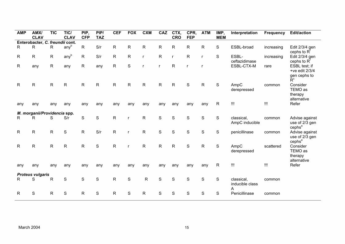
Resistance that encompasses cefoxitin and cefotetan as well as to third-generation cephalosporins in Enterobacteriaceae most often indicates AmpC production.9,19 Derepression of chromosomal AmpC in Enterobacter spp., Citrobacter freundii, Morganella morganii and Serratia spp. arises readily by mutation, giving this phenotype. AmpC hyperproduction can also arise by mutation in E. coli, though it is much rarer. Plasmid-mediated AmpC enzymes are increasingly encountered in E. coli and Klebsiella spp. and are mostly constitutive.20 Inducible AmpC, as in the classical phenotypes of Enterobacter and Citrobacter freundii gives resistance to cefoxitin without obvious cross-resistance to oxyimino cephalosporins (or cefotetan); a confirmatory test is cefoxitin mediated antagonism of oxyimino-cephalosporins.9,13
Resistance only to cefoxitin and cefuroxime in E. coli and Klebsiella spp. is mostly due to impermeability and porin loss; and is especially likely if the isolate retains moderate susceptibility/low level resistance to ampicillin.
Klebsiella oxytoca isolates that hyperproduce their chromosomal Kl β-lactamase often are mistaken for ESBL producers, but are distinguished by being highly resistant to aztreonam and
March 2004 3

cefuroxime but not ceftazidime or cefotaxime.9,21 They consistently are resistant to inhibitor combinations, even though extracted Kl enzyme is susceptible to inhibition.9,12
Most resistance to β-lactams in Enterobacteriaceae is mediated by acquired or chromosomal β-lactamases (Table V) but efflux and impermeability are more important in P. aeruginosa and in fastidious Gram-negative bacteria (Table VI and VII). These non-ß-lactamase-mediated mechanisms mostly give low-level broad-spectrum resistances, often affecting quinolones as well as ß-lactams.22 Imipenem, but not meropenem, escapes the commonest form of efflux-mediated resistance in P. aeruginosa (up-regulation of MexAB-OprM) but is more strongly compromised than meropenem by mutational loss of the OprD (=D2) porin, which provides carbapenem-specific channels through the outer membrane.23 In cases where P. aeruginosa isolates are highly resistant both to carbapenems (MIC >32 mg/L or growth up to the disc) and to other ß-lactams it may be worth doing a metallo-ß-lactamase test, seeking synergy between imipenem and EDTA.8 These tests are, however, complicated by the facts that not all gene-positive isolates are obviously resistant to carbapenems and that synergy between imipenem and EDTA can arise, at least in P. aeruginosa, for reasons other than inhibition of metallo-ß-lactamases.8 Resistance to carbapenems in Enterobacteriaceae (except for low-level resistance to imipenem in Proteeae) is unusual, and deserves reference laboratory examination, as does carbapenem resistance in Acinetobacter spp. The role of mecA in giving resistance to all β-lactams in methicillin-resistant staphylococci is discussed elsewhere5 and no comment is needed here, except to stress that isolates found resistant to indicator agents (Table IV) should be reported as resistant to all ß-lactams. In pneumococci, β-lactam resistance accrues stepwise and affects all members of the antibiotic class.24 Oxacillin resistance can be taken as an indicator of the underlying penicillin-binding protein changes.25 Cefotaxime, ceftriaxone and meropenem generally remain more active than penicillin against strains with the mechanism (Table VIII), but the position is reversed for a few isolates at least for the cephalosporins.26 Rare pneumococci are resistant to oxacillin, but not penicillin.24 Glycopeptides At present, transferable glycopeptide resistance is exclusive to enterococci, the exceptions being two recent reports of VanA-positive S. aureus.27 The common forms of this resistance are ht VanA and VanB types; VanD and VanE are rare, whereas VanC is intrinsic to clinically-infrequent enterococci, specifically Enterococcus casseliflavus and E. gallinarum. E. faecalis and E. faecium strains with the classical VanA phenotype show resistance to vancomycin and resistance, or markedly reduced susceptibility, to teicoplanin; those with classical VanB are resistant to vancomycin but remain susceptible to teicoplanin.28 Teicoplanin remains acceptable therapy against strains inferred, on this basis, to have VanB. VanC confers low-level resistance to Vancomycin, but not Teicoplanin.24 From the limited data available for the two recorded isolates, VanA behaves variably similarly in S. aureus to enterococci, affecting both vancomycin and teicoplanin, though not always giving frank resistance to the latter agent. Intermediate glycopeptide resistance which does not involve Van determinants -remains very rare in S. aureus, although teicoplanin resistance is frequent in coagulase-negative staphylococci. MIC tests are required to detect these mechanisms, disc tests being inadequate. Aminoglycosides In contrast to the β-lactamases, aminoglycoside-modifying enzymes modify their substrate compounds at different positions, variously acetylating, nucleotidylating or phosphorylating amino or hydroxyl groups. There are different forms of some modifying enzymes, often with markedly different substrate specificities. This variation is particularly evident in the AAC(3) and AAC(6') families.29,30 Counterwise, unrelated enzymes, affecting different sites, can confer same resistance phenotypes. Despite these difficulties the enzymes produced by isolates can often be predicted from the antibiogram data, as illustrated in Table IX and X.
Because few organisms have chromosomally encoded aminoglycoside-modifying enzymes, it is not necessary to split bacteria into as many groups as for β-lactamases and, with a few exceptions, Enterobacteriaceae can be treated as a single group (Table IX). However, Klebsiella spp. are shown separately, because resistance is more frequent than in most other genera.31 Serratia is also shown separately because of its chromosomally encoded AAC(6') enzyme.32 This usually is expressed weakly and the organism remains susceptible to aminoglycosides, but mutational hyper-production gives a characteristically resistant phenotype.33 Providencia stuartii possesses a chromosomal AAC(2') enzyme, which usually is expressed weakly but nevertheless confers low-level resistance to its substrates.34 This enzyme is virtually unknown outside Providencia spp.
Many of the plasmid-encoded enzymes seen in Enterobacteriaceae also occur in P. aeruginosa (Table IX), but AAC(3)II is very rare whereas AAC(3)III and AAC(6')II are more frequent.35 Broad-spectrum resistance, normally low level, is frequent in pseudomonads and is presumed to reflect poor up-take,35,36 although efflux may also be a factor in some organisms.37 P. aeruginosa is inherently resistant to kanamycin and neomycin, (kanamycin MICs around 64 mg/L) owing to low-level APH(3') activity.36
March 2004 4
Resistance to amikacin (and isepamicin) in Acinetobacter spp. is often associated with APH(3’)VI.38 This enzyme cannot modify gentamicin, netilmicin or tobramycin, so producers may be susceptible to some or all of these agents; many nevertheless are resistant owing to co-production of other modifying enzymes. Gram-positive organisms have different aminoglycoside-modifying enzymes to Gram-negative ones (Table X). Bi-functional APH(2')/AAC(6’) is by far the most important and frequent35,39 conferring resistance to all aminoglycoside analogues except streptomycin. Enterococci characteristically have low-level resistance to all aminoglycosides, but detection of high-level resistance (MICs > 256 mg/L though more often >1024 mg/L), mostly mediated by AAC(6’)/APH(2’) is significant, since it contra-indicates synergy with cell wall active agents.
Streptomycin is omitted from Table IX since it is seldom tested or used, and because there is no cross-resistance with other aminoglycosides, except when resistance is caused by impermeability.36,40 Streptomycin resistance in Enterobacteriaceae mostly depends on ANT(3')I or APH(3').41 High-level resistance in enterococci mostly reflects ANT(6)39 which, like other streptomycin-modifying enzymes, does not give cross-resistance to other aminoglycosides.
Production of multiple enzymes is more frequent with aminoglycoside-modifying enzymes than with β-lactamases.35,40 The simultaneous production of APH(3') plus a gentamicin-modifying enzyme can often be inferred from the resistance pattern; however, it is difficult to more than guess at the identity of combinations of enzymes that modify gentamicin or tobramycin, e.g. AAC(3)II + AAC(6'), without resorting to use of experimental compounds such as the 2' and 6'-N-ethyl derivatives of netilmicin. Experimental drugs such as these are a powerful tool in the prediction of aminoglycoside modifying enzyme types,40,42 but are beyond the scope of this review.
Most laboratory susceptibility test results with aminoglycosides can be accepted without editing. Quinolones Quinolones differ in their activity against bacterial species, doubtless reflecting differences in their ability to permeate, evade efflux and bind to different topoisomerases. Resistance, however, is a class effect, and isolates resistant to one analogue invariably show reduced susceptibility or resistance to other members of the family. In these circumstances there is little scope for interpretative reading, but a few general principles can be proposed.
Firstly, based on recent literature,43,44 the most active analogues against different groups are: Enterobacteriaceae: ciprofloxacin Non-fermenters: ciprofloxacin Pneumococci: moxifloxacin, gemifloxacin Enterococci: no available analogue has convincing activity Staphylococci: high risk of mutational resistance to all analogues
Secondly, the differentials in activity between ciprofloxacin, ofloxacin, norfloxacin, levofloxacin and moxifloxacin against Enterobacteriaceae are small (four-fold MIC variation).43,44 If an isolate is resistant to one of these drugs, susceptibility to the others is likely to be marginal at best and, in these circumstances, quinolones should only be used if there are no alternatives in other therapeutic classes. If an isolate appears highly susceptible to one fluoroquinolone but highly resistant to others, a testing problem is likely.
Thirdly, non-fermenters and Gram-positive cocci have lower inherent susceptibility to quinolones than Enterobacteriaceae. Isolates (even of classical phenotypes) may be susceptible to some analogues but marginally resistant to others. The most active analogues should be recommended for therapy, since it is hardest for resistance to develop.
Lastly, the value of using nalidixic acid as an indicator for reduced susceptibility or resistance to fluoroquinolones in fastidious Gram-negative bacteria (Table IV) should be re-emphasized, especially in the light of growing ciprofloxacin resistance in gonococci. MLS drugs (macrolides, lincosamides and streptogramins) Table XI gives interpretative guidelines for these agents. The most important source of resistance is the macrolide, lincosamides, streptogramin B (MLSB) system encoded by the erm genes, which may be constitutive or inducible in expression.45,46 Expression is regulated further by the sequences upstream of erm, which vary among the host elements prevalent in different species.
In the case of staphylococci, 14- and 15-membered macrolides, (e.g. erythromycin, clarithromycin and azithromycin) are inducers of erm whereas clindamycin and 16-membered macrolides do not induce. MLSB-inducible strains consequently express resistance to erythromycin but not clindamycin, whereas MLSB-constitutive (MLSB/c) organisms express resistance to both drugs. For MLSB-inducible isolates, erythromycin antagonizes clindamycin, a phenomenon easily demonstrated in double disc tests. Distin-guishing MLSB/c resistance in staphylococci is important since the dosage frequency for quinupristin/dalfopristin is changed from twice to thrice daily in skin and soft tissue infections when this mechanism is inferred.47
March 2004 5
Whether to report MLSB-inducible staphylococci (erythromycin-resistant, clindamycin-susceptible) as clindamycin-resistant remains debatable. Some authors support this approach, since MLSB-inducible strains segregate clindamycin resistant MLSB/c mutants, which may be selected in therapy.48,49

Nevertheless, one of the most-cited examples50 of resistance emerging during clindamycin treatment concerns a staphylococcal strain that was susceptible to erythromycin and so was unlikely to have harboured an erm gene. Lincosamide inactivation is an occasional source of resistance to clindamycin (not macrolides) in coagulase-negative staphylococci, but is very rare in Staphylococcus aureus.46
MLS resistance occurs in streptococci as well as staphylococci and, once again, can be inducible or constitutive. However, clindamycin as well as erythromycin often acts as an inducer. Thus, cross-resistance to both erythromycin and clindamycin indicates MLSB but does not prove constitutive expression. Resistance to erythromycin but not clindamycin may indicate an MLSB -inducible phenotype, but may also be contingent on efflux mediated by the products of mef genes. In the case of enterococci, a critical point is that E. faecalis is resistant to quinupristin/dalfopristin whereas almost all E. faecium isolates are susceptible—a mirror image of the pattern for ampicillin. Microbiologists should be sceptical of any isolate that is resistant or susceptible to both of these drugs or susceptible to both; such organisms deserve reference investigation. Tetracyclines No interpretative reading table for tetracyclines is provided, since multiple analogues are rarely tested. Nevertheless, not all the analogues are equally affected by the prevalent efflux [tet(A)-tet(F), tet(K) and tet(L)] or ribosomal protection [tet(M) and tet(O)] mechanisms, and a system of interpretative reading could be devised. In Gram-negative bacteria, tet(B) and tet(E) confer high-level resistance to all tetracycline derivatives whereas tet(A), tet(C), tet{D), tet(K) and tet(L) provide little or no protection against doxycycline and minocycline.51 In the case of gram-positive bacteria minocycline retains activity against strains with tet(K), but is compromised agasint those with tet(M).52 The glycylcycline derivative, tigecycline52 evades both efflux and ribosomal modes of resistance in both Gram-negative and Gram-positive bacteria. Other antibiotics Those antibiotics not discussed above are either the sole analogues within a class (e.g. chloramphenicol, fosfomycin; nitrofurantoin, trimethoprim) or belong to classes with little differentiation in microbiological activity (sulphonamides), meaning that there is little or no scope for interpretative reading. Nevertheless, interpretation is possible to the extent of recognizing inherently unlikely combinations of organisms and antibiotic susceptibility or resistance (Tables I and II); moreover the microbiologist should be alert to the likelihood of resistance emerging to many of these agents (Table III). The limits of interpretative reading Interpretative reading can never be so complete a strategy as identifying resistance mechanisms by genetic and biochemical investigation, and its limits should be recognized. First, bacteria with multiple resistance determinants affecting the same class(es) of antibiotics are increasingly frequent. Shaw et al.40 found multiple determinants in over 70% of 4088 amino-glycoside-resistant enterobacteria examined, and Essack et al53 found 84 TEM and SHV ß-lactamase genes among a collection of 25 K. pneumoniae isolates, only 20 of which were ESBL producers. The resistance patterns of isolates with multiple mechanisms may be confusing or misleading. For example, there is little to reliably distinguish the resistance pattern of a Klebsiella with an AmpC enzyme from that of a strain with both an ESBL and a permeability lesion. Secondly, interpretative reading cannot identify new resistance mechanisms if these give a resistance profile identical to that given by a known mechanism.1,3 Thirdly, some species and genera, notably Acinetobacter spp. and Burdholdera spp. frequently have complex multi-resistance profiles that are difficult to relate reliably to genetically-defined mechanism. Nevertheless, despite these caveats, there can be little doubt that interpretative reading, with attention to identifying the unusual and unlikely, editing out of dubious sensitivities and potential for surveillance resistance mechanisms is a useful advance over the standard practice of accepting all resistance data at face value. Acknowledgements We are grateful to the following for their helpful comments in the preparation of this paper: S. P. Borriello, J. Brazier, D. F. J. Brown, B. D. Cookson, J. Frost, R. C. George, A. J. Herring, A. P. Johnson, E. B. Kaczmarski, E. J. Threlfall and R. Wise.
March 2004 6

References 1. Courvalin, P. (1992). Interpretive reading of antimicrobial susceptibility tests. ASM News 58, 368-75. 2. Vedel, G., Peyret, M., Gayral, J. P. & Millot, P. (1996). Evaluation of an expert system linked to a rapid antibiotic susceptibility testing system for the detection of ß-lactam resistance phenotypes. Research in Microbiology 147, 297-309. 3. Funke, G., Monnet, D., deBernardis, C., von Graevenitz, A. & Frency, J. (1998). Evaluation of the VITEK 2 system for rapid identification of medically relevant gram-negative rods. Journal of Clinical Microbiology 36,1948-52. 4. Livermore, D.M., Struelens, M., Amorim, J. et al. (2002). Multicentre evaluation of the VITEK 2 Advanced Expert System for interpretive reading of antimicrobial resistance tests. Journal of Antimicrobial Chemotherapy 49, 289-300. 5. Brown, D. F. (2001). Detection of methicillin/oxacillin resistance in staphylococci. Journal of Antimicrobial Chemotherapy 48, 65-70. 6. Skov, R., Smyth, R., Clausen, M. et al. (2003). Evaluation of a cefoxitin 30 µg disc on Iso-Sensitest agar for detection of methicillin-resistant Staphylococcus aureus. Journal of Antimicrobial Chemotherapy 52, 204-7. 7. Andrews, J. M. &BSAC Working Party on Susceptibility Testing. (2001). BSAC standardized disc susceptibility testing method. Journal of Antimicrobial Chemotherapy 48, Suppl. A, 43-57. 8. Livermore, D. M. & Brown, D. F. J. (2001). Detection of ß-lactamase-mediated resistance. Journal of Antimicrobial Chemotherapy 48, Supp 1, 59-64. 9. Livermore, D. M. (1995). ß-Lactamases in laboratory and clinical resistance. Clinical Microbiology Reviews 8, 557-84. 10. Livermore, D. M. & Yuan, M. (1996). Antibiotic resistance and production of extended-spectrum ß-lactamases amongst Klebsiella spp. from intensive care units in Europe. Journal of Antimicrobial Chemotherapy 38, 409-24. 11. Bush, K., Jacoby, G. A. & Medeiros, A. A. (1995). A functional classification scheme for ß-lactamases and its correlation with molecular structure. Antimicrobial Agents and Chemotherapy 39, 1211-33. 12. Livermore, D.M. (1993). Determinants of the activity of ß-lactamase inhibitor combinations. Journal of Antimicrobial Chemotherapy 31 Suppl A, 9-21. 13. Jacoby, G. A. & Carreras, I. (1990). Activities of ß-lactam antibiotics against Escherichia coli strains producing extended-spectrum ß-lactamases. Antimicrobial Agents and Chemotherapy 34, 858-62. 14. Bradford, P. A., Yang, Y., Sahm, D.et al. (1998). CTX-M-5, a novel cefotaxime-hydrolyzing ß-lactamase from an outbreak of Salmonella typhimurium in Latvia. Antimicrobial Agents and Chemotherapy 42,1980-4. 15. Nordmann, P. (1998). Trends in ß-lactam resistance among Enterobacteriaceae. Clinical Infectious Diseases 27, Suppl. 1, S100-6. 16. Alobwede, I., M’Zali, F.H., Livermore, D.M. et al (2003). CTX-M extended-spectrum ß-lactamase arrives in the UK. Journal of Antimicrobial Chemotherapy 51, 470.1. 17. Brenwald, N.P., Jevons, G., Andrews, J.M. et al (2003). An outbreak of a CTX-M-type ß-lactamase-producing Klebsiella pneumoniae: the importance of using cefpodoxime to detect extended-spectrum ß-lactamases. Journal of Antimicrobial Chemotherapy 51, 195-6. 18. Mushtaq, S., Woodford, N., Potz, N., & Livermore, D.M. (2003). Detection of CTX-M-15 extended-spectrum ß-lactamase in the united Kingdom. Journal of Antimicrobial Chemotherapy 52, 528-9. 19. Moritz, V. A. & Carson, P. B. (1986). Cefoxitin sensitivity as a marker for inducible ß-lactamases. Journal of Medical Microbiology 21, 203-7.
March 2004 7

20. Walther-Rasmussen, J. & Hoiby, N. (2002). Plasmid-borne AmpC ß-lactamases. Journal of Microbiology 48,479-93. 21. Gheorghiu, R., Yuan, M., Hall, L.M.C. & Livermore, D.M. (1997). Bases of variation in resistance to ß-lactams in Klebsiella oxytoca isolates hyperproducing K1 ß-lactamase. Journal of Antimicrobial Chemotherapy 40, 533-41. 22. Li, X.Z., Nikaido, H., & Poole, K. (1995). Role of mexA-mexB-oprM in antibiotic efflux in Pseudomonas aeruginosa. Antimicrobial Agents and Chemotherapy 39, 1948-53. 23. Livermore, D.M. & Yang, Y.J. (1989). Comparative activity of meropenem against Pseudomonas aeruginosa strains with well-characterized resistance mechanisms. Journal of Antimicrobial Chemotherapy 24 Suppl A, 149-159. 24. Spratt, B.G. (1994). Resistance to antibiotics mediated by target alterations. Science 264, 388-93. 25. Klugman, K.P. (1990). Pneumococcal resistance to antibiotics. Clinical Microbiology Review 3, 171-196. 26. Lonks, J. R., Durkin, M. R., Meyerhoff, A. N. & Medeiros, A. A. (1995). Meningitis due to ceftriaxone-resistant Streptococcus pneumoniae. New England Journal of Medicine 332, 893-4. 27. Bozdogan, B., Esel, D., Whitener, C. et al (2003) Antibacterial susceptibility of a Vancomycin-resistant Staphylococcus aureus strain isolated at the Hershey Medical Centre. Journal of Antimicrobial Chemotherapy 52, 864-8. 28. Woodford, N., Johnson, A. P., Morrison, D & Speller, D. C. E. (1995). Current perspectives on glycopeptide resistance. Clinical Microbiology Reviews 8, 585-615. 29. Shaw, K. J., Rather, P. N., Hare, R. S. & Miller, G. H. (1993). Molecular genetics of aminoglycoside resistance genes and familial relationships of the aminoglycoside modifying enzymes. Microbiological Reviews 57,138-63. 30. Mingeot-Leclercq, M.-P., Glupczynski, Y. & Tulkens, P. M. (1999). Aminoglycosides: activity and resistance. Antimicrobial Agents and Chemotherapy, 43, 727-37. 31. Phillips, I., King, A. & Shannon, K. (1986). Prevalence and mechanisms of aminoglycoside resistance. A ten-year study. American Journal of Medicine 80, 48-55. 32. Champion, H. M., Bennett, P. M., Lewis, D. A. & Reeves, D. S. (1988). Cloning and characterization of an AAC(6') gene from Serratia marcescens. Journal of Antimicrobial Chemotherapy 22, 587-96. 33. Hawkey, P. M. & Constable, H. K. (1988). Selection of netilmicin resistance, associated with increased 6' aminoglycoside acetyltransferase activity, in Serratia marcescens. Journal of Antimicrobial Chemotherapy 21,535-44. 34. Rather, P. N., Orosz, E., Shaw, K. J., et al (1993). Characterization and transcriptional regulation of the 2'-N-acetyltransferase gene from Providencia stuartii. Journal of Bacteriology 175, 6492-8. 35. Miller, G. H., Sabatelli, F. J., Hare, R. S., et al. (1997). The most frequent aminoglycoside resistance mechanisms—changes with time and geographic area: a reflection of aminoglycoside usage patterns? Clinical Infectious Diseases 24, Suppl. 1, S46-62. 36. Phillips, I., King, B. A. & Shannon, K. P. (1978). The mechanisms of resistance to aminoglycosides in the genus Pseudomonas. Journal of Antimicrobial Chemotherapy 4,121-9. 37. Westbrock-Wadman, S., Sherman, D. R., Hickey, M. J., et al. (1999). Characterization of a Pseudomonas aeruginosa efflux pump contributing to aminoglycoside impermeability. Antimicrobial Agents and Chemotherapy 43, 2975-83. 38. Lambert, T., Gerbaud, G., & Courvalin, P. (1988). Transferable amikacin resistance in Acinetobacter spp. due to a new type of 3’-aminoglycoside phosphotransferase. Antimicobial Agents and Chemotherapy 32, 15-9.
March 2004 8

39. Ounissi, H., Derlot, E., Carlier, C. & Courvalin, P. (1990). Gene homogeneity for aminoglycoside-modifying enzymes in gram-positive cocci. Antimicrobial Agents and Chemotherapy 34, 2164-8. 40. Shaw, K. J., Hare, R. S., Sabatelli, F. J., et al. (1991). Correlation between aminoglycoside resistance profiles and DNA hybridization of clinical isolates. Antimicrobial Agents and Chemotherapy 35, 2253-61. 41. Shannon, K. P., Phillips, I. & King, B. A. (1978). Aminoglycoside resistance among Enterobacteriaceae and Acinetobacter species. Journal of Antimicrobial Chemotherapy 4,131 -42. 42. Schindler, J. Ed. (2000). World Antibiotic Resistance Network: A step-by-step procedure for the identification of Ag R mechanisms in Gram-negative bacteria. http://www.warn.cas.cz/. 9 January 2004, date last accessed. 43. Blondeau, J. M. (1999). A review of the comparative in-vitro activities of 12 antimicrobial agents, with a focus on five new 'respiratory quinolones' Journal of Antimicrobial Chemotherapy 43, Suppl. B,1-11. 44. Thomson, C. J. (1999). The global epidemiology of resistance to ciprofloxacin and the changing nature of antibiotic resistance: a 10-year perspective. Journal of Antimicrobial Chemotherapy 43, Suppl.A, 31-40. 45. LeClercq, R. & Courvalin, P. (1991). Bacterial resistance to macrolide, lincosamide, and streptogramin antibiotics by target modification. Antimicrobial Agents and Chemotherapy 35,1267-72. 46. LeClercq, R. & Courvalin, P. (1991). Intrinsic and unusual resistance to macrolide, lincosamide and streptogramin antibiotics in bacteria. Antimicrobial Agents and Chemotherapy 35,1273-6. 47. Johnson, A. P. & Livermore, D. M. (1999). Quinupristin/dalfopristin, a new addition to the antimicrobial arsenal. Lancet 354, 2012-3. 48. Ayliffe, G. (1978). Staphylococcus aureus. In: Laboratory Methods in Antimicrobial Chemotherapy, (Reeves, D. S., Phillips, I., Williams, J. D. & Wise, R., Eds), pp. 112-4. Churchill Livingstone, Edinburgh. 49. Duncan, I. B. R. (1967). Development of lincomycin resistance by staphylococci. Antimicrobial Agents and Chemotherapy 7, 723-9. 50. Watanakunakorn, C. (1976). Clindamycin therapy of Staphylococcus aureus endocarditis. Clinical relapse and development of resistance to clindamycin, lincomycin and erythromycin. American Journal of Medicine 60, 419-25. 51. Rice, L. B. & Bonomo, R. A. (1996). Genetic and biochemical mechanisms of bacterial resistance to antimicrobial agents. In Antibiotics in Laboratory Medicine, (Lorian, V., Ed.), pp. 453-501. Williams & Wilkins, Baltimore, MD. 52. Petersen.P. J., Jacobus, N.V.,Weiss, W.J. et al (1999). In vitro and in vivo antibacterial activities of a novel glycylcycline, the 9-t-butylglycylamido derivative of minocycline (GAR-936). Antimicrobial Agents and Chemotherapy 43, 738-44. 53. Essack, S. Y., Hall, L. M. C., Pillay, D. G., et al (2001). Complexity and diversity of Klebsiella pneumoniae strains with extended-spectrum ß-lactamases in 1994 and 1996 at a teaching hospital in Durban, South Africa. Antimicrobial Agents and Chemotherapy 45, 88-95.
March 2004 9

Table I. Unusual resistances needing reference laboratory confirmation (see text for addresses) Organism Resistances requiring confirmation
Staphylococcus aureus Any of: vancomycin, teicoplanin, linezolid, quinupristin/dalfopristin
Coagulase-negative staphylococci
Any of: vancomycin, linezolid
Jeikeium coryneforms Any of: vancomycin, teicoplanin, linezolid
Streptococcus pneumoniae Any of: meropenem, vancomycin, teicoplanin, linezolid. Group A, B, C, G ß-haemolytic streptococci
Any of: penicillin, vancomycin, teicoplanin, linezolid.
Enterococci Both ampicillin and quinupristin/dalfopristin. Linezolid. Teicoplanin but not vancomycin.
Enterobacteriaceae Meropenem Haemophilus influenzae Any third-generation cephalosporin, or carbapenem Moraxella catarrhalis Ciprofloxacin Neiseria meningitides Any of: penicillin (high-level), ciprofloxacin Neisseria gonorrhoeae Any third-generation cephalosporin Acinetobacter; P. aeruginosa Colistin Anaerobes in general Metronidazole Bacteroides Any of: metronidazole, co-amoxiclav, carbapenems Clostridium difficile Any of: metronidazole, vancomycin
Note to all tables: β-lactam groups First generation cephalosporins: cephalexin, cephalothin, cephazolin and cephradine. Second generation cephalosporins: cefamandole, cefaclor and cefuroxime. Third generation cephalosporins: cefotaxime, cefpodoxime, ceftazidime and ceftriaxone. Fourth generation cephalosporins: cefepime and cefpirome. Oxyimino cephalosporins: cefepime, cefotaxime, cefpirome, cefpodoxime, ceftazidime, ceftriaxone and cefuroxime. Cephamycins: cefoxitin, cefotetan. Aminopenicillins: amoxycillin, ampicillin, mezlocillin and piperacillin. Carboxypenicillins: carbenicillin and ticarcillin.
March 2004 10

Table II. Natural resistances typical of common pathogens Organisms Natural resistances to
All Enterobacteriaceae Penicillin G, glycopeptides, fusidic acid, macrolides, clindamycin, linezolid, streptogramins (e.g. quinupristin/dalfopristin), mupirocin
Acinetobacter baumannii Ampicillin, amoxycillin, first-generation cephalosporins
Pseudomonas aeruginosa Ampicillin, amoxycillin, co-amoxiclav, first-generation cephalosporins, second-generation cephalosporins, cefotaxime, ceftriaxone, nalidixic acid, nitrofurantoin, trimethoprim
Burkholderia cepacia Ampicillin, amoxycillin, first-generation cephalosporins, colistin, aminoglycosides
Stenotrophomonas maltophilia All ß-lactams except ticarcillin/clavulanate, aminoglycosides
Flavobacterium (Chryseobacterium/Myroides)
Ampicillin, amoxycillin, first-generation cephalosporins
Salmonella spp. Cefuroxime and aminoglycosides (active in vitro, not active in vivo)
Klebsiella spp., Citrobacter diversus
Ampicillin, amoxycillin, carbenicillin, ticarcillin
Enterobacter spp., C. freundii Ampicillin, amoxycillin, co-amoxiclav, first-generation cephalosporins, cefoxitin
M. morganii Ampicillin, amoxycillin, co-amoxiclav, first-generation cephalosporins, cefuroxime, colistin, nitrofurantoin, tetracyclines
Providencia spp. Ampicillin, amoxycillin, co-amoxiclav, first-generation cephalosporins, cefuroxime, gentamicin, netilmicin, tobramycin, colistin, nitrofurantoin, tetracyclines
Proteus mirabilis Colistin, nitrofurantoin, tetracyclines
Proteus vulgaris Ampicillin, amoxycillin, cefuroxime, colistin, nitrofurantoin, tetracyclines
Serratia spp. Ampicillin, amoxycillin, co-amoxiclav, first-generation cephalosporins, cefuroxime, colistin
Yersinia enterocolitica Ampicillin, amoxycillin, carbenicillin, ticarcillin, first-generation cephalosporins
Campylobacter jejuni, Campylobacter coli
Trimethoprim
H. influenzae Penicillin G, erythromycin, clindamycin
M. catarrhalis Trimethoprim
All Gram-positive bacteria Aztreonam, temocillin, colistin, nalidixic acid
Streptococci Fusidic acid, aminoglycosidesa
S. pneumoniae Trimethoprim, aminoglycosides
Methicillin-resistant S. aureus All ß-lactams
Enterococci Carbenicillin, ticarcillin, all cephalosporins, aminoglycosides,a mupirocin
Listeria Third-generation cephalosporins, fluoroquinolones
See note relating to all tables at foot of Table I. aLow-level resistance: aminoglycosides are useful for synergy with penicillins against typical streptococci and enterococci.
March 2004 11

Table III: Antibiotic/organism combinations where mutational resistance is likely to develop Organism Antibiotic Staphylococci Fusidic acid, rifampicin, fluoroquinolones Erythromcyin-resistant staphylococci Clindamycin S. pneumoniae Ciprofloxacin P. aeruginosa All anti-pseudomonal antibiotics, except colistin
and, possibly, meropenem B. cepacia All relevant antibiotics Enterobacter, Citrobacer, Serratia, Morganella All third-generation cephalosporins Coliforms iwth ESBLs Cephamycins (via impermeability) All coliforms Fosfomycin, nalidixic acid (not
fluoroquinolones) Serratia marcescens Netilmicin, tobramycin, amikacin, kanamycin See note relating to all tables at foot of Table I. This table excludes rarely used antibiotic/organism combinations; it also only considers the risk of resistance arising in the original pathogen, not the likelihood of overgrowth by other species (e.g. enterococci and C. difficile), which may also be a significant clinical hazard. Table IV. Useful indicator antibiotics Organism Resistance to Inference /action Staphylococci oxacillin or methicillin,
cefoxitin Resistant to all ß-lactams.
Staphylococci erythromycin Inducible clindamycin resistance likely; avoid clindamycin, or use with caution.
Staphylococci erythromycin and clindamycin (lincomycin may be better indicator than clindamycin)
Constitutive MLSB/c resistance. Quinupristin/ dalfopristin likely to be bacteriostatic, not bactericidal; dosage should be increased to thrice daily even in skin and soft tissue infection
Pneumococci oxacillin (zone ≤18 mm) Probably penicillin resistant. Perform E-test for any penicillin or cephalosporin to be used
E. faecalis ampicillin Probably E. faecium, but may be less frequent species or (just possibly) may have acquired resistance: check speciation or refer
H. influenzae cefaclor Likely non-ß-lactamase-type resistance (better indicator than ampicillin)
Neisseria spp. H. influenzae Campylobacter spp.
nalidixic acid Indicates reduced susceptibility or resistance to fluoroquinolones
Klebsiella / E. coli Ceftazidime or cefpodoxime Likely ESBL producer86 Avoid all cephalosporins except cephamycins
Any Enterobacteriaceae any second-generation cephalosporin
Likely to have potent ß-lactamase; avoid first-generation cephalosporins
Any Enterobacteriaceae any third -generation cephalosporin
Likely to have potent ß-lactamase; avoid first-and second- generation cephalosporins except, possibly, cephamycins
Any Enterobacteriaceae resistant to any ureidopenicillins
Likely to have penicillinase, avoid all amino-, ureido- and carboxy-penicillins
Any Enterobacteriaceae resistant to any ß-lactamase inhibitor combinations
Assume resistance to the corresponding unprotected penicillin
See note relating to all tables at foot of Table I.
March 2004 12

Table V. Phenotypes: interpretation of mechanisms and editing of antibiograms: B-lactams versus Enterobacteriaceae AMP AMX/
CLAV TIC TIC/
CLAV PIP, CFP
PIP/ TAZ
CEF FOX CXM CAZ CTX,CRO
CPR, FEP
ATM IMP,MEM
Interpretation Frequency Edit/action
E. Coli, P. mirabilis, Salmonella, Shigella spp. S S S S S S S
S S S S S S Sa classical commonR S R S r S r S S S S S S S penicillinase-
low common Edit pip to R;
edit 1st gen cephs to R except in UTI where e.g. Cephalexin retains acceptable activity
R r/R R r/R R r/R R S S S S S S S penicillinase-high
common Edit 1st gen cephs to R except in UTI
R R r R R R R R R R r/R S r/R S AmpC high-plasmid or chromosomal
rare ConsiderTEMO as alternative to carbapenems
R anyb R anyb R anyb R S R R R R R S ESBL-broad rare ESBL test; if+ve, edit 2/3/4 gen cephs to Rc
R anyb R anyb R anyb R S r R r r r S ESBL-ceftazidimase
rare ESBL test; if +ve, edit 2/3/4 gen cephs to Rc
R any R any R any R S r r R r r ESBL-CTX-M increasing ESBL test; if+ve, edit 2/3/4 gen cephs to Rc
R R R R R r/R S S S S S S S S IRTd ???r r r r r r r R R S S S S S impermeability
rare
any any any any any any any any any any any any any R1 !!! !!! Refer
March 2004 13

AMP AMX/ CLAV
TIC TIC/ CLAV
PIP, CFP
PIP/ TAZ
CEF FOX CXM CAZ CTX, CRO
CPR, FEP
ATM IMP, MEM
Interpretation Frequency Edit/action
Klebsiella spp. R S R
S r S S S S S S S S S Classical-lowSHV-1 or K1
common Edit allpenicillins (except TEMO) to R. R r/R R anyb R r/R R S S S S S S S Penicillinase-
high common
R anyb R anyb R anyb R S R R R R R S ESBL-broad scattered ESBL test; if+ve, edit 2/3/4 gen cephs to Rc
R anyb R anyb R anyb R S r R r r r S ESBL-ceftazidimase
scattered ESBL test; if +ve, edit 2/3/4 gen cephs to Rc
R any R any R any R S r r R r r ESBL-CTX-M increasing ESBL test; if+ve, edit 2/3/4 gen cephs to Rc
R R R R R r/R S S S S S S S S IRTd ???R R R R R R R S R S S S R S K1, high, K.
oxytoca only scattered Edit CTX to R;
??? CAZ R R R R R R R R R R R S r/R S Plasmid AmpC Rare
R r R r r r r R R S S S S S impermeability
Rare any
any
any any
any
any
any
any
any
any
any
any
any
any
!!! !!! Refer
Enterobacter, C. freundii R R S S/r S S R R S/r S S S S S classical,
AmpC inducible common Advise against
use of 2/3 gen cephse
R R R anyb R S/r R R S S S S S S Penicillinase common Advise againstuse of 2/3 gen cephse
March 2004 14
AMP AMX/ CLAV
TIC TIC/ CLAV
PIP, CFP
PIP/ TAZ
CEF FOX CXM CAZ CTX, CRO
CPR, FEP
ATM IMP, MEM
Interpretation Frequency Edit/action
Enterobacter, C. freundii cont. R R R anyb
R S/r R R R R R R R S ESBL-broad increasing Edit 2/3/4 gencephs to Rf
R R R anyb R S/r R R r R r R r S ESBL-ceftazidimase
increasing Edit 2/3/4 gen cephs to Rf
R any R any R any R S r r R r r ESBL-CTX-M rare ESBL test; if+ve edit 2/3/4 gen cephs to Rc
R R R R R R R R R R R S R S AmpCderepressed
common ConsiderTEMO as therapy alternative
any
any
any
any
any
any
any
any
any
any
any
any
any
R !!! !!! Refer
M. morganii/Providencia spp. R R S S/r S S R r R S S S S S classical,
AmpC inducible common Advise against
use of 2/3 gen cephse
R R R S R S/r R r R S S S S S penicillinase common Advise againstuse of 2/3 gen cephse
R R R R R S R r R R R S R S AmpCderepressed
scattered ConsiderTEMO as therapy alternative
any
any
any
any
any
any
any
any
any
any
any
any
any
R !!! !!! Refer
Proteus vulgaris R S R S S S R S R S S S S S classical,
inducible class A
common
R S R S R S R S R S S S S S Penicillinase common
March 2004 15

AMP AMX/ CLAV
TIC TIC/ CLAV
PIP, CFP
PIP/ TAZ
CEF FOX CXM CAZ CTX, CRO
CPR, FEP
ATM IMP, MEM
Interpretation Frequency Edit/action
Proteus vulgaris cont. R S R S
R S R S R S R S S S Chromosomalderepressed
Rare
any
any
any
any
any
any
any
any
any
any
any
any
any
R !!! !!! Refer
Citrobacter diversus R S R S r S R S S S S S S S classical,
inducible class A
common Edit allpenicillins (except TEMO) to R. R R/R R anyb R r/R R S S S S S S S penicillinase-
high
common
any
any
any
any
any
any
any
any
any
any
any
any
any
any
!!! !!! Refer
Serratia spp. R R S S S S R r R S S S S S classical,
AmpC inducible common Advise against
use of 2/3 gen cephse
R R R anyb R any R r R S S S S S penicillinase common Advise againstuse of 2/3 gen cephse
R R R R r/R R r R S R S R S AmpCderepressed
rare ConsiderTEMO as therapy alternative
any any any any any any any any any any any any any R !!! !!! Refer See note relating to all tables at foot of Table I. General notes for Tables V-XI: Classical means the historic phenotype of the species, without acquired resistance; Common means seen in >10% of isolates; scattered means seen in 5-10% of isolates; uncommon means seen in 1-5% of isolates; and rare means seen in <1%; increasing is used in cases where a resistance is still rare, but is proliferating rapidly. Local frequencies may be very different, especially during outbreaks and in specialist units. Refer and !!! mean send to an appropriate reference or academic laboratory for confirmation (see text); ??? means uncertain as insufficient data.
March 2004 16

Abbreviations: AMK, amikacin; AMX, amoxicillin; AMP, ampicillin; AMX/CLAV, co-amoxiclav; ATM, aztrenam; CAZ, ceftaxzidime; Ceph, cephalosporins; FEP, cefepime; CLI, clindamycin; CEF, cephalothin; CF, cystic fibrosis; CFP, cefoperazone, CPR, cefpirome; CRO, ceftriaxone; CTX, cefotaxime; CXM, cefuroxime; ERY, erythromycin; FOX, cefoxitin; GEN, gentamicin; IMP, imipenem; KAN, kanamycin; MEM, meropenem; NEO, neomycin; NET, netilmicin; OXA, oxacillin/cloxacillin; PCG, penicillin G; PIP, Piperacillin; TZP, Piperacillin/tazobactam; Q-D, quinupristin/dalfopristin; TEMO, temocillin; TIC, ticarcillin;’ TIC/CLAV, ticarcillin/clavulanate; TOB, Tobramycin; 1st gen ceph, first generation cephalosporins; 2/3/4 gen ceph, second/third/fourth generation cephalosporins; R, resistant; r, reduced zones but likely to remain susceptible at BSAC breakpoints; B, borderline (MICs for typical strains of the species without acquired resistance, fall around the zone/MIC breakpoints); S, susceptible. aDiscount low-level imipenem resistance in P.mirabilis bVaries with amount of ß-lactamase produced. cSee Livermore and Brown8 for ESBL tests for these species. dIRT; inhibitor-resistant TEM mutant. eIf second or third generation cephalosporins are used, there is substantial risk of selection of derepressed, mutants during therapy. See Table III. fESBL tests8 are difficult with AmpC-inducible species since clavulanate-induced AmpC enzymes (which evade the action of clavulanate) are prone to attack the indicator cephalosporin, but cefepime/clavulanate may be useful, as cefepime is less likely to be affected by induced AmpC than are third-generation cephalosporins. The pattern of cefotetan-susceptible, ceftazidime-resistant would imply ESBL production, but this principle has not been evaluated critically.8
March 2004 17

Table VI. Phenotypes, interpretation of mechanism and editing of antibiograms; ß-lactams versus non-fermenters TIC TIC/CLAV PIP,CFP PIP/TAZ CAZ CPR,FEP ATM IMP MEM Interpretation Frequency Edit/ActionP. aeruginosa S S/r S
S S S S S S classical common Bewaremutational resistance; see Table III R any R any S S S S S penicillinase
rare
r R r r r r R S S AmpC partderepressed
common
r/R r/R R R R S/r R S S AmpC fullyderepressed
rare
R R r/R r/R r/R R r/R S r Increasedeffluxa
common
S S/r S S S S S R r loss of OprDporin
scattered
R R r/R r/R R R R R R Acquisition ofmultiple mutationsa or, rarely, metallo-ß-lactamases
Mutational form common e.g. in CF
Seek metallo-ß-lactamases by imipenem/EDTA synergy if highly R
Acinetobacter spp.
Relationships between antibiogram and mechanisms poorly defined. Carbapenems have the most consistent activity against the genus, but carbapenemases or the OXA and VIM/IMP classes are a growing concern. Isolates with carbapenem resistance should be referred for specialist investigation. ESBL tests do not work, as many isolates are susceptible to clavulanate alone.
S. maltophilia May appear susceptible to penicillins and cephalosporins on Iso-Sensitest agar, but is generally resistant on Mueller-Hinton agar. Among ß-lactams, ticarcillin/clavulanate has best provenance, although co-trimoxazole (not trimethoprim alone) is the usual drug of choice.
See note to all tables at foot of Table 1 and general notes and abbreviations for Tables V-XI at the foot of Table V. aIsolates typically also have r/R to quinolones
March 2004 18

Table VII. Phenotypes; interpretation of mechanism and editing of antibiograms; ß-lactams versus fastidious Gram-negative bacteria and M. catarrhalis PCG AMP, AMX AMX/CLAV CCL CTX,
CRO,CFIX MEM IMP Interpretation Frequency Edit/action
H. influenaze R S
S S S S S Classical CommonR R S S S S S ß-lactamase +ve Common Confirm with ß-lactamase
testa R r/r r/R R r S S/R8 Intrinsic resistance-altered
PBPs; impermeability or efflux
Rare
any any any any any R any !!! !!! Referany any any any R any any !!! !!! Refer
N. gonorrhoeae S S S - S - - Classical common
R R S - S - - ß-lactamase +ve common Confirm with ß-lactamase testa
r/R r/R r/R - S - - Impermeability or efflux
common any any any any R - any !!! !!! Refer
N. meningitides S S S - S - - Classical Common
r R R - S - - Impermeability or efflux CommonSubstantial R to any ß-lactam !!! !!! Refer
M. catarrhalis R S S S S S S Classical Common Confirm ß-lactamase
negative by direct test; if +ve, report as ampicillin/resistant-resistant R R S S S S S BRO-1/2 ß-lactamase +ve common
See note to all tables at foot of Table I and general notes and abbreviations for Tables V-XI at the foot of Table V. aSee Livermore & Brown8 for ß-lactamase tests bH. influenzae with intrinsic resistance to penicillins and cephalosporins are either fully susceptible to imipenem, or show a high level of resistance, implying that the group encompasses at least two different genotypes.
March 2004 19

Table VIII. Phenotypes; interpretation of mechanism and editing of antibiograms: ß-lactams versus Gram-positive cocci PCG AMP, AMX AMX/CLAV OXA Any ceph IMP, MEM Interpretation Frequency Edit/Action Staphylococci S S
S S S S Classical, nowuncommon
Scattered
R R S S S S ß-lactamase +ve Common Edit all penicillins except oxacillin and methicillin to R
any any any any any any Methicillin/oxacillinresistant
Common Edit all ß-lactams to R
S. pyogenes S S S S S S Classical
Common
R any any any any any !!! !!! ReferS. pneumoniae
S S S S S S Classicalany any any R any any PenR pneumococcus Common Determine MICs of drugs intended
for use. Cefotaxime and ceftriaxone, also meropenem, often remain active, with oral cephalosporins mostly less active than amoxicillin E. faecalis
r S S R R S Classical Common R R S R R S ß-lactamase +ve !!! Refer
R R R R R R Probably E. faecium
Error Check speciation E. faecium
R S S R R S Classical, now rare ScatteredR R R R R R Uses PBP-5 to cross-
link peptidoglycan common
See note to all tables at foot of Table I and general notes and abbreviations for Tables V-XI at the foot of Table V.
March 2004 20

Table IX. Phenotypes; interpretation of mechanism and editing of antibiograms: aminoglycosides versus Gram-negative bacteria
March 2004 21
GEN NET TOB AMK KAN NEO Interpretation Frequency Edit/action and comments E. coli and other Enterobacteriaceae not shown separately S S S S S S classical common R S S S S S AAC(3)I Rare Also R to fortimicin R R R S R S AAC(3)II rare Greater R to GEN than to TOB
or NET R R R S r R AAC(3)IV rare Also R to apramycin (used in
veterinary practice). Mostly in E. coli
S/r R R R R R AAC(6’) rare One component of GEN remains active but in vivo use best avoided.
R S R S R S ANT(2’) rare Equal R to GEN and TOB S S S S R R APH(3’) common Usually more R to KAN than
NEO. Was common, now rarely tested.
r/R r/R r/R r/R r/R r/R ‘impermeability’ rare Low-level R to all aminoglycosides
Klebsiella spp. S S S S S S classical common R S S S S S AAC(3)I rare Also R to fortimicin R R R S r S AAC(3)II scattered/rare Greater R to GEN than to TOB
or NET S/r R R R R R AAC(6’) rare One component of GEN
remains active, but in vivo use of GEN vs suspect isolates.
R S R S R S ANT(2’) scattered/rare Equal R to GEN and TOB S S S S R R APH(3’) common? Usually more R to KAN than
NEO. Was common, now rarely tested.
r/R r/R r/R r/R r/R r/R ‘impermeability’ rare Low-level R to all aminoglycosides
Serratia spp. S S S S S S classical common Chromosomal AAC(6’)
expressed weakly: risk of selection of over-producers in therapy with AMK, TOB, NET.
R S S S S S AAC(3)I rare Also R to fortimicin R R R S r S AAC(3)III rare Greater R to GEN than TOB or
NET S/r R R R R R AAC(6’) common Mutation causes over-
production of chromosomal AAC(6’)
R S R S R S ANT(2’) rare Equal R to GEN and TOB S S S S R R APH(3’) rare Usually more R to KAN than
NEO r/R r/R r/R r/R r/R r/R ‘impermeability’ rare Low-level R to all
aminoglycosides Providencia stuartii R R R S S R AAC(2’) classical Chromosomal AAC(2’); poorly
expressed R R R S S R AAC(2’) common Mutation causes
overproduction of AAC(2’) P. aeruginosa S S S S R R classical common R S S S R R AAC(3)I rare Also R to fortimicin R S R S R R AAC(3)III rare S/r R R R R R AAC(6’) rare One component of GEN remain
active, but in vivo use best avoided
R R R S R R AAC(6’)II rare R pattern not obviously predictable from enzyme activity

GEN NET TOB AMK KAN NEO Interpretation Frequency Edit/action and comments P. aeruginosa cont. R S R S R R ANT(2’) rare Equal levels of R to GEN and
TOB S S S S R R APH(3’) common Usually more R to KAN than to
NEO r/R r/R r/R r/R r/R r/R ‘impermeability’ scattered Low-level R to all
aminoglycosides See note to all tables at foot of Table I and general notes and abbreviations for Tables V-XI at the foot of Table V. Mechanism varies in level among isolates and may also involve efflux; Tobramycin retains best activity against most representatives.
March 2004 22

Table X. Phenotypes; interpretation of mechanism and editing of antibiograms: aminoglycosides versus Gram-positive bacteria GEN NET TOB AMK KAN NEO Interpretation Frequency Edit/action and
comments Staphylococci S S S S S S classical common S S R R R S ANT(4’)(4”)I rare Unlike ‘Gram-negative’
ANT(4’), also modifies dibekacin at 4”.
R R R r R R APH(2”) AAC(6’)
rare scattered
Greater R to TOB
S S S S R R APH(3’) common Usually more R to KAN than NEO
S S S R R R APH(3’)III rare Rare r/R r/R R/R r/R r/R r/R ‘impermeability’ rare Low-level to all
aminoglycosides E. faecalis R R R R R R Classical common Intrinsic low-level
resistance R R HLR HLR HLR R ANT(4’)(4”)I rare HLR R HLR R HLR R APH(2”)/AAC(6’) common Greater R to GEN than
TOB R R R R HLR HLR APH(3’) common Usually more R to Kan
than NEO R R R HLR HLR HLR APH(3’)III rare Rare E. faecium R R R R R R AAC(6’)I classical Chromosomal AAC(6’),
intrinsic to E. faecium R R HLR HLR HLR R ANT(4’)(4’) rare HLR R HLR R HLR R APH(2”)/AAC(6’) common Greater R to GEN than
TOB R R R R HLR HLR APH(3’) common Usually greater R to KAN
than NEO R R R HLR HLR HLR APH(3’)III rare See note to all tables at foot of Table I and general notes and abbreviations for Tables V-XI at the foot of Table V. HLR = high-level resistance in enterococci.
March 2004 23

March 2004 24
Table XI. Phenotypes; interpretation of mechanism and editing of antibiograms: MLS drugs versus Gram-positive bacteria ERYa CLI Q-D Interpretation Frequency Edit/action Staphylococci S S S classical common R S S May be MLSB
inducible may have macrolide efflux
common Check if erythromycin antagonizes clindamycin; if antagonism seen, isolate have MLSb and clindamycin should be used with caution (if at all)
R R S MLSB constitutive common Note specification of product characteristics recommendation that Q-D should be given thrice daily even in skin and soft tissue infection
any any R !!! refer Streptococci, including S. pneumoniae S S R classical common R R S MLSB
constitutive/inducible common NB – inducible resistance usually
affects clindamycin as well as erythromycin in streptococci
R S S Efflux; MLSB inducible
common
any Any R !!! refer E. faecalis S S R classical common R S R May be MLSB
inducible may have macrolide efflux
common Check if erythromycin antagonizes clindamycin, e.g. with a double disc test. If antagonism is seen, the isolate have MLSB and clindamycin should be used with caution (if at all)
R R R MLSB constitutive common any any S Probably mis-
speciation If also AMP resistant, almost certainly
E. faecium, not E. faecalis. Refer if confirmed as E. faecalis.
E. faecium S S S classical common R S S May be MLSB
inducible may have macrolide efflux
common Check if erythromycin antagonizes clindamycin; if antagonism seen, isolate have MLSb and clindamycin should be used with caution (if at all)
R R S MLSB constitutive common any any R Probable mis-
speciation; possible quinupristin efflux or modification
If also AMP susceptible, almost certainly E. faecalis, not E. faecium. Refer if confirmed as E. faecium.
See note to all tables at foot of Table I and general notes and abbreviations for Tables V-XI at the foot of Table V. aOther macrolides, e.g. clarithromycin and azithromycin behave similarly to erythromycin