Embed Size (px)
Citation preview

International Psychogeriatricshttp://journals.cambridge.org/IPG
Additional services for International Psychogeriatrics:
Email alerts: Click hereSubscriptions: Click hereCommercial reprints: Click hereTerms of use : Click here
Focal or generalized vascular brain damage and vulnerability to depression after stroke: a 1year prospective followup study
I. Aben, J. Lodder, A. Honig, R. Lousberg, A. Boreas and F. Verhey
International Psychogeriatrics / Volume 18 / Issue 01 / March 2006, pp 19 35DOI: 10.1017/S104161020500270X, Published online: 11 January 2006
Link to this article: http://journals.cambridge.org/abstract_S104161020500270X
How to cite this article:I. Aben, J. Lodder, A. Honig, R. Lousberg, A. Boreas and F. Verhey (2006). Focal or generalized vascular brain damage and vulnerability to depression after stroke: a 1year prospective followup study. International Psychogeriatrics,18, pp 1935 doi:10.1017/S104161020500270X
Request Permissions : Click here
Downloaded from http://journals.cambridge.org/IPG, IP address: 171.65.65.46 on 13 Aug 2012

International Psychogeriatrics (2006), 18:1, 19–35 C© 2006 International Psychogeriatric Associationdoi:10.1017/S104161020500270X Printed in the United Kingdom
2005 I PA R E S EARCH AWARDS S E COND - PR I Z EW INNER
Focal or generalized vascular brain damageand vulnerability to depression after stroke:a 1-year prospective follow-up study
..............................................................................................................................................................................................................................................................................
Aben I.,1 Lodder J.,2 Honig A.,1 Lousberg R.,1 Boreas A.,2
and Verhey F.21Department of Psychiatry & Neuropsychology and 2Department of Neurology, Institute Brain and Behavior,Maastricht University, Maastricht, Netherlands
ABSTRACT
Background: Both the lesion location hypothesis and the vascular depressionhypothesis have been proposed to explain the high incidence of depression instroke patients. However, research studying both hypotheses in a single cohortis, at present, scarce.
Objective: To test the independent effects of lesion location (left hemisphere,anterior region) and of co-occurring generalized vascular damage on thedevelopment of depression in the first year after ischemic stroke, while otherrisk factors for depression are controlled for.
Methods: One hundred and ninety consecutive patients with a first-ever,supratentorial infarct were followed up for one year. CT was performed inthe acute phase of stroke, while in 75 patients an additional MRI scan wasalso available. Depression was assessed at 1, 3, 6, 9, and 12 months after strokeusing self-rating scales as screening tools and the SCID-I to diagnose depressionaccording to DSM-IV criteria.
Results: Separate analyses of the lesion location hypothesis and the vasculardepression hypothesis failed to reveal significant support for either of thesebiological models of post-stroke depression. Similar negative results appearedfrom one overall, multivariate analysis including variables of both focal andgeneralized vascular brain damage, as well as other non-cerebral risk factors.In addition, level of handicap and neuroticism were independent predictors ofdepression in this cohort, as has been reported previously.
Correspondence should be addressed to: Frans R.J. Verhey, Department of Psychiatry & Neuropsychology Institute of Brainand Behavior, Maastricht University/University Hospital Maastricht, PO box 5800, 6202 AZ Maastricht, Netherlands.Tel: + 31 43 3877443; Fax: + 31 43 3875444 Email: [email protected]. Received 8 Sep 2005; Accepted 8 Sep2005. First published online 11 Jan 2006.
19

20 Aben et al.
Conclusion: This study supports neither the lesion location nor the vasculardepression hypothesis of post-stroke depression. A biopsychosocial modelincluding both premorbid (prior to stroke) vulnerability factors, such asneuroticism and (family) history of depression, as well as post-stroke stressors,such as level of handicap, may be more appropriate and deserves furtherstudy.
Key words: mood, affective, poststroke, cerebrovascular, risk, longitudinal, epidemiology, biopsychosocial
Introduction
During the last two decades extensive research has tried to identify the etiologicalrole of cerebral damage in the development of depression in stroke patients(Aben et al., 2001). Most attention has been paid to the “lesion locationhypothesis”, which suggests that post-stroke depression (PSD) is directly causedby focal damage to brain regions involved in the mood regulatory system. As analternative, the so-called “vascular depression hypothesis” emphasizes the roleof generalized vascular brain damage in the development of depression in elderlypatients.
Robinson was the first to report that left-hemisphere lesions and lesionslocated to the vicinity of the frontal pole were more frequently associated withPSD than lesions elsewhere in the brain (Robinson, 1998). Lateralizeddifferentiation of the organization of emotions, the importance of frontalstructures in the regulation of emotional behavior, and the characteristicdistribution of noradrenergic axons via the white matter of the frontal lobes wereproposed to underlie these preferential lesion locations in PSD. However, tworecent systematic reviews on the relationship between the side of stroke anddepression failed to confirm the association between left-sided strokes anddepression (Carson et al., 2000; Singh et al., 1998). In a reaction to thesepublications, a meta-analysis was carried out, which suggested that in left-hemisphere stroke, PSD is related to the vicinity of the stroke lesion towards thefrontal pole, whereas in right-hemisphere stroke this is not the case (Narushimaet al., 2003).
The vascular depression hypothesis has been postulated since differentvascular diseases (stroke, coronary artery disease, myocardial infarction,diabetes, etc.), as well as vascular risk factors are associated with depression(Alexopoulos et al., 1997). Krishnan was the first to present evidence thatdepression in patients with white matter lesions on MRI is characterized by olderage, later age at onset of depressive disorder, and a different symptom profile(fewer feelings of guilt, more anhedonia and motor retardation) (Krishnan et al.,1997). It may be considered an extension of the lesion-location hypothesis,
Posts-stroke depression caused by vascular brain damage? 21
emphasizing that not only single lesions but also an accumulation of (smaller)lesions may induce depression. However, in their cohort of 275 stroke patients,Vataja et al. did not find a risk increasing effect of white matter lesions on theincidence of PSD (Vataja et al., 2001).
While the evidence for specific biological models for PSD is still conflicting,other factors that need not specifically be stroke-related should be taken intoaccount. This is in line with the observation that the incidence of depressionin the course of other non-cerebral diseases is also increased (MI, rheumatoidarthritis, cancer) (Honig and Van Praag, 1997; Robertson and Katona, 1997).In all these conditions, pre-morbid vulnerability to depression, dysfunctionalcoping skills and personality traits, reduced quality of life with disabilities andhandicaps, and lack of social support may all contribute to the development ofdepression. Consistent with such a biopsychosocial model of depression, we haverecently shown that the personality trait of neuroticism and the extent to whichstroke patients become handicapped are independent risk factors for depressionafter stroke (Aben et al., 2002a).
In this study, we studied the cumulative one-year incidence of PSD in 190consecutive patients with a first-ever supratentorial infarct. Available CT or MRscans were used to evaluate focal lesion characteristics as well as the occurrenceof generalized vascular damage. First, we tried to replicate the finding that left-frontal strokes increase the risk for PSD. Secondly, we studied the occurrenceof generalized vascular damage as a risk factor for PSD. Subsequently, usingthe same cohort, these two factors were combined into one multivariate analysisincluding other well-established risk factors of depression, such as female sex,history of depression, neuroticism and level of handicap.
Methods
Patients
Between September 1, 1997 and September 1, 1999, 444 consecutive patientswere diagnosed with a first-ever supratentorial brain infarct at the EmergencyDepartment and the Outpatients Clinic of the University Hospital of Maastricht,the Netherlands. This University Hospital serves approximately 200,000inhabitants and is the only hospital in the region.
Stroke was diagnosed by a neurologist according to the WHO criteria.(National Institute of Neurological Disorders and Stroke, 1990) Patients’ datawere entered into a prospective stroke registry (Maastricht Stroke Registry-MSR), which has been described in detail elsewhere (Boon et al., 1994). Theischemic nature of stroke was verified by CT. Patients with other types of stroke(e.g. recurrent stroke, hemorrhage, or brainstem infarct) were not included inorder to increase the homogeneity of the study groups.

22 Aben et al.
First-eversupratentorial stroke
(n = 444)
Inclusion infollow-up study
(n = 190)
Available for analysis(n = 189)
Exclusion (n = 193):
Death (n = 37)Severe physical morbidity (n = 38)Severe cognitive morbidity (n = 54)Combined physical / cognitive morbidity (n = 12)Concurrent major psychiatric disorder (n = 12)Other (n = 40)
Refusers (n = 61)
Bilateral stroke (n = 1)
Figure 1. Patient selection and recruitment.
One hundred and ninety three (43.5%) patients were excluded. Exclusionof patients who were unable to communicate reliably (e.g., because of severeaphasia or cognitive dysfunction) was based on combined clinical judgment andMini-mental State Examination (MMSE) and Frenchay Aphasia Screening Test(FAST) results (see below). Reasons for exclusion are shown in Figure 1.
Sixty-one of the remaining 251 eligible stroke patients refused participation(24.3%). Refusers were somewhat older than participants (72.4±9.7 vs.68.6±11.7 years; t(2) = 2.6, p = 0.01). No sex difference was found betweenthese two groups. Moreover, 35 refusers (57.4%) who were willing to fill out twopsychiatric self-rating scales (SCL-90 and HADS, see below), did not reportsignificantly more depressive symptoms than participants.
Thus, 190 stroke patients participated in the study. Major characteristics ofthis cohort are summarized in Table 1. All participants gave written informedconsent. The study was approved by the Medical Ethics Committee of theUniversity Hospital Maastricht.
CT and MRI scans
All patients had CT in the acute phase of stroke. Of a subgroup of 75 patients, anadditional MRI scan was made as part of a research project on stroke and reactivehypertension (Boreas et al., 2002). All scans were assessed by a neurologist (JL)who was blind to the clinical details of the stroke. Subsequently, discrepancies
Posts-stroke depression caused by vascular brain damage? 23
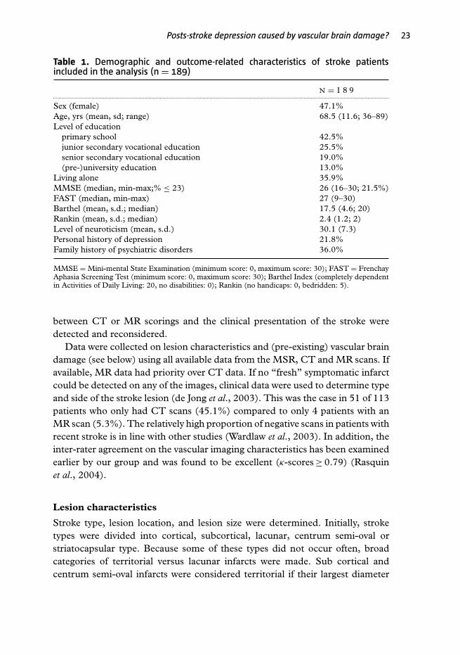
Table 1. Demographic and outcome-related characteristics of stroke patientsincluded in the analysis (n = 189)
N = 1 8 9............................................................................................................................................................................................................................................
Sex (female) 47.1%Age, yrs (mean, sd; range) 68.5 (11.6; 36–89)Level of education
primary school 42.5%junior secondary vocational education 25.5%senior secondary vocational education 19.0%(pre-)university education 13.0%
Living alone 35.9%MMSE (median, min-max;% ≤ 23) 26 (16–30; 21.5%)FAST (median, min-max) 27 (9–30)Barthel (mean, s.d.; median) 17.5 (4.6; 20)Rankin (mean, s.d.; median) 2.4 (1.2; 2)Level of neuroticism (mean, s.d.) 30.1 (7.3)Personal history of depression 21.8%Family history of psychiatric disorders 36.0%
MMSE = Mini-mental State Examination (minimum score: 0, maximum score: 30); FAST = FrenchayAphasia Screening Test (minimum score: 0, maximum score: 30); Barthel Index (completely dependentin Activities of Daily Living: 20, no disabilities: 0); Rankin (no handicaps: 0, bedridden: 5).
between CT or MR scorings and the clinical presentation of the stroke weredetected and reconsidered.
Data were collected on lesion characteristics and (pre-existing) vascular braindamage (see below) using all available data from the MSR, CT and MR scans. Ifavailable, MR data had priority over CT data. If no “fresh” symptomatic infarctcould be detected on any of the images, clinical data were used to determine typeand side of the stroke lesion (de Jong et al., 2003). This was the case in 51 of 113patients who only had CT scans (45.1%) compared to only 4 patients with anMR scan (5.3%). The relatively high proportion of negative scans in patients withrecent stroke is in line with other studies (Wardlaw et al., 2003). In addition, theinter-rater agreement on the vascular imaging characteristics has been examinedearlier by our group and was found to be excellent (κ-scores ≥ 0.79) (Rasquinet al., 2004).
Lesion characteristics
Stroke type, lesion location, and lesion size were determined. Initially, stroketypes were divided into cortical, subcortical, lacunar, centrum semi-oval orstriatocapsular type. Because some of these types did not occur often, broadcategories of territorial versus lacunar infarcts were made. Sub cortical andcentrum semi-oval infarcts were considered territorial if their largest diameter
24 Aben et al.
exceeded 15 mm. Infarcts including the cortex and striatocapsular infarcts wereconsidered as territorial infarcts.
Lesion location was both expressed in terms of hemisphere involvement (leftvs. right) and of involvement of the frontal region (further referred to as ‘anterior’vs. ‘posterior’). In the case of territorial infarcts with a positive scan, the centralsulcus was taken as the divider between the anterior and the remaining partof the brain. Striatocapsular infarcts were considered to involve the frontalcircuitry. In case of a negative scan, clinical stroke symptoms were used todecide about frontal involvement. Stroke syndromes restricted to visual fielddeficits, visuo-spatial deficits, agnosia, apraxia, or other type of “higher corticaldysfunction” were listed in the “posterior” category, whereas those with frontalsymptoms, such as Broca’s or mixed aphasia were considered as “anterior”. Inthe case of lacunar infarcts, involvement of the frontal region was considered tobe represented by damage to the head of the caudate nucleus, striate nucleus,and anterior leg of the internal capsule. If no infarct could be detected on CTor MRI scan, all regular lacunar syndromes were considered to have a posteriorlocalization because symptomatic lacunar infarcts are known to be mostly locatedin this region (Hupperts et al., 1994) and it prevented us from falsely allocatinginfarcts without frontal involvement to the hypothesized high-risk group.
In the case of a territorial infarct, size was rated on a semi-quantitative basis assmall, medium or large. For reasons of statistical power, data were subsequentlydichotomized into small vs. medium/large.
Generalized vascular brain damage
Leukoaraiosis can be defined as an abnormal appearance of the subcortical whitematter of the brain on neuroimaging. On CT, it is characterized by bilateralpatchy or diffuse areas of low attenuation, while on T2 MR it presents as hyper-intense areas in the white matter (Inzitari, 2003). On MR scans, such vascularwhite matter lesions were scored using the Fazekas scale. This scale separatelyrates the different types of hyper intense signal abnormalities surrounding theventricles and in deep white matter. Periventricular hyper intensity (PVH)is graded as 0 = absence, 1 = ‘caps’ or pencil-thin lining, 2 = smooth ‘halo’,3 = irregular PVH extending into the deep white matter. Deep white matterhyper-intensive signals (DWMH) are rated as 0 = absence, 1 = punctuate foci,2 = beginning confluence of foci, 3 = large confluent areas (Fazekas et al., 1987).
In case of CT data, leukoaraiosis was considered to be present if at least at oneof both sides (periventricular or in deep white matter) patchy or diffuse areas oflow attenuation were detected. Since CT scans are less sensitive to the detectionof vascular white matter lesions, and in order to enable pooling of CT and MRdata, we recoded the Fazekas ratings into one overall dichotomous measure of

Posts-stroke depression caused by vascular brain damage? 25
leukoaraiosis (present vs. absent). Only in the case of confluent lesions, wasleukoaraiosis rated as “present”.
Furthermore, asymptomatic (silent) infarcts were scored according toprocedures described in more detail elsewhere (Rasquin et al., 2004). OnMRI and CT, these are characterized by circumscriptive low-density areas(hyperdense in T2 MRI), compatible with infarction, but without a historyof any clinical signs or symptoms of stroke other than at study entry.
Initial assessment of depression
All patients were followed up during the first year after stroke. PSD was defined asan episode of major or minor depression according to DSM-IV criteria(AmericanPsychiatric Association, 1994) on at least one assessment during the 1-yearfollow-up period. After one month, all patients were interviewed using both thedepression section of the Structured Clinical Interview for DSM-IV (SCID-I-R) (First et al., 1996) and the Hamilton Depression Rating Scale (HAM-D)(Hamilton, 1960). The SCID is a structured psychiatric diagnostic interviewallowing for a DSM-IV diagnosis of major or minor depression. The HAM-Dis a clinical rating scale that measures the severity of depressive symptoms. Allinterviews were administered by the same clinician (IA), who was trained to usethese instruments. No formal test of inter-rater reliability was performed.
Follow-up assessment of depression
At 3, 6, 9, and 12 months after stroke, patients were asked to completethree psychiatric self-rating scales to screen for depression. These were theBeck Depression Inventory (BDI) (Beck et al., 1961), the Hospital Anxietyand Depression Scale (HADS) (Zigmond and Snaith, 1983), and the 90-itemSymptom Check List (SCL-90) (Arrindell and Ettema, 1981). Cut-off levelswere 9/10 for the BDI and 7/8 for both the depression and the anxiety subscaleof the HADS. In the case of the depression subscale of the SCL-90, the thresholdwas 22/23 for men and 27/28 for women. The predictive validity of theseinstruments in this cohort was previously analyzed (Aben et al., 2002b). Thesensitivity of this screening procedure was shown to be 93.8% at the one-monthassessment.
Patients whose scores exceeded the cut-off value for at least one of theself-rating scales were reinterviewed using the SCID and HAM-D in order todiagnose major or minor depression. In 50% of these cases, the interview waswithin two weeks; in 75% within 3–3.5 weeks.
In 33 patients the follow-up assessment of depression was incomplete, whilethe event of interest (depression) had not yet occurred. Of these, 16 withdrewtheir consent, 5 died, 8 had too severe co-morbidity, 1 was lost to follow-up, and3 did not respond at the final assessment (12 months).

26 Aben et al.
Potential confounders
Sex, age, personal history of depression, family history of psychiatric disorders,level of handicap, and neuroticism were predefined as potential confounders oreffect modifications in the hypothesized relation between focal or generalizedvascular brain damage and PSD.
Data concerning demographics, level of education, living situation, andfamily history of psychiatric disorders were collected on inquiry 1 month afterstroke. Level of disability and handicap were rated at the same time using theBarthel Index (Mahoney and Barthel, 1965) and Rankin score (Rankin, 1957),respectively. Furthermore, personal history of depression was measured usingthe SCID-I-R, while the MMSE (Folstein et al., 1975) and FAST (Enderbyet al., 1987) were administered to measure global cognitive functioning. Dataon personal history of depression was missing in 4 cases, on family history ofdepression in 11 cases, and on Rankin score in 1 case.
Neuroticism was assessed one month after stroke, using the NEO FiveFactor Inventory (NEO-FFI),(Costa and McCrae, 1985) which has beentranslated into Dutch (Hoekstra et al., 1996). This self-report questionnaireconsists of 60 statements covering the five main dimensions of personality:neuroticism, extraversion, openness to new experiences, agreeableness, andconscientiousness. Neuroticism has been related to depression most frequently(Enns and Cox, 1997) and is defined as a stable disposition to experiencepsychological distress across time and situations consisting of negative emotionssuch as fear, anger, and frustration (Costa and McCrae, 1985). Each statementis rated on a 5-point scale ranging from “strongly disagree” to “strongly agree”,resulting in total dimension scores between 12 and 60.
Non-response concerning the assessment of personality occurred in 36patients (18.9%), either because of study withdrawal or difficulty completingthe NEO-FFI.
Analysis
Since depressive outcome was measured prospectively on 5 different time-pointsduring the 1-year follow-up, a survival analysis technique was applied by meansof Cox regression. If data on depressive status on one assessment was missing,the patient was considered “not depressed” if the patient was not depressed atthe former assessment and if the depressive status at the next assessment wasvalidly measured. In all other situations, the case was excluded from furtheranalysis from the time point of the next assessment onward.
Cox regression was used to analyze the relative hazard (HR) of the differentmeasures of both focal and generalized vascular damage on the incidence ofPSD (major and minor depression combined). First, hemisphere involvement

Posts-stroke depression caused by vascular brain damage? 27
(left vs. right) and orientation towards the frontal pole (frontal region included vs.frontal region not included) were analyzed in an interaction model. Analogous,both measures of generalized vascular damage (leukoaraiosis and asymptomaticinfarcts) were analyzed in an interaction model.
Secondly, a multivariate model was tested including measures on both lesionlocation and generalized vascular damage, as well as potential confounders (sex,age, personal history of depression, family history of psychiatric disorders, levelof neuroticism, and level of handicap –i.e. Rankin score).
One case had bilateral stroke and was excluded from further analysis. In orderto optimize the statistical power, missing data on dichotomous variables wereimputed with value 0 (=absent), while missing data on continuous variables wereimputed with the mean value of that variable. Post-hoc analyses were carried outto explore the consequences of this technique on the results by using list-wisedeletion of cases with missing data on any of the variables in the equation. Nointeraction effects were hypothesized on an ‘a priori’ basis.
The output of the Cox analyses was checked for instability by influentialcases and for violation of both the proportional hazards assumption andthe assumption of linearity of effects. Where appropriate, Hazard ratios aregiven with their 95%-confidence intervals and 2-tailed p-values as: HR (95%-CI), p.
For group comparisons of descriptive sample characteristics, Student’st-test was used in the case of continuous normally distributed variables. Theχ2 test was used for all dichotomous variables. Finally, one-way ANOVA wasused to compare differences in HAMD scores between major depressed, minordepressed, and non-depressed patients. The level of significance was set at p<
0.05 (2-tailed) for all analyses. Where appropriate, results are given asmeans±SD.
Results
One-year cumulative incidence of depression
The 1-year cumulative incidence of depression was 38.7% (adjusted for caseswith incomplete follow-up). Cross-sectionally, the incidence rates were 21.6%(41/190) at 1 month, 5.1% (7/137.5) at 3 months, 6.0% (7/117) at 6 months,5.6% (6/107) at 9 months, and 7.1% (7/98) at 12 months. Of these, 41 patients(23.3%) met DSM-IV criteria for major depressive disorder and 27 (15.4%) metcriteria for minor depressive disorder. The mean HAMD score was 19.2±4.1for the patients with major depression, 13.2±4.3 for the patients with minordepression, and 7.3±4.1 for the non-depressed patients. These differences werestatistically significant (F(2) = 198.2; p<0.001).

28 Aben et al.
Figure 2. Distribution of lesion characteristics (hemisphere and frontal region involvement).
Distribution of measures of focal and generalized vascular damage
Of the 189 cases that were available for subsequent analysis on CT/MRI findings,89 (47.1%) had left-sided strokes and in 100 patients (52.9%) the strokewas due to a territorial infarct. In 64 strokes (33.9%) the frontal region wasinvolved. Figure 2 shows how these lesion characteristics were combined. Notethe relatively low “anterior” to “posterior” ratio (15/73) in lacunar infarcts ascompared to territorial infarcts (49/51). Size of infarct could only be rated in 75of 100 cortical infarcts (25 were not visible on scan). Seventeen of these wererated as small, 47 as moderate and 11 as large.
Posts-stroke depression caused by vascular brain damage? 29
Eighty-six patients (45.5%) showed one or more silent infarcts (i.e. notrelating to clinical symptoms), while 59 patients had leukoaraiosis (31.2%). Ofthese patients, 48 had a positive score on both of these measures of generalizedvascular damage, 11 only had leukoaraiosis, and 38 only had one or more silentinfarcts. Signs of generalized vascular damage occurred with equal frequencybetween territorial and lacunar infarcts: 49/100 patients with a territorial infarct(49.0%) showed any sign of generalized vascular damage compared to 48/89patients with a lacunar infarct (53.9%).
Lesion characteristics and generalized vascular damage: relation todepression
In an attempt to replicate the finding that left-sided strokes and/or strokes thatinvolve the frontal region of the brain increase the risk of PSD, Cox regressionwas performed with side of lesion (left vs. right) and frontal-region involvement(“anterior” vs. “posterior”) as independent variables. No significant interactioneffect of these lesion characteristics was found (left∗anterior: HR 0.60 (0.20–1.79), p=0.36). Nor was there evidence of a risk increasing effect of oneof the separate variables in a subsequent confounding model (left: HR 0.92(0.57–1.49), p=0.73; anterior: HR 0.74 (0.43–1.25), p=0.26). In addition, theeffect of infarct size could only be tested in 75 patients with a territorial infarct.Bivariate Cox regression analysis revealed no such effect (HR 0.73 (0.30–1.74),p=0.47).
Similarly, the attempt to replicate the finding that generalized vasculardamage increases the risk of depression, failed to reveal an interaction effectfor leukoaraiosis∗asymptomatic infarcts: HR 1.93 (0.40–9.38), p=0.42. Asubsequent confounding model also failed to reveal independent effects ofleukoaraiosis (HR 0.91 (0.51–1.63), p=0.75) or asymptomatic infarcts (HR1.30 (0.76–2.23), p=0.34) alone. Re-analysis of this model with one overallvariable for generalized vascular damage confirmed the absence of a relationshipwith PSD.
In accordance with our suggestion that PSD should be considered as havinga multifactorial path physiology, we subsequently tested one overall modelincluding both the variables of focal and generalized vascular damage as well asnon stroke-specific risk factors–sex, age, personal or family history of depression,neuroticism, and level of handicap.
Unexpectedly, in this multivariate model patients with “posterior” infarctshad a significant higher risk of depression than patients with strokes that involvedthe frontal region (see table 2). As expected from earlier analysis (Aben et al.,2002a), from the variables that are not specifically stroke-related, neuroticismand level of handicap showed to be independent risk factors for PSD. In addition,a trend was shown for a positive family history of psychiatric disorders.
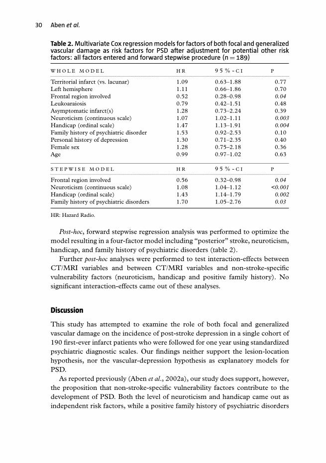
30 Aben et al.
Table 2.Multivariate Cox regressionmodels for factors of both focal and generalizedvascular damage as risk factors for PSD after adjustment for potential other riskfactors: all factors entered and forward stepwise procedure (n= 189)
W H O L E M O D E L H R 9 5 % - C I P.............................................................................................................................................................................................................................................
Territorial infarct (vs. lacunar) 1.09 0.63–1.88 0.77Left hemisphere 1.11 0.66–1.86 0.70Frontal region involved 0.52 0.28–0.98 0.04Leukoaraiosis 0.79 0.42–1.51 0.48Asymptomatic infarct(s) 1.28 0.73–2.24 0.39Neuroticism (continuous scale) 1.07 1.02–1.11 0.003Handicap (ordinal scale) 1.47 1.13–1.91 0.004Family history of psychiatric disorder 1.53 0.92–2.53 0.10Personal history of depression 1.30 0.71–2.35 0.40Female sex 1.28 0.75–2.18 0.36Age 0.99 0.97–1.02 0.63
S T E P W I S E M O D E L H R 9 5 % - C I P.............................................................................................................................................................................................................................................
Frontal region involved 0.56 0.32–0.98 0.04Neuroticism (continuous scale) 1.08 1.04–1.12 <0.001Handicap (ordinal scale) 1.43 1.14–1.79 0.002Family history of psychiatric disorders 1.70 1.05–2.76 0.03
HR: Hazard Radio.
Post-hoc, forward stepwise regression analysis was performed to optimize themodel resulting in a four-factor model including “posterior” stroke, neuroticism,handicap, and family history of psychiatric disorders (table 2).
Further post-hoc analyses were performed to test interaction-effects betweenCT/MRI variables and between CT/MRI variables and non-stroke-specificvulnerability factors (neuroticism, handicap and positive family history). Nosignificant interaction-effects came out of these analyses.
Discussion
This study has attempted to examine the role of both focal and generalizedvascular damage on the incidence of post-stroke depression in a single cohort of190 first-ever infarct patients who were followed for one year using standardizedpsychiatric diagnostic scales. Our findings neither support the lesion-locationhypothesis, nor the vascular-depression hypothesis as explanatory models forPSD.
As reported previously (Aben et al., 2002a), our study does support, however,the proposition that non-stroke-specific vulnerability factors contribute to thedevelopment of PSD. Both the level of neuroticism and handicap came out asindependent risk factors, while a positive family history of psychiatric disorders

Posts-stroke depression caused by vascular brain damage? 31
seemed to further contribute to the risk of PSD. Therefore, in studying the roleof biological (stroke-related) factors as independent predictors of PSD thesedeterminants should also be considered. Although in our study we found noevidence of interaction between vulnerability factors and measures of focal orgeneral vascular brain damage, we suggest that more powered studies couldreveal such risk-potentiating effects. For instance, vascular white matter lesionsmay contribute more to a person’s vulnerability for depression when the patienthas already suffered from depression in the past.
Much to our surprise, an unexpected protective effect of depression was foundfor infarctions with anterior involvement, when analyzing the overall model withboth stroke specific and non-specific factors. This finding opposes the proposedrole of damage to the frontal region of the brain in inducing post-stroke orvascular depression and needs further elaboration. A possible explanation forthis finding may be sought in the probable under-representation of left frontalstrokes by the exclusion of patients with severe aphasia or other severe cognitivedeficits from this study. As a consequence, other neurological deficits that arerelated to posterior brain dysfunction, such as visuo-spatial disturbance or hemi-neglect, may also trigger the development of PSD. As an alternative and moreconservative explanation, the finding may be based on a type I statistical error.It must be noted that the significant effect was only found in multivariate (11factor model and stepwise regression model) and not in bivariate analysis.
A variety of previous studies by others also failed to replicate the lesion-location hypothesis, but Robinson and colleagues carried out a meta-analysis(Narushima et al., 2003) and claimed to have found an interaction between lesionlocation and time of onset of PSD. Left-hemisphere lesions would increase therisk of PSD, especially in the first few months after stroke, whereas in the chroniccourse, psychosocial factors would become more important. We tried to test thismodification of his original hypothesis by controlling for time-dependency in theCox regression model but found no such effect. Gainotti had tested this sameassumption in 1999 and found that both the symptom profiles and anatomical-clinical correlates of major PSD were not different in the acute and more chronicstages of stroke (Gainotti et al., 1999).
Despite of the negative findings that undermine the plausibility of the lesion-location hypothesis, it cannot be rejected yet. A more precise delineation ofspecific brain structures that may be involved in mood regulation and, therefore,in the development of PSD, may result in more consistent evidence in favor of thelesion location hypothesis. Vataja et al. showed that this strategy seems hopeful,since in their study of 275 stroke patients, they found that infarcts affectingthe pre-frontosubcortical circuits, especially the caudate, pallidum, and genu ofinternal capsule (with left-sided predominance) were associated with a higherprevalence of PSD (Vataja et al., 2001).
32 Aben et al.
Concerning the vascular depression hypothesis, we argue that in a strokepopulation (pre-stroke) vascular brain damage, as measured by leukoaraiosis andsilent cerebral infarctions, does not significantly contribute to the developmentof PSD, since the direct consequences of stroke itself (either biological,psychological or social) are so strongly depressogenic that the contribution ofpre-existing small vascular lesions is overshadowed. In their study, Vataja et al.(2001) also failed to find an association between white matter lesions and PSD.This explanation is supported by the recent finding by Mast et al. (2004a; 2004b)that in a large cohort of 670 rehabilitation patients, depression was associatedwith increased burden of cerebrovascular risk factors in patients without strokebut not in stroke patients. It is additionally noted that our group also failed to finda significant contribution of generalized vascular damage to the development ofpost-stroke cognitive disorders (Rasquin et al., 2004).
The main shortcoming of the study lies in the limited number of availableMRI scans, so that we had to rely on CT data in a majority of cases. CTscans made in the acute phase of stroke failed to detect “fresh” infarction ofbrain tissue in 53 of 114 (46.5%) cases and they are also less sensitive indetecting vascular white matter lesions. Therefore, clinical data were used tocomplete data on lesion characteristics such as hemisphere involvement andtype of stroke (territorial vs. lacunar). Additionally, in pooling CT and MR data,variables were dichotomized in order to reduce the chance of systematic measureerrors.
Given the rather equal distribution of strokes over both hemispheres, it isnot likely that exclusions have led to essential under-representation of right-or left-hemisphere strokes. Because of the relatively low frequency of infarctswith cortical involvement, of which a minority of 43% were located in the lefthemisphere, it seems, however, that cortical strokes have been excluded relativelyfrequently, especially in the left hemisphere. One can imagine that patients withsevere aphasia or generalized cognitive disabilities are especially vulnerable todepression, so that the cumulative incidence of PSD as reported in this thesismay be underestimated. These limitations may have contributed to the negativeresults of this study.
In conclusion, we found no support for both the lesion-location hypothesisand the vascular-depression hypothesis in stroke patients. In order to appreciatethe lesion-location hypothesis for its true value, future research should aim toovercome the methodological difficulties in PSD research such as the exclusion ofpatients with severe aphasia or other cognitive deficits in our study. Furthermore,a more detailed determination of neuronal circuits that are involved in moodregulation may also prevent the influential hypothesis of Robinson being rejectedon immature research findings.

Posts-stroke depression caused by vascular brain damage? 33
Conflict of interest declaration
None.
Description of authors’ roles
I. Aben participated as a PhD student in all phases of the research and wrote thepaper. F. Verhey was his primary supervisor throughout; J. Lodder supervisedthe analysis of the CT and MR imaging and the preparation of the paper, andR. Lousberg was responsible for the statistical design and supervised thestatistical analysis. A. Boreas was involved in the recruitment of stroke patientsand in the management of the Maastricht Stroke Registry. All authors read,corrected, and approved with the contents of the paper.
References
Aben, I., Verhey, F., Honig, A., Lodder, J., Lousberg, R. and Maes, M. (2001). Researchinto the specificity of depression after stroke: a review on an unresolved issue. Progress inNeuropsychopharmacology and Biological Psychiatry, 25, 671–689.
Aben, I., Denollet, J., Lousberg, R., Verhey, F., Wojciechowski, F. and Honig, A. (2002a).Personality and vulnerability to depression in stroke patients: a 1-year prospective follow-upstudy. Stroke, 33, 2391–2395.
Aben, I., Verhey, F., Lousberg, R., Lodder, J. and Honig, A. (2002b). Validity of the BeckDepression Inventory, Hospital Anxiety and Depression Scale, SCL-90, and HamiltonDepression Rating Scale as screening instruments for depression in stroke patients.Psychosomatics, 43, 386–393.
Alexopoulos, G. S., Meyers, B. S., Young, R. C., Campbell, S., Silbersweig, D. andCharlson, M. (1997). ‘Vascular depression’ hypothesis. Archives of General Psychiatry, 54,915–922.
American Psychiatric Association (1994). Diagnostic and Statistical Manual of Mental Disorders(DSM-IV) (4th ed.). Washington, DC: American Psychiatric Press.
Arrindell, W. A. and Ettema, J. H. M. (1981). Dimensional structure, reliability and validityof the Dutch version of the Symptom Checklist (SCL-90). Nederlands Tijdshrift voorPsychologie, 43, 381–387.
Beck, A. T., Ward, C. H., Mendelson, M., Mock, J. and Erbaugh, J. (1961). An Inventoryfor measuring depression. Archives of General Psychiatry, 4, 561–571.
Boon, A., Lodder, J., Heuts-van Raak, L. and Kessels, F. (1994). Silent brain infarcts in 755consecutive patients with a first-ever supratentorial ischemic stroke. Relationship withindex-stroke subtype, vascular risk factors, and mortality. Stroke, 25, 2384–2390.
Boreas, A. M., Lodder, J., Kessels, F., de Leeuw, P. W. and Troost, J. (2002). Prognosticvalue of blood pressure in acute stroke. Journal of Human Hypertension, 16, 111–116.
Carson, A. J., et al. (2000). Depression after stroke and lesion location: a systematic review.Lancet, 356, 122–126.
Costa, P. T., Jr. and McCrae, R. R. (1985). The NEO Personality Inventory Manual. Odessa,FL: Psychological Assessment Resources Inc.

34 Aben et al.
de Jong, G., van Raak, L., Kessels, F. and Lodder, J. (2003). Stroke subtype andmortality. a follow-up study in 998 patients with a first cerebral infarct. Journal of ClinicalEpidemiology, 56, 262–268.
Enderby, P. M., Wood, V. A., Wade, D. T. and Hewer, R. L. (1987). The Frenchay AphasiaScreening Test: a short, simple test for aphasia appropriate for non-specialists. InternationalRehabilitation Medicine, 8, 166–170.
Enns, M. W. and Cox, B. J. (1997). Personality dimensions and depression: review andcommentary. Canadian Journal of Psychiatry, 42, 274–284.
Fazekas, F., Chawluk, J. B., Alavi, A., Hurtig, H. I. and Zimmerman, R. A. (1987). MRsignal abnormalities at 1.5 T in Alzheimer’s dementia and normal aging. American Journal ofRoentgenology, 149, 351–356.
First, M. B., Gibbon, M., Spitzer, R. L. and Williams, J. B. (1996). Users Guide for theStructured Clinical Interview for DSM-IV Axis I Disorders – Research Version – (SCID-I, Version2.0, February 1996 FINAL Version). New York, NY: New York State Psychiatric Institute.
Folstein, M. F., Folstein, S. E. and McHugh, P. R. (1975). ‘Mini-mental State’: a practicalmethod for grading the cognitive state of patients for the clinician. Journal of PsychiatricResearch, 12, 189–198.
Gainotti, G., Azzoni, A. and Marra, C. (1999). Frequency, phenomenology and anatomical-clinical correlates of major post-stroke depression. British Journal of Psychiatry, 175, 163–167.
Hamilton, M. (1960). A rating scale for depression. Journal of Neurology, Neurosurgery andPsychiatry, 23, 56–62.
Hoekstra, H. A., Ormel, J. and De Fruyt, F. (1996). NEO Personality QuestionnairesNEO-PI-R, NEO-FFI, Manual. Lisse (NL): Swets & Zeitlinger B.V.
Honig, A. and Van Praag, H. M. (Eds.) (1997). Depression. Neurobiological, Psychopathologicaland Therapeutic advances. Chichester, UK: John Wiley & Sons.
Hupperts, R. M., Lodder, J., Heuts-van Raak, E. P. and Kessels, F. (1994). Infarcts in theanterior choroidal artery territory. Anatomical distribution, clinical syndromes, presumedpathogenesis and early outcome. Brain, 117 (Pt 4), 825–834.
Inzitari, D. (2003). Leukoaraiosis: an independent risk factor for stroke? Stroke, 34, 2067–2071.
Krishnan, K. R., Hays, J. C. and Blazer, D. G. (1997). MRI-defined vascular depression.American Journal of Psychiatry, 154, 497–501.
Mahoney, F. I. and Barthel, D. W. (1965). Functional evaluation: the Barthel Index. StateMedical Journal, 14, 61–65.
Mast, B. T., MacNeill, S. E. and Lichtenberg, P. A. (2004a). Post-stroke and clinically-defined vascular depression in geriatric rehabilitation patients. American Journal of GeriatricPsychiatry, 12, 84–92.
Mast, B. T., Neufeld, S., MacNeill, S. E. and Lichtenberg, P. A. (2004b). Longitudinalsupport for the relationship between vascular risk factors and late-life depressive symptoms.American Journal of Geriatric Psychiatry, 12, 93–101.
Narushima, K., Kosier, J. T. and Robinson, R. G. (2003). A reappraisal of post-strokedepression, intra- and inter-hemispheric lesion location using meta-analysis. Journal ofNeuropsychiatry and Clinical Neuroscience, 15, 422–430.
National Institute of Neurological Disorders and Stroke (1990). Special report from theNational Institute of Neurological Disorders and Stroke. Classification of cerebrovasculardiseases III. Stroke, 21, 637–676.
Rankin, J. (1957). Cerebral vascular accidents in patients over the age of 60. 2. Prognosis.Scottish Medical Journal, 2, 200–215.
Rasquin, SM., Verhey, F. R. J., Van Oostenbrugge, R. J., Lousberg, R. and Lodder, J.Demographic and CT-scan features related to cognitive impairment in the first year afterstroke. Journal of Neurology, Neurosurgery and Psychiatry, 2004, 75, 1562–1567.

Posts-stroke depression caused by vascular brain damage? 35
Robertson, M. M. and Katona, C. L. E. (Eds.) (1997). Depression and Physical Illness.Chichester, UK: Wiley.
Robinson, R. G. (1998). The Clinical Neuropsychiatry of Stroke. Cognitive, Behavioral andEmotional Disorders following Vascular Brain Injury. Cambridge: Cambridge University Press.
Robinson, R. G. (2003). Poststroke depression: prevalence, diagnosis, treatment, and diseaseprogression. Biological Psychiatry, 54, 376–387.
Singh, A., Herrmann, N. and Black, S. E. (1998). The importance of lesion location inpoststroke depression: a critical review. Canadian Journal of Psychiatry, 43, 921–927.
Vataja, R., et al. (2001). Magnetic resonance imaging correlates of depression after ischemicstroke. Archives of General Psychiatry, 58, 925–931.
Wardlaw, J. M., West, T. M., Sandercock, P. A., Lewis, S. C. and Mielke, O. (2003).Visible infarction on computed tomography is an independent predictor of poor functionaloutcome after stroke, and not of haemorrhagic transformation. Journal of Neurology,Neurosurgery and Psychiatry, 74, 452–458.
Zigmond, A. S. and Snaith, R. P. (1983). The Hospital Anxiety and Depression Scale. ActaPsychiatrica Scandinavica, 67, 361–370.


![FOCAL POINT - CargillAg · tact your Cargill rep to reprice and lock in your Final Focal Point Price. Final Focal Point Price] - [Initial Focal Point Price] = [Focal Point Price Adjustment]](https://img.dokumen.tips/doc/110x75/5ea5a76ffc2e8d744054ad3b/focal-point-cargillag-tact-your-cargill-rep-to-reprice-and-lock-in-your-final.jpg)