Embed Size (px)
Citation preview

85
AM
BU
LAT
OR
Y S
UR
GER
Y
16.
4 D
ECEM
BER
R 2
010
Contents
The British Association of Day Surgery Directory of Procedures 87 I.J.B. Jackson, D. McWhinnie, M. Skues
Search for efficiency without neglecting safety in the design and construction of a 90 new ambulatory surgery centreW. Salgado
Is the prophylactic use of non-opioids for post-operative analgesia always indicated? 93A randomized controlled trial in breast surgeryM. Gehling, C. Arndt, I.C. Behrendt, H. Wulf, L.H.J. Eberhart
Spinal Anesthetic Block Failure due to the Hyperbaric Nature of 99 2% Chloroprocaine Local Anesthetic R. Raw, E.R. Nwaneri
Anesthetic Implications of Obstructive Sleep Apnea in the Ambulatory Setting 103 K.E. McGoldrick
87
AM
BU
LAT
OR
Y S
UR
GER
Y
16.
4 D
ECEM
BER
R 2
010
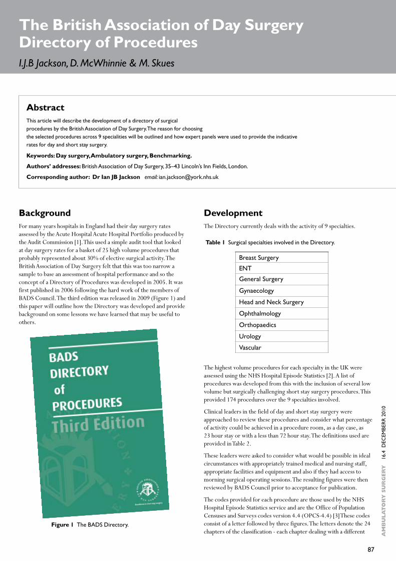
BackgroundFor many years hospitals in England had their day surgery rates assessed by the Acute Hospital Acute Hospital Portfolio produced by the Audit Commission [1]. This used a simple audit tool that looked at day surgery rates for a basket of 25 high volume procedures that probably represented about 30% of elective surgical activity. The British Association of Day Surgery felt that this was too narrow a sample to base an assessment of hospital performance and so the concept of a Directory of Procedures was developed in 2005. It was first published in 2006 following the hard work of the members of BADS Council. The third edition was released in 2009 (Figure 1) and this paper will outline how the Directory was developed and provide background on some lessons we have learned that may be useful to others.
DevelopmentThe Directory currently deals with the activity of 9 specialties.
The highest volume procedures for each specialty in the UK were assessed using the NHS Hospital Episode Statistics [2]. A list of procedures was developed from this with the inclusion of several low volume but surgically challenging short stay surgery procedures. This provided 174 procedures over the 9 specialties involved.
Clinical leaders in the field of day and short stay surgery were approached to review these procedures and consider what percentage of activity could be achieved in a procedure room, as a day case, as 23 hour stay or with a less than 72 hour stay. The definitions used are provided in Table 2.
These leaders were asked to consider what would be possible in ideal circumstances with appropriately trained medical and nursing staff, appropriate facilities and equipment and also if they had access to morning surgical operating sessions. The resulting figures were then reviewed by BADS Council prior to acceptance for publication.
The codes provided for each procedure are those used by the NHS Hospital Episode Statistics service and are the Office of Population Censuses and Surveys codes version 4.4 (OPCS-4.4) [3] These codes consist of a letter followed by three figures. The letters denote the 24 chapters of the classification - each chapter dealing with a different
AbstractThis article will describe the development of a directory of surgical procedures by the British Association of Day Surgery. The reason for choosing the selected procedures across 9 specialities will be outlined and how expert panels were used to provide the indicative rates for day and short stay surgery.
Keywords: Day surgery, Ambulatory surgery, Benchmarking.
Authors’ addresses: British Association of Day Surgery, 35–43 Lincoln’s Inn Fields, London.
Corresponding author: Dr Ian JB Jackson email: [email protected]
Breast Surgery
ENT
General Surgery
Gynaecology
Head and Neck Surgery
Ophthalmology
Orthopaedics
Urology
Vascular
Table 1 Surgical specialties involved in the Directory.
Figure 1 The BADS Directory.
The British Association of Day Surgery Directory of ProceduresI.J.B Jackson, D. McWhinnie & M. Skues

88
AM
BU
LAT
OR
Y S
UR
GER
Y
16.
4 D
ECEM
BER
201
0
part, or ‘system’ of the body. There are more than 6,000 codes altogether, but for many purposes it is acceptable to group codes at the 3-character level. For example, all codes beginning B34 are for “operations on duct of breast”. However where more precision is required it is necessary to use the sub-division indicated by the final character. Feedback received following the publication of the first edition confirmed the importance of this as the Directory needed to be more specific about the accepted codes for each procedure and to also provide exclusion codes for certain procedures. The reason for this is best given by an example. The code T24 is used for the repair of an umbilical hernia which can have a high day surgery rate. However it is divided into the sub codes T24.1, T24.2, T24.3, T24.4, T24.8 and T24.9. T24.4 is has been introduced as an exclusion code as this codes for the more complex revision of a previous repair involving the removal of prosthetic material. This is hopefully not a common procedure but if included it could skew what is achievable as a day case.
In the 2007 edition Healthcare Resource Groups (HRG) codes were provided for each procedure as these are increasingly being used in England as part of Payment by Results. [4] The NHS uses HRGs as a means of ‘determining fair and equitable reimbursement for care services rendered’. Unfortunately, though, the 4th update to HRG coding in 2008 meant that these codes had to be provided as an electronic cross comparison tool in a complementary resource to the published Directory, due to their increased complexity.
An example page from the Directory is shown in Figure 2.
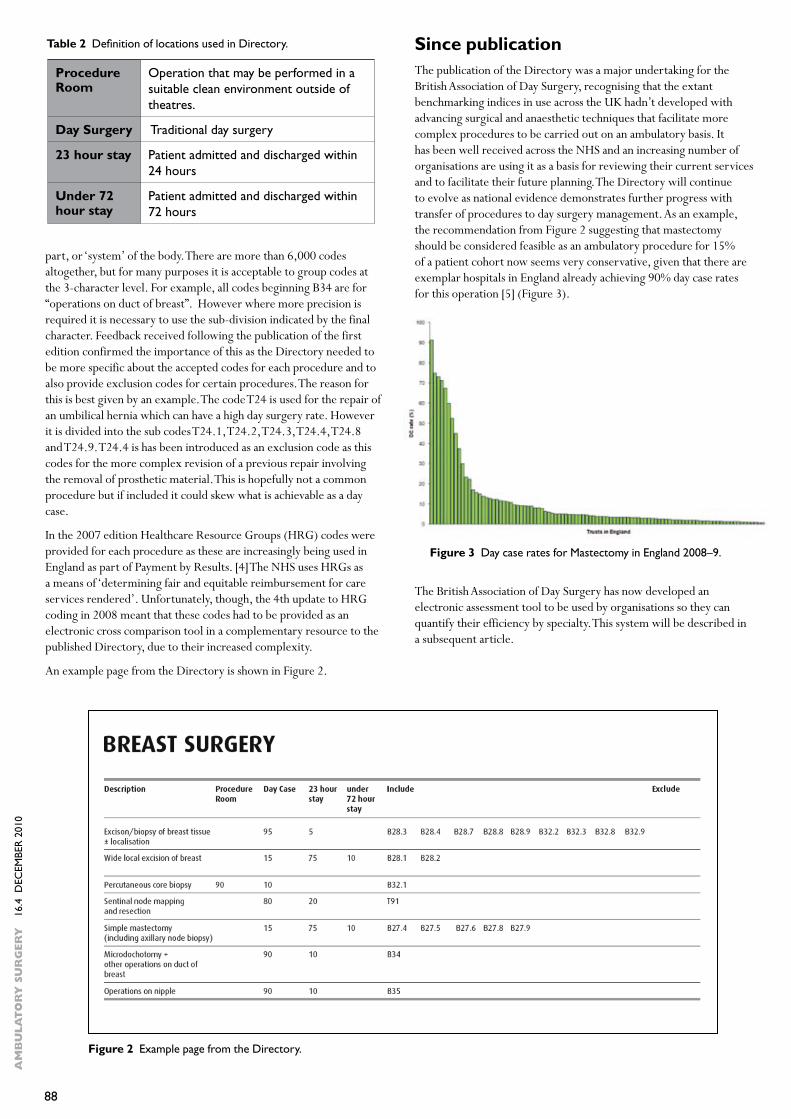
Since publicationThe publication of the Directory was a major undertaking for the British Association of Day Surgery, recognising that the extant benchmarking indices in use across the UK hadn’t developed with advancing surgical and anaesthetic techniques that facilitate more complex procedures to be carried out on an ambulatory basis. It has been well received across the NHS and an increasing number of organisations are using it as a basis for reviewing their current services and to facilitate their future planning. The Directory will continue to evolve as national evidence demonstrates further progress with transfer of procedures to day surgery management. As an example, the recommendation from Figure 2 suggesting that mastectomy should be considered feasible as an ambulatory procedure for 15% of a patient cohort now seems very conservative, given that there are exemplar hospitals in England already achieving 90% day case rates for this operation [5] (Figure 3).
The British Association of Day Surgery has now developed an electronic assessment tool to be used by organisations so they can quantify their efficiency by specialty. This system will be described in a subsequent article.
Procedure Room
Operation that may be performed in a suitable clean environment outside of theatres.
Day Surgery Traditional day surgery
23 hour stay Patient admitted and discharged within 24 hours
Under 72 hour stay
Patient admitted and discharged within 72 hours
Table 2 Definition of locations used in Directory.
Figure 3 Day case rates for Mastectomy in England 2008–9.
Figure 2 Example page from the Directory.
89
AM
BU
LAT
OR
Y S
UR
GER
Y
16.
4 D
ECEM
BER
R 2
010
ReferencesAudit Commission. Acute Hospital Portfolio. Day Surgery. 2001 1. www.audit-commission.gov.uk/health/nationalstudies/other/pages/daysurgery_copy.aspx#downloadsHESonline. www.hesonline.org.uk2. OPCS-4.4 Intervention Classification www.connectingforhealth.nhs.3. uk/systemsandservices/data/clinicalcoding/codingstandards/opcs4Healthcare Resource Groups. www.ic.nhs.uk/casemix4. Day case and short stay surgery performance assessment. www.5. audit-commission.gov.uk/health/trustpractice/ourservices/Pages/daycase.aspx

90
AM
BU
LAT
OR
Y S
UR
GER
Y
16.
4 D
ECEM
BER
201
0
IntroductionMore than 60% of elective surgery procedures in the United States were being performed as outpatient procedures by the year 2001[1], and in 2008, more than 22 million ambulatory surgery procedures were performed in that country [2]. There are many reasons justifying these numbers such as: improved surgical instruments, anaesthetic drugs and techniques; less invasive surgical techniques; multidisciplinary pre-operative preparation; improved pain and nausea control; post-operative protocols [3].
Ambulatory surgery has many advantages compared to inpatient surgery. There is minimal alteration to the routine of patients and their families and the care provided is individualized. Among other benefits, the risk of hospital infection and the costs of the procedures are reduced [4].
However, there is a constant concern about patient safety since there are aspects of ambulatory surgery that cannot be fully controlled, such as adherence to all pre-operative guidelines and to post-operative home care [4]. This topic should be taken into consideration, especially so when the goal is to structure a centre of ambulatory surgery with high efficiency and productivity.
Three years ago, when we received the responsibility to organize a public ambulatory surgery centre, all of these aspects were carefully planned. Special attention was devoted to the following parameters: compliance with current norms, humanization of care, technical and personnel habilitation, and protocol elaboration.
The objective of the present paper is to present the experience with the organization of the ambulatory surgery centre of the State Hospital of Ribeirão Preto and to describe the general results obtained after 2 years of effective functioning.
MethodsThe State Hospital of Ribeirão Preto was built in order to deal with patients with diseases considered to be of medium complexity, and started its surgical activities on May 12th, 2008.
Data was obtained from a retrospective survey of the data bank of the State Hospital. Almost all patient data is recorded in an informatized system (registration of presence, clinical observation and evolution, surgical file card, prescriptions, request for surgery,etc.), a fact that facilitates data retrieval.
ResultsThe State Hospital of Ribeirão Preto is a small hospital equiped with 4 operating rooms, 6 recovery beds, 10 ambulatory rooms and 50 beds mostly used by the internal medicine staff for emergency admission of medium complexity. In addition, facilities such as a sterilization centre, pharmacy and catering are provided on the premises. However, laundry and laboratory services are outsourced.
From the time when the hospital started its activities to April 2010, 8872 patients were operated upon in the following surgical specialties: ophthalmology, otorhinolaryngology, orthopaedics, general surgery, plastic surgery, urology, paediatric surgery, proctology, dermatology, dentistry, and vascular surgery. A total of 218 procedures across all specialties were performed. Most of them were surgical procedures of medium complexity (e.g. inguinal hernia repair, laparoscopic cholecystectomy, fasciectomy, tonsillectomy, knee arthroscopy, etc.). Some procedures were minor, such as excision of skin lesions, but some were more complex, such as major incisional herniorrhaphy, reconstruction of complex shoulder ligament injuries, reconstruction of intestinal transit after colostomy, and bilateral saphenectomy, among others.
AbstractAim: To describe the experience of a recently created ambulatory
surgery centre. Methods: Report of the experience and the routine implemented in the ambulatory surgery centre and survey of retrospective data in the data bank of the hospital.
Results: Our hospital has high surgical productivity, with highly satisfactory hospital quality indices such as a low rate of surgical site infection, minimal mortality and very high user satisfaction.
Conclusion: Even after a short functioning time, it is possible to structure a highly productive ambulatory surgery centre of excellent quality. Attention to the elaboration of processes, routines and participation and training of the staff is important.
Keywords: Ambulatory surgery; efficiency; safety.
Author’s address: a Dept. Surgery and Anatomy, Clinical Hospital of the Faculty of Medicine of Ribeirão Preto, University of São Paulo, Brazil. b State Hospital of Ribeirão Preto, Brazil.
Corresponding author: W. Salgado Jr. Tel: +55 16 36027117 Fax: +55 16 36022593 E-mail: [email protected]
Search for efficiency without neglecting safety in the design and construction of a new ambulatory surgery centreW. Salgado Jr.a,b
91
AM
BU
LAT
OR
Y S
UR
GER
Y
16.
4 D
ECEM
BER
R 2
010
When surgical production was analyzed on a six month basis, activity was found to be reduced during the end of the year period, but even so the number of procedures tended to increase over the two year period (Table 1).
Despite the large surgical volume, so far only one death has occurred. This involved a patient submitted to correction of gynaecomastia. In this case, the probable cause of death, as determined at autopsy, was severe cardiac arrhythmia secondary to the administration of ondasetrone for the control of post-operative nausea.
In order to be able to operate on this number of patients during this period we attended 24,029 patients in the outpatient department.
A total of 435 patients required hospitalization, most of them being patients submitted to more complex procedures who were admitted in order to receive better analgesia and the re-introduction of feeding.
We are constantly striving to reduce the rate of suspended surgeries. During the study period, 1351 surgeries were suspended, 463 of them due to institutional reasons (broken equipment, surgical delays, etc.) and 888 due to patient reasons (no-show, patient with no clinical conditions on the scheduled day, lack of pre-operative fasting, among others).
An outpatient clinic for discharged patients was created, with all operated patients required to return for removal of sutures and at least once more up to the 30th post-operative day in order to guarantee the notification of cases of surgical site infection. The Hospital Infection Committee actively participates in this clinic. On this basis, 76% of the patients returned during the post-operative period. Since we called all patients who failed to come for the first return, different justifications were given for their no-show such as lack of transportation and removal of sutures in the city of origin as an option chosen by the patient himself, among others.
The rates of surgical wound infection, considered to be very low, are presented in Table 2.
DiscussionA hospital that strives for quality and efficiency of care must work with indicators, written operational protocols, and well established routines. More importantly, these factors must be constantly updated and communicated to the entire staff, who must be trained to follow such routines [5].
One of the first steps in the elaboration of the care process and of the physical structure of an ambulatory surgery centre is to comply with prevailing norms. In the case of our hospital, two sources of regulation have to be obeyed: Resolution SS- 002 of January 6, 2006 of the Health Secretariat of the State of São Paulo, and Resolution CFM nº 1.409/94 of the Federal Council of Medicine, later replaced in 2008.
Several approaches were established in order to try to reduce the number of suspended operations. Protocols were first elaborated and sent to all towns that send patients to be operated upon at the hospital. These protocols established rigid criteria for patient selection based on the complexity of the procedures and on the physical status classification system of the American Society of Anesthesiology [6].
On the same day when he is examined by the surgeon the patient also has a consultation with the anaesthetist, who evaluates his surgical risk. All information regarding the day of surgery, pre-operative fasting, the use of medication etc. is given verbally and in writing to the patient and his accompanying person by these two doctors. At the end of these visits, the patient undergoes a post-consultation with the nursing staff for a re-statement of all the guidelines. In addition, in order to fully assimilate the information, the patient watches a 5 minute video that confirms all the information. Finally the patient receives guidelines from the social service in order to solve possible problems related to his job and transportation to the hospital.
It should be also mentioned that, one day before the scheduled surgery, every patient will receive a phone call from our social service in order to be reminded of all instructions and to be questioned about possible problems such as diseases, transportation etc. Despite these measures, our rate of surgery suspension due to patient reasons are still high, but would definitely be much higher without these precautionary measures. The standardization of conducts and routines is always mentioned in the literature as an important factor for the improvement of the efficiency of a surgical centre [7, 8].
Our time for room exchange is about 20 minutes and many anaesthetic procedures are performed in a room for anaesthetic induction, so that the patient will arrive at the operating room ready for the surgical act. More than 60% of anaesthetics are local or loco-regional, including those for inguinal hernia repair, and proctological and urological surgery.
In parallel to this special attention to effectiveness and productivity, we never relax our emphasis on the quality of care and on patient satisfaction. The constant presence of anaesthetists in the operating room even for less complex surgery and also in the anaesthetic recovery room is of fundamental importance in order to guarantee proper care for the patients.
Whenever a low rate of infection of the surgical site is reported, the question immediately raised is that undernotification may exist. A complete action strategy was elaborated to minimize this fact. A clinic for discharged patients was created, with every post-operative patient being required to return to the hospital during the first post-operative month. Informatized control of this return schedule was created, and when a patient failed to return he was contacted by telephone.
Half-year periods Number of operations performed
May to October 2008 1476
November 2008 to April 2009 2155
May to October 2009 2742
November 2009 to April 2010 2499
Table 1 Surgical production of the State Hospital divided by half-year periods.
Year Clean surgery All surgery
2008* 0.67% 0.6%
2009 1.1% 1%
Table 2 Rate of surgical site infection during the two years of functioning of the Hospital. The results are presented according to the classification of the surgical wound.
* starting May 2008.

92
AM
BU
LAT
OR
Y S
UR
GER
Y
16.
4 D
ECEM
BER
201
0
All doctors are encouraged by the Committee of Hospital Infection to notify cases of infection, with emphasis on the fact that doctors reporting such cases will not be punished. The form used for all return cases is informatized and one of the fields to be obligatorily filled out concerns the presence or absence of symptoms of surgical site infection. Finally, at the end of his visit, each patient participates in a post-visit session with the nursing staff, who are trained and advised to notify any case of suspected infection.
Crowning all of this work, the fact that most enriches our activity is the recognition shown by the patients treated at our hospital. In 2010, competing with 630 other hospitals, the State Hospital received the title of “Best Public Hospital in the State of São Paulo” according to the view of the patients.
ConclusionWe conclude that it is possible to deal with high surgical productivity without neglecting the quality, and especially the humanization, of care. For everything to occur according to plan, all employees should actively participate, whether they are from the support area, from the health area, or from the administration.
ReferencesCosta MJ. The lived perioperative experience of ambulatory surgery 1. patients. AORN J. 2001;74(6): 874–881.Gerencher K. Doctor-owned surgery centers spark controversy. 2. Market Watch. March 25, 2008. http://www.marketwatch.com/story/doctor-owned-surgery-centers-spark-conflict-of-interest-debate.AORN guidance statement: Preoperative patient care in the 3. ambulatory surgery setting. AORN J. 2005;81(4):871–883.Meta-analysis of randomized controlled trials on the safety 4. and effectiveness of day-case laparoscopic cholecystectomy. Gurusamy K, Junnarkar S, Farouk M, Davidson BR. Br J Surg. 2008 Feb;95(2):161–8.Frezza EE, Girnys RP, Silich RJ et al. Commentary: quality of care 5. and cost containment are the hospital based ambulatory surgery- challenges for the future. American Journal of Medical Quality 2000;15;114http://www.asahq.org/clinical/physicalstatus.htm6. Correll DJ, Bader AM, Hull MW, et al. Value of preoperative clinic 7. visits in identifying issues with potential impact on operating room efficiency. Anesthesiology 2006;105:1254–1259.Joshi GP. Efficiency in ambulatory surgery center. 8. Current Opinion in Anaesthesiology 2008;21:695–698.
93
AM
BU
LAT
OR
Y S
UR
GER
Y
16.
4 D
ECEM
BER
R 2
010
IntroductionIn the search for increased patient satisfaction with peri-operative pain management, the prevention of pain has evolved as a field of major clinical interest especially in ambulatory surgery. Several trials describe the successful prophylactic use of non-opioid analgesics for post-operative pain reduction, i.e. ketorolac, lornoxicam or tenoxicam [1–6]. More recently, similar prophylactic analgesic effects have been described for coxibs [7,8]. Even parecoxib and paracetamol pre-operatively may prevent post-operative pain [9,10].
To optimize prophylactic non-opioid analgesia, we conducted a prospective, randomized, double blind, clinical trial comparing the analgesic effects of parceocib/ vadecoxib, paracetamol or their combination with placebo in patients after breast surgery. Since valdecoxib was withdrawn from the market in 2005, we left the publication of the results aside. However, in the light of growing literature recommending the prophylactic use of non-opioids we felt that the results of our study may be important for the clinical decision to use analgesics prophylactically.
Planning the trial, our intention was an improvement of non-opioid analgesia and a reduction of opioid induced sedation, PONV, constipation and respiratory depression. Combining two non-opioid analgesics was supposed to increase the benefit, if an additive effect could be achieved [11]. Since non-opioid analgesics were not associated with a risk of respiratory depression, we applied them as basic analgesia, i.e. at a fixed regimen irrespective of pain complaints.
We investigated the analgesic efficacy of a coxib and paracetamol alone and combined. The combination of a non-steroidal antiinflammatory drug (NSAID/ Coxib) with paracetamol was chosen, because different sites of action might result in additive effects. In an autoradiographic investigation, Brune et al. found paracetamol in a lower concentration in inflamed tissues than classical non-steroidal antiinflammatory drugs (NSAID) [12]. Thus, paracetamol was supposed to have central effects, whereas NSAIDs
were considered to act in inflamed tissues. Since the distribution of cyclooxygenase-inhibitors differs substantially, additive analgesic effects of the combination of an NSAID and paracetamol may be expected. However, a systematic review of controlled clinical trials did not prove an additive analgesic effect of paracetamol and NSAIDs [13]. Therefore, we looked for a new way of combining non-opioids.
NSAIDs inhibit the enzymes cyclooxygenase –I and II. Only the inhibition of cyclooxygenase-II is involved in analgesic, anti-inflammatory and antipyretic effects of NSAIDs. The reduced activity of cyclooxygenase-I is associated with adverse events from NSAIDs - gastrointestinal bleeding and platelet dysfunction. Selective cyclooxygenase-II-inhibitors – a subgroup of NSAIDs - act only at the isoenzyme cyclooxygenase-II reducing cyclooxygenase-I-inhibition related adverse events. Selective cyclooxygenase-II-inhibitors are associated with decreased gastrointestinal side effects and no inhibition of platelet function. Parecoxib is a specific cyclooxygenase-II-inhibitor acting as a prodrug of valdecoxib. We assumed, that the combination of the selective cyclooxygenase-II-inhibitor parecoxib/ valdecoxib and paracetamol may offer an effective way to treat post-operative pain and avoid adverse events.
Since the combination of paracetamol and the selective cycloxygenase-II-inhibitor parecoxib/ valdecoxib had not been investigated for prophylactic pain management before, we conducted a clinical trial investigating analgesic effects of a coxib, paracetamol, and their combination in patients undergoing breast surgery.
MethodsThis prospective, randomized, double blind trial was conducted at a university hospital in Germany, and it was approved by the local ethics committee. All included patients gave written and informed consent.
ParticipantsPatients were enrolled in the study, if they were scheduled for elective
AbstractAim: To assess the analgesic efficacy of prophylactic parecoxib/
valdecoxib, paracetamol or the combination of both compared to placebo in patients undergoing elective breast surgery.
Methods: We conducted a randomized double-blind clinical trial. We measured opioid consumption over 24 hours post-operatively.
Results: After breast surgery, patients in the placebo group required
very low amounts of additional piritramide (dplacebo= 3.51 mg + 6.34 mg). The preventive use of parecoxib/ valdecoxib, paracetamol, or the combination did not significantly reduce opioid requirements (p>0.05).
Conclusion: The prophylactic use of non-opioids is not generally recommended, because patients may not require specific post-operative analgesia at all.
Keywords: Parecoxib; Paracetamol; Valdecoxib; Post-operative pain.
Author’s address: a Department of Anesthesiology and Intensive Care Medicine, Philipps-University Marburg, Germany. b Department of Anaesthesiology, Intensive Care Medicine and Pain Therapy, Klinikum Kassel, Germany.
Corresponding author: TM Gehling Tel: +49 561 98017111 Fax: +49 561 9806968 E-mail: [email protected]
Is the prophylactic use of non-opioids for post-operative analgesia always indicated? A randomized controlled trial in breast surgeryM. Gehlinga,b, C. Arndta, I.C. Behrendta, H. Wulfa, L.H.J. Eberharta

94
AM
BU
LAT
OR
Y S
UR
GER
Y
16.
4 D
ECEM
BER
201
0
breast surgery, if they were aged between 18 and 80 years and in good health (American-Society-of-Anesthesiologists-classification ASA < 3).
We excluded patients with heart failure, liver failure or renal dysfunction, coagulopathy or a history of adverse events after NSAID, paracetamol, parecoxib, valdecoxib, celecoxib or sulfonamides. Patients with severe bronchial asthma, i.e. after previous hospital administration, long-term medication with bronchodilators and corticosteriods were not eligible for this trial. Subjects under current NSAID intake ten days before surgery were not eligible. We precluded subjects who refused to consent.
InterventionsThe day before surgery, all included patients were informed and gave written consent to the study. Patients were introduced to the documentation of post-operative pain or adverse effects on numerical analogue scales (NRS) or visual analogue scales VAS.
Premedication, induction and maintenance of general anaesthesia as well as PONV prophylaxis were standardized in all participants.
Oral standard medications were discontinued except for antihypertensive drugs.
We used 7.5 mg midazolam mg for premedication and induced general anaesthesia with 0.1 to 0.25 mg Fentanyl and 1-3 mg/kg bodyweight (BW) Propofol. The intubation of the trachea was facilitated with 0.25 mg/ kg BW mivacurium. We maintained anaesthesia with 3-8 mg/kg BW/h Propofol as required. Routine antiemetic prophylaxis using 8 mg dexamethasone and 12.5 mg dolasetrone were administered after induction of anaesthesia.
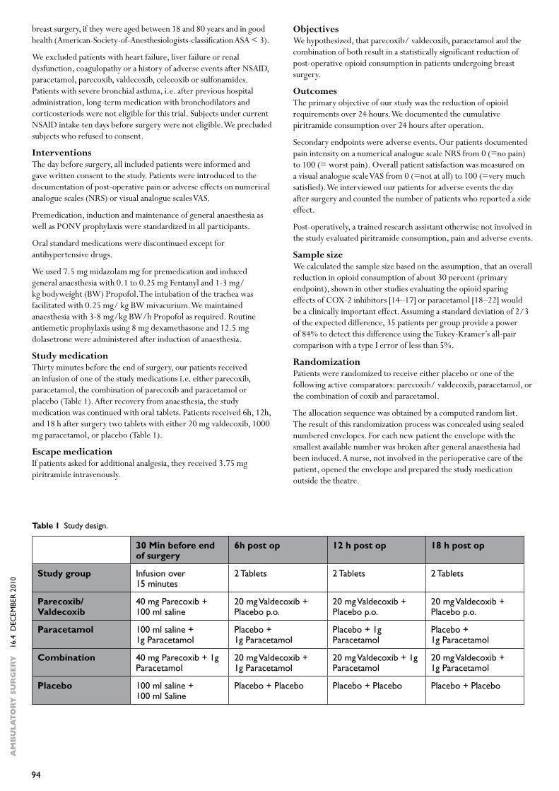
Study medicationThirty minutes before the end of surgery, our patients received an infusion of one of the study medications i.e. either parecoxib, paracetamol, the combination of parecoxib and paracetamol or placebo (Table 1). After recovery from anaesthesia, the study medication was continued with oral tablets. Patients received 6h, 12h, and 18 h after surgery two tablets with either 20 mg valdecoxib, 1000 mg paracetamol, or placebo (Table 1).
Escape medicationIf patients asked for additional analgesia, they received 3.75 mg piritramide intravenously.
ObjectivesWe hypothesized, that parecoxib/ valdecoxib, paracetamol and the combination of both result in a statistically significant reduction of post-operative opioid consumption in patients undergoing breast surgery.
OutcomesThe primary objective of our study was the reduction of opioid requirements over 24 hours. We documented the cumulative piritramide consumption over 24 hours after operation.
Secondary endpoints were adverse events. Our patients documented pain intensity on a numerical analogue scale NRS from 0 (=no pain) to 100 (= worst pain). Overall patient satisfaction was measured on a visual analogue scale VAS from 0 (=not at all) to 100 (=very much satisfied). We interviewed our patients for adverse events the day after surgery and counted the number of patients who reported a side effect.
Post-operatively, a trained research assistant otherwise not involved in the study evaluated piritramide consumption, pain and adverse events.
Sample sizeWe calculated the sample size based on the assumption, that an overall reduction in opioid consumption of about 30 percent (primary endpoint), shown in other studies evaluating the opioid sparing effects of COX-2 inhibitors [14–17] or paracetamol [18–22] would be a clinically important effect. Assuming a standard deviation of 2/3 of the expected difference, 35 patients per group provide a power of 84% to detect this difference using the Tukey-Kramer’s all-pair comparison with a type I error of less than 5%.
RandomizationPatients were randomized to receive either placebo or one of the following active comparators: parecoxib/ valdecoxib, paracetamol, or the combination of coxib and paracetamol.
The allocation sequence was obtained by a computed random list. The result of this randomization process was concealed using sealed numbered envelopes. For each new patient the envelope with the smallest available number was broken after general anaesthesia had been induced. A nurse, not involved in the perioperative care of the patient, opened the envelope and prepared the study medication outside the theatre.
Table 1 Study design.
30 Min before end of surgery
6h post op 12 h post op 18 h post op
Study group Infusion over 15 minutes
2 Tablets 2 Tablets 2 Tablets
Parecoxib/ Valdecoxib
40 mg Parecoxib + 100 ml saline
20 mg Valdecoxib + Placebo p.o.
20 mg Valdecoxib + Placebo p.o.
20 mg Valdecoxib + Placebo p.o.
Paracetamol 100 ml saline + 1g Paracetamol
Placebo + 1g Paracetamol
Placebo + 1g Paracetamol
Placebo + 1g Paracetamol
Combination 40 mg Parecoxib + 1g Paracetamol
20 mg Valdecoxib + 1g Paracetamol
20 mg Valdecoxib + 1g Paracetamol
20 mg Valdecoxib + 1g Paracetamol
Placebo 100 ml saline + 100 ml Saline
Placebo + Placebo Placebo + Placebo Placebo + Placebo
95
AM
BU
LAT
OR
Y S
UR
GER
Y
16.
4 D
ECEM
BER
R 2
010
BlindingPatients and researchers were not aware of the study medication. Study medications were clear, colourless fluids and white tablets of identical shape avoiding visible differences between the study drugs.
StatisticsData of piritramide consumption and pain intensities were treated as continuous data. The results are given as median (25th and 75th percentile) or mean (+ standard deviation). The analysis of the data was performed using the Tukey’s all pairs test, the Wilcoxon U-test, and χ²-test where appropriate. P-values of p < 0.05 were considered statistically significant.
ResultsParticipant flowA total of 182 patients were eligible for our study and recruited from October 2003 to May 2004. Seventeen women met exclusion criteria and were not enrolled. 165 patients were included in this trial. Five patients were withdrawn because of cancelled surgery (n=1), unexpected extension of surgery (n=2), severe vomiting in the PACU (n=1) followed by refusal of the patient to continue the trial, and major study violation (n=1). Therefore, 160 data sets could be included in the final analysis (Fig. 1)
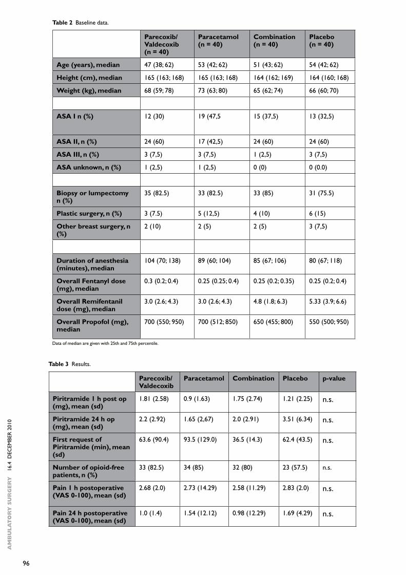
Baseline dataThe four groups were comparable with respect to demographic data, kind of surgery and anaesthesia (Table 2). No statistically significant differences for total remifentanil dose or desflurane-requirement were observed between study groups.
OutcomesThe overall piritramide request (mean + SD) during the 24 hours after breast surgery was very low with 3.51 mg + 6.34 mg in the placebo group. Coxibs and paracetamol reduced the opioid requirement to 2.2 + 2.92 and 1.65 mg + 2.67 mg respectively (Table 3). However, the differences between placebo and treated groups were not statistically significant (Fig. 2). The combination of both active drugs was not associated with a further decrease of opioid consumption (2.0 mg + 2.91 mg).
The performance of the study was not associated with increased pain intensity in any study group supporting the notion of post-operative pain management as being equally sufficient over all study groups. Placebo patients documented post-operative pain intensity one and 24 hours after surgery on a NRSplacebo 1h= 2.83 ± 2.0, and NRSplacebo 24h= 1.69 ± 4.29. One and twenty-four hours after surgery, patients with a coxib (parecoxib/ valdecoxib), paracetamol or the combination of both did not report significantly reduced pain compared to placebo (Table 3).
The average time until first opioid request was 62.4 + 43.5 min in the placebo group. Paracetamol but not valdecoxib or the combination prolonged the time until first additional opioid requirement to 93.5 + 129.0 min (p<0.05), 63.6 + 90.4 min and 36.5 + 14.3 min (Table 2).
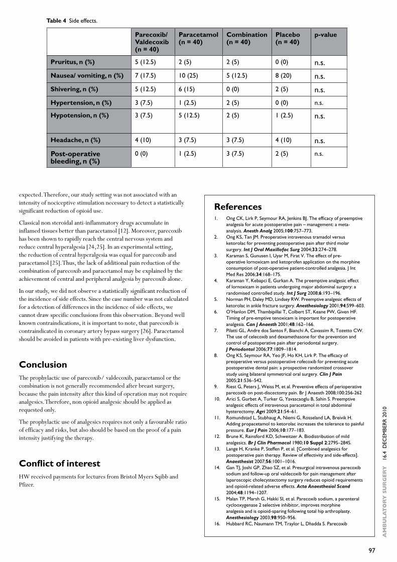
Side effectsWe documented adverse events during the 24 hours after surgery (Table 4). In the placebo group, no patient reported pruritus, 20 % nausea or vomiting, 5 % shivering, 0 % elevated blood pressure or palpitation, 2.5 % hypotension and 10 % headache after surgery. No significant differences were observed between placebo and treated groups (Table 4).
Bleeding occurred in 5% of placebo patients, in no coxib-treated patient, one paracetamol and three participants in the combination group. No other serious adverse event was observed during the trial period.
Discussion The reduction of opioid requirements in patients after surgery is an important intention of peri-operative non opioid analgesic use reducing sedation, impaired pulmonary function, PONV and constipation. We investigated the influence of prophylactic parecoxib/ valdecoxib, paracetamol and their combination on post-operative piritramide consumption in a randomized double blind placebo-controlled trial. Patients included in this analysis underwent elective breast surgery under general anaesthesia.
Placebo patients reported very low pain intensity and required almost no opioid analgesics after breast surgery. Therefore, the prophylactic use of non-opioids was not always necessary in the investigated group of patients. Parecoxib/ valdecoxib, paracetamol and their combination did not reduce post-operative opioid requirement significantly. The secondary outcome parameter of this trial was pain intensity after surgery. One and twenty-four hours after surgery we did not find a significant difference in pain intensity between placebo and treated groups.
It is an important issue to note thatthe preventive use of analgesics requires not only a favourable ratio of efficacy and risks, but also should be based on the proof of a pain intensity justifying the therapy.
In a study of the analgesic effects of paracetamol and the combination with codeine, Bjune at al. showed the importance of post-operative pain intensity in studies evaluating analgesic efficacy [23]. Patients with moderate baseline pain (VAS 40 to 60 mm) did not have analgesic efficacy of any tested drug, whereas patients with strong baseline pain (VAS > 60) had significant analgesic effects of either drug. Since the pain intensity of our patients always was very low, the assay- sensitivity of analgesia in our trial is obviously lower than
Figure 1 Participant workflow.
Figure 2 Piritramide consumption (mg/24h).
Data are given as mean and standard deviation.
96
AM
BU
LAT
OR
Y S
UR
GER
Y
16.
4 D
ECEM
BER
201
0
Table 2 Baseline data.
Data of median are given with 25th and 75th percentile.
Parecoxib/ Valdecoxib(n = 40)
Paracetamol(n = 40)
Combination(n = 40)
Placebo (n = 40)
Age (years), median 47 (38; 62) 53 (42; 62) 51 (43; 62) 54 (42; 62)
Height (cm), median 165 (163; 168) 165 (163; 168) 164 (162; 169) 164 (160; 168)
Weight (kg), median 68 (59; 78) 73 (63; 80) 65 (62; 74) 66 (60; 70)
ASA I n (%) 12 (30) 19 (47,5 15 (37,5) 13 (32,5)
ASA II, n (%) 24 (60) 17 (42,5) 24 (60) 24 (60)
ASA III, n (%) 3 (7,5) 3 (7,5) 1 (2,5) 3 (7,5)
ASA unknown, n (%) 1 (2,5) 1 (2,5) 0 (0) 0 (0.0)
Biopsy or lumpectomy n (%)
35 (82.5) 33 (82.5) 33 (85) 31 (75.5)
Plastic surgery, n (%) 3 (7.5) 5 (12,5) 4 (10) 6 (15)
Other breast surgery, n (%)
2 (10) 2 (5) 2 (5) 3 (7,5)
Duration of anesthesia (minutes), median
104 (70; 138) 89 (60; 104) 85 (67; 106) 80 (67; 118)
Overall Fentanyl dose (mg), median
0.3 (0.2; 0.4) 0.25 (0.25; 0.4) 0.25 (0.2; 0.35) 0.25 (0.2; 0.4)
Overall Remifentanil dose (mg), median
3.0 (2.6; 4.3) 3.0 (2.6; 4.3) 4.8 (1.8; 6.3) 5.33 (3.9; 6.6)
Overall Propofol (mg), median
700 (550; 950) 700 (512; 850) 650 (455; 800) 550 (500; 950)
Table 3 Results.
Parecoxib/ Valdecoxib
Paracetamol Combination Placebo p-value
Piritramide 1 h post op (mg), mean (sd)
1.81 (2.58) 0.9 (1.63) 1.75 (2.74) 1.21 (2.25) n.s.
Piritramide 24 h op (mg), mean (sd)
2.2 (2.92) 1.65 (2,67) 2.0 (2.91) 3.51 (6.34) n.s.
First request of Piritramide (min), mean (sd)
63.6 (90.4) 93.5 (129.0) 36.5 (14.3) 62.4 (43.5) n.s.
Number of opioid-free patients, n (%)
33 (82.5) 34 (85) 32 (80) 23 (57.5) n.s.
Pain 1 h postoperative (VAS 0-100), mean (sd)
2.68 (2.0) 2.73 (14.29) 2.58 (11.29) 2.83 (2.0) n.s.
Pain 24 h postoperative (VAS 0-100), mean (sd)
1.0 (1.4) 1.54 (12.12) 0.98 (12.29) 1.69 (4.29) n.s.
97
AM
BU
LAT
OR
Y S
UR
GER
Y
16.
4 D
ECEM
BER
R 2
010
expected. Therefore, our study setting was not associated with an intensity of nociceptive stimulation necessary to detect a statistically significant reduction of opioid use.
Classical non steroidal anti-inflammatory drugs accumulate in inflamed tissues better than paracetamol [12]. Moreover, parecoxib has been shown to rapidly reach the central nervous system and reduce central hyperalgesia [24,25]. In an experimental setting, the reduction of central hyperalgesia was equal for parecoxib and paracetamol [25]. Thus, the lack of additional pain reduction of the combination of parecoxib and paracetamol may be explained by the achievement of central and peripheral analgesia by parecoxib alone.
In our study, we did not observe a statistically significant reduction of the incidence of side effects. Since the case number was not calculated for a detection of differences in the incidence of side effects, we cannot draw specific conclusions from this observation. Beyond well known contraindications, it is important to note, that parecoxib is contraindicated in coronary artery bypass surgery [26]. Paracetamol should be avoided in patients with pre-existing liver dysfunction.
ConclusionThe prophylactic use of parecoxib/ valdecoxib, paracetamol or the combination is not generally recommended after breast surgery, because the pain intensity after this kind of operation may not require analgesics. Therefore, non opioid analgesic should be applied as requested only.
The prophylactic use of analgesics requires not only a favourable ratio of efficacy and risks, but also should be based on the proof of a pain intensity justifying the therapy.
Conflict of interestHW received payments for lectures from Bristol Myers Sqibb and Pfizer.
Table 4 Side effects.
Parecoxib/ Valdecoxib(n = 40)
Paracetamol(n = 40)
Combination(n = 40)
Placebo (n = 40)
p-value
Pruritus, n (%) 5 (12.5) 2 (5) 2 (5) 0 (0) n.s.
Nausea/ vomiting, n (%) 7 (17.5) 10 (25) 5 (12.5) 8 (20) n.s.
Shivering, n (%) 5 (12.5) 6 (15) 0 (0) 2 (5) n.s.
Hypertension, n (%) 3 (7.5) 1 (2.5) 2 (5) 0 (0) n.s.
Hypotension, n (%) 3 (7.5) 5 (12.5) 2 (5) 1 (2.5) n.s.
Headache, n (%) 4 (10) 3 (7.5) 3 (7.5) 4 (10) n.s.
Post-operative bleeding, n (%)
0 (0) 1 (2.5) 3 (7.5) 2 (5) n.s.
ReferencesOng CK, Lirk P, Seymour RA, Jenkins BJ. The efficacy of preemptive 1. analgesia for acute postoperative pain – management: a meta-analysis. Anesth Analg 2005;100:757–773.Ong KS, Tan JM. Preoperative intravenous tramadol versus 2. ketorolac for preventing postoperative pain after third molar surgery. Int J Oral Maxillofac Surg 2004;33:274–278.Karaman S, Gunusen I, Uyar M, Firat V. The effect of pre-3. operative lornoxicam and ketoprofen application on the morphine consumption of post-operative patient-controlled analgesia. J Int Med Res 2006;34:168–175.Karaman Y, Kebapci E, Gurkan A. The preemptive analgesic effect 4. of lornoxicam in patients undergoing major abdominal surgery: a randomised controlled study. Int J Surg 2008;6:193–196.Norman PH, Daley MD, Lindsey RW. Preemptive analgesic effects of 5. ketorolac in ankle fracture surgery. Anesthesiology 2001;94:599–603.O’Hanlon DM, Thambipillai T, Colbert ST, Keane PW, Given HF. 6. Timing of pre-emptive tenoxicam is important for postoperative analgesia. Can J Anaesth 2001;48:162–166.Pilatti GL, Andre dos Santos F, Bianchi A, Cavassim R, Tozetto CW. 7. The use of celecoxib and dexamethasone for the prevention and control of postoperative pain after periodontal surgery. J Periodontol 2006;77:1809–1814.Ong KS, Seymour RA, Yeo JF, Ho KH, Lirk P. The efficacy of 8. preoperative versus postoperative rofecoxib for preventing acute postoperative dental pain: a prospective randomized crossover study using bilateral symmetrical oral surgery. Clin J Pain 2005;21:536–542.Riest G, Peters J, Weiss M, et al. Preventive effects of perioperative 9. parecoxib on post-discectomy pain. Br J Anaesth 2008;100:256-262Arici S, Gurbet A, Turker G, Yavascaoglu B, Sahin S. Preemptive 10. analgesic effects of intravenous paracetamol in total abdominal hysterectomy. Agri 2009;21:54–61.Romundstad L, Stubhaug A, Niemi G, Rosseland LA, Breivik H. 11. Adding propacetamol to ketorolac increases the tolerance to painful pressure. Eur J Pain 2006;10:177–183.Brune K, Rainsford KD, Schweitzer A. Biodistribution of mild 12. analgesics. Br J Clin Pharmacol 1980;10 Suppl 2:279S–284S.Lange H, Kranke P, Steffen P, et al. [Combined analgesics for 13. postoperative pain therapy. Review of effectivity and side-effects]. Anaesthesist 2007;56:1001–1016.Gan TJ, Joshi GP, Zhao SZ, et al. Presurgical intravenous parecoxib 14. sodium and follow-up oral valdecoxib for pain management after laparoscopic cholecystectomy surgery reduces opioid requirements and opioid-related adverse effects. Acta Anaesthesiol Scand 2004;48:1194–1207.Malan TP, Marsh G, Hakki SI, et al. Parecoxib sodium, a parenteral 15. cyclooxygenase 2 selective inhibitor, improves morphine analgesia and is opioid-sparing following total hip arthroplasty. Anesthesiology 2003;98:950–956.Hubbard RC, Naumann TM, Traylor L, Dhadda S. Parecoxib 16.
98
AM
BU
LAT
OR
Y S
UR
GER
Y
16.
4 D
ECEM
BER
201
0
sodium has opioid-sparing effects in patients undergoing total knee arthroplasty under spinal anaesthesia. Br J Anaesth 2003;90:166–172.Reynolds LW, Hoo RK, Brill RJ, et al. The COX-2 specific inhibitor, 17. valdecoxib, is an effective, opioid-sparing analgesic in patients undergoing total knee arthroplasty. J Pain Symptom Manage 2003;25:133–141.Cobby TF, Crighton IM, Kyriakides K, Hobbs GJ. Rectal paracetamol 18. has a significant morphine-sparing effect after hysterectomy. Br J Anaesth 1999;83:253–256.Korpela R, Korvenoja P, Meretoja OA. Morphine-sparing effect 19. of acetaminophen in pediatric day-case surgery. Anesthesiology 1999;91:442–447.Remy C, Marret E, Bonnet F. Effects of acetaminophen on morphine 20. side-effects and consumption after major surgery: meta-analysis of randomized controlled trials. Br J Anaesth 2005;94:505–513.Viitanen H, Tuominen N, Vaaraniemi H, Nikanne E, Annila P. 21. Analgesic efficacy of rectal acetaminophen and ibuprofen alone or in combination for paediatric day-case adenoidectomy. Br J Anaesth 2003;91:363–367.Aubrun F, Kalfon F, Mottet P, et al. Adjunctive analgesia with 22. intravenous propacetamol does not reduce morphine-related adverse effects. Br J Anaesth 2003;90:314–319.Bjune K, Stubhaug A, Dodgson MS, Breivik H. Additive analgesic 23. effect of codeine and paracetamol can be detected in strong, but not moderate, pain after Caesarean section. Baseline pain-intensity is a determinant of assay-sensitivity in a postoperative analgesic trial. Acta Anaesthesiol Scand 1996;40:399–407.Mehta V, Johnston A, Cheung R, Bello A, Langford RM. Intravenous 24. parecoxib rapidly leads to COX-2 inhibitory concentration of valdecoxib in the central nervous system. Clin Pharmacol Ther 2008;83:430–435.Koppert W, Wehrfritz A, Korber N, et al. The cyclooxygenase 25. isozyme inhibitors parecoxib and paracetamol reduce central hyperalgesia in humans. Pain 2004;108:148–153.EMEA. EMEA public statement on Valdecoxib and Parecoxib. In: 26. EMEA; 2004
99
AM
BU
LAT
OR
Y S
UR
GER
Y
16.
4 D
ECEM
BER
R 2
010
IntroductionSpinal anesthesia is not a 100% certain successful technique. Failure rates of 0.72%% to 16.0% have been reported. [1,2,3] The cause of some failures may be due to technical inability to identify the subarachnoid space and that is obvious at the moment and understandable. The explanation for spinal block failure that occurs despite apparent technically correct injection of the correct drug can be mystifying.
The under reporting of specific cases with block failure and mechanisms of failure may reflect a general attitude that a regional anesthetic failure is although unfortunate, is “normal” and not a complication deserving investigation. A repeat spinal block as a remedy for a failed spinal block may be contraindicated depending on the first drug used due to the risk of neurotoxicity.
The following case report is of a failed spinal block that mystified the anesthesiologist at first but simple assessment provided a clear probable explanation. A following discussion will highlight causes and remedies. The patient gave telephonic consent to present this case on the day after his discharge from hospital.
Case ReportA 58-year-old male arrived for outpatient ambulatory surgery in the early afternoon, to undergo removal of two screws from the lateral side of his right ankle that had been inserted for fractures incurred seven months prior. He was allergic to sulfa drugs. He was a non-smoker. He was using oral opioids for ankle pain, metoclopramide for intermittent nausea, and loperamide for irritable bowel syndrome. He had undergone several uncomplicated general anesthetics for a variety of small surgeries (hernia repair, tonsillectomy), and was otherwise healthy.
However, previous regional and local anesthetic blocks had only been occasionally successful. At the time of the initial injury seven months earlier, emergency room physicians administered a sciatic nerve block that failed. The patient subsequently had a successful 0.75% hyperbaric bupivacaine spinal anesthesia for the surgery, administered by anesthesiologists. The patient reported that when past dentists administered lidocaine to him it often resulted in partial analgesia. He did note though that his current dentist’s tooth nerve injections were
consistently effective.
For the spinal anesthetic being reported here, anesthesia was induced via a 25 G Whitacre point needle inserted at the L4-5 interspinous space. The patient was in an upright sitting position. The subarachnoid space was at a depth of six centimeters from the skin, and it was identified by clear fluid considered to be cerebrospinal fluid dripping from the needle. Two milliliters of 2% Chloroprocaine was injected. After injection of one milliliter of drug, a half-milliliter of cerebrospinal fluid was aspirated into the needle and then re-injected with the remaining chloroprocaine. After completion of the injection, clear fluid considered to be cerebrospinal fluid still dripped from the back of the needle.
The anesthesiologist made a point of keeping the patient sitting for two additional minutes as per advice from a peer anesthesiologist who had recommended this technique and drug. The clinical goal was to have a short acting spinal anesthetic for foot surgery. The patient was returned to the supine position. After fifteen minutes had passed there was no evidence of sensory anesthesia. Ice applied to the mid-anterior thigh bilaterally produced the same cold sensation as that applying the ice to the upper chest and neck region. The toes were equally able to sense cold. The patient was thought to have slight reduced foot plantar flexion strength, but this was not compared to pre-spinal block strength. In addition the patient could not flex or extend toes bilaterally suggesting a degree of motor block was present. The chloroprocaine ampule of drug used was inspected and it was verified to be the intended drug to be used and corresponding amount of drug had been removed from the ampule. All other drugs in the anesthesiologist’s possession had appropriate syringe labels and the expected content volumes. There was nothing to suggest a wrong spinal drug had been injected, as cause of the failed block.
It was decided to induce general anesthesia with mask inhalational anesthesia using sevoflurane vapor and then place a laryngeal mask airway.
The surgery proceeded uneventfully. On emergence the patient was pain free. His spinal block was assessed again about an hour after block insertion. He still had intact sensation for ice induced coldness on the toes and in the mid anterior thigh region. He could move both sides’ toes and foot. He however had no sensation at all about his anus and posterior aspect of the scrotum. No other areas were tested.
It was diagnosed that he had a saddle block of the sacral dermatomes.
Financial Support: None
Previous Presentations of this Report: None
Keywords: Parecoxib; Paracetamol; Valdecoxib; Post-operative pain.
Authors’ addresses: a Department of Anesthesia, Carver College of Medicine, University of Iowa.
Corresponding author: Robert M Raw Department of Anesthesia, University of Iowa, 200 Hawkins Drive, Iowa City, IA52242, USATel 319-356-2633 Fax 319-356-2940 Email: [email protected]
Spinal Anesthetic Block Failure due to the Hyperbaric Nature of 2% Chloroprocaine Local AnestheticRobert M Raw MD, FCA & Enyinnaya R Nwaneri MD
100
AM
BU
LAT
OR
Y S
UR
GER
Y
16.
4 D
ECEM
BER
201
0
This is compatible with use of a hyperbaric local anesthetic drug (2% Chloroprocaine) in a patient where subarachnoid injection was made in the sitting position and where the patient was kept in the sitting position for few minutes after injection before reclining to the supine position.
DiscussionHorlocker and Wedel Human reported the density of many local anesthetics adjusted for temperature to match human normal temperature. [30] Increasing the drug temperature from room temperature to 37 degrees centigrade decreases the drug’s density. Human cerebrospinal fluid (CSF) has a specific gravity of 1.00063 to 1.00075 at 37 degree centigrade generally, and 1.00030 gram per milliliter in term pregnant woman a. [4,5] Preservative free chloroprocaine 2% and 3% solutions have specific gravities of 1.00123 and 1.00257 gm per milliliter respectively, which makes them hyperbaric without any added dextrose. Kopacz investigated the effect of added dextrose to 2% chloroprocaine in volunteers with a crossover spinal blocks protocol . With their technique of patient positioning during and immediately after administration of spinal anesthesia they found adding dextrose to 2% chloroprocaine did not increase the thoracic block extent of T4 seen when using dextrose free 2% chloroprocaine. The group who received the dextrose-enhanced 2% chloroprocaine group however had longer duration sacral block, on the basis of indirect evidence with delayed normalization of bladder function. Accordingly addition of 10% dextrose did not alter the pre-existing hyperbaric characteristics of 2% chloroprocaine significantly.
In this case report the anesthesiologist intentionally kept the patient sitting for a period after injection of the spinal chloroprocaine, on wrong advice. The chloroprocaine shifted to the most dependant part of the subarachnoid sack to only effectively block the lowest sacral spinal nerves supplying the perineum leaving the lumbar nerve roots supplying the legs unblocked. This is called a saddle block. No dextrose had been added to the chloroprocaine. This confirms the known hyperbaric nature of 2% chloroprocaine solution. [29,30]
This patient’s substantial history of failed regional anesthesia in the hands of dentists and emergency room physicians may raise the question of resistance to local anesthetic drugs. Sebrechts in 1934 noted apparent differences in resistance to spinal anesthesia between Italians and Anglo-Saxons and proposed the term ‘rachi-resistance”. [7] A pharmacogenetic mutation of sodium channels associated with reduced lidocaine sensitivity has been described but the frequency or relevance of genetic based local anesthesia resistance in the large population is unknown. [8] In this case report the patient’s history of other failed regional anesthesia blocks more likely reflects technical failures of the practitioners involved with the failed nerve blocks, as another different dentist and anesthesiologist produced successful blocks on other occasions.
A failed nerve block is not widely considered a “complication”. Regardless a failed block can force a change in anesthesia care plan that can be suboptimal or detrimental to the patient. Textbooks neglect failed spinal anesthesia as a topic or do not consider it a complication. Spinal And Epidural Anesthesia by Wong (McGrawhill 2007) offers no discussion or listing of failed spinal anesthesia at all. In the book Complications In Regional Anesthesia And Pain Medicine by editors Neal and Rathmell (Saunders 2007), block failure is not regarded as a complication. The only mention made of spinal block failure is incidental in the context of spinal drug maldistribution (to the cauda equina) and the use of repeat doses of chloroprocaine as
potential cause of neurotoxicity. The book references work by A Gissen and K Drasner. A Gissen’s 1984 editorial speculated on the possible causes of the neural complications then recently observed during chloroprocaine epidural blocks including consideration of inherent drug neurotoxicity. [11] Drasner studied how baricity of various local anesthetic drugs affected the propensity and potential of the drug to pool in the dependant parts of the subarachnoid space such as the cauda equina. [9] In 1991 Drasner recommended NOT repeating a chloroprocaine dose for failed spinal block in case the first dose was in fact administered subarachnoid and the total of the two doses would then exceed safe total dose limits for avoiding neurotoxicity. [10]
The dose of chloroprocaine above which the drug may be neurotoxic in spinal doses is quoted as 60 mg. [11–16] Complications of Regional Anesthesia by Finucane (Churchill Livingston 1999) says “it seems unnecessary to list failure (of neuraxial blocks) as a complication . . ..” The book however does cover the topic of failed spinal anesthesia in one paragraph, mentioning drug maldistribution and arachnoid cysts as causes of block failure. The book Regional Anesthesia and Analgesia by Brown (Saunders 1996) has a chapter on regional anesthesia complications that presents Drasner’s recommendations for managing failed spinal anesthesia (see below). More recently in 2009 the subject of failed spinal anesthesia enjoyed its first large review Failed spinal anaesthesia: mechanisms, management, and prevention by Fettes. [17] A recent case report by Hoppe of four failed obstetric spinal blocks gives a good summary of anatomical reasons and ligamentous cysts that can cause technical failure. [18] The Fettes’ review and Hoppe’s case report are recommended reading.
Causes of failed spinal anesthesia can be classified as
1. Successfully injected drugs that are maldistributed relative to the needs of the planned surgery.
2. Unrecognized failed injection of drug, partial or total.
3. Technical failure to enter the subarachnoid space, with no drug injection.
4. Drug errors, as wrong drugs and inappropriate additives.
5. Local anesthetic resistance.
6. Pseudo block failure, due to excessive expectations for speed of block onset.
7. Subdural injection of a spinal dose is conceptually a possible cause of spinal block failure, but has never been reported, recognized or studied in this context of small volume injections.
The evidence that 2% chloroprocaine is potentially neurotoxic comes from anecdotal human case reports, animal studies, and cell studies. Evidence of clinical toxicity is not clear cut. Laboratory evidence of toxicity is indirect and dependant upon study methodologies far removed from replication of clinical practice. Human cases reports associating cauda equina syndrome with use of chloroprocaine epidural anesthesia accumulated in the 80s. [19] One theory was that in these cauda equina syndrome cases large doses of chloroprocaine had transferred to the subarachnoid space. Some research attributed the cauda equina syndrome cases to the additive Sodium bisulphite. Sodium Bisulfite in laboratory studies was shown to have neurotoxicity potential. [20] Research by RS Ravindran in dogs suggested chloroprocaine was neurotoxic itself in the subarachnoid space in a dose related fashion. [21] A similar study in rats by DF Li also suggested 2-chloroprocaine had a dose related neurotoxicity. [22] A study in cats by DJ Ford assessing peripheral nerve toxicity of 2-chloroprocaine and bisulfite suggested that pH of the drug solution as well as bisulfite concentration was critical
101
AM
BU
LAT
OR
Y S
UR
GER
Y
16.
4 D
ECEM
BER
R 2
010
in the addition of bisulfite to chloroprocaine to cause evidence of neurotoxicity. Evidence in this last study even suggested that the combination of 2-chloroprocaine with bisulfite could reduce the magnitude of bisulfite’s neurotoxicity when administered alone. Masahiko Taniguchi conversely in 2004 clearly showed bisulfite could actually reduce the neurotoxicity of 2-chloroprocaine when used in combination . Taniguchi suggested chloroprocaine was the more neurotoxic single substance, of the two substances, namely sodium bisulfite and chlorprocaine, and that unexpectedly they were least neurotoxic when used as combination. In 2005 the resurgent new popularity of 2-chloroprocaine as replacement for lidocaine in spinal anesthesia evidenced by four human spinal 2-chloroprocaine studies was reviewed and discussed in an editorial by Kenneth Drasner . He stated “there is little doubt that large doses of subarachnoid chloroprocaine . . . can induce permanent neurological injury”. We note that comment, and accordingly concur with the recommendation of Drasner from 1991 to limit the maximum dose of chlorprocaine to 60 mg [10]. An apparently clinically failed chloroprocaine spinal block, as in our case reported here, does not exclude the possibility of the drug having been injected subarachnoid and the addition of second full subarachnoid dose could result in a potentially neurotoxic dose of chloroprocaine being administered.
In the initial assessment of failed chloroprocaine spinal block we recommend Drasner’s 1991 guideline’s with modification. [16] Namely;
1. Visualize Cerebro Spinal Fluid (CSF) before and after injecting spinal drugs. This may be done by observation of spontaneous CSF like clear fluid dripping from the needle or by aspiration of CSF like clear fluid into the syringe.
2. Examine sacral dermatomes as well when assessing any failed spinal block,
3. If a spinal block has failed despite pre- and post-injection visualization of CSF, regard the drugs as actually administered into CSF. This may modify the dose of a repeat spinal block depending in the drug first used. There is no way to assess the fraction of the injected amount of drug actually delivered into the subarachnoid space, in this circumstance, and it should be assumed, for safety purposes, to be the full dose.
4. If chloroprocaine is being used, and repeat injection is considered in the presence of suggestive evidence of correctly injected spinal drugs (CSF observed or saddle block present), reduce the second chloroprocaine dose to stay under a total dose of 60mg. Alternatively switch to using an entirely different drug administered at its own full dose. There are two case reports where a repeated spinal injection of dibucaine local anesthetic likely caused neurotoxicity by exceeding that drug’s recommended subarachnoid safe dose , .
5. If CSF was not seen after the spinal drug injection and the spinal block fails, repeat a “full spinal dose” if a saddle block has been excluded. Absence of a saddle block suggests the spinal drug never reach the subarachnoid space at all. Alternatively switch to using an entirely different drug at its own full dose.
A failed spinal block may be an apparent complete failure or only a partial failure. Management of a failed spinal anesthetic could include (i) abandonment of the procedure, (ii) repeat spinal anesthetic, (iii) use of supplementary sedation and analgesia, (iv) conversion to general anesthesia, or (v) addition of distal peripheral nerve blocks. Specific circumstance and patient considerations would determine the wisest course to follow.
A failed spinal block has previously been reported related to use of dextrose enhanced bupivacaine and the sitting position. [27] One series analysis has shown difference in failure rates of 19.4% when using hyperbaric solutions and 2.9% when using isobaric solution within one institution. [28] This suggests isobaric solutions are inherently more reliable. The lowest reported spinal block failure rate of 0.72% was in urological surgery series where intentional saddle blocks using hyperbaric solutions were done for perineal urological procedures. These all suggest that the unintentional saddle block is the largest cause of failure after successful intrathecal drug injection, and that avoidance of hyperbaric solutions and sitting position would favor higher success rates with spinal block for non-perineal surgery, for example orthopedic surgery as in this case report. Spinal anesthesia for Caesarean section on the other hand enjoys high success rates with hyperbaric local anesthetic solutions that are gravitationally directed, in a controlled fashion, to the thoracic dermatomes.
In conclusion, it is important to remember that 2-chloroprocaine is hyperbaric relative to human cerebrospinal fluid even without added dextrose, especially since 2-chloroprocaine is being widely promoted currently as a short acting spinal anesthesia drug in ambulatory surgery. [29, 30] Secondly we wish to caution against a second chloroprocaine spinal dose as a means to correct a failed first chloroprocaine spinal anesthetic lest the cauda equina nerve be exposed to a double dose of a potential neurotoxic drug. We however find no reason to discourage use of chloroprocaine spinal anesthesia as a short acting anesthetic, if the above considerations are kept in mind.
Lastly we would argue that any failed nerve block deserves to be considered a “complication” of an intervention. A complication has consequences that can force alternate interventions or therapies to be utilized which may be less favorable for the patient. In addition any outcome (such as failed spinal block) treated as a complication will receive more attention for analysis, discussion, prevention and education, all of which should benefit patients ultimately.
ACKNOWLEDGEMENTS: None
SUPPLEMENTARY INFORMATION: Time line.Monitoring initiated 13H45.Midazolam 2mg administered 13H50.Spinal block inserted 13H55.Ketamine 10 mg administered 14H00Ketamine 15mg administered 14H05Gas induction at 14H15 due to absence of loss of sensation to cold on both anterior mid thighs compared to cold sensation on chest. Re-examination in PACU 15H00
102
AM
BU
LAT
OR
Y S
UR
GER
Y
16.
4 D
ECEM
BER
201
0
ReferencesShrestha AB, Shresha CK, et al. Failure of subarachnoid block in 1. Cesarean section. Nepal Med Coll J. 2009 Mar;11(1):50–1.Harrisson DA. Spinal anaesthesia for urological surgery. A survey of 2. failure rate, post dural puncture headache and patient satisfaction. Anaesthesia 1992;47(10):902–3.Guinard JP, et al. A prospective evaluation of the failure rate of spinal 3. anesthesia for transurethral prostatic resection. Eur J Anaesthesiol 1992 Janurary;9(11):7–13.Levin E. Density of normal human cerebrospinal fluid and tetracaine 4. solutions. Anesth Analg. 1981 November;60(11):814–7.Richardson MG, Wissler RN. Density of lumbar cerebrospinal 5. fluid in pregnant and non-pregnant humans. Anesthesiology 1996 Aug;85(2):326–30. Warren DT, Kopacz DJ. 6. Anesth Analg. 2004 Jan;98(1):95–101.Sebrechts J. Spinal Anaesthesia. 7. Br. J Anaesthesia. 1934;12:4–27.Sheets PL, Jackson JO, et al. A Nav 1.7 channel mutation 8. associated with hereditary erythromelalgia contributes to neuronal hyperexcitability and displays reduced lidocaine sensitivity. J Physiol 2007;581:1019–1031.Drasner K. Models for local anesthetic toxicity from continuous 9. spinal anesthesia. Regional Anesthesia. 1993;18:434–438.Drasner K., Rigler M. Repeat injection after a “failed spinal”: At times 10. a potentially unsafe practice. Anesthesiology 1991;75:713–714. Gissen AJ, Datta S, et al. The chloroprocaine controversy: 1. A 11. hypothesis to explain the neural complications of chloroprocaine epidural. Regional Anesthesia. 1984;9:124–134. Taniguchi M, Bollen, AW, Drasner K. Sodium bisulphate. Scapegoat 12. for chloroprocaine neurotoxicity? Anesthesiology 2004;100:85–91.Lambert HD, et al. Letter: In defense of in Vitro findings. 13. Anesthesiology, November 2004;101(5):1246–7.Baker MT. Chloroprocaine or sulfite toxicity? 14. Anesthesiology November 2004;101(5):1247.Drasner K, Taniguchi T, et al. Chloroprocaine of sulfite toxicity? In 15. Reply: Anesthesiology November 2004;101(5):1247–8.Drasner K, Rigler M. Repeat injection after a “failed spinal”: At times, 16. a potentially unsafe practice. Anesthesiology 1991;75:713–4.Fettes PDW. Failed spinal anesthesia: mechanisms, management, and 17. prevention. Br J Anaesth 2009;102:739–48. Hoppe J, Popham P. Complete failure of spinal anaesthesia in 18. obstetrics. Int J Obstet Anes 2007;16:250–255.Moore DC, Spierdijk, et al. Chloroprociane neurotoxicity: four 19. additional cases. Anesth Analg 1982;61(2):155–9.Seravalli E, Lear E. Toxicity of chloroprocaine and sodium bisulfite 20. on human neuroblastoma cells. Anesth Analg 1987;66(10):954–8. Ravindran RS, Turner MS, Muller J. Neurologic effects of 21. subarachnoid administration of 2-chloroprocaine-CE, bupivacaine, and low pH normal saline in dogs. Anesth Analg 1982;61(3):279–83.Li DF, Bahar M, Cole G, et al. Neurological toxicity of 22. the subarachnoid infusion of bupivacaine, lignocaine, or 2-chloroporocaine in the rat. Br. J Anaesth. 1985;57(4):424–9.Taniguchi M, Bollen AW, et al. Sodium Bisulfite. Scapegoat for 23. chloroprocaine neurotoxicity? Anesthesiol 2004;100:85–91.Drasner K. Chloroprocaine spinal anesthesia: back to the future. 24. Anesth Analg 2005;109:549–52.Yoshihiro A, Koniushi R. Neurologic symptom associated with a 25. repeated injection after failed spinal anesthesia. Anesthesiology November 1998;89(5):1294–5.Yoshimura Y, Nomoto Y. Irreversible damage to the cauda equina 26. following repeated intrathecal injection of hyperbaric dibucaine. J Anesth. 2002;16:176–178.Barbosa FT, Borges EL et al. General anesthesia alter 27. failed spinal block for emergency surgery in a patient with mucopolysaccharidosis: case report. Rev Bras Anesthesiol 2007;57(6):658–64.Brun-Buisson V. Failure of spinal anesthesia. Evaluation of the 28. practice at a university hospital. Ann Fr. Anesth Reanim. 1988;7(5):383–6.Na KB, Kopacz DJ. Spinal chloroprocaine solutions: density at 37 29. degrees C and pH titration. Anesth Analg 2004 January;98(1):70–4.Horlocker TT, Wedel DJ. Density, specific gravity and baricity of 30. spinal anesthetic solutions at body temperature. Anesth Analgesia 1993;76:1015–8.
103
AM
BU
LAT
OR
Y S
UR
GER
Y
16.
4 D
ECEM
BER
R 2
010
IntroductionAlthough not all patients with obstructive sleep apnea (OSA) are obese nor do all obese patients have OSA, nonetheless a discussion of OSA would be incomplete without including some introductory remarks about obesity. Obesity (defined as a body mass index [BMI] > 30) is reaching epidemic proportions in the United States and has become a major public health hazard. Morbid obesity, defined as a BMI >35 or a weight that is twice ideal body weight (ideal body weight [kg] = height [cm] – 100), affects approximately 5% of Americans and creates notable problems for patients, surgeons, and anesthesiologists alike. Technical challenges abound when one is caring for a morbidly obese patient. Venous access may be very difficult to establish, and noninvasive blood pressure determination may be hampered by an improperly fitting cuff or one that takes too long to inflate. Mask ventilation may be extremely troublesome or impossible, and endotracheal intubation may be challenging. Additionally, patient positioning for surgical procedures often proves vexing, and optimal surgical exposure may prove elusive.
In addition to the technical challenges presented by the morbidly obese patient, the clinician is often confronted with a wide variety of associated medical problems that must be managed perioperatively. These may include diabetes mellitus, osteoarthritis, psychological disturbances, systemic and pulmonary hypertension, restrictive ventilatory dysfunction and hypoxemia, left ventricular and/or right ventricular failure, liver disease, an increased risk of aspiration owing to delayed gastric emptying time and hiatus hernia, hypercoagulabililty, wound infections, and OSA. Indeed, morbidity and mortality rates are high in morbidly obese patients primarily because of associated cardiovascular and respiratory abnormalities and their propensity for deep vein thrombosis and pulmonary embolism. Clearly, OSA plays an important role in contributing to the troubling morbidity and mortality rates encountered in the patient with morbid obesity.
This review article will focus on current knowledge and controversies surrounding the management of patients with OSA.
Sleep Apnea: Definitions and DemographicsSleep patterns disturbed by snoring are thought to occur in approximately 25% of the population.[1] However, most patients who snore do not have apnea or associated episodes of notable hypoxemia. Nonetheless, OAS is a relatively common disorder among middle-aged adults, especially (obese) Americans. Obesity is a critical independent causative risk factor. The majority of people who have OSA are obese, and the severity of the condition seems to correlate with the patient’s neck circumference.[2] In the minority of OSA patients who are nonobese, causative risk factors include craniofacial and orofacial bony abnormalities, nasal obstruction, and hypertrophied tonsils. Importantly, Young and colleagues [3] estimated that 93% of women and 82% of men with moderate to severe OSA have not been clinically diagnosed.
OSA is defined as cessation of airflow for >10 sec despite continuing ventilatory effort, occurring five or more times per hour of sleep, and is usually associated with a decrease in arterial oxygen saturation of >4%. Although this review will focus predominantly on OSA, it should be noted for the sake of completeness that the three types of sleep apnea are obstructive, central, and mixed. Central sleep apnea, much rarer than OSA, is also known as Ondine’s curse, an allusion to the mythological water nymph who cursed her unfaithful husband to cease breathing if he ever fell asleep. Unlike OSA, respiratory efforts temporarily stop in central sleep apnea. Diagnosis is established definitively with polysomnography.
It is generally accepted that many patients with OSA have resultant pathologic daytime sleepiness associated with performance decrements. It has also been well established that patients with severe
AbstractThe purpose of this review article is to summarize our current knowledge concerning the anesthetic management of patients with obstructive sleep apnea (OSA) in the ambulatory setting.The pathophysiology, detection, and management of OSA are presented. Although minimal data exist to guide perioperative management in an
evidence-based fashion, current guidelines and recommendations are discussed. Depending on the type of surgery, anticipated postoperative analgesic (opioid) requirement, severity of the OSA, associated comorbidities, and the resources of the facility, outpatient surgery may be imprudent.
Keywords: obstructive sleep apnea, ambulatory surgery, morbid obesity.
Author’s address: New York Medical College, Valhalla, New York 10595, USA.
Corresponding author: Kathryn E. McGoldrick MD New York Medical College, Valhalla, New York 10595, USA.Tel 914-493-7693 Fax 914-493-7927 Email: [email protected]
Anesthetic Implications of Obstructive Sleep Apnea in the Ambulatory SettingKathryn E. McGoldrick MD
104
AM
BU
LAT
OR
Y S
UR
GER
Y
16.
4 D
ECEM
BER
201
0
apnea suffer major health consequences as a result of their condition. Yet, it remains somewhat controversial whether patients with less severe forms of this disease incur the same detrimental consequences, owing to methodological problems and failure to control for confounding factors. Thus, few absolute conclusions can be drawn at this time about the long-term consequences of mild to moderate OSA. However, findings from the Sleep Heart Health Study, [4] the Copenhagen City Heart Study,[5] and others [6] demonstrate a firm association between sleep apnea and systemic hypertension, even after other important patient characteristics, such as age, gender, race, consumption of alcohol, and use of tobacco products are controlled for.
Few definitive data exist to guide perioperative management of patients with OSA. It is not surprising that many anesthesiologists question whether OSA patients are appropriate candidates for ambulatory surgery. The risks of caring for these challenging patients in the ambulatory venue are further amplified by the unfortunate fact that 80 to 95% of people with OSA are undiagnosed;[3,7] they have neither a presumptive clinical and/or a sleep study diagnosis of OSA. This is concerning because these patients may suffer perioperatively from life-threatening desaturation and postoperative airway obstruction. Moreover, serious comorbidities may be present because prolonged apnea results in hypoxemia and hypercarbia, which can lead to increased systemic and pulmonary artery pressures and dysrhythmias. Cor pulmonale, polycythemia, and congestive heart failure may develop.
Pathophysiology and TherapySleep apnea occurs when the negative airway pressure that develops during inspiration is greater than the muscular distending pressure. Obstruction can occur throughout the upper airway, above, below, or at the level of the uvula.[8,9] Because there is an inverse relationship between obesity and pharyngeal area, the smaller size of the upper airway in the obese patient causes a more negative pressure to develop for the same inspiratory flow. [9,10] Kuna has also postulated that there may be a neurological basis for the disease in that the neural drive to the airway dilator muscles is insufficient or not coordinated appropriately with the drive to the diaphragm. [9] Indeed, it has been hypothesized that OSA is a state-dependent disease possibly caused by complicated neuroanatomical interactions. During wakefulness OSA patients have increased basal genioglossus activity to compensate for their narrower, more collapsible airway. However, neural compensation for anatomic abnormalities that are operative during wakefulness is lost during sleep. [11] Isono has underscored that pharyngeal wall collapsibility is exacerbated by the reduced lung volumes associated with obesity. [12] The caudal tracheal traction that occurs during inspiration is reduced in obese, supine adults. This traction is thought to increase longitudinal tension of the pharyngeal airway wall, thereby stiffening the airway. [13] Isono, therefore, is emphatic that the safe management of OSA should focus on improving the pharyngeal anatomical imbalance and maintaining lung volume. [12]
Obstruction can occur during any sleep state, but is often noted during rapid eye movement (REM) sleep. Nasal continuous positive airway pressure (CPAP) can ameliorate the situation by keeping the pressure in the upper airway positive, thus acting as a “splint” to maintain airway patency and increasing the cross-sectional area. Interestingly, it has also been shown that CPAP reduces leptin levels and facilitates weight loss.
The site(s) of obstruction can be determined preoperatively by such
techniques as magnetic resonance imaging, computed tomography studies, and intraluminal pressure measurements during sleep. [14] Some studies suggest that the major site of obstruction in most patients is at the oropharynx, but obstruction can also occur at the nasopharynx, the hypopharynx, and the epiglottis. [15] Obviously, if the surgery is designed to relieve obstruction at one area but pathology extends to other sites, [16] postoperative obstruction is not only possible but probable, especially when one allows for edema associated with airway instrumentation.
CPAP devices, at least until the recent past, were often not well tolerated by patients. However, many technological advances have been made with positive airway pressure devices, making these gadgets more easily tolerated. Additionally, weight loss may improve OSA. Recently atrial overdrive pacing has shown promising results in patients with central or OSA. [17] French investigators serendipitously observed that some patients who had received a pacemaker with atrial overdrive pacing to reduce the incidence of atrial dysrhythmias reported a reduction in sleep disorders after pacemaker implantation. These cardiologists, therefore, initiated a study to investigate the efficacy of atrial overdrive pacing in the treatment of sleep apnea symptoms in consecutive patients who required a pacemaker for conventional indications. They found that atrial pacing at a rate 15 beats per minute faster than the mean nocturnal heart rate resulted in a significant reduction in the number of episodes of both central and obstructive apnea. [17] Postulating that enhanced vagal tone may be associated with (central) sleep apnea, the investigators acknowledged, however, that the mechanism of the amelioration of OSA by atrial overdrive pacing is unclear. Moreover, whether these unexpected findings are germane to the sleep apnea patient with normal cardiac function is uncertain. Gottlieb [18] has tantalizingly suggested that a central mechanism affecting both respiratory rhythm and pharyngeal motor neuron activity would offer the most plausible explanation for the reported equivalence in the improvement of central and OSA during atrial overdrive pacing. Do cardiac vagal afferents also inhibit respiration? Perhaps identification of specific neural pathways might also advance efforts to develop pharmacologic treatment for sleep apnea.
A variety of surgical approaches to treating sleep-related airway obstruction are available. They include classic procedures, such as tonsillectomy, that directly enlarge the upper airway, as well as more specialized procedures to accomplish the same objective. Examples of the latter include uvulopalatopharyngoplasty (UPPP), uvulopalatal flap (UPF), uvulopalatopharyngoglossoplasty (UPPGP), laser midline glossectomy (LMG), lingualplasty (LP), inferior sagittal mandibular osteotomy and genioglossal advancement (MOGA), hyoid myotomy (HM) and suspension, and maxillomandibular osteotomy and advancement (MMO). Another approach is to bypass the pharyngeal part of the airway with a tracheotomy.
Although physicians and surgeons have been treating OSA for more than 25 years, a paucity of long-term, standardized results about the efficacy of different therapies are available. One recent report, however, suggests that at least 50% of patients with sleep apnea syndrome can be managed effectively with one or a combination of therapies. Nasal CPAP, tracheotomy, MMO, and tonsillectomy typically receive high marks for efficacy, [19] and a recent study of UPPP showed positive results that were maintained for a minimum of one year. [20] Another study, combining UPPP with genioglossus and hyoid advancement, reported encouraging results in patients with mild and moderate OSA and multilevel obstruction. [21] However, concern about the long-term results of laser-assisted uvulopalatoplasty (LAUP) for the management of OSA was recently voiced. [22] The response has been characterized as varied and
105
AM
BU
LAT
OR
Y S
UR
GER
Y
16.
4 D
ECEM
BER
R 2
010
unpredictable. It appears that the favorable subjective short-term results of LAUP deteriorated in time. Postoperative polysomnography revealed that LAUP might lead to deterioration of existing apnea. These findings are probably related to velopharyngeal narrowing and progressive palatal fibrosis inflicted by the laser beam.
Should Patients with OSA Undergo Ambulatory Surgery?There is serious and thoughtful ongoing debate about whether OSA patients should undergo surgery as outpatients. Clearly, there is no one-size-fits-all solution. [7] According to Guidelines published in 2006 by the American Society of Anesthesiologists (ASA), [23] when deciding a management strategy it is important to consider the patient’s body mass index and neck circumference, the severity of the OSA, the presence or absence of associated cardiopulmonary disease, the nature of the surgery, and the anticipated postoperative opioid requirement. The extent of fat accumulation in the intraabdominal region, which is associated with the metabolic syndrome and the secretion of hormones and proinflammatory cytokines that may perhaps influence breathing in obese OSA patients, should also be considered. [24] Although multiple screening tests for OSA are available, such as the Berlin questionnaire, the STOP-Bang instrument, the ASA checklist, and the Kushida morphometric index, they are highly accurate for detecting only severe OSA, producing high false negative rates for identifying mild OSA. [25] Polysomnography is the gold standard – and a cumbersome and expensive one – for detecting and quantifying the severity of OSA. Absent polysomnography results, the clinician must rely heavily on the clinical history and physical examination.
It seems reasonable to expect that OSA patients without multiple risk factors who are having relatively noninvasive procedures (carpal tunnel repair, breast biopsy, knee arthroscopy, etc) typically associated with minimal postoperative pain may be candidates for ambulatory status. However, those individuals with multiple risk factors, or those OSA patients having airway surgery, most probably will benefit from a more conservative approach that includes postoperative admission and careful monitoring. [23] Indeed, the ASA Guidelines specifically state that adult airway surgery, tonsillectomy in children < 3 years of age, and laparoscopic surgery involving the upper abdomen are inadvisable outpatient procedures for patients with OSA. It is imperative to appreciate that OSA patients are exquisitely sensitive to the respiratory depressant effects of opioids. Additionally, the risk of prolonged apnea is increased for as long as one week postoperatively.
Is perioperative risk related to the type of anesthesia (general, regional, or monitored anesthesia care) administered? The limited evidence suggests that the type of surgery probably supercedes in importance the selection of anesthetic technique. Certainly, the use of regional anesthesia, although strongly recommended by the ASA, may not necessarily obviate the need for securing the airway, and may even require emergency airway intervention if excess sedative-hypnotics or opioids are administered, an intravascular injection inadvertently occurs, or if a high level of neuraxial blockade is obtained. Regardless of the type of anesthesia selected, sedation should be administered judiciously. CPAP or noninvasive positive pressure ventilation (NIPPV) should be administered as soon as feasible after surgery to patients who were receiving it preoperatively. The supine position should be avoided when possible during recovery. The sitting position allows for improved lung volumes, considered beneficial in terms of minimizing pharyngeal collapsibility. Moreover, it is important to be aware that the ASA Guidelines state OSA patients should be monitored postoperatively for 3 hours longer than usual, and for 7
hours after the last episode of obstruction or room air hypoxemia. Patients should be awake and alert, have an oxygen saturation within 2% of baseline, and have minimal pain and postoperative nausea/vomiting at the time of discharge. If the patient requires an oral opioid for analgesia, the effect of this medication should be observed for at least an hour before discharge
When confronted with an especially challenging OSA patient requiring general anesthesia, a judicious approach may include awake fiberoptic intubation, administering very low-dose, short-acting opioids, short-acting muscle relaxants, and a low solubility inhalational agent, as well as infiltrating the surgical site with a long-acting local anesthetic. Extubation should be performed only when the patient is without residual neuromuscular blockade and fully awake, leaving a tube changer or catheter until the patency of the airway is established.. Indications for admission include a difficult intraoperative or postoperative course, the requirement for parenteral opioids to manage pain postoperatively, and severe OSA with notable comorbidities. In the interest of patient safety, one should have a high index of suspicion and a low threshold for admitting patients. “When in doubt, admit.” Obviously, this option is best implemented when the surgery is conducted in a hospital-based ambulatory facility. Hence, different criteria for patient and procedure selection may – or should – apply in free-standing facilities.
SummaryAnesthetic care of the OSA patient is especially challenging, and few definitive data are available to guide perioperative management. Recommendations are based more on expert opinion and consensus than on evidence. The anesthesiologist should begin by having a high index of suspicion for the diagnosis, and then seek to identify and quantify associated comorbidities. The major focus of the anesthesiologist of necessity must be on establishing and maintaining the airway, a challenge that will extend well into the postoperative period. Depending on the type of surgery, the anticipated amount of opioid required postoperatively to manage pain, and the patient’s condition, outpatient surgery may not be prudent. The resources of the facility must also be factored into the decision whether to accept an OSA patient. The roles that effective communication, monitoring, vigilance, judgment, and contingency planning play cannot be overemphasized.
ReferencesOstermeier AM, Roizen MF, Hautkappe M, et al. Three sudden 1. postoperative arrests associated with epidural opioids in patients with sleep apnea. Anesth Analg 1997;85:452–460.Young T, Palta M, Dempsey J, et al. The occurrence of sleep-2. disordered breathing among middle-aged adults. N Engl J Med 1993;328:1230–1235.Young T, Evans L, Finn L, Palta M. Estimation of the clinically 3. diagnosed proportion of sleep apnea syndrome in middle-aged men and women. Sleep 1997;20(9):705–706.Nieto FJ, Young TB, Lind BK, et al. Association of sleep-disordered 4. breathing, sleep apnea, and hypertension in a large community-based study: Sleep Heart Health Study. JAMA 2000;283:1829–1836.Nymann P, Backer V, Dirksen A, Lange P. Increased diastolic blood 5. pressure associated with obstructive sleep apnea independently of overweight (abstract). Sleep 2000;23:A61.Bixler EO, Vgontzas AN, Lucas T, et al. The association between 6. sleep-disordered breathing and cardiovascular abnormalities (abstract). Sleep 2000;23:A59.Benumof JL. Obstructive sleep apnea in the adult obese patient: 7. Implications for airway management. J Clin Anesth 2001;13:144–156.Hudgel DW. Mechanisms of obstructive sleep apnea. 8. Chest

106
AM
BU
LAT
OR
Y S
UR
GER
Y
16.
4 D
ECEM
BER
201
0
1992;101:541–549.Kuna ST, Sant’Ambrogio G. Pathophysiology of upper airway closure 9. during sleep. JAMA 1991;266:1384–1389.Beydon L, Hassapopoulos J, Quera MA, et al. Risk factors for oxygen 10. desaturation during sleep after abdominal surgery. Br J Anaesth 1992;69: 137–142.Mezzanotte WS, Tangel DJ, White DP. Waking genioglossal 11. electromyogram in sleep apnea patients versus normal controls (a neuromuscular compensatory mechanism). J Clin Invest 1992;89:1571–1579.Isono S. Obstructive sleep apnea of obese adults: Pathophysiology 12. and perioperative airway management. Anesthesiology 2009;110:908–921.Thut DC, Schwartz AR, Roach D, et al. Tracheal and neck position 13. influence upper airway airflow dynamics by altering airway length. J Appl Physiol 1993;75:2084–2-90.Boudewyns AN, DeBacker WA, Van de Heyning PH. Pattern 14. of upper airway obstruction during sleep before and after uvulopalatopharyngoplasty in patients with obstructive sleep apnea. Sleep Med 2001;2:309–315.Catalfumo FJ, Golz A, Westerman ST, et al. The epiglottis and 15. obstructive sleep apnoea syndrome. J Laryngol Otol 1998;112:940–943.Farmer WC, Giudici SC. Site of airway collapse in obstructive sleep 16. apnea after uvulopalatopharyngoplasty. Ann Otol Rhinol Laryngol 2000;109: 581–584.Garrigue S, Bordier P, Jais P, et al. Benefit of atrial pacing in sleep 17. apnea syndrome. N Engl J Med 2002;346:404–412.Gottlieb DJ. Cardiac pacing – A novel therapy for sleep apnea? 18. (editorial). N Engl J Med 2002;346:444–5.Pirsig W, Verse T. Long-term results in the treatment of obstructive 19. sleep apnea. Eur Arch Otorhinolaryngol 2000;257:570–577.Itasaka Y, Miyazaki S, Tanaka T, et al. Uvulopalatopharyngoplasty for 20. obstructive sleep-related breathing disorders: one-year follow-up. Psychiatry Clin Neurosci 2001:55:261–264.Vilaseca I, Morello A, Montserrat JM, Santamaria J, Iranzo A. 21. Usefulness of uvulopalatopharyngoplasty with genioglossus and hyoid advancement in the treatment of obstructive sleep apnea. Arch Otolaryngol Head Neck Surg 2002;128:435–440.Finkelstein Y, Stein G, Ophir D, Berger R, Berger G. Laser-assisted 22. uvulopalatoplasty for the management of obstructive sleep apnea: Myths and facts. Otolaryngol Head Neck Surg 2002;128:429–434.23. Gross JB, Bachenberg KL, Benumof JL, et al. Practice guidelines 23. for the perioperative management of patients with obstructive sleep apnea: a report by the American Society of Anesthesiologists Task Force on the periopertive management of patients with obstructive sleep apnea. Anesthesiology 2006;104:1081–1093.Alam I, Lewis K, Stephens JW, Baxter JN. Obesity, metabolic 24. syndrome, and sleep apnoea: all proinflammatory states. Obes Rev 2007;8:119–127.Ramachandran SK, Josephs LA. A meta-analysis of clinical screening 25. tests for obstructive sleep apnea. Anesthesiology 2009;110:908–921.