Embed Size (px)
Citation preview

C a r e P r o c e s s M o d e l A P R I L 2 0 2 1
WHAT’S INSIDE?Through its Intermountain Imaging Criteria Project, Intermountain Healthcare has developed a suite of standardized care process models (CPMs) for the use of advanced imaging procedures in eight priority clinical areas. These evidence-based guidelines are intended to be widely implemented in order to improve patient safety, improve outcomes, and reduce unnecessary medical spending for the Medicare population and the U.S. health system overall.
Why Focus ON INTERMOUNTAIN IMAGING CRITERIA?Advanced imaging procedures, including MRI, CT, PET, and nuclear medicine, facilitate rapid and accurate detection and / or diagnosis of disease. The volume of advanced imaging procedures prescribed to patients in the U.S. increased three- to four-fold from 1996 – 2010 as the technologies became widely available.SMI The inflating costs of advanced imaging outstripped that of any other medical service.IGL, GAO These inflating costs resulted in up to $20 – 30 billion in unnecessary advanced imaging spending each year.NYDH
• High cost. Although the spending growth in advanced imaging dropped off after the early 2000s, 2014 costs to Medicare Part B for advanced imaging exceeded $2.4 billion for common conditions alone.LEV, CMS1
• Limited effectiveness. Multiple studies suggest that up to a third of advanced imaging procedures fail to contribute to diagnosis or are clinically inappropriate.NYDH
• Patient safety. Advanced diagnostic imaging often exposes the patient to ionizing radiation and / or contrast media, posing additional medical risks that must be weighed against the potential benefits of the imaging procedure.
• Overdiagnosis and overtreatment. There is an unrecognized risk of overdiagnosis and subsequent overtreatment that carries associated risks (e.g., drug reactions or unnecessary surgical interventions) if advanced imaging is performed in patients with low pretest probability. The Intermountain Imaging Criteria approach seeks to avoid these risks.
GOALS AND MEASURES This CPM was developed by Intermountain clinical experts to outline appropriate use criteria (AUC) for advanced imaging for headache. These guidelines, together with those for other priority clinical areas, will improve the quality of care provided to patients by:
• Increasing adherence to evidence-based AUC for the use of advanced imaging
• Reducing imaging tests that do not conform to AUC or for which there are no guidelines
Indicates an Intermountain measure
• Decreasing system-wide spending on unnecessary advanced imaging services• Reducing the risk of harm from unwarranted radiation exposure• Documenting the incidence of a significant positive on advanced imaging tests
and aligning with downstream care
OVERVIEW: INTERMOUNTAIN IMAGING CRITERIA AUC CONTENT . . . . . . . . 2
CARE PATHWAYS (ALGORITHMS)Existing HA + clinical progression . . . . . 5Chronic HA + refractory / debilitating pain . . . . . . . . . . . . . . . . . 6HA + focal neurologic deficit(s) . . . . . . . 7HA + elevated bleeding risk . . . . . . . . . . . . 9Suspected subarachnoid hemorrhage . . 10HA + known or suspected cancer . . . . . 12HA + suspected elevated intracranial
pressure or papilledema . . . . . . . . . . 13HA + suspected meningitis . . . . . . . . . . 15HA + suspected cervical artery dissection . . . . . . . . . . . . . . . . . . . . . 16HA + head and / or neck trauma . . . . . . 19HA + suspicion for giant cell / temporal
arteritis . . . . . . . . . . . . . . . . . . . . . . . 21HA + trigeminal distribution . . . . . . . . . 22
POINT-OF-ORDER CHECKLISTS . . . 23
RESOURCES . . . . . . . . . . . . . . . . . . 29
BIBLIOGRAPHY . . . . . . . . . . . . . . . 30
REFERENCES . . . . . . . . . . . . . . . . . 33
I n t e r m o u n t a i n I m a g i n g C r i t e r i a :
Headache

© 2017-2021 INTERMOUNTAIN INTELLECTUAL ASSET MANAGEMENT, LLC, A WHOLLY OWNED SUBSIDIARY OF INTERMOUNTAIN HEALTHCARE. ALL RIGHTS RESERVED. 2
INTERMOUNTAIN IMAGING CRITERIA FOR Headache (HA)
OVERVIEW: INTERMOUNTAIN IMAGING CRITERIA APPROPRIATE USE CRITERIA CONTENT
Intermountain Imaging Criteria appropriate use criteria (AUC) support clinicians in providing evidence-based care to the patients they serve. Although appropriate use of Intermountain Imaging Criteria fulfills compliance requirements under PAMA, patients only fully benefit from their use as they are deployed within the framework of a locally driven quality improvement program. To learn more about Intermountain’s process for developing and maintaining AUC, visit: https://intermountainhealthcare .org/services/imaging-services/intermountain-imaging-criteria/ .
The care process model approachDesigned as Care Process Models (CPMs), the Intermountain Imaging Criteria AUC content is a blueprint that logically guides the delivery of evidence-based care via an algorithmic visual presentation (see pages 5 through 22). Although these Intermountain Imaging Criteria CPMs specifically focus on the appropriate use of advanced imaging, they can be viewed as portions of broader CPMs that guide not only diagnostic but therapeutic interventions for a specific disease or condition.
Ideally, Intermountain Imaging Criteria CPMs are engaged early in the patient encounter and guide the various considerations that lead to the ultimate decision regarding ordering of an imaging study. Point-of-order checklists are also included (beginning on page 23). These checklist-based guidelines are logically equivalent to the algorithms from which they are derived.
Knowing that local factors will invariably impact decisions about selecting the most appropriate exam, Intermountain Imaging Criteria CPMs specify the generally preferred exam but also provide alternative choices that may be appropriate in certain clinical settings.
Relative imaging cost and radiation risk rankingsTo further aid providers, each algorithm includes a ranking of relative costs and radiation risk for each advanced imaging test recommended. The cost scale is derived using global non-facility relative-value units (RVUs) published by the Centers for Medicare and Medicaid Services (CMS) as a surrogate for cost.CMS2 The radiation risk is derived from data published in 2010 by the Health Physics Society.ACR, HPS
Evidentiary review and rankingIntermountain used the following two conceptual frameworks for evidentiary review of relevant literature: 1. The 2011 revision of the Oxford Centre for Evidence-Based Medicine (OCEBM) 2011 Levels of Evidence standard. This
standard includes categorical leveling grades relevant to diagnostic studies and rates individual sources of evidence (published papers or other research data) on a five-point scale.OCE
2. The extensively used Fryback and Thornbury conceptual framework, which uses six levels for assessing the efficacy of diagnostic imaging.FRY
Each algorithmic presentation provides both rankings for the decision node (pairing of AUC and recommended / alternative tests).
Using the algorithms and checklistsUnder “Care Pathways” on page 3, there is an annotated algorithmic sample for a typical clinical scenario found in this CPM. Under “Point-of-Order Checklist” on page 4, there is an annotated sample of a typical point-of-order checklist for an imaging procedure recommended within the above sample algorithm.
Abbreviations used in this CPM
AUC = appropriate use criteria CMS = Centers for Medicare and Medicaid Services CPG = clinical practice guideline
CPM = care process model
CSF = cerebral spinal fluid
CT = computed tomography
CTA = computed tomographic angiography
ENT = ear, nose, and throat
HA = headache
ICP = intracranial pressure
LP = lumbar puncture
MRA = magnetic resonance angiography
MRI = magnetic resonance imaging
OCEBM = Oxford Centre for Evidence-based Medicine PCP = primary care provider
PET = positron emission tomography RVU = relative-value units TA = temporal arteritis
TN = trigeminal nerve
V1 = ophthalmic nerve
V2 = maxillary nerve
V3 = mandibular nerve
© 2017-2021 INTERMOUNTAIN INTELLECTUAL ASSET MANAGEMENT, LLC, A WHOLLY OWNED SUBSIDIARY OF INTERMOUNTAIN HEALTHCARE. ALL RIGHTS RESERVED. 2

© 2017-2021 INTERMOUNTAIN INTELLECTUAL ASSET MANAGEMENT, LLC, A WHOLLY OWNED SUBSIDIARY OF INTERMOUNTAIN HEALTHCARE. ALL RIGHTS RESERVED. 3
INTERMOUNTAIN IMAGING CRITERIA FOR Headache (HA)
An alternate imaging recommendation has been included for when the primary recommendation is contraindicated or the alternative recommendation may be clinically appropriate .
The Arabic number in the green box indicates an evidence ranking derived from the OCEBM scale .OCE For this scale, the lower the number, the stronger the evidence ranking .
The Roman numeral in the orange box indicates an evidence ranking derived from the Fryback & Thornbury scale .FRY For this scale, the higher the number, the stronger the evidence ranking .
Cost rankings are indicated based on a range developed from the CMS Global Relative Value Units (RVUs) as follows:CMS2
$ = 0 – 5 RVU$$ = 5 – 10 RVU
$$$ = 10 – 15 RVU$$$$ = 15+ RVU
Radiation risk rankings use the scale developed by the American College of Radiology .ACR This rating framework offers the following six levels for adult effective dose range risk:R0 = 0 mSvR1 = < 0 .1 mSvR2 = 0 .1 – 1 mSv
R3 = 1 – 10 mSvR4 = 10 – 30 mSvR5 = 30 – 100 mSv
Imaging: primary recommendation
CT brain / head w / o contrast2 II $ R3
Imaging: alternative recommendation
MRI brain w / and w / o contrast
2 II $$ R0
This red flag signifies an urgent or
emergency situation (sometimes this
red flag indicates a scenario that may
require bypassing the AUC logic) .
The decision node box encompasses recommended advanced imaging based on the presence of evidence-based appropriate use criteria (AUC) or expert consensus (where evidence does not exist) .
This symbol indicates an Intermountain internal measure . Intermountain measures incidence of significant positive
results on advanced imaging tests .
This symbol indicates a common clinical scenario .
Care pathwaysFor each clinical scenario included (e.g., headache plus suspected infection), there is an algorithmic presentation of the care pathway context for the imaging decisions made. This pathway contains not only the appropriate use criteria (AUC) and evidence-based advanced imaging recommendations, but also what constitutes significant positive imaging results and downstream care recommendations. Note that performing neuroimaging studies for chronic but stable headache (i.e., no new features and normal neurologic exam) is not recommended.
This page presents the elements of the care pathway below and key information provided in each test recommendation box at right. There is a legend at the bottom of each care pathway page.
Downstream care recommendations are
general guidelines and are subject to
the discretion of individual healthcare
providers and the providers’ system
protocols .
DECISION NODE #8
no
yesAUC met (IF EITHER)• Fever• Nuchal rigidity
yes
no
yes
Significant positive result?
Findings suggestive of:• Meningitis• Encephalitis
Significant positive result?
Abscess
PROVIDE additional care as clinically warranted.
Imaging not recommended.
CONSULT with neurosurgery
if brain abscess (EMERGENCY)
Imaging: primary recommendation
CT brain / head w / o contrast
2 II $ R3
Imaging: alternative recommendation
MRI brain w / and w / o contrast 2 II $$ R0
HA + suspected meningitis
CONSIDER EMERGENCY REFERRAL
for acute symptoms
CONSIDER REFERRAL to medicine, ICU,
infectious disease, and / or neurology
PERFORM lumbar puncture to fully evaluate
for meningitis
© 2017-2021 INTERMOUNTAIN INTELLECTUAL ASSET MANAGEMENT, LLC, A WHOLLY OWNED SUBSIDIARY OF INTERMOUNTAIN HEALTHCARE. ALL RIGHTS RESERVED. 3

© 2017-2021 INTERMOUNTAIN INTELLECTUAL ASSET MANAGEMENT, LLC, A WHOLLY OWNED SUBSIDIARY OF INTERMOUNTAIN HEALTHCARE. ALL RIGHTS RESERVED. 4
INTERMOUNTAIN IMAGING CRITERIA FOR Headache (HA)
Point-of-order checklistsFor each advanced imaging test (e.g., MRI and CT), there is a checklist that compiles all of the appropriate use criteria from each clinical scenario (shown in the care pathways) for that test. These are presented in a checklist format for the provider to select the appropriate scenario AND the criteria that apply to the patient’s situation.
Tables included on pages 23 through 28 indicate if the test is a primary recommendation or alternative recommendation.
See abbreviations on page 2 .
TABLE 8. MRI cervical spine WITHOUT CONTRAST (trauma protocol) appropriate use indications
(PRIMARY recommendation)
� HA + head and / or neck trauma (WITHOUT suspicion of cervical artery trauma): �CT brain / head or CT cervical spine completed
(WITH ANY OF THE FOLLOWING): � Suspected brain contusion � Suspicion for occult fracture or ligamentous injury � Known spondyloarthropathy (AS or DISH) � Persistent neurologic deficit
© 2017-2021 INTERMOUNTAIN INTELLECTUAL ASSET MANAGEMENT, LLC, A WHOLLY OWNED SUBSIDIARY OF INTERMOUNTAIN HEALTHCARE. ALL RIGHTS RESERVED. 4

©2018 INTERMOUNTAIN INTELLECTUAL ASSET MANAGEMENT, LLC, A WHOLLY OWNED SUBSIDIARY OF INTERMOUNTAIN HEALTHCARE. ALL RIGHTS RESERVED. 5
INTERMOUNTAIN IMAGING CRITERIA FOR Headache (HA)
LEGEND
OCEBMLevel of Evidence2 Fryback & Thornbury
Level of EvidenceII Intermountain Measure $ (0 – 5 RVUs) $ $ (5 – 10 RVUs) $ $ $ (10 – 15 RVUs) $ $ $ $ (15+ RVUs)
R0 (0 mSv) R 3 (1 – 10 mSv) R 4 (10 – 30 mSv) See page 2 – 3 for explanation.Urgent or EmergencySituation
ClinicalScenario
Existing HA disorder + clinical
progression
AUC met?
Significant increase in headache frequency, severity, or duration
yes yes
no
DECISION NODE #1
Imaging: primary recommendation
MRI brain w / o contrast NA** NA** $ R0
Imaging: alternative recommendation
CT brain / head w / o contrast* NA** NA** $ R3
Significant positive result?
Secondary cause of HA identified
no
FOLLOW UP in outpatient setting
ANDCONSIDER referral to neurology
CONSULT with neurology OR
REFER to neurology (URGENT)
HEADACHE (HA) CARE PATHWAY ALGORITHMS See abbreviations on page 2 .
* MRI rather than CT should be performed for HA, except in emergency situations or when MRI is contraindicated.
** Based on expert opinion in the absence of literature-based evidence.
PROVIDE additional care as clinically warranted.
Imaging not recommended.
© 2017-2021 INTERMOUNTAIN INTELLECTUAL ASSET MANAGEMENT, LLC, A WHOLLY OWNED SUBSIDIARY OF INTERMOUNTAIN HEALTHCARE. ALL RIGHTS RESERVED. 5

©2018 INTERMOUNTAIN INTELLECTUAL ASSET MANAGEMENT, LLC, A WHOLLY OWNED SUBSIDIARY OF INTERMOUNTAIN HEALTHCARE. ALL RIGHTS RESERVED. 6
INTERMOUNTAIN IMAGING CRITERIA FOR Headache (HA)
LEGEND
OCEBMLevel of Evidence2 Fryback & Thornbury
Level of EvidenceII Intermountain Measure $ (0 – 5 RVUs) $ $ (5 – 10 RVUs) $ $ $ (10 – 15 RVUs) $ $ $ $ (15+ RVUs)
R0 (0 mSv) R 3 (1 – 10 mSv) R 4 (10 – 30 mSv) See page 2 – 3 for explanation.Urgent or EmergencySituation
ClinicalScenario
Chronic HA + refractory / debilitating
pain
AUC met?
Headache persistent for at least 3 months
yes yes
no
DECISION NODE #2
Imaging: primary recommendation
MRI brain w / o contrast NA** NA** $ R0
Imaging: alternative recommendation
CT brain / head w / o contrast*
NA** NA** $ R3
Significant positive result?
Secondary cause of HA identified
no
FOLLOW UP in outpatient setting
ANDCONSIDER referral to neurology
CONSULT with neurology OR
REFER to neurology (URGENT)
See abbreviations on page 2 .
* MRI rather than CT should be performed for HA, except in emergency situations or when MRI is contraindicated.
** Based on expert opinion in the absence of literature-based evidence.
PROVIDE additional care as clinically warranted.
Imaging not recommended.
© 2017-2021 INTERMOUNTAIN INTELLECTUAL ASSET MANAGEMENT, LLC, A WHOLLY OWNED SUBSIDIARY OF INTERMOUNTAIN HEALTHCARE. ALL RIGHTS RESERVED. 6
©2018 INTERMOUNTAIN INTELLECTUAL ASSET MANAGEMENT, LLC, A WHOLLY OWNED SUBSIDIARY OF INTERMOUNTAIN HEALTHCARE. ALL RIGHTS RESERVED. 7
INTERMOUNTAIN IMAGING CRITERIA FOR Headache (HA)
LEGEND
OCEBMLevel of Evidence2 Fryback & Thornbury
Level of EvidenceII Intermountain Measure $ (0 – 5 RVUs) $ $ (5 – 10 RVUs) $ $ $ (10 – 15 RVUs) $ $ $ $ (15+ RVUs)
R0 (0 mSv) R 3 (1 – 10 mSv) R 4 (10 – 30 mSv) See page 2 – 3 for explanation.Urgent or EmergencySituation
ClinicalScenario
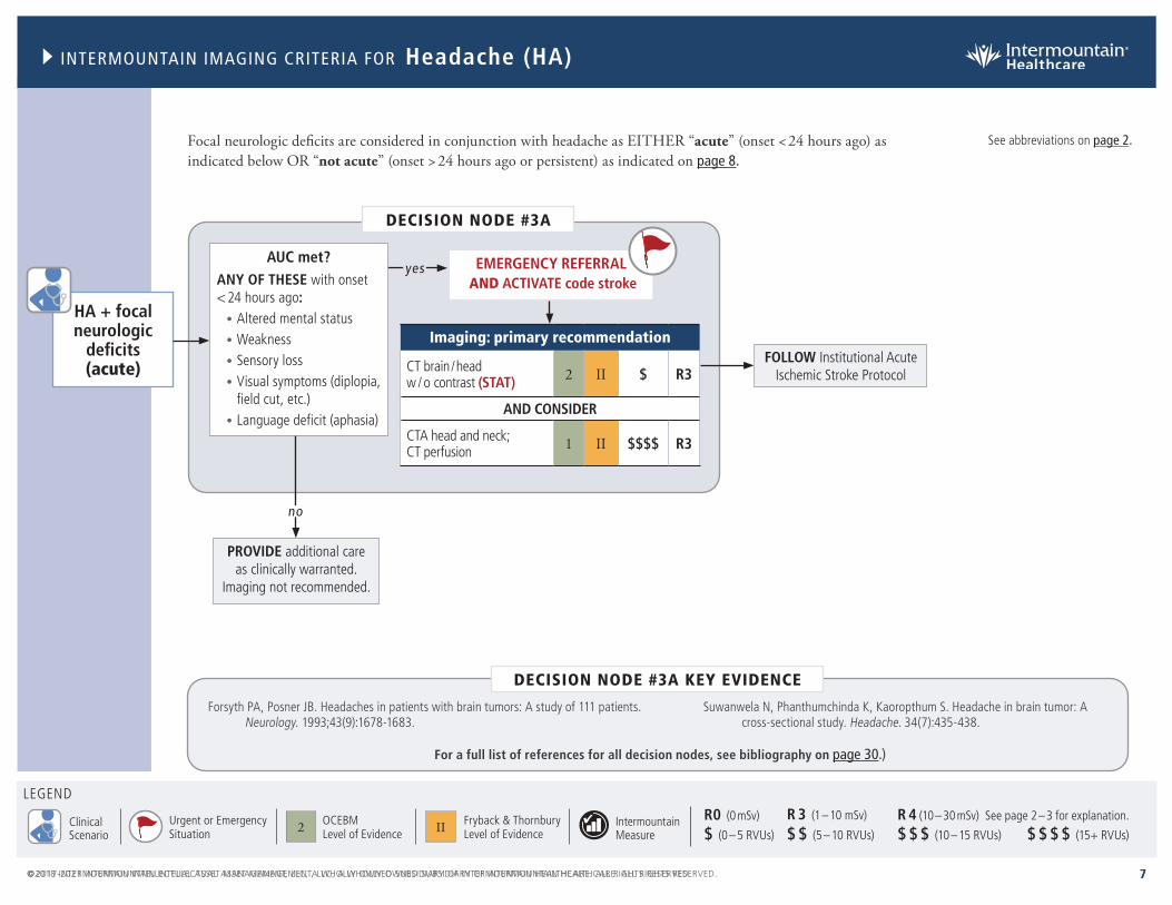
Forsyth PA, Posner JB. Headaches in patients with brain tumors: A study of 111 patients. Neurology. 1993;43(9):1678-1683.
Suwanwela N, Phanthumchinda K, Kaoropthum S. Headache in brain tumor: A cross-sectional study. Headache. 34(7):435-438.
DECISION NODE #3A KEY EVIDENCE
DECISION NODE #3A
HA + focal neurologic
deficits (acute)
no
yes EMERGENCY REFERRAL AND ACTIVATE code stroke
AUC met?ANY OF THESE with onset < 24 hours ago:
• Altered mental status• Weakness• Sensory loss• Visual symptoms (diplopia,
field cut, etc.)• Language deficit (aphasia)
Imaging: primary recommendation
CT brain / head w / o contrast (STAT) 2 II $ R3
AND CONSIDER
CTA head and neck; CT perfusion 1 II $$$$ R3
Focal neurologic deficits are considered in conjunction with headache as EITHER “acute” (onset < 24 hours ago) as indicated below OR “not acute” (onset > 24 hours ago or persistent) as indicated on page 8 .
See abbreviations on page 2 .
For a full list of references for all decision nodes, see bibliography on page 30 .)
PROVIDE additional care as clinically warranted.
Imaging not recommended.
FOLLOW Institutional Acute Ischemic Stroke Protocol
© 2017-2021 INTERMOUNTAIN INTELLECTUAL ASSET MANAGEMENT, LLC, A WHOLLY OWNED SUBSIDIARY OF INTERMOUNTAIN HEALTHCARE. ALL RIGHTS RESERVED. 7

©2018 INTERMOUNTAIN INTELLECTUAL ASSET MANAGEMENT, LLC, A WHOLLY OWNED SUBSIDIARY OF INTERMOUNTAIN HEALTHCARE. ALL RIGHTS RESERVED. 8
INTERMOUNTAIN IMAGING CRITERIA FOR Headache (HA)
LEGEND
OCEBMLevel of Evidence2 Fryback & Thornbury
Level of EvidenceII Intermountain Measure $ (0 – 5 RVUs) $ $ (5 – 10 RVUs) $ $ $ (10 – 15 RVUs) $ $ $ $ (15+ RVUs)
R0 (0 mSv) R 3 (1 – 10 mSv) R 4 (10 – 30 mSv) See page 2 – 3 for explanation.Urgent or EmergencySituation
ClinicalScenario
DECISION NODE #3B
HA + focal neurologic
deficits (NOT acute)
no
yes
yes
Significant positive result?
• Stroke• Hemorrhage• Mass lesion(s)• White matter changes
consistent with demyelination
no
CONSIDER EMERGENCY REFERRAL
AUC met?ANY OF THESE with onset > 24 hours ago or persistent:
• Altered mental status• Weakness• Sensory loss• Visual symptoms (diplopia,
field cut, etc.)• Language deficit (aphasia)
REFER to neurology
Imaging: primary recommendation
MRI brain w / o contrast 2 II $ R0
AND CONSIDER
MRA head and neck 4 II $$$$ R0
Imaging: alternative recommendation
CT brain / head w / o contrast 2 II $ R3
AND CONSIDER
CTA head and neck* 1 II $$$$ R3
CONSULT with neurology
See abbreviations on page 2 .
Forsyth PA, Posner JB. Headaches in patients with brain tumors: A study of 111 patients. Neurology. 1993;43(9):1678-1683.
Suwanwela N, Phanthumchinda K, Kaoropthum S. Headache in brain tumor: A cross-sectional study. Headache. 34(7):435-438.
DECISION NODE #3B KEY EVIDENCE
For a full list of references for all decision nodes, see bibliography on page 30 .)
PROVIDE additional care as clinically warranted.
Imaging not recommended.
* Include CT cervical spine reformats from CTA data set.
© 2017-2021 INTERMOUNTAIN INTELLECTUAL ASSET MANAGEMENT, LLC, A WHOLLY OWNED SUBSIDIARY OF INTERMOUNTAIN HEALTHCARE. ALL RIGHTS RESERVED. 8

©2018 INTERMOUNTAIN INTELLECTUAL ASSET MANAGEMENT, LLC, A WHOLLY OWNED SUBSIDIARY OF INTERMOUNTAIN HEALTHCARE. ALL RIGHTS RESERVED. 9
INTERMOUNTAIN IMAGING CRITERIA FOR Headache (HA)
LEGEND
OCEBMLevel of Evidence2 Fryback & Thornbury
Level of EvidenceII Intermountain Measure $ (0 – 5 RVUs) $ $ (5 – 10 RVUs) $ $ $ (10 – 15 RVUs) $ $ $ $ (15+ RVUs)
R0 (0 mSv) R 3 (1 – 10 mSv) R 4 (10 – 30 mSv) See page 2 – 3 for explanation.Urgent or EmergencySituation
ClinicalScenario
DECISION NODE #4
HA + elevated
bleeding risk*
no
yes
yes
Significant positive result?
Hemorrhage
no
CONSIDER EMERGENCY REFERRAL
for acute symptoms
AUC met (IF BOTH)?• New or worsening
headache• Patient currently taking
an anticoagulant
FOLLOW UP in outpatient setting
Imaging: primary recommendation
CT brain / head w / o contrast NA** NA** $ R3
CONSULT with neurology
and / or neurosurgery (EMERGENCY)
See abbreviations on page 2 .
* Risk factors include anticoagulant treatment, low platelets, liver dysfunction, etc.
** Based on expert opinion in the absence of literature-based evidence.
PROVIDE additional care as clinically warranted.
Imaging not recommended.
© 2017-2021 INTERMOUNTAIN INTELLECTUAL ASSET MANAGEMENT, LLC, A WHOLLY OWNED SUBSIDIARY OF INTERMOUNTAIN HEALTHCARE. ALL RIGHTS RESERVED. 9

©2018 INTERMOUNTAIN INTELLECTUAL ASSET MANAGEMENT, LLC, A WHOLLY OWNED SUBSIDIARY OF INTERMOUNTAIN HEALTHCARE. ALL RIGHTS RESERVED. 10
INTERMOUNTAIN IMAGING CRITERIA FOR Headache (HA)
LEGEND
OCEBMLevel of Evidence2 Fryback & Thornbury
Level of EvidenceII Intermountain Measure $ (0 – 5 RVUs) $ $ (5 – 10 RVUs) $ $ $ (10 – 15 RVUs) $ $ $ $ (15+ RVUs)
R0 (0 mSv) R 3 (1 – 10 mSv) R 4 (10 – 30 mSv) See page 2 – 3 for explanation.Urgent or EmergencySituation
ClinicalScenario
DECISION NODE #5
Suspected subarachnoid hemorrhage
no
yes
yes
Presence of both?
• Negative CT head < 6 hours from onset of symptoms
• Normal neuro exam
no
AUC met?• Sudden severe headache
ANY OF THESE:• Peak pain within 1 hour of onset• Age > 40 years• Neck pain or stiffness • Witnessed loss of consciousness• Sudden or severe headache
triggered by cough / sneeze, Valsalva, sex, or exercise / exertion
• Limited neck flexion on exam
PROVIDE additional care as clinically warranted
CONSIDER lumbar puncture vs additional imaging. USE shared decision making to select
the best modality for each patient after weighing the potential for false-positive imaging and the pros and cons associated with lumbar puncture.
Imaging: primary recommendation
CT brain / head w / o contrast (STAT) 1 II $ R3
No further imaging indicated.**
PROVIDE additional care as clinically
warranted.
Significant positive result?
• Hemorrhage• Stroke
yes
See abbreviations on page 2 .
* Based on expert opinion in the absence of literature-based evidence.
** Normal CT brain w / o contrast (3rd generation scanner) within 6 hours of symptom onset in an ED headache patient with a normal neuro exam can rule out non-traumatic subarachnoid hemorrhage.
CONSIDER EMERGENCY REFERRAL
for acute symptoms
no
AUC met?ANY OF THESE:• Negative CT head > 6
hours from onset of symptoms
• Abnormal neuro exam• High clinical suspicion
yesImaging: primary recommendation
CTA head and neck 2 II $ R3
Imaging: alternative recommendation
MRI brain w / o contrast 2 II $ R0
MRA brain / head w /o contrast NA* NA* $$ R0
MRA neck w /o contrast 1 II $$$$ R0
Significant positive result?• Hemorrhage• Stroke• Aneurysm
PROVIDE additional care as clinically warranted
yes
no
CONSULT with neurology and / or
neurosurgery (EMERGENCY)
no
CONSULT with neurology and / or
neurosurgery (EMERGENCY)
© 2017-2021 INTERMOUNTAIN INTELLECTUAL ASSET MANAGEMENT, LLC, A WHOLLY OWNED SUBSIDIARY OF INTERMOUNTAIN HEALTHCARE. ALL RIGHTS RESERVED. 10

©2018 INTERMOUNTAIN INTELLECTUAL ASSET MANAGEMENT, LLC, A WHOLLY OWNED SUBSIDIARY OF INTERMOUNTAIN HEALTHCARE. ALL RIGHTS RESERVED. 11
INTERMOUNTAIN IMAGING CRITERIA FOR Headache (HA)
LEGEND
OCEBMLevel of Evidence2 Fryback & Thornbury
Level of EvidenceII Intermountain Measure $ (0 – 5 RVUs) $ $ (5 – 10 RVUs) $ $ $ (10 – 15 RVUs) $ $ $ $ (15+ RVUs)
R0 (0 mSv) R 3 (1 – 10 mSv) R 4 (10 – 30 mSv) See page 2 – 3 for explanation.Urgent or EmergencySituation
ClinicalScenario
Dubosh NM, Bellolio MF, Rabinstein AA, Edlow JA. Sensitivity of early brain computed tomography to exclude aneurysmal subarachnoid hemorrhage: A systematic review and meta-analysis. Stroke. 2016;47(3):750-755.
Jayaraman MV, Mayo-Smith WW, Tung GA, et al. Detection of intracranial aneurysms: Multi-detector row CT angiography compared with DSA. Radiology. 2004;230(2):510-518.
Pascual J, Iglesias F, Oterino A, Vázquez-Barquero A, Berciano J. Cough, exertional, and sexual headaches: An analysis of 72 benign and symptomatic cases. Neurology. 1996;46(6):1520-1524.
DECISION NODE #5 KEY EVIDENCE
For a full list of references for all decision nodes, see bibliography on page 30 .)
© 2017-2021 INTERMOUNTAIN INTELLECTUAL ASSET MANAGEMENT, LLC, A WHOLLY OWNED SUBSIDIARY OF INTERMOUNTAIN HEALTHCARE. ALL RIGHTS RESERVED. 11

©2018 INTERMOUNTAIN INTELLECTUAL ASSET MANAGEMENT, LLC, A WHOLLY OWNED SUBSIDIARY OF INTERMOUNTAIN HEALTHCARE. ALL RIGHTS RESERVED. 12
INTERMOUNTAIN IMAGING CRITERIA FOR Headache (HA)
LEGEND
OCEBMLevel of Evidence2 Fryback & Thornbury
Level of EvidenceII Intermountain Measure $ (0 – 5 RVUs) $ $ (5 – 10 RVUs) $ $ $ (10 – 15 RVUs) $ $ $ $ (15+ RVUs)
R0 (0 mSv) R 3 (1 – 10 mSv) R 4 (10 – 30 mSv) See page 2 – 3 for explanation.Urgent or EmergencySituation
ClinicalScenario
Jin J, Zhou X, Liang X, et al. A study of patients with brain metastases as the initial manifestation of their systemic cancer in a Chinese population. J Neurooncol. 2011;103(3):649-655.
Yokoi K, Kamiya N, Matsuguma H, et al. Detection of brain metastasis in potentially operable non-small cell lung cancer: a comparison of CT and MRI. Chest. 1999;115(3):714-719.
DECISION NODE #6 KEY EVIDENCE
HA + known or suspected
cancer
AUC met?New headache
yes yes
no
DECISION NODE #6
Imaging: primary recommendation
MRI brain w / and w / o contrast 2 II $$ R0
Imaging: alternative recommendation
CT brain / head w / and w / o contrast 2 II $ R3
Significant positive result?
Intracranial malignancy
no
CONSIDER consult or referral to neurology
CONSULT with neurosurgery, neuro-oncology,
and / or neurology (URGENT)
CONSIDER EMERGENCY REFERRAL
for acute symptoms
See abbreviations on page 2 .
For a full list of references for all decision nodes, see bibliography on page 30 .)
PROVIDE additional care as clinically warranted.
Imaging not recommended.
© 2017-2021 INTERMOUNTAIN INTELLECTUAL ASSET MANAGEMENT, LLC, A WHOLLY OWNED SUBSIDIARY OF INTERMOUNTAIN HEALTHCARE. ALL RIGHTS RESERVED. 12

©2018 INTERMOUNTAIN INTELLECTUAL ASSET MANAGEMENT, LLC, A WHOLLY OWNED SUBSIDIARY OF INTERMOUNTAIN HEALTHCARE. ALL RIGHTS RESERVED. 13
INTERMOUNTAIN IMAGING CRITERIA FOR Headache (HA)
LEGEND
OCEBMLevel of Evidence2 Fryback & Thornbury
Level of EvidenceII Intermountain Measure $ (0 – 5 RVUs) $ $ (5 – 10 RVUs) $ $ $ (10 – 15 RVUs) $ $ $ $ (15+ RVUs)
R0 (0 mSv) R 3 (1 – 10 mSv) R 4 (10 – 30 mSv) See page 2 – 3 for explanation.Urgent or EmergencySituation
ClinicalScenario
DECISION NODE #7A
no
yes
AUC met (IF ANY)?• Visual symptoms• Increased pain when
lying down• Increased pain in the morning• Pain aggravated by Valsalva
yes
no
yes
Significant positive result?
Mass lesion(s)
Significant positive result?
Hydrocephalus
Imaging: primary recommendation
MRI brain w / and w / o contrast 4 I $$ R0
Imaging: alternative recommendation
CT brain / head w /o contrast NA** NA** $ R3
CONSULT with neurosurgery, neuro-oncology,
and / or neurology (URGENT)
CONSULT with neurosurgery and / or neurology
(URGENT)
CONSIDER EMERGENCY REFERRAL
for acute symptoms
Headache with elevated intracranial pressure (ICP) or papilledema are considered in terms of chronicity – EITHER acute or subacute / chronic (see below) – AND in terms of known or suspected hypercoagulable state (see page 14).
REFER to ophthalmology for papilledema AND CONSIDER neurology
referral for evaluation of idiopathic intracranial
hypertension
Suspected elevated
intracranial pressure or papilledema(no hyper-coagulable
state*)
See abbreviations on page 2 .
* Including dehydration.
** Based on expert opinion in the absence of literature-based evidence.
PROVIDE additional care as clinically warranted
no
© 2017-2021 INTERMOUNTAIN INTELLECTUAL ASSET MANAGEMENT, LLC, A WHOLLY OWNED SUBSIDIARY OF INTERMOUNTAIN HEALTHCARE. ALL RIGHTS RESERVED. 13

©2018 INTERMOUNTAIN INTELLECTUAL ASSET MANAGEMENT, LLC, A WHOLLY OWNED SUBSIDIARY OF INTERMOUNTAIN HEALTHCARE. ALL RIGHTS RESERVED. 14
INTERMOUNTAIN IMAGING CRITERIA FOR Headache (HA)
LEGEND
OCEBMLevel of Evidence2 Fryback & Thornbury
Level of EvidenceII Intermountain Measure $ (0 – 5 RVUs) $ $ (5 – 10 RVUs) $ $ $ (10 – 15 RVUs) $ $ $ $ (15+ RVUs)
R0 (0 mSv) R 3 (1 – 10 mSv) R 4 (10 – 30 mSv) See page 2 – 3 for explanation.Urgent or EmergencySituation
ClinicalScenario
yesyes
no
DECISION NODE #7B
Imaging: primary recommendation
MRI brain w / and w / o contrast 4 I $$ R0
AND
MR venogram brain / head w / and w / o contrast
4 I $$$ R0
Imaging: alternative recommendation
CT brain / head w / o contrast
NA** NA** $ R3
ANDCT venogram brain / head 5 I $$ R3
Significant positive result?
Venous sinus thrombosis
no
CONSULT with neurology (EMERGENCY)
HA + suspected elevated
intracranial pressure or papilledema(known or suspected
hyper-coagulable
state*)
AUC met (IF ANY)?• Visual symptoms• Increased pain when
lying down• Increased pain in
the morning• Pain aggravated by Valsalva
REFER to ophthalmology for papilledema, AND CONSIDER neurology
referral
See abbreviations on page 2 .
Chiewvit P, Piyapittayanan S, Poungvarin N. Cerebral venous thrombosis: Diagnosis dilemma. Neurol Int. 2011;3(3):e13.
Kumral E, Polat F, Uzunköprü C, Callı C, Kitis Ö. The clinical spectrum of intracerebral hematoma, hemorrhagic infarct, non-hemorrhagic infarct, and non-lesional venous stroke in patients with cerebral sinus-venous thrombosis. Eur J Neurol. 2012;19(4):537-543.
Zafar A, Ali Z. Pattern of magnetic resonance imaging and magnetic resonance venography changes in cerebral venous sinus thrombosis. J Ayub Med Coll Abbottabad. 24(1):63-67.
DECISION NODE #7 KEY EVIDENCE
For a full list of references for all decision nodes, see bibliography on page 30 .)
* Including dehydration.
** Based on expert opinion in the absence of literature-based evidence.
PROVIDE additional care as clinically warranted.
Imaging not recommended.
© 2017-2021 INTERMOUNTAIN INTELLECTUAL ASSET MANAGEMENT, LLC, A WHOLLY OWNED SUBSIDIARY OF INTERMOUNTAIN HEALTHCARE. ALL RIGHTS RESERVED. 14

©2018 INTERMOUNTAIN INTELLECTUAL ASSET MANAGEMENT, LLC, A WHOLLY OWNED SUBSIDIARY OF INTERMOUNTAIN HEALTHCARE. ALL RIGHTS RESERVED. 15
INTERMOUNTAIN IMAGING CRITERIA FOR Headache (HA)
LEGEND
OCEBMLevel of Evidence2 Fryback & Thornbury
Level of EvidenceII Intermountain Measure $ (0 – 5 RVUs) $ $ (5 – 10 RVUs) $ $ $ (10 – 15 RVUs) $ $ $ $ (15+ RVUs)
R0 (0 mSv) R 3 (1 – 10 mSv) R 4 (10 – 30 mSv) See page 2 – 3 for explanation.Urgent or EmergencySituation
ClinicalScenario
Hasbun R, Abrahams J, Jekel J, Quagliarello VJ. Computed tomography of the head before lumbar puncture in adults with suspected meningitis. N Engl J Med. 2001;345(24):1727-1733.
Zhang J, Hu X, Hu X, et al. Clinical features, Outcomes and molecular profiles of drug resistance in tuberculous meningitis in non-HIV patients. Sci Rep. 2016;6(1):19072.
DECISION NODE #8 KEY EVIDENCE
DECISION NODE #8
no
yesAUC met (IF EITHER)?• Fever• Nuchal rigidity
yes
no
yes
Significant positive result?
Findings suggestive of:• Meningitis• Encephalitis
Significant positive result?
Abscess
CONSULT with neurosurgery
if brain abscess (EMERGENCY)
Imaging: primary recommendation
CT brain / head w / o contrast*
2 II $ R3
Imaging: alternative recommendation
MRI brain w / and w / o contrast 2 II $$ R0
HA + suspected meningitis
CONSIDER EMERGENCY REFERRAL
for acute symptomsCONSIDER referral
to medicine, ICU, infectious disease, and / or neurology
PERFORM lumbar puncture to fully evaluate
for meningitis
* CT head is not required prior to every lumbar puncture, but is recommended if have clinical suspicion of elevated intracranial pressure or altered mental status.
See abbreviations on page 2 .
For a full list of references for all decision nodes, see bibliography on page 30 .)
PROVIDE additional care as clinically warranted.
Imaging not recommended.
no
© 2017-2021 INTERMOUNTAIN INTELLECTUAL ASSET MANAGEMENT, LLC, A WHOLLY OWNED SUBSIDIARY OF INTERMOUNTAIN HEALTHCARE. ALL RIGHTS RESERVED. 15

©2018 INTERMOUNTAIN INTELLECTUAL ASSET MANAGEMENT, LLC, A WHOLLY OWNED SUBSIDIARY OF INTERMOUNTAIN HEALTHCARE. ALL RIGHTS RESERVED. 16
INTERMOUNTAIN IMAGING CRITERIA FOR Headache (HA)
LEGEND
OCEBMLevel of Evidence2 Fryback & Thornbury
Level of EvidenceII Intermountain Measure $ (0 – 5 RVUs) $ $ (5 – 10 RVUs) $ $ $ (10 – 15 RVUs) $ $ $ $ (15+ RVUs)
R0 (0 mSv) R 3 (1 – 10 mSv) R 4 (10 – 30 mSv) See page 2 – 3 for explanation.Urgent or EmergencySituation
ClinicalScenario
no
REFER to the Concussion CPM and / or Neck Pain CPM or other system-
wide protocol
DECISION NODE #9A
Imaging: primary recommendation
CT brain / head w / o contrast NA** NA** $ R3
AND
CTA head and neck* 1 II $$$$ R3
SEE Decision Node #9B on page 17
yesAUC met?• CTA completed
OR • Clinical suspicion with
negative CTA
no
Imaging: primary recommendation
MRI brain w / o contrast 5 II $ R0
MRA head and neck 4 II $$$$ R0
no
yes
Significant positive result?
Stroke
yes
Headache in conjunction with suspected cervical artery dissection is considered EITHER "with acute trauma" (see below) OR "without acute trauma" (see page 17).
HA + suspected
cervical artery dissection
(WITH acute trauma)
See abbreviations on page 2 .
* Include CT cervical spine reformats from CTA data set.
** Based on expert opinion in the absence of literature-based evidence.
no
AUC met (IF ANY)?• Facial or neck pain • Neurologic deficit(s)
and / or stroke • Horner syndrome:
Miosis, ptosis, anhidrosis
CONSIDER EMERGENCY REFERRAL for acute symptoms
yes
CONSULT with neurology and / or
interventional radiology
CONSULT with neurosurgery and / or
orthopedic spine
yes
Significant positive result?
Intracranial and / or cervical artery dissection
no
Significant positive result?
Cervical spine injury
© 2017-2021 INTERMOUNTAIN INTELLECTUAL ASSET MANAGEMENT, LLC, A WHOLLY OWNED SUBSIDIARY OF INTERMOUNTAIN HEALTHCARE. ALL RIGHTS RESERVED. 16

©2018 INTERMOUNTAIN INTELLECTUAL ASSET MANAGEMENT, LLC, A WHOLLY OWNED SUBSIDIARY OF INTERMOUNTAIN HEALTHCARE. ALL RIGHTS RESERVED. 17
INTERMOUNTAIN IMAGING CRITERIA FOR Headache (HA)
LEGEND
OCEBMLevel of Evidence2 Fryback & Thornbury
Level of EvidenceII Intermountain Measure $ (0 – 5 RVUs) $ $ (5 – 10 RVUs) $ $ $ (10 – 15 RVUs) $ $ $ $ (15+ RVUs)
R0 (0 mSv) R 3 (1 – 10 mSv) R 4 (10 – 30 mSv) See page 2 – 3 for explanation.Urgent or EmergencySituation
ClinicalScenario
DECISION NODE #9B
Consider EMERGENCY referral
Imaging: primary recommendation
CTA head and neck* 1 II $$$$ R3
yes
yes
no
no
no
CONSULT with neurology and / or
interventional radiology
REFER to the Concussion CPM and / or Neck Pain CPM or other
system-wide protocol
Significant positive result?
Stroke
AUC met (IF ANY)?• CTA completed
• Clinical suspicion with negative CTA
yes
AUC met (IF ANY)?• Neurologic deficit(s)
and / or stroke• Horner syndrome: Miosis,
ptosis, anhidrosis
HA with
neck / facial pain +
suspected cervical artery
dissection(WITHOUT
acute trauma)
SEE Decision Node #10 on page 19
See abbreviations on page 2 .
*Include CT cervical spine reformats from CTA data set.
yes
Significant positive result?
Intracranial and / or cervical artery dissection
CONSULT with neurology and /or
interventional radiology
no
Imaging: primary recommendation
MRI brain w / o contrast 5 II $ R0
MRA head and neck 4 II $$$$ R0
© 2017-2021 INTERMOUNTAIN INTELLECTUAL ASSET MANAGEMENT, LLC, A WHOLLY OWNED SUBSIDIARY OF INTERMOUNTAIN HEALTHCARE. ALL RIGHTS RESERVED. 17
©2018 INTERMOUNTAIN INTELLECTUAL ASSET MANAGEMENT, LLC, A WHOLLY OWNED SUBSIDIARY OF INTERMOUNTAIN HEALTHCARE. ALL RIGHTS RESERVED. 18
INTERMOUNTAIN IMAGING CRITERIA FOR Headache (HA)
LEGEND
OCEBMLevel of Evidence2 Fryback & Thornbury
Level of EvidenceII Intermountain Measure $ (0 – 5 RVUs) $ $ (5 – 10 RVUs) $ $ $ (10 – 15 RVUs) $ $ $ $ (15+ RVUs)
R0 (0 mSv) R 3 (1 – 10 mSv) R 4 (10 – 30 mSv) See page 2 – 3 for explanation.Urgent or EmergencySituation
ClinicalScenario
Bromberg WJ, Collier BC, Diebel LN, et al. Blunt cerebrovascular injury practice management guidelines: The Eastern Association for the Surgery of Trauma. J Trauma. 2010;68(2):471-477.
Manabe H, Yonezawa K, Kato T, Toyama K, Haraguchi K, Ito T. Incidence of intracranial arterial dissection in non-emergency outpatients complaining of headache: Preliminary investigation with MRI / MRA examinations. Acta Neurochir Suppl. 2010;107:41-44.
Patterson BO, Holt PJ, Cleanthis M, et al. Imaging vascular trauma. Br J Surg. 2012;99(4):494-505.
DECISION NODE #9 KEY EVIDENCE
For a full list of references for all decision nodes, see bibliography on page 30 .)
© 2017-2021 INTERMOUNTAIN INTELLECTUAL ASSET MANAGEMENT, LLC, A WHOLLY OWNED SUBSIDIARY OF INTERMOUNTAIN HEALTHCARE. ALL RIGHTS RESERVED. 18

©2018 INTERMOUNTAIN INTELLECTUAL ASSET MANAGEMENT, LLC, A WHOLLY OWNED SUBSIDIARY OF INTERMOUNTAIN HEALTHCARE. ALL RIGHTS RESERVED. 19
INTERMOUNTAIN IMAGING CRITERIA FOR Headache (HA)
LEGEND
OCEBMLevel of Evidence2 Fryback & Thornbury
Level of EvidenceII Intermountain Measure $ (0 – 5 RVUs) $ $ (5 – 10 RVUs) $ $ $ (10 – 15 RVUs) $ $ $ $ (15+ RVUs)
R0 (0 mSv) R 3 (1 – 10 mSv) R 4 (10 – 30 mSv) See page 2 – 3 for explanation.Urgent or EmergencySituation
ClinicalScenario
DECISION NODE #10
yes
yes
no
yes
no
no
CONSULT with neurology and/or
neurosurgery
REFER to the Concussion CPM and / or Neck Pain CPM or other
system-wide protocol
Significant positive result?
Cranial or spinal injury
AUC met?• CT brain / head or CT
cervical spine completed (WITH ANY OF THE
FOLLOWING):• Suspected brain contusion• Suspicion for occult fracture
or ligamentous injury• Known spondyloarthropathy
(AS or DISH)• Persistent neurologic deficit
yesAUC met (IF ANY)?
Acute or subacute head and / or neck trauma
HA + head and / or neck trauma
(without suspicion of cervical
artery trauma)
Significant positive result?
Cranial or spinal injuryImaging: primary recommendation
CT brain / head w / o contrast 3 II $ R3
AND / OR
CT cervical spine w / o contrast 3 II $$ R3
See abbreviations on page 2 .
Imaging: primary recommendation
MRI brain w / o contrast 4 II $ R0
AND / OR
MRI cervical spine w / o contrast (trauma protocol) 4 II $$ R0
CONSIDER EMERGENCY REFERRAL for acute symptoms
no
© 2017-2021 INTERMOUNTAIN INTELLECTUAL ASSET MANAGEMENT, LLC, A WHOLLY OWNED SUBSIDIARY OF INTERMOUNTAIN HEALTHCARE. ALL RIGHTS RESERVED. 19

©2018 INTERMOUNTAIN INTELLECTUAL ASSET MANAGEMENT, LLC, A WHOLLY OWNED SUBSIDIARY OF INTERMOUNTAIN HEALTHCARE. ALL RIGHTS RESERVED. 20
INTERMOUNTAIN IMAGING CRITERIA FOR Headache (HA)
LEGEND
OCEBMLevel of Evidence2 Fryback & Thornbury
Level of EvidenceII Intermountain Measure $ (0 – 5 RVUs) $ $ (5 – 10 RVUs) $ $ $ (10 – 15 RVUs) $ $ $ $ (15+ RVUs)
R0 (0 mSv) R 3 (1 – 10 mSv) R 4 (10 – 30 mSv) See page 2 – 3 for explanation.Urgent or EmergencySituation
ClinicalScenario
Abdul Rahman YS, Al Den ASS, Maull KI. Prospective study of validity of neurologic signs in predicting positive cranial computed tomography following minor head trauma. Prehosp Disaster Med. 25(1):59-62.
Schwedt TJ, Chong CD, Peplinski J, Ross K, Berisha V. Persistent post-traumatic headache vs. migraine: An MRI study demonstrating differences in brain structure. J Headache Pain. 2017;18(1):87.
DECISION NODE #10 KEY EVIDENCE
For a full list of references for all decision nodes, see bibliography on page 30 .)
© 2017-2021 INTERMOUNTAIN INTELLECTUAL ASSET MANAGEMENT, LLC, A WHOLLY OWNED SUBSIDIARY OF INTERMOUNTAIN HEALTHCARE. ALL RIGHTS RESERVED. 20

©2018 INTERMOUNTAIN INTELLECTUAL ASSET MANAGEMENT, LLC, A WHOLLY OWNED SUBSIDIARY OF INTERMOUNTAIN HEALTHCARE. ALL RIGHTS RESERVED. 21
INTERMOUNTAIN IMAGING CRITERIA FOR Headache (HA)
LEGEND
OCEBMLevel of Evidence2 Fryback & Thornbury
Level of EvidenceII Intermountain Measure $ (0 – 5 RVUs) $ $ (5 – 10 RVUs) $ $ $ (10 – 15 RVUs) $ $ $ $ (15+ RVUs)
R0 (0 mSv) R 3 (1 – 10 mSv) R 4 (10 – 30 mSv) See page 2 – 3 for explanation.Urgent or EmergencySituation
ClinicalScenario
HA + suspicion for giant
cell / temporal arteritis
AUC met (IF ALL)?• New or progressive
headache• Age > 50• Visual symptoms
yes yes
no
DECISION NODE #11
Significant positive result?
Secondary cause of HA identified
no
PROVIDE additional care as clinically
warranted. Imaging not recommended
CONSULT with neurology
FOLLOW UP in outpatient setting, AND CONSIDER
referral to neurology and / or rheumatology
Imaging: primary recommendation
MRI brain w / o contrast 2 II $ R0
Imaging: alternative recommendation
CT brain / head w / o contrast
NA* NA* $ R3
Bley TA, Reinhard M, Hauenstein C, et al. Comparison of duplex sonography and high-resolution magnetic resonance imaging in the diagnosis of giant cell (temporal) arteritis. Arthritis Rheum. 2008;58(8):2574-2578.
Klink T, Geiger J, Both M, et al. Giant cell arteritis: diagnostic accuracy of MR imaging of superficial cranial arteries in initial diagnosis: Results from a multicenter trial. Radiology. 2014;273(3):844-852.
DECISION NODE #11 KEY EVIDENCE
See abbreviations on page 2 .
For a full list of references for all decision nodes, see bibliography on page 30 .)
* Based on expert opinion in the absence of literature-based evidence.
© 2017-2021 INTERMOUNTAIN INTELLECTUAL ASSET MANAGEMENT, LLC, A WHOLLY OWNED SUBSIDIARY OF INTERMOUNTAIN HEALTHCARE. ALL RIGHTS RESERVED. 21

©2018 INTERMOUNTAIN INTELLECTUAL ASSET MANAGEMENT, LLC, A WHOLLY OWNED SUBSIDIARY OF INTERMOUNTAIN HEALTHCARE. ALL RIGHTS RESERVED. 22
INTERMOUNTAIN IMAGING CRITERIA FOR Headache (HA)
LEGEND
OCEBMLevel of Evidence2 Fryback & Thornbury
Level of EvidenceII Intermountain Measure $ (0 – 5 RVUs) $ $ (5 – 10 RVUs) $ $ $ (10 – 15 RVUs) $ $ $ $ (15+ RVUs)
R0 (0 mSv) R 3 (1 – 10 mSv) R 4 (10 – 30 mSv) See page 2 – 3 for explanation.Urgent or EmergencySituation
ClinicalScenario
DECISION NODE #12
HA + trigeminal
distribution*
no
yes
AUC met?Positive neurologic symptoms (including but not limited to altered sensation)**
CONSULT with neurology and / or
neurosurgery
Imaging: primary recommendation
MRI brain w / and w / o contrast (trigeminal protocol)
2 II $$ R0
Imaging: primary recommendation
CT brain / head w / and w / o contrast***
2 II S R3
Antonini G, Di Pasquale A, Cruccu G, et al. Magnetic resonance imaging contribution for diagnosing symptomatic neurovascular contact in classical trigeminal neuralgia: A blinded case-control study and meta-analysis. Pain. 2014;155(8):1464-1471.
DECISION NODE #12 KEY EVIDENCE
yes
yes
Significant positive result?
Secondary cause of trigeminal pain identified
Significant positive result?
Intracranial complication of sinusitis / mastoiditis
FOLLOW UP in outpatient setting AND CONSIDER referral to neurology for medically refractory TN
CONSULT with ENT / neurosurgery
no
See abbreviations on page 2 .
For a full list of references for all decision nodes, see bibliography on page 30 .)
* V1: Orbital, periorbital, frontal / ethmoid sinuses.
** Imaging not generally needed in patients with TN symptoms and a normal exam. Consider alternative diagnoses (sinusitis, mastoiditis, and / or dental pathology).
*** MRI rather than CT should be performed for HA, except in emergency situations or when MRI is contra-indicated.
PROVIDE additional care as clinically
warranted. Imaging not recommended
no
© 2017-2021 INTERMOUNTAIN INTELLECTUAL ASSET MANAGEMENT, LLC, A WHOLLY OWNED SUBSIDIARY OF INTERMOUNTAIN HEALTHCARE. ALL RIGHTS RESERVED. 22

© 2017-2021 INTERMOUNTAIN INTELLECTUAL ASSET MANAGEMENT, LLC, A WHOLLY OWNED SUBSIDIARY OF INTERMOUNTAIN HEALTHCARE. ALL RIGHTS RESERVED. 23
INTERMOUNTAIN IMAGING CRITERIA FOR Headache (HA)
TABLE 1. MRI brain WITHOUT CONTRAST appropriate use indications(PRIMARY recommendation) (ALTERNATIVE recommendation)
� Existing HA disorder + clinical progression: � Significant increase in headache frequency, severity, or duration
� Chronic HA + refractory / debilitating pain: � Headache persistent for at least 3 months
� HA + focal neurologic deficits (NOT acute):
ANY OF THESE: with onset > 24 hours ago or persistent:
� Altered mental status
� Weakness
� Sensory loss
� Visual symptoms (diplopia, field cut, etc.)
� Language deficit (aphasia)
� HA + suspected cervical artery dissection (WITH ACUTE trauma) (IF ANY):
� Facial or neck pain � Neurologic deficit(s) and / or stroke � Horner syndrome: Miosis, ptosis, anhidrosis
AND EITHER:
� CTA completed
OR
� Clinical suspicion with negative CTA
� HA + head and / or neck trauma (WITHOUT suspicion of cervical artery trauma):
� CT brain / head or CT cervical spine completed
(WITH ANY OF THE FOLLOWING):
� Suspected brain contusion
� Suspicion for occult fracture or ligamentous injury
� Known spondyloarthropathy (AS or DISH)
� Persistent neurologic deficit
� HA + suspicion for giant cell / temporal arteritis (IF ALL):
� New or progressive headache
� Age > 50 � Visual symptoms
� HA w/ neck / facial pain + suspected cervical artery dissection (WITHOUT acute trauma):
� Neurologic deficit (s) and / or stroke � Horner syndrome: Miosis, ptosis, anhidrosis
AND EITHER:
� CTA completedOR
� Clinical suspicion with negative CTA
� Suspected subarachnoid hemorrhage (IF ANY): � Negative CT head > 6 hours from onset of symptoms
� Abnormal neuro exam
� High clinical suspicion
The provider must check BOTH:
1. The box next to the relevant clinical scenario
2. EACH AUC box that applies to the patient’s situation
See abbreviations on page 2 . POINT-OF-ORDER CHECKLISTS
© 2017-2021 INTERMOUNTAIN INTELLECTUAL ASSET MANAGEMENT, LLC, A WHOLLY OWNED SUBSIDIARY OF INTERMOUNTAIN HEALTHCARE. ALL RIGHTS RESERVED. 23

© 2017-2021 INTERMOUNTAIN INTELLECTUAL ASSET MANAGEMENT, LLC, A WHOLLY OWNED SUBSIDIARY OF INTERMOUNTAIN HEALTHCARE. ALL RIGHTS RESERVED. 24
INTERMOUNTAIN IMAGING CRITERIA FOR Headache (HA)
POINT-OF-ORDER CHECKLISTS, CONTINUED See abbreviations on page 2 .
TABLE 2. MRA head and neck appropriate use indications
(PRIMARY recommendation)
� HA + focal neurologic deficits (NOT acute) (ANY OF THESE with onset > 24 hours ago or persistent):
� Altered mental status
� Weakness
� Sensory loss
� Visual symptoms (diplopia, field cut, etc.)
� Language deficit (aphasia)
� HA + suspected cervical artery dissection (WITH acute trauma) (IF ANY):
� Facial or neck pain
� Neurologic deficit(s) and / or stroke
� Horner syndrome: Miosis, ptosis, anhidrosisAND EITHER:
� CTA completed
OR
� Clinical suspicion with negative CTA
� HA + suspected cervical artery dissection (WITHOUT acute trauma) (IF ANY):
� Neurologic deficit(s) and / or stroke
� Horner syndrome: Miosis, ptosis, anhidrosisAND EITHER:
� CTA completedOR
� Clinical suspicion with negative CTA
TABLE 3. MRA brain / head WITHOUT CONTRAST appropriate use indications
(ALTERNATIVE recommendation)
� Suspected subarachnoid hemorrhage (IF ANY): � Negative CT head > 6 hours from onset of symptoms
� Abnormal neuro exam
� High clinical suspicion
TABLE 4. MRA neck WITHOUT CONTRAST appropriate use indications
(ALTERNATIVE recommendation)
� Suspected subarachnoid hemorrhage (IF ANY): � Negative CT head > 6 hours from onset of symptoms
� Abnormal neuro exam
� High clinical suspicion
© 2017-2021 INTERMOUNTAIN INTELLECTUAL ASSET MANAGEMENT, LLC, A WHOLLY OWNED SUBSIDIARY OF INTERMOUNTAIN HEALTHCARE. ALL RIGHTS RESERVED. 24

© 2017-2021 INTERMOUNTAIN INTELLECTUAL ASSET MANAGEMENT, LLC, A WHOLLY OWNED SUBSIDIARY OF INTERMOUNTAIN HEALTHCARE. ALL RIGHTS RESERVED. 25
INTERMOUNTAIN IMAGING CRITERIA FOR Headache (HA)
See abbreviations on page 2 .
TABLE 6. MR venogram brain / head WITH AND WITHOUT CONTRAST appropriate use indications
(PRIMARY recommendation)
� HA + suspected elevated ICP or papilledema (known or suspected hypercoagulable state)* (IF ANY):
� Visual symptoms
� Increased pain when lying down
� Increased pain in the morning
� Pain aggravated by Valsalva
TABLE 7. MRI cervical spine WITHOUT CONTRAST (trauma protocol) appropriate use indications
(PRIMARY recommendation)
� HA + head and / or neck trauma (WITHOUT suspicion of cervical artery trauma):
� CT brain / head or CT cervical spine completed (WITH ANY OF THE FOLLOWING):
� Suspected brain contusion � Suspicion for occult fracture or ligamentous injury � Known spondyloarthropathy (AS or DISH) � Persistent neurologic deficit
TABLE 5. MRI brain WITH AND WITHOUT CONTRAST appropriate use indications
(PRIMARY recommendation) (ALTERNATIVE recommendation)
� HA + known or suspected cancer: � New headache
� Suspected elevated ICP or papilledema (NO hypercoagulable state)* (IF ANY):
� Visual symptoms
� Increased pain when lying down
� Increased pain in the morning
� Pain aggravated by Valsalva
� HA + suspected elevated ICP or papilledema (known or suspected hypercoagulable state)* (IF ANY):
� Visual symptoms � Increased pain when lying down � Increased pain in the morning � Pain aggravated by Valsalva
TRIGEMINAL PROTOCOL � HA + trigeminal distribution**:
� Positive neurologic symptoms (including but not limited to altered sensation***)
� HA + suspected meningitis (IF EITHER): � Fever
� Nuchal rigidity
* Including dehydration.** V1: Orbital, periorbital, frontal / ethmoid sinuses; V2: Cheek, maxillary sinus, upper teeth; V3: Jaw, lower teeth. *** Imaging not generally needed in patients with TN symptoms and a normal exam. Consider alternative diagnoses (sinusitis, mastoiditis,
and / or dental pathology).
POINT-OF-ORDER CHECKLISTS, CONTINUED
© 2017-2021 INTERMOUNTAIN INTELLECTUAL ASSET MANAGEMENT, LLC, A WHOLLY OWNED SUBSIDIARY OF INTERMOUNTAIN HEALTHCARE. ALL RIGHTS RESERVED. 25

© 2017-2021 INTERMOUNTAIN INTELLECTUAL ASSET MANAGEMENT, LLC, A WHOLLY OWNED SUBSIDIARY OF INTERMOUNTAIN HEALTHCARE. ALL RIGHTS RESERVED. 26
INTERMOUNTAIN IMAGING CRITERIA FOR Headache (HA)
See abbreviations on page 2 .
TABLE 8. CT brain / head WITHOUT CONTRAST appropriate use indications*(PRIMARY recommendation) (ALTERNATIVE recommendation)
� HA + elevated bleeding risk** (IF BOTH):
� New or worsening headache � Patient currently taking an anticoagulant
� HA + suspected meningitis*** (IF EITHER):
� Fever � Nuchal rigidity
� HA + suspected cervical artery dissection (WITH ACUTE trauma) (IF ANY):
� Facial or neck pain � Neurologic deficit(s) and / or stroke � Horner syndrome: Miosis, ptosis, anhidrosis
� HA+ head and / or neck trauma (WITHOUT suspicion of cervical artery trauma):
� Acute or subacute head and / or neck trauma
STAT
� HA + focal neurologic deficits (ACUTE): ANY OF THESE (with onset < 24 hours ago):
� Altered mental status � Weakness � Sensory loss � Visual symptoms (diplopia, field cut, etc.)
� Language deficit (aphasia)
� Suspected subarachnoid hemorrhage:
� Sudden severe headache
AND ANY OF THESE:
� Peak pain within 1 hour of onset
� Age > 40
� Neck pain or stiffness
� Witnessed loss of consciousness
� Sudden or severe headache triggered by: cough / sneeze, Valsalva, sex, or exercise / exertion
� Limited neck flexion on exam
� Existing HA disorder + clinical progression*:
� Significant increase in headache frequency, severity, or duration
� Chronic HA + refractory / debilitating pain:
� Headache persistent at least 3 months
� HA + focal neurologic deficits (NOT acute): ANY OF THESE: with onset > 24 hours ago or persistent:
� Altered mental status � Weakness � Sensory loss � Visual symptoms (diplopia, field cut, etc.) � Language deficit (aphasia)
� Suspected elevated ICP or papilledema (NO hypercoagulable state)**** (IF ANY):
� Visual symptoms � Increased pain when lying down � Increased pain in the morning � Pain aggravated by Valsalva
� HA + suspected elevated ICP or papilledema (known or suspected hypercoagulable state)**** (IF ANY):
� Visual symptoms � Increased pain when lying down � Increased pain in the morning � Pain aggravated by Valsalva
� HA + suspicion for giant cell / temporal arteritis (IF ALL):
� New or progressive headache
� Age > 50
� Visual symptoms
* MRI rather than CT should be performed for headache, except in emergency situations or when MRI is contraindicated.** Risk factors include anticoagulant treatment, low platelets, liver dysfunction. etc. *** CT head is not required prior to every lumbar puncture, but is recommended if have clinical suspicion of elevated intracranial pressure or altered mental status. **** Including dehydration.
POINT-OF-ORDER CHECKLISTS, CONTINUED
© 2017-2021 INTERMOUNTAIN INTELLECTUAL ASSET MANAGEMENT, LLC, A WHOLLY OWNED SUBSIDIARY OF INTERMOUNTAIN HEALTHCARE. ALL RIGHTS RESERVED. 26

© 2017-2021 INTERMOUNTAIN INTELLECTUAL ASSET MANAGEMENT, LLC, A WHOLLY OWNED SUBSIDIARY OF INTERMOUNTAIN HEALTHCARE. ALL RIGHTS RESERVED. 27
INTERMOUNTAIN IMAGING CRITERIA FOR Headache (HA)
See abbreviations on page 2 .
TABLE 11. CT brain / head WITH AND WITHOUT CONTRAST appropriate use indications*
(ALTERNATIVE recommendation)
� HA + known or suspected cancer � New headache
TRIGEMINAL PROTOCOL � HA + trigeminal distribution**:
� Positive neurologic symptoms (including but not limited to altered sensation***)
* MRI rather than CT should be performed for headache, except in emergency situations or when MRI is contraindicated.
** V1: Orbital, periorbital, frontal / ethmoid sinuses; V2: Cheek, max-illary sinus, upper teeth; V3: Jaw, lower teeth.
*** Imaging not generally needed in patients with TN symptoms and a normal exam. Consider alternative diagnoses (sinusitis, mastoiditis, and / or dental pathology).
TABLE 10. CTA head and neck appropriate use indications
(PRIMARY recommendation)
� Suspected subarachnoid hemorrhage (IF ANY): � Negative CT head > 6 hours from onset of symptoms � Abnormal neuro exam
� High clinical suspicion
� HA + suspected cervical artery dissection (WITH ACUTE trauma)* (IF ANY):
� Facial or neck pain � Neurologic deficit(s) and / or stroke � Horner syndrome: Miosis, ptosis, anhidrosis
� HA + suspected cervical artery dissection (WITHOUT ACUTE trauma)* (IF ANY):
� Neurologic deficit(s) and / or stroke � Horner syndrome: Miosis, ptosis, anhidrosis
(ALTERNATIVE recommendation)
� HA + focal neurologic deficits (NOT acute): ANY OF THESE (with onset >24 hours ago or persistent):
� Altered mental status � Weakness � Sensory loss � Visual symptoms (diplopia, field cut, etc.) � Language deficit (aphasia)
TABLE 9. CTA head and neck, CT perfusion appropriate use indications
(PRIMARY recommendation)
� HA +focal neurologic deficits (ACUTE): ANY OF THESE (with onset < 24 hours ago):
� Altered mental status � Weakness � Sensory loss � Visual symptoms (diplopia, field cut, etc.) � Language deficit (aphasia)
* Include CT cervical spine reformats from CTA data set.
POINT-OF-ORDER CHECKLISTS, CONTINUED
© 2017-2021 INTERMOUNTAIN INTELLECTUAL ASSET MANAGEMENT, LLC, A WHOLLY OWNED SUBSIDIARY OF INTERMOUNTAIN HEALTHCARE. ALL RIGHTS RESERVED. 27

© 2017-2021 INTERMOUNTAIN INTELLECTUAL ASSET MANAGEMENT, LLC, A WHOLLY OWNED SUBSIDIARY OF INTERMOUNTAIN HEALTHCARE. ALL RIGHTS RESERVED. 28
INTERMOUNTAIN IMAGING CRITERIA FOR Headache (HA)
TABLE 12. CT venogram brain / head appropriate use indications
(ALTERNATIVE recommendation)
� HA + suspected elevated ICP or papilledema (known or suspected hypercoagulable state)* (IF ANY):
� Visual symptoms
� Increased pain when lying down
� Increased pain in the morning
� Pain aggravated by Valsalva
TABLE 13. CT cervical spine WITHOUT CONTRAST appropriate use indications
(PRIMARY recommendation)
� HA + head and / or neck trauma (WITHOUT suspicion of cervical artery trauma):
� Acute or subacute head and / or neck trauma
POINT-OF-ORDER CHECKLISTS, CONTINUED See abbreviations on page 2 .
* Including dehydration.
© 2017-2021 INTERMOUNTAIN INTELLECTUAL ASSET MANAGEMENT, LLC, A WHOLLY OWNED SUBSIDIARY OF INTERMOUNTAIN HEALTHCARE. ALL RIGHTS RESERVED. 28

© 2017-2021 INTERMOUNTAIN INTELLECTUAL ASSET MANAGEMENT, LLC, A WHOLLY OWNED SUBSIDIARY OF INTERMOUNTAIN HEALTHCARE. ALL RIGHTS RESERVED. 29
INTERMOUNTAIN IMAGING CRITERIA FOR Headache (HA)
Fact sheets:
• Computed Tomography (CT) Scan (English) / (Spanish) (English) / (
• Spine Injury and Orthotic Braces (English) / (Spanish)
Patient education:
• Managing Chronic Pain (English)
• Pain Medicine Tracker (English) / (Spanish)
Intermountain provides educational materials designed to support providers in their efforts to care for, educate, and engage patients and their families.
Intermountain's patient education materials complement and reinforce clinical team interventions by providing a means for patients to reflect and learn in another mode and at their own pace.
Intermountain's Care Process Models (CPMs) outline evidence-based guidelines for patient care. In addition to the suite of Intermountain Imaging Criteria CPMs, Intermountain provides topical CPMs that have been developed by expert clinical teams. They can be accessed by navigating to http://www .intermountainphysician .org and selecting Care Process Models in the Tools & Resources drop down menu.
To access Intermountain’s Imaging Criteria CPMs and supporting materials, visit: https://intermountainhealthcare .org/services/imaging-services/intermountain-imaging-criteria/ .
RESOURCES
Related Care Process Models (CPMs):
Neck Pain CPM Prescribing Opioids for Chronic Non-Cancer Pain CPM
Imaging Radiation Exposure CPM
http://www .intermountainphysician .org Intermountain Imaging Criteria web page
Concussion CPM
© 2017-2021 INTERMOUNTAIN INTELLECTUAL ASSET MANAGEMENT, LLC, A WHOLLY OWNED SUBSIDIARY OF INTERMOUNTAIN HEALTHCARE. ALL RIGHTS RESERVED. 29

© 2017-2021 INTERMOUNTAIN INTELLECTUAL ASSET MANAGEMENT, LLC, A WHOLLY OWNED SUBSIDIARY OF INTERMOUNTAIN HEALTHCARE. ALL RIGHTS RESERVED. 30
INTERMOUNTAIN IMAGING CRITERIA FOR Headache (HA)
BIBLIOGRAPHYNODE #1
Recommendations based on expert opinion in the absence of literature-based evidence.
NODE #2
Recommendations based on expert opinion in the absence of literature-based evidence.
NODE #3
1. Douglas AC, Wippold FJ, Broderick DF, et al. ACR Appropriateness Criteria® Headache. J Am Coll Radiol. 2014;11(7):657-667.
2. Forsyth PA, Posner JB. Headaches in patients with brain tumors: A study of 111 patients. Neurology. 1993;43(9):1678-1683.
3. Suwanwela N, Phanthumchinda K, Kaoropthum S. Headache in brain tumor: A cross-sectional study. Headache. 34(7):435-438.
NODE #4
Recommendations based on expert opinion in the absence of literature-based evidence.
NODE #5
1. Cheng YC, Kuo KH, Lai TH. A common cause of sudden and thunderclap headaches: Reversible cerebral vasoconstriction syndrome. J Headache Pain. 2014;15(1):13.
2. Douglas AC, Wippold FJ, Broderick DF, et al. ACR Appropriateness Criteria® Headache. J Am Coll Radiol. 2014;11(7):657-667.
3. Dubosh NM, Bellolio MF, Rabinstein AA, Edlow JA. Sensitivity of early brain computed tomography to exclude aneurysmal subarachnoid hemorrhage: A systematic review and meta-analysis. Stroke. 2016;47(3):750-755.
4. Grooters GS, Sluzewski M, Tijssen CC. How often is thunderclap headache caused by the reversible cerebral vasoconstriction syndrome? Headache. 2014;54(4):732-735.
5. Pascual J, Iglesias F, Oterino A, Vázquez-Barquero A, Berciano J. Cough, exertional, and sexual headaches: An analysis of 72 benign and symptomatic cases. Neurology. 1996;46(6):1520-1524.
6. Jayaraman MV, Mayo-Smith WW, Tung GA, et al. Detection of intracranial aneurysms: Multi-detector row CT angiography compared with DSA. Radiology. 2004;230(2):510-518.
7. Linn FH, Wijdicks EF, van der Graaf Y, Weerdesteyn-van Vliet FA, Bartelds AI, van Gijn J. Prospective study of sentinel headache in aneurysmal subarachnoid haemorrhage. Lancet. 1994;344(8922):590-593.
8. Lledo A, Calandre L, Martinez-Menendez B, Perez-Sempere A, Portera-Sanchez A. Acute headache of recent onset and subarachnoid hemorrhage: A prospective study. Headache. 1994;34(3):172-174.
9. Silbert PL, Mokri B, Schievink WI. Headache and neck pain in spontaneous internal carotid and vertebral artery dissections. Neurology. 1995;45(8):1517-1522.
10. Suarez JI, Tarr RW, Selman WR. Aneurysmal subarachnoid hemorrhage. N Engl J Med. 2006;354(4):387-396.
11. van der Wee N, Rinkel GJ, Hasan D, van Gijn J. Detection of subarachnoid haemorrhage on early CT: Is lumbar puncture still needed after a negative scan? J Neurol Neurosurg Psychiatry. 1995;58(3):357-359.
NODE #6
1. Douglas AC, Wippold FJ, Broderick DF, et al. ACR Appropriateness Criteria® Headache. J Am Coll Radiol. 2014;11(7):657-667.
2. Jin J, Zhou X, Liang X, et al. A study of patients with brain metastases as the initial manifestation of their systemic cancer in a Chinese population. J Neurooncol. 2011;103(3):649-655.
3. Yokoi K, Kamiya N, Matsuguma H, et al. Detection of brain metastasis in potentially operable non-small cell lung cancer: A comparison of CT and MRI. Chest. 1999;115(3):714-719.
NODE #7
1. Chiewvit P, Piyapittayanan S, Poungvarin N. Cerebral venous thrombosis: Diagnosis dilemma. Neurol Int. 2011;3(3):e13.
2. Douglas AC, Wippold FJ, Broderick DF, et al. ACR Appropriateness Criteria® Headache. J Am Coll Radiol. 2014;11(7):657-667.
3. Filippidis A, Kapsalaki E, Patramani G, Fountas KN. Cerebral venous sinus thrombosis: Review of the demographics, pathophysiology, current diagnosis, and treatment. Neurosurg Focus. 2009;27(5):E3.
4. Kumral E, Polat F, Uzunköprü C, Callı C, Kitis Ö. The clinical spectrum of intracerebral hematoma, hemorrhagic infarct, non-hemorrhagic infarct, and non-lesional venous stroke in patients with cerebral sinus-venous thrombosis. Eur J Neurol. 2012;19(4):537-543.
5. Leach JL, Fortuna RB, Jones B V, Gaskill-Shipley MF. Imaging of cerebral venous thrombosis: Current techniques, spectrum of findings, and diagnostic pitfalls. Radiographics. 2006;26 Suppl 1(suppl_1):S19-41; discussion S42-43.
6. Zafar A, Ali Z. Pattern of magnetic resonance imaging and magnetic resonance venography changes in cerebral venous sinus thrombosis. J Ayub Med Coll Abbottabad. 24(1):63-67.
© 2017-2021 INTERMOUNTAIN INTELLECTUAL ASSET MANAGEMENT, LLC, A WHOLLY OWNED SUBSIDIARY OF INTERMOUNTAIN HEALTHCARE. ALL RIGHTS RESERVED. 30

© 2017-2021 INTERMOUNTAIN INTELLECTUAL ASSET MANAGEMENT, LLC, A WHOLLY OWNED SUBSIDIARY OF INTERMOUNTAIN HEALTHCARE. ALL RIGHTS RESERVED. 31
INTERMOUNTAIN IMAGING CRITERIA FOR Headache (HA)
BIBLIOGRAPHY, CONTINUEDNODE #8
1. Chu KH, Howell TE, Keijzers G, et al. Acute headache presentations to the emergency department: A statewide cross-sectional study. Acad Emerg Med. 2017;24(1):53-62.
2. Douglas AC, Wippold FJ, Broderick DF, et al. ACR Appropriateness Criteria® Headache. J Am Coll Radiol. 2014;11(7):657-667.
3. Hasbun R, Abrahams J, Jekel J, Quagliarello VJ. Computed tomography of the head before lumbar puncture in adults with suspected meningitis. N Engl J Med. 2001;345(24):1727-1733.
4. Zhang J, Hu X, Hu X, et al. Clinical features, Outcomes and molecular profiles of drug resistance in tuberculous meningitis in non-HIV patients. Sci Rep. 2016;6(1):19072.
NODE #9
1. Biffl WL, Egglin T, Benedetto B, Gibbs F, Cioffi WG. Sixteen-slice computed tomographic angiography is a reliable noninvasive screening test for clinically significant blunt cerebrovascular injuries. J Trauma. 2006;60(4):745-751; discussion 751-752.
2. Bromberg WJ, Collier BC, Diebel LN, et al. Blunt cerebrovascular injury practice management guidelines: The Eastern Association for the Surgery of Trauma. J Trauma. 2010;68(2):471-477.
3. Douglas AC, Wippold FJ, Broderick DF, et al. ACR Appropriateness Criteria® Headache. J Am Coll Radiol. 2014;11(7):657-667.
4. Eastman AL, Chason DP, Perez CL, McAnulty AL, Minei JP. Computed tomographic angiography for the diagnosis of blunt cervical vascular injury: Is it ready for primetime? J Trauma. 2006;60(5):925-929; discussion 929.
5. Manabe H, Yonezawa K, Kato T, Toyama K, Haraguchi K, Ito T. Incidence of intracranial arterial dissection in non-emergency outpatients complaining of headache: Preliminary investigation with MRI / MRA examinations. Acta Neurochir Suppl. 2010;107:41-44.
6. Patterson BO, Holt PJ, Cleanthis M, et al. Imaging vascular trauma. Br J Surg. 2012;99(4):494-505.
NODE #10
1. Abdul Rahman YS, Al Den ASS, Maull KI. Prospective study of validity of neurologic signs in predicting positive cranial computed tomography following minor head trauma. Prehosp Disaster Med. 25(1):59-62.
2. Ahmadi K, Hashemian A, Pishbin E, Taheriniya A, Jafarpour S, Rahimi-movaghar V. Role of headache management in minor head injury before performing brain CT scan — Can intravenous morphine sulfate predict intracranial injury? Turkish J Trauma Emerg Surg. 2014;20(6):432-436.
3. Douglas AC, Wippold FJ, Broderick DF, et al. ACR Appropriateness Criteria® Headache. J Am Coll Radiol. 2014;11(7):657-667.
4. Schwedt TJ, Chong CD, Peplinski J, Ross K, Berisha V. Persistent post-traumatic headache vs. migraine: An MRI study demonstrating differences in brain structure. J Headache Pain. 2017;18(1):87.
5. Sheehan A, Batchelor JS. A retrospective cohort study to re-evaluate clinical correlates for intracranial injury in minor head injury. Emerg Med J. 2012;29(11):899-901.
6. Shetty VS, Reis MN, Aulino JM, et al. ACR Appropriateness Criteria® Head Trauma. J Am Coll Radiol. 2016;13(6):668-679.
NODE #11
1. Bley TA, Reinhard M, Hauenstein C, et al. Comparison of duplex sonography and high-resolution magnetic resonance imaging in the diagnosis of giant cell (temporal) arteritis. Arthritis Rheum. 2008;58(8):2574-2578.
2. Bley TA, Uhl M, Carew J, et al. Diagnostic value of high-resolution MR imaging in giant cell arteritis. Am J Neuroradiol. 2007;28(9):1722-1727.
3. Bley TA, Wieben O, Uhl M, Thiel J, Schmidt D, Langer M. High-resolution MRI in giant cell arteritis: Imaging of the wall of the superficial temporal artery. Am J Roentgenol. 2005;184(1):283-287.
4. Douglas AC, Wippold FJ, Broderick DF, et al. ACR Appropriateness Criteria® Headache. J Am Coll Radiol. 2014;11(7):657-667.
5. Klink T, Geiger J, Both M, et al. Giant cell arteritis: diagnostic accuracy of MR imaging of superficial cranial arteries in initial diagnosis: Results from a multicenter trial. Radiology. 2014;273(3):844-852.
NODE #12
1. Antonini G, Di Pasquale A, Cruccu G, et al. Magnetic resonance imaging contribution for diagnosing symptomatic neurovascular contact in classical trigeminal neuralgia: A blinded case-control study and meta-analysis. Pain. 2014;155(8):1464-1471.
2. Cai J, Xin ZX, Zhang YQ, Sun J, Lu JL, Xie F. Diagnostic value of 3D time-of-flight MRA in trigeminal neuralgia. J Clin Neurosci. 2015;22(8):1343-1348.
3. Douglas AC, Wippold FJ, Broderick DF, et al. ACR Appropriateness Criteria® Headache. J Am Coll Radiol. 2014;11(7):657-667.
4. Goh BT, Poon CY, Peck RH. The importance of routine magnetic resonance imaging in trigeminal neuralgia diagnosis. Oral Surg Oral Med Oral Pathol Oral Radiol Endod. 2001;92(4):424-429.
5. Gronseth G, Cruccu G, Alksne J, et al. Practice parameter: The diagnostic evaluation and treatment of trigeminal neuralgia (an evidence-based review): Report of the Quality Standards Subcommittee of the American Academy of Neurology and the European Federation of Neurological Societies. Neurology. 2008;71(15):1183-1190.
6. Majoie CB, Hulsmans FJ, Castelijns JA, et al. Symptoms and signs related to the trigeminal nerve: Diagnostic yield of MR imaging. Radiology. 1998;209(2):557-562.
7. Majoie CB, Hulsmans FJ, Verbeeten B, et al. Trigeminal neuralgia: Comparison of two MR imaging techniques in the demonstration of neurovascular contact. Radiology. 1997;204(2):455-460.

© 2017-2021 INTERMOUNTAIN INTELLECTUAL ASSET MANAGEMENT, LLC, A WHOLLY OWNED SUBSIDIARY OF INTERMOUNTAIN HEALTHCARE. ALL RIGHTS RESERVED. 32
INTERMOUNTAIN IMAGING CRITERIA FOR Headache (HA)
REFERENCES (from pages 1 and 2)
ACR American College of Radiology. ACR Appropriateness Criteria® Radiation Dose Assessment Introduction. https://www .acr .org/~/media/ACR/Documents/AppCriteria/RadiationDoseAssessmentIntro .pdf. Accessed July 26, 2017.
CMS1 Centers for Medicare & Medicaid Services (CMS). Medicare claims data set. https://www .cms .gov/Medicare/Quality-Initiatives-Patient-Assessment-Instruments/Appropriate-Use-Criteria-Program/data .html. Published 2016. Accessed June 27, 2017.
CMS2 Centers for Medicare & Medicaid Services (CMS). Physician fee schedule. https://www .cms .gov/Medicare/Medicare-Fee-for-Service-Payment/PhysicianFeeSched/ . Published July 17, 2017 . Accessed August 8, 2017 .
FRY Fryback DG, Thornbury JR. The efficacy of diagnostic imaging. Med Decis Making 1991;11(2):88-94.
GAO United States Government Accountability Office (GAO). Medicare Part B imaging services: Rapid spending growth and shift to physician offices indicate need for CMS to consider additional management practices. 2008;(GAO-08-452). http://www .gao .gov/products/GAO-08-452. Accessed June 13, 2017.
HPS Health Physics Society. Radiation Exposure from Medical Exams and Procedures: Fact Sheet. https://hps .org/documents/Medical_Exposures_Fact_Sheet .pdf. Accessed July 31, 2017.
IGL Iglehart JK. Health insurers and medical-imaging policy — A work in progress. N Engl J Med. 2009;360(10):1030-1037.
LEV Levin DC, Parker L, Palit CD, Rao VM. After nearly a decade of rapid growth, use and complexity of imaging declined, 2008-14. Health Aff (Millwood). 2017;36(4):663-670.
NYDH New York State Department of Health (NYDH). Advanced diagnostic imaging: Background on use, patient safety, costs and implications for the health care industry definition of advanced medical imaging. https://www .health .ny .gov/facilities/public_health_and_health_planning_council/meetings/2013-07-17/docs/2013-07-03_adv_diag_imag_backgrnd_papers .pdf. Accessed June 21, 2017.
OCE OCEBM Levels of Evidence Working Group; Oxford Centre for Evidence-Based Medicine. The Oxford 2011 Levels of Evidence. http://www .cebm .net/index .aspx?o=5653. Accessed July 31, 2017.
SMI Smith-Bindman R, Miglioretti DL, Johnson E, et al. Use of diagnostic imaging studies and associated radiation exposure for patients enrolled in large integrated health care systems, 1996-2010. JAMA. 2012;307(22):2400-2409.
This CPM presents a model of best care based on the best available scientific evidence at the time of publication. It is not a prescription for every physician or every patient, nor does it replace clinical judgment. All statements, protocols, and recommendations herein are viewed as transitory and iterative. Although physicians are encouraged to follow the CPM to help focus on and measure quality, deviations are a means for discovering improvements in patient care and expanding the knowledge base.
Development Group • Jordan Albritton, PhD
• Tom Belnap, MS
• Benjamin Fox, MD
• James Hellewell, PhD
• Robert Hoesch, MD
• Jeremy Hopkin, MD
• David Jackson, MPH (Medical Writer)
• Alyssa Lettich, MD
• Elisabeth Malmberg, MS
• Julie Martinez, MSN, RN, CCRP
• Krista Schonrock, MD
• Stephen Warner, MD
• Keith White, MD
© 2017 - 2021 Intermountain Intellectual Asset Management, LLC, a wholly owned subsidiary of Intermountain Healthcare. All rights reserved. Intermountain Healthcare developed and endorses the Appropriate Use Criteria contained in this Care Process Model as a Provider-Led Entity qualified by the Centers for Medicare and Medicaid Services. Patient and Provider Publications CPM3004 - 01/21


![headaches and the third occipital nerve 2[1] - Palatine, IL Criteria for CGH Unilateral nuchal-occipital headache, continuous or paroxysmal with or without anterior radiation. Headache](https://img.dokumen.tips/doc/110x75/5b0b42067f8b9ac7678dce43/headaches-and-the-third-occipital-nerve-21-palatine-il-criteria-for-cgh-unilateral.jpg)