Embed Size (px)
Citation preview
Interactions between adipose tissue around lymph nodes and lymphoid cells in vitro
Caroline M. Pond1 and Christine A. Mattacks Department of Biology, The Open University, Milton Keynes, MK7 6AA, United Kingdom
Abstract The functional relationships between lymphoid cells and the adipose tissue that surrounds lymph nodes were investigated in healthy adult guinea pigs. Lymphoid cells ex- tracted from healthy adult guinea pigs were cocultured for 48 h with adipose tissue explants from 18 sites defined by their anatomical relations to lymph nodes. Such explants from near a node suppressed lymphocyte proliferation stimu- lated with concanavalin A or lipopolysaccharide more than those from sites 5-10 mm from nodes. Inhibition was almost completely abolished by 500 pU insulin. The presence of lymphoid cells increased lipolysis (measured as glycerol re- lease) in adipose tissue from all depots containing lymph nodes (Le., except perirenal), especially in the presence of mitogens and with near-node samples from intermuscular and mesenteric depots. Inhibition of lymphocyte prolifera- tion by adipose tissue was proportional to the additional lipolysis stimulated by the presence of lymphoid cells. For all depots except the mesenteric, glycerol release stimulated by lymphoid cells was inversely proportional to spontaneous lipolysis in adipose tissue cultured alone. I These experi- ments demonstrate reciprocal interactions between lymphoid cells and adipose tissue, especially that around lymph nodes. The mediators of the action of adipose tissue on lymphoid cells probably include lipolytic products; mediators of the inverse effects are unknown.-Pond, C. M., and C. A. Mat- tacks. Interactions between adipose tissue around lymph nodes and lymphoid cells in vitro. J. Lipid Res. 1995. 36: 2219-2231.
Supplementary key words guinea pig site-specific differences local interactions lipolysis tissue culture concanavalin A lipopolysaccharide T-lymphocytes B-lymphocytes macrophages fatty acids unsaturation index
Lymph nodes as major sites of proliferation and dissemination of lymphocytes are a special feature of adult mammals; a few similar structures are found in certain amphibians and in birds but lymph nodes do not occur in lower vertebrates (1). A major contrast between the anatomy of mammalian adipose tissue and that of lower vertebrates is its partitioning into numerous de- pots (2), most of which contain lymph nodes (3). In mammals, nearly all large lymph nodes, and many smaller ones, always occur embedded in and firmly attached to adipose tissue (3-6). Small quantities of
adipose tissue occur around the popliteal lymph node even in mammals such as pinnipeds, in which almost all the rest of the adipose tissue is specialized to form superficial blubber. The adipose tissue arouna the lymph nodes is the last (apart from bone marrow and the structural and cardiac depots) to be depleted in very lean wild mammals (personal observations based upon ref. 7).
The older anatomical literature repeatedly mentions intimate associations between adipose tissue and lymph nodes in various mammals (3) including humans (8), many of which change during ontogeny and/or in pathological conditions. Another lymphoid structure, the thymus, is also closely associated with adipose tissue in guinea pigs (4), humans (5) , and other mammals at birth, and is gradually replaced by it, so that in guinea pigs by the age of 1 year, the site of the regressed thymus is mostly adipose tissue. Although well known to anato- mists, the biochemical and physiological properties of the adipose tissue associated with lymph nodes have never been investigated. With a few exceptions (8, 9), anatomical and physiological studies of lymph nodes (1, 3-6) take no account of the proximity of the lymphoid tissue to adipose tissue; the adipose tissue is nearly always separated from the lymphatic tissue, or removed entirely (5, 10, 11).
Acute systemic immune responses are almost invari- ably accompanied by anorexia (12) and major changes in whole body lipid metabolism (13, 14). The fatty acids thus released into the circulation are believed to be important to lymphoid tissue as an energy source, as components of membranes, and as substrates for the
'To whom correspondence should be addressed. Abbreviations: TAG, triacylglycerol; FFA, free fatty acid; TAG FA,
triacylglycerol fatty acid; con A, concanavalin A LPS, lipopoly- saccharide; FAME, fatty acid methyl ester; PBS, phosphatebuffered saline.
Journal of Lipid Research Volume 36, 1995 2219
by guest, on May 17, 2018
ww
w.jlr.org
Dow
nloaded from
synthesis of complex lipids such as leukotrienes, pro- stacyclins, thromboxanes, and prostaglandins ( 15- 17). In vitro studies have demonstrated that single fatty acids can act as both promoters and inhibitors of mitogen- stimulated proliferation of rat and human lymphocytes (15, 17-19), and that unsaturated fatty acids are selec- tively incorporated into proliferating lymphocytes (20). The fatty acid composition of dietary lipids affects sev- eral different aspects of the immune response (21-23).
In spite of all this circumstantial evidence for its involvement in the immune response, the role of adi- pose tissue in the body’s response to infection remains unclear. Although in tissue culture, 3T3 cell lines that mature into ‘adipocytes’ respond vigorously to various cytokines secreted by lymphoid cells (24, 25), Griinfeld et al. (26) were disappointed by their attempts to dem- onstrate a direct effect in vivo of tumor necrosis factor (TNF) on lipoprotein lipase activity in the perirenal and ‘subcutaneous’ (presumably inguinal) adipose tissue of young adult rats. The action of TNF seems to depend upon its source within the body as well as its overall concentration in venous blood, strongly implicating local interactions between adjacent tissues (27), but there have been no systematic attempts to identify site- specific differences in the involvement of different adi- pose depots, or parts of depots, in interactions with the immune system. This study reports evidence for local interactions between lymphoid cells and adipose tissue from 18 sites defined by their anatomical relations to lymph nodes that may help to explain the apparent lack of involvement of adipose tissue in immune responses (26).
METHODS
Animals
Adult guinea pigs were chosen for this study primarily because they are much bigger, and spontaneously be- come fatter, than laboratory rats (28). The guinea pigs used were old enough for the thymus to be fully re- gressed and the lymph nodes to have matured as the major sites of proliferation of lymphocytes (1,4). By this age, the adipose tissue is also fully mature and body composition has reached a plateau (29). The specimens were large enough and fat enough that all the major adipose depots were big enough for several well-sepa- rated, clearly defined samples of adipose tissue to be taken from two or more distinct sites defined by their anatomical relations to lymph nodes. Female guinea pigs were preferred because adults could be kept in single-sex groups without risk of fighting or otherwise imposing stresses upon each other that could affect immune function.
Virgin female Bolivian guinea pigs were born and raised at the Open University in standard cages (area: 0.45 m2) in which breeding-grade guinea pig chow (sup- plied by Special Diet Services, Waltham, UK) and water with added vitamin C (0.1 mg/ml) were available ad lib. They were given hay every day and cabbage, carrot, and apple on 5 days per week. They were transferred to permanent groups at weaning and used as mature adults aged 11-16 months (mean: 14.3 f 0.3 months), with body mass 900-1200 g (mean: 1049 f 15 g). Guinea pigs with visible local or systemic infections, imperfect coat or skin, or that showed signs of stress were rejected, as were those in which the lymph nodes were found to be enlarged ht dissection. The body composition of guinea pigs of the same strain raised under conditions similar to those applied to those used for these experiments is about 15-16% dissectible adipose tissue (28). Each ani- mal was isolated with food and water available for 24 h before being killed, with minimal stress and excitement, by means of an IP injection of pentabarbitone dissolved in sterile 25 mM phosphate-buffered saline (PBS). The dissection was begun at once, and completed within 1 h.
Dissection and preparation of adipose tissue
A method was devised to co-culture lymphoid cells with explanted pieces of adipose tissue. To determine the optimum size of the adipose tissue explants, the uptake of “C-labeled glucose in the medium and its incorporation into triacylglycerols were measured from fragments of various sizes during the first hour after excision and after 48 h in culture. It was found that pieces of approximately 1 mm3 (about 1000 adipocytes), washed thoroughly with PBS to remove lipid and cell fragments from the cut surfaces, maintained their me- chanical integrity and biochemical activity most effi- ciently under these conditions.
Two such pieces were taken from sites near to a lymph node, and two from as far as possible from lymph nodes or knots of blood vessels in eight fairly large, physiologi- cally characterized adipose depots (28-3 1). The abun- dance and exact arrangement of most lymph nodes are variable in guinea pigs (4), as they are in most other mammals (1, 3). Published descriptions (32, 33) differ slightly in both terminology and the numbers and ar- rangements of lymph nodes reported, possibly because guinea pigs of different strains and ages were studied. None refers to the adipose tissue associated with the nodes.
The adipose depots studied that contain one or more lymph node(s) were 1) four superficial: forearm, adipose tissue in front of the arm near the elbow, containing the cubital lymph node; behind arm, adipose tissue on and behind the upper segment of forelimb containing up to three axillary nodes grouped together; interscapular,
2220 Journal of Lipid Research Volume 36. 1995
by guest, on May 17, 2018
ww
w.jlr.org
Dow
nloaded from

with several small nodes of variable arrangement; ingui- nal containing 2-5 inguinal (= subiliac) nodes in a tight group; 2) two intra-abdominal depots: mesenteric in the region of the small intestine and omental from the greater omentum, each of which contain several nodes of various sizes, located near confluences of blood ves- sels; 3) two intermuscular: popliteal, containing the single large popliteal node, and cervical, the adipose depot medial to the anterior trapezius muscle of neck and on and around the serratus ventralis cervicis muscle, that contains one large and several smaller nodes, called by Cooper and Schiller (4), respectively, the superficial dorsal cervical and deep cranial cervical node.
The third intra-abdominal depot, the perirenal, is the most massive in the guinea pig body, accounting for about 26% of the total dissectible adipose tissue (30,31), but it does not encase any lymph nodes. There are a few small nodes associated with the aorta and posterior vena cava but they make contact only with the extreme edge of the depot (32, 33). Two sets of samples were taken from the perirenal depot at its thickest point around the kidneys, one from as far as possible from visible blood vessels and the other from near a knot of blood vessels. The latter was chosen as the definition of a site compa- rable to that near a lymph node because most nodes occur at confluences of blood vessels. For each pair of explants of adipose tissue collected for incubation with lymphoid cells, a similar sample was taken from the immediately adjacent site for incubation under the same culture conditions but without lymphoid cells, and an- other set of samples was taken for separation and quan- tification of its triacylglycerol fatty acids. As in most other mammals, the abundance and exact
arrangement of many of the lymph nodes vary between individuals, and in non-septic, healthy animals, many nodes are inconspicuous because they are almost the same color as the surrounding adipose tissue. The nodes were located by feel (they are much firmer than warm, fresh adipose tissue) and by their association with con- fluences of large blood vessels. Most nodes are firmly attached to and almost completely enclosed by the surrounding adipose tissue, but the nodes in the ingui- nal depot are less firmly embedded in the surrounding tissue and are more easily separated from it. The exact dimensions of the adipose depots also varied with the size and fatness of the guinea pig. For most depots, the distances between the ‘near node’ and ‘far from node’ sample sites were about 5-10 mm, more in the case of the largest superficial depot, the inguinal. The dissection was always performed by the same experienced operator (CAM), who assessed each specimen carefully to ensure that the samples were taken from sites homologous for their anatomical relationships to lymph nodes.
Tissue culture
Lymphoid cells were isolated from the largest and most accessible lymph nodes of the same guinea pig from which the adipose tissue samples were derived; the superficial ventral cervical, medial retropharyngeal and axillary lymph nodes (nomenclature after ref. 4) from both sides together provided enough material for up to 120 aliquots of lymphoid cells. Pilot experiments showed that the node of origin of the lymphoid cells made no difference to the outcome of the co-incubation experiments.
A mixed suspension of T-lymphocytes, B-lymphocytes plus much smaller quantities of macrophages and other leukocytes was extracted using standard procedures (18). The lymph nodes were dissected free of adipose tissue, washed in PBS, and pushed through a sieve, thereby thoroughly mixing the cells from different nodes. The cells were spun at 400 g for 5 min, and washed twice in PBS before being resuspended in RPMI 1640 culture medium with 25 mM HEPES, plus 10% fetal calf serum. The cells were layered onto 5 ml histopaque 1077 (Sigma), again spun at 400 g for 5 min and the lymphocytes were collected as the middle layer and resuspended in fresh medium and serum.
After checking the cell count in 15#1 samples on a Coulter Counter, the suspension was diluted to produce approximately lo6 cells/ml. Aliquots of lymphoid cells of volume 0.5 ml (i.e., approximately 0.5 x lo5 cells) were cultured alone or with one 1 mm3 explant of adipose tissue taken from the sites described above and 10% fetal calf serum, 2 mM L-glutamine, antibiotics (penicillin: 200 units/ml; streptomycin: 100 units/ml) and for some experiments, either 40 pg/mlofconcanavalin A(conA), a mitogen that stimulates mainly T-lymphocytes, or 50 pg/ml of lipopolysaccharide (LPS), that stimulates mainly B-lymphocytes and macrophages (15, 17-19), although the latter do not divide. T-lymphocytes are about three times as abundant in unstimulated lymph nodes (15, 17,34), and hence generate more daughter cells when exposed to the mitogen. These concentrations of mitogens and glutamine were chosen on the basis of pilot experiments to produce maximallymphocyte prolif- eration. The total incubation volume was approximately 1 ml. For experiments to measure the effects of lymphoid cells on glycerol release, the mitogen was also present in the media used for incubation of adipose tissue alone. For someexperiments, 50or500pUofbovineinsulin(Sigma) was added to both the control cultures (i.e., those without any adipose tissue) and those withadipose tissueexplants. Bovine insulin is the most active of the commercially available insulins on guinea pig adipose tissue (30).
All incubations were carried out in quadruplicate
Pond and Mattacks Adipose tissue around lymph nodes 222 1
by guest, on May 17, 2018
ww
w.jlr.org
Dow
nloaded from

using four adipose tissue explants from homologous sites at 36.5-37.5”C in 5% carbon dioxide in air for 48 h. After this time, the adipose tissue explants were lifted out with a fine spatula. Two sets of wells were assayed for glycerol and for separation and identification of fatty acids. Aliquots of 0.1 pCi [3H]thymidine (2 Ci/mmol) (from Amersham International) were added to the other pair of wells and the incubation continued for a further 18 h. The cells from these wells were harvested onto filter paper using an automatic cell harvester and the incorporation of [3H]thymidine into cells was counted (17). Pilot experiments in which the incubation media were assayed for glycerol after 48 h and after 66 h showed that the concentration of glycerol was un- changed during this period.
Glycerol assay The glycerol kinase method developed by McGowan
et al. (35) for measuring glycerol at fairly low concentra- tions in small volumes of serum was applied to duplicate 10-pl samples using standards of 0-100 pM glycerol. The glycerol-3-phosphate produced from glycerol by glyc- erol kinase (from Sigma) coupled to ATP/ADP was converted to dihydroxyacetone phosphate by incuba- tion for 30 min at room temperature and hydrogen peroxide by bacterial a-glycerophosphate oxidase (Sigma). The hydrogen peroxide was assayed by adding 1.5 mM sodium-2-hydroxy-3,5-dichlorobenzene (Alpha products) and 1 mM 4-aminoantipyrine and measuring the red color spectrophotometrically at 510 nm.
Separation and quantification of fatty acids The triacylglycerol fatty acids (TAG FAs) in the adi-
pose tissue samples and the free fatty acids (FFA) in the media after incubation of adjacent samples with lympho- cytes were separated and identified by thin-layer chro- matography and gas chromatography using standard methods (36,37). As the volumes of incubation medium were only about 1 ml and the concentration of fatty acids in them was quite low, it proved impossible to remove the lymphocytes while retaining sufficient medium for accurate separation and quantification of the fatty acids and the other measurements. Therefore, this analysis was performed on the media containing the lymphoid cells after the adipose tissue explant had been removed and on extracts of cultures of the lymphoid cells alone (i.e., without any adipose tissue) and the tissue culture medium alone. Only traces of triacylglycerols were found in the incubation media, indicating that the preparation and incubation procedures used cause very little leakage of esterified fatty acids from damaged or dying adipocytes. Yields of fatty acids from the incuba- tion media around lymphoid cells cultured without adipose tissue and from the incubation medium con-
taining fetal calf serum alone were too low to be de- tected by this equipment, so the contribution of these components to FAs in the culture media must be veiy small compared to that of the adipose tissue. The sam- ples of fresh adipose tissue were stored at -15°C and wrapped tightly in foil, and those of the incubation medium were stored in sealed tubes, for up to 3 months (in most cases, for less than 3 weeks). To avoid contami- nation with plastics, all procedures were carried out in glass containers. The adipose tissue samples and incu- bation media samples from each guinea pig were ana- lyzed as a batch, running ‘near node’ and ‘far from node’ samples alternately.
Triacylglycerols were extracted from samples of about 10 mg of adipose tissue with a mixture of metha- nol-chloroform-water 2: 1:0.8, and the fatty acids from the incubation media were extracted with methanol and chloroform, with the medium serving as the aqueous phase. Concentrations of FFAs in the media after incu- bation of lymphoid cells and adipose tissue were obvi- ously lower than those obtained from extracts of fresh adipose tissue, but were always high enough to resolve the FA composition, although in about 10% of cases, the samples had to be run twice to obtain a good trace. The TAGS (from adipose tissue) or FFAs (from the media) in the chloroform phase were isolated by thin-layer chromatography, using diethyl ether-hexane-acetic acid 14: 10:0.8 as the eluant on silica G60 plates. The fatty acids were converted to methyl esters by heating each sample for 1 h at 70°C in sealed ampoules containing 14% boron trifluoride and hexane. The fatty acid methyl esters (FAMEs) were extracted using hexane-water 2: 1 and the hexane phase was removed and dried under nitrogen. When necessary, the samples were stored as FAMEs for up to 3 months at -15°C.
The FAMEs were separated on a 30-m polyethyle- neglycol-coated silica capillary column (ID 0.53 mm) in a Pye Unicam 4500 gas chromatograph with a flame ionization detector, using hydrogen as a carrier gas. Each sample was thawed and 10-100 p1 of dichloro- methane was added, from which samples of 100-500 nl were injected manually into the gas chromatograph in a 1 pl microsyringe. Peak integrations were performed with a JCL 6000 Jones chromatography data system. Each peak was identified against commercially available standard FAMEs, polyunsaturated fatty acids 1 and 2 from Supelco and from Sigma’s fatty acid standards that were prepared in the same way as the samples. Structural isomers were not identified separately. The complete set of standards were run weekly, with those for C16:O and C18:O fatty acids run each day to standardize the gas chromatograph’s operating conditions.
The data were summarized by calculating the unsatu- ration index (UI) for the eight fatty acids (C14:O; C15:O;
2222 Journal of Lipid Research Volume 36, 1995
by guest, on May 17, 2018
ww
w.jlr.org
Dow
nloaded from

C16:O; C16:l; C18:O; C18:l; C18:2; C18:3) that repre- sented 1% or more of the total extracted as: (% monoenoic + 2(% dienoic) + 3(% trienoic) ..... etc).
RESULTS
Adipose tissue pieces functioned well under these conditions: the rate of incorporation of labelled glucose into lipids was reduced by less than 14% after 48 h of incubation. Such explants also retained their mechani- cal integrity and could be easily lifted out of the tissue culture wells intact at the end of the incubation period. As expected (18-20), there was fairly wide variation between guinea pigs in the amount of lymphocyte pro- liferation stimulated under these culture conditions. Values from duplicate samples were all within 8% of each other, and many were within 1%. The design of these experiments, in which the effects of different
adipose tissue samples from the same guinea pig on identical aliquots of lymphoid cells were compared largely eliminates complications arising from inter-indi- vidual differences in spontaneous or stimulated activity of lymphoid cells. In the absence of adipose tissue, concanavalin A (con A) stimulated incorporation of an average of 40,617 k 626 dpm of [SH]thymidine into cells, and lipopolysaccharide, 17,541 k 470 dpm. The mini- mum incorporation of ["Hlthymidine, in the presence of the most inhibitory samples of adipose tissue, was never below 5,500 dpm, more than sufficient for accu- rate measurement. In the absence of mitogens, [3H]thymidine incorporation averaged only 1,933 f 149 dpm, indicating that few lymphocytes were dividing under these conditions.
The capacity of pieces of adipose tissue from different sources to inhibit spontaneous and mitogen-stimulated lymphocyte proliferation are summarized in Fig. 1. As expected, for healthy animals (15-17), the lymphoid
100 No mitogen, adipose tissue (AT) FAR from node NO mitogen, AT NEAR nod
1 Con A, AT FAR from node Con A, AT NEAR node " ' 1 ~ LPS, AT FAR from node T
1
dPs,
I
AT NEAR node
* *
Fig. 1. Site-specific differences in the effects of adipose tissue explants on spontaneous and mitogen-stimulated proliferation of lymphoid cells in culture. Means f SE of cell proliferation, measured as incorporation of [SH]thymidine (% counts in aliquots of lymphoid cells from the same source incubated under identical conditions without any adipose tissue) into lymphoid cells incubated with one explant of adipose tissue taken from far from lymph node(s) or, in the case of the perirenal depot, away from knots of blood vessels (light bars) and from near to lymph node(s) or near to knots of blood vessels (dark bars). Stippled bars: without mitogen, data from 7 guinea pigs, 100% = 1,933 f 149 dpm; horizontally striped bars: with concanavalin .4, data from 16 guinea pigs, 100% = 40,617 -k 626 dpm; diagonally striped bars: with lipopolysaccharide, data from 10 guinea pigs, 100% = 17.541 f 470 dpm. Pairs of samples were taken from four superficial (left group of bars), three intra-abdominal (center), and two intermuscular (right) adipose depots. Asterisks refer to differences between the 'near' and 'far' samples from the same adipose depot under the same culture conditions; **significantly different at P < 0.01; *significantly different at P < 0.05: NS, not significant.
Pond and Mattacks Adipose tissue around lymph nodes 2223
by guest, on May 17, 2018
ww
w.jlr.org
Dow
nloaded from

cells incorporated very little ["Hlthymidine in the ab- sence of mitogens. Con A stimulated about 2.3 times as much incorporation of ["]thymidine into cells as LPS. As with rat lymphocytes cultured in a similar way (18), the responses of the lymphoid tissue to the culture conditions differed a little between different guinea pigs: the standard deviation of the values for incorpora- tion of [3H]thymidine by lymphocytes proliferating with- out adipose tissue was about 5% of the mean in the experiments using con A and 15% when the mitogen was LPS. Although LPS stimulates incorporation of only 43% as much labeled thymidine as con A in lymphoid cells cultured alone, proliferation is more effectively inhibited by the presence of adipose tissue from all sources except perirenal. Adipose tissue explants from the mesenteric and intermuscular depots suppressed incorporation of [3H]thymidine into LPS-stimulated lymphocytes to 33% of the control values. In the absence of mitogens, thymidine incorporation was less than 5% as much as in systems stimulated with con A, but the maximum suppression induced by adipose tissue was almost identical (to 60% and 58% of the control values).
In all three culture conditions, adipose tissue samples from near lymph nodes were more inhibitory to lympho- cyte proliferation than those of the same depot taken from far from nodes (Fig. 1). In the presence of LPS, all
100 h
I T T
T
within-depot differences were significant, highly so for the mesenteric and intermuscular depots. In the con A-stimulated system, the within-depot differences were all small, and near-lymph node samples were signifi- cantly more inhibitory than those from far from nodes only in the shoulder, mesenteric, and intermuscular depots, but there were clear trends towards such intra- depot differences in the cases of the other three super- ficial depots and the omentum. Adipose tissue from the perirenal depot was the least inhibitory, especially in the absence of mitogens, and cell proliferation was reduced by only 12%. There were no significant differences between the effects of samples taken from sites near to or far from blood vessels in this depot under any of the conditions studied. The pattern of site-specific differ- ences after incubation without any mitogen was very similar to that produced with LPS-stimulated lymphoid cells, with large differences within the intermuscular, mesenteric, and forearm depots, although both maxi- mum lymphocyte proliferation and the maximum inhi- bition observed in the presence of adipose tissue were much smaller.
The effect of insulin on the capacity of adipose tissue to curtail mitogen-stimulated lymphocyte proliferation is shown in Fig. 2. The mean [?H]thymidine incorpora- tion in the control cultures for this experiment (without
50 pU insulin 1 500 p~ insulin I T
T T T I
Fig. 2. The effects of insulin on the capacity of adipose tissue explants from eight different depots to inhibit mitogen-stimulated proliferation of lymphoid cells in culture. %leans f SE of lymphocyte proliferation. measurecl as ["H]thymitIine incorporation (% counts without adipose tissue) in cultures of Ivniphoid cells with the mitogen concanavalin A and one adipose tissue explant from far from the lymph notle(s) o r knots of blood vessels of four superficial (left group of bars), two intra-ahtioniinal (center). and two intermuscular (right) depors without insulin (light stipple). 50 pU insulin (medium shading) and 500 pU instilin (black). .MI values are nieans of data from eight guinea pigs. 100% = 41.755 f 876 dpni. Asterisks refer to tlifyerences between measurements from incubations with explants from the same depot without insulin and with 50 pU insulin: *significantly different at f' < 0.05. All measurements from incubations with 500 pU insulin are significantlv dif'ferent from those untlrr the other two conditions.
2224 Journal of Lipid Research Volume 36, 1995
by guest, on May 17, 2018
ww
w.jlr.org
Dow
nloaded from

adipose tissue but with insulin) was 41,755 f 876 dpm, which is not significantly different (Student's t-test, t = 1.145, NS) from the control values measured from other con A-stimulated cultures of lymphoid cells without insulin (Fig. 1, stippled bars). At the lower concentration tested, inhibition of lymphocyte proliferation was sig- nificantly reduced only in the cultures with adipose tissue from the intermuscular depots, bur in the pres- ence of 500 pU insulin, which is sufficient to suppress lipolysis in adipose tissue almost completely, inhibition of lymphocyte proliferation is reduced to about 10% for all samples studied. Figure 3 shows the free glycerol concentrations in the
culture media at the end of the 48-h incubation period of adipose tissue explants from all the sites studied for Fig. 1 cultured alone, compared to those of adjacent explants after incubation with lymphoid cells stimulated with LPS, the mitogen with which site-specific differ-
160
140
120
z g 100 :
si. v
E a 5 80 E QI
C .- I
z 6 0 Q, u h - u
40
20
ences in inhibition of [3H]thymidine incorporation was most clearcut (Fig. 1). There were site-specific differ- ences between depots in the glycerol produced from adipose tissue explants incubated alone: samples from perirenal and inguinal depots released up to twice as much glycerol as those from the intermuscular depots, but there were no consistent differences between sam- ples from 'near' and 'far from' nodes of the same depot. Glycerol release from adipose tissue alone was also quite low in the mesenteric, omental, forearm, and in- terscapular depots, all of which were moderately or strongly inhibitory to lymphocyte proliferation (Fig. 1). The presence of lymphoid cells stimulated glycerol re- lease in all the samples taken from depots that contain lymph nodes, by from 25% ('far from' node, inguinal) to 245% ('near node' cervical). For each such depot, 23-40% more glycerol was present after incubation with adipose tissue from near the nodes than with that from
a Adipose Ilssue (AT) from FAR from lymph nodes alone 3 A T from FAR from lymph nodes + lymphoid cells
T AT from NEAR lymph nodes alone
a A T from NEAR lymph nodes + lymphoid cells
T
t
T
T T
Fig. 3. The effect of mitogen-stimulated lymphoid cells on the glycerol released during 48 h in culture from explants of adipose tissue taken from 18 different sites defined by their anatomical relations to lymph nodes. Means f SE of glycerol in the medium after incubation for 48 h with lipopolysaccharide and an explant of adipose tissue from far from lymph node(s) or knots of blood vessels (light bars) and from near to lymph node(s) or knots of blood vessels (dark bars) of four superficial (left group of bars), three intra-abdominal (center), and two intermuscular (right) adipose depots with (shaded bars) or without (striped bars) lymphoid cells. All values are means of data from 10 guinea pigs. Asterisks refer to differences between measurements from incubations of explants from the same site with or without lymphoid cells; *significantly different at P < 0.05. The differences between all other pairs of samples are highly significant at P < 0.001.
Pond and Mattacks Adipose tissue around lymph nodes 2225
by guest, on May 17, 2018
ww
w.jlr.org
Dow
nloaded from
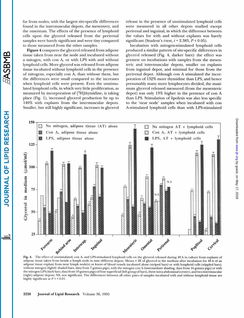
far from nodes, with the largest site-specific differences found in the intermuscular depots, the mesentery, and the omentum. The effects of the presence of lymphoid cells upon the glycerol released from the perirenal samples were barely significant and were tiny compared to those measured from the other samples. Figure 4 compares the glycerol released from adipose
tissue taken from near the node and incubated without a mitogen, with con A, or with LPS with and without lymphoid cells. More glycerol was released from adipose tissue incubated without lymphoid cells in the presence of mitogens, especially con A, than without them, but the differences were small compared to the increases when lymphoid cells were present. Even the unstimu- lated lymphoid cells, in which very little proliferation, as measured by incorporation of [3H]thymidine, is taking place (Fig. l), increased glycerol production by up to 140% with explants from the intermuscular depots. Smaller, but still highly significant, increases in glycerol
150
125
h z h 0
3. E 100
E a a, E 75
W
.C(
r=
0 L W
h
.I
I
v 50
W I
25
release in the presence of unstimulated lymphoid cells were measured in all other depots studied except perirenal and inguinal, in which the difference between the values for with and without explants was barely significant (Student's t-test, t = 2.383, P < 0.05).
Incubation with mitogen-stimulated lymphoid cells produced a similar pattern of site-specific differences in glycerol released (Fig. 4, darker bars): the effect was greatest on incubations with samples from the mesen- teric and intermuscular depots, smaller on explants from inguinal depot, and minimal for those from the perirenal depot. Although con A stimulated the incor- poration of 132% more thymidine than LPS, and hence presumably many more lymphocytes divided, the maxi- mum glycerol released measured (from the mesenteric depot) was only 13% higher in the presence of con A than LPS. Stimulation of lipolysis was also less specific to the 'near node' samples when incubated with con A-stimulated lymphoid cells than with LPS-stimulated
3 No mitogen, adipose tissue
a Con A, adipose tissue alone a LPS, adipose tissue alone
T
T
1
alone No mitogen Con A, AT LPS, AT +
T
AT + lymphoid cells + lymphoid cells
Fig. 4. The effect of unstimulated, con A- and LPS-stimulated lymphoid cells on the glycerol released during 48 h in culture from explants of adipose tissue taken from beside a lymph node in nine different depots. Means f SE of glycerol in the medium after incubation for 48 h of an adipose tissue explant from near lymph node(s) or knots of blood vessels incubated alone (striped bars) or with lymphoid cells (stippled bars), without mitogen (lightly shaded bars, data from 7 guinea pigs), with the mitogen con A (intermediate shading, data from 16 guinea pigs) or with the mitogen LPS (dark bars, data from 10 guinea pigs) of four superficial (left group of bars), three intra-abdominal (center), and two intermuscular (right) adipose depots; NS. not significant. The differences between all other pairs of samples incubated with and without lymphoid tissue are highly significant at P < < 0.01.
2226 Journal of Lipid Research Volume 36, 1995
by guest, on May 17, 2018
ww
w.jlr.org
Dow
nloaded from
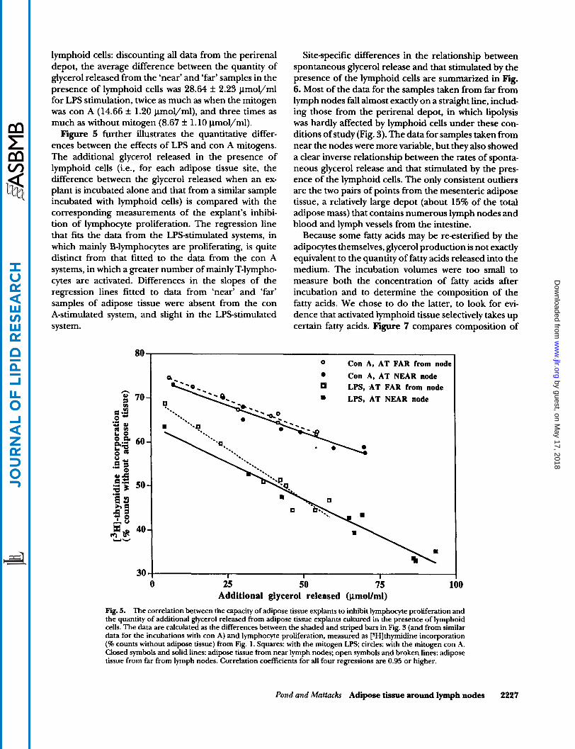
lymphoid cells: discounting all data from the perirenal depot, the average difference between the quantity of glycerol released from the ‘near’ and ‘far’ samples in the presence of lymphoid cells was 28.64 It 2.23 pmol/ml for LPS stimulation, twice as much as when the mitogen was con A (14.66 k 1.20 lmol/ml), and three times as much as without mitogen (8.67 f 1.10 pmol/ml). Figure 5 further illustrates the quantitative differ-
ences between the effects of LPS and con A mitogens. The additional glycerol released in the presence of lymphoid cells (i.e., for each adipose tissue site, the difference between the glycerol released when an ex- plant is incubated alone and that from a similar sample incubated with lymphoid cells) is compared with the corresponding measurements of the explant’s inhibi- tion of lymphocyte proliferation. The regression line that fits the data from the LPS-stimulated systems, in which mainly B-lymphocytes are proliferating, is quite distinct from that fitted to the data from the con A systems, in which a greater number of mainly T-lympho- cytes are activated. Differences in the slopes of the regression lines fitted to data from ‘near’ and ‘far’ samples of adipose tissue were absent from the con A-stimulated system, and slight in the LPS-stimulated system.
80
30
Site-specific differences in the relationship between spontaneous glycerol release and that stimulated by the presence of the lymphoid cells are summarized in Fig. 6. Most of the data for the samples taken from far from lymph nodes fall almost exactly on a straight line, includ- ing those from the perirenal depot, in which lipolysis was hardly affected by lymphoid cells under these con- ditions of study (Fig. 3). The data for samples taken from near the nodes were more variable, but they also showed a clear inverse relationship between the rates of sponta- neous glycerol release and that stimulated by the pres- ence of the lymphoid cells. The only consistent outliers are the two pairs of points from the mesenteric adipose tissue, a relatively large depot (about 15% of the total adipose mass) that contains numerous lymph nodes and blood and lymph vessels from the intestine.
Because some fatty acids may be re-esterified by the adipocytes themselves, glycerol production is not exactly equivalent to the quantity of fatty acids released into the medium. The incubation volumes were too small to measure both the concentration of fatty acids after incubation and to determine the composition of the fatty acids. We chose to do the latter, to look for evi- dence that activated lymphoid tissue selectively takes up certain fatty acids. Figure 7 compares composition of
Con A, AT FAR from node Con A, AT NEAR node LPS, AT FAR from node
9 LPS, AT NEAR node
25 50 75 Additional glycerol released (p.moVml)
100
Fig. 5. The correlation between the capacity of adipose tissue explants to inhibit lymphocyte proliferation and the quantity of additional glycerol released from adipose tissue explants cultured in the presence of lymphoid cells. The data are calculated as the differences between the shaded and striped bars in Fig. 3 (and from similar data for the incubations with con A) and lymphocyte proliferation, measured as [SHIthymidine incorporation (% counts without adipose tissue) from Fig. 1. Squares: with the mitogen LPS; circles: with the mitogen con A. Closed symbols and solid lines: adipose tissue from near lymph nodes: open symbols and broken lines: adipose tissue from far from lymph nodes. Correlation coefficients for all four regressions are 0.95 or higher.
Pond and Mattacks Adipose tissue around lymph nodes 2227
by guest, on May 17, 2018
ww
w.jlr.org
Dow
nloaded from

0
0
0
30 40 50 60 70 80
Me
LPS, AT from NEAR node LPS, AT from FAR from node Con A, AT from NEAR node Con A, AT from FAR from node
1
0
Fig. 6. The additional glycerol release stimulated by the presence of lymphoid cells from adipose tissues explants in culture (i.e., calculated as the differences between the shaded and striped bars in Fig. 3 and from similar data from incubations with con A) as a function of the glycerol measured from incubations without lymphoid cells of similar explants taken from the same depots. Symbols as in Fig. 5. Points representing measurements from particular samples are labeled: M, mesenteric adipose tissue; P, perirenal. Regression lines are fitted to all points except those from the mesenteric depot. ‘Near’ samples: r = 0.948; ‘far’ samples: r = 0.981.
the fatty acids in TAGS extracted from fresh adipose tissue with that of the FFAs in the culture media after incubation of adjacent samples with lymphoid tissue. Taking the data from all the depots together, the unsatu- ration indices of the TAG FAs in the fresh adipose tissue extracts were significantly higher than those of FFAs in the media after incubation of corresponding samples with lymphoid cells, particularly for the ‘near node’ samples, from which glycerol release in the presence of lymphoid tissue is greater (Figs. 3 and 4). The differ- ences in TAG FA composition between the ‘near node’ and ‘far from node’ adipose tissue are investigated in greater detail elsewhere (C. A. Mattacks and C. M. Pond, in preparation).
DISCUSSION
Site-specific properties of adipose tissue
By comparing the properties of adipose tissue from a depot that lacks lymph nodes and that from adjacent and distant areas of those that do contain nodes, we are able to highlight the special features of tissue that is immediately adjacent to the nodes. Under all conditions
studied, adipose tissue explants from near nodes inter- act with lymphoid cells more strongly than that from ‘far from node’ sites (Figs. 1, 3, 5, and 6). Among the superficial depots studied, the samples least inhibitory to lymphocyte proliferation (Fig. 1) were the ‘far from node’ samples from the inguinal depot, followed by those of the ‘behind arm’ depot. Both these depots are quite large (29-31) and their lymph nodes are localized into tight groups, and in the case of the inguinal depot, the ‘far from node’ samples were more than 10 mm from any nodes. The lack of any detectable interactions be- tween lymphoid cells and both perirenal samples (Figs. 1, 3, and 4) indicates that a lymph node, not merely a confluence of blood vessels, is the feature associated with the site-specific properties of adipose tissue.
The capacity of all samples of adipose tissue to inhibit lymphocyte proliferation was almost abolished by 500 pU insulin, and €or cultures containing explants from the most inhibitory depots, significantly reduced by smaller doses of insulin (Fig. 2). The concentrations tested are in the range at which lipolysis from isolated guinea pig adipocytes is most sensitive to bovine insulin (30). The minimal differences in free glycerol concen- tration among all cultures containing perirenal adipose tissue (Figs. 3 and 4) make it very unlikely that the
2228 Journal of Lipid Research Volume 36, 1995
by guest, on May 17, 2018
ww
w.jlr.org
Dow
nloaded from

120
110
100
90 3 a e .- g 80
a
.- Y Q L.
70
s 60
50
40
a Adipose tissue (AT) from FAR from node a Adipose tissue from NEAR node
Media extract after incubation with AT from FAR from node Q Media extract after incubation with AT from NEAR node
T T I T
T T
I
T T
1
* *
T
Fig. 7. The unsaturation indices of the triacylglycerol fatty acids extracted from adipose tissue collected during the first hour post mortem from 18 sites defined by their anatomical relations to lymph nodes and the free fatty acids in the tissue culture medium after incubation of adjacent samples with lymphoid cells. Means f SE of the unsaturation indices of triacylglycerol fatty acids of fresh adipose tissue (striped bars) taken from far from lymph node(s) or knots of blood vessels (light bars) and from near to lymph node(s) or knots of blood vessels (dark bars) and the free fatty acids in the incubation medium (brick-pattern bars) after incubation for 48 h of adipose tissue explants from adjacent sites with con A- or LPSstimulated lymphoid cells. Sets of samples were taken from four superficial (left group of bars), three intra-abdominal (center), and two intermuscular adipose depots (right group of bars) of 15 guinea pigs. The means f SE of data from all 'near node' samples and from all 'far from node' samples put together are shown on the far right. Asterisks refer to differences between measurements from fresh adipose tissue and in the culture media after incubation of lymphoid cells with adipose tissue explants taken from homologous sites; ** significantly different at P < 0.02; *significantly different at P < 0.05.
lymphoid cells themselves were releasing significant quantities of glycerol. This experiment suggests that lipolytic products, i.e., fatty acids and/or glycerol, are the major mediators of the effects of adipose tissue on lymphoid cells. This conclusion is supported by the inverse relationship between glycerol production and lymphocyte proliferation for the various adipose tissue explants (Fig. 5), and by the data in Fig. 7 that suggest that lymphoid tissue selectively sequesters unsaturated fatty acids from the culture medium. However, we can- not exclude the possibility that other agents released from adipocytes or endothelial tissue in the explants also interact with the lymphoid cells.
Glycerol release was significantly stimulated by the presence of lymphoid cells, even without any mitogen (Fig. 4). when cell proliferation was so slight as to
incorporate only a few hundred counts of labeled thymidine. At an increase of up to 4-fold, the capacity of lymphoid cells to stimulate lipolysis in near-lymph node samples (Figs. 4 and 5) is more than double the maximum increase in glycerol release measured from adipocytes in vitro isolated from homologous depots of similarly maintained guinea pigs and stimulated with supra-physiological concentrations of noradrenalin (no- repinephrine) (30). The capacity for such high rates of lipolysis in the presence of lymphoid tissue is found only in the small volume of adipose tissue immediately sur- rounding the lymph nodes. The response of the much more massive perirenal adipose tissue is minimal, al- though this depot interacts with all circulating hor- mones and locally secreted agents known to affect white adipose tissue (29-31), and is often regarded as repre-
Pond and Mattacks Adipose tissue around lymph nodes 2229
by guest, on May 17, 2018
ww
w.jlr.org
Dow
nloaded from
sentative of the adipose mass as a whole (38). Several cytokines secreted by lymphocytes and macro-
phages are known to modulate whole-body lipid meta- bolism (14) and adipocytes in vitro (24, 25), and the expression of tumor necrosis factor-a has been induced in mouse epididymal and parametrial adipose tissue (39), but these properties have not been sought in a wide range of adipose tissue samples of precisely defined origin. The clear-cut pattern of site-specific differences in response of adipose tissue to the presence of lym- phoid cells, particularly the absence of effects on the perirenal depot, provides a sensitive assay with which to identify the mediators of the interactions described here. Observations on 3T3 adipocytes treated with vari- ous cytokines (25) suggest that numerous different me- diators may be involved.
Effects of mitogens
T-lymphocytes are about three times as abundant as B-lymphocytes in unstimulated lymph nodes (15, 17, 34), and hence generate more daughter cells when exposed to the mitogen, con A. Although at the outset, the affected cells were much less numerous, LPS-stimu- lated cultures were more inhibited by adipose tissue (Fig. l), and stimulated greater glycerol release from explants of most depots than the con A-stimulated cul- tures (Fig. 4). The site-specific differences between adi- pose depots were also larger and more consistent in the presence of LPS. B-lymphocytes produce antibodies, an energetically demanding process, so they would be ex- pected to utilize more of the products of lipolysis than con A-stimulated T-lymphocytes. Production of cytoki- nes from macrophages activated by LPS may also con- tribute to the increase in lipolysis under these condi- tions, and the larger quantities of lipolytic products thus released may account for the greater inhibition of thymidine incorporation (Figs. 1 and 5). The fact that glycerol production was substantially increased by the presence of lymphoid cells even in the absence of any mitogen (Fig. 4) and when lymphocyte proliferation was minimal (Fig. 1) also implicates non-dividing cells such as macrophages in activating the near-node adipose tissue.
The small but consistent differences between the composition of TAG FAs in the original adipose tissue and that of the FFAs in the incubation media (Fig. 7) suggest that lymphoid tissue selectively incorporates unsaturated FAs from the mixture released from the adipose tissue. When applied individually to cultured rat lymphocytes, such FAs inhibit con A-stimulated prolif- eration (18) and are more readily incorporated into lymphocytes (20) than saturated FAs. Within-depot dif- ferences in capacity to inhibit lymphocyte proliferation were consistently smaller in the presence of con A than
with no mitogen or with LPS (Fig. l), although for near-node samples of many depots, glycerol production in the presence of lymphoid cells was only slightly lower with con A than with LPS (Fig. 4). This difference in response to the mixtures of FAs released from ‘near- node’ or ‘far from node’ adipose tissue may arise from the fact that T-lymphocytes, in contrast to other lympho- cytes, lack Gdesaturase and so do not convert stearic (C18:O) to oleic (C18:l) acid (40).
In conclusion, we suggest that the site-specific prop erties of adipose tissue around the lymph nodes de- scribed here equip the tissue to nourish or regulate (or both) the metabolism of the lymphoid cells in the nodes. Adaptive local interactions with peripheral lymph nodes might be a major reason for the evolution among mam- mals of the partitioning of adipose tissue into so many small depots (2). We urge that, in selectingadipose tissue samples for the study of lipid metabolism in relation to sepsis and other immunological activities, more atten- tion is paid to the anatomical relations of the samples sites to the lymph nodes. I
We thank Drs. Eric A. Newsholme, Philip C. Calder, and Richard H. Colby, and Mrs. Jane Bond and Mr. Graham Jeffs for technical advice, and Dr. Calder for the gift of sodium-2- hydroxy-3,5dichlorobenzene for the glycerol assay. We thank the Open University Research Committee for an equipment grant for the purchase of the automatic cell harvester and parts for the gas chromatograph, and Drs. Hilary MacQueen and Basiro Davey for helpful comments on the manuscript. Manuscript received 20 Murch 1995, in revised form 31 May 1995, and in re-rmisedform 11 July 1995.
REFERENCES
1. Yoffey, J. M., and F. C. Courtice. 1970. Lymphatics, Lymph and the Lymphomyeloid Complex. Academic Press, London and New York. 1-49.
2. Pond, C. M. 1978. Morphological aspects and the ecologi- cal and mechanical consequences of fat deposition in wild vertebrates. Annu. Rev. Ecol. Syst. 9: 519-570.
3. Kampmeier, 0. F. 1969. Evolution and Comparative Mor- phology of the Lymphatic System. Charles C. Thomas, Springfield, IL. 412-517.
4. Cooper, G., and A. L. Schiller. 1975. Anatomy of the Guinea Pig. Harvard University Press, Cambridge, MA.
5. Henry, K., and G. Farrer-Brown. 1981. A Colour Atlas of the Thymus and Lymph Nodes. Wolfe Medical Publica- tions, London. 74-80; 151; 221.
6. Kowala, M. C., and G. I. Schoefl. 1986. The popliteal lymph node of the mouse: internal architecture, vascular distribution and lymphatic supp1y.J. Anat. 148: 25-46.
7. Pond, C. M. 1994. The structure and organization of adipose tissue in naturally obese non-hibernating mam- mals. In Obesity in Europe ‘93. H. Ditschuneit, F. A. Cries, H. Hauner, V. Schusdziarra, and J. G. Wechsler, editors. Proceedings of the 5th European Congress of Obesity. J. Libbey & Co., London. 419-426.
213-233.
2230 Journal of Lipid Research Volume 36, 1995
by guest, on May 17, 2018
ww
w.jlr.org
Dow
nloaded from

8. Suzuki, T. 1952. Histological studies on lymphatic appa- ratus in human adipose tissue. Acta. Sch. Med. Uniu. Kyoto.
9. Heath, T., and R. Brandon. 1983. Lymphatic and blood vessels of the popliteal node in sheep. Anat. Rec. 207:
10. Hendriks, H. R. 1978. Occlusion of the lymph flow to rat popliteal lymph node for protracted periods. 2. Ver- suckstierk. Bd 20: 105-112.
11. Herman, P. G., D. Lyonnet, R. Fingerhut, andR. N.Tuttle. 1976. Regional blood flow to the lymph node during the immune response. Lymphology. 9 101-104.
12. McCarthy, D. O., M. J. Kluger, and A. J. Vander. 1985. The role of fever in appetite suppression after endotoxin administration. Am.J Clin. Nutr. 40: 310-316.
13. Beutler, B., and A. Cerami. 1988. Cachectin (tumor ne- crosis factor): a macrophage hormone governing cellular metabolism and inflammatory response. Endocrin. Rev. 9:
14. Griinfeld, C., and K. R. Feingold. 1992. Tumor necrosis factor, interleukin, and interferon induced changes in lipid metabolism as part of host defense (43424). Proc. SOC. Exp. Biol. Med 200: 224-227.
15. Buttke, T. M. 1984. Inhibition of lymphocyte proliferation by free fatty acids. 1. Differential effects on mouse B and T lymphocytes. Immunology. 5 3 235-242.
16. Ardawi, M. S. M., andE. A. Newsholme. 1985. Metabolism in lymphocytes and its importance in the immune re- sponse. Essays Biochem. 21: 1-43.
17. Calder, P. C. 1993. The effects of fatty acids on lympho- cyte functions. Brazilian J. Med. Biol. Res. 2 6 901-917.
18. Calder, P. C., J. A. Bond, S. J. Bevan, S. V. Hunt, and E. A. Newsholme. 1991. Effect of fatty acids on the prolifera- tion of concanavalin A-stimulated rat lymph node lymph* cytes. Int.J Biochem. 23: 579-588.
19. Calder, P. C., S. J. Bevan, and E. A. Newsholme. 1992. The inhibition of T-lymphocyte proliferation by fatty acids is via an eicosanoid-independent mechanism. Immunology
20. Calder, P. C., P. Yaqoob, D. J. Harvey, A. Watts, and E. A. Newsholme. 1994. Incorporation of fatty acids by conca- navalin A-stimulated lymphocytes and the effect on fatty acid composition and membrane fluidity. Biochem. J. 300:
21. Erickson, K. L., D. A. Adams, and C. J. McNeill. 1983. Dietary lipid modulation of immune responsiveness. Lip-
22. Yaqoob, P., E. A. Newsholme, and P. C. Calder. 1994. The effect of dietary lipid manipulation on rat lymphocyte subsets and proliferation. Immunology. 82: 603-6 10.
23. Guillou, P. J. 1993. The effects of lipids on some aspects of the cellular immune response. Proc. Nutr. SOC. 52: 9 1 - 100.
24. Patton, J. S., H. M. Shepard, H. Wilking, G. Lewis, B. B. Aggarwal, T. E. Eessalu, L. A. Gavin, and C. Griinfeld. 1986. Interferon and tumour necrosis factors have similar catabolic effects on 3T3-Ll cells. Proc. Natl. Acad. Sci. USA.
3 0 174-182.
461 -472.
57-66.
7 5 108-115.
509-518.
&. 18: 468-474.
83: 8313-8317.
25. Doerrler, W., K. R. Feingold, and C. Griinfeld. 1994. Cytokines induce catabolic effects in cultured adipocytes by multiple mechanisms. Cytokine. 6 478-484.
26. Griinfeld, C., R. Gulli, A. H. Moser, L. A. Gavin, and K. R. Feingold. 1989. Effect of tumor necrosis factor admini- stration in vivo on lipoprotein lipase activity in various tissues of the rat.J. Lipid Res. 3 0 579-585.
27. Tracey, K. J., and A. Cerami. 1992. Tumor necrosis factor and regulation of metabolism in infection: role of sys- temic versus tissue levels (43426). h o c . SOC. ExP. Biol. Med.
28. Pond, C. M., C. A. Mattacks, and D. Sadler. 1984. The effects of food restriction and exercise on site-specific differences in adipocyte volume and adipose tissue cellu- larity. 1. Superficial and intra-abdominal sites. Br.J. Nutr.
29. Mattacks, C. A., D. Sadler, and C. M. Pond. 1987. The effects of exercise on the activities of hexokinase and phosphofructokinase in superficial, intra-abdominal and intermuscular adipose tissue of guinea pigs. Comp. Bio- chem. Physiol. 87B: 533-542.
30. Pond, C. M., and C. A. Mattacks. 1991. The effects of noradrenaline and insulin on lipolysis in adipocytes iso- lated from nine different adipose depots of guinea pigs. Int. J. Obes. 15 609-618.
31. Pond, C. M., C. A. Mattacks, and D. Sadler. 1992. The effects of exercise and feeding on the activity of lipopro- tein lipase in nine different adipose depots of guinea pigs. Int. J. Biochem. 24: 1825-1831.
32. Hadek, R. 1951. The lymph nodes of the guinea pig. Br. Vet. J. 107: 487-493.
33. Maalouf, H., R. Gagnon, and P. Bois. 1967. ktude descrip tive et topographique des ganglions lymphatiques du cobaye. [Descriptive and topographic study of the lymph nodes of the guinea pig.] Rev. Can. Biol. 2 6 323-334.
34. Yaqoob, P., and P. C. Calder. 1993. The effects of fatty acids on lymphocyte functions. Int. J. Biochem. 25:
35. McGowan, M. W., J. D. Artiss, D. R. Strandberg, and B. Zak. 1983. A peroxidase-coupled method for the col- orimetric determination of serum triglycerides. Clin. Chem. 29: 538-542.
36. Hock, C. E., M. A. Holahan, and D. K. Reibel. 1987. Effect of dietary fish oil on myocardial phospholipids and my* cardial ischemic damage. Am. J. Physiol. 252: H554-H560.
37. Colby, R. H., and C. M. Pond. 1993. Site-specific differ- ences in the responses of guinea pig adipose tissue to changes in the fatty acid composition of the diet. Nutr.
38. Pond, C. M. 1992. An evolutionary and functional view of mammalian adipose tissue. Proc. Nutr. SOC. 51: 367-377.
39. Hotamisligil, G. S., N. S. Shargill, and B. M. Spiegelman. 1993. Adipose expression of tumor necrosis factora: direct role in obesity-linked insulin resistance. Science.
40. Tebbey, P. W., and T. M. Buttke. 1990. Molecular basis for the immunosuppressive action of stearic acid on T cells. Immunology. 70: 379-384.
200.233-239.
51: 415-424.
1705- 1714.
Res. 13: 1203-1212.
259: 87-91.
Pond and Mattacks Adipose tissue around lymph nodes 2231
by guest, on May 17, 2018
ww
w.jlr.org
Dow
nloaded from