Embed Size (px)
Citation preview

Published: April 01, 2011
r 2011 American Chemical Society 3336 dx.doi.org/10.1021/ac103129e |Anal. Chem. 2011, 83, 3336–3342
ARTICLE
pubs.acs.org/ac
Integrated Glass Microdevice for Nucleic Acid Purification,Loop-Mediated Isothermal Amplification, and Online DetectionQingqing Wu, Wei Jin, Chao Zhou, Sihai Han, Wenxiu Yang, Qiangyuan Zhu, Qinhan Jin, and Ying Mu*
Research Center for Analytical Instrumentation, Institute of Cyber-Systems and Control, State Key Laboratory of Industrial ControlTechnology, Zhejiang University, Hangzhou 310058, People's Republic of China
The purification of nucleic acids (NAs) is a precondition formost genetic analyses or diagnostics and their forensic
applications. However, the conventional methods for NA extrac-tion, such as phenol�chloroform extraction, have the disadvan-tage of being time-consuming and difficult to scale down to smallsample volumes. Compared to conventional methods, the micrototal analysis system (μ-TAS) offers several remarkable advan-tages, such as low reagent and sample consumption, increasedspeed, enhanced sensitivity, etc.1�4 Microfluidic approaches tonucleic acid isolation have therefore received great attention inrecent years. There are many investigations concerning theextraction of nucleic acids in microfluidic systems; these worksconcern mostly packing microchannels with silica beads5�10 orwith sol�gel and hybrid silica beads/sol�gel11,12 or fabricatingmicropillars and microposts in silicon microfluidic channels.13,14
NAs adsorb to silica in the presence of a high concentration of thechaotropic agent,15 and the extracted nucleic acids are elutedwith an aqueous low-salt buffer and concentrated into a verysmall volume.
One of the motivations for the development of on-chip NAextraction is to integrate other genetic analysis units such asnucleic acid amplification and detection into a single microchip.Legendre et al.16 demonstrated integrated online DNA purifica-tion and polymerase chain reaction (PCR) in a valveless glassmicrodevice. The chromatography required for solid-phase ex-traction (SPE) in the microfluidic sample preparation device wascarried out in a silica bead/sol�gel SPE bed, where the purified
DNA was eluted directly into a downstream chamber whereconventional thermocycling allowing for PCR amplification ofspecific DNA target sequences took place. Easley et al.17 devel-oped a fully integrated microfluidic system that can accept wholeblood as a crude biological sample. Upon loading the sample, theglass microfluidic genetic analysis system device carries out on-chip DNA purification and PCR-based amplification, followed byseparation and detection in a manner that allows for microlitersamples to be screened for infectious pathogens with the sample-in�answer-out mode in <30 min.
Although much progress has been made in integrating NApurification and PCR on a single microdevice, the complexity ofthe fabrication process for the silicon-based microchip, thepacking process for silica beads or sol�gel, and the demandsof a temperature cycling protocol for conventional PCR increasethe difficulties in chip fabrication. To simplify the fabrication ofthe microchip and better integrate NA purification with ampli-fication reactions, we seek glass alone as the material to fabricatethe NA purification unit. Microfabricated silica pillars were usedto increase the surface area available for DNA capture.13,14 In thisstudy, glass micropillars were designed to achieve the samepurpose as silica pillars did and the NA purification unit wasintegrated with subsequent amplification reactions.
Received: November 29, 2010Accepted: March 17, 2011
ABSTRACT: A microdevice made of glass for genetic analysishas been fabricated, for the first time, for integration ofextraction of nucleic acids and loop-mediated isothermal am-plification (LAMP), followed by online fluorescence detectionof amplification products on a single chip. The nucleic acid(NA) extraction region consists of a microfabricated serpentinechannel in which micropillars were etched to increase thechannel surface area and the capture efficiency of NAs. Nucleicacid molecules were bound to these pillars and channel surfacein the presence of the chaotropic salt guanidine hydrochlorideand eluted into a downstream amplification chamber with low ionic strength buffer where loop-mediated isothermal amplificationwas efficiently performed. Amplification can be detected online by the increase of fluorescence intensity at 540 nm when a lowconcentration of SYBR Green I, a fluorescent dsDNA intercalating dye, is employed. Flow control was accomplished by usinglaminar flow and differential channel flow resistances. Through passivation of the LAMP chamber and the channel between theextraction region and amplification domain, effective nucleic acid extraction and amplification were performed by just using adouble-channel syringe pump and a heating block. By using this integrated microdevice, the purification of nucleic acids fromcomplex biological matrixes and their subsequent amplification and detection online could be finished within 2 h.
3337 dx.doi.org/10.1021/ac103129e |Anal. Chem. 2011, 83, 3336–3342
Analytical Chemistry ARTICLE
To further simplify the system, we used loop-mediatedisothermal amplification (LAMP) for amplifying DNA insteadof conventional PCR. LAMP is a method for amplification ofnucleic acid sequences established by Notomi et al.18 whichutilizes a designed set of primers, termed inner and outer primers,to recognize specific gene sequences and a polymerase withstrand displacement activity to generate a large amount ofamplified product within 1 h.19 One significant advantage ofthe LAMP method is its ability to amplify specific DNA underisothermal conditions (60�65 �C) without thermocycling.Therefore, LAMP can be performed simply with a water bathor heating plate instead of an expensive thermocycler. Anotherimportant advantage of LAMP is its simplicity for detection ofthe amplification reaction. Visualization of DNA products on gelelectrophoresis is not required for assessing successful DNAamplification because a positive LAMP reaction yields whiteprecipitate due to the formation of magnesium pyrophosphatebyproduct, which increases the turbidity of the reactionmixture.20�22 For better visibility of the reaction result, afluorescent intercalating dye such as SYBR Green23,24 or propi-dium iodide24 is added to the solution after the reaction iscompleted. When the LAMP reaction is positive, a color changeis observed under ambient light or UV light. Tomita et al.developed a simple colorimetric assay for the detection of theLAMP reaction by adding calcein, a fluorescence metal indicator,to the prereaction solution.25
In this study, we have tried to make use of the property thatglass consists mainly of silica to capture NAs and integrated thisNA purification module with loop-mediated isothermal amplifi-cation, so the serpentine channels with an array of micropillarswere used as an SPE bed which we call a glass SPE (gSPE) bed.Nucleic acid purification through the gSPE bed, followed bytarget sequence amplification by LAMP and online fluorescencedetection, is accomplished in 2 h. Here we have shown that suchan integrated microfluidic genetic analysis system is simple tofabricate and highly functional.
’EXPERIMENTAL SECTION
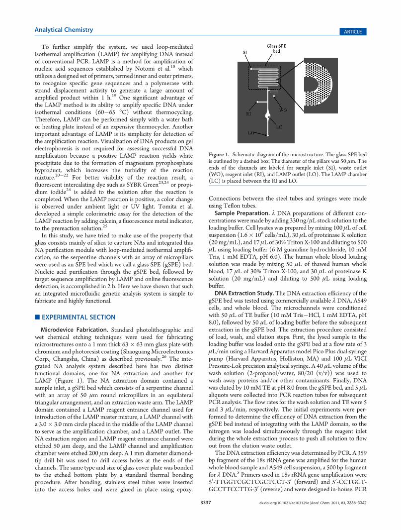
Microdevice Fabrication. Standard photolithographic andwet chemical etching techniques were used for fabricatingmicrostructures onto a 1 mm thick 63� 63 mm glass plate withchromium and photoresist coating (ShaoguangMicroelectronicsCorp., Changsha, China) as described previously.26 The inte-grated NA analysis system described here has two distinctfunctional domains, one for NA extraction and another forLAMP (Figure 1). The NA extraction domain contained asample inlet, a gSPE bed which consists of a serpentine channelwith an array of 50 μm round micropillars in an equilateraltriangular arrangement, and an extraction waste arm. The LAMPdomain contained a LAMP reagent entrance channel used forintroduction of the LAMPmaster mixture, a LAMP channel witha 3.0� 3.0 mm circle placed in the middle of the LAMP channelto serve as the amplification chamber, and a LAMP outlet. TheNA extraction region and LAMP reagent entrance channel wereetched 50 μm deep, and the LAMP channel and amplificationchamber were etched 200 μm deep. A 1 mm diameter diamond-tip drill bit was used to drill access holes at the ends of thechannels. The same type and size of glass cover plate was bondedto the etched bottom plate by a standard thermal bondingprocedure. After bonding, stainless steel tubes were insertedinto the access holes and were glued in place using epoxy.
Connections between the steel tubes and syringes were madeusing Teflon tubes.Sample Preparation. λ DNA preparations of different con-
centrations weremade by adding 330 ng/μL stock solution to theloading buffer. Cell lysates was prepared by mixing 100 μL of cellsuspension (1.6� 106 cells/mL), 30 μL of proteinase K solution(20mg/mL), and 17 μL of 30%Triton X-100 and diluting to 500μL using loading buffer (6 M guanidine hydrochloride, 10 mMTris, 1 mM EDTA, pH 6.0). The human whole blood loadingsolution was made by mixing 50 μL of thawed human wholeblood, 17 μL of 30% Triton X-100, and 30 μL of proteinase Ksolution (20 mg/mL) and diluting to 500 μL using loadingbuffer.DNA Extraction Study. The DNA extraction efficiency of the
gSPE bed was tested using commercially available λ DNA, A549cells, and whole blood. The microchannels were conditionedwith 50 μL of TE buffer (10 mM Tris�HCl, 1 mM EDTA, pH8.0), followed by 50 μL of loading buffer before the subsequentextraction in the gSPE bed. The extraction procedure consistedof load, wash, and elution steps. First, the lysed sample in theloading buffer was loaded onto the gSPE bed at a flow rate of 3μL/min using a Harvard Apparatus model Pico Plus dual-syringepump (Harvard Apparatus, Holliston, MA) and 100 μL VICIPressure-Lok precision analytical syringe. A 40 μL volume of thewash solution (2-propanol/water, 80/20 (v/v)) was used towash away proteins and/or other contaminants. Finally, DNAwas eluted by 10 mM TE at pH 8.0 from the gSPE bed, and 5 μLaliquots were collected into PCR reaction tubes for subsequentPCR analysis. The flow rates for the wash solution and TE were 5and 3 μL/min, respectively. The initial experiments were per-formed to determine the efficiency of DNA extraction from thegSPE bed instead of integrating with the LAMP domain, so thenitrogen was loaded simultaneously through the reagent inletduring the whole extraction process to push all solution to flowout from the elution waste outlet.TheDNA extraction efficiency was determined by PCR. A 359
bp fragment of the 18s rRNA gene was amplified for the humanwhole blood sample and A549 cell suspension, a 500 bp fragmentfor λ DNA.5 Primers used in 18s rRNA gene amplification were50-TTGGTCGCTCGCTCCT-30 (forward) and 50-CCTGCT-GCCTTCCTTG-30 (reverse) and were designed in-house. PCR
Figure 1. Schematic diagram of the microstructure. The glass SPE bedis outlined by a dashed box. The diameter of the pillars was 50 μm. Theends of the channels are labeled for sample inlet (SI), waste outlet(WO), reagent inlet (RI), and LAMP outlet (LO). The LAMP chamber(LC) is placed between the RI and LO.

3338 dx.doi.org/10.1021/ac103129e |Anal. Chem. 2011, 83, 3336–3342
Analytical Chemistry ARTICLE
master mixture was made to the following final concentrations:0.4 μM concentration of each primer, 0.3 mM dNTP, 1.25 unitsof Taq polymerase, 10 mM Tris�HCl (pH 8.3), 50 mM KCl,1.5 mM MgCl2, 5 μL of collected elution fractions, addingdoubly-distilled water (ddH2O) to 25 μL. The thermal cyclingprotocols used were 94 �C denaturation for 5 min, 30 cycles of94 �C for 30 s, 60 �C for 30 s (for both the 18s rRNA gene and λDNA), and 72 �C for 1 min, followed by a 10 min extension at72 �C. DNA amplification was confirmed by agarose gelelectrophoresis.Integrated Microfluidic LAMP and Detection Online. The
loading and wash steps for DNA extraction on the chip are thesame as mentioned in the section “DNA Extraction Study”. Thegas source which was connected to the LAMP reagent inlet wasevacuated and replaced by LAMP master mixture during DNAelution from the gSPE bed. Simultaneously, the waste outlet inthe NA extraction domain was sealed with a solid stainless steelplug. The eluting solution and LAMP reagents were controlled tomove into the LAMP chamber by using differential channel flowresistances and laminar flow. The syringe pump was stoppedwhen the amplification chamber had been full of the eluant andLAMP reagent mixture, and the entire chip was then placed on aheating block which was overlaid with the heat conducting oiland incubated at 65 �C for 60 min.
’RESULTS AND DISCUSSION
Microdevice Design. The design used for the integrated NAanalysis systems is shown in Figure 1. The gSPE bed consists of aserpentine channel containing an array of 50 μm round micro-pillars in an equilateral triangular arrangement which increasesthe surface area within the channel and forces fluid to changetrajectory and hence enhances the probability of DNA beingcaptured. The etched structure shows frustoconic or pyramidalpillars instead of circular micropillars due to the HF isotropicetching of glass (Figure 2a,b). Although the lateral surface area ofa pyramid is less than that of a cylinder, the surface roughnessresulting from the glass etching further increases the surface areawithin the channel.Two kinds of gSPE beds were fabricated by controlling the
etching time which have the same designed pillar diameter of 50μm but different channel depths of about 30 and 50 μm,respectively (as shown in Figure 2a,b). The pillar shape of theformer is frustoconic, and the latter one is typical “pyramidal”
resulting from a longer etching time. PCR on DNA extractedfrom 1 μL of whole blood was performed using the two differentgSPE beds. It is estimated, from Figure 2c, the the DNAextraction efficiency of the gSPE bed containing pyramidal pillarsis better. Although the reproducibility was not good enough, the
Figure 2. (a) The designed pillar diameter was 50 μm, and the approximate height of the pillars was 30 μm. (b) The designed pillar diameter was 50 μm,and the approximate height of the pillars was 50 μm. (c) Result of gel electrophoresis of PCR on DNA extracted from 1 μL of whole blood using twodifferent gSPE beds. M = DNA marker. Lanes 1�3: collected fractions 1�3 from the gSPE bed containing frustums with a height of approximately 30μm (a). Lanes 4�6: collected fractions 1�3 from the gSPE bed containing pyramidical pillars with a height of approximately 50 μm (b).
Figure 3. (a) Schematic diagram of the process for flow control:SigmaCote (blue) was loaded from the LAMP outlet and flowedthrough the channel to the elution waste outlet. Simultaneously, thesample inlet was connected to a nitrogen source using Teflon tubing topreclude the passivation reagent from entering the SPE bed (left).During the load and wash steps (center), nitrogen was connected to theLAMP reagent inlet, making extraction waste (yellow) flow out from thewaste outlet. During the DNA elution step (right), the waste outlet wasblocked to further increase the flow resistance, which is much highercompared with that of the LAMP domain because of the smaller channelsize of the elution waste arm. DNA eluted from the SPE domaincombined with the LAMP reagent mixture and flowed toward theLAMP chamber. (b) Dyes were placed in the channels for visualization.Left: selective passivation for the LAMP domain. Center: DNA elutedfrom the SPE domain flowed toward the LAMP domain. Right: eluentcombined with the LAMP reagent mixture.
3339 dx.doi.org/10.1021/ac103129e |Anal. Chem. 2011, 83, 3336–3342
Analytical Chemistry ARTICLE
poorer extraction efficiency was not seen in the gSPE bed havingtypical pyramidal pillars. On the contrary, for the design havingthe same pillar diameter and arrangement, we incline to the viewthat, within certain limits, the deeper the channel, the moreefficient the extraction efficiency. We speculate that pyramidalpillars promote the flow disturbance and hence enhance theprobability of DNA being captured. Although further worksassociated with reproducibility and statistical analysis for adefinite answer about the difference in the extraction efficiencyof the two above-mentioned gSPE beds are ongoing, it can beconfirmed that the extraction efficiency of a gSPE bed like theone shown in Figure 2b is high, so it was chosen to be integratedwith the LAMP domain for making an integrated genetic analysisdevice for subsequent experiments.Flow Control. Fluidic isolation of the NA extraction and
LAMP was accomplished for the two following purposes: (1) foravoiding the incompatibility of SPE reagents with the amplifica-tion process and (2) for selective passivation of th eLAMPdomain to avoid adsorption of Taq polymerase prior to andduring LAMP amplification. SigmaCote was selected as thepassivation reagent to provide a microchip passivation with aprocess that was one step and was complete in less than 5 min.16
Figure 3a illustrates the processes of flow control accomplishedby using laminar flow and differential channel flow resistancescaused by different channel sizes.16,17 If the microdevice is to bereused, a silanization treatment should to be done for the LAMPdomain prior to every reaction.Evaluation of the gSPE Bed for DNAExtraction. λDNAwas
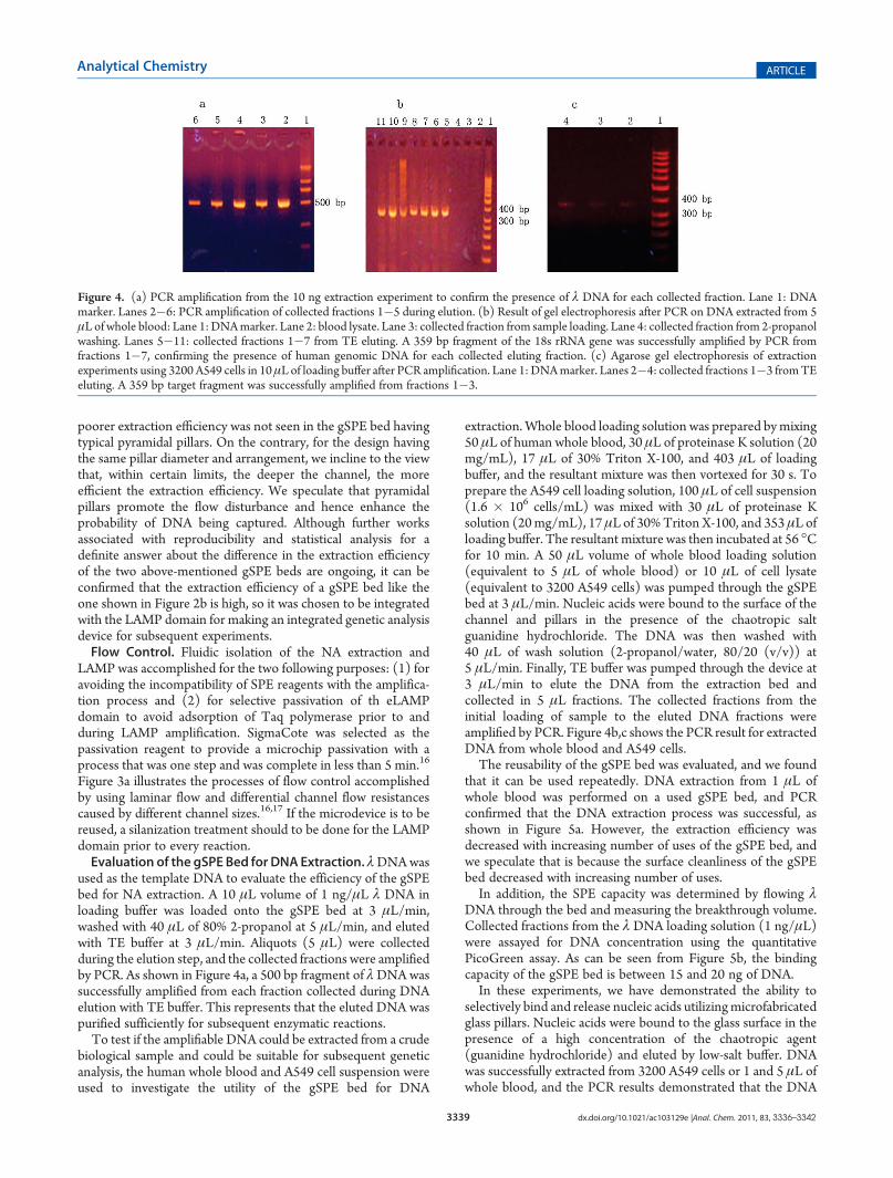
used as the template DNA to evaluate the efficiency of the gSPEbed for NA extraction. A 10 μL volume of 1 ng/μL λ DNA inloading buffer was loaded onto the gSPE bed at 3 μL/min,washed with 40 μL of 80% 2-propanol at 5 μL/min, and elutedwith TE buffer at 3 μL/min. Aliquots (5 μL) were collectedduring the elution step, and the collected fractions were amplifiedby PCR. As shown in Figure 4a, a 500 bp fragment of λDNA wassuccessfully amplified from each fraction collected during DNAelution with TE buffer. This represents that the eluted DNA waspurified sufficiently for subsequent enzymatic reactions.To test if the amplifiable DNA could be extracted from a crude
biological sample and could be suitable for subsequent geneticanalysis, the human whole blood and A549 cell suspension wereused to investigate the utility of the gSPE bed for DNA
extraction. Whole blood loading solution was prepared bymixing50 μL of human whole blood, 30 μL of proteinase K solution (20mg/mL), 17 μL of 30% Triton X-100, and 403 μL of loadingbuffer, and the resultant mixture was then vortexed for 30 s. Toprepare the A549 cell loading solution, 100 μL of cell suspension(1.6 � 106 cells/mL) was mixed with 30 μL of proteinase Ksolution (20mg/mL), 17 μL of 30%Triton X-100, and 353 μL ofloading buffer. The resultant mixture was then incubated at 56 �Cfor 10 min. A 50 μL volume of whole blood loading solution(equivalent to 5 μL of whole blood) or 10 μL of cell lysate(equivalent to 3200 A549 cells) was pumped through the gSPEbed at 3 μL/min. Nucleic acids were bound to the surface of thechannel and pillars in the presence of the chaotropic saltguanidine hydrochloride. The DNA was then washed with40 μL of wash solution (2-propanol/water, 80/20 (v/v)) at5 μL/min. Finally, TE buffer was pumped through the device at3 μL/min to elute the DNA from the extraction bed andcollected in 5 μL fractions. The collected fractions from theinitial loading of sample to the eluted DNA fractions wereamplified by PCR. Figure 4b,c shows the PCR result for extractedDNA from whole blood and A549 cells.The reusability of the gSPE bed was evaluated, and we found
that it can be used repeatedly. DNA extraction from 1 μL ofwhole blood was performed on a used gSPE bed, and PCRconfirmed that the DNA extraction process was successful, asshown in Figure 5a. However, the extraction efficiency wasdecreased with increasing number of uses of the gSPE bed, andwe speculate that is because the surface cleanliness of the gSPEbed decreased with increasing number of uses.In addition, the SPE capacity was determined by flowing λ
DNA through the bed and measuring the breakthrough volume.Collected fractions from the λ DNA loading solution (1 ng/μL)were assayed for DNA concentration using the quantitativePicoGreen assay. As can be seen from Figure 5b, the bindingcapacity of the gSPE bed is between 15 and 20 ng of DNA.In these experiments, we have demonstrated the ability to
selectively bind and release nucleic acids utilizingmicrofabricatedglass pillars. Nucleic acids were bound to the glass surface in thepresence of a high concentration of the chaotropic agent(guanidine hydrochloride) and eluted by low-salt buffer. DNAwas successfully extracted from 3200 A549 cells or 1 and 5 μL ofwhole blood, and the PCR results demonstrated that the DNA
Figure 4. (a) PCR amplification from the 10 ng extraction experiment to confirm the presence of λ DNA for each collected fraction. Lane 1: DNAmarker. Lanes 2�6: PCR amplification of collected fractions 1�5 during elution. (b) Result of gel electrophoresis after PCR on DNA extracted from 5μL of whole blood: Lane 1: DNAmarker. Lane 2: blood lysate. Lane 3: collected fraction from sample loading. Lane 4: collected fraction from 2-propanolwashing. Lanes 5�11: collected fractions 1�7 from TE eluting. A 359 bp fragment of the 18s rRNA gene was successfully amplified by PCR fromfractions 1�7, confirming the presence of human genomic DNA for each collected eluting fraction. (c) Agarose gel electrophoresis of extractionexperiments using 3200 A549 cells in 10μL of loading buffer after PCR amplification. Lane 1: DNAmarker. Lanes 2�4: collected fractions 1�3 fromTEeluting. A 359 bp target fragment was successfully amplified from fractions 1�3.

3340 dx.doi.org/10.1021/ac103129e |Anal. Chem. 2011, 83, 3336–3342
Analytical Chemistry ARTICLE
extracted by the device is amplifiable. The results show that sucha purification device is both simple to fabricate and highlyefficient.Comparison of the Efficiency of DNA Extraction Using the
gSPE Bed with That Using a Commercial Kit. The resultsdiscussed above establish that the gSPE bed can be utilized toretain PCR-amplifiable DNA. To make it more acceptable, acomparison of the efficiency of DNA extraction using the gSPEbed with that using a QIAamp DNA microkit (Qiagen) wascarried out. The whole blood loading solution for genomic DNAextraction using gSPE bed was prepared by mixing 50 μL ofhuman whole blood, 30 μL of proteinase K solution (20 mg/mL), 17 μL of 30% Triton X-100, and 403 μL of loading buffer,
and the resultant mixture was then vortexed for 30 s. A 50 μLvolume of whole blood loading solution (equivalent to 5 μL ofwhole blood) was loaded onto the gSPE bed, followed bywashing and elution steps, and 60 μL of eluent was collectedfor subsequent PCR. Three identical devices were used forexperimental replication. DNA extraction using the commercialkit was performed according to the manufacturer’s protocol witha little modification to make the data more comparable. A 50 μLvolume of the whole blood was lysed to a final volume of 260 μLaccording to the protocol, but 26 μL of lysate (equivalent to 5 μLof whole blood) was transferred to the QIAamp Minelutecolumn for genomic DNA extraction, and 60 μL of eluent wascollected for PCR. Figure 6 shows the PCR result on theextracted DNA from 5 μL of whole blood using gSPE bedsand the QIAamp DNA microkit. A 359 bp fragment of the 18srRNA gene was successfully amplified by PCR, confirming thepresence of human genomic DNA for each collected eluent. Inaddition, it is estimated that, from the brightness of the bands, theefficiency of DNA extraction using the gSPE bed is not worsethan that using the commercial kit or even may be better.LAMP in a PCR Tube.An initial study was carried out in a PCR
tube to identify wheter the DNA extracted from the gSPE bed isamplifiable for LAMP. A LAMP assay was carried out in a 25 μLreaction mixture containing a λ DNA template extracted fromthe gSPE bed (2 μL aliquots were collected during the elutionstep, and the collected fractions were amplified by LAMP), 1.6μM each FIP and BIP, 0.2 μM each F3 and B3, 8 units of BstDNA polymerase (New England Biolabs), 1.4 mM dNTP, 1 Mbetaine, 6mMMgSO4, 20mMTris�HCl (pH 8.8), 10mMKCl,10 mM (NH4)2SO4, and 0.1% Triton X-100. The reactionmixtures were incubated at 65 �C for 1 h, after which analysiswas done using 2% agarose gel electrophoresis as shown inFigure 7a. Lanes 2�7 show the amplified products resulting fromLAMP reaction by subjecting the basic LAMP sample solution to65 �C for 1 h. The LAMP reaction produced many bands ofdifferent sizes, forming a ladder-like pattern.18
Since the amplification efficiency of LAMP is higher than thatof PCR,18 the product concentration obtained by LAMP is
Figure 6. Result of gel electrophoresis after PCR on DNA extractedfrom 5 μL of whole blood using the gSPE bed and QIAamp DNAmicrokit. M = DNA marker. Lanes 1�3: PCR result of the extractedDNA using three identical gSPE beds. Lane 4: PCR result of theextracted DNA using the QIAamp DNA microkit.
Figure 7. (a) Results of gel electrophoresis after LAMP on DNAextracted from the gSPE bed. Lane 1: negative control. Lanes 2�7:amplified products resulting from DNA eluted from the SPE bed. (b)Fluorescence imaging of LAMP reaction tubes after addition of 10 μL of1:100 diluted original SYBR Green I. Tube 1: negative control. Tubes2�7: positive amplification.
Figure 5. (a) Result of gel electrophoresis after PCR on DNA extractedfrom 1 μL of whole blood on a used gSPE bed. M = DNA marker. Lane1: blood lysate. Lanes 2 and 3: collected fractions from TE precleaning.Lanes 4�7: collected fractions 1�4 from TE eluting. A 359 bp fragmentof the 18s rRNA gene was successfully amplified by PCR from fractions1�4, confirming the presence of human genomic DNA for eachcollected eluting fraction. (b) Collected fractions from the initial λDNA loading (1 ng/μL) were assayed for DNA concentration using thequantitative PicoGreen assay, and the capacity of the gSPE bed wasdetermined to be between 15 and 20 ng of DNA.

3341 dx.doi.org/10.1021/ac103129e |Anal. Chem. 2011, 83, 3336–3342
Analytical Chemistry ARTICLE
markedly higher than that obtained by PCR. Accordingly, it ispossible to check the existence of amplified DNA fragments bydirectly observing the color variance of fluorescence dye that hasbeen added into the reaction solution.27 SYBR Green I is anasymmetrical cyanine dye used for the quantification of double-strandedDNA in somemethods of real time PCR28,29 and is usedfor the visualization of the LAMP reaction.23,24,27 It is oftenadded into reaction tubes after LAMP reaction because theaddition of a high concentration of SYBR Green I could inhibitthe amplification. In the case of positive amplification, the colorof the dye changes to green, which can be judged under naturallight as well as under UV light.23,27 In this study, 10 μL ofSYBR Green I diluted 1:100 was added to each tube afteramplification,30 the reaction tubes were placed on a cassette andexcited with 455 nm excitation light, and fluorescence imageswere obtained by using CCD camera. Figure 7b shows theimaging results with 100� SYBRGreen I added into the negativecontrol solution and positive amplification products, respec-tively. Under 455 nm light excitation, the tubes with a positivereaction (tubes 2�7) show bright green fluorescence, which canbe distinguished from the yellow color of a negative reaction(tube 1). From the above-mentioned results, it could be pre-dicted that the DNA extracted from the gSPE bed is suitable forLAMP amplification.Integrated DNA Extraction, LAMP, and Detection on a
Chip. In the initial bulk LAMP analyses, the products weredetected visually by a color difference between the negativecontrol and positive amplification under 455 nm excitation lightafter addition of SYBR Green I dye (10 μL of 1:100 dilutedoriginal SYBR Green I) to tubes containing LAMP products(Figure 7b). However, the opening of the tubes (SYBR Green Iadded after LAMP reaction) increased the risk of contamination,and the LAMP reaction was inhibited if a high concentration ofSYBR Green I was added to the reaction premixture.27,31 There-fore, a study was carried out to test the influence of a lowerconcentration of SYBR Green I on the LAMP reaction. It wasfound that SYBR Green I at low concentration (1 μL of 1: 400diluted original SYBR Green I was added to the LAMP mastermixture) did not inhibit the LAMP reaction, and there was asignificant increase in fluorescence intensity in the tubes havingpositive amplification although a color difference between thenegative control and positive amplification could not be observedunder 455 nm excitation light when using a low concentration ofSYBR Green I as the indicator (data not shown here). With theinformation obtained from the initial study on bulk analysis, theLAMP reaction and online detection were integrated with DNApurification in a single microchip.λ DNA was chosen as the template DNA for testing the
integrated online DNA extraction, loop-mediated isothermalamplification, and detection of LAMP products on a single glassmicrochip. A 10 μL volume of 1 ng/μL λ DNA in loading bufferwas loaded onto the gSPE bed from the sample inlet at 3 μL/minfollowed by washing with 80% 2-propanol. Simultaneously, thegas source was connected to the LAMP reagent inlet to force theloading and washing waste to be directed toward the extractionwaste arm. After that, the syringe filled with washing solution wasremoved and the residual 2-propanol was blown away from theextraction domain. During the elution step, the gas source wasevacuated and replaced by LAMP master mixture containing alow concentration of SYBR Green I, and the Teflon tubingconnected to the waste outlet was blocked with a solid stainlesssteel plug for further increasing the resistance of the waste arm to
allow purified DNA to combine with the LAMP reagent mixtureand to flow toward the LAMP chamber. When the amplificationchamber was full of the converged flow and the two liquid flowsbecame stable, the syringe pumps were stopped (there was asmall difference between the time when eluent and LAMPreagents entered the amplification chamber so that the ratiobetween the two flows was unstable; therefore, the mixtureinitially entering the chamber was not used for amplification;besides, from Figures 4 and Figure 7a, the first elution fractionswere amplifiable, so it is not necessary to predetermine the DNAelution time offline), the syringes were removed, and the ends ofall Teflon tubings were blocked with solid stainless steel plugs.The entire chip was then placed on a heating block which wasoverlaid with heat conducting oil and incubated at 65 �C for 60min. Fluorescence spectra of the reaction solution in the LAMPchambers were obtained before and after the amplificationreaction, and the fluorescence intensities at 540 nm wererecorded for the histogram. Figure 8 shows that there is asignificant increase in fluorescence intensity in themicrochamberhaving positive amplification, while no obvious changes werefound in the negative control.
’CONCLUSION
We are dedicated to proposing a new pattern for nucleic acidextraction using a glass chip alone as the substrate and integratingthis gSPE bed with subsequent genetic analysis. The workpresented here represents the first application of a glass SPEbed on a chip for NA extraction and integration of DNApurification, loop-mediated isothermal amplification, and onlinedetection of products. The whole analysis from the sample lysisto the final result readout could be accomplished within 2 h.
Although our integrated microfluidic device has some simila-rities to other genetic analysis systems reported in theliterature,16,17 it is worth mentioning that this device has someimportant distinguishing characteristics. First, the design andfabrication of this glass microdevice is much easier: there is noneed to fill the SPE domain with the silica beads/sol�gel, andthere are no elastomeric valves being used in the system;however, the purpose of sample in�answer out is accomplished.The second distinction is the loop-mediated isothermal ampli-fication, which amplifies NAs under isothermal conditions and
Figure 8. Increased rate of fluorescence intensities of the negativecontrol (NC) and positive amplification (P). Fluorescence images of theLAMP chambers before and after LAMP were also obtained (inset).

3342 dx.doi.org/10.1021/ac103129e |Anal. Chem. 2011, 83, 3336–3342
Analytical Chemistry ARTICLE
can be accomplished on a common hot plate, circumventing theneed to use thermal cycling. In addition, a low concentration ofSYBR Green I was added to the LAMP premixture as theindicator without a significant effect on the LAMP reaction,and the whole system was closed during the amplification andonline fluorescence detection process, thereby reducing the riskof contamination of other subsequent LAMP reactions. With afurther evaluation and optimization of the device, we believe thatthe method describe here could be closer to practical application.
’AUTHOR INFORMATION
Corresponding Author*Phone: þ86-571-88208383. Fax: þ86-571-88208382. E-mail:[email protected].
’ACKNOWLEDGMENT
We are grateful for the kind help from the Institute ofMicroanalytical Systems, Zhejiang University. This work wassupported by the Innovation Method Fund of China (Grant2008IM040800), National Basic Research Program of China(Grants 2007CB714502 and 2007CB714503), National NaturalScience Foundation of China (Grant 31070772), Research Fundfor the Doctoral Program of Higher Education (Grant200901011110136), and Science and Technology Programs ofSuzhou (Grant ZXG0920).
’REFERENCES
(1) Reyes, D. R.; Iossifidis, D.; Auroux, P. A.; Manz, A. Anal. Chem.2002, 74, 2623–2636.(2) Auroux, P. A.; Iossifidis, D.; Reyes, D. R.; Manz, A. Anal. Chem.
2002, 74, 2637–2652.(3) Vilkner, T.; Janasek, D.; Manz, A. Anal. Chem. 2004,
76, 3373–3385.(4) Dittrich, P. S.; Tachikawa, K.; Manz, A. Anal. Chem. 2006,
78, 3887–3908.(5) Tian, H.; Huhmer, A. F.; Landers, J. P. Anal. Biochem. 2000,
283, 175–191.(6) Oleschuk, R. D.; Shultz-Lockyear, L. L.; Ning, Y.; Harrison, D. J.
Anal. Chem. 2000, 72, 585–590.(7) Wolfe, K. A.; Breadmore, M. C.; Ferrance, J. P.; Power, M. E.;
Conroy, J. F.; Norris, P. M.; Landers, J. P. Electrophoresis 2002,23, 727–733.(8) Zhong, R.; Liu, D.; Yu, L.; Ye, N.; Dai, Z.; Qin, J.; Lin, B.
Electrophoresis 2007, 28, 2920–2926.(9) Hagan, K. A.; Bienvenue, J. M.; Moskaluk, C. A.; Landers, J. P.
Anal. Chem. 2008, 80, 8453–8460.(10) Han, S. I.; Han, K. H.; Frazier, A. B.; Ferrance, J. P.; Landers,
J. P. Biomed. Microdevices 2009, 11, 935–942.(11) Breadmore, M. C.; Wolfe, K. A.; Arcibal, I. G.; Leung, W. K.;
Dickson, D.; Giordano, B. C.; Power, M. E.; Ferrance, J. P.; Feldman,S. H.; Norris, P. M.; Landers, J. P. Anal. Chem. 2003, 75, 1880–1886.(12) Wu, Q.; Bienvenue, J. M.; Hassan, B. J.; Kwok, Y. C.; Giordano,
B. C.; Norris, P. M.; Landers, J. P.; Ferrance, J. P. Anal. Chem. 2006,78, 5704–5710.(13) Christel, L. A.; Petersen, K.; McMillan, W.; Northrup, M. A.
J. Biomech. Eng. 1999, 121, 22–27.(14) Cady, N. C.; Stelick, S.; Batt, C. A. Biosens. Bioelectron. 2003,
19, 59–66.(15) Marko, M. A.; Chipperfield, R.; Birnboim, H. C. Anal. Biochem.
1982, 121, 382–387.(16) Legendre, L. A.; Bienvenue, J. M.; Roper, M. G.; Ferrance, J. P.;
Landers, J. P. Anal. Chem. 2006, 78, 1444–1451.
(17) Easley, C. J.; Karlinsey, J. M.; Bienvenue, J. M.; Legendre, L. A.;Roper, M. G.; Feldman, S. H.; Hughes, M. A.; Hewlett, E. L.; Merkel,T. J.; Ferrance, J. P.; Landers, J. P. Proc. Natl. Acad. Sci. U.S.A. 2006,103, 19272–19277.
(18) Notomi, T.; Okayama, H.; Masubuchi, H.; Yonekawa, T.;Watanabe, K.; Amino, N.; Hase, T. Nucleic Acids Res. 2000, 28, e63.
(19) Nagamine, K.; Hase, T.; Notomi, T. Mol. Cell. Probes 2002,16, 223–229.
(20) Mori, Y.; Nagamine, K.; Tomita, N.; Notomi, T. Biochem.Biophys. Res. Commun. 2001, 289, 150–154.
(21) Lee, S.; Huang, J.; Chuang, T.; Sheu, J.; Chuang, Y.; Holl, M.;Meldrum, D. R.; Lee, C.; Lin, C. Sens. Actuators, B 2008, 133, 493–501.
(22) Fang, X.; Liu, Y.; Kong, J.; Jiang, X. Anal. Chem. 2010,82, 3002–3006.
(23) Parida, M.; Horioke, K.; Ishida, H.; Dash, P. K.; Saxena, P.; Jana,A. M.; Islam, M. A.; Inoue, S.; Hosaka, N.; Morita, K. J. Clin. Microbiol.2005, 43, 2895–2903.
(24) Hill, J.; Beriwal, S.; Chandra, I.; Paul, V. K.; Kapil, A.; Singh, T.;Wadowsky, R. M.; Singh, V.; Goyal, A.; Jahnukainen, T.; Johnson, J. R.;Tarr, P. I.; Vats, A. J. Clin. Microbiol. 2008, 46, 2800–2804.
(25) Tomita, N.; Mori, Y.; Kanda, H.; Notomi, T. Nat. Protoc.2008, 3.
(26) Fang, Q.; Xu, G.; Fang, Z. Anal. Chem. 2002, 74, 1223–1231.(27) Hataoka, Y.; Zhang, L.; Mori, Y.; Tomita, N.; Notomi, T.; Baba,
Y. Anal. Chem. 2004, 76, 3689–3693.(28) Zipper, H.; Brunner, H.; Bernhagen, J.; Vitzthum, F. Nucleic
Acids Res. 2004, 32, e103.(29) Mackay, I. M.; Arden, K. E.; Nitsche, A.Nucleic Acids Res. 2002,
30, 1292–1305.(30) Goto, M.; Honda, E.; Ogura, A.; Nomoto, A.; Hanaki, K.
Biotechniques 2009, 46, 167–172.(31) Lam, L.; Sakakihara, S.; Ishizuka, K.; Takeuchi, S.; Arata, H.;
Fujita, H.; Noji, H. Biomed. Microdevices 2008, 10, 539.