Embed Size (px)
Citation preview

REVIEW ARTICLE
Insights into Mechanisms of Central NervousSystem Myelination Using Zebrafish
Tim Czopka
Myelin is the multi-layered membrane that surrounds most axons and is produced by oligodendrocytes in the central nervoussystem (CNS). In addition to its important role in enabling rapid nerve conduction, it has become clear in recent years thatmyelin plays additional vital roles in CNS function. Myelinating oligodendrocytes provide metabolic support to axons andactive myelination is even involved in regulating forms of learning and memory formation. However, there are still large gapsin our understanding of how myelination by oligodendrocytes is regulated. The small tropical zebrafish has become anincreasingly popular model organism to investigate many aspects of nervous system formation, function, and regeneration.This is mainly due to two approaches for which the zebrafish is an ideally suited vertebrate model—(1) in vivo live cell imagingusing vital dyes and genetically encoded reporters, and (2) gene and target discovery using unbiased screens. This reviewsummarizes how the use of zebrafish has helped understand mechanisms of oligodendrocyte behavior and myelination in vivoand discusses the potential use of zebrafish to shed light on important future questions relating to myelination in the contextof CNS development, function and repair.
GLIA 2016;64:333–349Key words: oligodendrocyte, live imaging, screening, target discovery
Introduction
The formation of myelinated axons is a key feature during
the evolution of complex nervous systems. In the central
nervous system (CNS) myelin is formed by oligodendrocytes,
which do so by iteratively “wrapping” tightly compacted
plasma membrane around the axon. This results in electrical
insulation of the myelinated axon and leads to fast
“salutatory” propagation of the action potential along the
short unmyelinated gaps between each myelin sheath, the
nodes of Ranvier. While the insulating properties of myelin
have long been known, it is now becoming increasingly clear
that oligodendrocytes play additional vital roles in regulating
CNS function. Myelinating oligodendrocytes metabolically
support axons and ensure their long-term survival
(F€unfschilling et al., 2012; Lee et al., 2012b; Saab et al.,
2013). Moreover, there is recent experimental evidence that
myelination is involved in regulating forms of learning and
memory. Mice reveal white matter changes and impaired
memory performance following social isolation (Liu et al.,
2012; Makinodan et al., 2012), and genetic ablation of newly
differentiating oligodendrocytes impairs learning of complex
motor tasks (McKenzie et al., 2014).
Disruption to myelination results in severe neurological
impairments. Impaired nerve conduction and ultimately neu-
rodegeneration due to failure of myelination or damage to
myelin are hallmarks of myelin diseases like multiple sclerosis
(MS), hereditary leukodystrophies (Pouwels et al., 2014), as
well as pathologies such as periventricular leukomalacia (dif-
fuse white matter injury in early born infants) (Volpe et al.,
2011). More recently, defects in oligodendrocytes and myeli-
nation have also been linked to neurodegenerative diseases
such as amyotrophic lateral sclerosis (ALS) and Huntingtons
disease (HD), which have both long been thought to be due
to autonomous defects in neurons (Kang et al., 2013; Philips
and Rothstein, 2014; Huang et al., 2015).
Despite the importance of myelin for nervous system
function, there are still large gaps in our understanding of the
mechanisms that regulate myelination in the CNS. We lack a
detailed understanding of the basic cellular principles underly-
ing the formation of myelinated axons. New myelin is formed
View this article online at wileyonlinelibrary.com. DOI: 10.1002/glia.22897
Published online August 6, 2015 in Wiley Online Library (wileyonlinelibrary.com). Received Apr 29, 2015, Accepted for publication July 15, 2015.
Address correspondence to Dr. Tim Czopka, Technische Universit€at M€unchen, Institute of Neuronal Cell Biology, Biedersteiner Str. 29, 80802 Munich, Germany.
E-mail: [email protected]
From the Institute of Neuronal Cell Biology, Technische Universit€at M€unchen, Munich, Germany
VC 2015 Wiley Periodicals, Inc. 333

over long time periods well into adulthood (Miller et al.,
2012; Young et al., 2013), but how this dynamic is reflected
at the cellular level, and where new myelin comes from, is
subject to ongoing debate. Some studies suggest only very lit-
tle turnover of oligodendrocytes and myelin at the global level
(Toyama et al., 2013; Yeung et al., 2014). However, cellular
analyses provide evidence for myelin turnover in adults
(Young et al., 2013). Furthermore, ultrastructural reconstruc-
tions recently revealed that cortical neurons often only show
intermitted myelination along the length of their axon (Tom-
assy et al., 2014), leaving space for dynamic myelination.
Indeed, a substantial amount of the adult brain (about 5%)
consists of undifferentiated oligodendrocyte precursor cells
(OPCs, also referred to as NG2 cells) (Dawson et al., 2003).
This cell population has been linked to several functions
including the production of new myelin. However, how
OPCs differentiate to myelinate specific axons is still largely
unknown.
The molecular mechanisms of CNS myelination are not
well defined. In contrast to the peripheral nervous system
(PNS), where axonal Neuregulin 1- Type III signals through
cognate ErbB receptors on Schwann cells to initiate axon
ensheathment (Michailov et al., 2004; Taveggia et al., 2005),
axo-glial signalling molecules essential for myelination by oli-
godendrocytes are yet to be identified. Numerous extrinsic
and intrinsic factors as well as intracellular signalling cascades
have been identified to regulate various aspects of oligoden-
drocyte lineage progression from their specification through
to their differentiation [for reviews, see (Emery, 2010; Liu
and Casaccia, 2010; Zuchero and Barres, 2013)]. However,
in many cases it is unclear how these molecules directly relate
to the myelination of target axons in vivo.
To understand how myelination is regulated, it is
important to (1) gain insight into the cellular principles
underlying oligodendrocyte behavior and myelination, and to
(2) identify the molecules crucial for mediating this behavior.
Two dimensional cell cultures have provided important infor-
mation on mechanisms of oligodendrocyte proliferation and
differentiation, but they have natural limitations when it
comes to the study of intricate cellular interactions such as
myelination so that in vivo studies are required. However,
one of the problems with studying myelination in vivo is that
it occurs relatively late during CNS development and contin-
ues over long time periods, which makes investigating oligo-
dendrocyte behavior over time technically challenging in
rodent models.
Zebrafish allow circumventing some of the inherent dif-
ficulties when studying myelination in vivo. They produce
large numbers of offspring, which develop rapidly and outside
the mother. This made the zebrafish a popular vertebrate
model to carry out genetic and chemical screens. In addition,
their small size and optical clarity make them a preeminent
model for in vivo live imaging using genetically encoded
markers. This review aims to provide an overview of zebrafish
as model system, to summarize insights on mechanisms of
myelination that have been obtained from zebrafish, and to
discuss how open questions relating to myelinated axon for-
mation can be addressed in the future.
Zebrafish as Vertebrate Model to StudyMyelinationOver the past decade the zebrafish has evolved as a popular
model organism with which to study myelinating glia. One
key characteristic is their rapid early development. Within
only 3 days a zebrafish develops from a fertilized egg to a
freely swimming animal of no more than five millimeters
length (Fig. 1), which undertakes complex behaviors such as
prey capture by 5 days of age. Myelination of axons is the
last major event during CNS development, both in mammals
and in zebrafish. However, while myelination primarily occurs
during postnatal stages in mammals, the first OPCs in the
zebrafish are already present as early as 48-h post fertilization
(hpf ) and oligodendrocytes, which express myelin genes such
as myelin basic protein (Mbp), appear shortly thereafter by
60 hpf (Br€osamle and Halpern, 2002; Buckley et al., 2010a;
Park et al., 2002). Despite the early oligodendrocyte specifica-
tion and onset of myelination this process continues over
long time, as is the case in higher vertebrates including
humans (Miller et al., 2012). Active oligodendrogenesis in
the zebrafish spinal cord has been reported to continue over a
period of a month (Park et al., 2007), and myelination pro-
gresses continuously into adulthood (Jung et al., 2009). In
our own work, we have demonstrated that new myelin
sheaths are produced over periods of weeks, concomitant with
the emergence of newly differentiating oligodendrocytes
[(Czopka et al., 2013), and unpublished observations]. This
work also showed that new myelin sheaths were often depos-
ited along the same axon gradually over a period of days,
meaning that these axons display transient patchy myelina-
tion. The phenomenon of patchy myelination has recently
been described along axons of pyramidal neurons in the
mouse cortex (Tomassy et al., 2014). Whether this non-
uniform pattern of myelination is a sign of mammalian neo-
cortical diversification or rather represents a snapshot of
ongoing myelination as observed in the zebrafish is subject to
further elucidation. However, work so far suggests that CNS
myelination in zebrafish and mammals follow common pat-
terns, but with the difference that myelination in the zebra-
fish occurs at a much faster rate when the entire animal
measures only a few millimeters.
Myelin is a vertebrate elaboration, which presumably
co-evolved with hinged jaws in placoderms (Zalc et al.,
334 Volume 64, No. 3

2008). Although zebrafish and mammals had hundreds of
millions of years of separate evolution, many genes are con-
served. All major myelin proteins such as Mbp, proteolipid
protein (Plp), and myelin protein zero (Mpz, or P0) are pres-
ent in fish myelin (Bai et al., 2014; Br€osamle and Halpern,
2002; Schweitzer et al., 2003, 2006). In fact, 70% of all
human genes have at least one zebrafish paralog and vice
versa (Howe et al., 2013). The study by Howe et al. also
revealed a comparable number of species specific genes for
zebrafish, human and mouse when duplicated genes are
regarded as one gene (Howe et al., 2013). Teleost fish have
undergone an additional round of whole genome duplication
and although many of these duplicated ohnologs got subse-
quently lost, about 30% of all zebrafish genes still have one
(Howe et al., 2013). The degree of conservation changes
from gene to gene, and it remains to be determined in many
cases whether both or only one of these duplicates are found
in a given cell type. For example, two ohnologs of zebrafish
mbp (mbpa and mbpb) are expressed in oligodendrocytes, and
although their amino acid sequences differ to some extent,
their cellular functions, such as membrane association and
compaction, remain conserved (Nawaz et al., 2013). Some
differences between zebrafish and mammalian myelin have
also been reported. Myelin protein zero (Mpz, or P0), which
is exclusive to PNS myelin in mammals, has been detected in
the PNS and CNS of zebrafish (Bai et al., 2014; Schweitzer
et al., 2003). Whether differences in such proteins, whose
prime function is often the compaction of myelin mem-
branes, also has functional consequences remains to be inves-
tigated. Also, no obvious paralog of the zebrafish myelin
protein 36k has yet been identified in mammals (Morris
et al., 2004). It will be helpful in the future for the commu-
nity to have a comparative transcriptome and proteome data-
base for zebrafish glia like the transcriptome databases that
have been assembled for mouse CNS cells using microarray
analysis (Cahoy et al., 2008), and more recently also RNA
sequencing (Zhang et al., 2014).
The vast majority of oligodendrocyte specific markers
and transcription factors used in mammals are the same in
zebrafish and their roles are highly conserved. As in mam-
mals, OPCs in the zebrafish spinal cord originate from the
pMN domain, which also gives rise to motor neurons in a
sonic hedgehog and Olig2 dependent manner (Park et al.,
2002). Key transcription factors known from mammalian
studies, such as Nkx2.2, Sox10, and Olig1, have all been
reported to function in oligodendrocyte differentiation in
zebrafish as well (Kirby et al., 2006; Kucenas et al., 2008; Li
et al., 2007; Schebesta and Serluca, 2009), meaning that pat-
terns and general principles of myelination are conserved
between fish and mammals. The function of numerous other
molecules on oligodendrocyte development have been studied
in zebrafish and some of them have recently been reviewed
by Preston and Macklin in great detail (Preston and Macklin,
2015). However, for the purpose of this review I want to
focus on the approaches where the properties of zebrafish as a
model have been of particular advantage to obtain insights on
myelination, and discuss how it can be used to address future
questions.
Insights into Oligodendrocyte Development andMyelination In Vivo Using Live Cell ImagingSeeing how cells interact and how they respond to experimen-
tal manipulation in a living animal in real time is of great
value to understand their behavior. The small size and optical
transparency of young zebrafish make them particularly suited
to study cellular behaviors using genetically encoded fluores-
cent proteins and non-invasive optical live imaging methods
(Fig. 2). Zebrafish live cell imaging studies have helped our
understanding of myelination, including the long standing
question of how oligodendrocytes “wrap” their plasma mem-
branes around axons to form a multilayered myelin sheath.
Although the ultrastructure of myelin is well described, the
cellular basis of myelin morphogenesis remained totally elu-
sive, essentially because of the technical and physical limita-
tions of visualizing myelin dynamics in vivo. In an integrative
approach using several models and manipulations, Snaidero
et al. could show and experimentally test for the first time
how myelin layers are assembled in the CNS. In this study,
high resolution live imaging in zebrafish was used in combi-
nation with in vivo and in vitro mouse studies using viral
labelling of newly integrated myelin membrane, three-
dimensional electron microscopic reconstructions and genetic
manipulations. Together, this work revealed that new layers of
myelin are always added at the innermost tongue of the grow-
ing myelin sheath immediately adjacent to the axon, from
where they extend laterally to form the multilayered internode
FIGURE 1: Major stages of zebrafish development. Images of a zebrafish at the one cell stage (left), at 3 days post fertilization (middle),and adulthood (right). Note the differences in scale.
Czopka: CNS Myelination in Zebrafish
March 2016 335
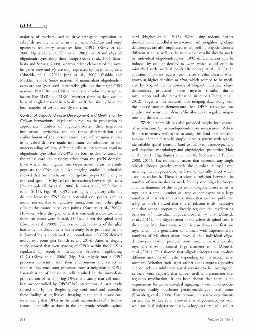
with its characteristic paranodal loops (Snaidero et al., 2014)
(Fig. 3A). This work very nicely exemplifies how in vivo live
imaging can help understand fundamental biological
principles.
A multitude of zebrafish reporters exist for virtually
every cell type and subcellular structure, primarily due to the
relative straightforwardness of generating reporter lines. The
dominating strategy in recent times is transgenesis via Tol2
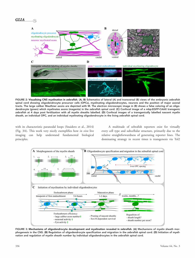
FIGURE 2: Visualising CNS myelination in zebrafish. (A, B) Schematics of lateral (A) and transversal (B) views of the embryonic zebrafishspinal cord showing oligodendrocyte precursor cells (OPCs), myelinating oligodendrocytes, neurons and the position of major axonaltracts. The large caliber Mauthner axons are depicted with M. The electron microscopic image in (B) shows a false coloring of an oligo-dendrocyte (green) which myelinates axons (magenta) in the zebrafish spinal cord. (C) Confocal image of a mbp:EGFP-CAAX transgeniczebrafish at 4 days post fertilization with all myelin sheaths labelled. (D) Confocal images of a transgenically labelled nascent myelinsheath, an individual OPC, and an individual myelinating oligodendrocyte in the living zebrafish spinal cord.
FIGURE 3: Mechanisms of oligodendrocyte development and myelination revealed in zebrafish. (A) Mechanisms of myelin sheath mor-phogenesis in the CNS. (B) Regulation of oligodendrocyte specification and migration in the zebrafish spinal cord. (D) Initiation of myeli-nation and regulation of myelin sheath number by individual oligodendrocytes in the zebrafish spinal cord.
336 Volume 64, No. 3

transposable elements (Kawakami, 2007). Tol2 sites have
been used to generate enhancer trap lines in which a reporter
(a fluorescent protein or a Gal4 transactivator) with a mini-
mal promoter randomly integrates into the genome. The
reporter gene will be expressed when it integrates in the prox-
imity of the enhancer of an endogenous gene (Korzh, 2007;
Asakawa and Kawakami, 2009). Collections of such enhancer
trap lines like the zTrap database provided by the Kawakami
laboratory (http://kawakami.lab.nig.ac.jp/ztrap/) (Kawakami
et al., 2010) consist of hundreds of different lines with
expression in distinct cell types. Lines with expression in sub-
sets of neurons have been used in several studies to elucidate
neuronal functions. Enhancer trap lines that are specific for
glial cells have not yet been reported, most likely because sys-
tematic screening for these cell types simply has not been car-
ried out so far. The identification of such reporters is thus
subject to further investigation and may even lead to the
detection of labelled subsets of oligodendrocytes as is the case
for neurons, furthering future studies on myelination using
zebrafish.
Toolsets to express any gene in a defined tissue and cell
type have also been developed. The most commonly used sys-
tem is the Tol2Kit developed by the Chien laboratory (Kwan
et al., 2007). This combines Tol2 transgenesis with recombi-
nation based Gateway cloning to generate transgenic expres-
sion constructs of any gene of interest under the control of a
desired upstream regulatory sequence in a simple overnight
reaction. Injection into fertilized zebrafish eggs results in
mosaic expression of the transgene, which allows analysis of
morphology and dynamics of single cells. Growing up
injected animals followed by screening for germline transmis-
sion of the transgene to the next generation can generate sta-
ble full transgenic lines. Suites of transgenic reagents have
been generated to visualize oligodendrocyte lineage cells at
different stages of their differentiation and neuronal subsets
using distinct fluorescent proteins (Table 1, Fig. 2). The vast
TABLE 1: Transgenic Reporter Lines to Label Oligodendrocyte Lineage Cells
Gene Labelled cell type(s) Line name, reference Notes
olig1 Oligodendrocyte lineage cells Tg(olig1:mEGFP)nv150Tg,Schebesta and Serluca , 2009
olig2 pMN domain, oligodendrocytelineage cells, motor neurons
Tg (olig2:EGFP)vu12Tg,Shin et al., 2003Tg(olig2:dsRed)vu19Tg,Kucenas et al., 2008Tg(olig2:Kaede)vu85Tg,Zannino and Appel, 2009
nkx2.2a Subset of OPCs and earlymyelinating oligodendrocytes,various non-related cell types
Tg(nkx2.2a:mEGFP)vu16Tg,Ng et al., 2005
sox10 Oligodendrocyte lineage cells,some interneurons, neural crestderivatives including Schwann cells
Tg(sox10:mRFP)vu234Tg,Kirby et al., 2006
Length of sox10 promoterfragment varies between lines
Tg(sox10:EGFP)ba4Tg,Dutton et al., 2008Tg(sox10:nls-Eos)w18Tg,Prendergast et al., 2012
mpz (p0) Myelinating oligodendrocytes,some non-related cell types
Tg(mpz[10kb]:EGFP)pt408Tg,Bai et al., 2014
plp Oligodendrocyte lineage cells,some non-related cell types
Tg(Mmu.Plp1:EGFP)cc1Tg,Yoshida and Macklin, 2005
Promoter fragmentof mouse plp gene
mbp Myelinating oligodendrocytesand Schwann cells
Tg(mbp:EGFP)ck1Tg,Jung et al., 2009Tg(mbp:EGFP)ue1Tg,Almeida et al., 2011Tg(mbp:EGFP-CAAX)ue2Tg,Almeida et al., 2011
claudinK Myelinating oligodendrocytesand Schwann cells
Tg(cldk:Gal4)ue101Tg,M€unzel et al., 2011
Czopka: CNS Myelination in Zebrafish
March 2016 337
majority of markers used to drive transgene expression in
zebrafish are the same as in mammals. Nkx2.2a and olig2
upstream regulatory sequences label OPCs (Kirby et al.,
2006; Ng et al., 2005; Shin et al., 2003), sox10 and olig1 all
oligodendrocytes along their lineage (Kirby et al., 2006; Sche-
besta and Serluca, 2009), whereas driver elements of the mye-
lin genes mbp and plp are only expressed by myelinating cells
(Almeida et al., 2011; Jung et al., 2009; Yoshida and
Macklin, 2005). Some markers of mammalian oligodendro-
cytes are not (yet) used in zebrafish glia like the major OPC
markers PDGFRa and NG2, and key myelin transcription
factors like MYRF (or MRF). Whether these markers cannot
be used as glial marker in zebrafish or if they simply have not
been established yet is presently not clear.
Control of Oligodendrocyte Development and Myelination by
Cellular Interactions. Myelination requires the production of
appropriate numbers of oligodendrocytes, their migration
into axonal territories, and the timed differentiation and
ensheathment of the correct axons. Live cell imaging studies
using zebrafish have made important contributions to our
understanding of how different cellular interactions regulate
oligodendrocyte behavior. OPCs are born in distinct areas (in
the spinal cord the majority arises from the pMN domain)
from where they migrate into target axonal areas to evenly
populate the CNS tissue. Live imaging studies in zebrafish
showed that one mechanism to regulate proper OPC migra-
tion and spacing is by cell-cell interactions between glial cells
[for example (Kirby et al., 2006; Kucenas et al., 2009; Smith
et al., 2014), Fig. 3B]. OPCs are highly migratory cells but
do not leave the CNS along potential exit points such as
motor nerves, due to repulsive interactions with other glial
cells at the motor nerve exit points (Kucenas et al., 2009).
However, when the glial cells that ensheath motor axons at
these exit zones were ablated, OPCs did exit the spinal cord
(Kucenas et al., 2009). The exact cellular identity of this glial
barrier is not clear, but it has recently been proposed that it
is formed by a specialized cell population of CNS derived
motor exit point glia (Smith et al., 2014). Another elegant
study showed that even spacing of OPCs within the CNS is
regulated by repulsive interactions between neighboring
OPCs (Kirby et al., 2006) (Fig. 3B). Highly motile OPC
processes constantly scan their environment and retract as
soon as they encounter processes from a neighboring OPC.
Laser-ablation of individual cells resulted in the immediate
proliferation of neighboring OPCs, indicating that cell num-
bers are controlled by OPC-OPC interactions. A later study
carried out by the Bergles group confirmed and extended
these findings using live cell imaging in the adult mouse cor-
tex showing that OPCs in the adult mammalian CNS behave
almost identically to those in the embryonic zebrafish spinal
cord (Hughes et al., 2013). Work using rodents further
showed that intercellular interactions with neighboring oligo-
dendrocytes are also implicated in controlling oligodendrocyte
differentiation as well as the number of myelin sheaths made
by individual oligodendrocytes. OPC differentiation can be
induced by cellular density in vitro, which could even be
mimicked with artificial beads (Rosenberg et al., 2008). In
addition, oligodendrocytes form fewer myelin sheaths when
grown at higher densities in vitro, which seemed to be medi-
ated by Nogo-A. In the absence of Nogo-A individual oligo-
dendrocytes produced more myelin sheaths during
myelination and also remyelination in mice (Chong et al.,
2012). Together, the zebrafish live imaging data along with
the mouse studies demonstrate that OPCs recognize one
another and sense their density/distribution to regulate migra-
tion and differentiation.
Work in zebrafish has also provided insight into control
of myelination by axon-oligodendrocyte interactions. Zebra-
fish are extremely well suited to study this kind of interaction
because of their relatively simple nervous system with readily
identifiable spinal neurons (and axons) with stereotypic and
well described morphology and physiological properties (Hale
et al., 2001; Higashijima et al., 2004; McLean and Fetcho,
2008, 2011). The number of axons that surround any single
oligodendrocyte greatly exceeds the number it myelinates,
meaning that oligodendrocytes have to carefully select which
axon to ensheath. There is a clear correlation between the
number of myelin sheaths made by any one oligodendrocyte
and the diameter of the target axon. Oligodendrocytes either
myelinate a small number of large caliber axons or a large
number of relatively thin axons. Work that we have published
using zebrafish showed that this correlation is also causative
and that axonal properties directly regulate the myelinating
behavior of individual oligodendrocytes in vivo (Almeida
et al., 2011). The biggest axon of the zebrafish spinal cord is
the unique Mauthner axon, which is also always the first one
myelinated. The generation of animals with supernumerary
numbers of Mauthner axons revealed that individual oligo-
dendrocytes readily produce more myelin sheaths to also
myelinate these additional large diameter axons (Almeida
et al., 2011). This showed that oligodendrocytes can produce
different amounts of myelin depending on the axonal envi-
ronment. Whether such larger caliber axons express a positive
cue or lack an inhibitory signal remains to be investigated.
In vitro work suggests that caliber itself is a parameter that
regulates myelination. It has been shown that there is no
requirement for active axo-glial signaling in vitro as oligoden-
drocytes readily myelinate paraformaldehyde fixed axons
(Rosenberg et al., 2008). Furthermore, innovative experiments
carried out by Lee et al. showed that oligodendrocytes even
wrap artificial polystyrene fibers, as long as they had a caliber
338 Volume 64, No. 3

greater than 0.4mm (Lee et al., 2012a). This indicates that
size is a simple biophysical property that contributes to regu-
lation of myelination by oligodendrocytes.
Zebrafish live imaging also provided novel information
into the cell biology of myelin sheath formation during the
lifetime of individual oligodendrocytes. New myelin is formed
over long periods of time and the number of myelin sheaths
produced by each cell is plastic. However, it remained unclear
when oligodendrocytes can exert such plasticity. We could
show that, regardless of when and where in the zebrafish spi-
nal cord an OPC differentiated, each individual cell had only
a short but very dynamic time window of no more than 5 h
to form its full complement of myelin sheaths after having
made its first one (Czopka et al., 2013). After this time, a
cell made no new myelin sheaths (Fig. 3C). It remains to be
seen how long such time window of myelination lasts for any
one oligodendrocyte in higher vertebrates, given that mamma-
lian development generally occurs at slower rates. However,
there is evidence from in vitro studies that such a short time
window also exists in rodents. Cultured retinal ganglion cell
axons become myelinated when co-cultured with isolated
OPCs but not when mature oligodendrocytes were used
(Watkins et al., 2008). Time-lapse imaging showed that also
these co-cultured OPCs stopped generating new sheaths 12 h
after the start of imaging. Furthermore, juvenile mice that
were socially isolated during a short critical period showed
behavioral deficits and impaired myelination due to reduced
myelin sheath numbers per oligodendrocyte (Makinodan
et al., 2012). Very interestingly, these defects were long lasting
and irreversible, indicating that myelinating oligodendrocytes
cannot form new myelin sheaths in mice in vivo. Irrespective
of the exact duration of myelination competence by an indi-
vidual oligodendrocyte, it will be interesting to determine
what triggers the first ensheathment and which mechanisms
set the clock that limit myelination competence. The exis-
tence of a short time window for an oligodendrocyte to gen-
erate new myelin sheaths means that the decision whether to
ensheath an axon or not has to occur during this time. Work
in zebrafish has shown that following their myelination phase,
oligodendrocyte dynamics are limited to occasional sheath
retractions, which has also been termed myelin pruning
(Czopka et al., 2013; Liu et al., 2013). The meaning of these
myelin sheath retractions is currently unclear, but one possi-
bility is that they reflect regulatory mechanisms to correct for
having ensheathed a “wrong” axon. This would be plausible
given that each oligodendrocyte can only form new myelin
sheaths for a short time, whereas it takes much longer to fully
myelinate an axon along its length. Therefore, more myelin
sheaths than required could initially be deposited along the
length of an axon, which then, as sheaths grow in length,
requires pruning of some sheaths to ultimately obtain appro-
priate nodal spacing and conduction speed.
Which signals tell right from wrong axon at a given
time? Axon caliber is a positive regulator of myelination fate.
However, in contrast to the PNS where only axons with a
cross-sectional diameter of �1 mm are myelinated, no such
threshold exists in the CNS where axons as thin as 200 nm
can get myelinated, indicating that other regulatory mecha-
nisms are likely to also play a role. There is accumulating evi-
dence that neuronal activity is one mechanism underlying
control of axonal myelination fate. Several rodent in vitro and
in vivo studies could demonstrate changes in OPC prolifera-
tion, differentiation and myelination upon interference with
neuronal firing (Barres and Raff, 1993; Demerens et al.,
1996; Fields, 2010; Gibson et al., 2014; Wake et al., 2011).
Two very recent zebrafish publications provided the first evi-
dence that neuronal activity directly affects myelination
behavior of oligodendrocytes in vivo (Hines et al., 2015;
Mensch et al., 2015). Blocking synaptic vesicle release using
tetanus toxin reduced myelination in zebrafish embryos, pri-
marily because individual oligodendrocytes generated fewer
myelin sheaths per cell during their time window of active
ensheathment (Mensch et al., 2015). When neuronal firing
was increased by pharmacological disinhibition, oligodendro-
cytes produced more myelin sheaths. In addition to these
global loss- and gain of function approaches of neuronal
activity, myelination fate of individual axons was also
impaired when synaptic vesicle release was blocked in individ-
ual axons (Hines et al., 2015; Mensch et al., 2015). The
study by Hines et al. further revealed that nascent sheaths
that did form on such single silenced axons were also shorter
in length. Very interestingly, this reduced sheath length was
rescued by suppressing the activity of neighboring axons,
indicating mechanisms of activity dependent competition for
myelination between axons (Hines et al., 2015). Time-lapse
analysis also showed that nascent sheaths were formed on
silenced axons at similar rates, but then failed to grow and
retracted more frequently, meaning that it is the stabilization
and maintenance of a newly formed ensheathment rather
than its initiation that is regulated by synaptic vesicle release
(Hines et al., 2015). Previous work in rodents demonstrated
the existence of synapse-like structures between OPCs and
axons, and it has also been shown that OPCs respond to glu-
tamate and other transmitters with inward currents (Bergles
et al., 2000, 2010; Maldonado and Angulo, 2015), indicating
active communication between axon and oligodendrocyte
prior to ensheathment. The precise role of these axon-OPC
synapses in regulating myelination is not known. NMDA-
type glutamate receptors in oligodendrocytes are dispensable
for myelination in mice (De Biase et al., 2011). In contrast,
it was shown that glutamate release can stimulate
Czopka: CNS Myelination in Zebrafish
March 2016 339
oligodendrocyte differentiation and myelination in a co-
culture system of OPCs and dorsal root ganglion neurons
(Wake et al., 2011). Wake et al. also demonstrated that this
glutamate mediated increase in myelin expression involved
activation of Fyn, a tyrosine kinase in oligodendrocytes which
acts as glial integrator of axonal cues by regulating cytos-
keletal dynamics as well as local mbp mRNA translation, at
least in vitro (Klein et al., 2002; Kr€amer-Albers and White,
2011; White et al., 2008a). Very interestingly, in a separate
study in zebrafish, we could show that increasing Fyn activity
in oligodendrocytes also increased myelin sheath number per
cell, whereas knock-down of Fyn had the opposite effect
(Czopka et al., 2013), very similar to what has been seen after
manipulation of neuronal activity by Mensch et al. It is
unclear if these effects are indeed mediated by glutamate and
Fyn as in vitro studies suggest, or by other neurotransmitters
that are known to act on oligodendrocytes, for example
GABA and ATP (Karadottir and Attwell, 2007). However,
zebrafish with their identifiable neurons with defined neuro-
transmitter phenotypes can be used to study individual
myelination events of neuronal subtypes. Optogenetic manip-
ulation of defined neurons is relatively straightforward in
zebrafish allowing for direct control of their activity. There-
fore, zebrafish represent a useful model for further and
detailed elucidation of the connection between neuronal firing
and myelination in the CNS.
Zebrafish as Screening Model Organism to DiscoverNew Molecular Functions
Forward Genetic Screening. Historically the zebrafish owes
its popularity to its suitability to carry out gene discovery
screens. It was the first vertebrate model organism in which
large scale forward genetic screens has been carried out. Two
parallel screens for disrupted tissue morphogenesis, one car-
ried out by the N€usslein-Volhard group (Haffter et al., 1996)
and another one by the Driever group (Driever et al., 1996),
led to the discovery of thousands of mutants with unique
phenotypes. With the analysis and identification of the causa-
tive mutations in subsequent years the zebrafish has become
well-established as a prime vertebrate model to carry out
genetic screens, a method that is still unchallenged for the
unbiased discovery of new gene functions for many biological
questions. The principle of a genetic screen is based on the
induction of random mutations, followed by phenotypic
screening, and the subsequent identification of the causative
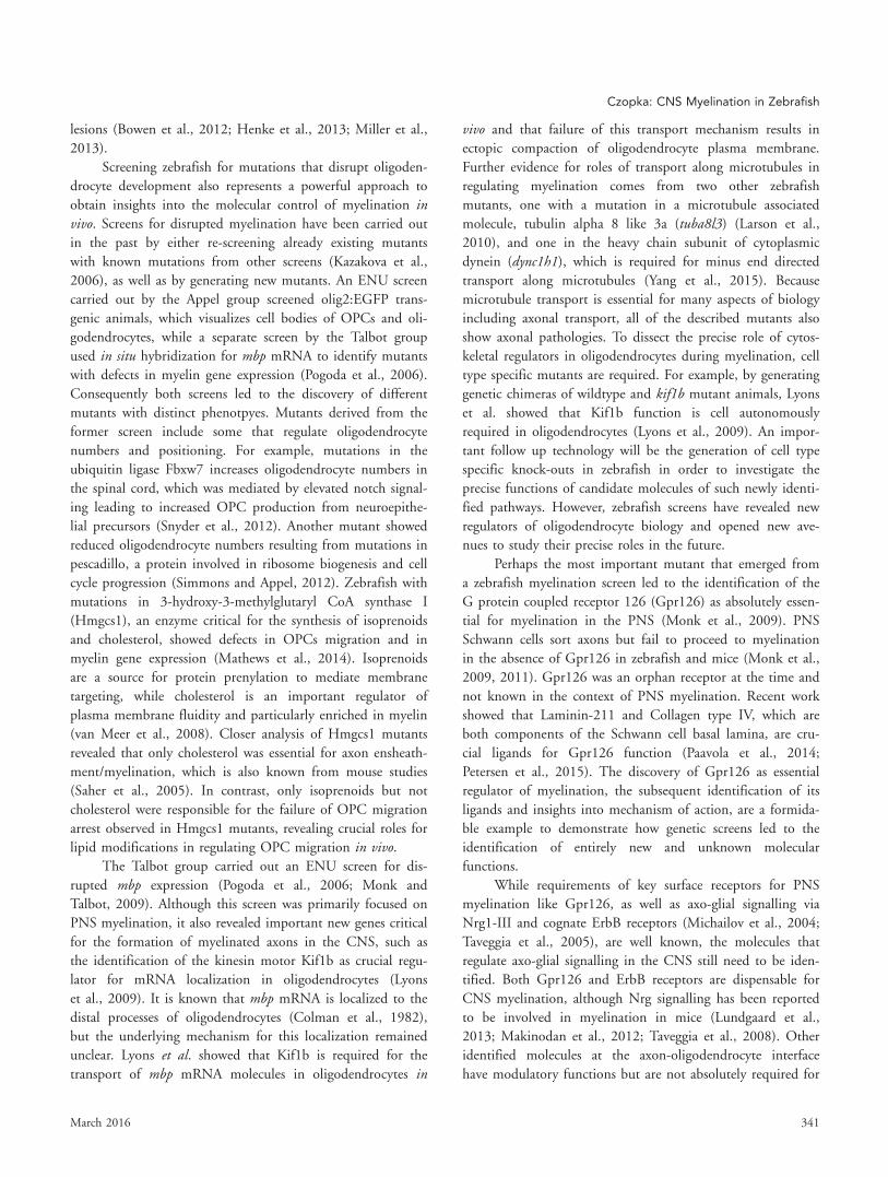
lesion in mutants of interest [reviewed by (Patton and Zon,
2001)]. In most genetic screens, random point mutations are
induced by N-ethyl-N-nitrosourea (ENU) in F0 animals and
then bred to homozygosity in a three-generation breeding
regime (Fig. 4). In such a breeding scheme, each F1 animal
that emerged from a mutagenized F0 parent represents a
unique mutagenized genome and is the foundation of F2
“families.” Intercrossing F2 individuals heterozygous for any
mutation will give 25% homozygous F3 animals. Screening
thousands of F3 animals derived from many different F2
families will allow to discover mutants with a phenotype of
interest (such as disrupted myelination). The causative muta-
tion in mutants of interest is identified retrospectively by
PCR based linkage mapping analysis and DNA sequencing
(Postlethwait and Talbot, 1997), and since more recently
whole genome/whole exome and RNA sequencing, which
have become efficient and affordable ways to map genetic
FIGURE 4: Methods of target discovery and validation in zebra-fish. The schematics show how new molecular targets can be dis-covered by genetic and chemical screening in zebrafish, followedby the subsequent validation using reverse genetic approaches.
340 Volume 64, No. 3
lesions (Bowen et al., 2012; Henke et al., 2013; Miller et al.,
2013).
Screening zebrafish for mutations that disrupt oligoden-
drocyte development also represents a powerful approach to
obtain insights into the molecular control of myelination invivo. Screens for disrupted myelination have been carried out
in the past by either re-screening already existing mutants
with known mutations from other screens (Kazakova et al.,
2006), as well as by generating new mutants. An ENU screen
carried out by the Appel group screened olig2:EGFP trans-
genic animals, which visualizes cell bodies of OPCs and oli-
godendrocytes, while a separate screen by the Talbot group
used in situ hybridization for mbp mRNA to identify mutants
with defects in myelin gene expression (Pogoda et al., 2006).
Consequently both screens led to the discovery of different
mutants with distinct phenotpyes. Mutants derived from the
former screen include some that regulate oligodendrocyte
numbers and positioning. For example, mutations in the
ubiquitin ligase Fbxw7 increases oligodendrocyte numbers in
the spinal cord, which was mediated by elevated notch signal-
ing leading to increased OPC production from neuroepithe-
lial precursors (Snyder et al., 2012). Another mutant showed
reduced oligodendrocyte numbers resulting from mutations in
pescadillo, a protein involved in ribosome biogenesis and cell
cycle progression (Simmons and Appel, 2012). Zebrafish with
mutations in 3-hydroxy-3-methylglutaryl CoA synthase I
(Hmgcs1), an enzyme critical for the synthesis of isoprenoids
and cholesterol, showed defects in OPCs migration and in
myelin gene expression (Mathews et al., 2014). Isoprenoids
are a source for protein prenylation to mediate membrane
targeting, while cholesterol is an important regulator of
plasma membrane fluidity and particularly enriched in myelin
(van Meer et al., 2008). Closer analysis of Hmgcs1 mutants
revealed that only cholesterol was essential for axon ensheath-
ment/myelination, which is also known from mouse studies
(Saher et al., 2005). In contrast, only isoprenoids but not
cholesterol were responsible for the failure of OPC migration
arrest observed in Hmgcs1 mutants, revealing crucial roles for
lipid modifications in regulating OPC migration in vivo.
The Talbot group carried out an ENU screen for dis-
rupted mbp expression (Pogoda et al., 2006; Monk and
Talbot, 2009). Although this screen was primarily focused on
PNS myelination, it also revealed important new genes critical
for the formation of myelinated axons in the CNS, such as
the identification of the kinesin motor Kif1b as crucial regu-
lator for mRNA localization in oligodendrocytes (Lyons
et al., 2009). It is known that mbp mRNA is localized to the
distal processes of oligodendrocytes (Colman et al., 1982),
but the underlying mechanism for this localization remained
unclear. Lyons et al. showed that Kif1b is required for the
transport of mbp mRNA molecules in oligodendrocytes in
vivo and that failure of this transport mechanism results in
ectopic compaction of oligodendrocyte plasma membrane.
Further evidence for roles of transport along microtubules in
regulating myelination comes from two other zebrafish
mutants, one with a mutation in a microtubule associated
molecule, tubulin alpha 8 like 3a (tuba8l3) (Larson et al.,
2010), and one in the heavy chain subunit of cytoplasmic
dynein (dync1h1), which is required for minus end directed
transport along microtubules (Yang et al., 2015). Because
microtubule transport is essential for many aspects of biology
including axonal transport, all of the described mutants also
show axonal pathologies. To dissect the precise role of cytos-
keletal regulators in oligodendrocytes during myelination, cell
type specific mutants are required. For example, by generating
genetic chimeras of wildtype and kif1b mutant animals, Lyons
et al. showed that Kif1b function is cell autonomously
required in oligodendrocytes (Lyons et al., 2009). An impor-
tant follow up technology will be the generation of cell type
specific knock-outs in zebrafish in order to investigate the
precise functions of candidate molecules of such newly identi-
fied pathways. However, zebrafish screens have revealed new
regulators of oligodendrocyte biology and opened new ave-
nues to study their precise roles in the future.
Perhaps the most important mutant that emerged from
a zebrafish myelination screen led to the identification of the
G protein coupled receptor 126 (Gpr126) as absolutely essen-
tial for myelination in the PNS (Monk et al., 2009). PNS
Schwann cells sort axons but fail to proceed to myelination
in the absence of Gpr126 in zebrafish and mice (Monk et al.,
2009, 2011). Gpr126 was an orphan receptor at the time and
not known in the context of PNS myelination. Recent work
showed that Laminin-211 and Collagen type IV, which are
both components of the Schwann cell basal lamina, are cru-
cial ligands for Gpr126 function (Paavola et al., 2014;
Petersen et al., 2015). The discovery of Gpr126 as essential
regulator of myelination, the subsequent identification of its
ligands and insights into mechanism of action, are a formida-
ble example to demonstrate how genetic screens led to the
identification of entirely new and unknown molecular
functions.
While requirements of key surface receptors for PNS
myelination like Gpr126, as well as axo-glial signalling via
Nrg1-III and cognate ErbB receptors (Michailov et al., 2004;
Taveggia et al., 2005), are well known, the molecules that
regulate axo-glial signalling in the CNS still need to be iden-
tified. Both Gpr126 and ErbB receptors are dispensable for
CNS myelination, although Nrg signalling has been reported
to be involved in myelination in mice (Lundgaard et al.,
2013; Makinodan et al., 2012; Taveggia et al., 2008). Other
identified molecules at the axon-oligodendrocyte interface
have modulatory functions but are not absolutely required for
Czopka: CNS Myelination in Zebrafish
March 2016 341
mediating ensheathment, like Lingo-1 (Lee et al., 2007; Mi
et al., 2005) and PSA-NCAM (Charles et al., 2000; Fewou
et al., 2007). Future genetic screens in zebrafish that are
designed to identify mutations that specifically disrupt CNS
myelination are a promising approach to identify new and
essential molecules to regulate axon ensheathment by oligo-
dendrocytes. However, approximately 30% of all zebrafish
genes exist as duplicated ohnologs and it is known that some
of them are co-expressed in the same cell where they fulfill
the same function. Thus, it cannot be excluded that mutagen-
esis screens in zebrafish will not reveal some genes with
important roles for CNS myelination. It also remains possible
that there might not exist a single molecule/receptor/ligand
that is absolutely required for oligodendrocytes to myelinate,
but that it is rather a combination of molecules, a protein
complex, or even multiple pathways that regulate whether to
myelinate an axon or not. For these reasons other screening
approaches will be required to identify new targets important
to myelination.
Chemical Screening. Like genetic screening, chemical
screening enables high throughput phenotyping of many dif-
ferent manipulations on a specific biological question. In
genetic screens, however, function disrupting mutations are
always assessed in a single gene at a time. In contrast, small
molecule compounds used in chemical screens can simultane-
ously target multiple proteins (including duplicated ones) and
signalling pathways by e.g., acting on common binding sites
required for function. Chemical screening also provides tem-
poral conditional flexibility of drug application, offering the
ability to identify distinct effects at early and late stages of
development which one would miss by genetic disruption at
all stages. Zebrafish are a pre-eminent model to carry out
chemical screens for driving discovery of new targets, investi-
gation of structure activity relationships, and toxicology stud-
ies in vivo (Zon and Peterson, 2005). The external
development, aquatic nature, and small size of embryonic
zebrafish allows simple drug application in small volumes.
There is certainly no other vertebrate model with which to
screen hundreds to thousands of different compounds in liv-
ing animals in 96-well formats (Figure 4). Over 60 chemical
screens on different aspects of vertebrate development have
been reported over the past 15 years [for a review, see (Ren-
nekamp and Peterson, 2015)]. Some led to the discovery of
new targets that are currently being tested to treat human dis-
eases. For example, a zebrafish chemical screen revealed that
compounds acting on Prostaglandin E2 synthesis regulate
hematopoietic stem cell homeostasis (North et al., 2007). A
respective drug called ProHema is currently in phase II clini-
cal trials for treatment of patients with leukemia and lym-
phoma (Hagedorn et al., 2014). This is the first example of a
drug initially discovered in a zebrafish screen that advanced
to use in humans.
Enhancing oligodendrocyte differentiation is an often
proposed strategy to improve functional recovery and to pre-
vent axon degeneration following demyelination after injury
or in diseases like MS (Franklin and ffrench-Constant, 2008).
Three independent rodent in vitro chemical screens have
recently been carried out leading to the identification of com-
pounds that enhance oligodendrocyte differentiation, myelina-
tion and remyelination (Deshmukh et al., 2013; Mei et al.,
2014; Najm et al., 2015). Identified compounds from two
studies act on muscarinic acetylcholine receptors (Deshmukh
et al., 2013; Mei et al., 2014), whereas compounds from the
third study involved glucocorticoid receptor signalling to
increase oligodendrocyte differentiation (Najm et al., 2015).
While glucocorticoids are known for regulating oligodendro-
cyte development (Barres et al., 1994), roles for muscarinic
acetylcholine receptors in regulating oligodendrocyte differen-
tiation had not been reported previously, which exemplifies
the power of unbiased screening to discover novel targets.
Chemical screens in zebrafish are an attractive strategy
to identify compounds that regulate myelination in a more
complex environment. In vivo screening allows immediate
detection of potentially deleterious side effects, which provide
important information on the specificity and toxicity of a
given compound. Buckley et al. have previously carried a
drug re-profiling screen for pro-myelinating compounds in
zebrafish (Buckley et al., 2010b). Screening of an olig2:EGFP
transgenic line showed that 2% of all screened compounds
altered the number of dorsally migrated olig2 expressing cells
in the spinal cord. Subsequent secondary and tertiary screens
revealed that also myelination was altered by some of these
compounds, for example, the Src family kinase inhibitor PP2,
showing that screening zebrafish for compounds that alter
myelination is possible. There is a continuously growing
number of available chemical libraries, some of known bioac-
tivity, some which are already approved by the US Food and
Drug Administration (FDA) to be safe for use in humans,
and even natural compound libraries. Screening such diverse
libraries will increase the spectrum to find novel targets and
pathways to regulate myelination. Moreover, automated
screening platforms such as the VAST Bioimager (for Verte-
brate Automated Screening Technology) which automatically
loads larval zebrafish from multiwell plates and positions
them under a microscope have been developed (Pardo-Martin
et al., 2010; Tamplin and Zon, 2010). The availability of
such automated platforms in combination with myelin spe-
cific zebrafish reporter lines may allow to carry out high
throughput in vivo screening and aid discover novel targets
that regulate myelination in the future. Following screening,
342 Volume 64, No. 3
identified candidate targets of such chemicals should be con-
firmed genetically by loss of function analyses.
Reverse Genetics. For a long time the zebrafish field has
lagged behind with technologies for targeted inactivation of a
desired gene. The use of synthetic antisense morpholinos to
either block mRNA splicing or translation of a protein of
interest was the most common approach for many years (Bill
et al., 2009; Nasevicius and Ekker, 2000). Morpholinos can
be a simple and efficient way to study gene functions; how-
ever, they are prone to give misleading results. It was recently
shown that 80% of published morphant phenotypes were not
recapitulated in zebrafish with function disrupting mutations
in the respective gene (Kok et al., 2015; Stainier et al., 2015).
While it remains to be seen to what extent this discrepancy
can be due to compensatory mechanisms in mutants, it is
clear (and not novel) that antisense morpholino approaches
always require careful controls [see for example (Eisen and
Smith, 2008)]. Furthermore, various reverse genetic
approaches are meanwhile available in zebrafish. Community-
wide efforts like the zebrafish mutation project at the Sanger
institute (Cambridge, UK) aim to generate mutant lines for
every protein coding gene and many mutants are already
available (Kettleborough et al., 2014). In addition, the devel-
opment of genome editing technologies using Zinc finger
nucleases, TALENs, and most recently CRISPR/Cas9 now
make it possible to generate functional knock-out and knock-
in zebrafish [for review, (Gaj et al., 2013)]. Very recently, also
the generation of cell type specific knockout zebrafish has
been reported using CRISPR/Cas9 (Ablain et al., 2015).
However, more work is needed in the future to address
potential pitfalls in the generation of such knock-outs.
The quick and straightforward generation of loss of
function animals will likely be the future standard to validate
roles of candidate molecules that emerged from genetic and
chemical screens, and enable to genetically test the involve-
ment of candidate pathways in regulating distinct aspects of
myelination. Zebrafish knock-outs in desired genes will also
allow for more detailed analysis of cell biological principles
underlying reported myelin phenotypes from rodent studies.
For example, Sox10 mutant mice do not have myelinating
oligodendrocytes (Stolt et al., 2002). Live imaging studies in
colorless mutant zebrafish, which lack functional Sox10 pro-
tein (Dutton et al., 2001), have shown that oligodendrocytes
lacking Sox10 develop normally but die shortly after they
ensheathed target axons (Takada et al., 2010). This suggests
that the role of Sox10 function in oligodendrocytes is differ-
ent prior to and after initiation of myelination and extends
our understanding of Sox10 function. In the future, the avail-
ability of (cell type specific) function disrupting mutants for
any protein coding gene will make zebrafish an even more
attractive vertebrate model to study cellular and genetic con-
trol of myelination.
Zebrafish as Model to Study RegenerationThe damaged mammalian CNS does not regenerate properly
due to two confounding factors. The first is that most differ-
entiated cells have an intrinsic inability to launch a regenera-
tive programme. The second factor is the formation of an
inhibitory environment surrounding the lesion, which pre-
vents regeneration from newly differentiating precursor cells.
In contrast to mammals, zebrafish do regenerate rather well
and mechanisms of tissue regeneration in zebrafish are widely
studied in many organs including heart, fin, and also the
CNS (Becker and Becker, 2014; Gemberling et al., 2013;
Kizil et al., 2012). Zebrafish do not form an inhibitory scar
like the glial scar in the injured CNS, which allows the study
of cellular responses to injury in an environment that is per-
missive for regeneration (scarless healing). However, in con-
trast to amphibians, which easily regenerate entire
appendages, zebrafish also have regenerative limitations. For
example, not all axons in the zebrafish spinal cord regenerate
equally. The large Mauthner axon does not regenerate after
transection although neighboring axons in the same tract do,
indicating that the Mauthner axon is situated in an environ-
ment permissive for regeneration. Bhatt et al. took advantage
of this circumstance to disentangle intrinsic limitations of
axon regeneration from environmental ones and revealed that
elevated cyclic AMP levels are sufficient to induce an intrinsic
axonal growth program (Bhatt, 2004), an approach initially
shown in mammalian models to promote axon regeneration
(Cai et al., 1999).
The repair of damaged myelin (re-myelination) after
injury and in disease is a regenerative process that can occur
in all vertebrates including humans and is one of the few
truly regenerative capacities of the CNS (Crawford et al.,
2013). However, remyelination is often less efficient than
developmental myelination. An often proposed strategy to
restore function and to prevent neurodegeneration is to
enhance remyelination, which requires that OPCs are avail-
able in sufficient numbers and readily myelinate demyelinated
axons (Franklin et al., 2012). Block of differentiation as well
as lack of OPC availability have been reported in demyeli-
nated lesions and are discussed as possible reasons for remye-
lination failure, and both can be a result of intrinsic and
extrinsic limitations.
Zebrafish can be used as a model to study remyelination
in a non-scaring environment. It is known from stab wound
lesion paradigms in the adult zebrafish telencephalon that
precursor cells dynamically respond to injury and that some
of these cells also express the OPC marker Olig2 (Baumgart
et al., 2012; Kroehne et al., 2011; M€arz et al., 2011).
Czopka: CNS Myelination in Zebrafish
March 2016 343

Chemical demyelination along optic nerves of adult zebrafish
using lysophosphaditylcholine (LPC) results in robust demye-
lination and subsequent remyelination within 4 weeks
(M€unzel et al., 2014). A main characteristic of mammalian
remyelination is the presence of thinner myelin (Blakemore,
1974). Interestingly, myelin thickness recovered fully in young
adults, but not in aged ones (M€unzel et al., 2014). Given
that zebrafish do not form inhibitory scars, it is possible that
‘old’ OPCs have an intrinsically reduced capacity to (re-
)myelinate. Evidence for cell intrinsic senescence comes from
analysis of long-term cultured rat OPCs, which show
increased cell cycle and differentiation times with increased
time of cultivation, very similar to freshly isolated cells that
come from animals of different ages (Tang et al., 2000).
However, the study by M€unzel et al. also reported that
reduced remyelination in aged zebrafish was accompanied by
a reduced macrophage response to LPC induced remyelina-
tion. Work in rodents has shown that macrophages secrete
cytokines that can enhance remyelination (Miron and Frank-
lin, 2014; Miron et al., 2013). In addition, heterochronic
parabiosis studies revealed that monocytes derived from the
young parabiotic partner enhance remyelination in old mice
(Ruckh et al., 2012). It thus remains unclear whether OPCs
in older animals/zebrafish have an intrinsically reduced myeli-
nation capacity.
Analysis of basic cellular principles of oligodendrocyte
behavior following damage and repair to myelinated axons
would help address whether these are similar or fundamen-
tally different to early developmental myelination. Models of
experimental demyelination by targeted cell ablation have
been reported using transgenic expression of nitroreductase in
oligodendrocytes in zebrafish (Chung et al., 2013) and also in
Xenopus tadpoles (Kaya et al., 2012). Nitroreductase induces
cell death by generating a DNA-crosslinking agent upon
exposure to metronidazole (Curado et al., 2008). The avail-
ability of zebrafish pigmentation mutants like casper which
are essentially translucent even as adults (White et al., 2008b)
allow for long-term in vivo live imaging to study basic cellular
principles following damage to myelin. It is still unclear how
exactly OPCs respond to myelin damage, whether there is for
example a minimal threshold of myelin damage to elicit an
OPC response, and if the cellular basis of remyelination (sec-
ondary myelination, or repeated myelination of the same
axon) is the same as during primary myelination. Such studies
may help address the long-standing question of whether
regeneration is a simple recapitulation of development or if
fundamental differences exist.
An increasing number of publications use zebrafish to
model human diseases. While this can be a perfectly reasona-
ble approach in monogenetic diseases in which known muta-
tions lead to a clear phenotype, zebrafish (like many other
animal models) are most likely not suitable to model more
complex human diseases. Myelin diseases like for example
MS, to which genetic as well as environmental risk factors
contribute to, can almost certainly not be modeled in zebra-
fish embryos or larvae. The innate immune response is well
studied in young zebrafish, very similar to that of mammali-
ans, and has helped a great deal to understand aspects of
microglial and macrophage biology (Oosterhof et al., 2014;
Sieger and Peri, 2012). Macrophages and microglia play
important roles in remyelination and work in zebrafish may
help obtaining insight into the interplay between oligoden-
drocytes and innate immune response following experimental
damage to myelin. However, mechanisms of B- and T-cell
biology, which are of great relevance for autoimmune diseases
like MS, still need to be investigated in greater detail in
zebrafish regarding their similarity to higher vertebrates (Iwa-
nami, 2014; Langenau and Zon, 2005). Lastly, the presence
of an environment permissive to regeneration as discussed
above is a major difference and should be taken into account
when using zebrafish as model. It is important to not ignore
but rather take advantage of these differences and to use the
zebrafish to address open questions that cannot be disen-
tangled in other models otherwise.
Concluding Remarks and Future Directions
The use of zebrafish has made significant contributions to
our understanding of myelinating cell biology in the CNS.
Live imaging studies have provided fundamental insights into
the dynamics of myelination and how oligodendrocyte behav-
ior is regulated by cellular interactions between glia and neu-
rons in vivo. We are, however, still only beginning to
understand how this leads to the formation of a myelinated
CNS as it is and many open questions need to be addressed
where the zebrafish will serve as a helpful model.
What triggers oligodendrocyte differentiation and the
initiation of axonal ensheathment? Over the last few years,
various genetically encoded reporters and indicators have been
developed to study cellular physiology using optical imaging
methods such as the calcium indicator GCaMP6 (Chen
et al., 2013), neurotransmitter sensors like the glutamate indi-
cator GluSnFR (Marvin et al., 2013). These reporters have
already proven to be of great help for the study of neuronal
function in zebrafish in vivo and equally offer the opportunity
to study direct communication between oligodendrocytes and
neurons to form and maintain a myelinated CNS.
How is myelinating cell behavior regulated by interac-
tions with non-neural cells? Immune cells and the vascula-
ture play important roles during myelination and
remyelination but how they directly affect oligodendrocyte
behavior is not well understood. Innate immunity as well as
vascular biology are intensely studied areas in the zebrafish
344 Volume 64, No. 3

and the availability of respective mutant and reporter lines
may help elucidate how such intersystems interactions regu-
late myelination.
New imaging technologies available such as light-sheet
microscopy, for which no other vertebrate model is better
suited than zebrafish, enables long-term fluorescence imaging
of whole organs and even entire animals (Keller and Ahrens,
2015). This will allow investigation of myelination at a sys-
tems level with high spatial and temporal resolution and may
also help address long-standing questions relating to circuit
formation, heterogeneity of oligodendrocytes and their func-
tional diversity within the CNS.
Genetic screens in the zebrafish have led to the discov-
ery of new molecules important to PNS and CNS myelina-
tion in the past. However, the molecules that mediate
myelinating cell behavior in vivo are still to be elucidated. For
example, axo-glial signaling molecules absolutely essential for
CNS myelination are still not known. Future genetic zebrafish
screens for CNS myelination may help identify such essential
molecular cues. Chemical screens in zebrafish have been
powerful approaches in research areas like hematopoiesis and
will likely be of equal potential when studying CNS myelina-
tion. Furthermore, chemical screening is less sensitive to
genetic redundancy (multiple copies and isoforms) and is
thus very complementary to genetic screening. In contrast to
chemical and genetic approaches, proteomic analysis in zebra-
fish still seems understudied. Recently, however, it was shown
that zebrafish are well suited for analysis of organ specific
proteome maps (Nolte et al., 2014), and also a proteomic
screen for molecules involved in fin regeneration using differ-
ential metabolic labelling using SILAC (stable isotope labeling
by amino acids in cell culture) has been reported (Nolte
et al., 2015). Such approaches may help reveal new targets in
the context of CNS regeneration and remyelination in the
future. Lastly, the development of efficient genome editing
technologies now enables generation of even cell type specific
knock-outs, making the zebrafish also attractive for reverse
genetic approaches.
Together, the zebrafish has been a useful model to study
mechanisms of myelinated axon formation in the past and
will most likely be of even bigger help in the future by com-
bining in vivo live cell analyses with unbiased screening and
reverse genetic approaches.
Acknowledgment
Grant sponsor: Emmy-Noether Fellowship of the Deutsche
Forschungsgemeinschaft; Grant number: DFG, CZ 226/1-1
The author thanks David Lyons, Thomas Misgeld, Leanne
Godinho, and Laura Hoodless for comments on the
manuscript.
ReferencesAblain J, Durand EM, Yang S, Zhou Y, Zon LI. 2015. A CRISPR/Cas9 vectorsystem for tissue-specific gene disruption in zebrafish. Dev Cell 32:756–764.
Almeida RG, Czopka T, ffrench-Constant C, Lyons DA. 2011. Individual axonsregulate the myelinating potential of single oligodendrocytes in vivo. Devel-opment 138:4443–4450.
Asakawa K, Kawakami K. 2009. The Tol2-mediated Gal4-UAS method forgene and enhancer trapping in zebrafish. Methods 49:275–281.
Bai Q, Parris RS, Burton EA. 2014. Different mechanisms regulate expressionof zebrafish myelin protein zero (P0) in myelinating oligodendrocytes and itsinduction following axonal injury. J Biol Chem 289:24114–24128.
Barres BA, Lazar MA, Raff MC. 1994. A novel role for thyroid hormone, glu-cocorticoids and retinoic acid in timing oligodendrocyte development. Devel-opment 120:1097–1108.
Barres BA, Raff MC. 1993. Proliferation of oligodendrocyte precursor cellsdepends on electrical activity in axons. Nature 361:258–260.
Baumgart EV, Barbosa JS, Bally-Cuif L, G€otz M, Ninkovic J. 2012. Stabwound injury of the zebrafish telencephalon: A model for comparative analy-sis of reactive gliosis. Glia 60:343–357.
Becker T, Becker CG. 2014. Axonal regeneration in zebrafish. Curr Opin Neu-robiol 27:186–191.
Bergles DE, Jabs R, Steinh€auser C. 2010. Neuron-glia synapses in the brain.Brain Res Rev 63:130–137.
Bergles DE, Roberts JDB, Somogyi P, Jahr CE. 2000. Glutamatergic synapseson oligodendrocyte precursor cells in the hippocampus. Nature 405:187–191.
Bhatt DH. 2004. Cyclic AMP-induced repair of zebrafish spinal circuits. Sci-ence 305:254–258.
Bill BR, Petzold AM, Clark KJ, Schimmenti LA, Ekker SC. 2009. A primer formorpholino use in zebrafish. Zebrafish 6:69–77.
Blakemore WF. 1974. Pattern of remyelination in the CNS. Nature 249:577–578.
Bowen ME, Henke K, Siegfried KR, Warman ML, Harris MP. 2012. Efficientmapping and cloning of mutations in zebrafish by low-coverage whole-genome sequencing. Genetics 190:1017–1024.
Br€osamle C, Halpern ME. 2002. Characterization of myelination in the devel-oping zebrafish. Glia 39:47–57.
Buckley CE, Marguerie A, Alderton WK, Franklin RJ. 2010a. Temporal dynam-ics of myelination in the zebrafish spinal cord. Glia 58:802–812.
Buckley CE, Marguerie A, Roach AG, Goldsmith P, Fleming A, Alderton WK,Franklin RJM. 2010b. Drug reprofiling using zebrafish identifies novel com-pounds with potential pro-myelination effects. Neuropharmacology 59:149–159.
Cahoy JD, Emery B, Kaushal A, Foo LC, Zamanian JL, Christopherson KS,Xing Y, Lubischer JL, Krieg PA, Krupenko SA, Thompson WJ, Barres BA.2008. A transcriptome database for astrocytes, neurons, and oligodendro-cytes: A new resource for understanding brain development and function.J Neurosci 28:264–278.
Cai D, Shen Y, De Bellard M, Tang S, Filbin MT. 1999. Prior exposure to neu-rotrophins blocks inhibition of axonal regeneration by MAG and myelin via acAMP-dependent mechanism. Neuron 22:89–101.
Charles P, Hernandez MP, Stankoff B, Aigrot MS, Colin C, Rougon G, Zalc B,Lubetzki C. 2000. Negative regulation of central nervous system myelinationby polysialylated-neural cell adhesion molecule. Proc Natl Acad Sci USA 97:7585–7590.
Chen T-W, Wardill TJ, Sun Y, Pulver SR, Renninger SL, Baohan A, SchreiterER, Kerr RA, Orger MB, Jayaraman V, Looger LL, Svoboda K, Kim DS. 2013.Ultrasensitive fluorescent proteins for imaging neuronal activity. Nature 499:295–300.
Chong SYC, Rosenberg SS, Fancy SPJ, Zhao C, Shen Y-AA, Hahn AT, McGeeAW, Xu X, Zheng B, Zhang LI, Rowitch DH, Franklin RJM, Lu QR, Chan JR.
Czopka: CNS Myelination in Zebrafish
March 2016 345

2012. Neurite outgrowth inhibitor Nogo-A establishes spatial segregationand extent of oligodendrocyte myelination. Proc Natl Acad Sci USA 109:1299–1304.
Chung A-Y, Kim P-S, Kim S, Kim E, Kim D, Jeong I, Kim H-K, Ryu J-H, Kim C-H,Choi J, Seo J-H, Park H-C. 2013. Generation of demyelination models by tar-geted ablation of oligodendrocytes in the zebrafish CNS. Mol Cells 36:82–87.
Colman DR, Kreibich G, Frey AB, Sabatini DD. 1982. Synthesis and incorpora-tion of myelin polypeptides into CNS myelin. J Cell Biol 95:598–608.
Crawford AH, Chambers C, Franklin RJM. 2013. Remyelination: The trueregeneration of the central nervous system. J Comp Pathol 149:242–254.
Curado S, Stainier DYR, Anderson RM. 2008. Nitroreductase-mediated cell/tissue ablation in zebrafish: A spatially and temporally controlled ablationmethod with applications in developmental and regeneration studies. NatProtocols 3:948–954.
Czopka T, ffrench-Constant C, Lyons DA. 2013. Individual oligodendrocyteshave only a few hours in which to generate new myelin sheaths in vivo. DevCell 25:599–609.
Dawson MRL, Polito A, Levine JM, Reynolds R. 2003. NG2-expressing glialprogenitor cells: An abundant and widespread population of cycling cells inthe adult rat CNS. Mol Cell Neurosci 24:476–488.
De Biase LM, Kang SH, Baxi EG, Fukaya M, Pucak ML, Mishina M, CalabresiPA, Bergles DE. 2011. NMDA receptor signaling in oligodendrocyte progeni-tors is not required for oligodendrogenesis and myelination. J Neurosci 31:12650–12662.
Demerens C, Stankoff B, Logak M, Anglade P, Allinquant B, Couraud F, ZalcB, Lubetzki C. 1996. Induction of myelination in the central nervous systemby electrical activity. Proc Natl Acad Sci USA 93:9887–9892.
Deshmukh VA, Tardif V, Lyssiotis CA, Green CC, Kerman B, Kim HJ,Padmanabhan K, Swoboda JG, Ahmad I, Kondo T, Gage FH, TheofilopoulosAN, Lawson BR, Schultz PG, Lairson LL. 2013. A regenerative approach tothe treatment of multiple sclerosis. Nature 512:327–332.
Driever W, Solnica-Krezel L, Schier AF, Neuhauss SC, Malicki J, Stemple DL,Stainier DY, Zwartkruis F, Abdelilah S, Rangini Z, Belak J, Boggs C. 1996. Agenetic screen for mutations affecting embryogenesis in zebrafish. Develop-ment 123:37–46.
Dutton JR, Antonellis A, Carney TJ, Rodrigues FSLM, Pavan WJ, Ward A,Kelsh RN. 2008. An evolutionarily conserved intronic region controls the spa-tiotemporal expression of the transcription factor Sox10. BMC Dev Biol 8:105–20.
Dutton KA, Pauliny A, Lopes SS, Elworthy S, Carney TJ, Rauch J, Geisler R,Haffter P, Kelsh RN. 2001. Zebrafish colourless encodes sox10 and specifiesnon-ectomesenchymal neural crest fates. Development 128:4113–4125.
Eisen JS, Smith JC. 2008. Controlling morpholino experiments: Don’t stopmaking antisense. Development 135:1735–1743.
Emery B. 2010. Regulation of oligodendrocyte differentiation and myelina-tion. Science 330:779–782.
Fewou SN, Ramakrishnan H, Bussow H, Gieselmann V, Eckhardt M. 2007.Down-regulation of polysialic acid is required for efficient myelin formation.J Biol Chem 282:16700–16711.
Fields RD. 2010. Neuroscience. Change in the brain’s white matter. Science330:768–769.
Franklin RJ, ffrench-Constant C. 2008. Remyelination in the CNS: From biol-ogy to therapy. Nat Rev Neurosci 9:839–855.
Franklin RJM, ffrench-Constant C, Edgar JM, Smith KJ. 2012. Neuroprotec-tion and repair in multiple sclerosis. Nat Rev Neurol 8:624–634.
F€unfschilling U, Supplie LM, Mahad D, Boretius S, Saab AS, Edgar J,Brinkmann BG, Kassmann CM, Tzvetanova ID, M€obius W, Diaz F, Meijer D,Suter U, Hamprecht B, Sereda MW, Moraes CT, Frahm J, Goebbels S, NaveK-A. 2012. Glycolytic oligodendrocytes maintain myelin and long-term axonalintegrity. Nature 485:521.
Gaj T, Gersbach CA, Barbas CF. 2013. ZFN, TALEN, and CRISPR/Cas-basedmethods for genome engineering. Trends Biotechnol 31:397–405.
Gemberling M, Bailey TJ, Hyde DR, Poss KD. 2013. The zebrafish as a modelfor complex tissue regeneration. Trends Genet 29:611–620.
Gibson EM, Purger D, Mount CW, Goldstein AK, Lin GL, Wood LS, Inema I,Miller SE, Bieri G, Zuchero JB, Barres BA, Woo PJ, Vogel H, Monje M. 2014.Neuronal activity promotes oligodendrogenesis and adaptive myelination inthe mammalian brain. Science. 344:1252304.
Haffter P, Granato M, Brand M, Mullins MC, Hammerschmidt M, Kane DA,Odenthal J, van Eeden FJ, Jiang YJ, Heisenberg CP, Kelsh RN, Furutani-SeikiM, Vogelsang E, Beuchle D, Schach U, Fabian C, Nusslein-Volhard C. 1996.The identification of genes with unique and essential functions in the devel-opment of the zebrafish, Danio rerio. Development 123:1–36.
Hagedorn EJ, Durand EM, Fast EM, Zon LI. 2014. Getting more for your mar-row_ Boosting hematopoietic stem cell numbers with PGE2. Exp Cell Res329:220–226.
Hale ME, Ritter DA, Fetcho JR. 2001. A confocal study of spinal interneuronsin living larval zebrafish. J Comp Neurol 437:1–16.
Henke K, Bowen ME, Harris MP. 2013. Perspectives for identification of muta-tions in the zebrafish: Making use of next-generation sequencing technolo-gies for forward genetic approaches. Methods 62:185–196.
Higashijima S-I, Schaefer M, Fetcho JR. 2004. Neurotransmitter properties of spi-nal interneurons in embryonic and larval zebrafish. J Comp Neurol 480:19–37.
Hines JH, Ravanelli AM, Schwindt R, Scott EK, Appel B. 2015. Neuronal activ-ity biases axon selection for myelination in vivo. Nat Neurosci 18:683–689.
Howe K, Clark MD, Torroja CF, Torrance J, Berthelot C, Muffato M, CollinsJE, Humphray S, McLaren K, Matthews L, McLaren S, Sealy I, Caccamo M,Churcher C, Scott C, Barrett JC, Koch R, Rauch G-J, White S, Chow W, KilianB, Quintais LT, Guerra-Assunc~ao JA, Zhou Y, Gu Y, Yen J, Vogel J-H, Eyre T,Redmond S, Banerjee R, Chi J, Fu B, Langley E, Maguire SF, Laird GK, LloydD, Kenyon E, Donaldson S, Sehra H, Almeida-King J, Loveland J, TrevanionS, Jones M, Quail M, Willey D, Hunt A, Burton J, Sims S, McLay K, Plumb B,Davis J, Clee C, Oliver K, Clark R, Riddle C, Elliot D, Threadgold G, HardenG, Ware D, Begum S, Mortimore B, Kerry G, Heath P, Phillimore B, Tracey A,Corby N, Dunn M, Johnson C, Wood J, Clark S, Pelan S, Griffiths G, SmithM, Glithero R, Howden P, Barker N, Lloyd C, Stevens C, Harley J, Holt K,Panagiotidis G, Lovell J, Beasley H, Henderson C, Gordon D, Auger K,Wright D, Collins J, Raisen C, Dyer L, Leung K, Robertson L, Ambridge K,Leongamornlert D, McGuire S, Gilderthorp R, Griffiths, C, Manthravadi D,Nichol S, Barker G, Whitehead S, Kay M, Brown J, Murnane C, Gray E,Humphries E, Sycamore N, Barker D, Saunders D, Wallis J, Babbage A,Hammond S, Mashreghi-Mohammadi M, Barr L, Martin S, Wray P, EllingtonA, Matthews N, Ellwood M, Woodmansey R, Clark G, Cooper JD, TromansA, Grafham D, Skuce C, Pandian R, Andrews R, Harrison E, Kimberley A,Garnett J, Fosker N, Hall R, Garner P, Kelly D, Bird C, Palmer S, Gehring I,Berger A, Dooley CM, Ersan-€Ur€un Z, Eser C, Geiger H, Geisler M, Karotki L,Kirn A, Konantz J, Konantz M, Oberl€ander M, Rudolph-Geiger S, Teucke M,Lanz C, Raddatz G, Osoegawa K, Zhu B, Rapp A, Widaa S, Langford C, YangF, Schuster SC, Carter NP, Harrow J, Ning Z, Herrero J, Searle SMJ, EnrightA, Geisler R, Plasterk RHA, Lee C, Westerfield M, de Jong PJ, Zon LI,Postlethwait JH, N€usslein-Volhard C, Hubbard TJP, Crollius HR, Rogers J,Stemple DL. 2013. The zebrafish reference genome sequence and its rela-tionship to the human genome. Nature 496:498–503.
Huang B, Wei W, Wang G, Gaertig MA, Feng Y, Wang W, Li X-J, Li S. 2015.Mutant huntingtin downregulates myelin regulatory factor-mediated myelingene expression and affects mature oligodendrocytes. Neuron 85:1212–1226.
Hughes EG, Kang SH, Fukaya M, Bergles DE. 2013. Oligodendrocyte pro-genitors balance growth with self-repulsion to achieve homeostasis in theadult brain. Nat Neurosci 16:668–676.
Iwanami N. 2014. Zebrafish as a model for understanding the evolution ofthe vertebrate immune system and human primary immunodeficiency. ExpHematol 42:697–706.
Jung S-H, Kim S, Chung A-Y, Kim H-T, So J-H, Ryu J, Park H-C, Kim C-H.2009. Visualization of myelination in GFP-transgenic zebrafish. Dev Dyn 239:592–597.
Kang SH, Li Y, Fukaya M, Lorenzini I, Cleveland DW, Ostrow LW, Rothstein JD,Bergles DE. 2013. Degeneration and impaired regeneration of gray matter oli-godendrocytes in amyotrophic lateral sclerosis. Nat Neurosci 16:571–579.
346 Volume 64, No. 3
Karadottir R, Attwell D. 2007. Neurotransmitter receptors in the life anddeath of oligodendrocytes. Neuroscience 145:1426–1438.
Kawakami K. 2007. Tol2: A versatile gene transfer vector in vertebrates.Genome Biol 8:S7.
Kawakami K, Abe G, Asada T, Asakawa K, Fukuda R, Ito A, Lal P, Mouri N,Muto A, Suster ML, Takakubo H, Urasaki A, Wada H, Yoshida M. 2010. zTrap:Zebrafish gene trap and enhancer trap database. BMC Dev Biol 10:105.
Kaya F, Mannioui A, Chesneau A, Sekizar S, Maillard E, Ballagny C, Houel-Renault L, Dupasquier D, Bronchain O, Holtzmann I, Desmazieres A, ThomasJ-L, Demeneix BA, Brophy PJ, Zalc B, Mazabraud A. 2012. Live imaging oftargeted cell ablation in xenopus: A new model to study demyelination andrepair. J Neurosci 32:12885–12895.
Kazakova N, Li H, Mora A, Jessen KR, Mirsky R, Richardson WD, Smith HK.2006. A screen for mutations in zebrafish that affect myelin gene expressionin Schwann cells and oligodendrocytes. Dev Biol 297:1–13.
Keller PJ, Ahrens MB. 2015. Visualizing whole-brain activity and developmentat the single-cell level using light-sheet microscopy. Neuron 85:462–483.
Kettleborough RNW, Busch-Nentwich EM, Harvey SA, Dooley CM, de BruijnE, van Eeden F, Sealy I, White RJ, Herd C, Nijman IJ, F�enyes F, Mehroke S,Scahill C, Gibbons R, Wali N, Carruthers S, Hall A, Yen J, Cuppen E, StempleDL. 2014. A systematic genome-wide analysis of zebrafish protein-codinggene function. Nature 496:494–497.
Kirby BB, Takada N, Latimer AJ, Shin J, Carney TJ, Kelsh RN, Appel B. 2006.In vivo time-lapse imaging shows dynamic oligodendrocyte progenitorbehavior during zebrafish development. Nat Neurosci 9:1506–1511.
Kizil C, Kaslin J, Kroehne V, Brand M. 2012. Adult neurogenesis and brainregeneration in zebrafish. Dev Neurobiol 72:429–461.
Klein C, Kramer E-M, Cardine A-M, Schraven B, Brandt R, Trotter J. 2002.Process outgrowth of oligodendrocytes is promoted by interaction of fynkinase with the cytoskeletal protein tau. J Neurosci 22:698–707.
Kok FO, Shin M, Ni C-W, Gupta A, Grosse AS, van Impel A, Kirchmaier BC,Peterson-Maduro J, Kourkoulis G, Male I, DeSantis DF, Sheppard-Tindell S,Ebarasi L, Betsholtz C, Schulte-Merker S, Wolfe SA, Lawson ND. 2015.Reverse genetic screening reveals poor correlation between morpholino-induced and mutant phenotypes in zebrafish. Dev Cell 32:97–108.
Korzh V. 2007. Transposons as tools for enhancer trap screens in vertebrates.Genome Biol 8:S8.
Kr€amer-Albers E-M, White R. 2011. From axon–glial signalling to myelination:The integrating role of oligodendroglial Fyn kinase. Cell Mol Life Sci 68:2003–2012.
Kroehne V, Freudenreich D, Hans S, Kaslin J, Brand M. 2011. Regenerationof the adult zebrafish brain from neurogenic radial glia-type progenitors.Development 138:4831–4841.
Kucenas S, Snell H, Appel B. 2008. nkx2.2a promotes specification and differ-entiation of a myelinating subset of oligodendrocyte lineage cells in zebra-fish. Neuron Glia Biol 4:71–81.
Kucenas S, Wang WD, Knapik EW, Appel B. 2009. A selective glial barrier atmotor axon exit points prevents oligodendrocyte migration from the spinalcord. J Neurosci 29:15187–15194.
Kwan KM, Fujimoto E, Grabher C, Mangum BD, Hardy ME, Campbell DS,Parant JM, Yost HJ, Kanki JP, Chien C-B. 2007. The Tol2kit: A multisitegateway-based construction kit for Tol2 transposon transgenesis constructs.Dev Dyn 236:3088–3099.
Langenau DM, Zon LI. 2005. The zebrafish: A new model of T-cell and thymicdevelopment. Nat Rev Immunol 5:307–317.
Larson TA, Gordon TN, Lau HE, Parichy DM. 2010. Defective adult oligoden-drocyte and Schwann cell development, pigment pattern, and craniofacialmorphology in puma mutant zebrafish having an alpha tubulin mutation. DevBiol 346:296–309.
Lee S, Leach MK, Redmond SA, Chong SYC, Mellon SH, Tuck SJ, Feng Z-Q,Corey JM, Chan JR. 2012a. A culture system to study oligodendrocyte myeli-nation processes using engineered nanofibers. Nat Meth 9:917–922.
Lee X, Yang Z, Shao Z, Rosenberg SS, Levesque M, Pepinsky RB, Qiu M,Miller RH, Chan JR, Mi S. 2007. NGF regulates the expression of axonalLINGO-1 to inhibit oligodendrocyte differentiation and myelination.J Neurosci 27:220–225.
Lee Y, Morrison BM, Li Y, Lengacher S, Farah MH, Hoffman PN, Liu Y,Tsingalia A, Jin L, Zhang P-W, Pellerin L, Magistretti PJ, Rothstein JD. 2012b.Oligodendroglia metabolically support axons and contribute to neurodegen-eration. Nature 487:443–448.
Li H, Lu Y, Smith HK, Richardson WD. 2007. Olig1 and Sox10 interact synerg-istically to drive myelin basic protein transcription in oligodendrocytes.J Neurosci 27:14375–14382.
Liu J, Casaccia P. 2010. Epigenetic regulation of oligodendrocyte identity.Trends Neurosci 33:193–201.
Liu J, Dietz K, DeLoyht JM, Pedre X, Kelkar D, Kaur J, Vialou V, Lobo MK,Dietz DM, Nestler EJ, Dupree J, Casaccia P. 2012. Impaired adult myelinationin the prefrontal cortex of socially isolated mice. Nat Neurosci 15:1621–1623.
Liu P, Du J-L, He C. 2013. Developmental pruning of early-stage myelin seg-ments during CNS myelination in vivo. Nat Methods 23:962–964.
Lundgaard I, Luzhynskaya A, Stockley JH, Wang Z, Evans KA, Swire M,Volbracht K, Gautier HOB, Franklin RJM, Charles ffrench-Constant, Attwell DK�arad�ottir RT. 2013. Neuregulin and BDNF induce a switch to NMDAreceptor-dependent myelination by oligodendrocytes. Plos Biol 11:e1001743.
Lyons DA, Naylor SG, Scholze A, Talbot WS. 2009. Kif1b is essential formRNA localization in oligodendrocytes and development of myelinatedaxons. Nat Genet 41:854–858.
Makinodan M, Rosen KM, Ito S, Corfas G. 2012. A critical period for socialexperience-dependent oligodendrocyte maturation and myelination. Science337:1357–1360.
Maldonado PP, Angulo MC. 2015. Multiple modes of communicationbetween neurons and oligodendrocyte precursor cells. Neuroscientist 21:266–276.
Marvin JS, Borghuis BG, Tian L, Cichon J, Harnett MT, Akerboom J, GordusA, Renninger SL, Chen T-W, Bargmann CI, Orger MB, Schreiter ER, DembJB, Gan W-B, Hires SA, Looger LL. 2013. An optimized fluorescent probe forvisualizing glutamate neurotransmission. Nat Meth 10:162–170.
Mathews ES, Mawdsley DJ, Walker M, Hines JH, Pozzoli M, Appel B. 2014.Mutation of 3-hydroxy-3-methylglutaryl CoA synthase i reveals requirementsfor isoprenoid and cholesterol synthesis in oligodendrocyte migration arrest,axon wrapping, and myelin gene expression. J Neurosci 34:3402–3412.
M€arz M, Schmidt R, Rastegar S, Str€ahle U. 2011. Regenerative response follow-ing stab injury in the adult zebrafish telencephalon. Dev Dyn 240:2221–2231.
McKenzie IA, Ohayon D, Li H, Paes de Faria J, Emery B, Tohyama K,Richardson WD. 2014. Motor skill learning requires active central myelination.Science 346:318–322.
McLean DL, Fetcho JR. 2008. Using imaging and genetics in zebrafish tostudy developing spinal circuitsin vivo. Dev Neurobiol 68:817–834.
McLean DL, Fetcho JR. 2011. Movement, technology and discovery in thezebrafish. Curr Opin Neurobiol 21:110–115.
Mei F, Fancy SPJ, Shen Y-AA, Niu J, Zhao C, Presley B, Miao E, Lee S,Mayoral SR, Redmond SA, Etxeberria A, Xiao L, Franklin RJM, Green A,Hauser SL, Chan JR. 2014. Micropillar arrays as a high-throughput screeningplatform for therapeutics in multiple sclerosis. Nat Med 20:954–960.
Mensch S, Baraban M, Almeida R, Czopka T, Ausborn J, Manira El A, LyonsDA. 2015. Synaptic vesicle release regulates myelin sheath number of individ-ual oligodendrocytes in vivo. Nat Neurosci 18:628–630.
Mi S, Miller RH, Lee X, Scott ML, Shulag-Morskaya S, Shao Z, Chang J, ThillG, Levesque M, Zhang M, Hession C, Sah D, Trapp B, He Z, Jung V, McCoyJM, Pepinsky RB. 2005. LINGO-1 negatively regulates myelination by oligo-dendrocytes. Nat Neurosci 8:745–751.
Michailov GV, Sereda MW, Brinkmann BG, Fischer TM, Haug B, BirchmeierC, Role L, Lai C, Schwab MH, Nave KA. 2004. Axonal neuregulin-1 regulatesmyelin sheath thickness. Science 304:700–703.
Czopka: CNS Myelination in Zebrafish
March 2016 347
Miller AC, Obholzer ND, Shah AN, Megason SG, Moens CB. 2013. RNA-seqbased mapping and candidate identification of mutations from forwardgenetic screens. Genome Res. 23:679–686.
Miller DJ, Duka T, Stimpson CD, Schapiro SJ, Baze WB, McArthur MJ, FobbsAJ, Sousa AMM, Sestan N, Wildman DE, Lipovich L, Kuzawa CW, Hof PR,Sherwood CC. 2012. Prolonged myelination in human neocortical evolution.Proc Natl Acad Sci USA 109:16480–16485.
Miron VE, Boyd A, Zhao J-W, Yuen TJ, Ruckh JM, Shadrach JL, vanWijngaarden P, Wagers AJ, Williams A, Franklin RJM, ffrench-Constant C.2013. M2 microglia and macrophages drive oligodendrocyte differentiationduring CNS remyelination. Nat Neurosci 16:1211–1218.
Miron VE, Franklin RJM. 2014. Macrophages and CNS remyelination.J Neurochem 130:165–171.
Monk KR, Naylor SG, Glenn TD, Mercurio S, Perlin JR, Dominguez C, MoensCB, Talbot WS. 2009. A G protein-coupled receptor is essential for Schwanncells to initiate myelination. Science 325:1402–1405.
Monk KR, Oshima K, Jors S, Heller S, Talbot WS. 2011. Gpr126 is essentialfor peripheral nerve development and myelination in mammals. Development138:2673–2680.
Monk KR, Talbot WS. 2009. Genetic dissection of myelinated axons in zebra-fish. Curr Opin Neurobiol 19:486–490.
Morris JK, Willard BB, Yin X, Jeserich G, Kinter M, Trapp BD. 2004. The 36K pro-tein of zebrafish CNS myelin is a short-chain dehydrogenase. Glia 45:378–391.
M€unzel EJ, Becker CG, Becker T, Williams A. 2014. Zebrafish regenerate fullthickness optic nerve myelin after demyelination, but this fails with increasingage. Acta Neuropathol 2:77.
M€unzel EJ, Schaefer K, Obirei B, Kremmer E, Burton EA, Kuscha V, BeckerCG, Br€osamle C, Williams A, Becker T. 2011. Claudin k is specificallyexpressed in cells that form myelin during development of the nervous sys-tem and regeneration of the optic nerve in adult zebrafish. Glia 60:253–270.
Najm FJ, Madhavan M, Zaremba A, Shick E, Karl RT, Factor DC, Miller TE,Nevin ZS, Kantor C, Sargent A, Quick KL, Schlatzer DM, Tang H, Papoian R,Brimacombe KR, Shen M, Boxer MB, Jadhav A, Robinson AP, Podojil JR,Miller SD, Miller RH, Tesar PJ. 2015. Drug-based modulation of endogenousstem cells promotes functional remyelination in vivo. Nature 522:216–220.
Nasevicius A, Ekker SC. 2000. Effective targeted gene “knockdown” in zebra-fish. Nat Genet 26:216–220.
Nawaz S, Schweitzer J, Jahn O, Werner HB. 2013. Molecular evolution of myelinbasic protein, an abundant structural myelin component. Glia 61:1364–1377.
Ng AN, de Jong-Curtain TA, Mawdsley DJ, White SJ, Shin J, Appel B, DongPD, Stainier DY, Heath JK. 2005. Formation of the digestive system in zebra-fish: III. Intestinal epithelium morphogenesis. Dev Biol 286:114–135.
Nolte H, H€olper S, Housley MP, Islam S, Piller T, Konzer A, Stainier DYR,Braun T, Kr€uger M. 2015. Dynamics of zebrafish fin regeneration using apulsed SILAC approach. Proteomics 15:739–751.
Nolte H, Konzer A, Ruhs A, Jungblut B, Braun T, Kr€uger M. 2014. Global pro-tein expression profiling of zebrafish organs based on in vivo incorporation ofstable isotopes. J Proteome Res 13:2162–2174.
North TE, Goessling W, Walkley CR, Lengerke C, Kopani KR, Lord AM,Weber GJ, Bowman TV, Jang I-H, Grosser T, Fitzgerald GA, Daley GQ, OrkinSH, Zon LI. 2007. Prostaglandin E2 regulates vertebrate haematopoietic stemcell homeostasis. Nature 447:1007–1011.
Oosterhof N, Boddeke E, van Ham TJ. 2014. Immune cell dynamics in theCNS: Learning from the zebrafish. Glia 63:719–735.
Paavola KJ, Sidik H, Zuchero JB, Eckart M, Talbot WS. 2014. Type IV colla-gen is an activating ligand for the adhesion G protein-coupled receptorGPR126. Sci Signal 7:ra76–ra76.
Pardo-Martin C, Chang T-Y, Koo BK, Gilleland CL, Wasserman SC, Yanik MF.2010. High-throughput in vivo vertebrate screening. Nat Meth 7:634–636.
Park HC, Mehta A, Richardson JS, Appel B. 2002. olig2 is required for zebra-fish primary motor neuron and oligodendrocyte development. Dev Biol 248:356–368.
Park HC, Shin J, Roberts RK, Appel B. 2007. An olig2 reporter gene marksoligodendrocyte precursors in the postembryonic spinal cord of zebrafish.Dev Dyn 236:3402–3407.
Patton EE, Zon LI. 2001. The art and design of genetic screens: Zebrafish.Nat Rev Genet 2:956–966.
Petersen SC, Luo R, Liebscher I, Giera S, Jeong S-J, Mogha A, Ghidinelli M,Feltri ML, Sch€oneberg T, Piao X, Monk KR. 2015. The adhesion GPCRGPR126 has distinct, domain-dependent functions in Schwann cell develop-ment mediated by interaction with Laminin-211. Neuron 85:755–769.
Philips T, Rothstein JD. 2014. Glial cells in amyotrophic lateral sclerosis. ExpNeurol 262:111–120.
Pogoda H-M, Sternheim N, Lyons DA, Diamond B, Hawkins TA, Woods IG,Bhatt DH, Franzini-Armstrong C, Dominguez C, Arana N, Jacobs J, Nix R,Fetcho JR, Talbot WS. 2006. A genetic screen identifies genes essential fordevelopment of myelinated axons in zebrafish. Dev Biol 298:118–131.
Postlethwait JH, Talbot WS. 1997. Zebrafish genomics: From mutants togenes. Trends Genet 13:183–190.
Pouwels PJW, Vanderver A, Bernard G, Wolf NI, Dreha-Kulczewksi SF, DeoniSCL, Bertini E, Kohlsch€utter A, Richardson W, ffrench-Constant C, K€ohler W,Rowitch D, Barkovich AJ. 2014. Hypomyelinating leukodystrophies: Transla-tional research progress and prospects. Ann Neurol 76:5–19.
Prendergast A, Linbo TH, Swarts T, Ungos JM, McGraw HF, Krispin S,Weinstein BM, Raible DW. 2012. The metalloproteinase inhibitor Reck isessential for zebrafish DRG development. Development 139:1141–1152.
Preston MA, Macklin WB. 2015. Zebrafish as a model to investigate CNSmyelination. Glia 63:177–193.
Rennekamp AJ, Peterson RT. 2015. ScienceDirect15 years of zebrafish chemi-cal screening. Curr Opin Chem Biol 24:58–70.
Rosenberg SS, Kelland EE, Tokar E, La Torre De AR, Chan JR. 2008. Thegeometric and spatial constraints of the microenvironment induce oligoden-drocyte differentiation. Proc Natl Acad Sci USA 105:14662–14667.
Ruckh JM, Zhao J-W, Shadrach JL, van Wijngaarden P, Rao TN, Wagers AJ,Franklin RJM. 2012. Rejuvenation of regeneration in the aging central nervoussystem. Cell Stem Cell 10:96–103.
Saab AS, Tzvetanova ID, Nave K-A. 2013. ScienceDirectThe role of myelinand oligodendrocytes in axonal energy metabolism. Curr Opin Neurobiol 23:1065–1072.
Saher G, Br€ugger B, Lappe-Siefke C, M€obius W, Tozawa R-I, Wehr MC,Wieland F, Ishibashi S, Nave K-A. 2005. High cholesterol level is essential formyelin membrane growth. Nat Neurosci 8:468–475.
Schebesta M, Serluca FC. 2009. Olig1 expression identifies developing oligo-dendrocytes in zebrafish and requires hedgehog and notch signaling. DevDyn 238:887–898.
Schweitzer J, Becker T, Becker CG, Schachner M. 2003. Expression of proteinzero is increased in lesioned axon pathways in the central nervous system ofadult zebrafish. Glia 41:301–317.
Schweitzer J, Becker T, Schachner M, Nave KA, Werner H. 2006. Evolution ofmyelin proteolipid proteins: Gene duplication in teleosts and expression pat-tern divergence. Mol Cell Neurosci 31:161–177.
Shin J, Park HC, Topczewska JM, Mawdsley DJ, Appel B. 2003. Neural cellfate analysis in zebrafish using olig2 BAC transgenics. Methods Cell Sci 25:7–14.
Sieger D, Peri F. 2012. Animal models for studying microglia: The first, thepopular, and the new. Glia 61:3–9.
Simmons T, Appel B. 2012. Mutation of pescadillo disrupts oligodendrocyteformation in zebrafish. PLoS One 7:e32317.
Smith CJ, Morris AD, Welsh TG, Kucenas S. 2014. Contact-mediated inhibi-tion between oligodendrocyte progenitor cells and motor exit point gliaestablishes the spinal cord transition zone. Plos Biol 12:e1001961–15.
Snaidero N, M€obius W, Czopka T, Hekking LHP, Mathisen C, Verkleij D,Goebbels S, Edgar J, Merkler D, Lyons DA, Nave K-A, Simons M. 2014.
348 Volume 64, No. 3
Myelin membrane wrapping of CNS axons by PI(3,4,5)P3-dependent polar-ized growth at the inner tongue. Cell 156:277–290.
Snyder JL, Kearns CA, Appel B. 2012. Fbxw7 regulates Notch to controlspecification of neural precursors for oligodendrocyte fate. Neural Dev 7:15.
Stainier DYR, Kontarakis Z, Rossi A. 2015. Making sense of anti-sense data.Dev Cell 32:7–8.
Stolt CC, Rehberg S, Ader M, Lommes P, Riethmacher D, Schachner M, BartschU, Wegner M. 2002. Terminal differentiation of myelin-forming oligodendro-cytes depends on the transcription factor Sox10. Genes Dev 16:165–170.
Takada N, Kucenas S, Appel B. 2010. Sox10 is necessary for oligodendrocytesurvival following axon wrapping. Glia 58:996–1006.
Tamplin OJ, Zon LI. 2010. Fishing at the cellular level. Nat Methods 7:600–601.
Tang DG, Tokumoto YM, Raff MC. 2000. Long-term culture of purified post-natal oligodendrocyte precursor cells: Evidence for an intrinsic maturationprogram that plays out over months. J Cell Biol 148:971–984.
Taveggia C, Thaker P, Petrylak A, Caporaso GL, Toews A, Falls DL, EinheberS, Salzer JL. 2008. Type III neuregulin-1 promotes oligodendrocyte myelina-tion. Glia 56:284–293.
Taveggia C, Zanazzi G, Petrylak A, Yano H, Rosenbluth J, Einheber S, Xu X, EsperRM, Loeb JA, Shrager P, Chao MV, Falls DL, Role L, Salzer JL. 2005. Neuregulin-1 type III determines the ensheathment fate of axons. Neuron 47:681–694.
Tomassy GS, Berger DR, Chen HH, Kasthuri N, Hayworth KJ, Vercelli A,Seung HS, Lichtman JW, Arlotta P. 2014. Distinct profiles of myelin distribu-tion along single axons of pyramidal neurons in the neocortex. Science 344:319–324.
Toyama BH, Savas JN, Park SK, Harris MS, Ingolia NT, Yates JRIII, HetzerMW. 2013. Identification of long-lived proteins reveals exceptional stability ofessential cellular structures. Cell 154:971–982.
van Meer G, Voelker DR, Feigenson GW. 2008. Membrane lipids: Wherethey are and how they behave. Nat Rev Mol Cell Biol 9:112–124.
Volpe JJ, Kinney HC, Jensen FE, Rosenberg PA. 2011. The developing oligo-dendrocyte: Key cellular target in brain injury in the premature infant. Int JDev Neurosci 29:423–440.
Wake H, Lee PR, Fields RD. 2011. Control of local protein synthesis and initialevents in myelination by action potentials. Science 333:1647–1651.
Watkins TA, Emery B, Mulinyawe S, Barres BA. 2008. Distinct stages of myeli-nation regulated by gamma-secretase and astrocytes in a rapidly myelinatingCNS coculture system. Neuron 60:555–569.
White R, Gonsior C, Kr€amer-Albers E-M, St€ohr N, H€uttelmaier S, Trotter J.2008a. Activation of oligodendroglial Fyn kinase enhances translation ofmRNAs transported in hnRNP A2-dependent RNA granules. J Cell Biol 181:579–586.
White RM, Sessa A, Burke C, Bowman T, LeBlanc J, Ceol C, Bourque C,Dovey M, Goessling W, Burns CE, Zon LI. 2008b. Transparent adult zebrafishas a tool for in vivo transplantation analysis. Cell Stem Cell 2:183–189.
Yang ML, Shin J, Kearns CA, Langworthy MM, Snell H, Walker MB, Appel B.2015. CNS myelination requires cytoplasmic dynein function. Dev Dyn 244:134–145.
Yeung MSY, Zdunek S, Bergmann O, Bernard S, Salehpour M, Alkass K, PerlS, Tisdale J, Possnert G, Lou Brundin, Druid H Fris�en J. 2014. Dynamics ofoligodendrocyte generation and myelination in the human brain. Cell 159:766–774.
Yoshida M, Macklin WB. 2005. Oligodendrocyte development and myelina-tion in GFP-transgenic zebrafish. J Neurosci Res 81:1–8.
Young KM, Psachoulia K, Tripathi RB, Dunn S-J, Cossell L, Attwell D,Tohyama K, Richardson WD. 2013. Oligodendrocyte dynamics in the healthyadult CNS: Evidence for myelin remodeling. Neuron 77:873–885.
Zalc B, Goujet D, Colman D. 2008. The origin of the myelination program invertebrates. Curr Biol 18:R511–R512.
Zannino DA, Appel B. 2009. Olig21 precursors produce abducens motorneurons and oligodendrocytes in the zebrafish hindbrain. J Neurosci 29:2322–2333.
Zhang Y, Chen K, Sloan SA, Bennett ML, Scholze AR, O’Keeffe S, PhatnaniHP, Guarnieri P, Caneda C, Ruderisch N, Deng S, Liddelow SA, Zhang C,Daneman R, Maniatis T, Barres BA, Wu JQ. 2014. An RNA-sequencing tran-scriptome and splicing database of glia, neurons, and vascular cells of thecerebral cortex. J Neurosci 34:11929–11947.
Zon LI, Peterson RT. 2005. In vivo drug discovery in the zebrafish. Nat RevDrug Discov 4:35–44.
Zuchero JB, Barres BA. 2013. Intrinsic and extrinsic control of oligodendro-cyte development. Curr Opin Neurobiol. 23:914–920.
Czopka: CNS Myelination in Zebrafish
March 2016 349