Embed Size (px)
Citation preview

Insectivore faunas from the Lower Miocene of Anatolia — Part 5:Talpidae
Lars W. van den Hoek Ostende
Hoek Ostende, L. W. van den. Insectivore faunas from the Lower Miocene of Anatolia — Part 5: Talp-idae. — Scripta Geol., 122: 1-45, 5 figs., 5 pls, Leiden, June 2001. L. W. van den Hoek Ostende. Nationaal Natuurhistorisch Museum, Postbus 9517, 2300 RA Leiden,The Netherlands.
Keywords: Anatolia, Talpidae, Uropsilinae, classification, new taxa, Early Miocene. Two new genera of Talpidae, Theratiskos gen. nov. and Suleimania gen. nov., and five new species, T.mechteldae sp. nov., T. rutgeri sp. nov., S. ruemkae sp. nov., Geotrypus haramiensis sp. nov. and G.kesekoeyensis sp. nov., are described from the Lower Miocene of Anatolia. The literature on the verydiverse genus Geotrypus is reviewed. The aberrant genus Suleimania is placed in a separate subfamily,Suleimaninae subfam. nov. Theratiskos is considered an uropsiline talpid. The taxonomy and classifi-cation of the Uropsilinae is discussed. On the basis of the morphology of the humerus the generaAsthenoscaptor and Mygatalpa are included in the Uropsilinae, showing that the diversity of this sub-family during the Early Miocene was much larger than previously assumed.
Contents
Introduction ............................................................................................................................. 1Methods and collections ........................................................................................................ 2Systematic part ........................................................................................................................ 3
Theratiskos gen. nov. ......................................................................................................... 3What are Uropsilinae? ................................................................................................... 17Geotrypus .......................................................................................................................... 18Suleimania gen. nov. ....................................................................................................... 26
Acknowledgements .............................................................................................................. 35
Introduction
The Talpidae formed the most diverse insectivore family during the EarlyMiocene. Ziegler (1985, 1990a, 1994) described a total of twenty species of moles fromthe Lower Miocene of southern Germany, and the locality Ulm-Westtangente alonehas eight different species.
In this paper the Early Miocene Talpidae from Anatolia are discussed. It is thefifth paper in a series on the Anatolian insectivores from this time interval. The Erina-ceidae were discussed in the first paper (van den Hoek Ostende, 1992), the secondand third dealt with the Heterosoricidae and the Dimylidae, respectively (van denHoek Ostende, 1995a,b). The talpid genus Desmanodon was already discussed in thefourth paper (van den Hoek Ostende, 1997) and the sixth paper will deal with theSoricidae (van den Hoek Ostende, 2001b). These papers concentrate on the taxonomyand the stratigraphic distribution of the various species. Our aim is to come to envi-ronmental reconstructions of the various localities and to make an attempt at moni-toring climatic changes during the Early Miocene. In order to do so, the taxonomy ofthe insectivores must first be elaborated.
pp 003-048 (SG 122-1) 15-01-2007 15:08 Pagina 1
Hoek Ostende, L.W. van den. Lower Miocene insectivores. Pt 5. Talpidae. Scripta Geol., 122 (2001)2
The geographical position of the localities is shown in Fig. 1. A total of eightassemblages has been studied: four from the Kilçak section (Ki 0, Ki 0”, Ki 3A and Ki3B), three assemblages from the Harami section (Ha 1, Ha 2 and Ha 3) and one assem-blage from Keseköy (Ke). The study of these assemblages is part of the project ‘Recon-struction of the changes in the continental Neogene of Anatolia’, a joint venture of theMineral Research and Exploration General Directorate of Turkey (M.T.A.) in Ankarawith Utrecht University, supported by NATO grant CRG 910750. The rodents of thevarious localities are discussed in a separate series. So far articles have appeared onvarious muroid genera (de Bruijn & Saraç, 1991, 1992, de Bruijn et al., 1993, de Bruijn& von Koenigswald, 1994) and on the Gliridae (Ünay, 1994).
Methods and collections
We follow van den Hoek Ostende (1989) for the nomenclature of parts of molarsand the method of measuring. The nomenclature for the parts of the humerus followsHutchison (1974). Measurements are given in millimetres. With the exception of theM3 of Suleimania, which lacks the talonid, the width given for lower molars is alwaysthe width of the talonid. The number in brackets in a description refers to the numberof specimens used in that description.
The material described in this paper was collected during joint fieldwork in theperiod 1987-1993 by Engin Ünay, Gerçek Saraç (both from the M.T.A.), and Hans deBruijn from Utrecht University. Teeth were obtained by wet-screening. All specimenswill be stored in the collections of the M.T.A. (Ankara). This is publication 990601 ofthe Netherlands Research School of Sedimentary Geology (NSG).
Fig. 1. Lower Miocene mammal localities in Anatolia.
pp 003-048 (SG 122-1) 15-01-2007 15:08 Pagina 2

3Hoek Ostende, L.W. van den. Lower Miocene insectivores. Pt 5. Talpidae. Scripta Geol., 122 (2001)
Systematic part
Talpidae Fisher von Waldheim, 1817Uropsilinae Dobson, 1883
Theratiskos gen. nov.
Derivatio nominis — Theratiskos (Greek) = small hunter; the slender humeri of thissmall shrew-mole indicate that it probably was an active hunter.
Diagnosis — The lower dentition is complete; the P2 is larger than the P3. The M1and M2 are of the same length. The oblique cristid of the M1 and M2 ends against themiddle of the protoconid-metaconid crest or lingually of that point, rarely reachingthe metaconid. The entocristid is well developed; a metacristid may be present. Theprotocone of the P4 is cone-shaped. The upper molars have undivided mesostyles,weakly developed protoconules and well-developed hypocones. The posterior side ofthe M1 and M2 may be slightly concave. The humerus is slender, with an open bicipi-tal groove.
Differential diagnosis — The slender humerus with an open bicipital groove is hereconsidered typical for the subfamily Uropsilinae. The discussion is therefore restrict-ed to the genera of this subfamily. The contents of the Uropsilinae will be discussedbelow.
Theratiskos differs from Desmanella in having a two-rooted P3 and in the directionof the oblique cristid of the lower molars, which ends more labially in Theratiskos. Theprotocone of the P4 is weaker in Theratiskos. Its M1 and M2 do not show the stronglyconcave posterior outline found in Desmanella and the protoconule is less developed.The humerus of Theratiskos is much more slender than that of Desmanella.
Theratiskos differs from Asthenoscaptor in the direction of the oblique cristid of thelower molars, which ends more labially in Theratiskos. The P4 of Theratiskos is relative-ly longer. The hypocone of the M1 and M2 of our new genus is better developed thanthat of Asthenoscaptor, and the posterior outline of these molars is less concave. Thehumerus of Theratiskos has a larger entepicondyle and olecranon fossa and shows amore pronounced pectoral process.
Theratiskos differs from Mygatalpa in having an undivided mesostyle. Thehypocone of the M1 and M2 of Theratiskos is better developed. The humerus of Ther-atiskos has a larger entepicondyle and olecranon fossa. The pectoral process is morepronounced.
Theratiskos differs from Mystipterus in having a complete dentition. The P4 of Ther-atiskos is longer and has a less developed protocone. The hypocone of the M1 is weak-er than that of Mystipterus and the posterior outline of the M1 and M2 is less concave.The humerus of Theratiskos differs from that of Mystipterus in having a longer terestubercle.
Theratiskos differs from Uropsilus in having a complete dentition. The hypocone ofthe M1 and M2 is weaker and the posterior outline of these molars is less concavethan in Uropsilus. The humerus of Theratiskos differs from that of Uropsilus in having alarger entepicondyle and a longer teres tubercle.
Type species — Theratiskos mechteldae sp. nov.
pp 003-048 (SG 122-1) 15-01-2007 15:08 Pagina 3

Hoek Ostende, L.W. van den. Lower Miocene insectivores. Pt 5. Talpidae. Scripta Geol., 122 (2001)4
Other species included — T. rutgeri sp. nov.; Desmanodon sp. from Sariçay (Engesser,1980).
Stratigraphical and geographical range — Lower-Middle Miocene of Anatolia.
Theratiskos mechteldae sp. nov. Pl. 1, figs. 1-7; Figs. 2-3.
Derivatio nominis — This species is named after Mechteld van den Hoek Ostende,the author’s daughter.
Diagnosis — All lower premolars are two-rooted. The P2 is larger than the P3. TheM1 and M2 are subequal in length. Both molars have an entocristid, but nometacristid; the oblique cristid ends against the middle of the protoconid-metaconidcrest. The M1 and M2 have large hypocones and poorly developed protoconules. Theposterior outline of the M1 and of the M2 is straight or slightly concave.
Type locality — Keseköy (MN 3).Holotype — Mandibula sin. with P4-M3 (Ke 6975) Pl. 1, fig. 3a-b; P4 = 1.09 × 0.65,
M1 = 1.69 × 0.99, M2 = 1.73 × 1.10, M3 = 1.25 × 0.61.Description of the holotype — The holotype is a damaged ramus horizontalis bear-
ing the P4, M1, M2 and M3, and showing the two alveoles of the P3. It is broken at theposterior alveole of the P2. The processus coronoides is broken off at het level of thedentition; the processus angularis is damaged. A small foramen mentale lies in themiddle of the ramus horizontalis below the trigonid of the M1.
The P4 consists mainly of the large protoconid, which has a rounded anterior sideand a rather straight posterior flank. Along the postero-lingual side of the protoconidruns a faint posterocristid. A short cingulum is present at the antero-lingual side ofthe protoconid. The talonid is short. The lingual side of this talonid is clearly higherthan the labial side. A low ridge runs along the posterior side of the premolar. A faintridge connects the middle of the posterior ridge to the posterocristid.
The M1 and M2 have about the same size. The trigonid of the M1 is clearly nar-rower than the talonid, giving the molar a subtriangular outline. Trigonid and talonidof the M2 are of similar width; the outline in occlusal view is subrectangular. Theentocristid is present in the M1 and M2; there are no metacristids. The oblique cristidends against the middle of the protoconid-metaconid crest in both molars. The anteri-or and posterior cingulums are well developed. The development of the anterior cin-gulum of the M2 is somewhat stronger than in the M1. The re-entrant valley is bor-dered by a well-developed labial cingulum. The entostylid of the M1 is somewhatlarger than that of the M2, which nevertheless is well developed.
The trigonid of the M3 resembles the trigonid of the M2. The talonid, however, isreduced. Hypoconid and entoconid are two low cusplets. The oblique cristid endssomewhat lingually of the middle of the protoconid-metaconid crest. The talonidbasin is bordered by an entocristid; there is no metacristid. The anterior cingulum isvery well developed. It continues along the base of the paraconid and ends near themetaconid. The re-entrant valley is bordered by a short labial cingulum.
Measurements — The measurements are listed in Table 1.
pp 003-048 (SG 122-1) 15-01-2007 15:08 Pagina 4
5Hoek Ostende, L.W. van den. Lower Miocene insectivores. Pt 5. Talpidae. Scripta Geol., 122 (2001)
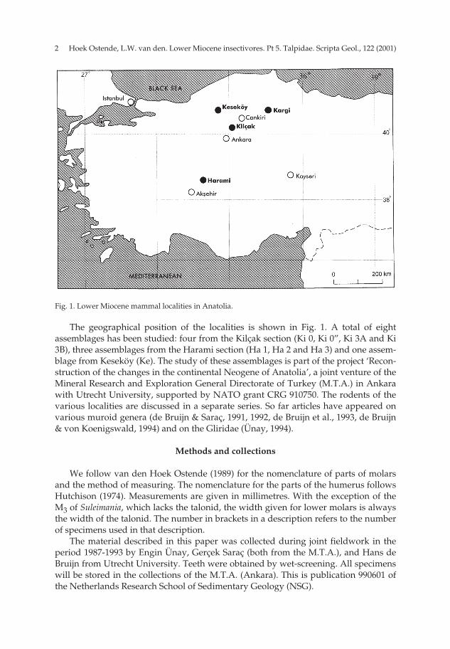
Fig.
2. M
and
ible
frag
men
t of T
hera
tisk
os m
echt
elda
esp
. nov
. wit
h th
e P 2
-P4
(Ke
6823
); a:
occ
lusa
l vie
w; b
: lab
ial v
iew
. Man
dib
le fr
agm
ent o
f The
rati
skos
mec
htel
-da
esp
. nov
. wit
h th
e P 4
and
the
alve
oles
of t
he a
nter
ior
den
titi
on (K
e 68
24);
c: o
cclu
sal v
iew
; d: l
abia
l vie
w (
× 12
.5).
a b
c d
pp 003-048 (SG 122-1) 15-01-2007 15:08 Pagina 5
Hoek Ostende, L.W. van den. Lower Miocene insectivores. Pt 5. Talpidae. Scripta Geol., 122 (2001)6
Description Mandible — The external temporal fossa is shallow. The ramus horizontalis is
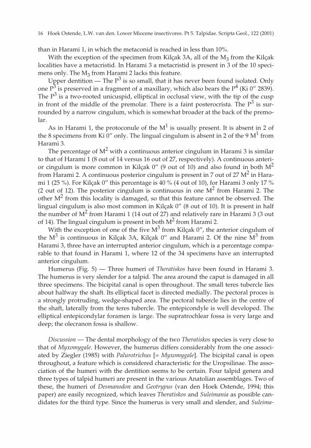
deepest under the M2/M3. There are two foramina mentale. The posterior one liesusually between the roots of the M1 (18), but may also be positioned under the trigon-id of that molar (8) or under the P4. The anterior foramen lies under the P1/P2. Awell-preserved anterior part of the mandible (Fig. 2c and d) shows ten alveoles infront of the P4. Four of these certainly belong to the P2 and P3, leaving six alveoles forthe remaining elements. The three anterior ones are small and presumably belong tothe incisors. The fourth, probably belonging to the canine, is the largest. This leavesthe small fifth and sixth alveole for the P1, which, like the other premolars, is there-fore two-rooted.
P2 (3), P3 (5) — A large number of ramus horizontalis fragments of Theratiskosmechteldae has been found. Some of these contain the P2 (1), the P3 (3) or both theseelements (2). Due to their small size, these premolars have not been found isolated.The P2 and P3 have a very similar morphology, the P2 being a somewhat larger ver-sion of the P3 (Fig. 2a and b). Both are two-rooted unicuspids. The outline in occlusalview is elliptical. The tip lies just in front of the middle of the premolar. Behind thetip lies a well-developed cingulum, which continues along the lingual and labial sidesand ends next to the tip.
D4 (30) — The outline of the occlusal surface is elliptical. The pyramid-shapedprotoconid is the only cusp. It occupies the front part of the milk molar. The edgesbetween the antero-lingual, antero-labial and posterior flank of the protoconid arerounded. The posterior part of the D4 consists of a large talonid, which is surroundedby a ridge. In some specimens this ridge is interrupted in the postero-labial corner.The lingual side of the talonid is clearly higher than the labial side. There is a smallcusplet close to the postero-lingual corner, which is incorporated in the posteriorridge. In 9 of the 30 specimens a central ridge is present, which connects the posteriorridge to the middle of the posterior flank of the protoconid.
P4 (48); M1 (71); M2 (79); M3 (77) — These elements have been described in thedescription of the holotype. The morphological variation in the lower dentition issmall.
D4 (25) — The outline of the occlusal surface is triangular. The paracone is thelargest cusp. Its tip lies in the front part of the milk molar, at about one third of itslength. A long posterocrista runs from the tip backwards. The anterior face of theparacone is rounded. The parastyle lies in front of the paracone on a rather largeshelf. Postero-lingually of the tip of the paracone lies the protocone. This is a low,blade-like cusp, which lies close to the flank of the paracone at its anterior end, but iswell separated from the paracone at its posterior end. The D4 is surrounded by widecingulums. The widest is the postero-lingual cingulum. The labial cingulum, whichruns from the parastyle to the back of the milk molar, is only somewhat narrower.The cingulum connecting the parastyle to the protocone becomes very narrow nearthe base of the paracone and is in some cases interrupted at this point.
P4 (53) — The outline of the occlusal surface is subtriangular. The labial side isconvex. The antero-lingual side is concave; the postero-lingual side is slightly concaveor straight. The paracone forms the largest part of the premolar. It has a strongly con-vex anterior side. The tip of the paracone lies in the middle of the P4. The straight
pp 003-048 (SG 122-1) 15-01-2007 15:08 Pagina 6
7Hoek Ostende, L.W. van den. Lower Miocene insectivores. Pt 5. Talpidae. Scripta Geol., 122 (2001)
Fig.
3. H
umer
us o
f The
rati
skos
mec
htel
dae
sp. n
ov. (
Ke
unnu
mbe
red
); a:
med
ial v
iew
; b: p
oste
rior
vie
w; c
: ant
erio
r vi
ew; d
: lat
eral
vie
w (
× 9)
.
ab
cd
pp 003-048 (SG 122-1) 15-01-2007 15:08 Pagina 7
Hoek Ostende, L.W. van den. Lower Miocene insectivores. Pt 5. Talpidae. Scripta Geol., 122 (2001)8
posterocrista runs from the tip backwards. The cone-shaped protocone lies linguallyof the tip of the paracone. It is a small cusplet that lies on a lingual extension. The pro-tocone is connected to the parastyle by the antero-lingual cingulum. The parastyle is avery small cusplet that lies in front of the paracone. It is even lower than the proto-cone. The postero-lingual cingulum connects the protocone to the posterior end of theposterocrista. This cingulum is strong and broad. The well-developed labial cingulumruns along the base of the posterocrista.
M1 (56) — The outline of the occlusal surface is irregularly quadrangular. Theprotocone is large. Its anterior arm runs along the anterior side of the molar and isconnected to the parastyle. The protoconule lies directly in front of the protocone. It islittle more than a thickening in the anterior arm of the protocone, and disappearsquickly with wear. It can only be observed in 19 out of the 56 specimens. The posteri-or arm of the protocone continues as a ridge that runs over the hypocone. Thehypocone is lower but only slightly smaller than the protocone. The parastyle liesdirectly in front of the paracone. It protrudes slightly. The posterior arm of the para-cone is S-curved. It is connected to the mesostyle. The mesostyle is undivided,although the two separate cusps can be discerned in unworn specimens. The meta-cone is the highest cusp of the M1. The arms of the metacone are straight. The anteriorarm is clearly shorter than the posterior arm. The latter extends to the postero-labialcorner of the M1. The posterior cingulum is well developed. It runs from thehypocone along the base of the posterior arm of the metacone. Specimens with asomewhat less developed cingulum usually have a concave posterior outline, in theothers the posterior side is straight. A weak labial cingulum may be present along themetacone. There is a small patch of lingual cingulum between the bases of the proto-cone and the hypocone.
M2 (73) — The outline of the occlusal surface is subrectangular. The large proto-cone lies in the antero-lingual corner of the molar. The tiny protoconule is usuallyincorporated in the anterior arm of the protocone and only recognizable in slightlyworn specimens. The anterior arm of the protoconule ends against the anterior flankof the paracone, but reaches the parastyle in 11 out of the 73 specimens. The posteriorarm of the protocone connects to the well-developed hypocone, which lies postero-labially of the protocone, making the lingual side of the molar slightly concave. Thehypocone is smaller and clearly lower than the protocone. The posterior arm of thehypocone continues as the posterior cingulum. This cingulum ends against the poste-rior flank of the metacone, either near the base or further lingually. In 17 out of 73specimens this cingulum reaches the postero-labial corner of the M2. The labial cuspsare of about the same size and height. The anterior arm of the paracone bends to formthe parastyle, which does not protrude. The anterior cingulum is wide near theparastyle. Mostly this cingulum is short and ends against the flank of the paracone.The posterior arm of the paracone is connected to the mesostyle, which is undivided.The posterior arm of the metacone bends to form the metastyle. The metastyle may beslightly protruding. In contrast to the parastyle, there is no cingulum near themetastyle. In 39 out of the 73 specimens there is a short lingual cingulum between theprotocone and the hypocone.
M3 (60) — The posterior outline of the occlusal surface is nearly half a circle. Thelarge protocone occupies the lingual part of the molar. The anterior arm of the proto-
pp 003-048 (SG 122-1) 15-01-2007 15:08 Pagina 8
9Hoek Ostende, L.W. van den. Lower Miocene insectivores. Pt 5. Talpidae. Scripta Geol., 122 (2001)
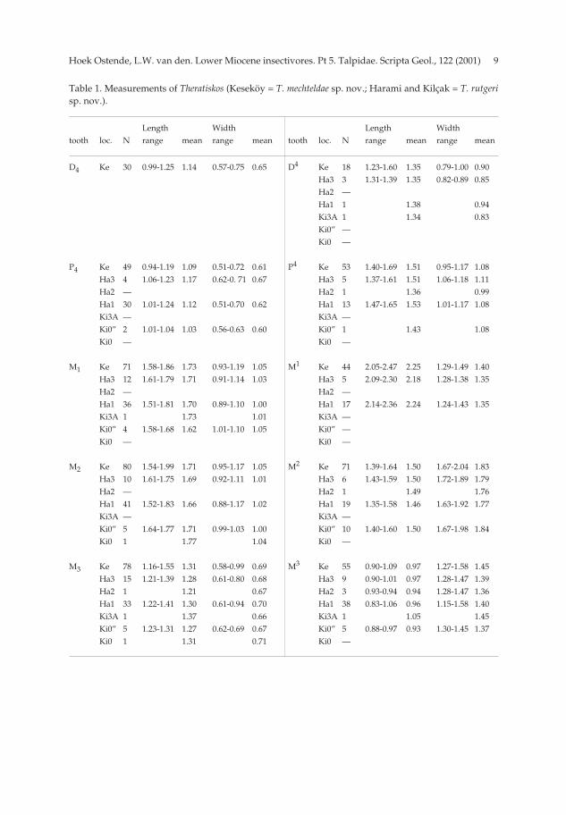
Table 1. Measurements of Theratiskos (Keseköy = T. mechteldae sp. nov.; Harami and Kilçak = T. rutgerisp. nov.).
Length Width Length Widthtooth loc. N range mean range mean tooth loc. N range mean range mean
D4 Ke 30 0.99-1.25 1.14 0.57-0.75 0.65 D4 Ke 18 1.23-1.60 1.35 0.79-1.00 0.90Ha3 3 1.31-1.39 1.35 0.82-0.89 0.85Ha2 —Ha1 1 1.38 0.94Ki3A 1 1.34 0.83Ki0” —Ki0 —
P4 Ke 49 0.94-1.19 1.09 0.51-0.72 0.61 P4 Ke 53 1.40-1.69 1.51 0.95-1.17 1.08Ha3 4 1.06-1.23 1.17 0.62-0. 71 0.67 Ha3 5 1.37-1.61 1.51 1.06-1.18 1.11Ha2 — Ha2 1 1.36 0.99Ha1 30 1.01-1.24 1.12 0.51-0.70 0.62 Ha1 13 1.47-1.65 1.53 1.01-1.17 1.08Ki3A — Ki3A —Ki0” 2 1.01-1.04 1.03 0.56-0.63 0.60 Ki0” 1 1.43 1.08Ki0 — Ki0 —
M1 Ke 71 1.58-1.86 1.73 0.93-1.19 1.05 M1 Ke 44 2.05-2.47 2.25 1.29-1.49 1.40Ha3 12 1.61-1.79 1.71 0.91-1.14 1.03 Ha3 5 2.09-2.30 2.18 1.28-1.38 1.35Ha2 — Ha2 —Ha1 36 1.51-1.81 1.70 0.89-1.10 1.00 Ha1 17 2.14-2.36 2.24 1.24-1.43 1.35Ki3A 1 1.73 1.01 Ki3A —Ki0” 4 1.58-1.68 1.62 1.01-1.10 1.05 Ki0” —Ki0 — Ki0 —
M2 Ke 80 1.54-1.99 1.71 0.95-1.17 1.05 M2 Ke 71 1.39-1.64 1.50 1.67-2.04 1.83Ha3 10 1.61-1.75 1.69 0.92-1.11 1.01 Ha3 6 1.43-1.59 1.50 1.72-1.89 1.79Ha2 — Ha2 1 1.49 1.76Ha1 41 1.52-1.83 1.66 0.88-1.17 1.02 Ha1 19 1.35-1.58 1.46 1.63-1.92 1.77Ki3A — Ki3A —Ki0” 5 1.64-1.77 1.71 0.99-1.03 1.00 Ki0” 10 1.40-1.60 1.50 1.67-1.98 1.84Ki0 1 1.77 1.04 Ki0 —
M3 Ke 78 1.16-1.55 1.31 0.58-0.99 0.69 M3 Ke 55 0.90-1.09 0.97 1.27-1.58 1.45Ha3 15 1.21-1.39 1.28 0.61-0.80 0.68 Ha3 9 0.90-1.01 0.97 1.28-1.47 1.39Ha2 1 1.21 0.67 Ha2 3 0.93-0.94 0.94 1.28-1.47 1.36Ha1 33 1.22-1.41 1.30 0.61-0.94 0.70 Ha1 38 0.83-1.06 0.96 1.15-1.58 1.40Ki3A 1 1.37 0.66 Ki3A 1 1.05 1.45Ki0” 5 1.23-1.31 1.27 0.62-0.69 0.67 Ki0” 5 0.88-0.97 0.93 1.30-1.45 1.37Ki0 1 1.31 0.71 Ki0 —
pp 003-048 (SG 122-1) 15-01-2007 15:08 Pagina 9
Hoek Ostende, L.W. van den. Lower Miocene insectivores. Pt 5. Talpidae. Scripta Geol., 122 (2001)10
cone continues as the anterior cingulum and is connected to the parastyle in 28 of the60 specimens. In the other specimens the anterior cingulum is interrupted near thebase of the paracone. The posterior arm of the protocone is connected to thehypocone. The latter is a small cusp, which lies lingually of the tip of the metacone.More often than not there is a small bulge in the outline at the position of thehypocone. The anterior arm of the paracone is somewhat longer than the posteriorarm. It bends slightly at the end to form a small parastyle. The posterior arm of theparacone is connected to the mesostyle, which is undivided. The metacone is small.There are no cingulums other than the anterior cingulum.
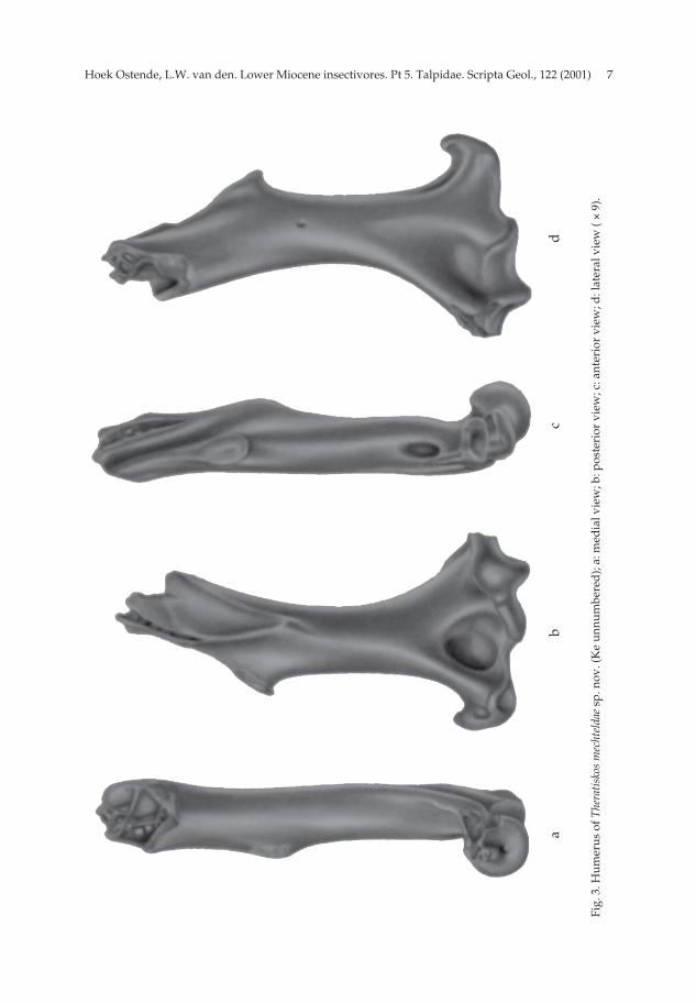
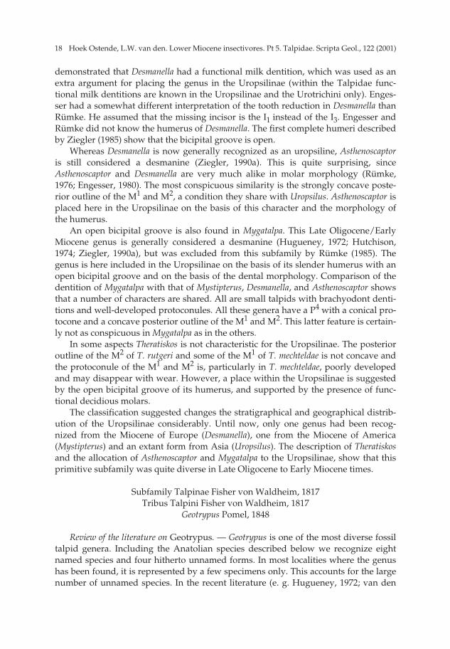
Humerus (4) (Fig. 3) — The humerus is very slender. The proximal part of thebone is damaged in all four specimens. The bicipital groove is open throughout. Theteres tubercle is small. It lies about halfway the shaft and has a medially directed,elliptical facet. The pectoral process is a protruding, wedge-shaped area. It lies in thecentre of the shaft. In three specimens it extends somewhat more distally than theteres tubercle, in the fourth the pectoral process lies directly laterally of the terestubercle. In one of the specimens the pectoral process is strongly developed, in theothers less so. The entepicondyle is small. The elliptical entepicondylar foramen islarge. The supratrochlear fossa is large and deep; the olecranon fossa is shallow.
Theratiskos rutgeri sp. nov. Pl. 1, figs. 8-16; Figs. 4-5.
Derivatio nominis — The species is named after Rutger van den Hoek Ostende, theauthor’s son.
Diagnosis — Dental formula unknown but probably complete. The M1 and M2 aresubequal in length. The entocristids of the M1 and M2 are well developed;metacristids are usually present on the M2, rarely on the M1. The oblique cristid endslingually of the middle of the protoconid-metaconid crest, and may even reach themetaconid. The hypocones of the M1 and M2 are well developed; the protoconule issmall. The posterior outline of the M1 is slightly concave.
Differential diagnosis — Theratiskos rutgeri differs from T. mechteldae in having ametacristid on the M2, and in rare cases also on the M1. The oblique cristid of the M1and M2 ends more lingually. The protoconule of the M1 and M2 is somewhat largerand the hypocone is smaller than in T. mechteldae.
Type locality — Harami 1 (MN 1).Other localities with T. rutgeri — Kilçak 0, Kilçak 0”, Kilçak 3A, Harami 2, Harami
3. Holotype — M2 sin. (Ha1 4057; Pl. 1, fig. 15); (1.58 × 1.78).Description of the holotype — The outline of the occlusal surface of the M2 is subrec-
tangular. The lingual part of the molar consists mainly of the protocone. The tip of theprotocone lies near the front of the tooth, making the outline slightly askew. The pro-toconule lies directly in front of the protocone. The anterior arm of the protoconebecomes very narrow between protocone and protoconule. The anterior arm of theprotoconule continues as the anterior cingulum, which is connected to the parastyle.The posterior arm of the protocone is connected to the small hypocone, which lies lin-gually of the tip of the metacone. The hypocone is lower than the protocone and the
pp 003-048 (SG 122-1) 15-01-2007 15:08 Pagina 10

11Hoek Ostende, L.W. van den. Lower Miocene insectivores. Pt 5. Talpidae. Scripta Geol., 122 (2001)
Fig. 4. Mandible dext. of Theratiskos rutgeri sp. nov. with M2 and the alveoles of the anterior dentition(Ha 1 3918); a: occlusal view; b: labial view. Mandible sin. Theratiskos rutgeri sp. nov. with M1 and thealveoles of the anterior dentition (Ki 0” 2814); c: occlusal view; d: labial view ( × 12.5).
a
b
c
d
pp 003-048 (SG 122-1) 15-01-2007 15:08 Pagina 11
Hoek Ostende, L.W. van den. Lower Miocene insectivores. Pt 5. Talpidae. Scripta Geol., 122 (2001)12
protoconule. The posterior arm of the hypocone continues as the posterior cingulumand ends against the flank of the metacone. The labial cusps are equal in size andheight. The anterior arm of the paracone bends to form the parastyle, which does notprotrude. The posterior arm of the paracone is connected to the undivided mesostyle.The posterior arm of the metacone bends to form the slightly protruding metastyle.There is a short lingual cingulum between the protocone and the hypocone.
Measurements — The measurements are listed in Table 1.
DescriptionMandible — Only a few mandibular fragments of T. rutgeri have been found. The
best-preserved specimen is a mandible with the M2 from Harami 1, showing 13 alve-oles in front of that molar (Fig. 4a-b). This number is corroborated by two mandibularfragments from Kilçak 0” (Ki 0” 2813, 2814; Fig 4c-d), which show 11 alveoles in frontof the M1. Determining the exact number of alveoles is difficult, since one cannot besure that all alveoles are preserved. If they are all preserved, the anterior dentition ofT. rutgeri would be slightly more reduced than that of T. mechteldae, which has 14alveoles in front of the M2. The fourth alveole of T. mechteldae is large and is interpret-ed as the alveole of the canine. In T. rutgeri all alveoles are of similar size. The inter-pretation is therefore difficult. The observed reduction in the number of alveoles mayeither mean that one single-rooted element is missing, or that one of the double-root-ed premolars of T. mechteldae has only one root in T. rutgeri. The external temporalfossa is shallow. The ramus horizontalis has its greatest depth below the M2/M3.There are two small foraminae mentale. The posterior one lies between the P4 and M1(6), the anterior one lies below the sixth alveole in front of the M1 (1).
P4 (32) — The outline of the occlusal surface is subrectangular. The labial side isslightly convex; the lingual side is straight. The P4 consists mainly of the large proto-conid. It has a rounded anterior side and a straight posterior flank. An indistinct pos-terocristid runs along the postero-lingual edge of the protoconid. There is a small cin-gulum along the antero-lingual side of the protoconid. A short talonid lies behind theprotoconid. The lingual side of this talonid is clearly higher than the labial side. A lowridge runs along the posterior side of the premolar. A second ridge connects the mid-dle of the posterior ridge to the posterocristid.
M1 (36) — The outline of the occlusal surface is subrectangular to subtriangular.The trigonid is clearly narrower than the talonid. A deep notch separates protoconidand metaconid. The paraconid is clearly lower than the protoconid and metaconid. Itlies at the end of a long paralophid. The trigonid basin is open. The entoconid andhypoconid are about the same height as the metaconid. The oblique cristid endsagainst the middle of the protoconid-metaconid crest or somewhat lingually of thatpoint. The entocristid is well developed. A very weak metacristid is present in 4 ofthe 36 specimens. The entostylid and posterior cingulum are well developed. The re-entrant valley is bordered by a short labial cingulum. The anterior cingulum, whichruns from the base of the paraconid to the base of the protoconid, is well developed.A parastylid may be present, but it is little more than a bulge on the anterior face ofthe paraconid. Some specimens show a short lingual cingulum along the base of theparaconid.
M2 (42) — The outline of the occlusal surface is subrectangular. The trigonid and
pp 003-048 (SG 122-1) 15-01-2007 15:08 Pagina 12
13Hoek Ostende, L.W. van den. Lower Miocene insectivores. Pt 5. Talpidae. Scripta Geol., 122 (2001)
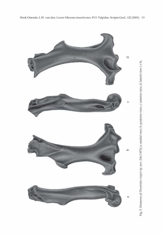
Fig.
5. H
umer
us o
f The
rati
skos
rut
geri
sp. n
ov. (
Ha
3 87
3); a
: med
ial v
iew
; b: p
oste
rior
vie
w; c
: ant
erio
r vi
ew; d
: lat
eral
vie
w (
× 9)
.
ab
cd
pp 003-048 (SG 122-1) 15-01-2007 15:08 Pagina 13
Hoek Ostende, L.W. van den. Lower Miocene insectivores. Pt 5. Talpidae. Scripta Geol., 122 (2001)14
talonid have about the same length and width. Protoconid and metaconid are sepa-rated by a deep notch. The metaconid is somewhat lower than the protoconid; theparaconid is clearly lower than the metaconid. The paralophid is straight or slightlybent. The trigonid basin is narrower than in the M1. The hypoconid and entoconid areof the same height. These cusps are somewhat lower than the metaconid, but higherthan the paraconid. The oblique cristid ends lingually of the middle of the proto-conid-metaconid crest, reaching the metaconid in 4 of the 42 specimens. Theentocristid is well developed; a clear metacristid is usually present. The anterior cin-gulum is well developed. The parastylid lies on this cingulum. As a rule, the anteriorcingulum bends along the base of the paraconid and continues as a narrow lingualcingulum that may reach the metaconid. The entostylid is well developed. The poste-rior cingulum is slightly narrower than the anterior cingulum. The re-entrant valley isbordered by a short labial cingulum.
M3 (32) — The outline of the occlusal surface is subrectangular. The talonid issomewhat shorter and somewhat narrower than the trigonid. The trigonid resemblesthat of the M2. Entoconid and hypoconid are low. They are incorporated in a longridge consisting of the oblique cristid, the posterior arm of the hypoconid and theentocristid. In some M3 a small metacristid is present. The oblique cristid ends closeto the metaconid. The anterior cingulum is well developed. It continues along thebase of the paraconid and ends halfway the trigonid basin. The re-entrant valley isbordered by a short labial cingulum.
D4 (2) — The outline of the occlusal surface is triangular. The paracone is thelargest cusp. Its tip lies in the front part of the milk molar, at about one third itslength. The posterocrista is straight. The anterior face of the paracone is rounded. Theparastyle lies in front of the paracone on a well-developed anterior cingulum. Thetwo arms of the parastyle end against the front of the paracone. The protocone lieslingually of the tip of the paracone. It is a low, blade-like cusp. The short anterior armof the protocone ends against the flank of the paracone. The posterior arm continuesas a ridge on the postero-lingual cingulum. It is connected at its end to the postero-crista. The labial cingulum runs from the flank of the paracone, just labially of its tip,to the back of the milkmolar. Thus the cingulums nearly surround the D4, with theexception of a part of the labial and lingual flanks of the paracone.
P4 (20) — The outline of the occlusal surface is subtriangular. The labial side isconvex. The paracone forms the largest part of the premolar. The tip of the paraconelies in the middle of the P4. It has a strongly convex anterior side. The posterocrista isstraight. The cone-shaped protocone lies directly lingual of the tip of the paracone. Itis a very small cusplet that lies on a lingual extension. The parastyle is even lowerthan the protocone. The protocone is connected to the parastyle by the antero-lingualcingulum. The postero-lingual cingulum is well developed and runs from the proto-cone to the posterior end of the posterocrista. There is a short labial cingulum thatruns along the base of the posterocrista.
M1 (22) — The outline of the occlusal surface is irregularly quadrangular. Theprotocone is large. Its anterior arm runs along the anterior side of the molar and isconnected to the parastyle. The posterior arm runs along the lingual side of the M1.The protoconule appears as a widening of the anterior arm and may disappear withwear. The hypocone lies behind the protocone. It is lower and smaller than the proto-
pp 003-048 (SG 122-1) 15-01-2007 15:08 Pagina 14
15Hoek Ostende, L.W. van den. Lower Miocene insectivores. Pt 5. Talpidae. Scripta Geol., 122 (2001)
cone. Due to the position of the hypocone the posterior side of the M1 is slightly con-cave. The slightly protruding parastyle lies directly in front of the paracone. The twocusplets of the mesostyle stand directly adjacent to each other. The anterior arm of themetacone is clearly shorter than the posterior arm. The posterior cingulum is welldeveloped. It runs from the hypocone along the base of the posterior arm of the meta-cone. There is a weak labial cingulum, which runs along the metacone. There is ashort lingual cingulum between the base of the protocone and the hypocone.
M2 (27) — The other M2 differ from the holotype mainly in the development ofthe cingulums. The anterior cingulum reaches the parastyle in 16 out of the 27 speci-mens; in the other 11 specimens it ends against the anterior flank of the paracone. Acontinuous posterior cingulum is found in 7 specimens. In the other 20 specimens itends against the posterior flank of the metacone. The short lingual cingulum betweenthe protocone and the hypocone is present in 14 specimens.
M3 (34) — The posterior outline of the occlusal surface is nearly half a circle. Thelarge protocone occupies the lingual part of the molar. The anterior arm of the proto-cone continues as the anterior cingulum and is connected to the parastyle. In 12 out ofthe 34 specimens the anterior cingulum is interrupted near the base of the paracone.The posterior arm of the protocone is connected to the hypocone. The latter is a smallto very small cusplet, which lies lingually of the tip of the metacone. The anterior armof the paracone is only slightly longer than the posterior arm. It bends slightly at theend to form a small parastyle. The posterior arm is connected to the mesostyle, whichis undivided. The posterior part of the mesostyle is connected to the small metacone.The anterior cingulum is the only cingulum present.
Material from other localities — The material of Theratiskos rutgeri found in the vari-ous other localites is given in Table 2. The morphological differences of these speci-mens with the material from the type locality are discussed below. In this section theP3 and the humerus, which have not been found in Harami 1, will be described.
Lower dentition — The metacristid of the M1 seems more frequently present inthe Kilçak localities and Harami 3 than in the type locality, where about 11 % showthis feature. This percentage is 33 % for Ki 0” and Ha 3 (2 out of 6 and 4 out of 12respectively) and 50% for Kilçak 3 A (1 out of 2).
The oblique cristid reaches the metaconid in 2 of the 6 M2 from Kilçak 0” and 2 ofthe 10 from Harami 3. In the only M2 from Kilçak 0 the oblique cristid ends just shortof the metaconid. Thus the oblique cristid of the M2 appears to end more lingually
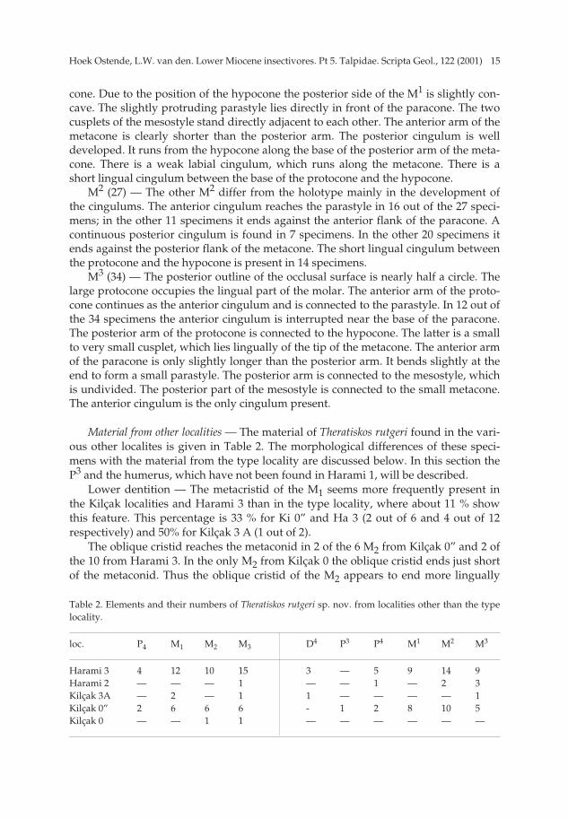
Table 2. Elements and their numbers of Theratiskos rutgeri sp. nov. from localities other than the typelocality.
loc. P4 M1 M2 M3 D4 P3 P4 M1 M2 M3
Harami 3 4 12 10 15 3 — 5 9 14 9Harami 2 — — — 1 — — 1 — 2 3Kilçak 3A — 2 — 1 1 — — — — 1Kilçak 0” 2 6 6 6 - 1 2 8 10 5Kilçak 0 — — 1 1 — — — — — —
pp 003-048 (SG 122-1) 15-01-2007 15:08 Pagina 15
Hoek Ostende, L.W. van den. Lower Miocene insectivores. Pt 5. Talpidae. Scripta Geol., 122 (2001)16
than in Harami 1, in which the metaconid is reached in less than 10%. With the exception of the specimen from Kilçak 3A, all of the M3 from the Kilçak
localities have a metacristid. In Harami 3 a metacristid is present in 3 of the 10 speci-mens only. The M3 from Harami 2 lacks this feature.
Upper dentition — The P3 is so small, that it has never been found isolated. Onlyone P3 is preserved in a fragment of a maxillary, which also bears the P4 (Ki 0” 2839).The P3 is a two-rooted unicuspid, elliptical in occlusal view, with the tip of the cuspin front of the middle of the premolar. There is a faint posterocrista. The P3 is sur-rounded by a narrow cingulum, which is somewhat broader at the back of the premo-lar.
As in Harami 1, the protoconule of the M1 is usually present. It is absent in 2 ofthe 8 specimens from Ki 0” only. The lingual cingulum is absent in 2 of the 9 M1 fromHarami 3.
The percentage of M2 with a continuous anterior cingulum in Harami 3 is similarto that of Harami 1 (8 out of 14 versus 16 out of 27, respectively). A continuous anteri-or cingulum is more common in Kilçak 0” (9 out of 10) and also found in both M2
from Harami 2. A continuous posterior cingulum is present in 7 out of 27 M2 in Hara-mi 1 (25 %). For Kilçak 0” this percentage is 40 % (4 out of 10), for Harami 3 only 17 %(2 out of 12). The posterior cingulum is continuous in one M2 from Harami 2. Theother M2 from this locality is damaged, so that this feature cannot be observed. Thelingual cingulum is also most common in Kilçak 0” (8 out of 10). It is present in halfthe number of M2 from Harami 1 (14 out of 27) and relatively rare in Harami 3 (3 outof 14). The lingual cingulum is present in both M2 from Harami 2.
With the exception of one of the five M3 from Kilçak 0”, the anterior cingulum ofthe M3 is continuous in Kilçak 3A, Kilçak 0” and Harami 2. Of the nine M3 fromHarami 3, three have an interrupted anterior cingulum, which is a percentage compa-rable to that found in Harami 1, where 12 of the 34 specimens have an interruptedanterior cingulum.
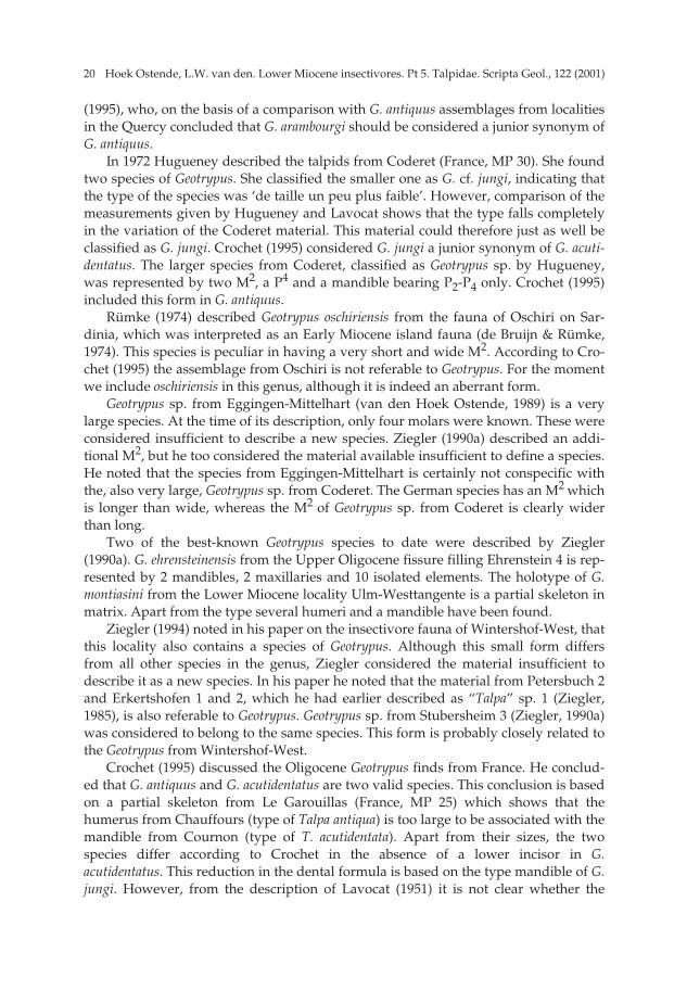
Humerus (Fig. 5) — Three humeri of Theratiskos have been found in Harami 3.The humerus is very slender for a talpid. The area around the caput is damaged in allthree specimens. The bicipital canal is open throughout. The small teres tubercle liesabout halfway the shaft. Its elliptical facet is directed medially. The pectoral proces isa strongly protruding, wedge-shaped area. The pectoral tubercle lies in the centre ofthe shaft, laterally from the teres tubercle. The entepicondyle is well developed. Theelliptical entepicondylar foramen is large. The supratrochlear fossa is very large anddeep; the olecranon fossa is shallow.
Discussion — The dental morphology of the two Theratiskos species is very close tothat of Myxomygale. However, the humerus differs considerably from the one associ-ated by Ziegler (1985) with Palurotrichus [= Myxomygale]. The bicipital canal is openthroughout, a feature which is considered characteristic for the Uropsilinae. The asso-ciation of the humeri with the dentition seems to be certain. Four talpid genera andthree types of talpid humeri are present in the various Anatolian assemblages. Two ofthese, the humeri of Desmanodon and Geotrypus (van den Hoek Ostende, 1994; thispaper) are easily recognized, which leaves Theratiskos and Suleimania as possible can-didates for the third type. Since the humerus is very small and slender, and Suleima-
pp 003-048 (SG 122-1) 15-01-2007 15:08 Pagina 16

17Hoek Ostende, L.W. van den. Lower Miocene insectivores. Pt 5. Talpidae. Scripta Geol., 122 (2001)
nia is a very large talpid, the humerus is associated with the smaller dental elementsof Theratiskos. Furthermore, three humeri have been found in the Keseköy assemblagein which Suleimania is rare and Theratiskos is abundant. The association of dentitionsand humeri seems therefore established.
The dentitions of Myxomygale and Theratiskos are also different. In Myxomygale allpremolars in front of the P4 are single-rooted. The P3 of Theratiskos has two roots. Fur-thermore, the deciduous molars attributed to Theratiskos show that the members ofthe genus had a functional milk dentition, whereas milk molars of Myxomygale havenot been found. The molar morphology of Theratiskos and Myxomygale is, however,very similar and isolated elements may easily be confused.
Engesser (1980) described two small M1 from Sariçay (MN 7/8) as Desmanodonsp., which were later transferred to Myxomygale sp. (van den Hoek Ostende, 1989).However, the two molars agree morphologically and metrically well with those ofTheratiskos mechteldae. Therefore the two molars may readily belong to Theratiskos.More material is needed to confirm this identification.
What are Uropsilinae?
Campbell (1939) noted in his discussion on the shoulder anatomy of extant Talpi-dae, that the humerus of Uropsilus is very primitive and resembles that of the shrewBlarina. Uropsilus is the only extant talpid with a humerus in which the walls of thebicipital groove have not fused to form a tunnel. An open bicipital groove is alsofound in the extinct American subfamily Gaillardinae and in the Proscalopidae. Thelatter used to be considered as a subfamily of the talpids, but was raised by Barnosky(1981) to family level. The humeri of these two groups are massive and also in otherrespects totally different from those of the Uropsilinae. The combination of an openbicipital groove and an overall slender humerus is considered typical for the Uropsili-nae.
The Uropsilinae is the most primitive subfamily of the Talpidae. Nevertheless fewfossil representatives have been recognized. The first extinct genus that has beenincluded in this subfamily is Mystipterus. This talpid from the Miocene of NorthAmerica was originally described as a bat (Hall, 1930) and later transferred to theSoricidae (Patterson & McGrew, 1939). Van Valen (1967) recognized its talpid affini-ties. Hutchison (1968) placed the genus in the Uropsilinae. In his description of thesubfamily Hutchison used the open bicipital groove, but he did not consider it typicalfor the subfamily. The feature is also found in Asthenoscaptor Hutchison, 1974. Hutchi-son placed Asthenoscaptor in his “Group I” of the Desmaninae, together with Des-manella. Although the humerus of true desmans is in many respects primitive, thebicipital canal in this subfamily is closed (Campbell, 1939).
On the basis of dental morphology and dental formula Rümke (1974) demonstrat-ed that Desmanella is not a desman. Contrary to Desmaninae, Desmanella presumablyhas lost its I3, while the I1 is much larger than the I2. Rümke’s arguments for placingthe genus in the Uropsilinae were the presence of the anterior and posterior cingu-lums in the lower molars, the length and position of the oblique cristid (extending tohalfway the protoconid-metaconid crest), the concave posterior outline of M1-M2 andthe presence of a labial cingulum in M1. Engesser (1980) agreed with Rümke and
pp 003-048 (SG 122-1) 15-01-2007 15:08 Pagina 17
Hoek Ostende, L.W. van den. Lower Miocene insectivores. Pt 5. Talpidae. Scripta Geol., 122 (2001)18
demonstrated that Desmanella had a functional milk dentition, which was used as anextra argument for placing the genus in the Uropsilinae (within the Talpidae func-tional milk dentitions are known in the Uropsilinae and the Urotrichini only). Enges-ser had a somewhat different interpretation of the tooth reduction in Desmanella thanRümke. He assumed that the missing incisor is the I1 instead of the I3. Engesser andRümke did not know the humerus of Desmanella. The first complete humeri describedby Ziegler (1985) show that the bicipital groove is open.
Whereas Desmanella is now generally recognized as an uropsiline, Asthenoscaptoris still considered a desmanine (Ziegler, 1990a). This is quite surprising, sinceAsthenoscaptor and Desmanella are very much alike in molar morphology (Rümke,1976; Engesser, 1980). The most conspicuous similarity is the strongly concave poste-rior outline of the M1 and M2, a condition they share with Uropsilus. Asthenoscaptor isplaced here in the Uropsilinae on the basis of this character and the morphology ofthe humerus.
An open bicipital groove is also found in Mygatalpa. This Late Oligocene/EarlyMiocene genus is generally considered a desmanine (Hugueney, 1972; Hutchison,1974; Ziegler, 1990a), but was excluded from this subfamily by Rümke (1985). Thegenus is here included in the Uropsilinae on the basis of its slender humerus with anopen bicipital groove and on the basis of the dental morphology. Comparison of thedentition of Mygatalpa with that of Mystipterus, Desmanella, and Asthenoscaptor showsthat a number of characters are shared. All are small talpids with brachyodont denti-tions and well-developed protoconules. All these genera have a P4 with a conical pro-tocone and a concave posterior outline of the M1 and M2. This latter feature is certain-ly not as conspicuous in Mygatalpa as in the others.
In some aspects Theratiskos is not characteristic for the Uropsilinae. The posterioroutline of the M2 of T. rutgeri and some of the M1 of T. mechteldae is not concave andthe protoconule of the M1 and M2 is, particularly in T. mechteldae, poorly developedand may disappear with wear. However, a place within the Uropsilinae is suggestedby the open bicipital groove of its humerus, and supported by the presence of func-tional decidious molars.
The classification suggested changes the stratigraphical and geographical distrib-ution of the Uropsilinae considerably. Until now, only one genus had been recog-nized from the Miocene of Europe (Desmanella), one from the Miocene of America(Mystipterus) and an extant form from Asia (Uropsilus). The description of Theratiskosand the allocation of Asthenoscaptor and Mygatalpa to the Uropsilinae, show that thisprimitive subfamily was quite diverse in Late Oligocene to Early Miocene times.
Subfamily Talpinae Fisher von Waldheim, 1817Tribus Talpini Fisher von Waldheim, 1817
Geotrypus Pomel, 1848
Review of the literature on Geotrypus. — Geotrypus is one of the most diverse fossiltalpid genera. Including the Anatolian species described below we recognize eightnamed species and four hitherto unnamed forms. In most localities where the genushas been found, it is represented by a few specimens only. This accounts for the largenumber of unnamed species. In the recent literature (e. g. Hugueney, 1972; van den
pp 003-048 (SG 122-1) 15-01-2007 15:08 Pagina 18

19Hoek Ostende, L.W. van den. Lower Miocene insectivores. Pt 5. Talpidae. Scripta Geol., 122 (2001)
Hoek Ostende, 1989; Ziegler, 1990a, 1994, 1998) material was often considered toolimited to describe a new species, even though the material could not be attributed toany of the known species. Since this cautious attitude is not found in the older litera-ture, some of the unnamed forms are better defined than some of the named species.
Geotrypus was already noted 150 years ago, presumably as a result of its large sizeand its robust humerus. It was defined by Pomel (1848), who combined a mandiblefrom Cournon and a humerus from Chaffours (both Upper Oligocene, France), whichhad been illustrated by de Blainville (1840) under the names Talpa acutidentata and T.antiqua, respectively. Pomel named this species Geotrypus antiquus. Ziegler (1990a)referred to this species as G. antiquus (Pomel, 1848). However, since Pomel based isdescription on the illustrations of de Blainville, the species name should be attributedto the latter author. Although G. antiquus sensu Pomel is based on the association of ahumerus and a mandible that originate from different localities, the association of thetwo remained unchallenged for a long time. Crochet (1995) suggested that themandible and the humerus represent two diffent species, which should be named G.acutidentatus and G. antiquus respectively. Here we follow Crochet’s classification.However, for the review of literature it is important to realise that up to 1995 G. acuti-dentatus was considered a synonym of G. antiquus.
During the second half of the 19th century more Geotrypus material was found,but placed in other genera. In 1877 Filhol described Protalpa cadurcensis from theQuercy. This species was synonymized with G. antiquus by Lavocat (1951). Crochet(1974) and Brunet et al. (1981) listed Geotrypus cadurcensis as a separate species, with-out however describing this form or indicating any differences with G. antiquus. Cro-chet (1995) synonimized P. cadurcensis pro partim with G. antiquus and pro partimwith G. acutidentatus.
Another species of Geotrypus initially assigned to Talpa is G. tomerdingensis (To-bien, 1939). The species is known from postcranial material only. Its type is a verylarge humerus from the German locality Tomerdingen (MN 1). Hugueney (1972) firstrecognized its true generic affinity. Ziegler (1990a) described various Geotrypusspecies from southern Germany, but did not find any dental elements referable to G.tomerdingensis. He included a fragment of a large ulna and a radius from the typelocality in the species. There can be little doubt that G. tomerdingensis is a goodspecies, since the bones found are huge in comparison to other species of the genus.However, the absence of molars makes it very difficult to compare G. tomerdingensiswith other species.
In his thesis on the mammals from the French Upper Oligocene locality CournonLavocat (1951) recognized three different species of Geotrypus, G. antiquus, G. jungiand G. arambourgi, all of which were based on scarce material. G. antiquus deBlainville, 1840 was only represented by the type. G. jungi Lavocat, 1951 wasdescribed on the basis of a mandible and part of a maxillary in matrix. G. arambourgiLavocat, 1951 was based on a crushed skull, in which the two M1 and one of the P4
were preserved. Although two different species of Geotrypus may occur in the samelocality (e. g. G. cf. jungi and Geotrypus sp. in Codoret; G. tomerdingensis and G. aff.montiasini in Tomerdingen), the presence of three large talpids belonging to onegenus in a single locality seems unlikely. Hugueney and Guerin (1981) suggested thatonly two species of Geotrypus are present in Cournon. This was confirmed by Crochet
pp 003-048 (SG 122-1) 15-01-2007 15:08 Pagina 19
Hoek Ostende, L.W. van den. Lower Miocene insectivores. Pt 5. Talpidae. Scripta Geol., 122 (2001)20
(1995), who, on the basis of a comparison with G. antiquus assemblages from localitiesin the Quercy concluded that G. arambourgi should be considered a junior synonym ofG. antiquus.
In 1972 Hugueney described the talpids from Coderet (France, MP 30). She foundtwo species of Geotrypus. She classified the smaller one as G. cf. jungi, indicating thatthe type of the species was ‘de taille un peu plus faible’. However, comparison of themeasurements given by Hugueney and Lavocat shows that the type falls completelyin the variation of the Coderet material. This material could therefore just as well beclassified as G. jungi. Crochet (1995) considered G. jungi a junior synonym of G. acuti-dentatus. The larger species from Coderet, classified as Geotrypus sp. by Hugueney,was represented by two M2, a P4 and a mandible bearing P2-P4 only. Crochet (1995)included this form in G. antiquus.
Rümke (1974) described Geotrypus oschiriensis from the fauna of Oschiri on Sar-dinia, which was interpreted as an Early Miocene island fauna (de Bruijn & Rümke,1974). This species is peculiar in having a very short and wide M2. According to Cro-chet (1995) the assemblage from Oschiri is not referable to Geotrypus. For the momentwe include oschiriensis in this genus, although it is indeed an aberrant form.
Geotrypus sp. from Eggingen-Mittelhart (van den Hoek Ostende, 1989) is a verylarge species. At the time of its description, only four molars were known. These wereconsidered insufficient to describe a new species. Ziegler (1990a) described an addi-tional M2, but he too considered the material available insufficient to define a species.He noted that the species from Eggingen-Mittelhart is certainly not conspecific withthe, also very large, Geotrypus sp. from Coderet. The German species has an M2 whichis longer than wide, whereas the M2 of Geotrypus sp. from Coderet is clearly widerthan long.
Two of the best-known Geotrypus species to date were described by Ziegler(1990a). G. ehrensteinensis from the Upper Oligocene fissure filling Ehrenstein 4 is rep-resented by 2 mandibles, 2 maxillaries and 10 isolated elements. The holotype of G.montiasini from the Lower Miocene locality Ulm-Westtangente is a partial skeleton inmatrix. Apart from the type several humeri and a mandible have been found.
Ziegler (1994) noted in his paper on the insectivore fauna of Wintershof-West, thatthis locality also contains a species of Geotrypus. Although this small form differsfrom all other species in the genus, Ziegler considered the material insufficient todescribe it as a new species. In his paper he noted that the material from Petersbuch 2and Erkertshofen 1 and 2, which he had earlier described as “Talpa” sp. 1 (Ziegler,1985), is also referable to Geotrypus. Geotrypus sp. from Stubersheim 3 (Ziegler, 1990a)was considered to belong to the same species. This form is probably closely related tothe Geotrypus from Wintershof-West.
Crochet (1995) discussed the Oligocene Geotrypus finds from France. He conclud-ed that G. antiquus and G. acutidentatus are two valid species. This conclusion is basedon a partial skeleton from Le Garouillas (France, MP 25) which shows that thehumerus from Chauffours (type of Talpa antiqua) is too large to be associated with themandible from Cournon (type of T. acutidentata). Apart from their sizes, the twospecies differ according to Crochet in the absence of a lower incisor in G.acutidentatus. This reduction in the dental formula is based on the type mandible of G.jungi. However, from the description of Lavocat (1951) it is not clear whether the
pp 003-048 (SG 122-1) 15-01-2007 15:08 Pagina 20
21Hoek Ostende, L.W. van den. Lower Miocene insectivores. Pt 5. Talpidae. Scripta Geol., 122 (2001)
incisor was absent or only missing in the fossil. Crochet gave extensive diagnoses forthe genus and for the species G. antiquus and G. acutidentatus. Unfortunately, he didnot give any descriptions, on which his conclusions and diagnoses are based,announcing that he intends to do so in a separate review.
Ziegler (1998) described Geotrypus sp. from the fissure fillings from Herrlingen 8(MP 28) and Herrlingen 9 (MP 29). He considered the material too scarce to classify itwith certainty as any known or new species. Below the species assigned to Geotrypusin this paper are listed. Geotrypus acutidentatus (de Blainville, 1840)Geotrypus antiquus (de Blainville, 1840) Geotrypus ehrensteinensis Ziegler, 1990 Geotrypus haramiensis sp. nov. Geotrypus montinasini Ziegler, 1990Geotrypus oschiriensis Rümke, 1974 Geotrypus kesekoeyensis sp. nov. Geotrypus tomerdingensis (Tobien, 1939)Geotrypus sp. from Eggingen-Mittelhart (van den Hoek Ostende, 1989)Geotrypus sp. from Stubersheim 3 (Ziegler, 1990)Geotrypus sp. from Wintershof-West (Ziegler, 1994)Geotrypus sp. from Herrlingen 8 and 9 (Ziegler, 1998)
Van Valen (1967) included Geotrypus in the Scaptonychini. This tribe, intended tohold the primitive Talpinae and their relatively unmodified descendants, forms anunnatural group, containing genera that have since been included in the Urotrichini(Myxomygale) and the Uropsilinae (Mygatalpa). Ziegler (1990a) already indicated thatGeotrypus also should not be included in the Scaptonychini. Because of its stronglydeveloped humeri, Geotrypus is by no means a primitive talpid. On the basis of thehumeri, which indicate a fossorial mode of live, Ziegler included the genus in theTalpini. This is corroborated by the caniniform P1, a feature only found in the Talpiniand in the enigmatic genus Scaptonyx.
The youngest record of Geotrypus is from Petersbuch 2 and Erkertshofen 1 and 2(MN 4). The only fossil Talpini known from younger localities is Talpa, a genus whichappears for the first time in MN 2 (Ulm-Westtangente). In the timespan that Geotrypusand Talpa coexisted, the two can be found in the same localities (Ulm-Westtangente,Wintershof-West). In localities older than MN 2, a larger and a smaller species ofGeotrypus often co-occur (Cournon, Coderet, Tomerdingen). Apparently Talpa slowlyreplaced Geotrypus, first taking over the niche of the smaller Talpini. In younger local-ities, Talpa also fills the niche of the large Talpini. In Pliocene and Pleistocene locali-ties it is quite common to find two species of Talpa.
The Anatolian species of Geotrypus
Geotrypus haramiensis sp. nov. Pl. 2, figs. 1-7.
Derivatio nominis — This species is named after its type locality.
pp 003-048 (SG 122-1) 15-01-2007 15:08 Pagina 21
Hoek Ostende, L.W. van den. Lower Miocene insectivores. Pt 5. Talpidae. Scripta Geol., 122 (2001)22
Diagnosis — Geotrypus haramiensis is a large species of Geotrypus (M2 = 2.36). Thelower dental formula is ?.?.3.3. The P2 is missing. The mesostyle of the upper molarsis slightly divided. The M2 has no hypocone. Its labial side is straight. The L/W ratioof the M2 is c. 0.85.
Differential diagnosis — Geotrypus haramiensis is smaller than G. tomerdingensis andGeotrypus sp. from Eggingen-Mittelhart. It is larger than G. acutidentatus, G. oschirien-sis, Geotrypus sp. from Wintershof-West, and Geotrypus sp. from Erkertshofen. It iscomparable in size to G. antiquus, G. montiasini and G. ehrensteinensis.
The absence of the P2 distinguishes Geotrypus haramiensis from G. ehrensteinensis,G. oschiriensis, G. acutidentatus and G. antiquus. G. montiasini also lacks the P2; the den-tal formula of the other Geotrypus species is not known.
The slight division of the mesostyle of the M2 and M3 distinguishes G. haramiensisfrom G. ehrensteinensis, G. acutidentatus, Geotrypus sp. from Eggingen-Mittelhart, andGeotrypus sp. from Wintershof-West. The labial side of the latter is concave, a featureit shares with Geotrypus sp. from Stubersheim 3. The labial side of the M2 of G.haramiensis is straight. The M2 of G. haramiensis is relatively wider than that of Geotry-pus sp. from Eggingen-Mittelhart, G. montiasini and G. ehrensteinensis, and relativelynarrower than the M2 of G. oschiriensis.
G. haramiensis differs from G. montiasini in the better developed anterior cingulumof the M2. The M2 of G. oschiriensis has no anterior cingulum and, in contrast to G.haramiensis, the talonid is wider than the trigonid.
Type locality — Harami 3 (MN 1). Other localities with G. haramiensis sp. nov. — Kilçak 3A, Harami 1. Holotype — Part of a mandible with P1, P3-M3 (Ha 3 781; Pl. 2, fig. 1). Description of the holotype — The ramus horizontalis and the lower part of the
processus coronoides are preserved only. The processus coronoides appears to havebeen narrow. The ramus horizontalis is slender and has three foramina on the labialside, one below the P3, one below the M1 and one just in front of and below the exter-nal temporal fossa of the processus coronoides.
The mandible bears six teeth. The M1 and M3 are damaged. The trigonid of theM1 and the talonid of the M3 are broken. The M2 is well preserved. The P3 and P4 areset obliquely in the jaw. The large, caniniform element in front of the P3 is consideredto be the P1. This premolar has two roots and a high, peak-like cusp that curves back-wards. The P3 is clearly smaller than the P4, but these two premolars have a very sim-ilar morphology. The main cusp is high. Its tip lies in front of the middle of theocclusal surface. Both P3 and P4 have a posterior flattening, which bears a cusplet.The P4 also has a small anterior flattening.
The cusps of the lower molars are high and pointed. The M1 has a well-developedentostylid. The talonid of the M2 is somewhat shorter and narrower than the trigonid.The oblique cristid runs in the direction of the metaconid and ends at about two-thirds of the protoconid-metaconid crest. There is neither a metacristid, nor anentocristid. The M2 has a well-developed entostylid and a strong parastylid, whichcontinues labially as the anterior cingulum. The M3 has a similarly strong parastylid.
Measurements — The measurements are listed in Table 3.
pp 003-048 (SG 122-1) 15-01-2007 15:08 Pagina 22
23Hoek Ostende, L.W. van den. Lower Miocene insectivores. Pt 5. Talpidae. Scripta Geol., 122 (2001)
DescriptionM2 (1) — The outline of the occlusal surface is triangular. The cusps are high and
pointed. The protocone is large. Its anterior arm ends against the base of the para-cone, the posterior arm ends against the base of the metacone. There is neither a pro-toconule nor a hypocone. The labial cusps are higher than the protocone. The anteriorarm of the paracone bends and forms a protruding parastyle. The posterior arm isconnected to the mesostyle, which is undivided. The posterior arm of the metacone isslightly longer than the anterior arm. There is no metastyle and there are no cingu-lums.
M3 (1) — The outline of the occlusal surface is subtriangular. The paracone is thehighest cusp. Its anterior arm is slightly longer than its posterior arm. The anteriorarm bends at its end to form a very weak parastyle. The posterior arm is connected tothe mesostyle, which is indistinctly divided. The metacone and protocone are of thesame height. The metacone has an S-shaped anterior arm, which is connected to themesostyle. The anterior arm of the protocone ends against the base of the paracone.The protocone does not have a posterior arm. There are no cingulums.
Humerus (3) — The humerus is very robust, indicating a fossorial mode of life. Inall three specimens the proximal side is damaged. The greater tuberosity, lessertuberosity and pectoral crest have not been preserved. The pectoral process is pro-nounced. The pectoral ridge is well defined. It reaches to about one third of the widthof the bone and it is only slightly inclined towards the side of the tuberculum teres.The pectoral tubercle is large and protruding. The supratrochlear fossa and olecranonfossa are large and deep.
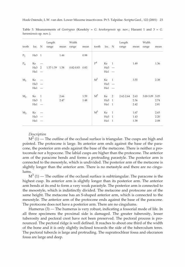
Table 3. Measurements of Geotrypus (Keseköy = G. kesekoeyensis sp. nov.; Harami 1 and 3 = G.haramiensis sp. nov.).
Length Width Length Widthtooth loc. N range mean range mean tooth loc. N range mean range mean
P1 Ha3 1 1.44 0.98
P4 Ke — P4 Ke 1 1.49 1.36Ha3 2 1.57-1.59 1.58 0.82-0.83 0.83 Ha3 —Ha1 — Ha1 —
M1 Ke — M1 Ke 1 3.55 2.38Ha3 — Ha3 —Ha1 — Ha1 —
M2 Ke 1 2.66 1.59 M2 Ke 2 2.62-2.64 2.63 3.00-3.09 3.05Ha3 1 2.47 1.48 Ha3 1 2.36 2.74Ha1 — Ha1 1 2.42 2.81
M3 Ke — M3 Ke 1 1.67 2.65Ha3 — Ha3 1 1.43 2.20Ha1 — Ha1 1 1.38 2.08
pp 003-048 (SG 122-1) 15-01-2007 15:08 Pagina 23
Hoek Ostende, L.W. van den. Lower Miocene insectivores. Pt 5. Talpidae. Scripta Geol., 122 (2001)24
Geotrypus haramiensis from other localitiesIn Harami 1 Geotrypus haramiensis is represented by one M2, one M3, and a frag-
ment of a humerus only. The molars show only minor differences with the corre-sponding elements from the type locality. The two arms of the paracone of the M3
are, in contrast to the situation in the specimen from Harami 3, of about the samelength. In Kilçak 3A only two damaged humeri of this species have been found. Oneof these is somewhat more complete than the best preserved specimen from Harami3, the other is only a small fragment. The tuberculum teres is long. The bicipitalgroove is rather wide. The pectoral ridge reaches to halfway the width of thehumerus. The pectoral process is less pronounced and the supratrochlear fossa andolecranon fossa are smaller than in the specimens from the type locality.
Geotrypus kesekoeyensis sp. nov. Pl. 3, figs. 1-6.
Derivatio nominis — The species is named after its type locality Keseköy. Diagnosis — Very large species of Geotrypus (M2 = 2. 63). The P4 is relatively short
and has a large protoconal flange. The mesostyle of the upper molars is undivided. Avery small hypocone may be present on the M1 and M2.
Differential diagnosis — Geotrypus kesekoeyensis is larger than all other species ofGeotrypus, with the possible exception of G. tomerdingensis. Since the latter is knownby its humerus only, and since the humerus of G. kesekoeyensis is not known, the twospecies cannot be compared.
Apart from its size G. kesekoeyensis differs from other species of Geotrypus in hav-ing a relatively short P4 (the P4 of G. tomerdingensis, G. antiquus, G. haramiensis, Geotry-pus sp. from Wintershof-West, and Geotrypus sp. from Eggingen-Mitelhart are notknown). The undivided mesostyle of the upper molars distinguishes G. kesekoeyensisfrom G. ehrensteinensis, G. acutidentatus, Geotrypus sp. from Eggingen-Mittelhart, andGeotrypus sp. from Wintershof-West.
Type locality — Keseköy (MN 3).Holotype — M2 sin. (Ke 6712) (2.64 × 3.09; Pl. 3, fig. 5).Description of the holotype — The outline of the occlusal surface is subtriangular.
The postero-lingual side is concave. The protocone is small. Its very short anteriorarm ends against the base of the paracone. The posterior side of the protocone bears avery faint ridge. This ridge ends against the tiny hypocone, which lies lingually of thebase of the metacone and is separated from that cusp by a clear trench. The paraconeis broken. Its base extends far towards the lingual side. The anterior arm of the para-cone bends at its end and forms a small, protruding parastyle. The posterior arm isconnected to the undivided mesostyle. The two arms of the metacone are of the samelength. The posterior arm bends at its end and forms a slightly protruding metastyle.There are no cingulums.
Measurements — The measurements are listed in Table 3.
DescriptionM1 (1) — The only available specimen is damaged. The area of the paraconid is
broken. The outline of the occlusal surface is subrectangular. The cusps are high and
pp 003-048 (SG 122-1) 15-01-2007 15:08 Pagina 24

25Hoek Ostende, L.W. van den. Lower Miocene insectivores. Pt 5. Talpidae. Scripta Geol., 122 (2001)
pointed. The posterior arm of the protoconid slopes downward and is connected tothe metaconid. The talonid is long and wider than the trigonid. The hypoconid issomewhat higher than the entoconid. The long oblique cristid ends against the mid-dle of the protoconid-metaconid crest. There is neither a metacristid nor anentocristid. The entostylid is well developed. There are no cingulums.
M2 (1) — The outline of the occlusal surface is subrectangular. Even though thespecimen is worn, the cusps are very high. The anterior and posterior arms of theprotoconid are connected to the paraconid and metaconid, respectively. The twoarms are straight and run parallel to each other. There is a notch in the protoconid-metaconid crest at about two-thirds of its length. The talonid is somewhat narrowerthan the trigonid. The talonid and the trigonid are of about the same length. Thecusps of the talonid are clearly lower than those of the trigonid. The oblique cristid isconnected to the protoconid-metaconid crest below the notch. There is neither ametacristid nor an entocristid. The parastylid and the entostylid are well developed.There is a short anterior cingulum that runs from the parastylid to halfway the anteri-or side of the molar.
P4 (1) — This specimen is preserved in a piece of the maxillary, which also con-tains the anterior alveoles of the M1. The outline of the occlusal surface is triangular.The P4 consists mainly of the very high and very large paracone. The tip of the cusplies in the middle of the premolar. The anterior face of the paracone is rounded. Thesharp posterocrista is slightly curved and runs to the postero-labial corner of the P4.There is a large lingual shelf that bears a tiny protocone. A small parastyle lies infront of the paracone. The postero-lingual cingulum is the only cingulum. It is wide atits posterior end and becomes narrow near the lingual shelf.
M1 (1) — The outline of the occlusal surface is triangular. The cusps are high andpointed. The only preserved specimen is unworn. The anterior and posterior arms ofthe protocone form a straight line. The anterior arm is short and ends close to the baseof the paracone. The posterior arm is longer and ends lingually of the base of themetacone leaving a large trench between the protocone and the metacone. There is avery small elevation (hypocone?) near the end of the posterior arm of the protocone.This elevation may be expected to disappear with wear. The paracone is somewhatlower than the metacone. Directly in front of the paracone lies a very well-developedparastyle. The mesostyle is undivided. The posterior arm of the metacone is onlysomewhat longer than the anterior arm. The posterior arm of the metacone bends atits end and forms a small metastyle. The only cingulum is the very narrow posteriorcingulum that runs from the metastyle to halfway the posterior flank of the metacone.
M2 (2) — In addition to the holotype there is a second M2. The ridge on the poste-rior side of the protocone is even more faint and does not connect to the hypocone.The latter cusp is somewhat better developed than in the holotype.
M3 (2) — The outline of the occlusal surface is triangular. The protocone is small.Its short anterior arm ends against the base of the paracone. The posterior arm is long.It runs along the postero-lingual side of the M3 and ends against the base of the meta-cone. The paracone is the largest cusp. Its anterior arm is clearly longer than its poste-rior arm. The latter connects to the mesostyle, which is undivided. The molar has nocingulums.
Remarks — The two Anatolian species of Geotrypus seem to represent one evolu-
pp 003-048 (SG 122-1) 15-01-2007 15:08 Pagina 25
Hoek Ostende, L.W. van den. Lower Miocene insectivores. Pt 5. Talpidae. Scripta Geol., 122 (2001)26
tionary lineage; the main difference between G. haramiensis and G. kesekoeyensis is thelarger size of the latter. Unfortunately, the comparison between the two is limited tothree elements (M2, M2 and M3). We do not know whether G. haramiensis possesed ashort P4, which is typical for G. kesekoeyensis, neither do we know whether G.kesekoeyensis had three lower premolars like G. haramiensis. Little can be said about therelationships between the Anatolian and European representatives of the genus, sincethe material available of most species is very limited.
Suleimaninae subfam. nov.
The Lower Miocene localities of Anatolia have yielded a dimylid-like talpid,which is so unlike any other fossil or Recent form, that a new subfamily has beenestablished. Since this subfamily contains as yet one genus only, the diagnosis anddifferential diagnosis for the subfamily are the same as for the type genus.
Suleimania gen. nov.
Derivatio nominis — The genus is named after Suleiman the Magnificent, Sultan ofthe Ottoman Empire between 1520 and 1566.
Type species — Suleimania ruemkae gen. nov. et sp. nov. Diagnosis — Suleimania is a large-sized talpid. The M3 is missing and the M3 is
small, lacking the talonid. The premolars have sharp cutting edges. The mesostyle ofthe M1 and M2 is strongly divided. The hypocone of the M1 is very well developed.The cusps of the M1 and M1 are slightly inflated.
Differential diagnosis — Suleimania differs from all other Talpidae in the loss of theM3 and in the degree of reduction of the M3. It differs from all Dimylidae in havingsharp premolars and lower molars. In contrast to the Dimylidae, the oblique cristid ofthe lower molars ends halfway the protoconid-metaconid crest. The development ofthe hypocone is strong for a talpid, but considerably less than found in the Dimyli-dae.
Stratigraphic and geographic distribution — Lower Miocene (MN 1- MN 3) of Anato-lia.
Suleimania ruemkae gen. nov. et sp. nov. Pl. 3, figs. 7-10; Pl. 4, figs. 1-8; Pl. 5, figs. 1-10.
Derivatio nominis — The species is named after Dr C. G. Rümke, who introducedthe author to the study of fossil insectivores.
Diagnosis — See diagnosis of the genus. Type locality — Harami 3 (MN 1).Other localities with Suleimania ruemkae — Kilçak 0, Kilçak 0”, Kilçak 3A, Kilçak
3B, Harami 1, Harami 2, Keseköy.Holotype — M1 dext. (Ha 3 769) (3.83 × 2.25; Pl. 5. fig. 10).Description of the holotype — The outline of the occlusal surface is irregularly quad-
rangular, the labial side being much longer than the lingual side. The protocone isvery large. Its anterior arm connects to the parastyle. The posterior arm of the proto-
pp 003-048 (SG 122-1) 15-01-2007 15:08 Pagina 26
27Hoek Ostende, L.W. van den. Lower Miocene insectivores. Pt 5. Talpidae. Scripta Geol., 122 (2001)
cone runs along the lingual side of the molar and connects to the hypocone. The pro-toconule is a mere thickening in the anterior arm of the protocone. The hypocone is awell-developed, crescent-shaped cusp. The anterior arm of the hypocone ends againstthe base of the metacone; the posterior arm ends against the posterior flank of themetacone. The paracone is of the same height as the protocone. Its posterior armcurves to the mesostyle. The parastyle is a small but distinct cusp directly in front ofthe paracone. The metacone is the largest and highest cusp of the M1. Its anterior andposterior arms are straight. The posterior arm is somewhat longer than the anteriorarm. The anterior arm connects to the mesostyle, the posterior arm extends to the pos-tero-labial corner of the tooth. The two cusps of the mesostyle are clearly separated.The trigon basin is very deep. There are no cingulums. The tooth has three stout, flatroots, one under each of the main cusps.
Measurements — The measurements are listed in Table 4.
Description Premolars — The premolars of Suleimania are easily recognizable due to their
large size. Unfortunately, practically no jaw fragments have been found with theanterior dentition in place, so that we do not know the position of the various premo-lars in the dentition. Therefore, the various types of premolars are indicated by aroman number except for the P4 and P4, which can readily be recognized on basis oftheir size. The only mandible fragment with a premolar in place is a piece ofmandible carrying a Type I premolar. The fragment is broken directly in front of thepremolar. There are three alveoli in this vertical section. These alveoli run lengthwiseinto the mandible and presumably belong to the foremost elements. Directly behindthe premolar there is a large alveolus. Between the two roots of the premolar there isa very large foramen mentale which lies in a gutter-like depression in the mandible.
Two groups of premolars can be distinguished. The first group has the tip of themain cusp in front of the middle of the tooth. These premolars have a straight lingualside and a convex labial side. The second group has the tip of the main cusp in or justin front of the centre and the premolars are more or less elliptical in occlusal view.Premolars of the first group (Type I, II, IV, VI) are considered to represent the lowerdentition and the largest of these is considered to be the P4. This implies that we havefive premolariform elements in the lower jaw, suggesting that one of the elements isin fact the canine. Premolars with a more central position of the tip (Type III, V, VII)are believed to be the upper premolars. The P4 is easily recognizable due to its verylarge size. The somewhat enigmatic Type VIII antemolars, which have been found inKeseköy only, may represent the upper canine.
Type I (Pl. 4, fig. 1) (9). The outline of the occlusal surface is subelliptical. The pre-molar is much longer than wide. There is only one cusp. Its tip lies far to the front ofthe tooth. The centrocristid runs in the middle of the premolar over the tip of thecusp. The lingual flank of the cusp is straight; the labial side is slightly convex. Thecingulum is well developed at the lingual and labial sides. At the front and at theback of the premolar, where the centrocristid reaches the side, the cingulum is inter-rupted. The premolar has two long, slender roots that are of about the same lengthand thickness.
Type II (Pl. 3, fig. 9) (1). The outline of the occlusal surface is subelliptical. The
pp 003-048 (SG 122-1) 15-01-2007 15:08 Pagina 27

Hoek Ostende, L.W. van den. Lower Miocene insectivores. Pt 5. Talpidae. Scripta Geol., 122 (2001)28
premolar is very narrow. It consists mainly of one large, blade-like cusp. The tip ofthis cusp lies just in front of the middle of the premolar. The labial side is convex; thelingual side is straight. There is a small cusplet directly in front of the main cusp anda second directly behind it. The sharp centrocristid runs over the tips of the threecusps. The premolar stands on two plank-shaped roots. The posterior root is some-what thicker than the anterior one.
Type III (Pl. 4, fig. 4) (6). The outline of the occlusal surface is elliptical. The pre-molar consists mainly of a large blade-like cusp, with a small cusplet directly in front
Table 4. Measurements of Suleimania ruemkae gen. et sp. nov.
Length Width Length Widthtooth loc. N range mean range mean tooth loc. N range mean range mean
D4 Ha1 2 2.00-2.10 2.05 0.92-0.94 0.93 D4 Ke —Ki3A 2 1.80-1.99 1.90 0.86-1.00 0.93 Ha3 4 2.35-2.63 2.46 1.28-1.42 1.33Ki3B 1 1.87 0.93 Ha1 2 2.52-2.55 2.54 1.30-1.31 1.31Ki0” 7 1.76-1.87 1.82 0.83-1.05 0.92 Ki3A —
Ki0” 4 2.21-2.52 2.32 1.18-1.30 1.24Ki0 —
P4 Ke 2 2.13-2.20 2.17 1.05-1.06 1.06 P4 Ke 3 2.68-2.68 2.68 1.48-1.50 1.49Ha3 13 1.99-2.33 2.18 0.90-1.08 0.97 Ha3 9 2.41-2.87 2.70 1.38-1.65 1.52Ha1 12 1.96-2.17 2.04 0.83-0.98 0.92 Ha1 19 2.54-3.00 2.70 1.31-1.74 1.46Ki3A 2 2.08-2.16 2.12 0.87-0.91 0.89 Ki3A 2 2.47-2.65 2.56 1.34-1.52 1.43Ki0” 7 1.80-2.11 1.97 0.80-0.91 0.85 Ki0” 11 2.43-2.67 2.56 1.29-1.53 1.43Ki0 2 2.13-2.18 2.16 0.92-1.04 0.98 Ki0 7 2.56-2.97 2.79 1.46-1.69 1.54
M1 Ke 1 2.80 1.77 M1 Ke 2 3.83-3.85 3.84 2.09-2.13 2.11Ha3 12 2.85-3.19 3.03 1.76-1.98 1.90 Ha3 5 3.76-4.09 3.94 2.15-2.25 2.22Ha2 1 2.83 1.39 Ha1 7 3.60-4.07 3.89 2.64-2.83 2.75Ha1 8 2.73-3.01 2.84 1.76-1.96 1.83 Ki3A —Ki3A 1 3.06 1.82 Ki0” 3 3.66-3.72 3.68 2.02-2.06 2.04Ki3B 1 3.08 1.80 Ki0 —Ki0” 6 2.63-3.05 2.76 1.58-1.80 1.67Ki0 2 2.85-2.87 2.86 1.77-1.84 1.81
M2 Ke — M2 Ke —Ha3 6 2.71-2.86 2.78 1.38-1.51 1.43 Ha3 9 2.13-2.32 2.25 2.62-2.87 2.74Ha1 6 2.63-2.86 2.76 1.49-1.62 1.39 Ha1 8 1.91-2.31 2.07 2.35-2.83 2.65Ki3A — Ki3A —Ki0” 7 2.46-2.72 2.59 1.22-1.45 1.35 Ki0” 9 1.65-2.26 1.99 2.24-2.70 2.48Ki0 3 2.68-2.92 2.84 1.34-1.46 1.38 Ki0 2 2.14-2.30 2.22 2.62-2.89 2.76
M3 Ke —Ha3 8 1.21-1.30 1.27 0.66-0.79 0.73Ha1 7 1.26-1.69 1.38 0.77-0.93 0.85Ki3A 4 1.26-1.52 1.35 0.71-0.85 0.79Ki0” 10 1.11-1.29 1.19 0.63-0.81 0.72Ki0 1 1.07 0.72
pp 003-048 (SG 122-1) 15-01-2007 15:08 Pagina 28
29Hoek Ostende, L.W. van den. Lower Miocene insectivores. Pt 5. Talpidae. Scripta Geol., 122 (2001)
of it. The tip of the main cusp lies just in front of the middle of the premolar. The labi-al and lingual flanks are slightly convex. The posterocrista is sharp. It slopes downfrom the tip of the main cusp and ends horizontally. In unworn specimens the anteri-or side of the main cusp is rounded. In worn specimens there is a sharp anterocrista.The labial cingulum is weak. Two out of the six specimens have also a weak lingualcingulum. The type III premolar has two flat roots, which stand far apart.
Type IV (Pl. 3, fig. 8) (1). The outline of the occlusal surface is subelliptical. Thepremolar is somewhat longer than wide. There is one cusp, the tip of which lies infront of the middle of the premolar. The centrocristid runs over the tip. The labialflank is convex, the lingual flank is straight. The premolar is surrounded by a cingu-lum that is only interrupted at the front of the tooth. The cingulum is irregular, welldeveloped in some places, but nearly absent in others. There are two roots that areelliptical in cross section. The posterior root is clearly thicker than the anterior one.
Type V (Pl. 3, fig. 10) (2). The outline of the occlusal surface is elliptical. The pre-molar has one cusp, the tip of which lies just in front of the middle of the tooth. Thelingual and labial sides are convex. There is a very weak posterocrista at the back ofthe premolar. The posterocrista is not connected to the tip of the cusp. The anteriorcingulum is very well developed. The premolar has two stout roots. The posteriorroot is thicker than the anterior one.
D4 (1) — The outline of the occlusal surface is subelliptical. The milkmolar isarrow-shaped in labial view, with a posterior side that is clearly lower than the anteri-or side. The tip of the main cusp lies in front of the middle of the milkmolar. A sharpcentrocristid runs over this tip. The centrocristid is forked near its posterior end. Oneof these ridges ends in the postero-lingual corner, the other one in the postero-labialcorner of the D4. The labial side is convex. A short second ridge runs from the tip ofthe protoconid backwards and ends near the lingual side of the tooth. The lingualflank of the main cusp is straight in front of this ridge and concave between the trans-verse ridge and the centrocristid. The lingual cingulum is weak along the straightflank, but well developed along the concave part. The antero-labial cingulum is welldeveloped. The posterolabial cingulum is weak.
P4 (13) — The outline of the occlusal surface is subelliptical. The tooth is muchlonger than wide. The protoconid is the only cusp. Its tip lies in front of the middle ofthe P4. The sharp centrocristid runs just lingually of the axis, giving the premolar ablade-like appearance. Just in front of and behind the tip of the protoconid there arethickenings in the centrocristid. In unworn specimens these thickenings appear assmall cusplets. There is a weak lingual cingulum. The premolar has two strong roots.
M1 (12) — The outline of the occlusal surface is subtriangular. The trigonid is con-siderably narrower and somewhat shorter than the talonid. The protoconid is thehighest cusp. It is connected to the metaconid and paraconid by notched ridges. Themetaconid is somewhat lower than the protoconid; the paraconid is lower than themetaconid. The very small parastylid lies in front of the paraconid. The cusps of thetalonid are of the same height as the metaconid. The hypoconid is very large. Theoblique cristid ends close to the protoconid, at about one-thirds of the protoconid-metaconid crest. The entocristid is well developed. The talonid basin is bordered by asmall and low metacristid. The entocristid and metacristid are not connected. Directlybehind the entoconid lies a small entostylid. A weak labial cingulum borders the re-
pp 003-048 (SG 122-1) 15-01-2007 15:08 Pagina 29
Hoek Ostende, L.W. van den. Lower Miocene insectivores. Pt 5. Talpidae. Scripta Geol., 122 (2001)30
entrant valley. In 5 out of the 12 specimens there is a weak anterior cingulum. Thereare no other cingulums. The molar has two strong roots with a triangular cross-sec-tion.
M2 (6) — The outline of the occlusal surface is subrectangular. The trigonid issomewhat shorter and wider than the talonid. The protoconid is very high. The twoarms of the protoconid are sharp. The low parastylid lies directly in front of the para-conid. It lies on an anterior cingulum, which is broad on the lingual side and narrowslabially. The cusps of the talonid are lower than the metaconid. The oblique cristidends lingually of the middle of the protoconid-metaconid crest. The entocristid is welldeveloped; the metacristid is very small. The entostylid is well developed in 1 out ofthe 6 specimens. In the others it is small to very small. The molar has two strong rootswith a triangular cross-section.
M3 (8) — The M3 is very small. The outline of the occlusal surface is subelliptical.The molar consists of the trigonid only. The highest cusp is the protoconid. It is con-nected to the paraconid and to the metaconid by sharp ridges. The posterior arm ofthe protoconid is shorter than its anterior arm. The metaconid is lower than the para-conid. The M3 has a broad anterior cingulum. It has only one root.
I1 (4) — The crown is rectangular in labial view. The antero-labial corner is wornin 1 out of the 4 specimens, giving the crown a more triangular outline. There is asmall pinnacle on the labial side of the crown that disappears quickly with wear. Theenamel is thickened along the base of the crown. In medial view the crown is triangu-lar. The enamel-dentine boundery curves up sharply on the medial side. The innerface of the crown is concave. Unlike the labial face, the enamel is not thickened nearthe base. The incisor has one strong, straight root.
D4 (5) — The outline of the occlusal surface is subelliptical. The anterolingual andposterolingual sides are fairly straight. The paracone is the largest cusp. Its tip lies infront of the middle of the tooth. A slightly curved posterocrista runs from the tip ofthe paracone to the back of the tooth. In one out of the five specimens there is a smallcusplet at the posterior end of the posterocrista. The small protocone lies on the lin-gual side of the paracone, directly lingual from the tip of the latter cusp. A well-developed ridge slopes from the tip of the protocone downward and ends at the backof the tooth. Directly in front of the paracone lies the parastyle. It is well developedand crescent-shaped in two out of the five specimens. In the other three it is less pro-nounced. There is a well-developed cingulum at the labial side of the tooth, which isonly interrupted directly off the tip of the paracone.
P4 (10) — The outline of the occlusal surface is subelliptical. The postero-lingualside is straight. The tooth consists mainly of the very large paracone. The tip of thiscusp lies in front of the middle of the tooth. The straight posterocrista runs from thetip of the paracone to the back of the tooth. In front of the paracone lies a well-devel-oped parastyle. The parastyle is small in two out of the ten specimens, well devel-oped in the others. The postero-lingual cingulum is strong; it starts lingually of the tipof the paracone and ends at the back of the tooth.
The premolar has four strong roots: one in front, one at the back and a labial andlingual root in the middle of the premolar.
M1 (7) — See the description of the holotype. Two out of the seven specimenshave a short transverse ridge between the anterior arm of the protocone and the base
pp 003-048 (SG 122-1) 15-01-2007 15:08 Pagina 30
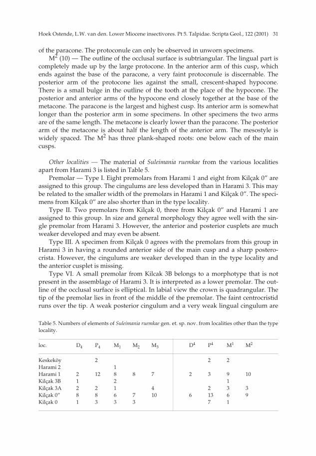
31Hoek Ostende, L.W. van den. Lower Miocene insectivores. Pt 5. Talpidae. Scripta Geol., 122 (2001)
of the paracone. The protoconule can only be observed in unworn specimens. M2 (10) — The outline of the occlusal surface is subtriangular. The lingual part is
completely made up by the large protocone. In the anterior arm of this cusp, whichends against the base of the paracone, a very faint protoconule is discernable. Theposterior arm of the protocone lies against the small, crescent-shaped hypocone.There is a small bulge in the outline of the tooth at the place of the hypocone. Theposterior and anterior arms of the hypocone end closely together at the base of themetacone. The paracone is the largest and highest cusp. Its anterior arm is somewhatlonger than the posterior arm in some specimens. In other specimens the two armsare of the same length. The metacone is clearly lower than the paracone. The posteriorarm of the metacone is about half the length of the anterior arm. The mesostyle iswidely spaced. The M2 has three plank-shaped roots: one below each of the maincusps.
Other localities — The material of Suleimania ruemkae from the various localitiesapart from Harami 3 is listed in Table 5.
Premolar — Type I. Eight premolars from Harami 1 and eight from Kilçak 0” areassigned to this group. The cingulums are less developed than in Harami 3. This maybe related to the smaller width of the premolars in Harami 1 and Kilçak 0”. The speci-mens from Kilçak 0” are also shorter than in the type locality.
Type II. Two premolars from Kilçak 0, three from Kilçak 0” and Harami 1 areassigned to this group. In size and general morphology they agree well with the sin-gle premolar from Harami 3. However, the anterior and posterior cusplets are muchweaker developed and may even be absent.
Type III. A specimen from Kilçak 0 agrees with the premolars from this group inHarami 3 in having a rounded anterior side of the main cusp and a sharp postero-crista. However, the cingulums are weaker developed than in the type locality andthe anterior cusplet is missing.
Type VI. A small premolar from Kilcak 3B belongs to a morphotype that is notpresent in the assemblage of Harami 3. It is interpreted as a lower premolar. The out-line of the occlusal surface is elliptical. In labial view the crown is quadrangular. Thetip of the premolar lies in front of the middle of the premolar. The faint centrocristidruns over the tip. A weak posterior cingulum and a very weak lingual cingulum are
Table 5. Numbers of elements of Suleimania ruemkae gen. et. sp. nov. from localities other than the typelocality.
loc. D4 P4 M1 M2 M3 D4 P4 M1 M2
Keskeköy 2 2 2Harami 2 1Harami 1 2 12 8 8 7 2 3 9 10Kilçak 3B 1 2 1Kilçak 3A 2 2 1 4 2 3 3Kilçak 0” 8 8 6 7 10 6 13 6 9Kilçak 0 1 3 3 3 7 1
pp 003-048 (SG 122-1) 15-01-2007 15:08 Pagina 31
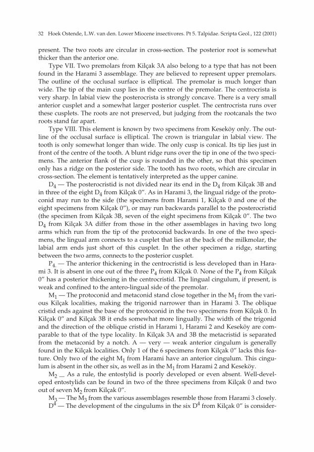
Hoek Ostende, L.W. van den. Lower Miocene insectivores. Pt 5. Talpidae. Scripta Geol., 122 (2001)32
present. The two roots are circular in cross-section. The posterior root is somewhatthicker than the anterior one.
Type VII. Two premolars from Kilçak 3A also belong to a type that has not beenfound in the Harami 3 assemblage. They are believed to represent upper premolars.The outline of the occlusal surface is elliptical. The premolar is much longer thanwide. The tip of the main cusp lies in the centre of the premolar. The centrocrista isvery sharp. In labial view the posterocrista is strongly concave. There is a very smallanterior cusplet and a somewhat larger posterior cusplet. The centrocrista runs overthese cusplets. The roots are not preserved, but judging from the rootcanals the tworoots stand far apart.
Type VIII. This element is known by two specimens from Keseköy only. The out-line of the occlusal surface is elliptical. The crown is triangular in labial view. Thetooth is only somewhat longer than wide. The only cusp is conical. Its tip lies just infront of the centre of the tooth. A blunt ridge runs over the tip in one of the two speci-mens. The anterior flank of the cusp is rounded in the other, so that this specimenonly has a ridge on the posterior side. The tooth has two roots, which are circular incross-section. The element is tentatively interpreted as the upper canine.
D4 — The posterocristid is not divided near its end in the D4 from Kilçak 3B andin three of the eight D4 from Kilçak 0”. As in Harami 3, the lingual ridge of the proto-conid may run to the side (the specimens from Harami 1, Kilçak 0 and one of theeight specimens from Kilçak 0”), or may run backwards parallel to the posterocristid(the specimen from Kilçak 3B, seven of the eight specimens from Kilçak 0”. The twoD4 from Kilçak 3A differ from those in the other assemblages in having two longarms which run from the tip of the protoconid backwards. In one of the two speci-mens, the lingual arm connects to a cusplet that lies at the back of the milkmolar, thelabial arm ends just short of this cusplet. In the other specimen a ridge, startingbetween the two arms, connects to the posterior cusplet.
P4. — The anterior thickening in the centrocristid is less developed than in Hara-mi 3. It is absent in one out of the three P4 from Kilçak 0. None of the P4 from Kilçak0” has a posterior thickening in the centrocristid. The lingual cingulum, if present, isweak and confined to the antero-lingual side of the premolar.
M1 — The protoconid and metaconid stand close together in the M1 from the vari-ous Kilçak localities, making the trigonid narrower than in Harami 3. The obliquecristid ends against the base of the protoconid in the two specimens from Kilçak 0. InKilçak 0” and Kilçak 3B it ends somewhat more lingually. The width of the trigonidand the direction of the oblique cristid in Harami 1, Harami 2 and Keseköy are com-parable to that of the type locality. In Kilçak 3A and 3B the metacristid is separatedfrom the metaconid by a notch. A — very — weak anterior cingulum is generallyfound in the Kilçak localities. Only 1 of the 6 specimens from Kilçak 0” lacks this fea-ture. Only two of the eight M1 from Harami have an anterior cingulum. This cingu-lum is absent in the other six, as well as in the M1 from Harami 2 and Keseköy.
M2 — As a rule, the entostylid is poorly developed or even absent. Well-devel-oped entostylids can be found in two of the three specimens from Kilçak 0 and twoout of seven M2 from Kilçak 0”.
M3 — The M3 from the various assemblages resemble those from Harami 3 closely. D4 — The development of the cingulums in the six D4 from Kilçak 0” is consider-
pp 003-048 (SG 122-1) 15-01-2007 15:08 Pagina 32
33Hoek Ostende, L.W. van den. Lower Miocene insectivores. Pt 5. Talpidae. Scripta Geol., 122 (2001)
ably weaker than in Harami 3, particularly on the lingual side. The parastyle is only asmall cusp. In 1 of the 6 specimens the tip of the paracone lies in the centre of the milkmolar, and not in front of the middle, as in the other D4. The development of cingu-lums on the only complete D4 found in Harami 1 resembles that of Kilçak 0”. The tipof the paracone in this D4 also lies in the centre.
P4 — The tip of the paracone lies in the middle of the P4 in all localities older thanHarami 3. Only in Keseköy and Harami 3 itself the tip lies in front of the middle. Thetwo P4 from Keseköy also agree with Harami 3 in having a well-developed parastyleand a strong postero-lingual cingulum. In the other localities the parastyle is small.The postero-lingual cingulum is generally well developed, but it is weak in two of theseven specimens from Kilçak 0, 3 of the 13 from Kilçak 0”, both specimens fromKilçak 3A and in two of the three specimens from Harami 1.
M1 — The anterior arm of the protocone usually connects to the paracone (Kilçak3A, Keseköy) or ends just short of it (Kilçak 0”, Harami 1). This arm connects to theparastyle in two of the six M1 from Kilçak 0”. The anterior arm of the protocone issplit into two ridges in the specimen from Kilçak 0, the one from Kilçak 3B and one ofthe nine specimens from Harami 1. In these cases a thick ridge connects to the para-cone, and a narrower one runs in the direction of the parastyle. The protoconule issmall and can be observed in two unworn specimens only, one from Kilçak 0” andone from Harami 1. The development of the hypocone varies. This cusp is very welldeveloped in Kilçak 0, Kilçak 3A and Kilçak 3B. The development of the hypocone inthe other localities resembles that of Harami 3. A widely-spaced mesostyle, resem-bling the configuration found in Harami 3, is found in the specimen from Kilçak 3B,as well as in two of the nine specimens from Harami 1 and two of the six specimensfrom Kilçak 0”. In the other specimens the mesostyle is well divided.
M2 — An indistinct protoconule is found in two of the ten specimens from Hara-mi 1 only. The development of the hypocone varies. The hypocone is well developedin all specimens from Kilçak 0 and Kilçak 3A, but only in three out of nine of the M2
from Kilçak 0” and in five out of ten specimens from Harami 1. The arms of the para-cone in the Kilçak 0” assemblage seem somewhat longer than those from the otherlocalities. They stand close together, leaving only a narrow valley in between. Two ofthe M2 from Harami 1 stand out by having very short arms of the paracone. The divi-sion of the mesostyle is most pronounced in the type locality. In the other localitiesthe two cusps of the mesostyle even touch near their bases in some cases.
Remarks — Some of the differences between the various assemblages of Suleimaniaruemkae can be interpreted as an evolutionary trend. However, the differences are notvery clear-cut. There seems to be an increase in size through time, especially in theM1/M1. Morphological differences are the stronger development of cingulums on theP4 and a somewhat less developed hypocone of the M1 in the younger localities. Thetrigonid of the M1 seems to become wider in time. However, these trends are not con-sistent. The hypocone of the M1 in Kilçak 0”, for instance, resembles that of theyounger localities. Since there is no clear gap in size or morphology between the vari-ous assemblages, all of them have been assigned to a single species.
At first sight, Suleimania ruemkae might be identified as a dimylid. The M1 with itsquadrangular outline and slightly inflated cusps can be easily confused with the M1
pp 003-048 (SG 122-1) 15-01-2007 15:08 Pagina 33

Hoek Ostende, L.W. van den. Lower Miocene insectivores. Pt 5. Talpidae. Scripta Geol., 122 (2001)34
of primitive dimylids. The M3 has lost its talonid and resembles the M3 of theOligocene dimylid Exaeonodus. The M3 is absent. Dimylidae are, however, not theonly insectivores in which the M3 and M3 are reduced. The shrew Amblycoptus alsolacks the last molars. In addition to the reduced last molars and the morphology ofthe upper molars, S. ruemkae shares the presence of a functional milk dentition withthe Dimylidae. Within the Talpidae this feature is only found in the shrew-moles(Uropsilinae and Urotrichini sensu Hutchison, 1968).
Other features suggest that Suleimania ruemkae is a talpid rather than a dimylid.The oblique cristid of Dimylidae always ends against the protoconid, whereas thisridge ends against the protoconid-metaconid crest in Suleimania as it does in Talpidae.Although the hypocone of the M1 is large in comparison to other Talpidae, it is con-siderably smaller than the large cone-shaped hypocone of Dimylidae. Although theP4, M1 and M1 show a tendency to inflate their cusps, this inflation is much less pro-nounced than in the exaenodont dentitions of Dimylidae. Furthermore, the tendencyis restricted to the above mentioned elements. In contrast, the M2 is characterized bysharp ridges. The premolars too have very sharp cutting edges, whereas Dimylidaeare defined as having blunt premolars with inflated cusps. All these features indicatethat Suleimania is best classified as a talpid. The resemblence to Dimylidae is consid-ered to be the result of convergence.
The combination of the characters mentioned above does not allow a classificationof Suleimania ruemkae in any of the known subfamilies (Uropsilinae, Talpinae, Des-maninae, Gaillardinae). Therefore a new subfamily has been erected for this peculiarspecies.
The inflation of the P4, M1 and M1 can be seen as an indication that Suleimaniaruemkae, like dimylids, may have fed on molluscs. It is, however, unlikely that mol-luscs were its main diet. The premolars and M2 still have sharp ridges, which sug-gests that the species also fed on nimbler prey, such as (water) insects and/or smallvertebrates. Furthermore, the long paralophid of the M1 and the long posterolingualflank of the P4 provide a shearing action rather than a crushing action, as we wouldexpect in true mollusc eaters. As a result of the reduction of the last molars, the cut-ting edge of the P4 and M1 lies relatively close to the condyle, which provides a pow-erful shearing apparatus. Such a strong cutting edge would have been useful forinstance when eating small vertebrates as frogs and lizards.
The Suleimaninae is the first extinct subfamily of moles recognized in Eurasia.The discovery of an extinct stock of talpids confirms that the diversity of the familywas once much higher than it is today. This is also the case for the talpids of NorthAmerica (Hutchison, 1968).
Conclusions
Four different genera of Talpidae, Desmanodon, Theratiskos, Geotrypus and Suleiman-ia, were found in Lower Miocene strata from Anatolia. This means that the Talpidaewere the most diverse insectivore family in the Early Miocene of that area. In south-ern Germany too, talpids are the most diverse insectivore group in Early Miocenetimes (Ziegler, 1985; 1990a). Talpid diversity in southern Germany is higher than inAnatolia, but this holds true for all insectivore families.
pp 003-048 (SG 122-1) 15-01-2007 15:08 Pagina 34
35Hoek Ostende, L.W. van den. Lower Miocene insectivores. Pt 5. Talpidae. Scripta Geol., 122 (2001)
Two of the genera found show a connection with the European insectivore fau-nas. Geotrypus is a common element in Late Oligocene/ Early Miocene insectivorefaunas from Europe. Desmanodon appears in Europe sometime during the EarlyMiocene (Van den Hoek Ostende, 1997), but the closely related genus Paratalpa wasalready found in Europe during the Late Oligocene. The other two genera, Theratiskosand Suleimania, are thus far known from Anatolia only. Possibly these genera havetheir origin in Asia.
Acknowledgements
I am grateful to the Mineral Research and Exploration General Directorate ofTurkey (M.T.A.) in Ankara for their support and promoting international collabora-tion in studying the fossil mammal faunas of Anatolia. Various versions of the manu-script were read by Hans de Bruijn, Albert van der Meulen, Thijs Freudenthal, andJohan Meulenkamp. Their comments and discussions were of great value. Jaap Luteijnmade the drawings; the SEM photographs were made by Wil den Hartog.
References
For References see Part 8 in this volume.
Manuscript received 30 September 1998.
pp 003-048 (SG 122-1) 15-01-2007 15:08 Pagina 35

Hoek Ostende, L.W. van den. Lower Miocene insectivores. Pt 5. Talpidae. Scripta Geol., 122 (2001)36
Plate 1
Figs. 1-7. Theratiskos mechteldae sp. nov. from Keseköy. 1: D4 sin., Ke 6901. 2: Mandible sin. with M1, Ke 6941. 3: Mandible sin. with P4-M2, Ke 6975, holotype; a: occlusal view; b: labial view. 4: D4 sin., Ke 7163. 5: Maxillary fragment sin. with P4 and M1, Ke 7224. 6: M2 sin., Ke 7304. 7: M3 sin. Ke 7366.
Figs. 8-16. Theratiskos rutgeri sp. nov. from Harami 1. 8: P4 sin., Ha 1 3811. 9: M1 sin., Ha 1 3853. 10: M2 sin., Ha 1 3894. 11: M3 sin., Ha 1 3936. 12: D4 sin., Ha 1 3981. 13: P4 sin., Ha 1 3997.14: M1 sin., Ha 1 4028.15: M2 sin., Ha 1 4057, holotype. 16: M3 sin., Ha 1 4108.
All specimens × 12.5.
pp 003-048 (SG 122-1) 15-01-2007 15:08 Pagina 36
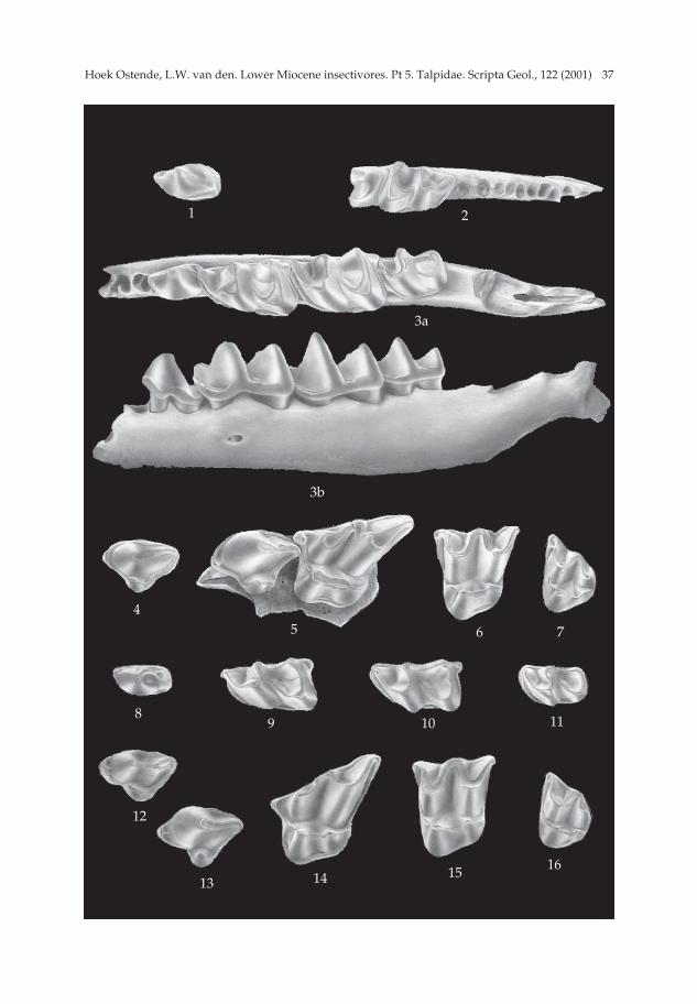
37Hoek Ostende, L.W. van den. Lower Miocene insectivores. Pt 5. Talpidae. Scripta Geol., 122 (2001)
1 2
3a
3b
45 6 7
89 10 11
12
13 14 15 16
pp 003-048 (SG 122-1) 15-01-2007 15:08 Pagina 37

Hoek Ostende, L.W. van den. Lower Miocene insectivores. Pt 5. Talpidae. Scripta Geol., 122 (2001)38
Plate 2
Figs. 1-7. Geotrypus haramiensis sp. nov. from Harami 1 and 3. 1: Mandible sin. with P1-M3, Ha 3 781, holotype; a: occlusal view; b: labial view (× 6.2). 2: P4 sin., Ha 3 782.3: Humerus, Ha 3 876 (× 4). a: posterior view; b: anterior view.4: M2 sin., Ha 3 787. 5: M3 sin., Ha 3 788. 6: M2 sin., Ha 1 3801. 7: M3 sin., Ha 1 3802.
Specimens × 12.5 unless indicated otherwise.
pp 003-048 (SG 122-1) 15-01-2007 15:08 Pagina 38

39Hoek Ostende, L.W. van den. Lower Miocene insectivores. Pt 5. Talpidae. Scripta Geol., 122 (2001)
1a
1b
2
3a
3b
4
6
5
7
pp 003-048 (SG 122-1) 15-01-2007 15:08 Pagina 39
Hoek Ostende, L.W. van den. Lower Miocene insectivores. Pt 5. Talpidae. Scripta Geol., 122 (2001)40
Plate 3
Figs. 1-6. Geotrypus kesekoeyensis sp. nov. from Keseköy. 1: M1 sin., Ke 6703. 2: M2 sin., Ke 6704. 3: P4 dext., Ke 6706. 4: M1 sin., Ke 6708. 5: M2 sin., Ke 6712, holotype. 6: M3 sin., Ke 6715.
Figs. 7-10. Suleimania ruemkae gen. et sp. nov. from Harami 3. 7: I1 dext., Ha 1, 3631.8: Premolar Type IV, Ha 3 696; a: occlusal view; b: labial view. 9: Premolar Type II, Ha 3 691; a: occlusal view; b: labial view. 10: Premolar Type V, Ha 3 699; a: occlusal view; b: labial view.
All specimens × 12.5.
pp 003-048 (SG 122-1) 15-01-2007 15:08 Pagina 40
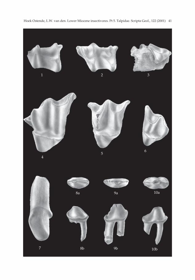
41Hoek Ostende, L.W. van den. Lower Miocene insectivores. Pt 5. Talpidae. Scripta Geol., 122 (2001)
1 2 3
4
8b
10a9a8a
7
56
9b 10b
pp 003-048 (SG 122-1) 15-01-2007 15:08 Pagina 41
Hoek Ostende, L.W. van den. Lower Miocene insectivores. Pt 5. Talpidae. Scripta Geol., 122 (2001)42
Plate 4
Figs. 1-8. Suleimania ruemkae gen. et sp. nov. from Harami 1 and 3. 1: Premolar Type I with part of mandible, Ha 3 686; a: occlusal view; b: labial view. 2: D4 sin., Ha 1 3644. 3: D4 dext., Ha 3 706; a: occlusal view; b: labial view. 4: Premolar Type III, Ha 3 693. 5: P4 sin., Ha 3 720. 6: P4 sin., Ha 3 659. 7: D4 sin., Ha 3 751. 8: D4 sin., Ha 1 3734.
All specimens × 12.5.
pp 003-048 (SG 122-1) 15-01-2007 15:08 Pagina 42
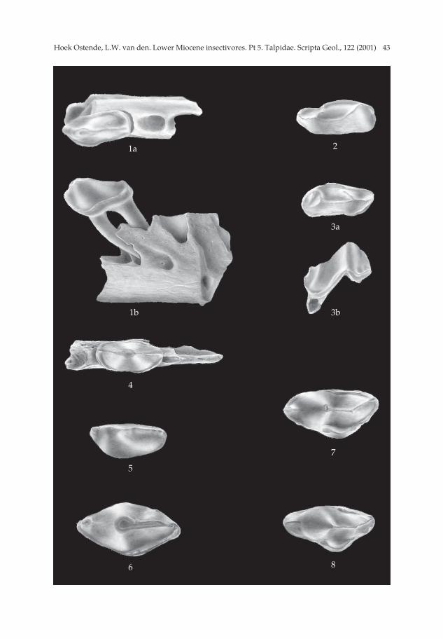
43Hoek Ostende, L.W. van den. Lower Miocene insectivores. Pt 5. Talpidae. Scripta Geol., 122 (2001)
1a
1b
4
5
6
2
3a
3b
7
8
pp 003-048 (SG 122-1) 15-01-2007 15:08 Pagina 43
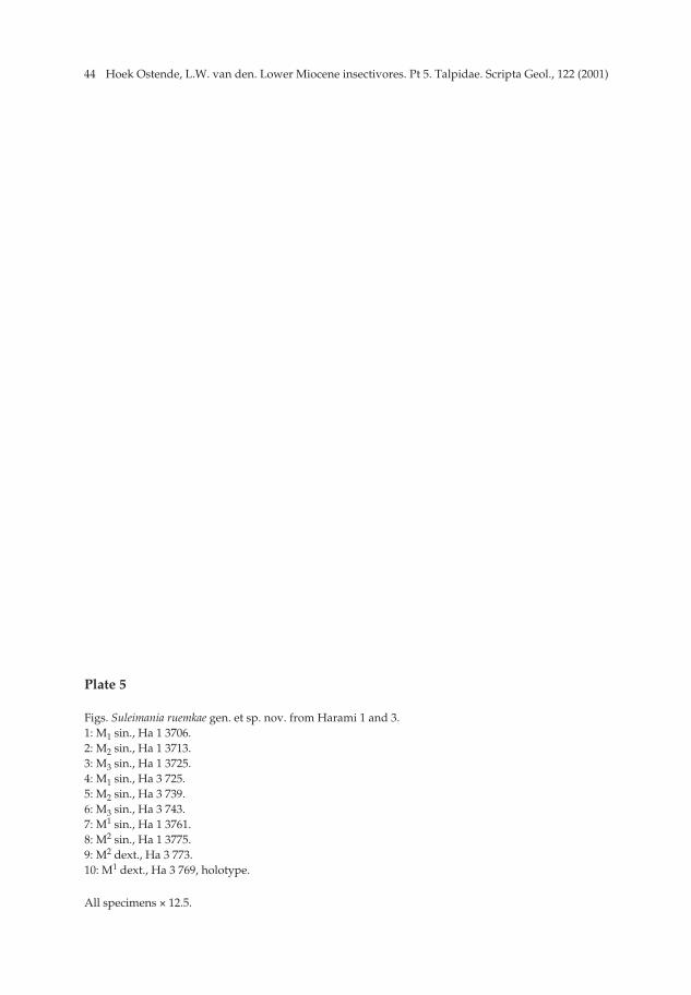
Hoek Ostende, L.W. van den. Lower Miocene insectivores. Pt 5. Talpidae. Scripta Geol., 122 (2001)44
Plate 5
Figs. Suleimania ruemkae gen. et sp. nov. from Harami 1 and 3. 1: M1 sin., Ha 1 3706. 2: M2 sin., Ha 1 3713. 3: M3 sin., Ha 1 3725. 4: M1 sin., Ha 3 725. 5: M2 sin., Ha 3 739. 6: M3 sin., Ha 3 743. 7: M1 sin., Ha 1 3761. 8: M2 sin., Ha 1 3775. 9: M2 dext., Ha 3 773.10: M1 dext., Ha 3 769, holotype.
All specimens × 12.5.
pp 003-048 (SG 122-1) 15-01-2007 15:08 Pagina 44

45Hoek Ostende, L.W. van den. Lower Miocene insectivores. Pt 5. Talpidae. Scripta Geol., 122 (2001)
1 2 3
4 5 6
78
9 10
pp 003-048 (SG 122-1) 15-01-2007 15:08 Pagina 45

pp 003-048 (SG 122-1) 15-01-2007 15:08 Pagina 46