Embed Size (px)
Citation preview
Guest Editors: Andrea Lauterio, Luciano De Carlis, Irinel Popescu, and Juan García-Valdecasas
HPB Surgery
Innovative Strategies and Recent Advances in Liver Surgery

Innovative Strategies and Recent Advances inLiver Surgery

HPB Surgery
Innovative Strategies and Recent Advances inLiver Surgery
Guest Editors: Andrea Lauterio, Irinel Popescu,Juan Carlos Garcıa-Valdecasas, and Luciano De Carlis

Copyright © 2013 Hindawi Publishing Corporation. All rights reserved.
This is a special issue published in “HPB Surgery.” All articles are open access articles distributed under the Creative Commons Attribu-tion License, which permits unrestricted use, distribution, and reproduction in any medium, provided the original work is properly cited.

Editorial Board
Shushma Aggarwal, USAMichael Bartels, GermanyChristoph E. Broelsch, GermanyJavier Bueno, SpainJuli Busquets, SpainDaniel Casanova, SpainRichard Charnley, UKVito R. Cicinnati, GermanyUta Dahmen, GermanyAshley Denison, UKChristos G. Dervenis, GreeceVincent Donckier, BelgiumOlivier Farges, France
Peter Faybik, AustriaLaureano Fernandez-Cruz, SpainLuis Grande, SpainJeffrey Halldorson, USAHobart W. Harris, USAShuji Isaji, JapanJ. R. Izbicki, GermanyPiotr Kalicinski, PolandTatsuya Kin, CanadaGuy J. Maddern, AustraliaMarcello Maestri, ItalyUmberto Maggi, ItalyAttila Olah, Hungary
Eugenia Pareja, SpainG. J. Poston, UKGuntars Pupelis, LatviaPablo Ramırez, SpainB. Krishna Rau, IndiaGina R. Rayat, CanadaMagnus Rizell, SwedenHarald Schrem, GermanyUtz Settmacher, GermanyJames Shapiro, CanadaDuncan Spalding, UKRobin C. Williamson, UKShu-Sen Zheng, China

Contents
Innovative Strategies and Recent Advances in Liver Surgery, Andrea Lauterio, Irinel Popescu,Juan Carlos Garcıa-Valdecasas, and Luciano De CarlisVolume 2013, Article ID 517279, 2 pages
Living-Donor Liver Transplantation and Hepatitis C, Nobuhisa Akamatsu and Yasuhiko SugawaraVolume 2013, Article ID 985972, 13 pages
Bleeding in Hepatic Surgery: Sorting through Methods to Prevent It, Fabrizio Romano, Mattia Garancini,Fabio Uggeri, Luca Degrate, Luca Nespoli, Luca Gianotti, Angelo Nespoli, and Franco UggeriVolume 2012, Article ID 169351, 12 pages
Contemporary Strategies in the Management of Hepatocellular Carcinoma, Shirin Elizabeth Khorsandiand Nigel HeatonVolume 2012, Article ID 154056, 8 pages
Surgical Options for Initially Unresectable Colorectal Liver Metastases, Irinel Popescu andSorin Tiberiu AlexandrescuVolume 2012, Article ID 454026, 13 pages
Initial Experiences of Simultaneous Laparoscopic Resection of Colorectal Cancer and Liver Metastases,L. T. Hoekstra, O. R. C. Busch, W. A. Bemelman, T. M. van Gulik, and P. J. TanisVolume 2012, Article ID 893956, 6 pages
Initial Experience in Single-Incision Transumbilical Laparoscopic Liver Resection: Indications, PotentialBenefits, and Limitations, Giovanni Dapri, Livia DiMarco, Guy-Bernard Cadiere, and Vincent DonckierVolume 2012, Article ID 921973, 9 pages
Laparoscopy in Liver Transplantation: The Future Has Arrived, Quirino Lai, Rafael S. Pinheiro,Giovanni B. Levi Sandri, Gabriele Spoletini, Fabio Melandro, Nicola Guglielmo, Marco Di Laudo,Fabrizio M. Frattaroli, Pasquale B. Berloco, and Massimo RossiVolume 2012, Article ID 148387, 7 pages
Hindawi Publishing CorporationHPB SurgeryVolume 2013, Article ID 517279, 2 pageshttp://dx.doi.org/10.1155/2013/517279
EditorialInnovative Strategies and Recent Advances in Liver Surgery
Andrea Lauterio,1 Irinel Popescu,2 Juan Carlos García-Valdecasas,3 and Luciano De Carlis1
1 Dipartimento di Chirurgia Generale e Trapianti Addominali, Ospedale Niguarda, Piazza Ospedale Maggiore, 3 20162 Milano, Italy2 Center of General Surgery and Liver Transplantation, Fundeni Clinical Institute, Bucharest, Romania3 Department of Surgery, Hospital Clınic, University of Barcelona, Barcelona, Catalunya, Spain
Correspondence should be addressed to Andrea Lauterio; [email protected]
Received 6 March 2013; Accepted 6 March 2013
Copyright © 2013 Andrea Lauterio et al.This is an open access article distributed under theCreativeCommonsAttribution License,which permits unrestricted use, distribution, and reproduction in any medium, provided the original work is properly cited.
Techniques for hepatic surgery have evolved over the past fewdecades and have broadened indications for liver resection(LR) for liver tumors. New strategies including downsizingchemotherapy, two-stage LR with or without portal veinembolization, and resection combined with ablative methodsallow tailoring the treatment to each patient depending oncondition of the liver and tumor burden. In the recentyears, the new dissector devices have been developed andtogether with the use of intraoperative ultrasound allowa new approach to the anatomical ultrasound-guided liverresection, even for large tumors located in challenging posi-tions.
Improvements in imaging evaluation with high-resolu-tion CT scan or MRI allow new methods for the studyof the future remnant liver and play an important role inthe planning of the resection strategy reducing the risk ofmajor complications and liver failure, especially in patientswho undergo major resection. In addition, development ofnew technology in local ablative therapies for liver tumors isposing a competition to LR.
The incidence of hepatocellular carcinoma (HCC) isclimbing rapidly and in a current climate of organ shortagehas led to the re-evaluation of locoregional therapies andresectional surgery tomanage the case load.The introductionof biological therapies has had a new dimension to care,adding to the complexities of multidisciplinary teamworkingin the management of HCC. S. E. Khorsandi and N. Heatongive a very comprehensive overview of the present daymanagement strategies and decisionmaking for patients withHCC.
Simultaneous resection of primary colorectal carcinoma(CRC) and synchronous liver metastases (SLM) is subject ofdebate with respect to morbidity in comparison to stagedresection. In contrast to the extensive literature on stagedlaparoscopic colorectal and laparoscopic liver surgery, thereare only a few reports on combined laparoscopic colorectaland liver resection.
L. T. Hoekstra and colleagues report their initial experi-ence of simultaneous laparoscopic resection of primary CRCand SLM. According to the modern literature, the authorsconclude that patient selection and expertise are essentialfor this complex type of surgery and the multidisciplinaryteam should decide on optimal timing within multimodalityschedules.
I. Popescu and S. T. Alexandrescu challenge recent evi-dence in the different surgical options for initially unre-sectable colorectal liver metastases. The authors illustrate theavailable oncosurgical modalities including liver resectionfollowing portal vein ligation/embolization, “two-stage” liverresection, one-stage ultrasonically guided liver resection,hepatectomy following conversion chemotherapy, and liverresection combined with thermal ablation. The authorsdiscuss the role of liver transplantation (LT) as a futureopportunity in the treatment of unresectable CRLM inselected patients, taking into account the related ethicalconsiderations especially in case of LT from living donoror LT with marginal grafts. Although the available data donot support liver transplantation as a routine procedure inpatients with CRLM, this paper could promote the debate onthis issue.

2 HPB Surgery
Partial liver transplantation, including split-liver and liv-ing donor liver transplantation, represents another importantapplication of these advantages applied in the field of organtransplantation.
N. Akamatsu and Y. Sugawara provide a review articleon the current trends and controversies in living donor livertransplantation (LDLT) for patients with HCV in relation tothe perspectives from deceased donor.
They focused their attention on the recent advances inantiviral treatment for the recurrent hepatitis C after LTreporting the different strategies from the Japanese LDLTcenters.
Minimally invasive approach in the field of HPB surgeryis gaining popularity due to the availability of new laparo-scopic instruments for liver transection. Laparoscopic LR hasevolved significantly over the past decade moving from anexperimental procedure to a standard part of the hepaticsurgeon’s armamentarium. Most recently, robotic-assistedtechnology offers solutions to overcome the limitations ofconventional laparoscopic resection.
With the review paper titled “Laparoscopy in liver trans-plantation:The future has arrived,”Q. Lai and colleagues shedfurther light on the role of the laparoscopy in this field ofsurgery. Intent of the review is to underline the current roleof diagnostic and therapeutic laparoscopy in patients waitingfor LT, in the living donor LT and in LT recipients.
G. Dapri and colleagues report their initial experiencewith single-incision transumbilical laparoscopic liver resec-tion (SITLLR) with a detailed technical paper and discussthe future of this approach in terms of indications, poten-tial benefits, and limitations in comparison with multiportlaparoscopic technique.
Single incision transumbilical laparoscopy represents thelatest advance of the laparoscopic approach; however, itsuse in LR still remains limited to small reported series,and further evaluation is required to assess the potentialadvantages and the improvement in the patients outcome.As reported by the authors, at this point of the experi-ence, several questions on SITLLR remain to be addressed,concerning the feasibility and mostly the reproducibility ofthis technique, the indications, selection criteria, limitations,effect on postoperative outcomes, and long-term results.
F. Romano et al. provide a summarizing paper illustratingmethods to prevent bleeding in hepatic surgery. This is ofparticular interest, as bleeding inHPB surgery represents oneof the most common features associated with poor outcome.The paper is based on the literature information and author’sexperience, and the aim of the study is to investigate theprincipal solutions to the problem of high blood loss inLR focusing on technological approach to the parenchymatransection.
The aim of this special issue is to update and promoteinterchange of the current knowledge and recent progressfocusing on innovative strategies and recent advances inliver surgery. These manuscripts represent an exciting andinsightful snapshot of the recent advances in liver surgery.State-of-the-art, existing challenges, and emerging futuretopics are highlighted in this special issue, which may inspirethe reader and help advance in this field of surgery.We would
like to thank all the authors and reviewers for making thisspecial issue in HPB surgery possible.
Andrea LauterioIrinel Popescu
Juan Carlos Garcıa-ValdecasasLuciano De Carlis

Hindawi Publishing CorporationHPB SurgeryVolume 2013, Article ID 985972, 13 pageshttp://dx.doi.org/10.1155/2013/985972
Review ArticleLiving-Donor Liver Transplantation and Hepatitis C
Nobuhisa Akamatsu1, 2 and Yasuhiko Sugawara2
1 Department of Hepato-Biliary-Pancreatic Surgery, SaitamaMedical Center, SaitamaMedical University, 1981 Tsujido-cho, Kamoda,Kawagoe, Saitama 350-8550, Japan
2Arti�cial �rgan and Transplantation Division, Department of Surgery, �raduate School of Medicine, University of Tokyo,7-3-1 Hongo, Bunkyo-ku, Tokyo 113-8655, Japan
Correspondence should be addressed to Yasuhiko Sugawara; [email protected]
Received 17 May 2012; Accepted 1 January 2013
Academic Editor: Andrea Lauterio
Copyright © 2013 N. Akamatsu and Y. Sugawara. is is an open access article distributed under the Creative CommonsAttribution License, which permits unrestricted use, distribution, and reproduction in any medium, provided the original work isproperly cited.
Hepatitis-C-virus- (HCV-) related end-stage cirrhosis is the primary indication for liver transplantation in many countries.Unfortunately, however, HCV is not eliminated by transplantation and gra reinfection is universal, resulting in �brosis, cirrhosis,and �nally gra decompression. In areas with low deceased-donor organ availability like Japan, living-donor liver transplantation(LDLT) is similarly indicated forHCV cirrhosis as deceased-donor liver transplantation (DDLT) inWestern countries and acceptedas an established treatment for HCV-cirrhosis, and the results are equivalent to those of DDLT. To prevent gra failure dueto recurrent hepatitis C, antiviral treatment with pegylated-interferon and ribavirin is currently considered the most promisingregimen with a sustained viral response rate of around 30% to 35%, although the survival bene�t of this regimen remains to beinvestigated. In contrast to DDLT, many Japanese LDLT centers have reported modi�ed treatment regimens as best e�orts tosecure �rst gra, such as aggressive preemptive antiviral treatment, escalation of dosages, and elongation of treatment duration.
1. Introduction
Since the �rst successful application of living donor livertransplantation (LDLT) in 1990 [1] and subsequent success-ful LDLT for adult recipient in 1994 [2], the use of livedonors for liver transplantation has been widely applied toadult recipients where the availability of deceased-donors isseverely restricted, like in Japan [3], and also accepted as asolution to the cadaveric donor shortage inWestern countries[4].
End-stage liver disease caused by chronic hepatitis C virus(HCV) infection is the leading cause of liver transplantationin developed countries [5, 6], including Japan [7]. Unfor-tunately, liver transplantation does not cure HCV-infectedrecipients, but re-infection of HCV universally occurs anddisease progression is accelerated compared with that in thenontransplant population, resulting in poor outcomes forHCV-infected recipients [8].
e aim of this paper was to overview the current trendsand controversies in LDLT for patients with HCV in relation
to the perspectives fromdeceased-donor liver transplantation(DDLT).
2. Natural History of Hepatitis C afterOrthotopic Liver Transplantation
Accumulating perspectives of disease recurrence in HCV-infected recipients have been obtained in DDLT withinthe last two decades. HCV reinfection occurs just aerreperfusion followed by a rapid increase in HCV ribonucleicacid (RNA) levels within 4 postoperative months [9]. ehistologic features of liver injury usually resemble those ofnontransplant HCV hepatitis typically developing aer 3months, but the clinical presentation, severity, and outcomeare extremely heterogeneous and more profound comparedto those in immune competent patients [10]. Progressionto cirrhosis usually takes 9 to 12 years aer liver trans-plantation with a linear progression of histologic �brosis[10, 11]. A less common, but well-documented, form of
2 HPB Surgery
recurrence is called �brosing cholestatic hepatitis (<10%),possibly mediated by a direct cytopathic mechanism underan extremely high viral load and immune-compromisedcondition. Gra failure occurs in 50% of recipients within afewmonths aer �brosing cholestatic hepatitis develops [12].Some HCV-reinfected recipients, however, show no apparentdisease progression for at least the �rst decade and theirgra injury remains mild or even absent despite a high viraburden.
Overall, cirrhosis develops in approximately 25% of livertransplant recipients (range 8%−44%) aer 5 to 10 years andthis percentage is likely to increase with an increase in thefollow-upperiod [10, 11].Once cirrhosis is complete, survivaltime is severely decreased and decompression is encounteredwith cumulative rates at 1 and 3 years of 40% and 60%,respectively, which �nally results in gra failure [11, 13].
e development of decompensated cirrhosis due torecurrent hepatitis C is now the most frequent cause of grafailure, patient death, and the need for retransplantation inHCV-infected recipients [11, 13–17]. As a result, survival issigni�cantly decreased compared with other indications, anoverall 10% difference at 3 years [18]. In the most recentUnited Network for Organ Sharing/Organ Procurement andTransplantation Network (UNOS/OPTN) study from theUnited States, 3-year survival is 78% among 7459 HCV-positive recipients compared with 82% among 20734 HCV-negative recipients (𝑃𝑃 < 𝑃𝑃𝑃𝑃𝑃𝑃; http://www.unos.org) [19].
e poor outcome of HCV-positive recipients hasresulted in the divergence in transplant outcomes betweenHCV-positive recipients and HCV-negative recipients.Improvements in organ preservation, surgical techniques,and postoperative care have dramatically improved thesurvival of HCV-negative recipients over the last twodecades, whereas this has not been the case in HCV-positiverecipients for whom outcome has remained unchanged oreven worsened over time [19–22].
3. Current Status of LDLT
In areas with low deceased-donor organ availability likeJapan, the indication of LDLT for HCV cirrhosis is similarto that of DDLT [7], whereas in Western countries, LDLTis conducted in an attempt to alleviate the shortage ofdonor organs and decrease the mortality among patientsawaiting transplants, accounting for only 3% to 4% of all livertransplants [23].
According to the Japan Liver Transplantation Society[24], a total of 6097 LDLTs, comprising 98% of all livertransplants, have been performed till the end of 2010 in Japan.Among those, 3796 were adult cases including 1200 (32%)cases of HCV-related disease as a leading indication for adultLDLT. e 1, 3, 5, and 10 year survival rates of all adultLDLT and those of HCV-positive adults were 81%, 75%, 72%,and 66%, and 78%, 72%, 68%, and 59%, respectively, withoutdifference.
In the United States, nearly 3000 LDLTs have beenperformed by the end of 2009, with decreased number of
cases annually, comprising only 4.5% of all liver transplants[23, 25, 26].
4. LDLT as a Risk Factor for RecurrentHepatitis C Studies Comparing Outcomes ofLDLT and DDLT
Based on the signi�cant negative impact of recurrent hepati-tis C on recipients’ outcome, it is critical to identify the factorsrelated to severe recurrent hepatitis C [8, 13]. In the trans-plant setting, many factors contribute to disease progressioncompared with nontransplant patients [13], including, viral-related factors [10, 27–36], donor age [17, 37–43], recipient-related factors [32, 44–49], gra and surgical factors [40,50–57], and immunosuppressive agents [58–75] (Table 1)however, many aspects remain unclear and require furtherinvestigation [8]. Among those, the possibility of increasedseverity of recurrent HCV in LDLT patients had been one ofthe hottest debates. e bene�t of LDLT might be offset if theoutcome of LDLT for HCV-positive recipients is worse thanthat of DDLT.
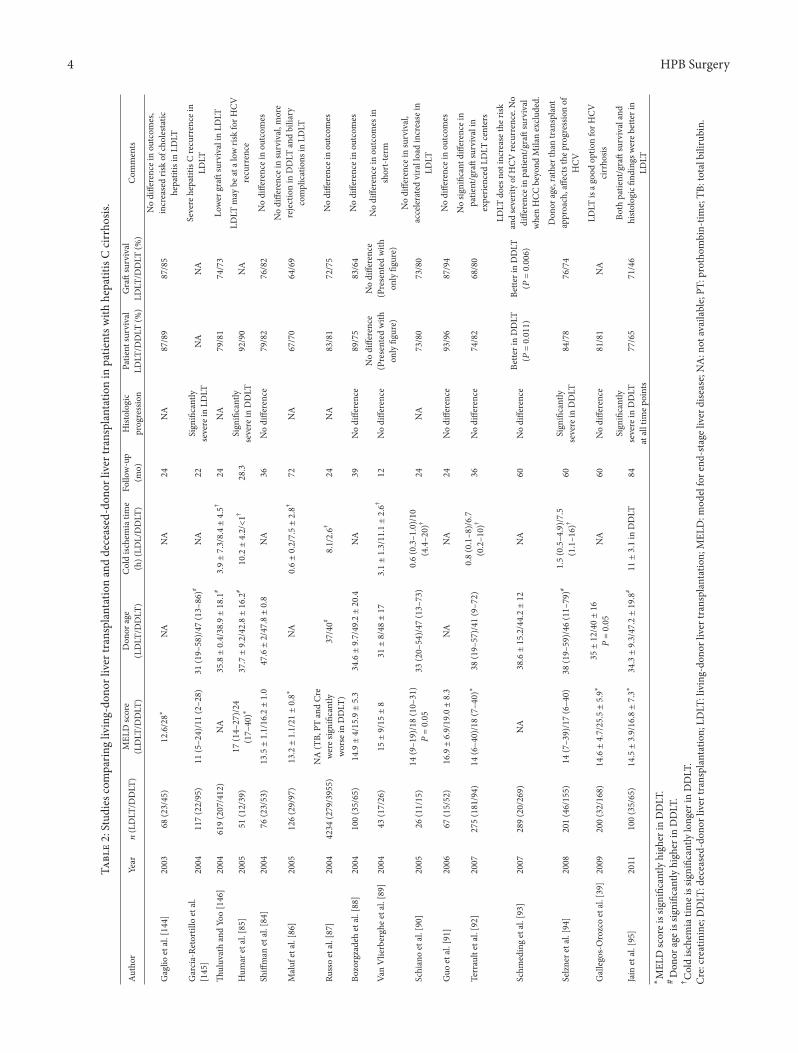
Early studies raised some negative concerns regarding theoutcomes of LDLT in HCV patients, such as a poorer graoutcome and earlier and more aggressive HCV recurrenceaer LDLT comparedwithDDLT [144–146]. Several theorieshave been proposed to explain the differences in HCV recur-rence between LDLT and DDLT recipients. One possibleexplanation is that the intense hepatocyte proliferation thatoccurs in partial liver gras may lead to increased viraltranslation and replication [145, 147–149]. Genetic donor-recipient similarity is another proposed mechanism for moresevere HCV recurrence [150, 151]. Recent studies, however,comparing outcomes of LDLT and DDLT in HCV-infectedpatients have not only failed to identify LDLT as a risk factorformore intense viral recurrence with impaired outcome, butalso revealed improved results in LDLT recipients [39, 84–95], which do not support the aforementioned speculations.Alternatively, recent studies favored the theory that outcomesof LDLT for HCV cirrhosis could be better than those ofDDLT due to the younger donor age and shorter ischemictime of LDLT gras. e studies comparing outcomesbetween LDLT and DDLT in HCV-infected recipients aresummarized in Table 2.
While several earlier studies demonstrated impairedpatient/gra survival and severe histologic �ndings in LDLT[144–146], the majority of studies reported equal or evenimproved outcomes both in patient/gra survival and in�brosis progression in LDLT [39, 84–95]. Since the largeUNOS database study [87] demonstrated comparable short-term (24 months) survival between LDLT and DDLT, sub-sequent studies with considerable follow-up period havebeen published demonstrating comparable or even superioroutcome in LDLT. Five-year patient survival ranged 71% to84% in HCV-positive LDLT recipients among studies withsufficient follow-up period [39, 86, 94, 95]. Additionally, asTerrault et al. [92] reported, the learning curve for the LDLTproceduremay have a considerable impact on the outcome ofLDLT for HCV cirrhosis, which has been repeatedly pointed

HPB Surgery 3
T 1: Factors associated with the severity of recurrent hepatitis C aer liver transplantation.
Variables Effect on recurrent hepatitis CDonor and gra factors
Age [17, 37–43] More severe disease (>40, >50, >65)Steatosis [56, 57, 76–79] Few studiesProlonged ischemic time [54, 55, 80–83] More severe diseaseHCV+ gra [6, 22, 40, 50–53, 76] No in�uenceReduced size versus whole liver (LDLT versus DDLT) [39, 84–95] No difference
Pretransplant recipient factorsGenotype 1b [8, 32, 33, 35, 40] ControversialPre-LT higher viral load [21, 28, 96, 97] UnclearAge [32, 44, 98] Few studiesRace [45, 46, 99] Few studiesSex [20, 47, 48] Few studiesHIV coinfection [100–107] No in�uenceIL-28B gene polymorphism [49, 108–111] More severe disease in CT and TT genotype
Posttransplant recipient factorsPost-LT higher viral load [10, 27–31] More severe diseaseCMV infection [22, 29, 32, 112–116] UnclearDiabetes mellitus (Metabolic syndrome) [29, 117–121] More severe disease
ImmunosuppressionSteroid bolus/OKT3 [6, 21, 22, 58, 59, 122–124] More severe diseaseMaintenance steroid [34, 60–62, 122] Severe disease when rapidly taperedSteroid free regimen [63–68, 125–127] No in�uenceTacrolimus versus cyclosporine [69–75] No differenceAnti-IL-2 receptor antibodies [63, 126, 128–131] ControversialAzathioprine/mycophenolate mofetil [132–140] ControversialmTOR inhibitors [141–143] Few studies
CMV: cytomegalovirus; DDLT: deceased-donor liver transplantation; HCV: hepatitis C virus; HIV: human immunode�ciency virus; LDLT: living-donor livertransplantation; LT: liver transplantation; mTOR: mammalian target of rapamycin.
out by recent authors. Actually, none of reports aer 2005 hasfound impaired outcome in LDLT.
ese data should be interpreted with caution, however,because of the important clinical distinction between LDLTand DDLT. At the time of transplantation, DDLT recipientsare far sicker than LDLT recipients as represented by asigni�cantly higher MELD score, donor age is higher, andgra ischemic time is longer. Indeed, signi�cantly poorer pre-operative condition and older donor age in DDLT recipientswere indicated in 7 and 6 studies, respectively, among 16studies listed in Table 2. Additionally, cold ischemia time issigni�cantly longer in DDLT than that in LDLT in all studies.All these factors, as presented in Table 1, are consideredindependent prognostic factors for severe HCV recurrenceand impaired patient/gra outcome. Actually, Jain et al. [95],who recently reported that both patient/gra survival andhistologic �ndings are better in LDLT, found in a subanalysisof the study that adjusting for MELD score (<25) and donorage (<50) resulted in similar outcomes.
Based on accumulating reports demonstrating compa-rable outcome of LDLT and DDLT for HCV cirrhosis, andre�nement of surgical techniques and management in LDLT,hepatitis C recurrence by itself does not seem to explainthe differences in patient/gra survival between LDLT and
DDLT, and even improved outcomes could be achieved inLDLT due to the better quality of the gra, younger donors,and less sick recipient condition at the time of transplanta-tion. Furthermore, based on these bene�ts of LDLT, donorselection to improve outcome of LDLT for HCV positiverecipients could be assumed. Selecting younger donors [17]or donors with favorable IL-28B genotype [108, 109] couldbe possible future issues; however, with the severe lack oflive donors, it seems impractical in clinical setting at present.Anyway, LDLT could be strongly recommended for HCV-positive patients whenever it is available.
5. Antiviral Treatment
Antiviral treatments for recurrent hepatitis C aer livertransplantation include eradication of the HCV virus beforetransplantation with the use of pretransplant antiviral treat-ment, eradication of HCV virus early aer transplantationpreemptively to prevent gra damage, and treatment forestablished recurrent hepatitis C in the acute, or more com-monly, chronic phase. Regardless of the antiviral treatmenttiming, interferon (INF), especially pegylated-INF (PEG-INF), in conjunction with ribavirin (RBV), is currentlyaccepted as a standard key drug in achieving high sustained
4 HPB Surgery
T
2:
Stud
iesc
ompa
ringliv
ing-
dono
rliver
tran
splant
atio
nan
dde
ceas
ed-d
onor
liver
tran
splant
atio
nin
patie
ntsw
ithhe
patit
isC
cirrho
sis.
Auth
orYe
arn
(LDLT
/DDLT
)M
ELD
scor
e(L
DLT
/DDLT
)Don
orag
e(L
DLT
/DDLT
)Coldisc
hem
iatim
e(h
)(LD
L/DDLT
)Fo
llow-u
p(m
o)Hist
olog
icpr
ogre
ssio
nPa
tient
surv
ival
LDLT
/DDLT
(%)
Gra
su
rvival
LDLT
/DDLT
(%)
Com
men
ts
Gag
lioet
al.[
144]
2003
68(2
3/45
)12
.6/2
8∗NA
NA
24NA
87/8
987
/85
Nodi
ffere
ncei
nou
tcom
es,
incr
ease
dris
kof
choles
tatic
hepa
titis
inLD
LT
Gar
cia-
Reto
rtill
oet
al.
[145
]20
0411
7(2
2/95
)11
(5–2
4)/1
1(2
–28)
31(1
9–58
)/47
(13–
86)#
NA
22Sign
i�ca
ntly
seve
rein
LDLT
NA
NA
Seve
rehe
patit
isC
recu
rren
cein
LDLT
ul
uvath
andYo
o[1
46]
2004
619(2
07/4
12)
NA
35.8±
0.4/
38.9±
18.1
#3.9±
7.3/
8.4±
4.5†
24NA
79/8
174
/73
Lower
gra
surv
ival
inLD
LT
Hum
aret
al.[
85]
2005
51(1
2/39
)17
(14–
27)/24
(17–
40)∗
37.7±
9.2/
42.8±
16.2
#10
.2±
4.2/<1
†28
.3Sign
i�ca
ntly
seve
rein
DDLT
92/9
0NA
LDLT
may
beat
alow
riskfo
rHCV
recu
rren
ce
Shiff
man
etal.[
84]
2004
76(2
3/53
)13
.5±
1.1/
16.2±
1.0
47.6±
2/47
.8±
0.8
NA
36Nodi
ffere
nce
79/8
276
/82
Nodi
ffere
ncei
nou
tcom
es
Maluf
etal.[
86]
2005
126(2
9/97
)13
.2±
1.1/
21±
0.8∗
NA
0.6±
0.2/
7.5±
2.8†
72NA
67/7
064
/69
Nodi
ffere
ncei
nsu
rvival,m
ore
rejectio
nin
DDLT
andbi
liary
com
plicatio
nsin
LDLT
Russoet
al.[
87]
2004
4234
(279
/395
5)NA
(TB,
PTan
dCr
ewer
esigni
�can
tlywor
sein
DDLT
)37
/40#
8.1/
2.6†
24NA
83/8
172
/75
Nodi
ffere
ncei
nou
tcom
es
Bozo
rgza
deh
etal.[
88]
2004
100(3
5/65
)14
.9±
4/15
.9±
5.3
34.6±
9.7/
49.2±
20.4
NA
39Nodi
ffere
nce
89/7
583
/64
Nodi
ffere
ncei
nou
tcom
es
Van
Vlie
rber
ghee
tal.[8
9]20
0443
(17/
26)
15±
9/15
±8
31±
8/48
±17
3.1±
1.3/
11.1±
2.6†
12Nodi
ffere
nce
Nodi
ffere
nce
(Pre
sent
edwith
only
�gur
e)
Nodi
ffere
nce
(Pre
sent
edwith
only
�gur
e)
Nodi
ffere
ncei
nou
tcom
esin
shor
t-ter
m
Schi
anoet
al.[
90]
2005
26(1
1/15
)14
(9–1
9)/1
8(1
0–31
)P
=0.05
33(2
0–54
)/47
(13–
73)
0.6(0
.3–1
.0)/10
(4.4–2
0)†
24NA
73/8
073
/80
Nodi
ffere
ncei
nsu
rvival,
acce
lera
tedvira
lloa
din
crea
sein
LDLT
Guo
etal.[
91]
2006
67(1
5/52
)16
.9±
6.9/
19.0±
8.3
NA
NA
24Nodi
ffere
nce
93/9
687
/94
Nodi
ffere
ncei
nou
tcom
es
Terrau
ltet
al.[
92]
2007
275(1
81/9
4)14
(6–4
0)/1
8(7
–40)∗
38(1
9–57
)/41
(9–7
2)0.8(0
.1–8
)/6.7
(0.2–1
0)†
36Nodi
ffere
nce
74/8
268
/80
Nosig
ni�c
antd
iffer
ence
inpa
tient
/gra
su
rvival
inex
perie
nced
LDLT
cent
ers
Schm
edin
get
al.[
93]
2007
289(2
0/26
9)NA
38.6±
15.2/4
4.2±
12NA
60Nodi
ffere
nce
Bette
rin
DDLT
(P=
0.01
1)Be
tteri
nDDLT
(P=
0.00
6)
LDLT
does
noti
ncre
aset
heris
kan
dse
verit
yofH
CVre
curren
ce.N
odi
ffere
ncei
npa
tient
/gra
su
rvival
whe
nHCC
beyo
ndM
ilan
exclu
ded.
Selzn
eret
al.[
94]
2008
201(4
6/15
5)14
(7–3
9)/1
7(6
–40)
38(1
9–59
)/46
(11–
79)#
1.5(0
.5–4
.9)/7.5
(1.1–1
6)†
60Sign
i�ca
ntly
seve
rein
DDLT
84/7
876
/74
Don
orag
e,ra
ther
than
tran
splant
appr
oach
,affe
ctst
hepr
ogre
ssio
nof
HCV
Gallego
s-Oro
zcoet
al.[
39]
2009
200(3
2/16
8)14
.6±
4.7/
25.5±
5.9∗
35±
12/4
0±
16P
=0.05
NA
60Nodi
ffere
nce
81/8
1NA
LDLT
isag
oodop
tion
forH
CVcirrho
sis
Jain
etal.[
95]
2011
100(3
5/65
)14
.5±
3.9/
16.8±
7.3∗
34.3±
9.3/
47.2±
19.8
#11
±3.1in
DDLT
84Sign
i�ca
ntly
seve
rein
DDLT
atallt
imep
oint
s77
/65
71/4
6Bo
thpa
tient
/gra
su
rvival
and
histo
logic�
ndin
gswer
ebetteri
nLD
LT∗M
ELD
scor
eiss
igni
�can
tlyhi
gher
inDDLT
.#Don
orag
eiss
igni
�can
tlyhi
gher
inDDLT
.† C
oldisc
hem
iatim
eiss
igni
�can
tlylong
erin
DDLT
.Cr
e:cr
eatin
ine;
DDLT
:dec
ease
d-do
norl
iver
tran
splant
atio
n;LD
LT:livin
g-do
norl
iver
tran
splant
atio
n;M
ELD:m
odel
fore
nd-stage
liver
dise
ase;
NA:n
otav
ailable;
PT:p
roth
ombi
n-tim
e;TB
:tot
albi
lirub
in.

HPB Surgery 5
viral response (SVR) rate according to the perspectivesobtained in nontransplant populations.
Former two strategies, however, have almost been aban-doned in Western countries. Pretransplant treatment isseverely limited by poor liver function, a high prevalence ofnonresponders, severe cytopenia, and complications, includ-ing life-threatening infections [152], and to date, only sixstudies [153–158] have been published in this phase withdifferences in the treatment duration (6–14 months versus2-3 months) and in regimens used (INF only, INF/RBV, orPEG-INF/RBV). Regardless of the approach used, the resultsare similar, resulting in the prevention of HCV re-infectionin about 20% of treated patients with high discontinuationrate and high dose reduction rate [152]. Considering theless severe disease of LDLT recipients as discussed earlier,pretransplant antiviral treatment seems more preferable forLDLT recipients to improve outcome; however, no such trialhas been published so far in LDLT setting. is issue alsoseems remain to be investigated in future studies as with thecase in live donor selection issues.
Prophylactic or preemptive antiviral treatment generallymeans antiviral treatment with INF/PEG-INF and RBVstarted early posttransplant, without requiring evidence ofrecurrent hepatitis C. In published studies [159–164] ofpreemptive antiviral therapy, SVR rates are reported torange from 8% to 34% (5% to 43% for genotype 1 and14% to 100% for genotypes 2 or 3), with the rates of dosereduction and drug discontinuation are approximately 70%and 30%, respectively, due to the existence of cytopenia,renal dysfunction, rejection, or extrahepatic complications,and high levels of immunosuppression in this time win-dow. e most recently published prospective, multicenter,randomized study (PHOENIX study) by Bzowej et al. [165]was designed to compare the efficacy, tolerability, and safetyof an escalating dose regimen of PEG-INF alpha 2a/RBVfor 48 weeks for preemptive antiviral treatment versus notreatment, which showed only 22% SVR in the prophylaxispatients with the rate of marked HCV recurrence at 120weeks (62% in prophylaxis patients versus 65% in observationpatients), and comparable �brosis progression 120 weeksas well as similar patient/gra survival in both study arms.Dose reduction and discontinuation were required in 70%and 28%, respectively. Based on these results, European andUnited States transplant societies do not support the routineuse of preemptive antiviral therapy.
Consequently, initiating antiviral therapy with PEG-INF/RBV aer the con�rmation of recurrent hepatitis C inthe gra by liver biopsies is the mainstay for the treatment ofrecurrent disease in Western countries [35, 166–190]. Mostof the data come from uncontrolled studies with differentdesigns regarding time to start treatment, regimen used,and follow-up, but treatment duration is generally 48 to 52weeks. erefore, the results were also very different, withSVR rates ranging 0% to 56% (median: 33%), discontin-uation rates ranging 4% to 58%, and dose reduction rateranging 28% to 100%. In addition, the survival bene�t ofthe treatment has not been con�rmed in most studies sofar, and it is compelling to conclude that there is currentlyno evidence to support the antiviral treatment for recurrent
gra hepatitis C due to the lack of clinical bene�t andfrequent adverse effects, as concluded by the recent Cochranemeta-analysis [191]. On the other hand, recent retrospectivecohort studies with a considerable follow-up duration foundimproved patient/gra survival in patients who obtainedan SVR aer antiviral treatment [35, 192–194]. Furtherrandomized clinical trials with appropriate trialmethodologyand adequate follow-up duration are necessary to con�rm anactual survival bene�t of antiviral treatment.
6. Reports from Japanese LDLT Centers
Although retransplantation is the only potentially curativeoption for those with decompressed cirrhosis due to recur-rent hepatitis C, in contrast to Western countries where re-DDLT is spared as a last resort [195, 196], it is extremelyunlikely in Japan to perform retransplantation for patientswith recurrent end-stage hepatitis C, if not absolutely impos-sible. ese backgrounds might have led to various modi�edstrategies for the treatment of recurrent disease as best effortsto secure �rst gra, such as aggressive preemptive antiviraltreatment, escalation of dosages, and elongation of treatmentduration.
We have reported preemptive INF/RBV treatment forHCV-positive LDLT recipients [161, 197–199]. Preemptivetreatment was started just aer recipient’s condition hadbecome stable (approximately one month aer LDLT) withlow-dose INF alpha 2b and RBV (400mg/day) followedby escalation to PEG-INF (1.5 𝜇𝜇g/kg per week) and RBV(800mg/day) depending onpatient’s tolerance.e treatmentduration was not settled, and was continued for additional12 months aer the serum HCV-RNA became negative.e response was considered to be SVR provided negativeserologic results for another 6 months aer discontinuationof therapy. at is, nonstopping peg-INF/RBV approachwas applied for non-responders. Among 122HCV-positiveLDLT recipients, 42 (34%) achieved SVR and those withSVR showed signi�cantly improved survival when comparedto those without SVR (cumulative 5-year survival rate; 97%versus 66%) [199].
�yoto group also reported modi�ed PEG-INF/RBVtreatment with individualized extension, while they startedantiviral treatment for cases with biopsy-proven recurrentdisease [200–202]. ey started with PEG-INF (1.5 𝜇𝜇g/kgper week) and RBV (400–800mg/day) for 12 months for allpatients with recurrent hepatitis. en, full dose treatmentwas continued for additional 8–22 months for those whoseserum HCV-RNA became negative within 12 months, whilepatients who did not become negative for serum HCV RNAwithin 12 months continued to receive a low-dose PEG-INF (0.5–0.75 𝜇𝜇g/kg per week) with or without reduced RBV(200mg/day) as maintenance treatment. Among 80 patientswith recurrent hepatitis C aer LDLT, SVR was achieved in31 (39%), while remaining 49 (61%) received maintenancetherapy among those 26 (53%) discontinued. In comparisonto �brosis progression, no difference was observed betweenSVR group and maintenance treatment group with improvedor stable �brosis in both groups, while those who withdrew

6 HPB Surgery
from maintenance showed signi�cantly deteriorated �brosis[202].
Kyushu group performed antiviral treatment for 80patients among 106 consecutive HCV-positive recipients,excluding 26 cases of early death, negative HCV RNA,and refusal for treatment [203]. Basically, they started withPEG-INF (0.5 𝜇𝜇g/kg per week) and RBV (200mg/day),then escalated to PEG-INF (1.5 𝜇𝜇g/kg per week) and RBV(800mg/day), with the treatment duration of 48 weeks andover 72 weeks for those with early viral response and forthose without it, respectively. ey reported overall SVR rateof 35%. ey found both signi�cantly severe �brosis andimpaired gra survival in those who did not show viral norbiochemical response.
Other Japanese centers [204–207] have also reportedsimilar modi�ed antiviral treatment with PEG-INF and RBVincluding dose escalation, treatment for all HCV-positivecases, and extension of treatment. Additionally, simulta-neous splenectomy during LDLT operation in an attemptto improve tolerance to antiviral treatment, SVR rate andfurther gra survival should be noticed [198, 208, 209].
7. Conclusion
Hepatitis C is here to stay and will remain the most commonindication for liver transplantation. In the areas where cadav-eric organs are extremely limited like in Japan, indicationof LDLT is same as that of DDLT, and recent studies haveproved that LDLT can be performed as safely and effectivelyas DDLT for HCV-infected patients in experienced centers.Further investigation formore effective and tolerable antiviraltreatment is warranted to secure the �rst live donor gra tothe possible extent.
Abbreviations
DDLT: Deceased-donor liver transplantationHCV: Hepatitis C virusHIV: Human immunode�ciency virusINF: InterferonLDLT: Living-donor liver transplantationMELD: Model for end-stage liver diseaseMMF: Mycophenolate mofetilmTOR: Mammalian target of rapamycinPEG-INF: Pegylated-interferonRBV: RibavirinRNA: Ribonucleic acidSVR: Sustained viral responseUNOS/OPTN: e United Network for Organ
Sharing/Organ Procurement andTransplantation Network.
References
[1] R. W. Strong, S. V. Lynch, T. H. Ong, H. Matsunami, Y. Koido,and G. A. Balderson, “Successful liver transplantation from aliving donor to her son,” New England Journal of Medicine, vol.322, no. 21, pp. 1505–1507, 1990.
[2] Y. Hashikura, M. Makuuchi, S. Kawasaki et al., “Successfulliving-related partial liver transplantation to an adult patient,”Lancet, vol. 343, no. 8907, pp. 1233–1234, 1994.
[3] Y. Sugawara and M. Makuuchi, “Advances in adult livingdonor liver transplantation: a review based on reports fromthe 10th anniversary of the adult-to-adult living donor livertransplantation meeting in Tokyo,” Liver Transplantation, vol.10, no. 6, pp. 715–720, 2004.
[4] R. M. Merion, “Current status and future of liver transplan-tation,” Seminars in Liver Disease, vol. 30, no. 4, pp. 411–421,2010.
[5] R. Adam, P. McMaster, J. G. O’Grady et al., “Evolution ofliver transplantation in Europe: report of the European livertransplant registry,” Liver Transplantation, vol. 9, no. 12, pp.1231–1243, 2003.
[6] R. H. Wiesner, M. Sorrell, F. Villamil et al., “Report of the�rst international liver transplantation society expert panelconsensus conference on liver transplantation and hepatitis C,”Liver Transplantation, vol. 9, no. 11, pp. S1–S9, 2003.
[7] Y. Sugawara and M. Makuuchi, “Living donor liver transplanta-tion to patients with hepatitis C virus cirrhosis,” World Journalof Gastroenterology, vol. 12, no. 28, pp. 4461–4465, 2006.
[8] M. Berenguer, R. Charco, J. M. Pascasio et al., “Spanish societyof liver transplantation (SETH) consensus recommendationson hepatitis C virus and liver transplantation,” Liver Interna-tional, vol. 32, no. 5, pp. 712–713, 2012.
[9] M. Garcia-Retortillo, X. Forns, A. Feliu et al., “Hepatitis C viruskinetics during and immediately aer liver transplantation,”Hepatology, vol. 35, no. 3, pp. 680–687, 2002.
[10] E. J. Gane, B. G. Portmann, N. V. Naoumov et al., “Long-termoutcome of hepatitis C infection aer liver transplantation,”New England Journal of Medicine, vol. 334, no. 13, pp. 815–820,1996.
[11] M. Berenguer, M. Prieto, J. M. Rayón et al., “Natural history ofclinically compensated hepatitis C virus-related gra cirrhosisaer liver transplantation,”Hepatology, vol. 32, no. 4, part 1, pp.852–858, 2000.
[12] T. K. Narang, W. Ahrens, and M. W. Russo, “Post-livertransplant cholestatic hepatitis C: a systematic review of clinicaland pathological �ndings and application of consensus criteria,”Liver Transplantation, vol. 16, no. 11, pp. 1228–1235, 2010.
[13] B. Roche andD. Samuel, “Risk factors for hepatitis C recurrenceaer liver transplantation,” Journal of Viral Hepatitis, vol. 14, no.1, pp. 89–96, 2007.
[14] L. M. Forman, J. D. Lewis, J. A. Berlin, H. I. Feldman, and M. R.Lucey, “e association between hepatitis C infection and sur-vival aer orthotopic liver transplantation,” Gastroenterology,vol. 122, no. 4, pp. 889–896, 2002.
[15] M. Ghabril, R. Dickson, and R. Wiesner, “Improving outcomesof liver retransplantation: an analysis of trends and the impactof hepatitis C infection,” American Journal of Transplantation,vol. 8, no. 2, pp. 404–411, 2008.
[16] M. Berenguer, “Natural history of recurrent hepatitis C,” LiverTransplantation, vol. 8, no. 10, supplement 1, pp. S14–S18, 2002.
[17] M. Berenguer, M. Prieto, F. S. Juan et al., “Contribution ofdonor age to the recent decrease in patient survival amongHCV-infected liver transplant recipients,” Hepatology, vol. 36,no. 1, pp. 202–210, 2002.
[18] A. Rubin, V. Aguilera, andM. Berenguer, “Liver transplantationand hepatitis C,” Clinics and Research in Hepatology andGastroenterology, vol. 35, no. 12, pp. 805–812, 2011.

HPB Surgery 7
[19] P. J. uluvath, K. L. Krok, D. L. Segev, and H. Y. Yoo, “Trendsin post-liver transplant survival in patients with hepatitis Cbetween 1991 and 2001 in the United States,” Liver Transplan-tation, vol. 13, no. 5, pp. 719–724, 2007.
[20] L. S. Belli, A. K. Burroughs, P. Burra et al., “Liver trans-plantation for HCV cirrhosis: improved survival in recentyears and increased severity of recurrent disease in femalerecipients: results of a long term retrospective study,” LiverTransplantation, vol. 13, no. 5, pp. 733–740, 2007.
[21] M. Berenguer, L. Ferrell, J. Watson et al., “HCV-related �brosisprogression following liver transplantation: increase in recentyears,” Journal of Hepatology, vol. 32, no. 4, pp. 673–684, 2000.
[22] D. Samuel, X. Forns, M. Berenguer et al., “Report of themonothematic EASL conference on liver transplantation forviral hepatitis,” Journal of Hepatology, vol. 45, no. 1, pp.127–143, 2006.
[23] K. M. Olthoff, M. M. Abecassis, J. C. Emond et al., “Outcomesof adult living donor liver transplantation: comparison of theadult-to-adult living donor liver transplantation cohort studyand the national experience,” Liver Transplantation, vol. 17, no.7, pp. 789–797, 2011.
[24] e Japanese Liver Transplantation Society, “Liver transplan-tation in Japan. Registry by the Japanese liver transplantationsociety,” Japanese Journal of Transplantation, vol. 46, no. 6, pp.524–536, 2011.
[25] M. G. Ghany, D. B. Strader, D. L. omas, and L. B. Seeff,“Diagnosis, management, and treatment of hepatitis C: anupdate,” Hepatology, vol. 49, no. 4, pp. 1335–1374, 2009.
[26] P. A. Vage�, N. L. Ascher, C. E. Freise et al., “Use of living donorliver transplantation varies with the availability of deceaseddonor liver transplantation,” Liver Transplantation, vol. 18, no.2, pp. 160–165, 2012.
[27] E. J. Gane, N. V. Naoumov, K. P. Qian et al., “A longitudinalanalysis of hepatitis C virus replication following liver trans-plantation,”Gastroenterology, vol. 110, no. 1, pp. 167–177, 1996.
[28] R. Sreekumar, A. Gonzalez-Koch, Y.Maor-Kendler et al., “Earlyidenti�cation of recipients with progressive histologic recur-rence of hepatitis C aer liver transplantation,”Hepatology, vol.32, no. 5, pp. 1125–1130, 2000.
[29] I. A. Hanouneh, A. E. Feldstein, A. J. McCullough et al., “esigni�cance of metabolic syndrome in the setting of recurrenthepatitis C aer liver transplantation,” Liver Transplantation,vol. 14, no. 9, pp. 1287–1293, 2008.
[30] G. V. Papatheodoridis, S. G. Barton, D. Andrew et al., “Longi-tudinal variation in hepatitis C virus (HCV) viraemia and earlycourse of HCV infection aer liver transplantation for HCVcirrhosis: the role of different immunosuppressive regimens,”Gut, vol. 45, no. 3, pp. 427–434, 1999.
[31] N. A. Shackel, J. Jamias, W. Rahman et al., “Early high peakhepatitis C viral load levels independently predict hepatitis C-related liver failure post-liver transplantation,” Liver Transplan-tation, vol. 15, no. 7, pp. 709–718, 2009.
[32] C. Feray, L. Caccamo, G. J. Alexander et al., “Europeancollaborative study on factors in�uencing outcome aer livertransplantation for hepatitis C. European concerted action onviral hepatitis (EUROHEP) group,” Gastroenterology, vol. 117,no. 3, pp. 619–625, 1999.
[33] H. E. Vargas, T. Laskus, L. F. Wang et al., “e in�uence of hep-atitis C virus genotypes on the outcome of liver transplantation,”Liver Transplantation and Surgery, vol. 4, no. 1, pp. 22–27, 1998.
[34] M. Berenguer, J. Crippin, R. Gish et al., “A model to predictsevere HCV-related disease following liver transplantation,”Hepatology, vol. 38, no. 1, pp. 34–41, 2003.
[35] N. Selzner, E. L. Renner, M. Selzner et al., “Antiviral treatmentof recurrent Hepatitis C aer liver transplantation: predictors ofresponse and long-term outcome,” Transplantation, vol. 88, no.10, pp. 1214–1221, 2009.
[36] C. F�ray, M. Gigou, D. Samuel et al., “In�uence of the genotypesof hepatitis C virus on the severity of recurrent liver diseaseaer liver transplantation,” Gastroenterology, vol. 108, no. 4, pp.1088–1096, 1995.
[37] D. G. Maluf, E. B. Edwards, R. T. Stravitz, and H. M. Kauman,“Impact of the donor risk index on the outcome of hepatitisC virus-positive Liver transplant recipients,” Liver Transplanta-tion, vol. 15, no. 6, pp. 592–599, 2009.
[38] K. Rifai, M. Sebagh, V. Karam et al., “Donor age in�uences10-year liver gra histology independently of hepatitis C virusinfection,” Journal of Hepatology, vol. 41, no. 3, pp. 446–453,2004.
[39] J. F. Gallegos-Orozco, A. Yosephy, B. Noble et al., “Naturalhistory of post-liver transplantation hepatitis C: a review offactors that may in�uence its course,” Liver Transplantation, vol.15, no. 12, pp. 1872–1881, 2009.
[40] A. P. Khapra, K. Agarwal, M. I. Fiel et al., “Impact of donor ageon survival and �brosis progression in patients with hepatitis Cundergoing liver transplantation using HCV+ allogras,” LiverTransplantation, vol. 12, no. 10, pp. 1496–1503, 2006.
[41] S. C. Rayhill, Y. M. Wu, D. A. Katz et al., “Older donor liversshow early severe histological activity, �brosis, and gra failureaer liver transplantation for hepatitis C,” Transplantation, vol.84, no. 3, pp. 331–339, 2007.
[42] V. I. Machicao, H. Bonatti, M. Krishna et al., “Donor age affects�brosis progression and gra survival aer liver transplantationfor hepatitis C,” Transplantation, vol. 77, no. 1, pp. 84–92, 2004.
[43] A. W. Avolio, U. Cillo, M. Salizzoni et al., “Balancing donor andrecipient risk factors in liver transplantation: the value of D-MELD with particular reference to HCV recipients,” AmericanJournal of Transplantation, vol. 11, no. 12, pp. 2724–2736, 2011.
[44] M. Selzner, A. Kash�, N. Selzner et al., “Recipient age affectslong-term outcome and hepatitis C recurrence in old donorlivers following transplantation,” Liver Transplantation, vol. 15,no. 10, pp. 1288–1295, 2009.
[45] P. S. Pang, A. Kamal, and J. S. Glenn, “e effect of donorrace on the survival of black Americans undergoing livertransplantation for chronic hepatitis C,” Liver Transplantation,vol. 15, no. 9, pp. 1126–1132, 2009.
[46] V. Saxena, J. C. Lai, J. G. O. ’Leary et al., “Donor-recipientrace mismatch in African-American ant patients with chronichepatitis C,” Liver Transplantation, vol. 18, no. 5, pp. 524–531,2012.
[47] T. Walter, J. Dumortier, O. Guillaud, V. Hervieu, J. Y. Scoazec,and O. Boillot, “Factors in�uencing the progression of �brosisin patients with recurrent hepatitis C aer liver transplantationunder antiviral therapy: a retrospective analysis of 939 liverbiopsies in a single center,” Liver Transplantation, vol. 13, no.2, pp. 294–301, 2007.
[48] J. C. Lai, E. C. Verna, R. S. Brown et al., “Hepatitis C virus-infectedwomenhave a higher risk of advanced �brosis and graloss aer liver transplantation than men,” Hepatology, vol. 54,no. 2, pp. 418–424, 2011.
[49] M. R. Charlton, A.ompson, B. J. Veldt et al., “Interleukin-28Bpolymorphisms are associated with histological recurrence and

8 HPB Surgery
treatment response following liver transplantation in patientswith hepatitis C virus infection,” Hepatology, vol. 53, no. 1, pp.317–324, 2011.
[50] P. G. Northup, C. K. Argo, D. T. Nguyen et al., “Liver allograsfrom hepatitis C positive donors can offer good outcomesin hepatitis C positive recipients: a us national transplantregistry analysis,” Transplant International, vol. 23, no. 10, pp.1038–1044, 2010.
[51] J. I. Arenas, H. E. Vargas, and J. Rakela, “e use of hepatitis C-infected gras in liver transplantation,” Liver Transplantation,vol. 9, no. 11, pp. S48–S51, 2003.
[52] R. Ballarin, A. Cucchetti,M. Spaggiari et al., “Long-term follow-up and outcome of liver transplantation from anti-hepatitisC virus-positive donors: a European multicentric case-controlstudy,” Transplantation, vol. 91, no. 11, pp. 1265–1272, 2011.
[53] A. T. Burr, Y. Li, J. F. Tseng et al., “Survival aer liver transplan-tation using hepatitis C virus-positive donor allogras: case-controlled analysis of the UNOS database,” World Journal ofSurgery, vol. 35, no. 7, pp. 1590–1595, 2011.
[54] K. D. S. Watt, E. R. Lyden, J. M. Gulizia, and T. M. McCashland,“Recurrent hepatitis C posttransplant: early preservation injurymay predict poor outcome,” Liver Transplantation, vol. 12, no.1, pp. 134–139, 2006.
[55] P. W. Baron, D. Sindram, D. Higdon et al., “Prolongedrewarming time during allogra implantation predisposes torecurrent hepatitis C infection aer liver transplantation,” LiverTransplantation, vol. 6, no. 4, pp. 407–412, 2000.
[56] D. Brandman, A. Pingitore, J. C. Lai et al., “Hepatic steatosis at1 year is an additional predictor of subse�uent �brosis severityin liver transplant recipients with recurrent hepatitis C virus,”Liver Transplantation, vol. 17, no. 12, pp. 1380–1386, 2011.
[57] V. Subramanian, A. B. Seetharam, N. Vachharajani et al.,“Donor gra steatosis in�uences immunity to hepatitis C virusand allogra outcome aer liver transplantation,” Transplanta-tion, vol. 92, no. 11, pp. 1259–1268, 2011.
[58] J. R. Lake, “e role of immunosuppression in recurrence ofhepatitis C,” Liver Transplantation, vol. 9, no. 11, pp. S63–S66,2003.
[59] P. A. Sheiner, M. E. Schwartz, E. Mor et al., “Severe or multiplerejection episodes are associated with early recurrence ofhepatitis C aer orthotopic liver transplantation,” Hepatology,vol. 21, no. 1, pp. 30–34, 1995.
[60] M. Berenguer, V. Aguilera, M. Prieto et al., “Signi�cantimprovement in the outcome ofHCV-infected transplant recip-ients by avoiding rapid steroid tapering and potent inductionimmunosuppression,” Journal of Hepatology, vol. 44, no. 4, pp.717–722, 2006.
[61] S. Brillanti, M. Vivarelli, N. de Ruvo et al., “Slowly tapering offsteroids protects the gra against hepatitis C recurrence aerliver transplantation,” Liver Transplantation, vol. 8, no. 10, pp.884–888, 2002.
[62] M. Vivarelli, P. Burra, G. L. Barba et al., “In�uence of steroids onHCVrecurrence aer liver transplantation: a prospective study,”Journal of Hepatology, vol. 47, no. 6, pp. 793–798, 2007.
[63] G. B. Klintmalm,G. L.Davis, L. Teperman et al., “A randomized,multicenter study comparing steroid-free immunosuppressionand standard immunosuppression for liver transplant recipientswith chronic hepatitis C,” Liver Transplantation, vol. 17, no. 12,pp. 1394–1403, 2011.
[64] G. Sgourakis, A. Radtke, I. Fouzas et al., “Corticosteroid-freeimmunosuppression in liver transplantation: a meta-analysis
and meta-regression of outcomes,” Transplant International,vol. 22, no. 9, pp. 892–905, 2009.
[65] C. Margarit, I. Bilbao, L. Castells et al., “A prospective random-ized trial comparing tacrolimus and steroids with tacrolimusmonotherapy in liver transplantation: the impact on recurrenceof hepatitis C,” Transplant International, vol. 18, no. 12, pp.1336–1345, 2005.
[66] T. Kato, J. J. Gaynor, H. Yoshida et al., “Randomized trialof steroid-free induction versus corticosteroid maintenanceamong orthotopic liver transplant recipients with hepatitis Cvirus: impact on hepatic �brosis progression at one year,”Transplantation, vol. 84, no. 7, pp. 829–835, 2007.
[67] L. Lladó, J. Fabregat, J. Castellote et al., “Impact of immuno-suppression without steroids on rejection and hepatitis C virusevolution aer liver transplantation: results of a prospectiverandomized study,” Liver Transplantation, vol. 14, no. 12, pp.1752–1760, 2008.
[68] P. Manousou, D. Samonakis, E. Cholongitas et al., “Outcomeof recurrent hepatitis C virus aer liver transplantation ina randomized trial of tacrolimus monotherapy versus tripletherapy,” Liver Transplantation, vol. 15, no. 12, pp. 1783–1791,2009.
[69] “Cyclosporine a-based immunosuppression reduces relapserate aer antiviral therapy in transplanted patients with hep-atitis C virus infection: a large multicenter cohort study,”Transplantation, vol. 92, no. 3, pp. 334–340, 2011.
[70] P. Martin, R. W. Busuttil, R. M. Goldstein et al., “Impact oftacrolimus versus cyclosporine in hepatitis C virus-infectedliver transplant recipients on recurrent hepatitis: a prospective,randomized trial,” Liver Transplantation, vol. 10, no. 10, pp.1258–1262, 2004.
[71] J. G. O’Grady, P. Hardy, A. K. Burroughs et al., “Random-ized controlled trial of tacrolimus versus microemulsi�edcyclosporin (TMC) in liver transplantation: poststudy surveil-lance to 3 years,”American Journal of Transplantation, vol. 7, no.1, pp. 137–141, 2007.
[72] G. Levy, G. L. Grazi, F. Sanjuan et al., “12-month follow-up analysis of a multicenter, randomized, prospective trial inde novo liver transplantation recipients (LIS2T) comparingcyclosporine microemulsion (C2 monitoring) and tacrolimus,”Liver Transplantation, vol. 12, no. 10, pp. 1464–1472, 2006.
[73] M. Berenguer, V. Aguilera, F. San Juan et al., “Effect ofcalcineurin inhibitors in the outcome of liver transplantation inhepatitis C virus-positive recipients,” Transplantation, vol. 90,no. 11, pp. 1204–1209, 2010.
[74] M. Berenguer, A. Royuela, and J. Zamora, “Immunosuppres-sion with calcineurin inhibitors with respect to the outcome ofHCV recurrence aer liver transplantation: results of a meta-analysis,” Liver Transplantation, vol. 13, no. 1, pp. 21–29, 2007.
[75] W. D. Irish, S. Arcona, D. Bowers, and J. F. Trotter,“Cyclosporine versus tacrolimus treated liver transplant recip-ients with chronic hepatitis C: outcomes analysis of theUNOS/OPTN database,” American Journal of Transplantation,vol. 11, no. 8, pp. 1676–1685, 2011.
[76] M. Berenguer, “Risk of extended criteria donors in hepatitisC virus-positive recipients,” Liver Transplantation, vol. 14,supplement 2, pp. S45–S50, 2008.
[77] A. Nocito, A. M. El-Badry, and P. A. Clavien, “When is steatosistoo much for transplantation?” Journal of Hepatology, vol. 45,no. 4, pp. 494–499, 2006.
[78] S. M. Strasberg, T. K. Howard, E. P. Molmenti, and M. Hertl,“Selecting the donor liver: risk factors for poor function aer

HPB Surgery 9
orthotopic liver transplantation,”Hepatology, vol. 20, no. 4, part1, pp. 829–838, 1994.
[79] R. J. Ploeg, A.M. D’Alessandro, S. J. Knechtle et al., “Risk factorsfor primary dysfunction aer liver transplantation—a multi-variate analysis,” Transplantation, vol. 55, no. 4, pp. 807–813,1993.
[80] S. Feng, N. P. Goodrich, J. L. Bragg-Gresham et al., “Character-istics associated with liver gra failure: the concept of a donorrisk index,” American Journal of Transplantation, vol. 6, no. 4,pp. 783–790, 2006.
[81] J. Briceño, R. Ciria, M. Pleguezuelo et al., “Contribution ofmarginal donors to liver transplantation for hepatitis C virusinfection,” Transplantation Proceedings, vol. 39, no. 7, pp.2297–2299, 2007.
[82] A. M. Cameron, R. M. Ghobrial, H. Yersiz et al., “Optimalutilization of donor graswith extended criteria: a single-centerexperience in over 1000 liver transplants,”Annals of Surgery, vol.243, no. 6, pp. 748–753, 2006.
[83] R. Hernandez-Alejandro, K. P. Croome, D. Quan et al.,“Increased risk of severe recurrence of hepatitis C virus in livertransplant recipients of donation aer cardiac death allogras,”Transplantation, vol. 92, no. 6, pp. 686–689, 2011.
[84] M. L. Shiffman, R. T. Stravitz, M. J. Contos et al., “Histologicrecurrence of chronic hepatitis C virus in patients aer livingdonor and deceased donor liver transplantation,” Liver Trans-plantation, vol. 10, no. 10, pp. 1248–1255, 2004.
[85] A. Humar, K. Horn, A. Kalis, B. Glessing, W. D. Payne, andJ. Lake, “Living donor and split-liver transplants in hepatitisC recipients: does liver regeneration increase the risk forrecurrence?” American Journal of Transplantation, vol. 5, no. 2,pp. 399–405, 2005.
[86] D. G. Maluf, R. T. Stravitz, A. H. Cotterell et al., “Adult livingdonor versus deceased donor liver transplantation: a 6-yearsingle center experience,” American Journal of Transplantation,vol. 5, no. 1, pp. 149–156, 2005.
[87] M. W. Russo, J. Galanko, K. Beavers, M. W. Fried, and R.Shrestha, “Patient and gra surivival in hepatitis C recipientsaer adult living donor liver transplantation in the UnitedStates,” Liver Transplantation, vol. 10, no. 3, pp. 340–346, 2004.
[88] A. Bozorgzadeh, A. Jain, C. Ryan et al., “Impact of hepatitis Cviral infection in primary cadaveric liver allogra versus pri-mary living-donor allogra in 100 consecutive liver transplantrecipients receiving tacrolimus,” Transplantation, vol. 77, no.712, pp. 1066–1070, 2004.
[89] H. vanVlierberghe, R. Troisi, I. Colle, S. Ricciardi,M. Praet, andB. de Hemptinne, “Hepatitis C infection-related liver disease:patterns of recurrence and outcome in cadaveric and living-donor liver transplantation in adults,” Transplantation, vol. 77,no. 2, pp. 210–214, 2004.
[90] T. D. Schiano, J. A. Gutierrez, J. L. Walewski et al., “Acceleratedhepatitis C virus kinetics but similar survival rates in recipientsof liver gras from living versus deceased donors,” Hepatology,vol. 42, no. 6, pp. 1420–1428, 2005.
[91] L.Guo,M.Orrego,H. Rodriguez-Luna et al., “Living donor livertransplantation for hepatitis C-related cirrhosis: no difference inhistological recurrence when compared to deceased donor livertransplantation recipients,” Liver Transplantation, vol. 12, no. 4,pp. 560–565, 2006.
[92] N. A. Terrault, M. L. Shiffman, A. S. F. Lok et al., “Outcomes inhepatitis C virus-infected recipients of living donor vs. deceaseddonor liver transplantation,” Liver Transplantation, vol. 13, no.1, pp. 122–129, 2007.
[93] M. Schmeding, U. P. Neumann, G. Puhl, M. Bahra, R. Neuhaus,and P. Neuhaus, “Hepatitis C recurrence and �brosis progres-sion are not increased aer living donor liver transplantation: asingle-center study of 289 patients,” Liver Transplantation, vol.13, no. 5, pp. 687–692, 2007.
[94] N. Selzner, N. Girgrah, L. Lilly et al., “e difference in the�brosis progression of recurrent hepatitis C aer live donor livertransplantation versus deceased donor liver transplantation isattributable to the difference in donor age,” Liver Transplanta-tion, vol. 14, no. 12, pp. 1778–1786, 2008.
[95] A. Jain, A. Singhal, R. Kashyap et al., “Comparative analysisof hepatitis C recurrence and �brosis progression betweendeceased-donor and living-donor liver transplantation: 8-yearlongitudinal follow-up,” Transplantation, vol. 92, no. 4, pp.453–460, 2011.
[96] M. Charlton, E. Seaberg, R.Wiesner et al., “Predictors of patientand gra survival following liver transplantation for hepatitisC,” Hepatology, vol. 28, no. 3, pp. 823–830, 1998.
[97] G. W. McCaughan and A. Zekry, “Mechanisms of HCVreinfection and allogra damage aer liver transplantation,”Journal of Hepatology, vol. 40, no. 3, pp. 368–374, 2004.
[98] R. J. Firpi, M. F. Abdelmalek, C. Soldevila-Pico et al., “One-yearprotocol liver biopsy can stratify �brosis progression in livertransplant recipients with recurrent hepatitis C infection,” LiverTransplantation, vol. 10, no. 10, pp. 1240–1247, 2004.
[99] M. Moeller, A. Zalawadia, A. Alrayes, G. Divine, K. Brown, andD. Moonka, “e impact of donor race on recurrent hepatitis Caer liver transplantation,” Transplantation Proceedings, vol. 42,no. 10, pp. 4175–4177, 2010.
[100] Y. Sugawara, S. Tamura, and N. Kokudo, “Liver transplantationin HCV/HIV positive patients,” World Journal of Gastrointesti-nal Surgery, vol. 3, no. 2, pp. 21–28, 2011.
[101] J. C. Duclos-Vallee, C. Feray, M. Sebagh et al., “Survival andrecurrence of hepatitis C aer liver transplantation in patientscoinfected with human immunode�ciency virus and hepatitisC virus,” Hepatology, vol. 47, no. 2, pp. 407–417, 2008.
[102] J. C. Duclos-Vallee, B. Falissard, and D. Samuel, “Liver trans-plant outcomes in HIV-infected patients: a systematic reviewand meta-analysis with a synthetic cohort,” AIDS, vol. 25, no.13, pp. 1675–1676, 2011.
[103] A. Moreno, C. Cervera, J. Fortun et al., “Epidemiology and out-come of infections in human immunode�ciency virus/hepatitisC virus-coinfected liver transplant recipients: a FIPSE/GESIDAprospective cohort study,” Liver Transplantation, vol. 18, no. 1,pp. 70–81, 2012.
[104] M. E. de Vera, I. Dvorchik, K. Tom et al., “Survival of livertransplant patients coinfected with HIV and HCV is adverselyimpacted by recurrent hepatitis C,” American Journal of Trans-plantation, vol. 6, no. 12, pp. 2983–2993, 2006.
[105] J. Fung, B. Eghtesad, K. Patel-Tom, M. DeVera, H. Chapman,and M. Ragni, “Liver transplantation in patients with HIVinfection,” Liver Transplantation, vol. 10, no. 10, supplement 2,pp. S39–S53, 2004.
[106] K. Wojcik, M. Vogel, E. Voigt et al., “Antiviral therapy forhepatitis C virus recurrence aer liver transplantation in HIV-infected patients: outcome in the Bonn cohort,” AIDS, vol. 21,no. 10, pp. 1363–1365, 2007.
[107] N. M. Kemmer and K. E. Sherman, “Liver transplantationtrends in the HIV population,” Digestive Diseases and Sciences,vol. 56, no. 11, pp. 3393–3398, 2011.
[108] T. Fukuhara, A. Taketomi, T. Motomura et al., “Variants inIL28B in liver recipients and donors correlate with response to

10 HPB Surgery
peg-interferon and ribavirin therapy for recurrent hepatitis C,”Gastroenterology, vol. 139, no. 5, article e3, pp. 1577–1585, 2010.
[109] D. Eurich, S. Boas-Knoop, M. Ruehl et al., “Relationshipbetween the interleukin-28b gene polymorphism and the histo-logical severity of hepatitis C virus-induced gra in�ammationand the response to antiviral therapy aer liver transplantation,”Liver Transplantation, vol. 17, no. 3, pp. 289–298, 2011.
[110] C. M. Lange, D. Moradpour, A. Doehring et al., “Impact ofdonor and recipient IL28B rs12979860 genotypes on hepatitisC virus liver gra reinfection,” Journal of Hepatology, vol. 55,no. 2, pp. 322–327, 2011.
[111] M. Coto-Llerena, G. Crespo, P. Gonzalez et al., “Determinationof IL28B polymorphisms in liver biopsies obtained aer livertransplantation,” Journal of Hepatology, vol. 56, no. 2, pp.355–358, 2012.
[112] H. R. Rosen, S. Chou, C. L. Corless et al., “Cytomegalovirusviremia: risk factor for allogra cirrhosis aer liver transplanta-tion for hepatitis C,”Transplantation, vol. 64, no. 5, pp. 721–726,1997.
[113] A. Humara, D. Kumar, J. Raboud et al., “Interactions betweencytomegalovirus, human herpesvirus-6, and the recurrence ofhepatitis C aer liver transplantation,” American Journal ofTransplantation, vol. 2, no. 5, pp. 461–466, 2002.
[114] R. Teixeira, S. Pastacaldi, S. Davies et al., “e in�uence ofcytomegalovirus viraemia on the outcome of recurrent hepatitisC aer liver transplantation,” Transplantation, vol. 70, no. 10,pp. 1454–1458, 2000.
[115] G. Nebbia, F. M. Mattes, E. Cholongitas et al., “Exploring thebidirectional interactions between human cytomegalovirus andhepatitis C virus replication aer liver transplantation,” LiverTransplantation, vol. 13, no. 1, pp. 130–135, 2007.
[116] A. Humar, K. Washburn, R. Freeman et al., “An assessment ofinteractions between hepatitis C virus and herpesvirus reactiva-tion in liver transplant recipients using molecular surveillance,”Liver Transplantation, vol. 13, no. 10, pp. 1422–1427, 2007.
[117] S. Baid, A. B. Cosimi, M. Lin Farrell et al., “Posttransplantdiabetes mellitus in liver transplant recipients: risk factors,temporal, relationship with hepatitis C virus allogra hepatitis,and impact on mortality,” Transplantation, vol. 72, no. 6, pp.1066–1072, 2001.
[118] M. R. Foxton, A. Quaglia, R. Muiesan et al., “e impact of dia-betes mellitus on �brosis progression in patients transplantedfor hepatitis C,” American Journal of Transplantation, vol. 6, no.8, pp. 1922–1929, 2006.
[119] A. A. AlDosary, A. S. Ramji, T. G. Elliott et al., “Post-livertransplantation diabetes mellitus: an association with hepatitisC,” Liver Transplantation, vol. 8, no. 4, pp. 356–361, 2002.
[120] E. J. Gane, “Diabetes mellitus following liver transplantationin patients with hepatitis C virus: risks and consequences,”American Journal of Transplantation, vol. 12, no. 3, pp. 531–538,2012.
[121] B. J. Veldt, J. J. Poterucha, K. D. S. Watt et al., “Insulinresistance, serum adipokines and risk of �brosis progressionin patients transplanted for hepatitis C,” American Journal ofTransplantation, vol. 9, no. 6, pp. 1406–1413, 2009.
[122] D. N. Samonakis, C. K. Triantos, U. alheimer et al.,“Immunosuppresion and donor age with respect to severity ofHCV recurrence aer liver transplantation,” Liver Transplanta-tion, vol. 11, no. 4, pp. 386–395, 2005.
[123] M. Charlton and E. Seaberg, “Impact of immunosuppressionand acute rejection on recurrence of hepatitis C: results of
the national institute of diabetes and digestive and kidneydiseases liver transplantation database,” Liver Transplantationand Surgery, vol. 5, no. 4, supplement 1, pp. S107–S114, 1999.
[124] U. P. Neumann, T. Berg, M. Bahra et al., “Long-term outcomeof liver transplants for chronic hepatitis C: a 10-year follow-up,”Transplantation, vol. 77, no. 2, pp. 226–231, 2004.
[125] J. D. Eason, S. Nair, A. J. Cohen, J. L. Blazek, and G. E. Loss,“Steroid-free liver transplantation using rabbit antithymocyteglobulin and early tacrolimus monotherapy,” Transplantation,vol. 75, no. 8, pp. 1396–1399, 2003.
[126] F. Filipponi, F. Callea, M. Salizzoni et al., “Double-blindcomparison of hepatitis C histological recurrence rate in HCV+liver transplant recipients given basiliximab+steroids or basil-iximab+placebo, in addition to cyclosporine and azathioprine,”Transplantation, vol. 78, no. 10, pp. 1488–1495, 2004.
[127] D. L. Segev, S. M. Sozio, E. J. Shin et al., “Steroid avoidancein liver transplantation: meta-analysis and meta-regression ofrandomized trials,” Liver Transplantation, vol. 14, no. 4, pp.512–525, 2008.
[128] Y. Calmus, J. R. Scheele, I. Gonzalez-Pinto et al., “Immunopro-phylaxis with basiliximab, a chimeric anti-interleukin-2 recep-tor monoclonal antibody, in combination with azathioprine-containing triple therapy in liver transplant recipients,” LiverTransplantation, vol. 8, no. 2, pp. 123–131, 2002.
[129] G. B. G. Klintmalm, W. K. Washburn, S. M. Rudich et al.,“Corticosteroid-free immunosuppression with daclizumab inHCV+ liver transplant recipients: 1-year interim results ofthe HCV-3 study,” Liver Transplantation, vol. 13, no. 11, pp.1521–1531, 2007.
[130] P. Neuhaus, P. A. Clavien, D. Kittur et al., “Improved treatmentresponse with basiliximab immunoprophylaxis aer liver trans-plantation: results from a double-blind randomized placebo-controlled trial,” Liver Transplantation, vol. 8, no. 2, pp.132–142, 2002.
[131] D. R. Nelson, C. Soldevila-Pico, A. Reed et al., “Anti-interleukin-2 receptor therapy in combination with mycophe-nolate mofetil is associated with more severe hepatitis Crecurrence aer liver transplantation,” Liver Transplantation,vol. 7, no. 12, pp. 1064–1070, 2001.
[132] A. Jain, R. Kashyap, A. J. Demetris, B. Eghstesad, R. Pokharna,and J. J. Fung, “A prospective randomized trial of mycopheno-late mofetil in liver transplant recipients with hepatitis C,” LiverTransplantation, vol. 8, no. 1, pp. 40–46, 2002.
[133] R. H. Wiesner, J. S. Shorr, B. J. Steffen, A. H. Chu, R. D. Gordon,and J. R. Lake, “Mycophenolate mofetil combination therapyimproves long-term outcomes aer liver transplantation inpatients with and without hepatitis C,” Liver Transplantation,vol. 11, no. 7, pp. 750–759, 2005.
[134] F. Sánchez-Bueno, M. L. Ortiz, J. Bermejo et al., “Prognosticfactors for hepatitis C recurrence in patients undergoing ortho-topic liver transplantation,” Transplant Immunology, vol. 17, no.1, pp. 47–50, 2006.
[135] T. M. Manzia, R. Angelico, L. Toti et al., “Long-term, main-tenance MMF monotherapy improves the �brosis progressionin liver transplant recipients with recurrent hepatitis C,” Trans-plant International, vol. 24, no. 5, pp. 461–468, 2011.
[136] A. Kornberg, B. Küpper, J. Wilberg et al., “Conversion tomycophenolate mofetil for modulating recurrent hepatitis C inliver transplant recipients,” Transplant Infectious Disease, vol. 9,no. 4, pp. 295–301, 2007.
[137] A. Kornberg, B. Küpper, A. Tannapfel, M. Hommann, and J.Scheele, “Impact of mycophenolate mofetil versus azathioprine

HPB Surgery 11
on early recurrence of hepatitis C aer liver transplantation,”International Immunopharmacology, vol. 5, no. 1, pp. 107–115,2005.
[138] M. Bahra, U. I. F. P. Neumann, D. Jacob et al., “MMF andcalcineurin taper in recurrent hepatitis C aer liver trans-plantation: impact on histological course,” American Journal ofTransplantation, vol. 5, no. 2, pp. 406–411, 2005.
[139] A. Zekry, M. Gleeson, S. Guney, and G. W. McCaughan, “Aprospective cross-over study comparing the effect of mycophe-nolate versus azathioprine on allogra function and viral loadin liver transplant recipients with recurrent chronic HCVinfection,” Liver Transplantation, vol. 10, no. 1, pp. 52–57, 2004.
[140] G. Germani, M. Pleguezuelo, F. Villamil et al., “Azathioprine inliver transplantation: a reevaluation of its use and a comparisonwith mycophenolate mofetil,” American Journal of Transplanta-tion, vol. 9, no. 8, pp. 1725–1731, 2009.
[141] T. Kawahara, S. Asthana, and N. M. Kneteman, “m-TORinhibitors: what role in liver transplantation?” Journal of Hep-atology, vol. 55, no. 6, pp. 1441–1451, 2011.
[142] S. Asthana, C. Toso, G. Meeberg et al., “e impact of sirolimuson hepatitis C recurrence aer liver transplantation,” CanadianJournal of Gastroenterology, vol. 25, no. 1, pp. 28–34, 2011.
[143] S. J. F. Harper, W. Gelson, I. G. Harper, G. J. M. Alexander, andP. Gibbs, “Switching to sirolimus-based immune suppressionaer liver transplantation is safe and effective: a single-centerexperience,” Transplantation, vol. 91, no. 1, pp. 128–132, 2011.
[144] P. J. Gaglio, S. Malireddy, B. S. Levitt et al., “Increased risk ofcholestatic hepatitis C in recipients of gras from living versuscadaveric liver donors,” Liver Transplantation, vol. 9, no. 10, pp.1028–1035, 2003.
[145] M. Garcia-Retortillo, X. Forns, J. M. Llovet et al., “HepatitisC recurrence is more severe aer living donor compared tocadaveric liver transplantation,” Hepatology, vol. 40, no. 3, pp.699–707, 2004.
[146] P. J. uluvath and H. Y. Yoo, “Gra and patient survival aeradult live donor liver transplantation compared to a matchedcohort who received a deceased donor transplantation,” LiverTransplantation, vol. 10, no. 10, pp. 1263–1268, 2004.
[147] N. Fausto and J. S. Campbell, “e role of hepatocytes and ovalcells in liver regeneration and repopulation,” Mechanisms ofDevelopment, vol. 120, no. 1, pp. 117–130, 2003.
[148] M. A. Zimmerman and J. F. Trotter, “Living donor liver trans-plantation in patients with hepatitis C,” Liver Transplantation,vol. 9, no. 11, pp. S52–S57, 2003.
[149] K.M. Olthoff, “Hepatic regeneration in living donor liver trans-plantation,” Liver Transplantation, vol. 9, no. 10, supplement 2,pp. S35–S41, 2003.
[150] G. T. Everson and J. Trotter, “Role of adult living donor livertransplantation in patients with hepatitis C,” Liver Transplanta-tion, vol. 9, no. 10, supplement 2, pp. S64–S68, 2003.
[151] R. Manez, R. Mateo, J. Tabasco, S. Kusne, T. E. Starzl, and R.J. Du�uesnoy, “e in�uence of HLA donor-recipient compat-ibility on the recurrence of HBV and HCV hepatitis aer livertransplantation,” Transplantation, vol. 59, no. 4, pp. 640–642,1995.
[152] B. Roche and D. Samuel, “Hepatitis C virus treatment pre-and post-liver transplantation,” Liver International, vol. 32,supplement 1, pp. 120–128, 2012.
[153] J. A. Carrión, E. Martínez-Bauer, G. Crespo et al., “Antiviraltherapy increases the risk of bacterial infections in HCV-infected cirrhotic patients awaiting liver transplantation: a
retrospective study,” Journal of Hepatology, vol. 50, no. 4, pp.719–728, 2009.
[154] X. Forns, M. García-Retortillo, T. Serrano et al., “Antiviraltherapy of patients with decompensated cirrhosis to preventrecurrence of hepatitis C aer liver transplantation,” Journal ofHepatology, vol. 39, no. 3, pp. 389–396, 2003.
[155] J. S. Crippin, T. McCashland, N. Terrault, P. Sheiner, and M.R. Charlton, “A pilot study of the tolerability and efficacy ofantiviral therapy in hepatitis C virus-infected patients awaitingliver transplantation,” Liver Transplantation, vol. 8, no. 4, pp.350–355, 2002.
[156] G. T. Everson, J. Trotter, L. Forman et al., “Treatment ofadvanced hepatitis C with a low accelerating dosage regimen ofantiviral therapy,” Hepatology, vol. 42, no. 2, pp. 255–262, 2005.
[157] R. M. omas, J. J. Brems, G. Guzman-Hartman, S. Yong, P.Cavaliere, and D. H. vaniel, “Infection with chronic hepatitisC virus and liver transplantation: a role for interferon therapybefore transplantation,” Liver Transplantation, vol. 9, no. 9, pp.905–915, 2003.
[158] G. T. Everson, N. A. Terrault, A. S. Lok et al., “A randomizedcontrolled trial of pretransplant antiviral therapy to preventrecurrence of hepatitis C aer liver transplantation,” Hepatol-ogy.
[159] A. K. Shergill, M. Khalili, S. Straley et al., “Applicability,tolerability and efficacy of preemptive antiviral therapy inhepatitis C-infected patients undergoing liver transplantation,”American Journal of Transplantation, vol. 5, no. 1, pp. 118–124,2005.
[160] V. Mazzaferro, A. Tagger, M. Schiavo et al., “Prevention ofrecurrent hepatitis C aer liver transplantation with early inter-feron and ribavirin treatment,”Transplantation Proceedings, vol.33, no. 1-2, pp. 1355–1357, 2001.
[161] S. Tamura, Y. Sugawara, N. Yamashiki, J. Kaneko, N. Kokudo,and M. Makuuchi, “Pre-emptive antiviral therapy in livingdonor liver transplantation for hepatitis C: observation basedon a single-center experience,” Transplant International, vol. 23,no. 6, pp. 580–588, 2010.
[162] A. Kuo, V. Tan, B. Lan et al., “Long-term histological effects ofpreemptive antiviral therapy in liver transplant recipients withhepatitis C virus infection,” Liver Transplantation, vol. 14, no.10, pp. 1491–1497, 2008.
[163] N. Chalasani, C. Manzarbeitia, P. Ferenci et al., “Peginterferonalfa-2a for hepatitis C aer liver transplantation: two random-ized, controlled trials,” Hepatology, vol. 41, no. 2, pp. 289–298,2005.
[164] N. Singh, T. Gayowski, C. F. Wannstedt et al., “Interferon-𝛼𝛼for prophylaxis of recurrent viral hepatitis C in liver transplantrecipients: a prospective, randomized, controlled trial,” Trans-plantation, vol. 65, no. 1, pp. 82–86, 1998.
[165] N. Bzowej, D. R. Nelson, N. A. Terrault et al., “PHOENIX:a randomized controlled trial of peginterferon alfa-2a plusribavirin as a prophylactic treatment aer liver transplantationfor hepatitis C virus,” Liver Transplantation, vol. 17, no. 5, pp.528–538, 2011.
[166] B. Roche, M. Sebagh, M. L. Canfora et al., “Hepatitis C virustherapy in liver transplant recipients: response predictors, effecton �brosis progression, and importance of the initial stage of�brosis,” Liver Transplantation, vol. 14, no. 12, pp. 1766–1777,2008.
[167] M. Berenguer, V. Aguilera, M. Prieto et al., “Worse recentefficacy of antiviral therapy in liver transplant recipients with

12 HPB Surgery
recurrent hepatitis C: impact of donor age and baseline cirrho-sis,” Liver Transplantation, vol. 15, no. 7, pp. 738–746, 2009.
[168] H. Rodriguez-Luna, A. Khatib, P. Sharma et al., “Treatmentof recurrent hepatitis C infection aer liver transplantationwith combination of pegylated interferon 𝛼𝛼2b and ribavirin: anopen-label series,” Transplantation, vol. 77, no. 2, pp. 190–194,2004.
[169] G. W. Neff, M. Montalbano, C. B. O’Brien et al., “Treatment ofestablished recurrent hepatitis C in liver-transplant recipientswith pegylated interferon-alfa-2b and ribavirin therapy,” Trans-plantation, vol. 78, no. 9, pp. 1303–1307, 2004.
[170] A. S. Ross, A. K. Bhan, M. Pascual, M. iim, A. B. Cosimi, andR. T. Chung, “Pegylated interferon 𝛼𝛼-2b plus ribavirin in thetreatment of post-liver transplant recurrent hepatitis C,”ClinicalTransplantation, vol. 18, no. 2, pp. 166–173, 2004.
[171] M. Babatin, L. Schindel, and K.W. Burak, “Pegylated-interferonalpha 2b and ribavirin for recurrent hepatities C aer liver livertransplantation: from a Canadian experience to recommenda-tions for therapy,”Canadian Journal of Gastroenterology, vol. 19,no. 6, pp. 359–365, 2005.
[172] L. Castells, V. Vargas, H. Allende et al., “Combined treatmentwith pegylated interferon (𝛼𝛼-2b) and ribavirin in the acutephase of hepatitis C virus recurrence aer liver transplantation,”Journal of Hepatology, vol. 43, no. 1, pp. 53–59, 2005.
[173] P. Toniutto, C. Fabris, E. Fumo et al., “Pegylated versus standardinterferon-𝛼𝛼 in antiviral regimens for post-transplant recurrenthepatitis C: comparison of tolerability and efficacy,” Journal ofGastroenterology and Hepatology, vol. 20, no. 4, pp. 577–582,2005.
[174] M. Berenguer, A. Palau, A. Fernandez et al., “Efficacy, predictorsof response, and potential risks associatedwith antiviral therapyin liver transplant recipients with recurrent hepatitis C,” LiverTransplantation, vol. 12, no. 7, pp. 1067–1076, 2006.
[175] M. Biselli, P. Andreone, A. Gramenzi et al., “Pegylated inter-feron plus ribavirin for recurrentHepatitis C infection aer livertransplantation in naïve and non-responder patients on a stableimmunosuppressive regimen,” Digestive and Liver Disease, vol.38, no. 1, pp. 27–32, 2006.
[176] I. Fernández, J. C. Meneu, F. Colina et al., “Clinical andhistological efficacy of pegylated interferon and ribavirin ther-apy of recurrent hepatitis C aer liver transplantation,” LiverTransplantation, vol. 12, no. 12, pp. 1805–1812, 2006.
[177] S. Mukherjee and E. Lyden, “Impact of pegylated interferon𝛼𝛼-2B and ribavirin on hepatic �brosis in liver transplantpatients with recurrent hepatitis C: an open-label series,” LiverInternational, vol. 26, no. 5, pp. 529–535, 2006.
[178] U. Neumann, G. Puhl, M. Bahra et al., “Treatment of patientswith recurrent hepatitis C aer liver transplantation withpeginterferon alfa-2B plus ribavirin,” Transplantation, vol. 82,no. 1, pp. 43–47, 2006.
[179] E. Oton, R. Barcena, J. M. Moreno-Planas et al., “HepatitisC recurrence aer liver transplantation: viral and histologicresponse to full-dose peg-interferon and ribavirin,” AmericanJournal of Transplantation, vol. 6, no. 10, pp. 2348–2355, 2006.
[180] M. Angelico, A. Petrolati, R. Lionetti et al., “A randomizedstudy on Peg-interferon alfa-2a with or without ribavirin inliver transplant recipients with recurrent hepatitis C,” Journalof Hepatology, vol. 46, no. 6, pp. 1009–1017, 2007.
[181] J. A. Carrión,M. Navasa, M. García-Retortillo et al., “Efficacy ofantiviral therapy on hepatitis C recurrence aer liver transplan-tation: a randomized controlled study,” Gastroenterology, vol.132, no. 5, pp. 1746–1756, 2007.
[182] F. P. Picciotto, G. Tritto, A. G. Lanza et al., “Sustained virologicalresponse to antiviral therapy reduces mortality in HCV reinfec-tion aer liver transplantation,” Journal of Hepatology, vol. 46,no. 3, pp. 459–465, 2007.
[183] P. Sharma, J. A. Marrero, R. J. Fontana et al., “Sustainedvirologic response to therapy of recurrent hepatitis C aer livertransplantation is related to early virologic response and doseadherence,” Liver Transplantation, vol. 13, no. 8, pp. 1100–1108,2007.
[184] T. Zimmermann, W. O. Böcher, S. Biesterfeld et al., “Efficacyof an escalating dose regimen of pegylated interferon 𝛼𝛼-2aplus ribavirin in the early phase of HCV reinfection aer livertransplantation,” Transplant International, vol. 20, no. 7, pp.583–590, 2007.
[185] I. A. Hanouneh, C. Miller, F. N. Aucejo, R. Lopez, M. K.Quinn, and N. N. Zein, “Recurrent hepatitis C aer livertransplantation: on-treatment prediction of response to pegin-terferon/ribavirin therapy,” Liver Transplantation, vol. 14, no. 1,pp. 53–58, 2008.
[186] F. Lodato, S. Berardi, A. Gramenzi et al., “Clinical trial: peg-interferon alfa-2b and ribavirin for the treatment of genotype-1hepatitis C recurrence aer liver transplantation,” AlimentaryPharmacology and erapeutics, vol. 28, no. 4, pp. 450–457,2008.
[187] S. Dinges, I. Morard, M. Heim et al., “Pegylated interferon-alpha2a/ribavirin treatment of recurrent hepatitis C aer livertransplantation,”Transplant Infectious Disease, vol. 11, no. 1, pp.33–39, 2009.
[188] A. Jain, R. Sharma, C. Ryan et al., “Response to antiviral therapyin liver transplant recipients with recurrent hepatitis C viralinfection: a single center experience,” Clinical Transplantation,vol. 24, no. 1, pp. 104–111, 2010.
[189] S. C. Schmidt, M. Bahra, S. Bayraktar et al., “Antiviral treatmentof patients with recurrent hepatitis c aer liver transplantationwith pegylated interferon,” Digestive Diseases and Sciences, vol.55, no. 7, pp. 2063–2069, 2010.
[190] W. Al-Hamoudi, H. Mohamed, F. Abaalkhail et al., “Treatmentof genotype 4 hepatitis C recurring aer liver transplanta-tion using a combination of pegylated interferon alfa-2a andribavirin,” Digestive Diseases and Sciences, vol. 56, no. 6, pp.1848–1852, 2011.
[191] K. S. Gurusamy, E. Tsochatzis, B. R. Davidson, and A. K.Burroughs, “Antiviral prophylactic intervention for chronichepatitis C virus in patients undergoing liver transplantation,”Cochrane Database of Systematic Reviews, no. 12, Article IDCD006573, 2010.
[192] B. J. Veldt, J. J. Poterucha, K. D. S. Watt et al., “Impact ofpegylated interferon and ribavirin treatment on gra survivalin liver transplant patients with recurrent hepatitis C infec-tion,” American Journal of Transplantation, vol. 8, no. 11, pp.2426–2433, 2008.
[193] M. Berenguer, A. Palau, V. Aguilera, J. M. Rayón, F. S. Juan, andM. Prieto, “Clinical bene�ts of antiviral therapy in patients withrecurrent hepatitis C following liver transplantation,” AmericanJournal of Transplantation, vol. 8, no. 3, pp. 679–687, 2008.
[194] R. J. Firpi, V. Clark, C. Soldevila-Pico et al., “e naturalhistory of hepatitis C cirrhosis aer liver transplantation,” LiverTransplantation, vol. 15, no. 9, pp. 1063–1071, 2009.
[195] T. McCashland, K. Watt, E. Lyden et al., “Retransplantation forhepatitis C: results of a U.S. multicenter retransplant study,”Liver Transplantation, vol. 13, no. 9, pp. 1246–1253, 2007.

HPB Surgery 13
[196] J. Martí, R. Charco, J. Ferrer et al., “Optimization of liver grasin liver retransplantation: a European single-center experience,”Surgery, vol. 144, no. 5, pp. 762–769, 2008.
[197] Y. Sugawara, M. Makuuchi, Y. Matsui et al., “Preemptivetherapy for hepatitis C virus aer living-donor liver transplan-tation,” Transplantation, vol. 78, no. 9, pp. 1308–1311, 2004.
[198] Y. Kishi, Y. Sugawara, N. Akamatsu et al., “Splenectomy andpreemptive interferon therapy for hepatitis C patients aerliving-donor liver transplantation,” Clinical Transplantation,vol. 19, no. 6, pp. 769–772, 2005.
[199] S. Tamura, Y. Sugawara, N. Yamashiki, J. Kaneko, N. Kokudo,and M. Makuuchi, “Preemptive antiviral treatment for hepatitisC virus aer living donor liver transplantation,”TransplantationProceedings, vol. 44, no. 3, pp. 971–973, 2012.
[200] Y. Ueda, Y. Takada, H. Haga et al., “Limited bene�t of bio-chemical response to combination therapy for patients withrecurrent hepatitis C aer living-donor liver transplantation,”Transplantation, vol. 85, no. 6, pp. 855–862, 2008.
[201] Y. Ueda, Y. Takada, H. Marusawa, H. Egawa, S. Uemoto, and T.Chiba, “Individualized extension of pegylated interferon plusribavirin therapy for recurrent hepatitis C genotype 1b aerliving-donor liver transplantation,” Transplantation, vol. 90, no.6, pp. 661–665, 2010.
[202] Y. Ueda, H. Marusawa, T. Kaido et al., “Effect of maintenancetherapy with low-dose peginterferon for recurrent hepatitisC aer living donor liver transplantation,” Journal of ViralHepatitis, vol. 19, no. 1, pp. 32–38, 2012.
[203] T. Ikegami, A. Taketomi, Y. Soe�ima et al., “�e bene�tsof interferon treatment in patients without sustained viralresponse aer living donor liver transplantation for hepatitisC,” Transplantation Proceedings, vol. 41, no. 10, pp. 4246–4252,2009.
[204] S. Eguchi, M. Takatsuki, A. Soyama et al., “Intentional con-version from tacrolimus to cyclosporine for HCV-positivepatients on preemptive interferon therapy aer living donorliver transplantation,” Annals of Transplantation, vol. 12, no. 4,pp. 11–15, 2007.
[205] T. Kawaoka, N. Hiraga, S. Takahashi et al., “Prolongation ofinterferon therapy for recurrent hepatitis C aer living donorliver transplantation: analysis of predictive factors of sustainedvirological response, including amino acid sequence of the coreand NS5A regions of hepatitis C virus,” Scandinavian Journal ofGastroenterology, vol. 45, no. 12, pp. 1488–1496, 2010.
[206] Y. Masuda, Y. Nakazawa, K. Matsuda et al., “Clinicopathologi-cal features of hepatitis C virus disease aer living donor livertransplantation: relationship with in situ hybridisation data,”Pathology, vol. 43, no. 2, pp. 156–160, 2011.
[207] S. Marubashi, K. Dono, H. Nagano et al., “Steroid-free livingdonor liver transplantation in adults: impact on hepatitisC recurrence,” Clinical Transplantation, vol. 23, no. 6, pp.904–913, 2009.
[208] L. B. Jeng, C. C. Lee, H. C. Chiang et al., “Indication forsplenectomy in the Era of living-donor liver transplantation,”Transplantation Proceedings, vol. 40, no. 8, pp. 2531–2533, 2008.
[209] T. Yoshizumi, A. Taketomi, Y. Soe�ima et al., “�e bene�-cial role of simultaneous splenectomy in living donor livertransplantation in patients with small-for-size gra,” TransplantInternational, vol. 21, no. 9, pp. 833–842, 2008.

Hindawi Publishing CorporationHPB SurgeryVolume 2012, Article ID 169351, 12 pagesdoi:10.1155/2012/169351
Review Article
Bleeding in Hepatic Surgery: Sorting throughMethods to Prevent It
Fabrizio Romano, Mattia Garancini, Fabio Uggeri, Luca Degrate, Luca Nespoli,Luca Gianotti, Angelo Nespoli, and Franco Uggeri
Unit of Hepatobiliary and Pancreatic Surgery, Department of Surgery, San Gerardo Hospital, University of Milan-Bicocca,Via Donizetti 106, 20052 Monza, Italy
Correspondence should be addressed to Fabrizio Romano, [email protected]
Received 4 June 2012; Accepted 23 October 2012
Academic Editor: Andrea Lauterio
Copyright © 2012 Fabrizio Romano et al. This is an open access article distributed under the Creative Commons AttributionLicense, which permits unrestricted use, distribution, and reproduction in any medium, provided the original work is properlycited.
Liver resections are demanding operations which can have life threatening complications although they are performed byexperienced liver surgeons. The parameter “Blood Loss” has a central role in liver surgery, and different strategies to minimizeit are a key to improve results. Moreover, recently, new technologies are applied in the field of liver surgery, having one goal: saferand easier liver operations. The aim of this paper is to review the different principal solutions to the problem of blood loss inhepatic surgery, focusing on technical aspects of new devices.
1. Introduction
Liver resection is considered the treatment of choice for livertumours. Despite standardized techniques and technologicaladvancing for liver resections, an intraoperative haemor-rhage rate ranging from 700 to 1200 mL is reported with apostoperative morbidity rate ranging from 23% to 46% anda surgical death rate ranging from 4% to 5% [1–6].
The parameter “Blood loss” has a central role in liversurgery and different strategies to minimize it are a key toimprove these results. Bleeding has to be considered a majorconcern for the hepatic surgeon because of several reasons.At first, it is certainly the major intraoperative surgicalcomplication and cause of death and historically one of themajor postoperative complication together with bile leaksand hepatic failure [5–9].
Besides, a high intraoperative blood loss is associatedwith a higher rate of postoperative complication and shorterlong-term survival [10–13]. Furthermore, it is associatedwith an extensive use of vessel occlusion techniques, directlycorrelated with higher risk of postoperative hepatic failure.Last, a higher value of intraoperative blood loss is associated
with a higher rate of perioperative transfusions; and hostimmunosuppression associated with transfusions with adose-related relationship is correlated with a higher rateof complication (in particular infections) and recurrenceof malignancies in neoplastic patients [11, 12, 14–21]. Inorder to reduce transfusions, hepatic surgeon has also not tomisinterpret postoperative fluctuations of blood parameter:Torzilli et al. demonstrated that haemoglobin rate andhaematocrit after liver resection show a steady and significantdecrease until the third postoperative day and then anincrease, so this situation has to be explained as physiologicaland does not justifies blood administration [22].
2. How Can We Reduce Bleeding inLive Surgery?
This study is based on the literature information and ourown experience.
The aim of the study is to investigate the principalsolutions to the problem of high blood loss in hepaticresection.
2 HPB Surgery
2.1. The Role of the Surgeon. Most blood loss during liverresection occurs during parenchymal transection. Hepaticsurgeon has different ways to control bleeding.
2.1.1. Vessel Occlusion Techniques. Those techniques arebased on the idea that to limit the blood flow through theliver during parenchymal transection can reduce the haem-orrhage. Although various forms and modified techniques ofvascular control have been practiced, there are basically twomain strategies; inflow vascular occlusion and total vascularexclusion [23, 24]. Inflow vascular occlusions are techniquesthat limit anterograde blood flow with the clamping of all thetriad of the hepato-duodenal ligament (Pringle’s manoeuvre),only of the vascular pedicles (selective clamping of theportal vein and the hepatic artery or bismuth technique) orintravascular portal clamping. During Pringle’s maneuvre, thehepatoduodenal ligament is encircled with a tape, and then avascular clamp or tourniquet is applied until the pulse in thehepatic artery disappears distally. The PM has relatively littlegeneral haemodynamic effect and no specific anaestheticmanagement is required. However, bleeding can still occurfrom the backflow from the hepatic veins and from the livertransection plane during unclamping. The other concern isthe ischaemic-reperfusion injury to the liver parenchyma,especially in patients with underlying liver diseases [25].The continuous Pringle manoeuvre (CPM) can be safelyapplied to the normal liver under normothermic conditionsfor up to 60 minutes and up to 30 minutes in pathological(fatty or cirrhotic) livers, although much longer durationsof continuous clamping 127 minutes in normal livers and100 minutes in pathological livers have been reported to besafe [26, 27]. One way to extend the duration of clampingand to reduce ischaemia to the remnant liver is by theintermittent Pringle manoeuvre (IPM). It involves periodsof inflow clamping that last for 15–20 minutes followedby periods of unclamping for five minutes (mode 15/5 or20/5), or five minutes clamping followed by one minuteunclamping (mode 5/1) [28, 29]. IPM permits a doubling ofthe ischaemia time, when compared with CPM, and the totalclamping time can be extended to 120 minutes in normallivers and 60 minutes in pathological livers. The disadvantageof IPM is that bleeding occurs from the liver transactionsurface during the unclamping period and, thus, the overalltransection time is prolonged as more time is spent inachieving haemostasis. Belghiti et al. [28] revealed that therewas no significant difference in total blood loss or volumeof blood transfusion between CPM and IPM (mode 15/5).However, they noticed that pathological livers tolerated CPMpoorly.
A newer perspective on inflow occlusion comes fromthe concept of ischaemic preconditioning (IP). It refersto an endogenous self-protective mechanism by which ashort period of ischaemia followed by a brief period ofreperfusion produces a state of protection against subsequentsustained ischaemia-reperfusion injury [30, 31]. The IPis performed with ten minutes of ischaemia followed byten minutes of reperfusion before liver transaction withCPM [32]. Hemihepatic clamping (half-Pringle manoeuvre)
interrupts the arterial and portal inflow selectively to theright or left liver lobe that is to be resected [33, 34]. Itcan be performed with or without prior hilar dissection.It can also be combined with simultaneous occlusion ofthe ipsilateral major hepatic vein. The advantage of thistechnique is that it avoids ischaemia in the remnant liver,avoids splanchnic congestion, and allows clear demarcationof the resection margin. The disadvantage is that bleedingfrom the parenchymal cut surface can occur from thenonoccluded liver lobe.
Segmental vascular clamping entails the occlusion of theipsilateral hepatic artery branch and balloon occlusion of theportal branch of a particular segment. The portal branch isidentified by intraoperative ultrasound and puncture witha cholangiography needle through which a guide wire andballoon catheter are passed [35, 36].
Total vascular exclusion (TVE) combines total inflow andoutflow vascular occlusion of the liver, isolating it completelyfrom the systemic circulation. It is done with completemobilisation of the liver, encircling of the suprahepatic andinfrahepatic IVC, application of the Pringle manoeuvre, andthen clamping the infrahepatic IVC followed by clampingof the suprahepatic IVC. TVE is associated with significanthaemodynamic changes and warrants close invasive andanaesthetic monitoring. Occlusion of the IVC leads tomarked reduction of venous return and cardiac output, witha compensatory 80% increase in systemic vascular resistanceand 50% increase in heart rate and, thus, not every patientcan tolerate it. TVE can be applied to a normal liver forup to 60 minutes and for 30 minutes in a diseased liver.The ischaemic time can be extended when combined withhypothermic perfusion of the liver [37, 38]. Apart fromthe unpredictable haemodynamic intolerance, postoperativeabdominal collections or abscesses and pulmonary compli-cations are more common in TVE, when compared withCPM.
Inflow occlusion with extraparenchymal control of hep-atic veins is a modified way of performing TVE. The mainand any accessory right hepatic vein, the common trunk ofthe middle and left hepatic veins, or the separate trunks ofthe middle and left hepatic veins (15% of cases) are firstdissected-free and looped. It has been reported that thetrunks of the major hepatic veins can be safely looped in 90%of patients [39, 40]. The loops can then be tightened or thevessels can be clamped after inflow occlusion is applied, sothat the liver lobe is isolated from the systemic circulationwithout interrupting the caval flow. It can be applied in acontinuous or intermittent manner. The maximal ischaemiatime is up to 58 minutes under continuous occlusion. Thistechnique is more demanding than TVE, but it can avoidthe haemodynamic drawbacks of TVE while at the same timeprovide almost a bloodless field for liver transection.
2.1.2. Instruments and Technique for Resections. Althougha large part of improvements of these last decades inliver surgery can be correlated to a better knowledge ofthe surgical hepatic anatomy (Couinaud’s segmentationof liver [41]), better monitoring during anaesthesia, and

HPB Surgery 3
introduction of intraoperative ultrasonography and of otherimaging techniques, the choice of surgical technique forsectioning the liver has surely important repercussions on theintervention’s outcome.
There are two techniques we could define traditionally:the finger fracture method and the clamp crushing method.These are the oldest techniques for hepatic transection andare still employed especially by long-experienced surgeons.The use of traditional techniques to isolate bile ducts and vas-cular pedicles from the surrounding parenchyma providesfor employment of clips or sutures for sealing bile ductsand vascular vessels and for other haemostasis techniquesto stop haemorrhage from the resection’s surface. There areseveral studies that sustain that traditional methods are stillcompetitive with a new technique based on utilization ofspecial devices [1, 42, 43].
Introduction of new devices for liver dissection surelyhave an important role, in particular for reduction ofintraoperative blood loss. Actually the most importantdevices useful for liver resection are presented a technicalpoint of view and analysed to find the advantages (A) andthe disadvantages (D) correlated to their employment.
Harmonic Scalpel, HS (Johnson and Johnson Medical,Ethicon, Cincinnati, OH, USA), also known as “Ultrasoni-cally Activated Scalpel” or “Ultrasonic Coagulation Shears,”this instrument was introduced in the early 1990s. Theultrasound scissors system includes a generator with afoot switch, the reusable handle for the scalpel, and thecutting device with scissors. The scissors are composed bya moveable blade and by a fixed longitudinal blade thatvibrates with an ultrasonic frequency of 55,5 kHz (55.500vibrations per second). HS can simultaneously cut andcoagulate causing protein denaturation by destroying thehydrogen bonds in proteins and by generation of heat invibrating tissue. This generated heat denatures proteins andforms a sticky coagulum that covers the edges of dissection.Although the heat produces no smoke and thermal injury islimited, the depth of marginal necrosis is greater than thatincurred by either the water jet or CUSA. The lateral spreadof the energy is 500 micrometers.
A: HS is the only instrument that can simultaneouslycut and coagulate (it can coagulate vessel until 2-3 mmof diameter [44]); it is useful on cirrhotic liver [45]; noelectricity passes through the patient and there is no smokeproduction (especially useful in laparoscopic surgery); it canbe used in laparoscopic and laparotomic surgery. D: theinstrument results in a continuous bleeding risk related tothe blind tissue penetration to coagulate vessels hidden intothe hepatic parenchyma. Studies demonstrate that HS is notcapable of reducing blood loss and operating time comparedto traditional techniques [46, 47], cannot coagulate vesselover 2-3 mm of diameter which have to be clipped, and islegated or sealed with other instruments; HS is not easy touse as a blunt dissector and has substantially demonstratedits usefulness only during the resection of the superficial partof liver (2-3 cm) free from large vessels and bile ducts; besidessome studies have demonstrated that HS increases the rateof postoperative bile leaks [48, 49], raising the concern thatHS may not be effective in sealing bile ducts. The use of HS
in liver cirrhosis is controversial. The greatest concern withthe use of the harmonic scalpel is the risk of shearing [50].Slight errors of movement can shear parenchyma withoutcompletely coagulating vessels and/or ducts. Moreover, it isexpensive (the generator costs US$20.000 and the handleUS$250).
2.1.3. Cavitron Ultrasonic Surgical Aspirator, CUSA (Val-leylab). The use in liver surgery of this instrument, alsoknown as ultrasonic dissector, was described for the firsttime in the literature in 1979 by Hodgson [51]. CUSA is asurgical system in which a pencil-grip surgical hand piececontains a transducer that oscillates longitudinally at 23 kHzand to which a hollow conical titanium tip is attached.The vibrating tip of the instrument causes explosion ofcells with a high water content (just like hepatocytes) andfragmentation of parenchyma sparing blood and bile vesselbecause of their walls prevalently composed by connectivecells poor of water but rich of intracellular bonds. The deviceis equipped by a saline solution irrigation system that coolsthe hand piece and washes the transection plane and bya constant suction system that removes fragmented bits oftissue and permits excellent visualization. A: CUSA is capa-ble of dissecting, offering excellent visualization useful inparticular during nonanatomical resections and approachingthe deeper portion of the transaction plane [52, 53]. (1)The instrument allows surgeons to see clearly blood andbiliary vessels as they dissect through the liver [54], (2) use ofthe instrument allows them to avoid prolonged extrahepaticvascular control, and (3) the operation actually takes lesstime because the vessels are continuously controlled duringthe dissection and there is little need for a prolonged searchfor bleeding or biliary vessels after the specimen has beenremoved.
A previous retrospective study from Fan showed thatthe ultrasonic dissector resulted in lower blood loss, lowermorbidity, and lower mortality compared with the clampcrushing technique [55]. Furthermore, ultrasonic dissectionresulted in a wider tumor-free margin because of a moreprecise transection plane.
D: CUSA cannot coagulate or realize haemostasis andeven if some studies sustain it to be capable of reducingintraoperative blood loss, operating time, and duration ofvessel occlusion [56], important studies demonstrate thatCUSA cannot offer these advantages if compared with tra-ditional techniques; a prospective trial by Rau et al. showedno statistical difference in the reduction of blood loss withthe use of CUSA as compared to conventional methods [57];another trial by Takayama et al. [53], in fact, noted a greatermedian blood loss. CUSA causes more frequent tumourexposure at the surgical margin than traditional techniques[1] and it is less useful for cirrhotic livers because theassociated fibrosis prevents easy removal of hepatocytes [58];besides, some authors found using CUSA method (comparedto clamp crushing method) an increase of venous airembolism without evidence of hemodynamic compromisebut with increased risk of paradoxical embolism in cirrhoticpatients [59]. Moreover, CUSA should be used in association

4 HPB Surgery
with other devices which are able to perform hemostasis.The instrument seems cumbersome and complicated toinexperienced operating room personnel. Therefore, it iseasy for the instrument to malfunction. The fact that theinstrument works by removing a margin of liver tissue makesit, by nature, less attractive for harvesting liver for living-donor transplantation.
2.1.4. Tissuelink Monopolar Floating Ball, TMFB (FloatingBall, TissueLink Medical, Dover, NH, USA). This new instru-ment was put on the market in 2002 and it is a lineardevice that employs radiofrequency (RF) energy focused atthe tip to coagulate target tissue. The tip is provided witha low volume (4–6 mL/min) saline solution irrigation thatmakes easier the conduction of RF in surrounding tissue andcools the tip itself avoiding formation of chars. TMFB canseal vascular and bile structures up to 3 mm in diameter bycollagen fusion. These qualities make this device an excellentinstrument for achieving haemostasis and in particular forprecoagulating (with a painting movement) parenchyma andvessels prior to transection, preventing blood loss.
Otherwise, continuously heating tissue underneath acool layer, however, causes a buildup of steam that can resultin tissue destruction. The latter phenomenon is known assteam popping [60].
There are two models on the market, the DS3.0 withblunt tip that simply coagulates and the DS3.5-C DissectingSealer that is provided with sharp tip that can also dissect. A:the instrument is, in a sense, “friendlier” to most surgeons.In other words, surgeons, who are usually adept at usingcautery, can easily understand this mechanism of action anduse it accordingly. TMFB can coagulate (and the DissectingSealer can also cut) tissues and seals blood and bile ductsup to 3 mm in diameter, is able to reduce blood loss andthe recourse to vessel occlusion techniques if comparedto traditional techniques [61–63], offers good results alsoin cirrhotic livers and cystopericystectomy [64], and has asaline irrigation that avoids production of smoke, chars, andsticky coagulum to which the device could stick causingnew bleeding when it is moved away. TMFB, used on thecut liver surface after dissection, destroys eventual additionalcancer cells at the margin of resection; in order to assuresterile margins, extra tissue destruction at the margins ofresection may be desirable for tumor excisions. Otherwise,this could be a disadvantage in case of living-donor livertransplantation. It is available for both laparotomic andlaparoscopic surgery and it is quite cheap and compatiblewith most electrosurgical generator currently available.
D: TMFB is not able to coagulate vessel over 2-3 mmof diameter which has to be clipped, legated, or sealedwith other instruments [65]. Moreover, studies do notdemonstrate its efficacy to reduce operating time if comparedwith traditional techniques [66].
2.1.5. The Aquamantys System. The Aquamantys Systememploys transcollation technology to simultaneously deliverRF (radiofrequency) energy and saline for haemostaticsealing and coagulation of soft tissue and bone at the surgical
site. Transcollation technology is used in a wide varietyof surgical procedures, including orthopaedic joint replace-ment, spinal surgery, orthopaedic trauma, and surgicaloncology. Transcollation technology simultaneously inte-grates RF (radiofrequency) energy and saline to delivercontrolled thermal energy to the tissue. This allows the tissuetemperature to stay at or below 100◦C, the boiling pointof water. Unlike conventional electrosurgical devices whichoperate at high temperatures, transcollation technology doesnot result in smoke or char formation when put in contactwith tissue. Blood vessels contain Type I and Type III collagenwithin their walls. Heating these collagen fibers causes radialcompression, resulting in a decrease in vessel lumen diame-ter. Using the Aquamantys generator with patented bipolarand monopolar sealers, surgeons can achieve broad tissue-surface haemostasis by applying transcollation technology ina painting motion, or it can be used to spot-treat bleedingvessels. This is capable of sealing structures 3–6 mm indiameter without producing high temperature or excessivecharring and eschar. Structures more than 6 mm in diametershould be divided in conventional manner with clips or ties.Constant suction is required to clear the saline used forirrigation.
A: its use is “friendlier” to most surgeons, easy to learnmost surgeons are comfortable after 5-6 procedures. It sealsblood and bile ducts up to 6 mm in diameter and is ableto reduce blood loss and the recourse to vessel occlusiontechniques. Moreover, it offers good results also in cirrhoticlivers [67] and destroys eventual additional cancer cells at themargin of resection.
D: it is expensive and pace of liver transaction could below.
2.1.6. Bipolar Vessel Sealing Device, BVSD (LigaSure, ValleylabInc., Boulder, CO, USA). The use in liver surgery of thisinstrument was described for the first time in the literaturein 2001 by Horgan [68]. The LigaSure System includesa generator with a foot switch and a clamp-form handpiece that can be used for parenchymal fragmentation andisolation of blood and bile structures just like in clampcrushing technique before application of energy; it employsRF to realize permanent occlusion of vessels or tissuebundle. The LigaSure generator has a Valleylab’s InstantResponse technology, a feedback-controlled response systemthat diagnoses the tissue type in the instrument jaws anddelivers the appropriate amount of energy to effectively sealthe vessel: when the seal cycle is complete, a generator toneis sound, and the output to the handset is automaticallydiscontinued. BVSD is capable of obliterating the lumen ofveins and arteries up to 7 mm in diameter by the fusionof elastin and collagen proteins of the vessel walls and thatmakes BVSB the only safe and real alternative to sutures andclips for sealing vessel [69–71].
A: BVSD coagulates sealing vessels up to 7 mm indiameter with minimal charring, thermal spread, or smoke;it is capable of reducing blood loss and the recourse to vesselocclusion techniques if compared to traditional techniques[8, 72, 73]. A recently published randomized controlled

HPB Surgery 5
trial demonstrated that the use of LigaSure in combinationwith a clamp crushing technique resulted in lower bloodloss and faster transaction speed in minor liver resectionscompared with the conventional technique of electric cauteryor ligature for controlling vessels in the transection plane[74]. Otherwise, a more recent randomized trial from thesame team was not able to show a real difference between thetraditional techniques and the LigaSure vessel sealing system[75]. The instrument is available for both laparotomic andlaparoscopic surgery [76]. Furthermore, the use of LigaSureSystem is not correlated with an increase of the rate ofpostoperative bile leaks and in some studies bile leakage wasnihil [77] and that proves its effectiveness in obliterating alsobile vessel. D: after the application the coagulated tissue oftensticks to the instrument’s jaws causing new bleeding whenthe device is moved away; BVSD seems to be less effectivein presence of cirrhosis for two reasons: first the portalhypertension correlated with cirrhosis causes thinning of thedilated portal vein’s walls and makes their obliteration lesseffective; second cirrhosis makes crushing technique difficultand the hepatic tissue between the blades may disperse thepower applied causing vessel to bleed; moreover, it seemsto be ineffective in cystopericystectomy [78] (even if somesurgeons sustain its effectiveness in this surgery [79]).
2.1.7. Water Jet Scalpel: WJS. The WJS was introduced in1982 by Papachristou [80]. The device consists of a pressuregenerating pump and a flexible hose connected to thehand piece. The liquid (saline solution) flows at a steadystream and is projected through the nozzle at the tip ofthe hand piece. The jet hits the liver at the desired line oftransection and washes away the parenchyma, leaving theintrahepatic ducts and vessel undamaged, then the vascularand bile structures can be legated and the transection planecoagulated. The tip is reinforced by a suction tube whichremoves excess fluid, besides splashing is avoided by coveringthe area of dissection with a transparent sheet or a Petridish. Compared to the CUSA, the water jet leaves a smoothercut surface and little hepatic degeneration or necrosis at theborders.
A: WJS can dissect, offering excellent visualization, andis effective also in the cirrhotic liver. In the only availableprospective randomized trial of water jet in the literature,in which 31 patients underwent liver resection using waterjet and another 30 patients underwent liver resection usingCUSA, water jet transection reduced blood loss, bloodtransfusion, and transection time compared with CUSA[81]. Water jet techniques are quite good for dissecting outmajor hepatic veins when tumors are in proximity. Thisallows for delineation of hepatic veins, particularly at thejunction with the inferior vena cava, and prevents positivemargin.
D: WJS cannot coagulate or realize haemostasis and somestudies demonstrate that it cannot achieve a reduction ofintraoperative blood loss and operating time if comparedwith traditional techniques [82, 83]; using this techniqueis possible in cancerous seeding of the healthy abdominalorgans and infection of the operators by hepatic viruses;
moreover, in the literature some cases of gas embolism aredescribed using this device [84]. Furthermore, the instru-ment may be more effective than the CUSA with respectto operating in the presence of cirrhosis. Papachristou andBarters [80] initially reported that the water jet was likelyto be ineffective when there is increased fibrotic tissue.Later papers, however, describe successful resections withcirrhosis by using higher jet pressures. Une et al. [81]report that one does not need to use higher water jetpressures to dissect cirrhotic tissue effectively; instead, thesame pressures as for normal parenchyma just need to beapplied longer. The major concern of surgeons using thewater jet is the associated splash. The latter effect is caused bysolution bouncing off tissues. Besides, the obvious infectiousconcerns of the possibility of contaminating operating roompersonnel, the splash, bring up the notion of the possibilityof cancerous seeding. This possibility must be considered inoperations for malignancy and one needs to take additionalcare not to be exposed to the gross tumor during thedissection.
2.1.8. Staplers. Staplers can be used in liver surgery forcontrol of inflow and outflow vessels, or to divide liverparenchyma [85, 86]. The stapler is rarely used as theprincipal instrument in hepatic resection. The device can addspeed to the operation in open or laparoscopic surgery. Itsprimary use is for achieving control of hepatic vasculature,particularly the hepatic veins. Biliary radicals can be incor-porated efficiently into the staple line. Division of the hepaticveins with a stapler as opposed to direct ligation proffersseveral advantages. First, it eliminates the risk of dissectingthe hepatic veins and minimizes the risk of slipped ligature.Furthermore, the stapler simultaneously divides multiplevenous branches, especially on the right side, that are tooshort to allow for a safe, rapid and more traditional ligation.
It is particularly useful in dividing the major trunkof hepatic veins or the middle hepatic vein deep in thetransaction. Vascular staplers also can be used to divide thehepatic duct pedicle in the right or left hepatectomy [7]. Theprocedure starts by dividing the liver capsule by diathermy,the use of a stapler for transection of the liver parenchyma,followed by fracturing the liver tissue with a vascularclamp in a stepwise manner and subsequently divided withan ENDOGia vascular stapler. In a large series of 300stapler hepatectomies, including 193 major hepatectomies,mortality of 4% and morbidity of 33% were reported whichis comparable with conventional liver resection techniques.Vascular control was necessary in only 10% of the series, withan overall median blood loss of 700 mL [87]. Although thetechnique appears attractive, the financial cost is a seriousdrawback. One problem associated with the use of a staplerfor liver transection is the increased risk of bile leak, sincethe stapler is not very effective in sealing small bile ducts[88]. Moreover, the surgeon must also be selective in theuse of a stapler for the treatment of tumors particularlynear the hilum in order to obtain sufficient margin. In caseof stapler malfunction, the surgeon should be ready with

6 HPB Surgery
a backup technique to achieve vein control in case of asudden hemorrhage.
2.1.9. Habib’s Technique. This technique, invented by Habibin 2002, is also known as bloodless hepatectomy tech-nique [10, 89]. Resection is conducted using cooled tipradiofrequency probe that contains a 3 cm exposed tipto coagulate liver resection margins. Once a 2 cm widecoagulative necrosis zone is created by multiple applicationsof the probes in adjacent zones and at different depths, thedivision of the parenchyma with a surgical scalpel is possible.Both the remnant liver and the removed specimen have onthe margin of resection a portion of necrotic coagulated liver1 cm thick.
A: Habib’s technique allows hepatic resections withmarginal blood loss, without any vessel occlusion techniqueor intra- or postoperative transfusions. In a preliminarystudy of 15 cases of mainly segmental or wedge resectionreported by Weber et al., the mean blood loss was only30 ± 10 mL, and no complications such as bile leakage wereobserved [89]. Another group also reported low blood lossusing this technique in liver resection [90]. Haemostasis isobtained only by RF thermal energy: no additional deviceslike stitches, knots, clips, or fibrin glue are needed [10, 89,91, 92]; it is effective also in the cirrhotic liver and the1 cm thick of burned coagulated surface assures margins freefrom tumour. The technique has the advantage of simplicitycompared with the aforementioned transection techniques.
D: Habib’s technique cannot be applied near the hilum orthe cava vein for fear of damaging this structures and becausethe blood flow of large vessels subtracts RF energy andinvolves an incomplete coagulative necrosis [93, 94] (up tonow the technique has been experienced only for segmentalresection); the 1-cm-thick of burned coagulated layer in thesurface involves the loss of part of healthy parenchyma anda higher rate of postoperative abdominal abscesses [92, 95].Moreover, one potential disadvantage of this technique is thesacrifice of parenchymal tissue in the liver remnant, with a1 cm wide necrotic tissue at the transection margin, whichmay be critical in cirrhotic patients who require major liverresection.
2.1.10. Chang’s Needle Technique. This technique presentedby Chang in 2001 [96] is based on the utilization of aspecial instrument equipped with an 18 cm straight innerneedle with a hook near its top; Chang needle can be appliedrepeatedly to make overlapping interlocking mattress sutureswith N◦ l silks along the inner side of the division line.After this phase, liver parenchyma can be divided directlyby scissors, electrocautery or traditional resection methodsapplying new suture only for tubular structures of significantsize.
A: Chang’s needle technique can be safely used withoutvascular occlusion, without any other hemostatic techniquethus obtaining a reduction in blood trasfusion rates. Thismethod seems to be capable of reducing both intraoperativeblood loss and resection time; besides it is surely cheap andis reported to be simple too [43].
D: it cannot be applied if the lesion is too close to inferiorcava vein [97].
2.1.11. Gyrus PlasmaKinetic Pulsed Bipolar CoagulationDevice. Gyrus (Gyrus Medical Inc., Maple Groves, MN,USA) is a bipolar cautery device which seals the hepaticparenchyma using a combination of pressure and energythat results in the fusion of collagen and elastin in the wallsof the hepatic vasculature and bile ducts [98]. The devicecan reliably seal vessels up to 7 mm in diameter minimizingthe amount of blood loss during the transection of theliver. Thermal spread and sticking to tissues is reduced bya cooling period after each pulse as the impedance of thecoagulated tissue increased. This instrument has been widelyused in previous gynaecological procedures and its use inliver surgery is relatively new. It could be used in a similarmanner to the clamp-crush technique to transect hepaticparenchyma. After incising the hepatic capsule with Bovie,the instrument is inserted into the liver in an open mannerand bipolar energy is applied as the forceps are slowly closedover the parenchyma. In a recent series, median blood lossrate is compared favourably with those in several large seriesusing the traditional clamp-crush technique [99]. Moreover,blood loss and transfusion rates were comparable with thosecited in recent report of alternative parenchymal transaction,as showed by results of Tan et al. [100]. In this study, Gyrusis compared favourably with Harmonic scalpel in terms ofbile leakage and the author underlined the concorrential costof the device. Moreover, it seems to be useful even in caseof cirrhotic patients. Corvera et al. [98] have also reportedthe use of the Gyrus device in cirrhotic livers comparingit to the clamp and crush technique. They evaluated fivepatients in each group showing similar results between thetwo groups in terms of operating time, blood loss, and majorpostoperative complications.
2.1.12. Haemostasis Techniques. Coagulation of vessels over1 mm of diameter can be achieved by positioning clips orsutures before division, or using devices like LigaSure, TMFB,or HS for their target vessels or staplers for the largest veins.Clips and sutures are used especially during transactionthrough traditional techniques.
During and after liver’s transaction, haemostasis of thevascular structures under 1 mm of diameter is anotherimportant concern of the surgeon: firstly because the con-tinuous bleeding from the little vessels in the parenchymarepresents a considerable part of intraoperative blood loss,and secondly because it makes hard for the surgeon tovisualize the surgical field. The stop of tearing small vesselsthat causes oozing from the cut surface can be achievedwith normal monopolar or bipolar electrocoagulator, betterif equipped with saline irrigation that makes them lesstraumatic and avoids formation of sticky coagulum. Analternative is represented by employment of Argon BeamCoagulator or TMFB that probably is the best device forstopping tearing of small vessels on the cut surface of theliver.

HPB Surgery 7
After the resection, another two precautions can be taken:application of mattress sutures for providing a mechanicalcompression of the bare surface and application of biologicalglue for realizing complete haemostasis through a chemi-cal/biological action.
2.1.13. Choice of Surgical Strategy. The choice of surgicalstrategy is based on the preoperative evaluation and on thenow indispensable intraoperative ultrasonography (IOUS);in fact several studies have demonstrated that the IOUS iscapable of changing surgical strategy in over 40% of casesfinding new lesions or diagnosing as inoperable lesions thosewhich were thought as operable at the previous evaluation[101–104]. The kind of surgical strategy chosen for theintervention on the base of the effects strongly influencesthe operative outcome and the amount of operative bloodloss. The most considerable aspect is the amplitude of theresection: a large resection like a right hemihepatectomy (oranother typical resection) involves a higher bleeding and arisk of complications. From this point of view, the choiceof segmental or wedge limited resections, when they arepossible in respect of radical oncology standards, has tobe considered as the best option [105, 106]. Usual surgicalmargins for removal of liver tumours are 1 cm of healthyparenchyma surrounding the lesion. Kokudo et al. in 2002demonstrated that for colorectal metastases the surgicalmargin can be, in particular situations, lowered to 2 mm withincrease of the pathology recurrence rate from O% for 5 mmmargin to 6% for 2 mm margin [107].
This finding, combined with a contrast-enhanced IOUSduring the resection, could be a rationale incentive forpractising limited resections [108–110], and the possibilityof an accurate investigation of the remnant liver through theIOUS.
2.1.14. Drug Administration for Reducing Intraoperative BloodLoss. Liver resection may cause a variable degree of hyper-fibrinolytic states; this phenomenon occurs in the daysimmediately after hepatectomy and is more pronouncedin patients with a diseased liver or in patients who haveundergone to a wider hepatectomy extent [111–116]. Sosome authors propose the utilization of drugs with antifib-rinolytic effect like Aprotinin that is reported to be capableof reducing intraoperative blood loss (especially duringliver resection time) and transfusions [117–119]. Otherauthors propose utilization of the cheaper Tranexamic acidreporting similar results [120]. Although a theoretical risk ofthromboembolic complications is present, no adverse drugeffects like deep venous thrombosis, pulmonary embolism,or other circulatory disturbances were detected in both thesestudies.
3. Comparison of Different LiverTransection Techniques
The choice of transection techniques is currently a matter ofpreference for surgeons, as there are few data from prospec-tive randomized trials that compared different techniques.
It has been shown in small prospective randomized trialsthat clamp crushing or water jet may be preferable to CUSAin terms of quality of transection or speed of transaction[1, 121]. However, the results of these trials remain to bevalidated by larger-scale trials. CUSA dissection is still awidely used technique worldwide. Recently, a randomizedtrial compared four methods of liver transection, namely,clamp crushing, CUSA, Hydrojet, and dissecting sealer,with 25 patients in each group [122]. In that study, clampcrushing was associated with the fastest transection speed,lowest blood loss, and lowest blood transfusion requirement.Furthermore, clamp crushing was the most cost-effectivetechnique. However, in that study, clamp crushing wasperformed with the Pringle maneuvre, whereas the othertechniques were performed without the Pringle maneu-vre. This might have resulted in bias in favor of clampcrushing. Another recent comparative study between clampcrushing technique (CRUSH), ultrasonic dissection (CUSA),or bipolar device (LigaSure) failed to show any differencebetween the three techniques in terms of intraoperativeblood loss, blood transfusion, postoperative complications,and mortality [73]. Further prospective randomized studiesare needed to determine which transection technique is thebest. Moreover, a recent review of the Cochrane concludethat clamp-crush technique is advocated as the method ofchoice in liver parenchymal transection because it avoidsspecial equipment, whereas the newer methods do notseem to offer any benefit in decreasing the morbidity ortransfusion requirement. Otherwise in the comparison ofdifferent techniques, apart from the efficacy in transactionwith low blood loss, the relative speed of transection andthe potential complications are other parameters to be con-sidered [121]. Furthermore, the use of special instrumentsfor transection is costly, especially when two instruments areused in combination for transection and hemostasis. It isdifficult to compare the relative cost of different transectioninstruments because some are reusable whereas others aredesigned for single use, and the cost of the same instrumentvaries substantially in different countries. Nonetheless, thecost of these various techniques should play a part in thesurgeon’s decision as to whether to use them or not.
3.1. The Role of the Anaesthetist. Patients who are subjectedto liver surgery are usually pre- and intra-operationallytreated with infusion of liquids, plasma expanders, andblood products: normally hepatic resections are in factconduced in condition of euvolaemia or hypervolaemia toprotect patients from the risk of consistent haemorrhage andhaemodynamic’s instability.
Despite this idea, several studies have demonstrated thata condition of low central venous pressure (LCVP) canreduce bleeding, recourse to vessel occlusion techniques,and transfusions during resection [111–113]. It has beenscientifically demonstrated that intraoperative blood lossis correlated with inferior retrohepatic vena cava pressure[114].
Mendelez obtained very low blood loss results in majorhepatic resections and managed to keep the CVP under

8 HPB Surgery
5 mmHg: this is possible with abstention from practisingany infusion but intraoperative liquid infusion at the lowspeed of 75 mL/h and without any drug administration butemploying hypotensive effects of normal anaesthetics (likeIsoflurane, Morphine and Fentanyl). It is obvious that LCVPtechnique needs a strict monitoring of several parameters: inparticular, systolic arterial pressure has constantly to be keptover 90 mmHg and diuresis over 25 mL/h. After the specimenis removed and after the realization of complete haemostasisstarts, the infusion of liquids, and, if necessary, of plasmaexpanders and blood products until euvolaemia is obtainedand haemoglobin value is over 8–10 g/dL [115].
LCVP has to be abandoned in case of uncontrollablehaemorrhage (over 25% of total blood volume) or applica-tion of total vascular exclusion technique. Mendelez usingLCVP reports a 0,4% rate of gas embolism [116]. Thisillustrates the importance of collaboration between surgeonsand anaesthetists for a successful hepatectomy.
4. Conclusions
Improvement in the techniques of liver transection is one ofthe most important factors for improved safety of hepatec-tomy in recent years. The use of intraoperative ultrasoundaids delineation of the proper transection plane and allowsto transect tumor close to main vessels without bleeding.Clamp-crushing and ultrasonic dissection are currently thetwo most popular techniques of liver transection. The role ofnew instruments such as ultrasonic shear and RFA devices inliver transection remains unclear, with few data available inthe literature.
The role of vascular exclusion including Pringle’s maneu-vre seems to be decreasing with improved transection tech-nique. However, it remains a useful technique in reducingbleeding from inflow vessels, especially for surgeons with lessexperience in liver resection, and recent results show safetyof this technique even for prolonged total time of ischemia.Maintenance of low central venous pressure remains animportant adjunctive measure to reduce blood loss in livertransection.
As clear data for comparison of various liver transectiontechniques are lacking, currently the choice of technique isoften based on the individual surgeon’s preference. However,certain general recommendations can be made based onexisting data and the author’s experience. Clamp-crushingis a low-cost technique but it requires substantial experienceto be used effectively for liver transection, especially in thecirrhotic liver. CUSA can be used in both cirrhotic andnoncirrhotic liver, is associated with low blood loss, and hasa well-established safety record, with low risk of bile leak.It is particularly useful in major hepatic resections whendissection of the major branches of the hepatic veins isrequired, or in cases where the tumor is in close proximityto a major hepatic vein, as it allows clear dissection of thehepatic vein from the tumor. The main disadvantage of theCUSA technique is slow transection.
Newer instruments such as the Harmonic scalpel, Lig-aSure, and TissueLink Dissector enhance the capability of
hemostasis and allow faster transection. However, they lackthe preciseness of CUSA in dissection of major hepaticveins, and HS more than others may be associated withincreased risk of bile leak. Moreover, they are particularlyuseful in laparoscopic liver resection. They can also beused in combination with CUSA for sealing of vessels, butthis increases the cost substantially. RFA-assisted transectionis probably the most speedy liver transaction technique.However, the risk of thermal injury to major bile ductis a serious concern and its use is probably restricted tominor resection. Gyrus and Aquamantys are relatively newinstruments and the literature does not allow to draw anyconclusion about their efficacy and safety.
The experience of the surgeon in practising hepaticsurgery, whatever is the method to perform it, is still afactor of primary importance. In spite of that, the advent ofnew diagnostic instruments, new devices for resection andcoagulation, a better knowledge of the liver’s anatomy andpathology, and a closer collaboration with the anaesthetistmake the hepatic surgery a kind of surgery more definedand rational. From this point of view, new studies basedon the use of different surgical strategies, association ofdifferent devices, and employment of different diagnosticand anaesthetic techniques are desirable.
References
[1] L. Aldrighetti, C. Pulitano, M. Arru, M. Catena, R. Finazzi,and G. Ferla, ““Technological” approach versus clampcrushing technique for hepatic parenchymal transection: acomparative study,” Journal of Gastrointestinal Surgery, vol.10, no. 7, pp. 974–979, 2006.
[2] M. Rees, G. Plant, J. Wells, and S. Bygrave, “One hundredand fifty hepatic resections: evolution of technique towardsbloodless surgery,” British Journal of Surgery, vol. 83, no. 11,pp. 1526–1529, 1996.
[3] R. Doci, L. Gennari, P. Bignami et al., “Morbidity and mor-tality after hepatic resection of metastases from colorectalcancer,” British Journal of Surgery, vol. 82, no. 3, pp. 377–381,1995.
[4] J. Belghiti, K. Hiramatsu, S. Benoist, P. P. Massault, A.Sauvanet, and O. Farges, “Seven hundred forty-seven hepa-tectomies in the 1990s: an update to evaluate the actual riskof liver resection,” Journal of the American College of Surgeons,vol. 191, no. 1, pp. 38–46, 2000.
[5] G. Gozzetti, A. Mazziotti, G. L. Grazi et al., “Liver resectionwithout blood transfusion,” British Journal of Surgery, vol. 82,no. 8, pp. 1105–1110, 1995.
[6] J. D. Cunningham, Y. Fong, C. Shriver, J. Melendez, W.L. Marx, and L. H. Blumgart, “One hundred consecutivehepatic resections: blood loss, transfusion, and operativetechnique,” Archives of Surgery, vol. 129, no. 10, pp. 1050–1056, 1994.
[7] B. Descottes, F. Lachachi, S. Durand-Fontanier et al., “Righthepatectomies without vascular clamping: report of 87cases,” Journal of Hepato-Biliary-Pancreatic Surgery, vol. 10,no. 1, pp. 90–94, 2003.
[8] F. Romano, C. Franciosi, R. Caprotti, F. Uggeri, and F. Uggeri,“Hepatic surgery using the LigaSure vessel sealing system,”World Journal of Surgery, vol. 29, no. 1, pp. 110–112, 2005.

HPB Surgery 9
[9] W. R. Jarnagin, M. Gonen, Y. Fong et al., “Improvementin perioperative outcome after hepatic resection: analysisof 1,803 consecutive cases over the past decade,” Annals ofSurgery, vol. 236, no. 4, pp. 397–407, 2002.
[10] G. Navarra, D. Spalding, D. Zacharoulis et al., “Bloodlesshepatectomy technique,” HPB, vol. 4, no. 2, pp. 95–97, 2002.
[11] C. B. Rosen, D. M. Nagomey, H. F. 1‘aswell, S. I-Iegelson,D. Ilstrup, and J. A. Van Heerden, “Perioperative bloodtrasfusion and determinants of survival after liver resectionfor metastatic colorectal carcinama,” Annals of Surgery, vol.216, pp. 493–505, 1992.
[12] K. R. Stephenson, S. M. Steinberg, K. S. Hughes, J. T. Vetto,P. H. Sugarbaker, and A. E. Chang, “Perioperative bloodtransfusions are associated with decreased time to recurrenceand decreased survival after resection of colorectal livermetastases,” Annals of Surgery, vol. 208, no. 6, pp. 679–687,1988.
[13] G. Torzilli, M. Makuuchi, Y. Midorikawa et al., “Liverresection without total vascular exclusion: hazardous orbeneficial? An analysis of our experience,” Annals of Surgery,vol. 233, no. 2, pp. 167–175, 2001.
[14] D. A. Kooby, J. Stockman, L. Ben-Porat et al., “Influenceof transfusions on perioperative and long-term outcome inpatients following hepatic resection for colorectal metas-tases,” Annals of Surgery, vol. 237, no. 6, pp. 860–870, 2003.
[15] J. Fujimoto, E. Okamoto, N. Yamanaka, T. Tanaka, and W.Tanaka, “Adverse effect of perioperative blood transfusionson survival after hepatic resection for hepatocellular carci-noma,” Hepato-Gastroenterology, vol. 44, no. 17, pp. 1390–1396, 1997.
[16] M. Ghio, P. Contini, C. Mazzei et al., “Soluble HLA class I,HLA class II, and Fas ligand in blood components: a possiblekey to explain the immunomodulatory effects of allogeneicblood transfusions,” Blood, vol. 93, no. 5, pp. 1770–1777,1999.
[17] B. D. Tait, A. J. F. d’Apice, L. Morrow, and L. Kennedy,“Changes in suppressor cell activity in renal dialysis patientsafter blood transfusion,” Transplantation Proceedings, vol. 16,no. 4, pp. 995–997, 1984.
[18] J. Kaplan and S. Sarnaik, “Transfusion-induced immunologicabnormalities not related to AIDS virus,” The New EnglandJournal of Medicine, vol. 313, no. 19, p. 1227, 1985.
[19] P. K. Donnelly, B. K. Shenton, A. M. Alomran, D. M.Francis, G. Proud, and R. M. Taylor, “A new mechanism ofhumoral immunodepression in chronic renal failure and itsimportance to dialysis and transplantation,” Proceedings ofthe European Dialysis and Transplant Association. EuropeanDialysis and Transplant Association, vol. 20, pp. 297–304,1983.
[20] V. Lenhard, D. Gemsa, and G. Opelz, “Transfusion-inducedrelease of prostaglandin E2 and its role in the activation of Tsuppressor cells,” Transplantation Proceedings, vol. 17, no. 6,pp. 2380–2382, 1985.
[21] R. J. Lawrence, A. J. Cooper, M. Loizidou, P. Alexander, andI. Taylor, “Blood transfusion and recurrence of colorectalcancer: role of platelet derived growth factors,” British Journalof Surgery, vol. 77, no. 10, pp. 1106–1109, 1990.
[22] G. Torzilli, A. Gambetti, D. Del Fabbro et al., “Techniquesfor hepatectomies without blood transfusion, focusing oninterpretation of postoperative anemia,” Archives of Surgery,vol. 139, no. 10, pp. 1061–1065, 2004.
[23] E. K. Abdalla, R. Noun, and J. Belghiti, “Hepatic vascularocclusion: which technique?” Surgical Clinics of North Amer-ica, vol. 84, no. 2, pp. 563–585, 2004.
[24] V. Smyrniotis, C. Farantos, G. Kostopanagiotou, andN. Arkadopoulos, “Vascular control during hepatectomy:review of methods and results,” World Journal of Surgery, vol.29, no. 11, pp. 1384–1396, 2005.
[25] Y. I. Kim, “Ischemia-reperfusion injury of the humanliver during hepatic resection,” Journal of Hepato-Biliary-Pancreatic Surgery, vol. 10, no. 3, pp. 195–199, 2003.
[26] V. E. Smyrniotis, G. G. Kostopanagiotou, J. C. Contis et al.,“Selective hepatic vascular exclusion versus Pringle maneuverin major liver resections: prospective study,” World Journal ofSurgery, vol. 27, no. 7, pp. 765–769, 2003.
[27] D. J. Muilenburg, A. Singh, G. Torzilli, and V. P. Khatri,“Surgery in the Patient with Liver Disease,” AnesthesiologyClinics, vol. 27, no. 4, pp. 721–737, 2009.
[28] J. Belghiti, R. Noun, R. Malafosse et al., “Continuous versusintermittent portal triad clamping for liver resection: acontrolled study,” Annals of Surgery, vol. 229, no. 3, pp. 369–375, 1999.
[29] L. Capussotti, A. Muratore, A. Ferrero, P. Massucco, D.Ribero, and R. Polastri, “Randomized clinical trial of liverresection with and without hepatic pedicle clamping,” BritishJournal of Surgery, vol. 93, no. 6, pp. 685–689, 2006.
[30] P. A. Clavien, S. Yadav, D. Sindram, and R. C. Bentley,“Protective effects of ischemic preconditioning for liverresection performed under inflow occlusion in humans,”Annals of Surgery, vol. 232, no. 2, pp. 155–162, 2000.
[31] G. Nuzzo, F. Giuliante, M. Vellone et al., “Pedicle clampingwith ischemic preconditioning in liver resection,” LiverTransplantation, vol. 10, no. 2, pp. S53–S57, 2004.
[32] P. A. Clavien, M. Selzner, H. A. Rudiger et al., “A prospectiverandomized study in 100 consecutive patients undergoingmajor liver resection with versus without ischemic precon-ditioning,” Annals of Surgery, vol. 238, no. 6, pp. 843–852,2003.
[33] M. Makuuchi, T. Mori, and P. Gunven, “Safety of hemihep-atic vascular occlusion during resection of the liver,” SurgeryGynecology and Obstetrics, vol. 164, no. 2, pp. 155–158, 1987.
[34] P. G. Horgan and E. Leen, “A simple technique for vascularcontrol during hepatectomy: the half-Pringle,” AmericanJournal of Surgery, vol. 182, no. 3, pp. 265–267, 2001.
[35] D. Castaing, O. J. Garden, and H. Bismuth, “Segmental liverresection using ultrasound-guided selective portal venousocclusion,” Annals of Surgery, vol. 210, no. 1, pp. 20–23, 1989.
[36] N. Goseki, S. Kato, S. Takamatsu et al., “Hepatic resectionunder the intermittent selective portal branch occlusion byballoon catheter,” Journal of the American College of Surgeons,vol. 179, no. 6, pp. 673–678, 1994.
[37] C. Huguet, P. Addario-Chieco, A. Gavelli, E. Arrigo, J. Harb,and R. R. Clement, “Technique of hepatic vascular exclusionfor extensive liver resection,” American Journal of Surgery, vol.163, no. 6, pp. 602–605, 1992.
[38] D. Eyraud, O. Richard, D. C. Borie et al., “Hemodynamicand hormonal responses to the sudden interruption of cavalflow: insights from a prospective study of hepatic vascularexclusion during major liver resections,” Anesthesia andAnalgesia, vol. 95, no. 5, pp. 1173–1178, 2002.
[39] G. Torzilli, M. Makuuchi, Y. Midorikawa et al., “Liverresection without total vascular exclusion: hazardous orbeneficial? An analysis of our experience,” Annals of Surgery,vol. 233, no. 2, pp. 167–175, 2001.
[40] D. Elias, P. Dube, S. Bonvalot, B. Debanne, B. Plaud, and P.Lasser, “Intermittent complete vascular exclusion of the liverduring hepatectomy: technique and indications,” Hepato-Gastroenterology, vol. 45, no. 20, pp. 389–395, 1998.

10 HPB Surgery
[41] C. Couinaud, Le Foie: Etudes Anatomique et Chirurgicales,Masson, Paris, France, 1957.
[42] W. C. Meyers, S. Shekherdimian, S. M. Owen, B. H. Ringe,and A. D. Brooks, “Sorting through methods of dividing theliver,” European Surgery, vol. 36, no. 5, pp. 289–295, 2004.
[43] Y. C. Chang, N. Nagasue, C. S. Chen, and X. Z. Lin,“Simplified hepatic resections with the use of a Chang’sneedle,” Annals of Surgery, vol. 243, no. 2, pp. 169–172, 2006.
[44] S. Schmidbauer, K. K. Hallfeldt, G. Sitzmann, T. Kantelhardt,and A. Trupka, “Experience with ultrasound scissors andblades (UltraCision) in open and laparoscopic liver resec-tion,” Annals of Surgery, vol. 235, no. 1, pp. 27–30, 2002.
[45] H. Sugo, Y. Mikami, F. Matsumoto et al., “Hepatic resectionusing the harmonic scalpel,” Surgery Today, vol. 30, no. 10,pp. 959–962, 2000.
[46] J. Kim, S. A. Ahmad, A. M. Lowy et al., “Increased biliaryfistulas after liver resection with the harmonic scalpel,”American Surgeon, vol. 69, no. 9, pp. 815–819, 2003.
[47] T. Okamoto, Y. Nakasato, S. Yanagisawa, H. Kashiwagi, Y.Yamazaki, and T. Aoki, “Hepatectomy using the coagulatingshears type of ultrasonically activated scalpel,” DigestiveSurgery, vol. 18, no. 6, pp. 427–430, 2001.
[48] S. T. Fan, E. C. S. Lai, C. M. Lo, K. M. Chu, C. L. Liu,and J. Wong, “Hepatectomy with an ultrasonic dissector forhepatocellular carcinoma,” British Journal of Surgery, vol. 83,no. 1, pp. 117–120, 1996.
[49] H. Nakayama, H. Masuda, M. Shibata, S. Amano, and M.Fukuzawa, “Incidence of bile leakage after three types ofhepatic parenchymal transection,” Hepato-Gastroenterology,vol. 50, no. 53, pp. 1517–1520, 2003.
[50] W. Schweiger, A. El-Shabrawi, G. Werkgartner et al., “Impactof parenchymal transection by Ultracision� harmonicscalpel in elective liver surgery,” European Surgery, vol. 36, no.5, pp. 285–288, 2004.
[51] W. J. B. Hodgson and A. Aufses Jr., “Surgical ultrasonicdissection of liver,” Surgical Rounds, vol. 2, article 68, 1979.
[52] F. Fasulo, A. Giori, S. Fissi, F. Bozzetti, R. Doci, and L.Gennari, “Cavitron ultrasonic surgical aspirator (CUSA) inliver resection,” International Surgery, vol. 77, no. 1, pp. 64–66, 1992.
[53] T. Takayama, M. Makuuchi, K. Kubota et al., “Randomizedcomparison of ultrasonic vs clamp transection of the liver,”Archives of Surgery, vol. 136, no. 8, pp. 922–928, 2001.
[54] E. Felekouras, E. Prassas, M. Kontos et al., “Liver tissuedissection: ultrasonic or RFA energy?” World Journal ofSurgery, vol. 30, no. 12, pp. 2210–2216, 2006.
[55] S. T. Fan, E. C. S. Lai, C. M. Lo, K. M. Chu, C. L. Liu,and J. Wong, “Hepatectomy with an ultrasonic dissector forhepatocellular carcinoma,” British Journal of Surgery, vol. 83,no. 1, pp. 117–120, 1996.
[56] Y. Yamamoto, I. Ikai, M. Kume et al., “New simple techniquefor hepatic parenchymal resection using a Cavitron Ultra-sonic Surgical Aspirator� and bipolar cautery equipped witha channel for water dripping,” World Journal of Surgery, vol.23, no. 10, pp. 1032–1037, 1999.
[57] H. G. Rau, M. W. Wichmann, S. Schinkel et al., “Surgicaltechniques in hepatic resections: ultrasonic aspirator versusjet-cutter. A prospective randomized clinical trial,” Zentral-blatt fur Chirurgie, vol. 126, no. 8, pp. 586–590, 2001.
[58] W. R. Wrightson, M. J. Edwards, and K. M. McMasters, “Therole of the ultrasonically activated shears and vascular cuttingstapler in hepatic resection,” American Surgeon, vol. 66, no.11, pp. 1037–1040, 2000.
[59] B. N. Koo, H. K. Kil, J. S. Choi, J. Y. Kim, D. H. Chun, andY. W. Hong, “Hepatic resection by the Cavitron UltrasonicSurgical Aspirator increases the incidence and severity ofvenous air embolism,” Anesthesia and Analgesia, vol. 101, no.4, pp. 966–970, 2005.
[60] S. A. Topp, M. McClurken, D. Lipson et al., “Saline-linkedsurface radiofrequency ablation: factors affecting steam pop-ping and depth of injury in the pig liver,” Annals of Surgery,vol. 239, no. 4, pp. 518–527, 2004.
[61] Y. Sakamoto, J. Yamamoto, N. Kokudo et al., “Bloodless liverresection using the monopolar floating ball plus LigaSurediathermy: preliminary results of 16 liver resections,” WorldJournal of Surgery, vol. 28, no. 2, pp. 166–172, 2004.
[62] I. Di Carlo, F. Barbagallo, A. Toro, M. Sofia, T. Guastella,and F. Latteri, “Hepatic resections using a water-cooled,high-density, monopolar device: a new technology for safersurgery,” Journal of Gastrointestinal Surgery, vol. 8, no. 5, pp.596–600, 2004.
[63] T. A. Aloia, D. Zorzi, E. K. Abdalla, and J. N. Vauthey, “Two-surgeon technique for hepatic parenchymal transectionof the noncirrhotic liver using saline-linked cautery andultrasonic dissection,” Annals of Surgery, vol. 242, no. 2, pp.172–177, 2005.
[64] G. Torzilli, M. Donadon, M. Marconi et al., “Monopolarfloating ball versus bipolar forceps for hepatic resection: Aprospective randomized clinical trial,” Journal of Gastroin-testinal Surgery, vol. 12, no. 11, pp. 1961–1966, 2008.
[65] J. Arita, K. Hasegawa, N. Kokudo, K. Sano, Y. Sugawara, andM. Makuuchi, “Randomized clinical trial of the effect of asaline-linked radiofrequency coagulator on blood loss duringhepatic resection,” British Journal of Surgery, vol. 92, no. 8, pp.954–959, 2005.
[66] L. Sandonato, M. Soresi, C. Cipolla et al., “Minor hepaticresectio for hepatocellular carcinoma in cirrhotic patients:kelly clamp crushing resection versus heat coagulativenecrosis with bipolar radiofrequency devices,” The AmericanJournal of Surgery, pp. 1490–1495, 2011.
[67] D. A. Geller, A. Tsung, V. Maheshwari, L. A. Rutstein, J. J.Fung, and J. W. Marsh, “Hepatic resection in 170 patientsusing saline-cooled radiofrequency coagulation,” HPB, vol. 7,no. 3, pp. 208–213, 2005.
[68] P. G. Horgan, “A novel technique for parenchymal divisionduring hepatectomy,” American Journal of Surgery, vol. 181,no. 3, pp. 236–237, 2001.
[69] S. M. Strasberg, J. A. Drebin, and D. Linehan, “Use of abipolar vessel-sealing device for parenchymal transectionduring liver surgery,” Journal of Gastrointestinal Surgery, vol.6, no. 4, pp. 569–574, 2002.
[70] A. Nanashima, S. Tobinaga, T. Abo, T. Nonaka, T. Sawai,and T. Nagayasu, “Usefulness of the combination procedureof crash clamping and vessel sealing for hepatic resection,”Journal of Surgical Oncology, vol. 102, no. 2, pp. 179–183,2010.
[71] K. Tepetes, G. Christodoulidis, E. M. Spyridakis, and C.Chatzitheofilou, “Tissue preserving hepatectomy by a vesselsealing device,” Journal of Surgical Oncology, vol. 97, no. 2,pp. 165–168, 2008.
[72] L. Patrlj, S. Tuorto, and Y. Fong, “Combined blunt-clampdissection and LigaSure ligation for hepatic parenchyma dis-section: postcoagulation technique,” Journal of the AmericanCollege of Surgeons, vol. 210, no. 1, pp. 39–44, 2010.

HPB Surgery 11
[73] K. Doklestic, A. Karamarkovic, B. Stefanovic et al., “Theefficacy of three transection techniques of the liver resection:a randomized clinical trial,” Hepatogastroenterology, vol. 59,pp. 117–121, 2011.
[74] A. Saiura, J. Yamamoto, R. Koga et al., “Usefulness ofLigaSure for liver resection: analysis by randomized clinicaltrial,” American Journal of Surgery, vol. 192, no. 1, pp. 41–45,2006.
[75] M. Ikeda, K. Hasegawa, K. Sano et al., “The vessel sealing sys-tem (LigaSure) in hepatic resection: a randomized controlledtrial,” Annals of Surgery, vol. 250, no. 2, pp. 199–203, 2009.
[76] D. P. Slakey, “Laparoscopic liver resection using a bipolarvessel-sealing device: LigaSure,” HPB, vol. 10, no. 4, pp. 253–255, 2008.
[77] S. Evrard, Y. Becouarn, R. Brunet, M. Fonck, C. Larrue,and S. Mathoulin-Pelissier, “Could bipolar vessel sealersprevent bile leaks after hepatectomy?” Langenbeck’s Archivesof Surgery, vol. 392, no. 1, pp. 41–44, 2007.
[78] H. Andoh, T. Sato, O. Yasui, S. Shibata, and T. Kurokawa,“Laparoscopic right hemihepatectomy for a case of polycysticliver disease with right predominance,” Journal of Hepato-Biliary-Pancreatic Surgery, vol. 11, no. 2, pp. 116–118, 2004.
[79] M. Garancini, L. Gianotti, I. Mattavelli et al., “Bipolarvessel sealing system vs clamp crushing technique for liverparenchyma transection,” Hepato-Gastroenterology, vol. 58,no. 105, pp. 127–132, 2011.
[80] D. N. Papachristou and R. Barters, “Resection of the liverwith a water jet,” British Journal of Surgery, vol. 69, no. 2, pp.93–94, 1982.
[81] Y. Une, J. Uchino, T. Shimatura, T. Kamiyama, and I.Saiki, “Water jet scalpel for liver resection in hepatocellularcarcinoma with or without cirrhosis,” International Surgery,vol. 81, no. 1, pp. 45–48, 1996.
[82] R. Izumi, K. Yabushita, K. Shimizu et al., “Hepatic resectionusing a water jet dissector,” Surgery Today, vol. 23, no. 1, pp.31–35, 1993.
[83] H. G. Rau, M. W. Wichmann, S. Schinkel et al., “Surgicaltechniques in hepatic resections: ultrasonic aspirator versusjet-cutter. A prospective randomized clinical trial,” Zentral-blatt fur Chirurgie, vol. 126, no. 8, pp. 586–590, 2001.
[84] H. G. Rau, A. P. Duessel, and S. Wurzbacher, “The use ofwater-jet dissection in open and laparoscopic liver resection,”HPB, vol. 10, no. 4, pp. 275–280, 2008.
[85] Y. Fong and L. H. Blumgart, “Useful stapling techniques inliver surgery,” Journal of the American College of Surgeons, vol.185, no. 1, pp. 93–100, 1997.
[86] H. Kaneko, Y. Otsuka, S. Takagi, M. Tsuchiya, A. Tamura, andT. Shiba, “Hepatic resection using stapling devices,” AmericanJournal of Surgery, vol. 187, no. 2, pp. 280–284, 2004.
[87] P. Schemmer, H. Friess, U. Hinz et al., “Stapler hepatectomyis a safe dissection technique: analysis of 300 patients,” WorldJournal of Surgery, vol. 30, no. 3, pp. 419–430, 2006.
[88] W. X. Wang and S. T. Fan, “Use of the endo-GIA vascularstapler for hepatic resection,” Asian Journal of Surgery, vol.26, no. 4, pp. 193–196, 2003.
[89] J. C. Weber, G. Navarra, L. R. Jiao, J. P. Nicholls, S. L. Jensen,and N. A. Habib, “New technique for liver resection usingheat coagulative necrosis,” Annals of Surgery, vol. 236, no. 5,pp. 560–563, 2002.
[90] M. Stella, A. Percivale, M. Pasqualini et al., “Radiofrequency-assisted liver resection,” Journal of Gastrointestinal Surgery,vol. 7, no. 6, pp. 797–801, 2003.
[91] K. S. Haghighi, F. Wang, J. King, S. Daniel, and D. L.Morris, “In-line radiofrequency ablation to minimize blood
loss in hepatic parenchymal transection,” American Journal ofSurgery, vol. 190, no. 1, pp. 43–47, 2005.
[92] M. Pai, A. E. Frampton, S. Mikhail et al., “Radiofrequencyassisted liver resection: analysis of 604 consecutive cases,”European Journal of Surgical Oncology, vol. 38, pp. 274–280,2012.
[93] M. Pai, L. R. Jiao, S. Khorsandi, R. Canelo, D. R. C.Spalding, and N. A. Habib, “Liver resection with bipolarradiofrequency device: Habibtrade mark 4X,” HPB, vol. 10,no. 4, pp. 256–260, 2008.
[94] A. Ayav, L. Jiao, R. Dickinson et al., “Liver resection with anew multiprobe bipolar radiofrequency device,” Archives ofSurgery, vol. 143, no. 4, pp. 396–401, 2008.
[95] A. Ayav, P. Bachellier, N. A. Habib et al., “Impact of radiofre-quency assisted hepatectomy for reduction of transfusionrequirements,” American Journal of Surgery, vol. 193, no. 2,pp. 143–148, 2007.
[96] Y. C. Chang, N. Nagasue, X. Z. Lin, and C. S. Chen, “Easierhepatic resections with a straight needle,” American Journalof Surgery, vol. 182, no. 3, pp. 260–264, 2001.
[97] Y. C. Chang and N. Nagasue, “Blocking intrahepatic inflowand backflow using Chang’s needle during hepatic resection:Chang’s maneuver,” HPB, vol. 10, no. 4, pp. 244–248, 2008.
[98] C. U. Corvera, S. A. Dada, J. G. Kirkland, R. D. Garrett, L. W.Way, and L. Stewart, “Bipolar pulse coagulation for resectionof the cirrhotic liver,” Journal of Surgical Research, vol. 136,no. 2, pp. 182–186, 2006.
[99] M. R. Porembka, M. B. M. Doyle, N. A. Hamilton et al.,“Utility of the Gyrus open forceps in hepatic parenchymaltransection,” HPB, vol. 11, no. 3, pp. 258–263, 2009.
[100] J. Tan, A. Hunt, R. Wijesuriya, L. Delriviere, and A. Mitchell,“Gyrus PlasmaKinetic bipolar coagulation device for liverresection,” ANZ Journal of Surgery, vol. 80, no. 3, pp. 182–185, 2010.
[101] P. J. Shukla, D. Pandey, P. P. Rao et al., “Impact of intra-operative ultrasonography in liver surgery,” Indian Journal ofGastroenterology, vol. 24, no. 2, pp. 62–65, 2005.
[102] H. Bismuth, D. Castaing, and O. J. Garden, “The use ofoperative ultrasound in surgery of primary liver tumors,”World Journal of Surgery, vol. 11, no. 5, pp. 610–614, 1987.
[103] E. D. Staren, M. Gambla, D. J. Deziel et al., “Intraoperativeultrasound in the management of liver neoplasms,” AmericanSurgeon, vol. 63, no. 7, pp. 591–597, 1997.
[104] G. A. Parker, W. Lawrence, J. S. Horsley et al., “Intraoperativeultrasound of the liver affects operative decision making,”Annals of Surgery, vol. 209, no. 5, pp. 569–577, 1989.
[105] R. P. DeMatteo, C. Palese, W. R. Jarnagin, R. L. Sun, L. H.Blumgart, and Y. Fong, “Anatomic segmental hepatic resec-tion is superior to wedge resection as an oncologic operationfor colorectal liver metastases,” Journal of GastrointestinalSurgery, vol. 4, no. 2, pp. 178–184, 2000.
[106] N. Kokudo, K. Tada, M. Seki et al., “Anatomical majorresection versus nonanatomical limited resection for livermetastases from colorectal carcinoma,” American Journal ofSurgery, vol. 181, no. 2, pp. 153–159, 2001.
[107] N. Kokudo, Y. Miki, S. Sugai et al., “Genetic and histologicalassessment of surgical margins in resected liver metastasesfrom colorectal carcinoma: minimum surgical margins forsuccessful resection,” Archives of Surgery, vol. 137, no. 7, pp.833–840, 2002.
[108] G. Torzilli, D. Del Fabbro, N. Olivari, F. Calliada, M.Montorsi, and M. Makuuchi, “Contrast-enhanced ultra-sonography during liver surgery,” British Journal of Surgery,vol. 91, no. 9, pp. 1165–1167, 2004.

12 HPB Surgery
[109] G. Torzilli, N. Olivari, E. Moroni et al., “Contrast-enhancedintraoperative ultrasonography in surgery for hepatocellularcarcinoma in cirrhosis,” Liver Transplantation, vol. 10, no. 2,pp. S34–S38, 2004.
[110] G. Torzilli, D. Del Fabbro, A. Palmisano et al., “Contrast-enhanced intraoperative ultrasonography during hepatec-tomies for colorectal cancer liver metastases,” Journal ofGastrointestinal Surgery, vol. 9, no. 8, pp. 1148–1154, 2005.
[111] J. A. Melendez, V. Arslan, M. E. Fischer et al., “Perioperativeoutcomes of major hepatic resections under low centralvenous pressure anesthesia: blood loss, blood transfusion,and the risk of postoperative renal dysfunction,” Journal ofthe American College of Surgeons, vol. 187, no. 6, pp. 620–625,1998.
[112] C. Terai, H. Anada, S. Matsushima, S. Shimizu, and Y. Okada,“Effects of mild Trendelenburg on central hemodynamicsand internal jugular vein velocity, cross-sectional area, andflow,” American Journal of Emergency Medicine, vol. 13, no. 3,pp. 255–258, 1995.
[113] R. L. Hughson, A. Maillet, G. Gauquelin, P. Arbeille, Y.Yamamoto, and C. Gharib, “Investigation of hormonal effectsduring 10-h head-down tilt on heart rate and blood pressurevariability,” Journal of Applied Physiology, vol. 78, no. 2, pp.583–596, 1995.
[114] V. Smyrniotis, G. Kostopanagiotou, K. Theodoraki, D. Tsan-toulas, and J. C. Contis, “The role of central venous pressureand type of vascular control in blood loss during major liverresections,” American Journal of Surgery, vol. 187, no. 3, pp.398–402, 2004.
[115] H. Chen, N. B. Merchant, and M. S. Didolkar, “Hepaticresection using intermittent vascular inflow occlusion andlow central venous pressure anesthesia improves morbidityand mortality,” Journal of Gastrointestinal Surgery, vol. 4, no.2, pp. 162–167, 2000.
[116] M. Johnson, R. Mannar, and A. V. O. Wu, “Correlationbetween blood loss and inferior vena caval pressure duringliver resection,” British Journal of Surgery, vol. 85, no. 2, pp.188–190, 1998.
[117] G. V. Paputheodoridis and A. K. Burroughs, “Hemostasisin hepatic and biliary disorders,” in Surgery of the Liver andBiliary Tract, L. H. Blumgart and Y. Fong, Eds., pp. 199–213,Saunders, London, UK, 3rd edition, 2000.
[118] A. Oguro, H. Taniguchi, T. Daidoh, A. Itoh, N. Tsukuda, andT. Takahashi, “Factors relating to coagulation, fibrinolysisand hepatic drainage after liver resection,” HPB Surgery, vol.7, no. 1, pp. 43–49, 1993.
[119] C. Lentschener, D. Benhamou, F. J. Mercier et al., “Aprotininreduces blood loss in patients undergoing elective liverresection,” Anesthesia and Analgesia, vol. 84, no. 4, pp. 875–881, 1997.
[120] C. C. Wu, W. M. Ho, S. B. Cheng et al., “Perioperativeparenteral tranexamic acid in liver tumor resection: aprospective randomized trial toward “blood transfusion”-free hepatectomy,” Annals of Surgery, vol. 243, no. 2, pp. 173–180, 2006.
[121] K. S. Gurusamy, V. Pamecha, D. Sharma, and B. R. Davidson,“Techniques for liver parenchymal transection in liver resec-tion,” Cochrane Database of Systematic Reviews, no. 1, ArticleID CD006880, 2009.
[122] M. Lesurtel, M. Selzner, H. Petrowsky et al., “How shouldtransection of the liver be performed? A prospective ran-domized study in 100 consecutive patients: comparing fourdifferent transection strategies,” Annals of Surgery, vol. 242,no. 6, pp. 814–823, 2005.

Hindawi Publishing CorporationHPB SurgeryVolume 2012, Article ID 154056, 8 pagesdoi:10.1155/2012/154056
Review Article
Contemporary Strategies in the Management ofHepatocellular Carcinoma
Shirin Elizabeth Khorsandi and Nigel Heaton
Institute of Liver Studies, King’s College Hospital, Denmark Hill, London SE5 9RS, UK
Correspondence should be addressed to Shirin Elizabeth Khorsandi, [email protected]
Received 12 July 2012; Accepted 17 September 2012
Academic Editor: Andrea Lauterio
Copyright © 2012 S. E. Khorsandi and N. Heaton. This is an open access article distributed under the Creative CommonsAttribution License, which permits unrestricted use, distribution, and reproduction in any medium, provided the original work isproperly cited.
Liver transplantation is the treatment of choice for selected patients with hepatocellular carcinoma (HCC) on a background ofchronic liver disease. Liver resection or locoregional ablative therapies may be indicated for patients with preserved syntheticfunction without significant portal hypertension. Milan criteria were introduced to select suitable patients for liver transplant withlow risk of tumor recurrence and 5-year survival in excess of 70%. Currently the incidence of HCC is climbing rapidly and in acurrent climate of organ shortage has led to the re-evaluation of locoregional therapies and resectional surgery to manage the caseload. The introduction of biological therapies has had a new dimension to care, adding to the complexities of multidisciplinaryteam working in the management of HCC. The aim of this paper is to give a brief overview of present day management strategiesand decision making.
1. Introduction
Hepatocellular carcinoma (HCC) is the fifth most commoncancer in the world. Ninety percent of primary liver cancersare HCC, the majority of which develop on the backgroundof cirrhosis. Over the past decade, medical managementof the patient with chronic liver disease has improved. Inparallel, the prevalence of hepatitis B (HBV), hepatitis Cvirus (HCV), alcohol related liver disease, and NASH hasincreased and combined with an ageing population hasled to a surge in the number of cases worldwide [1–3].As a consequence, HCC is an important complication ofcirrhosis and a leading indication for liver transplantation(LT), accounting for approximately a third of patients ontransplant waiting lists [4]. The introduction of surveillanceusing alphafetoprotein and ultrasound has led to the earlierrecognition of HCC and increases the therapeutic optionsavailable [5]. In the absence of treatment the overall 5-year survival is <10% [6]. These include LT, resection,locoregional, and systemic therapies. For a solitary HCCwith preserved liver function and low hepatic vein pressuregradient, liver resection still remains the first choice.
Historically, survival rates were 35–62% at 3 years and17–50% at 5 years for patients with cirrhosis undergoingresection for HCC [6, 7]. However, tumor recurrence rateswere high, up to 70%, and progression to liver failure wascommon [6, 8–10]. LT is an attractive treatment option asit treats both the cancer and underlying liver disease. In the1980s, patients presenting with large HCC were consideredgood candidates for LT as they were in better conditionthan patients with chronic liver disease and were more likelyto survive the perioperative period but they had large ormultifocal tumors. This resulted in a high recurrence rateof up to 65%, a 5 year survival of 10–35%, and a mediansurvival of those with recurrence of 6 months, coupled withincreasing demand for donor livers led to a more restrictiveselection process [11–13]. Recognition that small tumorsappeared to fare better after LT led Mazzaferro to introducethe Milan criteria to select patients leading to improvedsurvival with low rates of tumor recurrence [14]. By adheringto the Milan criteria of undertaking LT for HCC with asolitary tumor ≤ 5 cm or 3 tumors ≤ 3 cm each, a 5 yearsurvival greater than 70% and recurrence rates of <10% wereproduced.
2 HPB Surgery
2. Staging
A staging system for HCC poses problems because thepresence of liver disease and tumor varies due to the differentepidemiological backgrounds and risk factors. The idealstaging system needs to include prognostic informationregarding both the cancer and liver functional status andtake account of clinical factors that influence response totreatment. The TNM classification is an oncology standarduseful in conjunction with the presence of microvascularinvasion of examined resection or explanted tumors/liverand provides information regarding the risk of tumorrecurrence but does not take account of the liver functionalstatus. As TNM requires pathological data (microvascularinvasion) and only 20% HCC are resected for which it isgood at discriminating stages for, its usability is limited. TheOkuda staging system has been widely used since 1985. Ituses four criteria of ascites, albumin, bilirubin, and tumorsize to assess liver functional status and tumor stage. It isa good system for stratifying advanced/symptomatic diseasebut less useful in early stage to guide treatment choices. Otheravailable systems are the French classification, the Cancerof the Liver Italian Program (CLIP) classification, and theBarcelona-Clınic Liver Cancer (BCLC) staging system; theChinese University Prognostic Index (CUPI score) and theJapan Integrated Staging (JIS) or bm-JIS if biomarkers areincluded [15]. The CUPI and CLIP scores mainly stratifypatients at advanced stages; only two include prognosticvariables (BCLC, CUPI) and only one allocates treatmentaccording to specific prognostic subclasses (BCLC). TheBCLC is emerging as the standard staging system for HCCin the West and has been externally validated and incorpo-rates prognostic variables related to tumor (size, number,vascular invasion, N1, M1), liver function (Child-Pugh), andhealth status (ECOG-Eastern Cooperative Oncology GroupPerformance Status). As well as incorporating variables thatinfluence therapy such as bilirubin, portal hypertension, andpresence of symptoms to assist in treatment decision making(see Figure 1).
3. Liver Transplant
LT is the treatment of choice for small multifocal HCC ≤ 3tumors and≤ 3 cm or a single tumor≤ 5 cm with significantliver functional impairment. Better selection of patients hasimproved 5 year survival to >70% and recurrence <10%. Themajor limitation to LT as treatment for HCC is the scarcity ofcadaveric donors and the associated waiting time that resultsin a 20% drop-out rate and potentially increases the risk ofrecurrence from extension of vascular invasion. The use oftumor size and number to try to reflect tumor biology hasbeen successful. However, it is clear that some patients withfavourable “biology” are excluded. A number of groups havetried to expand indications beyond the Milan criteria andclaim to achieve similar survival rates [16, 17]. The Univer-sity of California San Francisco (UCSF) criteria are probablythe best known and include one tumor ≤ 6.5 cm or multipletumors of which the largest is 4.5 cm and the sum of alldiameters is ≤8 cm [16]. More recently the up-to-7 criteria,
where the HCC scores 7 based on the sum of the largesttumor (diameter cm) and the total number of tumors, havebeen introduced [17]. The majority of the studies supportingextension of the Milan Criteria are based on retrospectivehistological analysis of the tumor burden in the explant liverand have not been validated prospectively [16–19].
Another area where the principles of the Milan Criteriahave been challenged is in salvage LT. Salvage LT has beenadvocated by some to manage HCC within Milan Criteriaafter resection [20]. In selected cases, similar overall 5year survival for salvage LT as primary LT for HCC hasbeen achieved (provided the comparison is from time listedfor LT rather than date of LT, that is, intention to treatbias). There is continuing debate regarding whether previousresection compromises the subsequent LT [21]. Other groupshave found salvage LT to have a high operative mortality,23.5% versus 2.1% for primary LT, higher recurrence rates,and poorer overall 5 year survival of 41% [22]. SalvageLT remains controversial at a time of a limited resourcewith tumor characteristics, background liver (cirrhotic ornoncirrhotic), and centre experience appearing to be themain determinants of recurrence and survival.
An alternative strategy to expanding the criteria for LTis to downstage to within Milan Criteria aiming to achievepatient survival and recurrence free survival rates similarto those treated at an earlier stage. This is distinct frombridging therapy. Bridging therapy is utilised to maintainthe tumor within listing criteria while a suitable graft isawaited for on the waiting list. Bridging therapy is a widelyaccepted practice whereas downstaging for LT is not [23, 24].To be eligible for downstaging locoregional therapy, thereshould be no radiological evidence of vascular invasion.There is no consensus limit to tumor number or size[25]. Predictors of downstaging failure are tumors with aninfiltrative pattern [26] and an AFP > 1000 ng/mL [27].There is evidence to suggest that downstaging of HCC towithin Milan Criteria can produce reasonable results [25,27]. But the data is difficult to interpret as the studiesutilise different inclusion criteria (tumor size and number),locoregional therapies either individually or in combination,and endpoints. Current published data reveal that afterdownstaging, surgical resection rates vary widely between7% and 18% producing 5 year survival rates of between 25%and 57% [28] and LT rates range between 24% and 90%, withan intention to treat post HCC treatment survival of between60% and 70% at 3 years [27, 29, 30].
Living donor LT (LDLT) is a good graft option forHCC as it allows neoadjuvant treatment to be organizedaround a LT. It provides a high-quality graft and removes acompeting HCC recipient from the waiting list. But higherrecurrence rates and reduced survival have been reportedwhen compared to cadaveric LT [31–33]. Explanations forthis observation include growth factors released from theregenerating liver may stimulate cancer cell growth. Theshorter waiting time for LDLT may remove the observationperiod that occurs on the waiting list to assess tumor biologyand a 3 month cooling off period has been advocated beforeundertaking LDLT. Surgical oncological clearance may alsobe compromised as the IVC has to be preserved for LDLT

HPB Surgery 3
Stage 0
HCC
Stage D
Very early stage (0) Early stage (A) Advanced stage (C)Intermediate stage (B) Terminal stage (D)
Single
Portal pressure/bilirubin
Associated diseases
Normal No Yes
Resection Liver transplantation (CLT/LDLT) RF/PEI TACE Sorafenib Best supportive care
Multinodular, PS 0
Increased
Stage A–C
PST 0, Child-Pugh A PST 0–2, Child-Pugh A-B PST >2, Child-Pugh C
Single or 3 nodules ≤3 cm,
3 nodules ≤3 cm
PS 0Portal invasion,N1, M1, PS 1-2
Single <2 cm,
carcinoma in situ
Figure 1: The Barcelona-Clınic Liver Cancer (BCLC) staging system for HCC. M: metastasis classification; N: node classification; PST:performance status; RFA: radiofrequency ablation; TACE: transarterial chemoembolization.
[34]. In addition, an element of institutional bias may lead toLDLT in HCC with a higher risk of recurrence. On multivari-ate analysis of published studies on LDLT versus cadaveric LT,graft type, and waiting time have not been found to be sig-nificant risk factors for recurrence post LT. If LDLT is under-taken for HCC outside regional criteria and the graft failsretransplantation with cadaveric LT is ethically contentious[35]. The donor risk and the degree of benefit to the recipientneeded to justify LDLT for advanced HCC are still unde-termined and for now many centres have adopted the samecriteria/therapeutic goals for LDLT as cadaveric LT [36].
4. Liver Resection
Resection is the treatment of choice in noncirrhotics.Noncirrhotic HCC accounts for 5% of cases in the Westand 40% of cases in Asia. Patients with cirrhosis suitablefor resection need preserved liver function and a hepaticvenous pressure gradient ≤10 mmHg. Anatomic resection isadvocated by some as being more preferable to nonanatomic,as it is thought to produce better outcome by eliminatingintrahepatic metastases in the related portal vein tributary[37]. In patients with cirrhosis selected on liver functionalstatus, the main predictors for survival are tumor size,multiplicity, and vascular invasion. Five-year survival fortumors ≤2 cm, 2–5 cm, and >5 cm are 66%, 52%, and 37%,respectively. For single tumors the 5 year survival is 57% andfor multiple 26% but some centres are achieving >50% inmultiple HCC within the Milan Criteria but otherwise are
not suitable for LT [38]. Recurrence remains problematicoccurring in 70% at 5 years; true recurrence/intrahepaticmetastases generally occur within 2 years of resection; ifgreater than 2 years it is generally regarded as a de novotumor or late recurrence [39]. At present there is noevidence that neoadjuvant/adjuvant therapy has any efficacyin reducing recurrence after resection [40]. Downstaginglocoregional therapy can be employed to facilitate resectionin disease which is initially regarded as unresectable andcan achieve reasonable outcome, with 5 year survivals of25–67%, with the possibility of cure [28]. Preoperativeportal vein embolisation can be employed to increase futureremnant liver volume to allow more extensive resections tobe undertaken but the complication rate in cirrhotics is 10–20% and its effectiveness in this patient group is not fullyestablished [41]. A laparoscopic approach to resection incirrhotics has been proposed by some to reduce the operativeinsult and the risk of decompensation [42].
5. Locoregional Therapy: Ablation,TACE, and Radiation
There are a number of different locoregional strategiesavailable or being developed but the largest experience iswith transarterial chemoembolization (TACE) and radiofre-quency ablation (RFA). Percutaneous ethanol injection (PEI)was the first chemical ablative technique utilised. Whenapplied to small tumors <2 cm PEI produces 90% necrosesand a 5-year survival of 47–53% but is limited by high

4 HPB Surgery
recurrence rates of approximately 40%. Chemical ablationhas now been superseded by thermal techniques such asradiofrequency ablation (RFA). RFA is the most well-studiedalternative to PEI producing better local tumor control witha 2 year recurrence of 2–18% and a 5 year survival of 40–70%or better when the treatment groups have been selected [43].Meta-analyses of randomised control trials have confirmedthat RFA is a more effective way to obtain local tumor controland survival benefit compared to PEI, establishing it as astandard locoregional treatment [44, 45].
RFA can be performed percutaneously, laparoscopically,or at open surgery depending on tumor location [46, 47].RFA is effective for early small HCC <3 cm when resectionor LT are not feasible [48–51], whereas larger tumors maybe inadequately treated. Overall 10–25% of tumors will notbe suitable for RFA because of location such as subcapsular,adjacent to the gall bladder or major vessels which increasesthe risk of complication and inadequate ablation because ofheat sink. The recurrence rate after RFA for selected earlysmall HCC can be comparable to that of surgery [50, 51].In highly selected HCC < 2 cm RFA has the potential to becurative with a rate of complete response approaching 97%and a 5 year survival of 68%. However, randomised controltrials of RFA against resection for small HCC < 3 cm havefailed to show that RFA is as effective as resection but themajority of studies were underpowered or had incompletefollow up [49, 52]. Increasingly, RFA is being consideredas an alternative initial “curative” treatment option forsmall centrally placed HCC as it offers the advantages ofpreserving parenchyma, potentially removes competitionfrom the transplant waiting list and based on location,effective tumor necrosis can be obtained [43, 48].
Solitary HCC > 3 cm but <5 cm RFA becomes lesseffective. But when TACE followed by RFA for this sizetumor is applied, the therapeutic effect of RFA is signif-icantly increased and reduces tumor progression rate to6% compared to 39% for RFA alone [53]. In larger HCC> 5 cm outside LT criteria or not suitable for resection,ablative strategies may not work in a predictable manner.TACE also has inconsistent results and no advantage has beendemonstrated by combining therapies [54]. When RFA isnot suitable either because of tumor location or size, novelthermal or nonthermal ablative techniques may overcomethe limits of RFA. Promising thermal ablative strategiesinclude microwave producing large areas of ablation withless heat sink and high intensity focused US (HIFU) thatcan be used in patients with ascites. Alternative non thermalablative techniques of interest include irreversible electropo-ration (IE). Ablative technology is improving and furtherexperience will determine its applicability. In assessing theeffectiveness of ablation radiologically, the widely usedRECIST (the response evaluation criteria in solid tumours)has limitations as it includes both necrotic and viable tumorareas [55] and the modified RECIST that includes theassessment of viable tumor showing uptake in the arterialphase is more reliable.
Based on a meta-analysis TACE is emerging as thestandard of care for asymptomatic HCC outside Milancriteria [56], demonstrating improved survival compared to
best supportive care. A partial response of 15–55% can beobserved producing a survival benefit, increasing mediansurvival time from 16 months to 20 months with 49%survival at 2 years [56]. But individually the studies did notclearly demonstrate a benefit, mainly because of heteroge-nous patient study groups and varying TACE techniques.This implies that good results with TACE are achievedwhen it is used on a selective basis. Generally TACE isnot suitable in decompensated liver disease where there isascites or jaundice to the avoid major complications andminimize treatment related deaths to less than 2% [57].For optimal results TACE needs to be as selective as possi-ble, producing sustained and high localised concentrationswithin the tumor minimizing systemic exposure. Alternativeways to be delivering chemotherapy instead of the standardethiodized oil (lipiodol) suspensions are drug-eluting beads[58]. In the PRECISION V trial [59], a randomized controltrial, comparing drug-eluting beads with doxorubicin toconventional TACE with doxorubicin found it was bettertolerated, with reduced liver toxicity and improved treatmentresponse. Owing to the improved safety and tolerance drug-eluting beads could be applicable in higher risk patientgroups. Further ways of optimising the therapeutic benefitof TACE is by combining with systemic drugs. Using agentsthat target the angiogenic pathways that are switched on bythe local hypoxia produced by TACE is being evaluated [45].Generally, if there is no response after two TACE sessions,alternative treatment strategies should be considered, whichin the majority will be systemic therapy. In highly selectedpatients consideration should be given to combination oftreatments such as ablation/radioembolization.
Advanced HCC that is symptomatic, exhibiting vascularinvasion and/or has extrahepatic disease have a short mediansurvival of 6 months with 25% surviving a year [60].Systemics are often the only treatment option for palliationbut there is a subset that benefit from locoregional therapysuch as where vascular invasion is limited to a venous branchreceiving intra-arterial therapies such as TACE [61] orradioembolization [62]. Radioembolization using yttrium-90 (90Y) labelled microspheres a beta emitter, appearspromising, and may also be effective as a precursor toradical therapy with outcomes similar to TACE [30, 63] andSorafenib [64]. There is a need to be aware of intestinaland lung shunting which may provoke serious complica-tions. There is a minimal embolic effect so when there ismain portal vein involvement and TACE is contraindicatedradioembolization with yttrium may be a good option. Inthe absence of portal vein involvement radioembolization inChild A survival is 15.5 months, Child B is 13 months, witha portal vein involvement survival of both Child A + B being5.6 months [65] with 25–50% response rates [64].
Cyberknife is a new stereotactic body radiation therapy(SBRT) or stereotactic ablative radiotherapy (SABR) incombination with a robotic system that tracks the tumorduring respiration and is able to deliver high dose radiationaccurately sparing adjacent normal tissue in a small numberof fractions. A number of studies in HCC not suitable forstandard locoregional treatment or resection have reportedpromising results. In HCC < 100 ml progression free survival

HPB Surgery 5
rates at 6 months, 1 year, and 3 years of 83%, 72%, and 68%respectively, with overall survival at 1 year, and 3 years of92.9% and 58.6% have been reported. It also has utility aslocal salvage treatment after TACE achieving local controlin 95% [66]. To this date no serious SBRT related toxicitiesbeing reported [67–69] but it is not clear whether it canbe applied to patients with more severe liver diseases as itsthreshold for tolerance is not defined.
6. Systemic Therapies:Antiviral Therapy, Immunosuppression,Biologicals, and Chemotherapy
Worldwide 78% of HCC are viral related with 53% attributedto HBV and 25% to HCV [70]. Risk of HCC recurrenceafter treatment is increased with progression of activehepatitis and fibrosis. Antiviral therapy is used as an adjuvanttreatment with the aim to reduce viral load and fibrosis withthe aim of halting progression of viral induced liver diseaseand reducing the risk of further HCC developing. HepatitisB (HBV) infection increases the risk of HCC recurrenceparticularly for the patient who is HBeAg + and/or has a highserum HBV DNA level [71, 72]. Treatment with nucleosideanalogues entecavir or tenofovir suppresses HBV DNA levelsimproving liver function in decompensated liver disease andmay reduce the risk of HCC development over time [73].HBV antiviral therapy reduces risk of recurrence by 41% andoverall mortality by 73%, mainly because death from liverfailure is reduced by 77% [74]. Use of longterm lamivudinetreatment resistance can occur in 70% over 5 years has givenway to alternative nucleoside analogues such as entecavir,telbivudine, and nucleotide analogues such as tenofovir butlongterm data is lacking for their effect on reducing HCCrecurrence.
Chronic infection with HCV appears to increase therate of HCC development in a similar way to HBV. Therisk does not change with genotype (G) but a recent meta-analysis suggests that HCV G1b maybe more at risk ofHCC transformation [75]. Meta-analysis of adjuvant alphaIFN shows reduction in HCC recurrence and mortality incuratively ablated viral hepatitis related HCC. Individuallythese studies report no effect. Antiviral potency and theability to produce a sustained viral response (SVR) inHCV appear to be associated with reducing the risk ofHCC recurrence [76]. More longterm data is needed fromthe newer protease inhibitors (boceprevir, telaprevir) todetermine whether the higher SVR they are able to producetranslates into lower risk of HCC longterm.
Immunosuppressive agents compound malignant behav-iour as immune surveillance for cancer cells is impaired.Mammalian target of rapamycin (mTOR) inhibitors, forexample, Sirolimus is an exception to this rule. mTOR isoverexpressed in approximately 2/3 of HCC making it anattractive therapeutic target [77]. To establish whether theimmunosuppressive regime affects recurrence rates, datafrom the SiLVER Study is awaited. The SiLVER Study is arandomised multicenter clinical trial comparing Sirolimuscontaining to a mTOR inhibitor free immunosuppressive
regimes. The study consists of a 3-year enrolment periodand a 5-year followup. At present, there is little evidenceon whether the immunosuppression regime should be com-pletely changed to a mTOR inhibitor or whether these agentsshould be added to the preexisting immunosuppressiveregime when recurrent HCC presents post LT [35, 78, 79].CNI exposure should be minimised as there is evidencethat this reduces the risk of tumor recurrence long term[80].
HCC is an unique chemoresistant tumor and until 2007no systemic drug was recommended for its management. Inthe early 1990s, a number of randomised controlled studiesassessed the role of adjuvant chemotherapy but no benefitor efficacy was demonstrated. Multiple agents have beenassessed but doxorubicin, an anthracycline, has been mostrigorously studied [81, 82]. The main patient groups thatshould be considered for adjuvant chemotherapy are thosebeing transplanted for extended criteria or have a high riskof recurrence based on the explant pathology. As patientsselected for LT should be at low risk of recurrence themajority will not gain any benefit from routine adjuvantchemotherapy.
Since 2007 Sorafenib, an oral tyrosine kinase inhibitor,has become the standard of care. Based upon the SHARPstudy that demonstrated Sorafenib improved median sur-vival from 7.9 months to 10.7 months and slowed timeto progression from 2.8 to 5.5 months [83]. It is welltolerated with diarrhoea in 8-9% and hand-foot skin reactionin 8–16%, with side effects leading to its discontinuationin 15%. Sorafenib is regarded as the standard therapy formetastatic disease and for HCC progressing despite optimallocoregional therapy [84]. A number of ongoing studiesare establishing Sorafenib’s adjuvant role in resection, localablation (Sorafenib as Adjuvant Treatment in the Preventionof Recurrence of Hepatocellular Carcinoma (STORM)), andTACE. Additionally, a phase 1 study is being undertakenin high-risk patients post-LT that on explant are outsideMilan criteria with microvascular or macrovascular invasionor histologically poorly differentiated HCC. At present thereis no evidence of increased toxicity in LT recipients andSorafenib can produce a response based on published casereports [85, 86]. Other biological agents entering phase 2 or3 trials for HCC include EGFR (erlotinib) and VEGFR/FGFR(brivanib) tyrosine kinase inhibitors [87].
7. Conclusions
Management of HCC continues to evolve and interventionalradiology in the form of TACE±RFA increasingly dominatesmanagement either as a bridge to LT or to downstage facil-itating LT or resection. As locoregional therapy technologyadvances patients that can be considered either for palliationor potential cure will increase. Criteria for LT listing needto become more sophisticated by incorporating tumorbiology in decision making, presently inferred from clinicalbehaviour but in the future by the use of molecular markers.This will facilitate stratification and individualization ofHCC treatment. Ultimately, the aim of LT, irrespective of

6 HPB Surgery
disease etiology is to give the maximum benefit from alimited organ pool.
References
[1] H. B. El-Serag and A. C. Mason, “Rising incidence of hepa-tocellular carcinoma in the United States,” The New EnglandJournal of Medicine, vol. 340, no. 10, pp. 745–750, 1999.
[2] F. X. Bosch, J. Ribes, M. Dıaz, and R. Cleries, “Primary livercancer: worldwide incidence and trends,” Gastroenterology,vol. 127, supplement 1, pp. S5–S16, 2004.
[3] H. B. El-Serag and K. L. Rudolph, “Hepatocellular carcinoma:epidemiology and molecular carcinogenesis,” Gastroenterol-ogy, vol. 132, no. 7, pp. 2557–2576, 2007.
[4] D. Samuel, M. Colombo, H. El-Serag, R. Sobesky, andN. Heaton, “Toward optimizing the indications for ortho-topic liver transplantation in hepatocellular carcinoma,” LiverTransplantation, vol. 17, supplement 2, pp. S6–S13, 2011.
[5] V. Santi, F. Trevisani, A. Gramenzi et al., “Semiannual surveil-lance is superior to annual surveillance for the detection ofearly hepatocellular carcinoma and patient survival,” Journalof Hepatology, vol. 53, no. 2, pp. 291–297, 2010.
[6] J. M. Llovet, J. Fuster, and J. Bruix, “Intention-to-treat analysisof surgical treatment for early hepatocellular carcinoma:resection versus transplantation,” Hepatology, vol. 30, no. 6,pp. 1434–1440, 1999.
[7] D. Franco, L. Capussotti, C. Smadja et al., “Resection of hepa-tocellular carcinomas. Reults in 72 European patients withcirrhosis,” Gastroenterology, vol. 98, no. 3, pp. 733–738, 1990.
[8] S. Okada, K. Shimada, J. Yamamoto et al., “Predictive factorsfor postoperative recurrence of hepatocellular carcinoma,”Gastroenterology, vol. 106, no. 6, pp. 1618–1624, 1994.
[9] M. Minagawa, M. Makuuchi, T. Takayama, and N. Kokudo,“Selection criteria for repeat hepatectomy in patients withrecurrent hepatocellular carcinoma,” Annals of Surgery, vol.238, no. 5, pp. 703–710, 2003.
[10] R. T. P. Poon, S. T. Fan, C. M. Lo, C. L. Liu, and J.Wong, “Intrahepatic recurrence after curative resection ofhepatocellular carcinoma: long-term results of treatment andprognostic factors,” Annals of Surgery, vol. 229, no. 2, pp. 216–222, 1999.
[11] J. G. O’Grady, R. J. Polson, K. Rolles, R. Y. Calne, andR. Williams, “Liver transplantation for malignant disease,”Annals of Surgery, vol. 207, no. 4, pp. 373–379, 1988.
[12] B. Ringe, R. Pichlmayr, C. Wittekind, and G. Tusch, “Surgicaltreatment of hepatocellular carcinoma: experience with liverresection and transplantation in 198 patients,” World Journalof Surgery, vol. 15, no. 2, pp. 270–285, 1991.
[13] P. Moreno, E. Jaurrieta, J. Figueras et al., “Orthotopic livertransplantation: treatment of choice in cirrhotic patients withhepatocellular carcinoma?” Transplantation Proceedings, vol.27, no. 4, pp. 2296–2298, 1995.
[14] V. Mazzaferro, E. Regalia, R. Doci et al., “Liver transplantationfor the treatment of small hepatocellular carcinomas inpatients with cirrhosis,” The New England Journal of Medicine,vol. 334, no. 11, pp. 693–699, 1996.
[15] K. M. Olthoff, A. Forner, S. Hubscher, and J. Fung, “Whatis the best staging system for hepatocellular carcinoma in thesetting of liver transplantation?” Liver Transplantation, vol. 17,supplement 2, pp. S26–S33, 2011.
[16] F. Y. Yao, L. Ferrell, N. M. Bass et al., “Liver transplantation forhepatocellular carcinoma: expansion of the tumor size limitsdoes not adversely impact survival,” Hepatology, vol. 33, no. 6,pp. 1394–1403, 2001.
[17] V. Mazzaferro, J. M. Llovet, R. Miceli et al., “Predicting survivalafter liver transplantation in patients with hepatocellular car-cinoma beyond the Milan criteria: a retrospective, exploratoryanalysis,” The Lancet Oncology, vol. 10, no. 1, pp. 35–43, 2009.
[18] S. Roayaie, J. S. Frischer, S. H. Emre et al., “Long-term resultswith multimodal adjuvant therapy and liver transplantationfor the treatment of hepatocellular carcinomas larger than 5centimeters,” Annals of Surgery, vol. 235, no. 4, pp. 533–539,2002.
[19] N. M. Kneteman, J. Oberholzer, M. Al Saghier et al.,“Sirolimus-based immunosuppression for liver transplanta-tion in the presence of extended criteria for hepatocellularcarcinoma,” Liver Transplantation, vol. 10, no. 10, pp. 1301–1311, 2004.
[20] P. E. Majno, F. P. Sarasin, G. Mentha, and A. Hadengue,“Primary liver resection and salvage transplantation or pri-mary liver transplantation in patients with single, smallhepatocellular carcinoma and preserved liver function: anoutcome-oriented decision analysis,” Hepatology, vol. 31, no.4, pp. 899–906, 2000.
[21] D. Fuks, S. Dokmak, V. Paradis, M. Diouf, F. Durand,and J. Belghiti, “Benefit of initial resection of hepatocellularcarcinoma followed by transplantation in case of recurrence:an intention-to-treat analysis,” Hepatology, vol. 55, no. 1, pp.132–140, 2012.
[22] R. Adam, D. Azoulay, D. Castaing et al., “Liver resection asa bridge to transplantation for hepatocellular carcinoma oncirrhosis: a reasonable strategy?” Annals of Surgery, vol. 238,no. 4, pp. 508–519, 2003.
[23] P. E. Majno, R. Adam, H. Bismuth et al., “Influence of preop-erative transarterial lipiodol chemoembolization on resectionand transplantation for hepatocellular carcinoma in patientswith cirrhosis,” Annals of Surgery, vol. 226, no. 6, pp. 688–703,1997.
[24] I. W. Graziadei, H. Sandmueller, P. Waldenberger et al.,“Chemoembolization followed by liver transplantation forhepatocellular carcinoma impedes tumor progression whileon the waiting list and leads to excellent outcome,” LiverTransplantation, vol. 9, no. 6, pp. 557–563, 2003.
[25] F. Y. Yao, S. Breitenstein, C. E. Broelsch et al., “Does apatient qualify for liver transplantation after the down-stagingof hepatocellular carcinoma?” Liver Transplantation, vol. 17,supplement 2, pp. S109–S116, 2011.
[26] O. Barakat, R. P. Wood, C. F. Ozaki et al., “Morphologicalfeatures of advanced hepatocellular carcinoma as a predictorof downstaging and liver transplantation: an intention-to-treat analysis,” Liver Transplantation, vol. 16, no. 3, pp. 289–299, 2010.
[27] F. Y. Yao, R. K. Kerlan Jr., R. Hirose et al., “Excellentoutcome following down-staging of hepatocellular carcinomaprior to liver transplantation: an intention-to-treat analysis,”Hepatology, vol. 48, no. 3, pp. 819–827, 2008.
[28] W. Y. Lau and E. C. H. Lai, “Salvage surgery followingdownstaging of unresectable hepatocellular carcinoma—astrategy to increase resectability,” Annals of Surgical Oncology,vol. 14, no. 12, pp. 3301–3309, 2007.
[29] M. Ravaioli, G. L. Grazi, F. Piscaglia et al., “Liver transplan-tation for hepatocellular carcinoma: results of down-stagingin patients initially outside the Milan selection criteria,”

HPB Surgery 7
American Journal of Transplantation, vol. 8, no. 12, pp. 2547–2557, 2008.
[30] R. J. Lewandowski, L. M. Kulik, A. Riaz et al., “A compar-ative analysis of transarterial downstaging for hepatocellularcarcinoma: chemoembolization versus radioembolization,”American Journal of Transplantation, vol. 9, no. 8, pp. 1920–1928, 2009.
[31] L. Kulik and M. Abecassis, “Living donor liver transplantationfor hepatocellular carcinoma,” Gastroenterology, vol. 127,supplement 1, pp. S277–S282, 2004.
[32] S. Di Sandro, A. O. Slim, A. Giacomoni et al., “Living donorliver transplantation for hepatocellular carcinoma: long-termresults compared with deceased donor liver transplantation,”Transplantation Proceedings, vol. 41, no. 4, pp. 1283–1285,2009.
[33] K. Vakili, J. J. Pomposelli, Y. L. Cheah et al., “Living donorliver transplantation for hepatocellular carcinoma: Increasedrecurrence but improved survival,” Liver Transplantation, vol.15, no. 12, pp. 1861–1866, 2009.
[34] R. S. Mangus, J. A. Fridell, R. M. Vianna, A. B. Cooper, D. T.Jones, and A. J. Tector, “Use of the piggyback hepatectomytechnique in liver transplant recipients with hepatocellularcarcinoma,” Transplantation, vol. 85, no. 10, pp. 1496–1499,2008.
[35] P. A. Clavien, M. Lesurtel, P. M. Bossuyt et al., “Recommen-dations for liver transplantation for hepatocellular carcinoma:an international consensus conference report,” The LancetOncology, vol. 13, pp. e11–e22, 2012.
[36] P. Bhangui, E. Vibert, P. Majno et al., “Intention-to-treatanalysis of liver transplantation for hepatocellular carcinoma:living versus deceased donor transplantation,” Hepatology, vol.53, no. 5, pp. 1570–1579, 2011.
[37] J. Chen, K. Huang, J. Wu et al., “Survival after anatomicresection versus nonanatomic resection for hepatocellularcarcinoma: a meta-analysis,” Digestive Diseases and Sciences,vol. 56, no. 6, pp. 1626–1633, 2011.
[38] R. T. P. Poon, S. T. Fan, C. M. Lo et al., “Extendedhepatic resection for hepatocellular carcinoma in patients withcirrhosis: is it justified?” Annals of Surgery, vol. 236, no. 5, pp.602–611, 2002.
[39] H. Imamura, Y. Matsuyama, E. Tanaka et al., “Risk factorscontributing to early and late phase intrahepatic recurrenceof hepatocellular carcinoma after hepatectomy,” Journal ofHepatology, vol. 38, no. 2, pp. 200–207, 2003.
[40] M. Samuel, P. K. Chow, E. Chan Shih-Yen, D. Machin, andK. C. Soo, “Neoadjuvant and adjuvant therapy for surgicalresection of hepatocellular carcinoma,” Cochrane Database ofSystematic Reviews, no. 1, article CD001199, 2009.
[41] A. Abulkhir, P. Limongelli, A. J. Healey et al., “Preoperativeportal vein embolization for major liver resection: a meta-analysis,” Annals of Surgery, vol. 247, no. 1, pp. 49–57, 2008.
[42] K. P. Croome and M. H. Yamashita, “Laparoscopic vs openhepatic resection for benign and malignant tumors: anupdated meta-analysis,” Archives of Surgery, vol. 145, no. 11,pp. 1109–1118, 2010.
[43] T. Livraghi, F. Meloni, M. Di Stasi et al., “Sustained com-plete response and complications rates after radiofrequencyablation of very early hepatocellular carcinoma in cirrhosis:is resection still the treatment of choice?” Hepatology, vol. 47,no. 1, pp. 82–89, 2008.
[44] Y. K. Cho, J. K. Kim, M. Y. Kim, H. Rhim, and J. K. Han,“Systematic review of randomized trials for hepatocellularcarcinoma treated with percutaneous ablation therapies,”Hepatology, vol. 49, no. 2, pp. 453–459, 2009.
[45] R. Lencioni, “Loco-regional treatment of hepatocellular carci-noma,” Hepatology, vol. 52, no. 2, pp. 762–773, 2010.
[46] S. Tanaka, M. Shimada, K. Shirabe et al., “Surgical radiofre-quency ablation for treatment of hepatocellular carcinoma: anendoscopic or open approach,” Hepato-Gastroenterology, vol.56, no. 93, pp. 1169–1173, 2009.
[47] F. Di Benedetto, G. Tarantino, R. Montalti et al., “Laparoscopicradiofrequency ablation in the caudate lobe for hepatocellularcarcinoma before liver transplantation,” Journal of Laparoen-doscopic & Advanced Surgical Techniques, vol. 22, no. 4, pp.400–402, 2012.
[48] Y. K. Cho, J. K. Kim, W. T. Kim, and J. W. Chung, “Hepaticresection versus radiofrequency ablation for very early stagehepatocellular carcinoma: a markov model analysis,” Hepatol-ogy, vol. 51, no. 4, pp. 1284–1290, 2010.
[49] M. S. Chen, J. Q. Li, Y. Zheng et al., “A prospective randomizedtrial comparing percutaneous local ablative therapy and par-tial hepatectomy for small hepatocellular carcinoma,” Annalsof Surgery, vol. 243, no. 3, pp. 321–328, 2006.
[50] C. Camma, V. Di Marco, A. Orlando et al., “Treatment of hep-atocellular carcinoma in compensated cirrhosis with radio-frequency thermal ablation (RFTA): a prospective study,”Journal of Hepatology, vol. 42, no. 4, pp. 535–540, 2005.
[51] R. Tateishi, S. Shiina, T. Teratani et al., “Percutaneous radiofre-quency ablation for hepatocellular carcinoma: an analysis of1000 cases,” Cancer, vol. 103, no. 6, pp. 1201–1209, 2005.
[52] J. Huang, L. Yan, Z. Cheng et al., “A randomized trialcomparing radiofrequency ablation and surgical resection forHCC conforming to the Milan criteria,” Annals of Surgery, vol.252, no. 6, pp. 903–912, 2010.
[53] M. Morimoto, K. Numata, M. Kondou, A. Nozaki, S.Morita, and K. Tanaka, “Midterm outcomes in patients withintermediate-sized hepatocellular carcinoma: a randomizedcontrolled trial for determining the efficacy of radiofrequencyablation combined with transcatheter arterial chemoem-bolization,” Cancer, vol. 116, no. 23, pp. 5452–5460, 2010.
[54] A. Veltri, P. Moretto, A. Doriguzzi, E. Pagano, G. Carrara,and G. Gandini, “Radiofrequency thermal ablation (RFA)after transarterial chemoembolization (TACE) as a combinedtherapy for unresectable non-early hepatocellular carcinoma(HCC),” European Radiology, vol. 16, no. 3, pp. 661–669, 2006.
[55] A. Forner, C. Ayuso, M. Varela et al., “Evaluation of tumorresponse after locoregional therapies in hepatocellular car-cinoma: are response evaluation criteria in solid tumorsreliable?” Cancer, vol. 115, no. 3, pp. 616–623, 2009.
[56] J. M. Llovet and J. Bruix, “Systematic review of randomizedtrials for unresectable hepatocellular carcinoma: chemoem-bolization improves survival,” Hepatology, vol. 37, no. 2, pp.429–442, 2003.
[57] J. L. Raoul, B. Sangro, A. Forner et al., “Evolving strategiesfor the management of intermediate-stage hepatocellularcarcinoma: available evidence and expert opinion on the use oftransarterial chemoembolization,” Cancer Treatment Reviews,vol. 37, no. 3, pp. 212–220, 2011.
[58] M. Varela, M. I. Real, M. Burrel et al., “Chemoembolization ofhepatocellular carcinoma with drug eluting beads: efficacy anddoxorubicin pharmacokinetics,” Journal of Hepatology, vol. 46,no. 3, pp. 474–481, 2007.
[59] J. Lammer, K. Malagari, T. Vogl et al., “Prospective random-ized study of doxorubicin-eluting-bead embolization in thetreatment of hepatocellular carcinoma: results of the PRECI-SION v study,” CardioVascular and Interventional Radiology,vol. 33, no. 1, pp. 41–52, 2010.
8 HPB Surgery
[60] G. Cabibbo, M. Enea, M. Attanasio, J. Bruix, A. Craxı, andC. Camma, “A meta-analysis of survival rates of untreatedpatients in randomized clinical trials of hepatocellular carci-noma,” Hepatology, vol. 51, no. 4, pp. 1274–1283, 2010.
[61] J. Luo, R. P. Guo, E. C. H. Lai et al., “Transarterial chemoem-bolization for unresectable hepatocellular carcinoma withportal vein tumor thrombosis: a prospective comparativestudy,” Annals of Surgical Oncology, vol. 18, no. 2, pp. 413–420,2011.
[62] L. M. Kulik, B. I. Carr, M. F. Mulcahy et al., “Safety and efficacyof 90Y radiotherapy for hepatocellular carcinoma with andwithout portal vein thrombosis,” Hepatology, vol. 47, no. 1, pp.71–81, 2008.
[63] L. M. Kulik, B. Atassi, L. van Holsbeeck et al., “Yttrium-90 microspheres (TheraSphere) treatment of unresectablehepatocellular carcinoma: downstaging to resection, RFA andbridge to transplantation,” Journal of Surgical Oncology, vol.94, no. 7, pp. 572–586, 2006.
[64] B. Sangro, M. Inarrairaegui, and J. I. Bilbao, “Radioemboliza-tion for hepatocellular carcinoma,” Journal of Hepatology, vol.56, no. 2, pp. 464–473, 2012.
[65] R. Salem, R. J. Lewandowski, L. Kulik et al., “Radioem-bolization results in longer time-to-progression and reducedtoxicity compared with chemoembolization in patients withhepatocellular carcinoma,” Gastroenterology, vol. 140, no. 2,pp. 497–507, 2011.
[66] J. K. Kang, M. S. Kim, C. K. Cho et al., “Stereotactic bodyradiation therapy for inoperable hepatocellular carcinomaas a local salvage treatment after incomplete transarterialchemoembolization,” Cancer, vol. 118, no. 21, pp. 5424–5431,2012.
[67] A. Takeda, M. Takahashi, E. Kunieda et al., “Hypofraction-ated stereotactic radiotherapy with and without transarterialchemoembolization for small hepatocellular carcinoma noteligible for other ablation therapies: preliminary results forefficacy and toxicity,” Hepatology Research, vol. 38, no. 1, pp.60–69, 2008.
[68] R. V. Tse, M. Hawkins, G. Lockwood et al., “Phase I study ofindividualized stereotactic body radiotherapy for hepatocellu-lar carcinoma and intrahepatic cholangiocarcinoma,” Journalof Clinical Oncology, vol. 26, no. 4, pp. 657–664, 2008.
[69] J. H. Kwon, S. H. Bae, J. Y. Kim et al., “Long-term effect ofstereotactic body radiation therapy for primary hepatocellularcarcinoma ineligible for local ablation therapy or surgicalresection. Stereotactic radiotherapy for liver cancer,” BMCCancer, vol. 10, article 475, 2010.
[70] J. F. Perz, G. L. Armstrong, L. A. Farrington, Y. J. F. Hutin,and B. P. Bell, “The contributions of hepatitis B virus andhepatitis C virus infections to cirrhosis and primary livercancer worldwide,” Journal of Hepatology, vol. 45, no. 4, pp.529–538, 2006.
[71] H. I. Yang, S. N. Lu, Y. F. Liaw et al., “Hepatitis B e antigenand the risk of hepatocellular carcinoma,” The New EnglandJournal of Medicine, vol. 347, pp. 168–174, 2002.
[72] H. L. Y. Chan, C. H. Tse, F. Mo et al., “High viral load andhepatitis B virus subgenotype Ce are associated with increasedrisk of hepatocellular carcinoma,” Journal of Clinical Oncology,vol. 26, no. 2, pp. 177–182, 2008.
[73] Y. F. Liaw, J. J. Sung, W. C. Chow et al., “Lamivudine forpatients with chronic hepatitis B and advanced liver disease,”The New England Journal of Medicine, vol. 351, pp. 1521–1531,2004.
[74] J. S. W. Wong, G. L. H. Wong, K. K. F. Tsoi et al., “Meta-analysis: the efficacy of anti-viral therapy in prevention ofrecurrence after curative treatment of chronic hepatitis B-related hepatocellular carcinoma,” Alimentary Pharmacologyand Therapeutics, vol. 33, no. 10, pp. 1104–1112, 2011.
[75] S. Raimondi, S. Bruno, M. U. Mondelli, and P. Maisonneuve,“Hepatitis C virus genotype 1b as a risk factor for hepato-cellular carcinoma development: a meta-analysis,” Journal ofHepatology, vol. 50, no. 6, pp. 1142–1154, 2009.
[76] L. T. Chen, M. F. Chen, L. A. Li et al., “Long-term resultsof a randomized, observation-controlled, phase III trial ofadjuvant interferon Alfa-2b in hepatocellular carcinoma aftercurative resection,” Annals of Surgery, vol. 255, no. 1, pp. 8–17,2012.
[77] F. Sahin, R. Kannangai, O. Adegbola, J. Wang, G. Su, and M.Torbenson, “mTOR and P70 S6 kinase expression in primaryliver neoplasms,” Clinical Cancer Research, vol. 10, no. 24, pp.8421–8425, 2004.
[78] A. Valdivieso, J. Bustamante, M. Gastaca et al., “Managementof hepatocellular carcinoma recurrence after liver transplanta-tion,” Transplantation Proceedings, vol. 42, no. 2, pp. 660–662,2010.
[79] J. M. Alamo, L. Barrera, M. D. Casado et al., “Efficacy,tolerance, and safety of mammalian target of rapamycin inhi-bitors as rescue immunosuppressants in liver transplantation,”Transplantation Proceedings, vol. 41, no. 6, pp. 2181–2183,2009.
[80] M. Vivarelli, A. Dazzi, M. Zanello et al., “Effect of differentimmunosuppressive schedules on recurrence-free survivalafter liver transplantation for hepatocellular carcinoma,”Transplantation, vol. 89, no. 2, pp. 227–231, 2010.
[81] G. Soderdahl, L. Backman, H. Isoniemi et al., “A prospec-tive, randomized, multi-centre trial of systemic adjuvantchemotherapy versus no additional treatment in liver trans-plantation for hepatocellular carcinoma,” Transplant Interna-tional, vol. 19, no. 4, pp. 288–294, 2006.
[82] H. Pokorny, M. Gnant, S. Rasoul-Rockenschaub et al., “Doesadditional doxorubicin chemotherapy improve outcome inpatients with hepatocellular carcinoma treated by liver trans-plantation?” American Journal of Transplantation, vol. 5, no. 4,pp. 788–794, 2005.
[83] J. M. Llovet, S. Ricci, V. Mazzaferro et al., “Sorafenib inadvanced hepatocellular carcinoma,” The New England Journalof Medicine, vol. 359, no. 4, pp. 378–390, 2008.
[84] European Association for the Study of the Liver and EuropeanOrganisation for Research and Treatment of Cancer, “EASL-EORTC clinical practice guidelines: management of hepato-cellular carcinoma,” Journal of Hepatology, vol. 56, no. 4, pp.908–943, 2012.
[85] R. Kim, G. El-Gazzaz, A. Tan et al., “Safety and feasibility ofusing sorafenib in recurrent hepatocellular carcinoma afterorthotopic liver transplantation,” Oncology, vol. 79, no. 1-2,pp. 62–66, 2010.
[86] M. Yeganeh, R. S. Finn, and S. Saab, “Apparent remission ofa solitary metastatic pulmonary lesion in a liver transplantrecipient treated with sorafenib,” American Journal of Trans-plantation, vol. 9, no. 12, pp. 2851–2854, 2009.
[87] A. Villanueva and J. M. Llovet, “Targeted therapies forhepatocellular carcinoma,” Gastroenterology, vol. 140, no. 5,pp. 1410–1426, 2011.
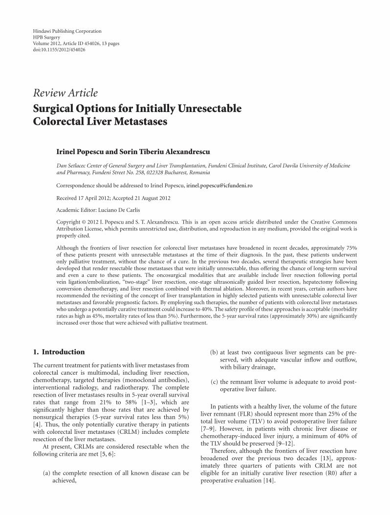
Hindawi Publishing CorporationHPB SurgeryVolume 2012, Article ID 454026, 13 pagesdoi:10.1155/2012/454026
Review Article
Surgical Options for Initially UnresectableColorectal Liver Metastases
Irinel Popescu and Sorin Tiberiu Alexandrescu
Dan Setlacec Center of General Surgery and Liver Transplantation, Fundeni Clinical Institute, Carol Davila University of Medicineand Pharmacy, Fundeni Street No. 258, 022328 Bucharest, Romania
Correspondence should be addressed to Irinel Popescu, [email protected]
Received 17 April 2012; Accepted 21 August 2012
Academic Editor: Luciano De Carlis
Copyright © 2012 I. Popescu and S. T. Alexandrescu. This is an open access article distributed under the Creative CommonsAttribution License, which permits unrestricted use, distribution, and reproduction in any medium, provided the original work isproperly cited.
Although the frontiers of liver resection for colorectal liver metastases have broadened in recent decades, approximately 75%of these patients present with unresectable metastases at the time of their diagnosis. In the past, these patients underwentonly palliative treatment, without the chance of a cure. In the previous two decades, several therapeutic strategies have beendeveloped that render resectable those metastases that were initially unresectable, thus offering the chance of long-term survivaland even a cure to these patients. The oncosurgical modalities that are available include liver resection following portalvein ligation/embolization, “two-stage” liver resection, one-stage ultrasonically guided liver resection, hepatectomy followingconversion chemotherapy, and liver resection combined with thermal ablation. Moreover, in recent years, certain authors haverecommended the revisiting of the concept of liver transplantation in highly selected patients with unresectable colorectal livermetastases and favorable prognostic factors. By employing such therapies, the number of patients with colorectal liver metastaseswho undergo a potentially curative treatment could increase to 40%. The safety profile of these approaches is acceptable (morbidityrates as high as 45%, mortality rates of less than 5%). Furthermore, the 5-year survival rates (approximately 30%) are significantlyincreased over those that were achieved with palliative treatment.
1. Introduction
The current treatment for patients with liver metastases fromcolorectal cancer is multimodal, including liver resection,chemotherapy, targeted therapies (monoclonal antibodies),interventional radiology, and radiotherapy. The completeresection of liver metastases results in 5-year overall survivalrates that range from 21% to 58% [1–3], which aresignificantly higher than those rates that are achieved bynonsurgical therapies (5-year survival rates less than 5%)[4]. Thus, the only potentially curative therapy in patientswith colorectal liver metastases (CRLM) includes completeresection of the liver metastases.
At present, CRLMs are considered resectable when thefollowing criteria are met [5, 6]:
(a) the complete resection of all known disease can beachieved,
(b) at least two contiguous liver segments can be pre-served, with adequate vascular inflow and outflow,with biliary drainage,
(c) the remnant liver volume is adequate to avoid post-operative liver failure.
In patients with a healthy liver, the volume of the futureliver remnant (FLR) should represent more than 25% of thetotal liver volume (TLV) to avoid postoperative liver failure[7–9]. However, in patients with chronic liver disease orchemotherapy-induced liver injury, a minimum of 40% ofthe TLV should be preserved [9–12].
Therefore, although the frontiers of liver resection havebroadened over the previous two decades [13], approx-imately three quarters of patients with CRLM are noteligible for an initially curative liver resection (R0) after apreoperative evaluation [14].
2 HPB Surgery
The most common causes of the initial unresectabilityare the following.
(1) A single, very large liver metastasis, the resection ofwhich would not spare a sufficient volume of liverparenchyma to avoid postoperative liver failure.
(2) Multiple bilobar liver metastases, the complete resec-tion of which would not preserve a sufficient volumeof functional liver parenchyma.
(3) CRLM involving or located in close proximity toeither the bifurcation of the portal vein or theconfluence of the three hepatic veins with the inferiorvena cava (IVC). In this case, the resection of theliver metastasis would not allow for the preservationof a minimum of two adjacent liver segments withadequate vascular inflow and outflow.
Until 20 years ago, the only available treatment for thesepatients was palliative chemotherapy, the goals of which wereto increase progression-free and overall survival; however,there was no prospect of a cure. Although survival ratesincreased with the advent of new chemotherapeutics (suchas Oxaliplatin and Irinotecan) and targeted therapies (e.g.,Bevacizumab, Cetuximab, and Panitumumab), the currentsurvival rates for these cases are still modest compared tothose that can be achieved by liver resection. Therefore,several therapeutic strategies were introduced to achieve acomplete resection in these patients [15].
2. Therapeutic Options
In Figure 1, we schematically present those situationsin which metastases are considered unresectable and thetherapeutic options that are available for conversion toresectability.
2.1. Liver Resection Following Portal Vein Embolization/Liga-tion. In certain instances, although a minimum of twoadjacent segments with appropriate vascular inflow andoutflow, and biliary drainage can be preserved following thecomplete resection of CRLM, the volume of the remainingliver parenchyma may be insufficient to avoid postoperativeliver failure. Such situations are generally encountered inpatients who (1) require a right trisectionectomy, or (2)when a right hemihepatectomy must be performed, but thevolume of the left hemiliver is prohibitively small (Figures1(a) and 1(b)). To avoid postoperative liver failure in thesepatients, it is advisable to attempt to increase the volume ofthe FLR prior to the liver resection.
This goal may be achieved by initially performing portalvein embolization (PVE) or ligation (PVL). If the volumeof the FLR following a PVE/PVL increases sufficiently toprevent the risk of postoperative liver failure, liver resectionshould be performed 4–8 weeks later.
This therapeutic strategy is based on the observation thatincreasing the volume of the FLR improves the function ofthe residual liver parenchyma following the hepatectomy [16,17].
The reports of Kinoshita and Makuuchi revealed thatthe ligation or embolization of the right portal vein inducesa process of atrophy-hypertrophy of the liver (increasingthe safety of liver resection) in patients with hepatocellularcarcinoma or hilar cholangiocarcinoma [18, 19]. Therefore,other authors applied the same procedure in patients withCRLM whose FLR was insufficient to avoid postoperativeliver failure [10, 20–22]. The rationale for such an approachis that the embolization or ligation of the right portal branchabolishes the portal inflow into the right hemiliver, leadingto its atrophy; alternatively, the portal inflow into the lefthemiliver increases, causing hypertrophy of the FLR.
This approach permits the performance of the scheduledhepatectomy (while concomitantly reducing the risk of fatalliver failure) in more than 50% of patients who wereotherwise unresectable due to a small FLR.
Concerns regarding the comparative effectiveness of PVEversus PVL have been raised by certain authors. In an animalmodel, Furrer et al. revealed that the hypertrophy of theleft hemiliver significantly increased following PVL versusPVE. These authors hypothesized that the entrapment ofa greater number of macrophages in the embolized liver(due to the foreign-body reaction that is induced by thematerial used for embolization) explains this result [23].Their conclusion that PVL is superior to PVE in inducinga regenerative response of the remnant liver is in contrastto that of Wilms et al., who stated that although PVLand PVE both induce liver hypertrophy, PVE is the mosteffective technique to increase the FLR [24]. These authorsstated that PVE-induced vascular occlusion is more durablethan that induced by PVL. Furthermore, the cause of theinferior regeneration in the ligation group was reported tobe the formation of collaterals between the occluded andnonoccluded portions of the liver. To avoid this undesirablesituation, certain authors recommended the transection andethanol injection into the ligated portal branch [20]. Lastly,in addition to these experimental studies, a retrospectivestudy of 35 patients revealed that PVL and PVE are similarin terms of both increasing the FLR and the conversion toresectability rate [25].
PVE- or PVL-induced liver hypertrophy involves bothsegments 2-3 and segment 4. Most patients that are subjectedto PVL/PVE require a right trisectionectomy. In suchpatients, the principal objective of this maneuver is toincrease the volume of segments 2-3 and not the volumeof segment 4 (which is resected). To primarily increase thevolume of the left lateral section, certain authors state thatthe optimum approach is to concomitantly occlude the rightportal vein and the portal branches to segment 4 (righttrisection portal vein embolization-R3PE) [26]. In 2000,Nagino et al. presented results that support this hypothesis,demonstrating that the volume gain of the left lateral sectionwas higher in patients with R3PE relative to patients whoreceived right portal vein embolization [26]. To date, wehave performed R3PL in one patient, and the results wereoutstanding: the percent of FLR gain was 16.22%, whereas

HPB Surgery 3
FLR less than 25–30% of TLV
(a)
Liver resection after PVE/PVL
(b)
Few CRLM >3 cm in the future liver remnant
(c)
“Two-stage” liver resection
(d)
Few large CRLM whose resection does not spare a volume of functional liver able to avoid
fatal postoperative liver failure
Liver resection after conversion chemotherapy
(e)
Three or less CRLM <3 cm in the remnant liver
Liver resection combined with thermal ablation
(f)
Multiple bilobar colorectal liver metastases
Liver transplantation (highly selected patients)
(g)
Figure 1: Strategies used for potentially curative treatment of the initially unresectable CRLM, depending on the location, number, and sizeof the lesions.
this metric was 10.8% in the patients who received a rightportal branch ligation [27]. However, another study that waspublished in 2005 failed to confirm these results, revealingthat the mean volume of segments 2-3 following emboliza-tion and the rate of the segments 2-3 volume increase weresimilar between the patients who received a R3PE and thosethat received a standard right portal vein embolization [7].Therefore, definitive conclusions cannot be drawn regardingthe usefulness of the embolization of the portal branches tosegment 4, and further studies are required to clarify thissubject. Most centers now prefer to routinely perform onlyright portal branch embolization/ligation in such patients.
To achieve a more marked and rapid hypertrophy ofthe FLR following portal vein ligation, Schnitzbauer et al.[28] and de Santibanes et al. [29] recently recommendedthe association of right portal vein ligation with “in situliver transection/splitting”. Using this approach, the authorsachieved a significant and more rapid hypertrophy of theFLR, enabling subsequent curative liver resection duringthe same hospitalization. The authors concluded that thistechnique induces the rapid and more robust growth of theFLR [28] than is reported with portal vein occlusion alone.Moreover, this approach allows for the performance of astaged liver resection during a single hospital stay [29].

4 HPB Surgery
Two formulas can be used to calculate the percent of theFLR gain following PVE/PVL:
(a) (Volume of the FLR following PVE − Volume of theFLR prior to PVE) × 100/Volume of the FLR prior toPVE [30],
(b) %FLR following PVE −%FLR prior to PVE [12].
In a series of 30 patients, the percent of the FLR gain(calculated using the first formula) was 42% [10], whereasthe FLR gain ranged from 9.7% to 13% in different seriesusing the second formula [21, 27, 30]. In most patients,these percentages are generally sufficient to allow for a saferesection of liver metastases.
When PVE is planned, Bevacizumab should be used withcaution given that Aussilhou et al. revealed the detrimentaleffect of this medication on the FLR gain; this effect isespecially strong in patients who are older than 60 years andreceived more than six cycles [31]. However, other authorshave demonstrated that this monoclonal antibody does notimpair liver regeneration following PVE [32].
The most severe complications of right PVE are liverhematoma, liver abscess, thrombosis of the left portal vein,portal hypertension, and cholangitis. In a meta-analysisthat was performed by Aboulkhir et al., the morbidity ratefollowing PVE was 2.2%, and the mortality was zero [30].
The resectability rate following PVE/PVL in differentcenters ranges from 60% to 88% [10, 21, 22]. The primaryreason of failure to perform the curative hepatectomy is notinsufficient hypertrophy of the FLR but rather the progres-sion of the disease. In our series, 5 of 13 patients (38%)exhibited disease progression following PVL, precluding acurative liver resection. The other 8 patients underwentsuccessful complete resection of the initially unresectableCRLM. The resectability rate was therefore 62% [27].
The morbidity and mortality rates that were observedfollowing curative hepatectomy were less than 35% and 4%,respectively, in most series [10, 22].
The 5-year survival rate of these patients was approxi-mately 38% [10, 21].
It must be noted that in patients with liver metastasesin the FLR, this approach is not recommended, due to therisk of rapid growth of these metastases. Such approach mayjeopardize the chances of a subsequent potentially curativeliver resection. Such patients should undergo a “two-stage”liver resection (see below) to clear the remnant liver prior tothe PVE [33].
2.2. Two-Stage Liver Resection. The term “two-stage” liverresection has been used by a small number of authors todefine a strategy that consists of a single liver resectionthat is performed following PVL and which does notinclude a sequential liver resection [20]. Herein, we shalluse the nomenclature of “two-stage” liver resection for thoseprocedures that consist of two consecutive hepatectomies.
This therapeutic strategy is used in patients with multiplebilobar CRLM, whose resection will not spare a sufficientamount of liver parenchyma to avoid postoperative liverfailure. These patients usually require a right hepatectomy ora right trisectionectomy along with wedge resections of the
metastases that are located in the left hemiliver or in the leftlateral section (Figures 1(c) and 1(d)).
To avoid such extensive resections, which are accom-panied by a high risk of postoperative fatal liver failure,it is recommended that a complete resection of the livermetastases be achieved in a two-stage surgical procedure.In the first stage, a limited resection of the metastasesfrom the left hemiliver or the left lateral section (futureliver remnant) is performed. In stage two (following theregeneration of the FLR), the bulk of the metastatic burdenis resected by a right hepatectomy or trisectionectomy. FLRregeneration is essential to minimize the risks of hepaticfailure following the second operation. Thus, PVE/PVL maybe suitable in patients with small FLR to increase the safetyof the second hepatectomy. To facilitate the second operationand to avoid disease progression between the first and thesecond intervention, it may be useful to deliver systemic orlocoregional chemotherapy to shrink the metastatic bulk.To minimize the inhibitory effects of the chemotherapeuticdrugs on liver regeneration, the chemotherapy should bebegun three weeks following the first hepatectomy. Thissequence is necessary, as liver regeneration is essential to thefeasibility of the second resection [34].
Such a therapeutic approach is especially useful inpatients with synchronous bilobar CRLM [35] given that(1) it avoids the cumulative risks of a simultaneous primarytumor resection and major hepatectomy, and (2) it allows forthe evaluation of the chemosensitivity of the liver metastasesand the guiding of the adjuvant therapy following the secondoperation. The resection of the primary tumor is performedin the first stage, along with a limited resection of themetastases from the future liver remnant (generally fromthe left hemiliver or the left lateral section). Occasionally, aright portal vein ligation is also performed during the firstoperation. Short-course chemotherapy (systemic or loco-regional) should begin three weeks later. If the residuallesions will be stable or responsive to chemotherapy, thesecond liver resection should be performed.
The results of such an approach were first publishedby the Paul Brousse group in 2000. This group reported aresectability rate of 81% [34]. In other series, the resectabilityrate ranged from 66% to 75% [27, 33].
Among the published series, the morbidity rates follow-ing the first resection were less than 31%, and the mortalityrates were zero [33, 34].
The morbidity rate following the second liver resectionranges from 45% to 56% [27, 33, 34]. Despite these relativehigh morbidity rates, the mortality was zero in most series[27, 33]. Nonetheless, a mortality rate of 15% following thesecond operation was reported by Adam et al. [34]. Theauthors explained that this result was a consequence of thecombination of (1) the diminished tolerance of such patientsto perioperative complications due to their advanced neo-plastic disease and (2) the effects of the adjuvant proceduresthat were used to facilitate liver resection (chemotherapy,PVE).
The 3-year survival rates of these patients ranged from35% to 54% [27, 33, 34], with a median survival of 44months from the diagnosis of liver metastases [34].

HPB Surgery 5
In selected patients with multiple bilobar colorectal livermetastases, a ”two-stage” liver resection could be avoided, byperforming ultrasonically-guided hepatectomy.
2.3. One-Stage Ultrasonically Guided Liver Resection. Theimplementation of ultrasonography in liver surgery dra-matically alters the approach to liver metastases, permittinga more accurate diagnosis and challenging the traditionalparadigms of liver resection.
Intraoperative ultrasound (IOUS) allows for the detec-tion of additional CRLM that were not revealed by preop-erative imaging methods and is the most accurate techniquefor detecting liver tumors [36, 37]. However, standard IOUSmay miss lesions that are smaller than 1 cm, especially inpatients who are undergoing preoperative chemotherapy,whose CRLM exhibit a similar echo-pattern to that ofthe surrounding liver parenchyma. The use of contrast-enhanced IOUS (CE-IOUS) was demonstrated to improvethe detection of CRLM and is the most sensitive and specificmethod for the diagnosis of CRLM [38].
In the mid 1980s, IOUS was first used to guide thepuncture and balloon occlusion of the portal branch thatfeeds the portion of the liver to be resected, allowingfor limited anatomical liver resections instead of majorhepatectomies [39–41]. This technique decreased the risk ofpostoperative liver failure and was recommended principallyin patients who have HCCs on liver cirrhosis.
In addition, IOUS offers a better estimation of the spatialrelationships between the liver tumors and the intrahepaticvessels, permitting the resection of liver masses with thepreservation of intrahepatic vascular structures even whenthe tumors are located in close proximity to major intrahep-atic vessels. Furthermore, even when major hepatic vein(s)must be resected, color Doppler IOUS findings providereliable information that may lead to the preservation of aportion of the liver parenchyma that is drained by thosevein(s), avoiding major hepatectomies [42–45]. Thus, a novelliver resection technique was developed in recent years thatis referred to as “ultrasonically guided hepatectomy”. Thistechnique opened the door to new procedures that allowfor radical but conservative liver resections, reducing therequirement for major hepatic resections [46–48].
Because many patients exhibit colorectal liver metastasesthat are considered unresectable due to the insufficientremnant liver parenchyma following major hepatectomies,the use of this surgical technique (which spares a significantamount of functional liver parenchyma) allows for thecomplete resection of the metastases, reducing the risk ofdeveloping postoperative liver failure. Thus, this techniquewas used more frequently in patients with CRLM.
Moreover, ultrasonically guided liver resection decreasesthe requirement for major hepatectomies, obviating therequirement for portal vein occlusion prior to the liverresection and/or the necessity of a “two-stage” liver resectionin selected patients.
In patients with CRLM that are located in close proximityto major hepatic veins or near the first-degree portalbranches, a major hepatectomy is still the main surgicaloption at most centers. If the remnant liver volume following
a major hepatectomy is critically small, a liver resectionfollowing portal vein occlusion, either by PVE or PVL,is generally recommended. In such instances, the patientis exposed to an interventional radiology procedure or alaparotomy prior to the curative liver resection. Each of theseprocedures presents additional risks of morbidity [30]. Thedevelopment of the ultrasonically guided hepatectomy inrecent years permits a more limited liver resection of poorlylocated CRLM, avoiding the necessity of a prehepatectomyPVE/PVL. In a series of 22 patients who presented poorlylocated liver tumors and who were scheduled for initialultrasonically guided liver resection, a limited resection withor without hepatic vein preservation was achieved in 91%cases, providing lower morbidity rates than major resectionsfollowing PVE and no mortality [43]. The rate of localrecurrence (at the transection surface) was zero at a meanfollow-up period of 23 months. Because this approach avoidsportal vein occlusion (and its associated morbidity), thecomfort of the patient is also improved. Moreover, theresectability rate following portal vein occlusion does notexceed 60–88%, due to either insufficient hypertrophy of theremnant liver or disease progression in the interval betweenthe portal vein occlusion and the liver regeneration [21, 22,49]. When an initial ultrasonically guided hepatectomy isperformed, the risk of disease progression is avoided, andthe hypertrophy of the FLR is no longer necessary. Thus, theresectability rate that is achieved by the ultrasonically guidedapproach appears to be higher than those that are achievedby PVE/PVL, broadening the indications for curative surgeryin cases of CRLM [43].
In patients with multiple bilobar CRLM, the ultra-sonically guided technique may also represent an effectivealternative to the “two-stage” hepatectomy, permitting acurative and conservative liver resection [44]. The advantagesof this approach over the “two-stage” liver resection are thecomfort of the patient, a lower morbidity rate [44], andan increased possibility of repeat resections if the patientdevelops recurrent metastases [50–52]. Furthermore, therecurrence rate following one-stage ultrasonically guidedliver resection was similar to that reported after “two-stageliver resection”.
Due to the aforementioned benefits, the one-stageultrasonically guided liver resection should be part of thearmamentarium of the liver surgeon, especially in the contextof patients with complex tumoral presentations.
2.4. Liver Resection Following Conversion Chemotherapy. Thistherapeutic strategy was first presented by the Paul Broussegroup in 1996 [53] and is recommended in patients witha small number of large CRLM, the resection of whichwould not spare a sufficient amount of functional liver toprevent postoperative liver failure (Figure 1(e)). The goalof this approach is to “downsize” the liver metastases to anextent that allows for their complete resection. Therefore, achance of a potentially curative liver resection is availableto patients who otherwise may have only benefited frompalliative treatment.
Until 20 years ago, the only efficient chemotherapeu-tic regimen that was used in patients with unresectable

6 HPB Surgery
CRLM consisted of 5-Fluorouracil (5-FU) and Folinic acid.Although this chemotherapy increases the overall survivalrates and the progression-free survival rates of these patients,the response rates were less than 23%, and only anecdotalcases of liver metastases that shrink sufficiently to allow for asubsequent curative hepatectomy were reported [54–56].
The advent of new chemotherapeutic agents such asOxaliplatin and Irinotecan led to significantly better results.The response rates that have been achieved by FOLFOXand FOLFIRI regimens range from 40% to 56% [57–59]. Astrong correlation was observed between the response ratesand the resection rates of patients with initially unresectableCLRM [60]. Therefore, more patients became resectable fol-lowing so-called conversion chemotherapy. Folprecht et al.thus concluded that resectability should be considered a newendpoint for preoperative chemotherapy, focusing on thecurative potential of this oncosurgical treatment [60].
The Paul-Brousse group published their updated resultsin 2004, reporting a 12.5% rate of conversion to resectabilityin 1104 patients with initially unresectable CRLM (followingan average of 10 courses of chemotherapy) [14]. Apartfrom these very large series of patients, other centers havesubsequently reported similar results in smaller numbers ofselected patients who presented with initially unresectableCRLM that were rendered resectable by different chemother-apy regimens [61–63].
In many reports, the morbidity rates following hepatec-tomy in patients with initially unresectable CRLM [14, 62,64] ranges from 23% to 28%, which are similar to thoserates that are observed in patients with initially resectableCRLM. However, certain authors have reported significantlyhigher incidences of postoperative complications in patientswho receive resections following “downsizing” chemother-apy, raising concerns regarding the deleterious effects ofthe preoperative chemotherapy on the liver parenchyma(see below) [65, 66]. The postoperative mortality rates arereported to be less than 2% in most centers, which are similarto those rates that have been achieved in patients who did notreceive preoperative chemotherapy [14, 53, 62, 66].
The 5-year survival rate of patients who were renderedresectable by chemotherapy was 33% in the Paul Broussegroup, a rate that was higher than those that were achieved bynew palliative chemotherapeutic regimens in similar patients[14]. Although this survival rate is significantly lower thanwhat can be achieved in patients with initially resectableCRLM (P value = 0.01), the 5-year disease-free survival rateof 22% that was reported in initially unresectable patientsappears to fully justify the efforts to render to resectability,these patients with otherwise dismal prognosis [14].
The addition of targeted therapies (e.g., Bevacizumab,Cetuximab, and Panitumumab) to chemotherapy regimensmay be useful in further increasing the rate of conversion toresectability in initially unresectable lesions. This hypothesiswas confirmed in a series of patients whose liver metas-tases were refractory to previous rounds of conventionalchemotherapy. The advent of Cetuximab to the next-linechemotherapy rendered 7% of these patients resectable,with morbidity and mortality rates of 50% and 3.7%,respectively, and a median survival of 20 months [67]. This
study, similar to those of Zorzi et al., demonstrated thatmonoclonal antibodies in combination with conventionalchemotherapy have no detrimental effects on the safetyof liver resection [32]. Furthermore, Gruenberger et al.revealed that Bevacizumab has little detrimental impact onliver regeneration following hepatectomy [68]. However,it should be noted that the use of vascular endothelialgrowth factor inhibitors (e.g., Bevacizumab) prior to majorsurgery increases the risks of bleeding and wound healingcomplications [69]. This therapy should be discontinued 5–8weeks prior to the surgical intervention [68, 70].
Several issues should be kept in mind when deciding totake this therapeutic approach.
(i) The response to chemotherapy cannot be assumed topersist. Metastases occasionally shrink and become resectablefollowing several cycles of chemotherapy. However, if thechemotherapy is continued, the metastases may regrowand again become unresectable, closing the “window ofopportunity” for a potentially curative hepatectomy [71].Therefore, if the systemic disease is controlled, the liverresection should be scheduled as soon as the metastasesbecome resectable.
(ii) If the chemotherapy is continued beyond the pointwhen the metastases become resectable, it is possible theliver metastases will become smaller and will no longer bevisible on imaging (CT/MR/PET scans). Such metastasesare referred to as “vanishing metastases.” Unfortunately,this “radiological complete response” or “clinical completeresponse” [72] does not indicate a cure, as a “pathologiccomplete response” is achieved in fewer than 20% of cases[73, 74]. In one-third of patients with radiological completeresponse, a laparotomy may reveal small metastases thatwere missed by the imaging methods or residual scars,the resection of which would reveal viable tumor cells.Alternatively, in patients without macroscopic residual tissue(on laparotomy) and negative (contrast-enhanced) intraop-erative ultrasound, pathologic examination of the resectedspecimens, including liver segments where the metastaseswhere initially located, revealed viable metastatic cells in 75%of cases [74, 75]. An indirect confirmation of the presence ofviable tumor cells at the sites of the former metastases (whichwere invisible on laparotomy) is given by certain reports. Ina series of patients with 31 CRLM that disappeared followingchemotherapy and which were not observed on laparotomy,the resection of the initial metastases site was not performed.After a one-year follow-up, 23 (74%) metastases recurred insitu [74].
The survival benefit that is achieved by performingliver resection in patients with clinical complete responsefollowing chemotherapy was revealed by another study.Fourteen patients with radiological complete response fol-lowing chemotherapy were not subjected to a liver resection,achieving a 5-year overall survival of 14% and a mediansurvival of 30 months. In 25 patients who suffered frominitially unresectable CRLM that were rendered to resectabil-ity by a FOLFOXIRI regimen and further resected, the 5-year survival rate was 43%, and the median survival wassignificantly higher (61 months, P value = 0.006) [62].

HPB Surgery 7
For these reasons, laparotomy is mandatory in patientswith vanishing metastases, with the aim of resecting themacroscopic residual metastatic tissue or the sites of theinitial CRLM (“blind resection”).
The resection of the metastases sites is a very demandingoperation, especially in patients with (initially) multiplemetastases located deep in the liver. In such cases, computer-based virtual surgery planning is very useful, mergingpre- and postchemotherapy computed tomography data.Recently, Oldhafer et al. presented such a surgical approach.Information that is processed using a computer is thenintraoperatively transferred to the liver surface using animage-guided stereotactically navigated ultrasound dissector,enabling the surgeon to perform the resection [75].
In patients with initially multiple bilobar CRLM thatbecome “invisible” following chemotherapy and that cannotbe identified intraoperatively, it is frequently impossible toassume a complete resection of the metastatic sites. Thus, asmall number of these “missing metastases” will remain. Insuch situations, Elias et al. recommended the placement of achemotherapy catheter in the hepatic artery, allowing for ahepatic arterial infusion (HAI) with Oxaliplatin, along withsystemic 5-FU and Folinic acid [76]. Using this approach,following a median follow-up period of 51 months, themissing metastases did not recur in 62% of the patients.The recurrence rate following HAI with Oxaliplatin wassignificantly lower than those that were noted in patientswho were treated by systemic chemotherapy alone (P value =0.01) [76].
Alternatively, it should be noted that a small number (4–15%) of CRLM that were treated by systemic chemotherapyprior to the liver resection achieved a complete pathologicresponse [72, 73, 77]. The complete pathologic responsewas observed both in patients with or without a radiologiccomplete response. The predictive factors for a completepathologic response were age less than 60 years, maximummetastasis diameter of less than 3 cm, CEA levels at diagnosisbelow 30 ng/mL, an objective response following chemother-apy [72] and the use of hepatic arterial infusion chemother-apy [76]. The addition of Bevacizumab to the Oxaliplatin-based chemotherapeutic regimens did not appear to increasethe incidence of the complete pathologic response (11.3%versus 11.6%; P value = 0.59) [78]. Patients with completepathologic response achieved uncommonly high survivalrates (76% at 5 years) [72].
(iii) Treatment with new chemotherapeutic drugsinduces alterations of the nontumoral liver parenchyma,potentially impacting the results of the liver resection.
The initial belief was that use of Irinotecan may causenonalcoholic fatty liver disease (NAFLD), which representsa spectrum of diseases. The mildest form of NAFLD ismacrovesicular steatosis, and the most severe form is non-alcoholic steatohepatitis (NASH) [79]. Although a study thatwas published in 2003 revealed a correlation between priortreatment with chemotherapy and steatosis and highlightedthe fact that morbidity rates following liver resections weresignificantly higher in patients with marked steatosis [80],more recent studies have reported differing results. In 2006,a multicenter trial revealed that Irinotecan was associated
with steatohepatitis but not with steatosis [81]. Moreover,this latter study noted that the mortality rate was significantlyhigher in patients with steatohepatitis (14.7%) than inpatients without steatohepatitis (1.6%, P value = 0.001).
The first study to reveal a correlation between Oxaliplatinand non-tumoral liver parenchyma injury was published in2004 [82]. The results indicated that 78% of the patientswho were preoperatively treated with Oxaliplatin exhib-ited sinusoidal alterations. These results were subsequentlyconfirmed by other reports [83–85], which revealed thatOxaliplatin-based preoperative chemotherapy was associatedwith sinusoidal dilatation and congestion, peliosis, andvenoocclusive disease. One of these studies reported thatonly long-course Oxaliplatin-based chemotherapy (6 ormore cycles) is significantly associated with sinusoidal injury[85]. However, in a series that was presented by Vautheyet al., the risk of sinusoidal dilatation did not appear toincrease with the duration of chemotherapy (although theirpatients received relatively short-course treatments) [81].Interestingly, the addition of Bevacizumab to Oxaliplatin-based regimens appears to reduce the incidence and severityof hepatic injury [78]. The impact of these liver injurieson the clinical outcome of the patients who receivedresections following Oxaliplatin-based chemotherapy wasassessed in several reports. None of these studies reportedincreased mortality rates following liver resection in patientswith Oxaliplatin-related sinusoidal injury [79]. Two trialsrevealed that a limited course (fewer than 6 cycles) ofOxaliplatin-based therapy was not associated with increasedmorbidity rates following liver resection [81, 86]. However,Karoui et al. observed a statistically higher incidence ofpostoperative complications in patients who underwent amajor hepatectomy following preoperative chemotherapywhen compared with the patients who were subjected to asimilar liver resection without preoperative chemotherapy[66]. Similarly, Nakano et al. observed that sinusoidal injurywas significantly associated with increased morbidity andlonger hospital stays in patients who underwent a majorhepatectomy [85].
The above-mentioned pitfalls that are associated withpreoperative chemotherapy justify the scheduling of the liverresection as soon as the metastases become resectable.
2.5. Liver Resection Combined with Thermal Ablation forUnresectable CRLM. This type of approach is especiallyrecommended in patients with multiple bilobar CRLM whopresent with fewer than 3 liver metastases in the FLR, witheach of these metastases being less than 3 cm in maximumdiameter (Figure 1(f)). Another indication for this approachis when one or a small number of metastases are anatomicallypoorly located (e.g., in close proximity to the confluence ofthe three hepatic veins and the inferior vena cava or at thebifurcation of the portal vein) [87].
The operation consists of the resection of the main tumorbulk (generally by a right trisectionectomy) and thermalablation of the unresected metastases from the remnant liver(frequently the left lateral section) [88].
This approach could be performed in a “two-stage”manner in patients with synchronous unresectable CRLM,

8 HPB Surgery
as described by Lygidakis et al. [89]. In the first stage, thefollowing procedures are performed: (1) the resection of theprimary colorectal tumor, (2) the ligation and transectionof the relevant (right or left) primary portal branch, (3)the ablation of the metastatic nodules in the contralateralhemiliver, and (4) the insertion of an arterial catheter intothe hepatic artery for locoregional chemo(immuno)therapy.The second stage of the operation consists of the resectionof the tumoral liver (usually by right hemihepatectomy ortrisectionectomy).
Thermal ablation can be achieved using radiofrequen-cies, microwaves, lasers, or cryotherapy.
To increase the chances of a complete hyperthermicablation, the Pringle maneuver can be performed during theablation [88].
The morbidity and mortality rates (44% and 2.3%, resp.)[27, 90] of patients undergoing combined liver resection andthermal ablation of unresectable CRLM appear to be similarto those of patients with initially unresectable CRLM that arerendered resectable by other therapeutic strategies.
A retrospective study that was published in 2004 reporteda significantly higher local recurrence rate following resec-tion combined with RFA (5%) than was observed followingcomplete resection of the CRLM (2%). However, the patientswere not stratified according to the maximum diameter ofthe ablated CRLM in this study [90]. It was recently demon-strated that the best results achieved by thermoablation areobserved in patients whose CRLM were less than 3 cm inmaximum diameter. Thus, a significantly higher rate (Pvalue = 0.0001) of sustained complete ablation was achievedin patients whose lesions were less than 3 cm (66.7%) thanfor the patients with metastases that were larger than 3 cm(33.3%) [91]. One retrospective study (including resectableand unresectable CRLM) revealed that the local recurrencerate following the radiofrequency ablation (RFA) of metas-tases that were less than 3 cm in maximum diameter was1.3% after a follow-up period of 33 months [92]. Moreover,another retrospective study (including patients with singleCRLM treated by RFA or liver resection) determined that the5-year overall and local recurrence-free survival rates weresimilar for patients with CRLM that were smaller than 3 cmwho were treated either with RFA or liver resection [87].
However, in patients with multiple bilobar CRLM whoare treated with a combination of RFA and liver resection,the overall and disease-free survival rates were significantlylower than for patients who underwent a complete resectionof CRLM but significantly higher than in the patientswho were treated by chemotherapy alone [90]. Moreover,Rivoire et al. revealed in a study of 57 patients with initiallyunresectable CRLM that the overall survival rates weresimilar for patients who underwent a complete liver resectionfollowing conversion chemotherapy and those who receivedliver resection combined with cryotherapy [93].
These results justify liver resection combined with ther-mal ablation of initially unresectable CRLM in patients whofulfill the above-mentioned criteria.
However, due to the lower recurrence rates achievedby the ultrasonically guided liver resection technique, this
approach may be more suitable than liver resection com-bined with thermal ablation.
2.6. Liver Transplantation—A Future Opportunity? Unfortu-nately, there are still many patients with multiple bilobarcolorectal cancer liver metastases who are not amenableto complete resection by any of the above-mentionedtherapeutic strategies. In such patients, the only chance ofcomplete removal of the liver metastases is total hepatectomyfollowed by liver transplantation (Figure 1(g)).
This approach was used in the early period of livertransplantation, achieving 1- and 5-year overall survival ratesof 62% and 18%, respectively [94].
Due to organ shortages, it was considered that theallocation of an organ to a patient with such a short lifeexpectancy following the transplantation was not ethicallyacceptable.
Currently, unresectable CRLM are considered to be acontraindication to liver transplantation.
However, the above-mentioned survival rates appear tobe higher than those that can be achieved by palliative treat-ment, suggesting that even using the therapeutic options thatwere available thirty years ago, liver transplantation offered ahigher survival benefit than the best palliative treatment thatis currently available. Furthermore, due to recent progress inthe fields of posttransplant immunosuppression and medicaloncology and due to the more refined methods that are usedin selecting patients to receive tailored therapies (based onreliable pathologic and biologic markers), improved survivalrates could be achieved for selected patients who undergoliver transplantation.
Moreover, the ethical issues could be challenged by theuse of a living donor liver transplantation given that, in suchinstances, the willing donation is directed toward a certainpatient and not to the community [95]. Meanwhile, it is alsoconsidered unethical to offer a marginal graft to a patientwith a good chance of long-term survival following livertransplantation. Because the number of available marginalgrafts has increased in recent years, it may be acceptable toallocate such organs in selected patients with CRLM, at leastin the setting of controlled trials.
Although the available data do not support liver trans-plantation as a routine procedure in patients with CRLM,we believe that a discussion of the current advances in thisfield and of the recently published results is worthwhile andshould encourage debate on this issue.
By reviewing the largest series of patients undergoingliver transplantation for unresectable CRLM [96], it wasrevealed that 66% of patients with histologically negativelymph nodes were genetically positive for micrometastaseswhen mutant allele-specific amplification (MASA) methodwas used to search for micrometastases in DNA from theregional lymph nodes of the primary colorectal cancer [97].Those patients who were both genetically and histologicallynegative exhibited a significantly longer overall survival (Pvalue = 0.011) than the other patients. Thus, Kappel et al.concluded that the genetic detection of micrometastases byMASA may be a powerful prognostic indicator for selecting

HPB Surgery 9
patients with colorectal liver metastases who could benefitfrom liver transplantation [97].
Over the previous two decades, certain further devel-opments have emerged that may improve patient selectionand the results of liver transplantation. For example, theadvent of MDCT, gadolinium-enhanced MRI, and PET/CTscans has improved the detection of extrahepatic metastases,permitting the better selection of patients with colorectalcancer that had metastasized only to the liver. Recentstudies have identified several biological parameters (suchas the expression of p53, thymidylate synthase, Ki-67, K-ras, and human telomerase reverse transcriptase, as well asthe type, density, and location of immune cells within thetumor) that may be more sensitive predictors of outcome inpatients with CRLM than are the current histopathologicalmethods that are used to stage colorectal cancer [98, 99].Using a panel that incorporates these parameters, it may bepossible to identify a highly selected group of patients whocould greatly benefit from liver transplantation. Moreover,it has been hypothesized that the progress in posttransplantimmunosuppressive therapy may decrease recurrence ratesand improve the survival of patients who undergo livertransplantation for malignant disease, primarily due tothe use of m-TOR inhibitors. Such immunosuppressiveagents (sirolimus, temsirolimus) inhibit tumor growth andproliferation and exhibit antiangiogenic effects. These effectsare in contrast to traditional immunosuppressive drugs,which appear to promote malignant cell proliferation [100,101].
Over the past several years, taking into account the betterexpertise of transplant surgeons and the above-mentionedprogress in both the selection of patients with CRLM and inthe efficacy of posttransplant immunosuppressive regimens,certain authors have argued that the outcome of selectedpatients who undergo liver transplantation for unresectableliver metastases from colorectal cancer may be significantlyimproved [102]. For these reasons, certain authors haveproposed a rational revisitation of the concept of liver trans-plantation in such patients. Thus, a pilot study (SECA-study)that aims to assess the survival and quality of life in patientsreceiving pretransplant chemotherapy, liver transplantationfor unresectable CRLM, and posttransplant Sirolimus-basedimmunosuppressive regimen began in Norway in November2006. The preliminary data of this study reveal a 94% survivalrate after a median 25 months of postoperative follow-up andan excellent quality of life [102]. However, only 40% of thesepatients are disease-free after a median follow-up period of25 months.
Favorable results were also recently reported in twopatients with CRLM who were treated with liver resec-tion followed by hepatic artery infusion chemotherapyand who underwent a liver transplantation for intraarte-rial chemotherapy-induced sclerosing cholangitis. The twopatients were disease-free at 2 and 5 years following trans-plantation, respectively.
Based on these disparate results, definitive conclusionscannot be drawn; however, due to ethical considerations(i.e., organ shortage), liver transplantation with grafts from
brain-dead donors cannot be accepted until 5-year survivalrates exceed 50% [102].
However, if the results that are achieved by liver trans-plantation will become significantly higher than those canbe achieved by nonsurgical therapies, it may eventually bedifficult, in the future, to defend the prohibition of living-donor liver transplantation or liver transplantation withmarginal grafts in highly selected patients with unresectableCRLM. Such a position would be difficult given that ethicalconsiderations would no longer be valid in such situations.
3. Conclusions
Selected patients with initially unresectable CRLM may berendered resectable following portal vein embolization orligation, resulting in an important survival benefit or evena cure.
“Two-stage” hepatectomies (with/without PVE/PVL)may be performed safely, achieving complete resection ofliver metastases and long-term survival.
The use of ultrasonographically guided hepatectomiesdecreases the requirement for major hepatectomies, portalvein occlusion and “two-stage” liver resections in patientswith CRLM that are close to the hepatocaval confluence orin cases of multiple bilobar disease. This approach provides(1) an improved comfort and safety profile over “two-stage”liver resections and major hepatectomies following PVE/PVLand (2) a similar oncological benefit to other strategies.
Liver resection following conversion chemotherapy inpreviously unresectable patients may offer a considerablesurvival benefit. RFA could be combined with liver resectionto increase the number of patients who are eligible forcomplete removal and ablation of CRLM.
In the future, highly selected patients with unresectableCRLM and favorable prognostic factors who receive livertransplantations with grafts from marginal donors or fromliving donors could achieve better survival rates than wouldbe possible with palliative treatment. However, further stud-ies and perioperative treatment improvements are requiredbefore this procedure achieves social acceptance.
Acknowledgments
This paper is partly supported by the Sectoral OperationalProgramme Human Resources Development (SOPHRD),financed from the European Social Fund, and by theRomanian Government under the Contract number POS-DRU/89/1.5/S/64153.
References
[1] M. A. Choti, J. V. Sitzmann, M. F. Tiburi et al., “Trendsin long-term survival following liver resection for hepaticcolorectal metastases,” Annals of Surgery, vol. 235, no. 6, pp.759–766, 2002.
[2] I. Popescu, M. Ionescu, S. Alexandrescu et al., “Surgical treat-ment of liver metastases from colorectal cancer,” Chirurgia,vol. 101, no. 1, pp. 13–24, 2006.
[3] J. Scheele and A. Altendorf-Hofmann, “Resection of colorec-tal liver metastases,” Langenbeck’s Archives of Surgery, vol. 384,no. 4, pp. 313–327, 1999.

10 HPB Surgery
[4] M. Van den Eynde and A. Hendlisz, “Treatment of colorectalliver metastases: a review,” Reviews on Recent Clinical Trials,vol. 4, no. 1, pp. 56–62, 2009.
[5] C. Charnsangavej, B. Clary, Y. Fong, A. Grothey, T. M. Pawlik,and M. A. Choti, “Selection of patients for resection ofhepatic colorectal metastases: expert consensus statement,”Annals of Surgical Oncology, vol. 13, no. 10, pp. 1261–1268,2006.
[6] T. M. Pawlik, R. D. Schulick, and M. A. Choti, “Expandingcriteria for resectability of colorectal liver metastases,” Oncol-ogist, vol. 13, no. 1, pp. 51–64, 2008.
[7] L. Capussotti, A. Muratore, A. Ferrero, G. C. Anselmetti,A. Corgnier, and D. Regge, “Extension of right portal veinembolization to segment IV portal branches,” Archives ofSurgery, vol. 140, no. 11, pp. 1100–1103, 2005.
[8] T. De Baere, A. Roche, D. Elias, P. Lasser, C. Lagrange,and V. Bousson, “Preoperative portal vein embolization forextension of hepatectomy indications,” Hepatology, vol. 24,no. 6, pp. 1386–1391, 1996.
[9] E. K. Abdalla, M. E. Hicks, and J. N. Vauthey, “Portal veinembolization: rationale, technique and future prospects,”British Journal of Surgery, vol. 88, no. 2, pp. 165–175, 2001.
[10] D. Azoulay, D. Castaing, A. Smail et al., “Resection ofnonresectable liver metastases from colorectal cancer afterpercutaneous portal vein embolization,” Annals of Surgery,vol. 231, no. 4, pp. 480–486, 2000.
[11] R. Kianmanesh, O. Farges, E. K. Abdalla, A. Sauvanet, P.Ruszniewski, and J. Belghiti, “Right portal vein ligation: anew planned two-step all-surgical approach for completeresection of primary gastrointestinal tumors with multiplebilateral liver metastases,” Journal of the American College ofSurgeons, vol. 197, no. 1, pp. 164–170, 2003.
[12] K. Kubota, M. Makuuchi, K. Kusaka et al., “Measurementof liver volume and hepatic functional reserve as a guide todecision-making in resectional surgery for hepatic tumors,”Hepatology, vol. 26, no. 5, pp. 1176–1181, 1997.
[13] M. Minagawa, M. Makuuchi, G. Torzilli et al., “Extension ofthe frontiers of surgical indications in the treatment of livermetastases from colorectal cancer: long-term results,” Annalsof Surgery, vol. 231, no. 4, pp. 487–499, 2000.
[14] R. Adam, V. Delvart, G. Pascal et al., “Rescue surgeryfor unresectable colorectal liver metastases downstaged bychemotherapy: a model to predict long-term survival,”Annals of Surgery, vol. 240, no. 4, pp. 644–658, 2004.
[15] D. Jaeck, P. Bachellier, J. C. Weber, E. Oussoultzoglou, andM. Greget, “Progrs dans la chirurgie d’exrse des mtastaseshpatiques des cancers colorectaux,” Bulletin de l’AcademieNationale de Medecine, vol. 187, no. 5, pp. 863–876, 2003.
[16] M. Ijichi, M. Makuuchi, H. Imamura, and T. Takayama, “Por-tal embolization relieves persistent jaundice after completebiliary drainage,” Surgery, vol. 130, no. 1, pp. 116–118, 2001.
[17] K. Uesaka, Y. Nimura, and M. Nagino, “Changes in hepaticlobar function after right portal vein embolization: anappraisal by biliary indocyanine green excretion,” Annals ofSurgery, vol. 223, no. 1, pp. 77–83, 1996.
[18] H. Kinoshita, K. Sakai, and K. Hirohashi, “Preoperativeportal vein embolization for hepatocellular carcinoma,”World Journal of Surgery, vol. 10, no. 5, pp. 803–808, 1986.
[19] M. Makuuchi, B. L. Thai, K. Takayasu et al., “Preoperativeportal embolization to increase safety of major hepatectomyfor hilar bile duct carcinoma: a preliminary report,” Surgery,vol. 107, no. 5, pp. 521–527, 1990.
[20] N. J. Lygidakis, L. Vlachos, S. Raptis et al., “New frontiers inliver surgery. Two-stage liver surgery for the management of
advanced metastatic liver disease,” Hepato-Gastroenterology,vol. 46, no. 28, pp. 2216–2228, 1999.
[21] D. Elias, J. F. Ouellet, T. De Baere, P. Lasser, and A. Roche,“Preoperative selective portal vein embolization before hepa-tectomy for liver metastases: long-term results and impact onsurvival,” Surgery, vol. 131, no. 3, pp. 294–299, 2002.
[22] D. Jaeck, P. Bachellier, H. Nakano et al., “One or two-stagehepatectomy combined with portal vein embolization forinitially nonresectable colorectal liver metastases,” AmericanJournal of Surgery, vol. 185, no. 3, pp. 221–229, 2003.
[23] K. Furrer, Y. Tian, T. Pfammatter et al., “Selective portalvein embolization and ligation trigger different regenerativeresponses in the rat liver,” Hepatology, vol. 47, no. 5, pp.1615–1623, 2008.
[24] C. Wilms, L. Mueller, C. Lenk et al., “Comparative studyof portal vein embolization versus portal vein ligation forinduction of hypertrophy of the future liver remnant using amini-pig model,” Annals of Surgery, vol. 247, no. 5, pp. 825–834, 2008.
[25] B. Aussilhou, M. Lesurtel, A. Sauvanet et al., “Right portalvein ligation is as efficient as portal vein embolization toinduce hypertrophy of the left liver remnant,” Journal ofGastrointestinal Surgery, vol. 12, no. 2, pp. 297–303, 2008.
[26] M. Nagino, J. Kamiya, M. Kanai et al., “Right trisegment por-tal vein embolization for biliary tract carcinoma: techniqueand clinical utility,” Surgery, vol. 127, no. 2, pp. 155–160,2000.
[27] I. Popescu, S. Alexandrescu, A. Croitoru, and M. Boros,“Strategies to convert to resectability the initially unre-sectable colorectal liver metastases,” Hepato-Gastroenterol-ogy, vol. 56, no. 91-92, pp. 739–744, 2009.
[28] A. A. Schnitzbauer, S. A. Lang, H. Goessmann, S. Nadalin,J. Baumgart, S. A. Farkas et al., “Right portal vein ligationcombined with in situ splitting induces rapid left lateral liverlobe hypertrophy enabling 2-staged extended right hepaticresection in small-for-size settings,” Annals of Surgery, vol.255, no. 3, pp. 405–414, 2012.
[29] S. E. De, F. A. Alvarez, and V. Ardiles, “How to avoidpostoperative liver failure: a novel method,” World Journal ofSurgery, vol. 36, no. 1, pp. 125–128, 2012.
[30] A. Abulkhir, P. Limongelli, A. J. Healey et al., “Preoperativeportal vein embolization for major liver resection: a meta-analysis,” Annals of Surgery, vol. 247, no. 1, pp. 49–57, 2008.
[31] B. Aussilhou, S. Dokmak, S. Faivre, V. Paradis, V. Vilgrain,and J. Belghiti, “Preoperative liver hypertrophy induced byportal flow occlusion before major hepatic resection forcolorectal metastases can be impaired by bevacizumab,”Annals of Surgical Oncology, vol. 16, no. 6, pp. 1553–1559,2009.
[32] D. Zorzi, Y. S. Chun, D. C. Madoff, E. K. Abdalla, andJ. N. Vauthey, “Chemotherapy with bevacizumab does notaffect liver regeneration after portal vein embolization in thetreatment of colorectal liver metastases,” Annals of SurgicalOncology, vol. 15, no. 10, pp. 2765–2772, 2008.
[33] D. Jaeck, E. Oussoultzoglou, E. Rosso et al., “A two-stagehepatectomy procedure combined with portal vein emboliza-tion to achieve curative resection for initially unresectablemultiple and bilobar colorectal liver metastases,” Annals ofSurgery, vol. 240, no. 6, pp. 1037–1051, 2004.
[34] R. Adam, A. Laurent, D. Azoulay et al., “Two-stage hepate-ctomy: a planned strategy to treat irresectable liver tumors,”Annals of Surgery, vol. 232, no. 6, pp. 777–785, 2000.
[35] N. J. Lygidakis, A. D. Bhagat, P. Vrachnos, and L. Grigo-rakos, “Challenges in everyday surgical practice: synchronous

HPB Surgery 11
bilobar hepatic colorectal metastases—newer multimodalityapproach,” Hepato-Gastroenterology, vol. 54, no. 76, pp.1020–1024, 2007.
[36] D. V. Sahani, S. P. Kalva, K. K. Tanabe et al., “IntraoperativeUS in patients undergoing surgery for liver neoplasms:comparison with MR imaging,” Radiology, vol. 232, no. 3, pp.810–814, 2004.
[37] G. Torzilli and M. Makuuchi, “Intraoperative ultrasonog-raphy in liver cancer,” Surgical Oncology Clinics of NorthAmerica, vol. 12, no. 1, pp. 91–103, 2003.
[38] M. Donadon, F. Botea, D. Del Fabbro, A. Palmisano, M.Montorsi, and G. Torzilli, “The surgical policy predicts theimpact of contrast enhanced intraoperative ultrasound forcolorectal liver metastases,” European Journal of Radiology,vol. 67, no. 1, pp. 177–178, 2008.
[39] M. Makuuchi, H. Hasegawa, and S. Yamazaki, “Ultrasonicallyguided subsegmentectomy,” Surgery Gynecology and Obstet-rics, vol. 161, no. 4, pp. 346–350, 1985.
[40] D. Castaing, O. J. Garden, and H. Bismuth, “Segmental liverresection using ultrasound-guided selective portal venousocclusion,” Annals of Surgery, vol. 210, no. 1, pp. 20–23, 1989.
[41] Y. Shimamura, P. Gunven, and Y. Takenaka, “Selective portalbranch occlusion by balloon catheter during liver resection,”Surgery, vol. 100, no. 5, pp. 938–941, 1986.
[42] A. Muratore, P. Conti, M. Amisano, H. Bouzari, and L.Capussotti, “Bisegmentectomy 7-8 as alternative to moreextensive liver resections,” Journal of the American College ofSurgeons, vol. 200, no. 2, pp. 224–228, 2005.
[43] G. Torzilli, M. Montorsi, F. D. Del, A. Palmisano, M.Donadon, and M. Makuuchi, “Ultrasonographically guidedsurgical approach to liver tumours involving the hepatic veinsclose to the caval confluence,” British Journal of Surgery, vol.93, no. 10, pp. 1238–1246, 2006.
[44] G. Torzilli, F. Procopio, F. Botea, M. Marconi, F. D. Del,M. Donadon et al., “One-stage ultrasonographically guidedhepatectomy for multiple bilobar colorectal metastases: afeasible and effective alternative to the 2-stage approach,”Surgery, vol. 146, no. 1, pp. 60–71, 2009.
[45] K. Sano, M. Makuuchi, K. Miki et al., “Evaluation of hepaticvenous congestion: proposed indication criteria for hepaticvein reconstruction,” Annals of Surgery, vol. 236, no. 2, pp.241–247, 2002.
[46] T. Takayama and M. Makuuchi, “Intraoperative ultrasonog-raphy and other techniques for segmental resections,” Surgi-cal Oncology Clinics of North America, vol. 5, no. 2, pp. 261–269, 1996.
[47] G. Torzilli, A. Palmisano, D. Del Fabbro, M. Donadon, andM. Montorsi, “Technical tricks for radical but conservativeliver resection: the ultrasound guidance,” Minerva Chirur-gica, vol. 60, no. 3, pp. 159–165, 2005.
[48] G. Torzilli, M. Montorsi, M. Donadon et al., ““Radical butconservative” is the main goal for ultrasonography-guidedliver resection: prospective validation of this approach,”Journal of the American College of Surgeons, vol. 201, no. 4,pp. 517–528, 2005.
[49] D. Azoulay, D. Castaing, A. Smail et al., “Resection ofnonresectable liver metastases from colorectal cancer afterpercutaneous portal vein embolization,” Annals of Surgery,vol. 231, no. 4, pp. 480–486, 2000.
[50] A. Muratore, R. Polastri, H. Bouzari, V. Vergara, A. Ferrero,and L. Capussotti, “Repeat hepatectomy for colorectal livermetastases: a worthwhile operation?” Journal of SurgicalOncology, vol. 76, no. 2, pp. 127–132, 2001.
[51] S. Suzuki, T. Sakaguchi, Y. Yokoi et al., “Impact of repeat hep-atectomy on recurrent colorectal liver metastases,” Surgery,vol. 129, no. 4, pp. 421–428, 2001.
[52] J. Yamamoto, T. Kosuge, K. Shimada, S. Yamasaki, Y.Moriya, and K. Sugihara, “Repeat liver resection for recurrentcolorectal liver metastases,” American Journal of Surgery, vol.178, no. 4, pp. 275–281, 1999.
[53] H. Bismuth, R. Adam, F. Levi et al., “Resection of nonre-sectable liver metastases from colorectal cancer after neoad-juvant chemotherapy,” Annals of Surgery, vol. 224, no. 4, pp.509–522, 1996.
[54] W. C. Fowler, B. L. Eisenberg, and J. P. Hoffman, “Hepaticresection following systemic chemotherapy for metastaticcolorectal carcinoma,” Journal of Surgical Oncology, vol. 51,no. 2, pp. 122–125, 1992.
[55] “Modulation of fluorouracil by leucovorin in patients withadvanced colorectal cancer: evidence in terms of responserate,” Journal of Clinical Oncology, vol. 10, no. 6, pp. 896–903,1992.
[56] A. Wein, C. Riedel, F. Kockerling et al., “Impact of surgeryon survival in palliative patients with metastatic colorectalcancer after first line treatment with weekly 24-hour infusionof high-dose 5-fluorouracil and folinic acid,” Annals ofOncology, vol. 12, no. 12, pp. 1721–1727, 2001.
[57] J. Y. Douillard, A. Sobrero, C. Carnaghi et al., “Metastaticcolorectal cancer: integrating irinotecan into combinationand sequential chemotherapy,” Annals of Oncology, vol. 14,Supplement 2, pp. ii7–ii12, 2003.
[58] R. M. Goldberg, D. J. Sargent, R. F. Morton et al., “Arandomized controlled trial of fluorouracil plus leucovorin,irinotecan, and oxaliplatin combinations in patients withpreviously untreated metastatic colorectal cancer,” Journal ofClinical Oncology, vol. 22, no. 1, pp. 23–30, 2004.
[59] C. Tournigand, T. Andre, E. Achille et al., “FOLFIRI followedby FOLFOX6 or the reverse sequence in advanced colorectalcancer: a randomized GERCOR study,” Journal of ClinicalOncology, vol. 22, no. 2, pp. 229–237, 2004.
[60] G. Folprecht, A. Grothey, S. Alberts, H. R. Raab, and C. H.Kohne, “Neoadjuvant treatment of unresectable colorectalliver metastases: correlation between tumour response andresection rates,” Annals of Oncology, vol. 16, no. 8, pp. 1311–1319, 2005.
[61] T. Delaunoit, S. R. Alberts, D. J. Sergent et al., “Chemother-apy permits resection of metastatic colorectal cancer: expe-rience from intergroup N9741,” Annals of Oncology, vol. 16,no. 3, pp. 425–429, 2005.
[62] G. Masi, F. Loupakis, L. Pollina et al., “Long-term out-come of initially unresectable metastatic colorectal cancerpatients treated with 5-fluorouracil/leucovorin, oxaliplatin,and irinotecan (FOLFOXIRI) followed by radical surgery ofmetastases,” Annals of Surgery, vol. 249, no. 3, pp. 420–425,2009.
[63] C. Pozzo, M. Basso, A. Cassano et al., “Neoadjuvant treat-ment of unresectable liver disease with irinotecan and 5-fluorouracil plus folinic acid in colorectal cancer patients,”Annals of Oncology, vol. 15, no. 6, pp. 933–939, 2004.
[64] R. Adam, E. Avisar, A. Ariche et al., “Five-year survivalfollowing hepatic resection after neoadjuvant therapy fornonresectable colorectal [liver] metastases,” Annals of Surgi-cal Oncology, vol. 8, no. 4, pp. 347–353, 2001.
[65] D. Elias, P. Lasser, P. Rougier, M. Ducreux, C. Bognel, and A.Roche, “Frequency, technical aspects, results, and indicationsof major hepatectomy after prolonged intra-arterial hepaticchemotherapy for initially unresectable hepatic tumors,”

12 HPB Surgery
Journal of the American College of Surgeons, vol. 180, no. 2,pp. 213–219, 1995.
[66] M. Karoui, C. Penna, M. Amin-Hashem et al., “Influence ofpreoperative chemotherapy on the risk of major hepatectomyfor colorectal liver metastases,” Annals of Surgery, vol. 243, no.1, pp. 1–7, 2006.
[67] R. Adam, T. Aloia, F. Levi et al., “Hepatic resection afterrescue cetuximab treatment for colorectal liver metastasespreviously refractory to conventional systemic therapy,”Journal of Clinical Oncology, vol. 25, no. 29, pp. 4593–4602,2007.
[68] B. Gruenberger, D. Tamandl, J. Schueller et al., “Beva-cizumab, capecitabine, and oxaliplatin as neoadjuvant ther-apy for patients with potentially curable metastatic colorectalcancer,” Journal of Clinical Oncology, vol. 26, no. 11, pp. 1830–1835, 2008.
[69] F. A. Scappaticci, L. Fehrenbacher, T. Cartwright et al., “Sur-gical wound healing complications in metastatic colorectalcancer patients treated with bevacizumab,” Journal of SurgicalOncology, vol. 91, no. 3, pp. 173–180, 2005.
[70] M. D’Angelica, P. Kornprat, M. Gonen et al., “Lack ofevidence for increased operative morbidity after hepatectomywith perioperative use of bevacizumab: a matched case-control study,” Annals of Surgical Oncology, vol. 14, no. 2, pp.759–765, 2007.
[71] G. Mentha, A. D. Roth, S. Terraz et al., “’Liver first’ approachin the treatment of colorectal cancer with synchronous livermetastases,” Digestive Surgery, vol. 25, no. 6, pp. 430–435,2009.
[72] R. Adam, D. A. Wicherts, R. J. De Haas et al., “Completepathologic response after preoperative chemotherapy forcolorectal liver metastases: myth or reality?” Journal ofClinical Oncology, vol. 26, no. 10, pp. 1635–1641, 2008.
[73] M. C. B. Tan, D. C. Linehan, W. G. Hawkins, B. A. Siegel,and S. M. Strasberg, “Chemotherapy-induced normalizationof FDG uptake by colorectal liver metastases does notusually indicate complete pathologic response,” Journal ofGastrointestinal Surgery, vol. 11, no. 9, pp. 1112–1119, 2007.
[74] S. Benoist, A. Brouquet, C. Penna et al., “Complete responseof colorectal liver metastases after chemotherapy: does itmean cure?” Journal of Clinical Oncology, vol. 24, no. 24, pp.3939–3945, 2006.
[75] K. J. Oldhafer, G. A. Stavrou, G. Prause, H. O. Peitgen, T.C. Lueth, and S. Weber, “How to operate a liver tumor youcannot see,” Langenbeck’s Archives of Surgery, vol. 394, no. 3,pp. 489–494, 2009.
[76] D. Elias, D. Goere, V. Boige et al., “Outcome of posthepa-tectomy-missing colorectal liver metastases after completeresponse to chemotherapy: impact of adjuvant intra-arterialhepatic oxaliplatin,” Annals of Surgical Oncology, vol. 14, no.11, pp. 3188–3194, 2007.
[77] D. G. Blazer, Y. Kishi, D. M. Maru et al., “Pathologic responseto preoperative chemotherapy: a new outcome end pointafter resection of hepatic colorectal metastases,” Journal ofClinical Oncology, vol. 26, no. 33, pp. 5344–5351, 2008.
[78] D. Ribero, H. Wang, M. Donadon et al., “Bevacizumabimproves pathologic response and protects against hepaticinjury in patients treated with oxaliplatin-based chemother-apy for colorectal liver metastases,” Cancer, vol. 110, no. 12,pp. 2761–2767, 2007.
[79] E. K. Abdalla and J. N. Vauthey, “Chemotherapy prior tohepatic resection for colorectal liver metastases: helpful untilharmful?” Digestive Surgery, vol. 25, no. 6, pp. 421–429, 2009.
[80] D. A. Kooby, Y. Fong, A. Suriawinata et al., “Impact of steato-sis on perioperative outcome following hepatic resection,”Journal of Gastrointestinal Surgery, vol. 7, no. 8, pp. 1034–1044, 2003.
[81] J. N. Vauthey, T. M. Pawlik, D. Ribero et al., “Chemotherapyregimen predicts steatohepatitis and an increase in 90-daymortality after surgery for hepatic colorectal metastases,”Journal of Clinical Oncology, vol. 24, no. 13, pp. 2065–2072,2006.
[82] L. Rubbia-Brandt, V. Audard, P. Sartoretti et al., “Severehepatic sinusoidal obstruction associated with oxaliplatin-based chemotherapy in patients with metastatic colorectalcancer,” Annals of Oncology, vol. 15, no. 3, pp. 460–466, 2004.
[83] T. Aloia, M. Sebagh, M. Plasse et al., “Liver histologyand surgical outcomes after preoperative chemotherapywith fluorouracil plus oxaliplatin in colorectal cancer livermetastases,” Journal of Clinical Oncology, vol. 24, no. 31, pp.4983–4990, 2006.
[84] N. N. Mehta, R. Ravikumar, C. A. Coldham et al., “Effect ofpreoperative chemotherapy on liver resection for colorectalliver metastases,” European Journal of Surgical Oncology, vol.34, no. 7, pp. 782–786, 2008.
[85] H. Nakano, E. Oussoultzoglou, E. Rosso et al., “Sinusoidalinjury increases morbidity after major hepatectomy inpatients with colorectal liver metastases receiving preoper-ative chemotherapy,” Annals of Surgery, vol. 247, no. 1, pp.118–124, 2008.
[86] B. Nordlinger, H. Sorbye, B. Glimelius et al., “Perioperativechemotherapy with FOLFOX4 and surgery versus surgeryalone for resectable liver metastases from colorectal cancer(EORTC Intergroup trial 40983): a randomised controlledtrial,” The Lancet, vol. 371, no. 9617, pp. 1007–1016, 2008.
[87] H. Hur, Y. T. Ko, B. S. Min et al., “Comparative studyof resection and radiofrequency ablation in the treatmentof solitary colorectal liver metastases,” American Journal ofSurgery, vol. 197, no. 6, pp. 728–736, 2009.
[88] D. Elias, A. Goharin, A. El Otmany et al., “Usefulnessof intratoperative radiofrequency thermoablation of livertumours associated or not with hepatectomy,” EuropeanJournal of Surgical Oncology, vol. 26, no. 8, pp. 763–769, 2000.
[89] N. J. Lygidakis, G. Singh, E. Bardaxoglou et al., “Two-stageliver surgery for advanced liver metastasis synchronous withcolorectal tumor,” Hepato-Gastroenterology, vol. 51, no. 56,pp. 413–418, 2004.
[90] E. K. Abdalla, J. N. Vauthey, L. M. Ellis et al., “Recurrenceand outcomes following hepatic resection, radiofrequencyablation, and combined resection/ablation for colorectal livermetastases,” Annals of Surgery, vol. 239, no. 6, pp. 818–827,2004.
[91] A. Veltri, P. Sacchetto, I. Tosetti, E. Pagano, C. Fava, and G.Gandini, “Radiofrequency ablation of colorectal liver metas-tases: small size favorably predicts technique effectivenessand survival,” CardioVascular and Interventional Radiology,vol. 31, no. 5, pp. 948–956, 2008.
[92] P. Abitabile, U. Hartl, J. Lange, and C. A. Maurer, “Radiofre-quency ablation permits an effective treatment for colorectalliver metastasis,” European Journal of Surgical Oncology, vol.33, no. 1, pp. 67–71, 2007.
[93] M. Rivoire, F. De Cian, P. Meeus, S. Ngrier, H. Sebban, and P.Kaemmerlen, “Combination of neoadjuvant chemotherapywith cryotherapy and surgical resection for the treatmentof unresectable liver metastases from colorectal carcinoma:long-term results,” Cancer, vol. 95, no. 11, pp. 2283–2292,2002.

HPB Surgery 13
[94] E. Hoti and R. Adam, “Liver transplantation for primary andmetastatic liver cancers,” Transplant International, vol. 21, no.12, pp. 1107–1117, 2008.
[95] B. Kocman, D. Mikulic, S. Jadrijevic, M. Poljak, I. Kocman, S.Gasparov et al., “Long-term survival after living-donor livertransplantation for unresectable colorectal metastases to theliver: case report,” Transplantation Proceedings, vol. 43, no.10, pp. 4013–4015, 2011.
[96] F. Muhlbacher and F. Piza, “Orthotopic liver transplantationfor secondary malignancies of the liver,” TransplantationProceedings, vol. 19, no. 1, Part 3, pp. 2396–2398, 1987.
[97] S. Kappel, D. Kandioler, R. Steininger et al., “Genetic detec-tion of lymph node micrometastases: a selection criterion forliver transplantation in patients with liver metastases aftercolorectal cancer,” Transplantation, vol. 81, no. 1, pp. 64–70,2006.
[98] J. Galon, A. Costes, F. Sanchez-Cabo et al., “Type, density,and location of immune cells within human colorectaltumors predict clinical outcome,” Science, vol. 313, no. 5795,pp. 1960–1964, 2006.
[99] T. M. Pawlik and M. A. Choti, “Shifting from clinical tobiologic indicators of prognosis after resection of hepaticcolorectal metastases,” Current Oncology Reports, vol. 9, no.3, pp. 193–201, 2007.
[100] M. Guba, P. Von Breitenbuch, M. Steinbauer et al.,“Rapamycin inhibits primary and metastatic tumor growthby antiangiogenesis: involvement of vascular endothelialgrowth factor,” Nature Medicine, vol. 8, no. 2, pp. 128–135,2002.
[101] J. J. Fung, D. Kelly, Z. Kadry, K. Patel-Tom, and B. Eght-esad, “Immunosuppression in liver transplantation beyondcalcineurin inhibitors,” Liver Transplantation, vol. 11, no. 3,pp. 267–280, 2005.
[102] A. Foss, R. Adam, and S. Dueland, “Liver transplantation forcolorectal liver metastases: revisiting the concept,” TransplantInternational, vol. 23, no. 7, pp. 679–685, 2010.

Hindawi Publishing CorporationHPB SurgeryVolume 2012, Article ID 893956, 6 pagesdoi:10.1155/2012/893956
Clinical Study
Initial Experiences of Simultaneous Laparoscopic Resection ofColorectal Cancer and Liver Metastases
L. T. Hoekstra, O. R. C. Busch, W. A. Bemelman, T. M. van Gulik, and P. J. Tanis
Department of Surgery, Academic Medical Center, University of Amsterdam, Meibergdreef 9, 1105 AZ Amsterdam, The Netherlands
Correspondence should be addressed to P. J. Tanis, [email protected]
Received 4 March 2012; Accepted 30 August 2012
Academic Editor: Andrea Lauterio
Copyright © 2012 L. T. Hoekstra et al. This is an open access article distributed under the Creative Commons Attribution License,which permits unrestricted use, distribution, and reproduction in any medium, provided the original work is properly cited.
Introduction. Simultaneous resection of primary colorectal carcinoma (CRC) and synchronous liver metastases (SLMs) is subjectof debate with respect to morbidity in comparison to staged resection. The aim of this study was to evaluate our initial experiencewith this approach. Methods. Five patients with primary CRC and a clinical diagnosis of SLM underwent combined laparoscopiccolorectal and liver surgery. Patient and tumor characteristics, operative variables, and postoperative outcomes were evaluatedretrospectively. Results. The primary tumor was located in the colon in two patients and in the rectum in three patients. TheSLM was solitary in four patients and multiple in the remaining patient. Surgical approach was total laparoscopic (2 patients) orhand-assisted laparoscopic (3 patients). The midline umbilical or transverse suprapubic incision created for the hand port and/orextraction of the specimen varied between 5 and 10 cm. Median operation time was 303 (range 151–384) minutes with a totalblood loss of 700 (range 200–850) mL. Postoperative hospital stay was 5, 5, 9, 14, and 30 days. An R0 resection was achieved in allpatients. Conclusions. From this initial single-center experience, simultaneous laparoscopic colorectal and liver resection appearsto be feasible in selected patients with CRC and SLM, with satisfying short-term results.
1. Introduction
The liver is the most common site of hematogenous spreadof primary colorectal carcinoma (CRC) [1, 2] and is affectedin approximately 10–25% of patients having surgery [3].Surgical resection is the most effective and potential curativetherapy for metastatic CRC to the liver. The treatmentstrategies and outcomes for these patients have undergonemany evolutionary changes [4–6]. Technical innovations inthe field of surgery continue to evolve. Minimally inva-sive laparoscopic surgery improves postoperative recovery,diminishes postoperative pain, reduces wound infections,shortens hospitalization, propagates rapid return to fullactivity, and yields superior cosmetic results, without com-promising oncological outcome [7, 8]. Currently, laparo-scopic resection of primary CRC is performed in more than40% of all patients in The Netherlands according to theDutch Surgical Colorectal Audit [9]. However, the use oflaparoscopy in liver surgery is still limited in The Netherlands[10].
There are several treatment options for CRC patients pre-senting with synchronous liver metastases (SLMs) depending
on primary tumor location (rectum or colon) and extentof hepatic disease. Planning of perioperative treatmentand type of surgery are discussed in a multidisciplinaryteam. Performing a simultaneous or staged resection of theprimary tumor and liver metastasis is one of the itemsbeing discussed. Recently, a systematic review showed thatcombined resection resulted in shorter hospitalization andfewer complications in comparison with staged resectionalthough a tendency was seen towards a higher postoperativemortality after simultaneous resection [11]. In contrast tothe extensive literature on staged laparoscopic colorectal andlaparoscopic liver surgery, there are only a few reports oncombined laparoscopic colorectal and liver resection. Theaim of this study is therefore to evaluate our initial expe-riences of simultaneous laparoscopic resection of primaryCRC and SLM.
2. Materials and Methods
Five patients with primary CRC and a clinical diagnosisof SLM underwent combined laparoscopic colorectal and

2 HPB Surgery
liver surgery between March 2011 and January 2012 in theAcademic Medical Center, Amsterdam. Patients charts andoperative notes were reviewed retrospectively for the presentstudy. Patient and tumor characteristics, operative variables,and postoperative outcome were evaluated.
2.1. Combined Modality Treatment of SLM. Treatment deci-sions in patients with CRC and SLM were based on locationand complexity of resection of the primary tumor, extentof liver resection, feasibility of a laparoscopic approach,and physical condition of the patient. The institutionalprotocol of the Academic Medical Center indicates thatsimultaneous resection is preferred in patients who requireminor liver resection (<3 segments) independent of primarytumor location. Neoadjuvant treatment preceding simul-taneous resection is given for rectal primaries, consistingof short-course radiotherapy (5 × 5 Gy) followed by 3to 6 courses of systemic chemotherapy (capecitabine andoxaliplatin). If major hepatectomy is indicated, a “liverfirst approach” is preferred with neoadjuvant treatmentconsisting of 3 to 6 courses of systemic chemotherapywhich is preceded by short-course radiotherapy in case of arectal primary. Systemic chemotherapy is completed in theadjuvant setting to a total number of 8 courses in patientswith adequate clinical condition. All patients are discussed bythe multidisciplinary team, and treatment is tailored to theindividual patient with well-documented arguments for notfollowing the institutional protocol (i.e., simultaneous righthemihepatectomy with right hemicolectomy in a fit youngpatient).
2.2. Surgery. Laparoscopic colorectal surgery was performedusing a medial to lateral approach with intracorporealdissection and vascular control. For right hemicolectomy,a vertical umbilical incision was performed for specimenextraction and extracorporeal anastomosis. A Pfannenstielincision was used for specimen extraction in left-sidedresections followed by an intracorporeal anastomosis usingthe double-stapling technique with circular stapler. Thespecimen was extracted through the perineum after laparo-scopic abdominoperineal excision. Complete laparoscopicliver resection was performed with the surgeon in betweenthe patient’s legs. For metastasectomy in segments 7 and 8,a hand-assisted laparoscopic approach was performed withthe surgeon standing at the left side of the patient. Totallaparoscopic liver resection was started with insertion of a10 mm trocar at the umbilicus followed by insufflation. Fortumorectomy, two 5 mm trocars were positioned in the rightand left upper abdomen. For left lateral sectionectomy, two10/12 trocars were placed in each upper quadrant and anadditional 5 mm trocar left subcostally. For tumorectomyin segments 7 and 8, a hand port was placed via a verticalumbilical incision followed by a 10/12 mm trocar in the rightlower quadrant and a 5 mm trocar in the midline abovethe hand port. In one patient, an additional 5 mm trocarwas placed right subcostally. The placement of trocars isshown in Figure 1. Parenchymal transection was performedby using an ultrasonic dissection device with additional
haemostasis using bipolar diathermy. The left segmental(2/3) portal pedicle and left hepatic vein were transectedusing a laparoscopic 60 mm stapler in case of left lateralsectionectomy. The entire liver was systematically examinedto identify occult lesions using laparoscopic ultrasound. Theliver specimen was put in a plastic bag in case of totallaparoscopic resection and extracted via the umbilical orPfannenstiel incision.
3. Results
Four men and one woman with a pathological diagnosisof CRC and clinical or pathological diagnosis of SLM wereincluded. The median age was 72 (range 56–77) years.Characteristics of each patient are displayed in Table 1. Theaverage body mass index was 29.0 (range 23.9–30.1) kg/m2.One patient (number 5) underwent laparoscopic resection ofright-sided renal cell carcinoma two months earlier. Duringlaparoscopy, a liver lesion was found and subsequent PETscanning and endoscopy revealed an additional sigmoidtumor. Preoperative treatment of rectal cancer consisted ofshort-course radiotherapy (5 fractions of 5 Gy) followedby three to four courses of systemic chemotherapy (oxali-platin and capecitabine). All laparoscopic resections weresuccessful without conversion to open surgery. Surgicaloutcomes are depicted in Table 2. The following procedureswere performed: total laparoscopic right hemicolectomywith tumorectomy of segment 2 (extraction via umbilicalincision), total laparoscopic sigmoid resection with tumorec-tomy of segment 4/5 including the gallbladder and tumorec-tomy of segment 3 (extraction via Pfannenstiel), assistedlaparoscopic low anterior resection and diverting ileostomywith total laparoscopic left lateral sectionectomy (extractionvia Pfannenstiel), and two laparoscopic intersphinctericabdominoperineal excision with hand-assisted laparoscopictumorectomy of segment 7 and segment 8, respectively(extraction via the umbilical hand port). The incision createdfor hand port and/or extraction of the specimen variedbetween 5 and 10 cm.
Median operation time was 303 minutes (range 151–384) with an estimated total blood loss of 700 mL (range200–850). Intraoperative complications consisted of a smallperforation of the right hepatic vein during liver mobilisationin one patient, which was sutured using an additional5 mm trocar. One patient had surgery-related complicationswith perineal wound infection and delayed gastric emptying(patient number 3). Other complications consisted of post-operative myocardial infarction necessitating reanimationand angioplasty (patient number 1), and pneumonia withdelirium managed by intravenous antibiotics and haloperi-dol (patient number 2). Median postoperative hospital staywas 9 days (range 5–30). There was no postoperative mortal-ity. An R0 resection of the primary tumor and liver lesionswas achieved in all patients. Details of the pathologicalexamination are displayed in Table 3. Definitive pathology ofthe liver in the patient with renal cell carcinoma and sigmoidcarcinoma showed an adenocarcinoma originating from theupper gastrointestinal tract. At present, the origin of this

HPB Surgery 3
5 mm
Extraction site10 cm
5 mm
5 mm5 mm
10 mm
Patient number 1
(a)
Hand port
5 mm5 mm
5 mm7 cm
colostomy site5 mm12 mm
Patient number 2
(b)
5 mm
10 mm
12 mm
5 mm
Hand portColostomy site5 mm
8 cm
Patient number 3
(c)
12 mm12 mm
5 mm
5 mm10 mm
Ileostomy
Hand port
Patient number 4
10 cm
Pfannenstiel
(d)
5 mm
5 mm
5 mm
12 mm 10 mm
5 cm
extractionPatient number 5
Pfannenstiel for
(e)
Figure 1: For simultaneous laparoscopic resection of colorectal cancer and liver metastases, a 10 mm subumbilical trocar was placed forpneumoperitoneum. An umbilical midline incision was created for specimen extraction in the patient that underwent a right hemicolectomy(patient number 1). In two patients, this vertical incision was used for the handport (patient numbers 2 and 3). For left-sided resections, aPfannenstiel incision was used for specimen extraction. Four 5/12 mm trocars were positioned in the four quadrants for dissection. An extra5 or 10 mm trocar was placed in the midline above the hand port for tumorectomy in segments 7 and 8. In one patient, an additional 5 mmtrocar was placed right subcostal (patient number 2).
third primary tumor is unknown and this patient receivespalliative chemotherapy because of progressive liver disease.In two of the three patients who already had inductionchemotherapy, systemic treatment was continued postopera-tively. The patient with myocardial infarction did not receiveany (neo)adjuvant systemic chemotherapy because of hispoor physical condition.
4. Discussion
The advantages of performing laparoscopic colorectal [7] orhepatic resections [8] by experienced surgeons have led to aprominent increase for these operative approaches in recentyears. Separately for primary CRC and CRC liver metastases,laparoscopic resection has been shown to result in enhancedrecovery and reduced morbidity with similar oncologicaloutcome [7, 8]. This suggests that a laparoscopic combinedapproach will also benefit patients who are candidates forsimultaneous resection. Open resection of both primarytumor and synchronous liver metastases often requires anextensive incision, especially if the location of the liver
metastasis is opposite to the primary tumor location (i.e.,right liver lobe and rectum). Using a laparoscopic approach,exposure can be improved due to a magnified visualizationfrom different angles, even in the narrow pelvis or not easilyaccessible places of the upper abdomen. The feasibility of asimultaneous laparoscopic approach has been demonstratedby our initial experience in five patients, and the resultspresented confirm findings from the limited literature on thistopic (Table 4).
Patients with a solitary, peripherally located metastasis insegments 2–6 are the most ideal candidates for simultaneouslaparoscopic resection. Two additional trocars can provideadequate access to the liver besides the standard trocarplacement for the colorectal procedure. Both specimens canbe extracted via a single incision. Two patients in this serieswere diagnosed with a peripheral lesion in the posteriorsegments 7 and 8, requiring full mobilization of the rightliver. Both liver mobilization and parenchymal transectioncould be accomplished in these patients using an umbilicalhand port. Others also described the use of a hand portplaced in an upper midline incision for liver mobilization[12, 17]. If major hepatectomy is indicated, the midline

4 HPB Surgery
Table 1: Patient characteristics and preoperative data.
Patientnumber
Sex/age(year)
Medical history Location CRC Location SLMPreoperativeradiotherapy
Preoperativechemotherapy
1 M/75
Angina pectoris,paroxysmal atrial fibrillation,hypercholesterolemia, intermittentclaudication, severe coronary artery disease
Ascending colon Segment 2 No No
2 M/77Hypertension, appendectomy,inguinal hernia repair
Rectum Segment 75 × 5 Gy Oxaliplatin and
capecitabine
3 M/72 Hypertension Rectum Segment 8 5 × 5 GyOxaliplatin and
capecitabine
4 M/56 None Rectum Segment 2/3 5 × 5 GyOxaliplatin and
capecitabine
5 V/64
Hypertension, intermittent claudication,hypothyroidism,hypercholesterolemia,resection renal cell carcinoma
Sigmoid Segments 3, 4/5 No No
Table 2: Surgical results.
Patientnumber
OperationIncision
(cm)/locationOperation time
(min)Blood loss
(mL)Postoperative
hospital stay (d)
1Right hemicolectomy with tumorectomy ofsegment 2
10/midline 151 200 30
2Abdominoperineal excision with hand-assistedlaparoscopic tumorectomy of segment 7
7/midline 310 700 9
3Abdominoperineal excision with hand-assistedlaparoscopic tumorectomy of segment 8
8/midline 384 850 14
4Low anterior resection and diverting ileostomywith total laparoscopic left lateralsectionectomy
10/Pfannenstiel 189 800 5
5Sigmoid resection with tumorectomy ofsegment 4/5 including gallbladder andtumorectomy of segment 3
5/Pfannenstiel 303 300 5
incision can subsequently be used for vascular control andparenchymal transection. But even total laparoscopic majorhepatectomy in combination with colorectal resection hasbeen shown to be feasible in three patients (Table 4) [23, 25].This allows for a small Pfannenstiel incision to extract thespecimens, which has been proven to result in the lowestincidence of incisional hernia [26].
Simultaneous resection in synchronously metastasizedCRC is still controversial. There are no randomized con-trolled trials comparing simultaneous and staged resectionsand the existing comparative studies are obviously difficultto interpret due to selection bias. Theoretical argumentsagainst simultaneous resection are the combination of aclean and contaminated procedure, and the impaired proteinsynthesis of the liver increasing the risk of infection andcompromising anastomotic healing. Furthermore, venouscongestion by Pringle maneuver may result in bowel edema.However, according to a systematic review based on 14comparative studies, combined resection was associated withlower morbidity [11]. This led the authors to concludethat simultaneous resection can be undertaken in selectedpatients by surgeons specialized in both fields of colorectal
and hepatobiliary surgeries. Patient selection and expertiseare essential for this complex type of surgery, and themultidisciplinary team should decide on optimal timingwithin multimodality treatment schedules.
Life expectancy of patients with liver metastasis from col-orectal origin has been increasing as a result of improvementsin liver surgery and systemic chemotherapy. Repeat surgeryfor recurrent liver metastasis has been shown to have similaroutcome compared with the first liver resection [27–29].An initial laparoscopic approach reduces adhesion formationand facilitates repeat resection, which has also been shownto be beneficiary in patients who need subsequent livertransplantation in case of patients with HCC [30]. Given theimproved oncological outcome, quality-of-life issues relatedto abdominal wall integrity and cosmesis become moreimportant, underlining the potential benefits of laparoscopy.
In conclusion, our initial experience in combination withthe previously published data indicate that simultaneouslaparoscopic resection of primary CRC and synchronousliver metastases is feasible and advisable in selected patients,provided that appropriate expertise is available.

HPB Surgery 5
Table 3: Pathological examination.
Patient number (y)pTN stage/radicality Circumferential resection margin (mm) Diameter SLM (cm) Resection margin SLM (mm)
1 pT2N2/R0 — 1.5 >10
2 ypT3N0/R0 1.5 1.9 8
3 ypT0N1/R0 Not applicable (complete response) 0.6 8
4 ypT3N0/R0 >10 7.0 6
5 pT1N0/R0 — 2.7 3
Table 4: Case reports and small cohort series describing laparoscopic colorectal surgery in combination with liver surgery using differentapproaches. Indication was colorectal cancer except for Inagaki et al. [12] (diverticular disease with cystic liver tumor).
Author Year NLiver resection
Time (min) Blood loss (mL) LOS (days)Laparoscopic assisted Total laparoscopic
Inagaki et al. [12] 2003 1 1 LH 0 331 930 16
Geiger et al. [13] 2006 1 0 1 LLS 330 600 4
Leung et al. [14] 2006 1 0 1 LLS 350 500 7
Vibert et al. [15] 2006 8 0 8 NR NR NR
Law et al. [16] 2008 4 0 4 NR NR NR
Kim et al. [17] 2008 32 S 0 362 300 10
1 T (210–450) (300-300) (9–16)
Pessaux and Panaro [18] 2009 1 0 1T + RFA NR NR NR
Bretagnol et al. [19] 2008 3 01 LLS NR NR NR
2 T
Sasaki et al. [20] 2009 9 02 LLS 418 219 9
7 T (215–520) (32–745) (7–26)
Akiyoshi et al. [21] 2009 33 T 0 372 45 16
(300–453) (30–60) (16–23)
Casaccia et al. [22] 2010 1 0 1 LLS 455 NR 12
Lee et al. [23] 2010 10∗ 0
6 LLS 401 500 10
5 T (230–620) (60–1000) (7–15)
1 S
1 RH
Hayashi et al. [24] 2011 4 2 2 378 138 11
(270–575) (40–330) (7–14)
Tranchart et al. [25] 2011 2 01 LH 310, 345 200, 200 4, 6
1 RH
LOS: length of postoperative hospital stay, NR: not reported, LH: left hemihepatectomy, LLS: left lateral sectionectomy, T: tumorectomy, S: segmentectomy,RH: right hemihepatectomy, RFA: radio frequency ablation, ∗13 resections in 10 patients.
Conflict of Interests
There is no conflict of interests disclosure or declaration offunding sources.
References
[1] L. C. Cummings, J. D. Payes, and G. S. Cooper, “Survival afterhepatic resection in metastatic colorectal cancer: a population-based study,” Cancer, vol. 109, no. 4, pp. 718–726, 2007.
[2] R. T. Grundmann, P. Hermanek, S. Merkel et al., “Diagnosisand treatment of colorectal liver metastases—workflow,”Zentralblatt fur Chirurgie, vol. 133, no. 3, pp. 267–284, 2008.
[3] B. Cady, D. O. Monson, and N. W. Swinton, “Survival ofpatients after colonic resection for carcinoma with simultane-ous liver metastases,” Surgery Gynecology and Obstetrics, vol.131, no. 4, pp. 697–700, 1970.
[4] H. D. Gonzalez and J. Figueras, “Practical questions inliver metastases of colorectal cancer: general principles oftreatment,” HPB, vol. 9, no. 4, pp. 251–258, 2007.
[5] M. Hebbar, F. R. Pruvot, O. Romano, J. P. Triboulet, andA. de Gramont, “Integration of neoadjuvant and adjuvantchemotherapy in patients with resectable liver metastases fromcolorectal cancer,” Cancer Treatment Reviews, vol. 35, no. 8, pp.668–675, 2009.
[6] A. D. Yang, A. Brouquet, and J. N. Vauthey, “Extending limitsof resection for metastatic colorectal cancer: risk benefit ratio,”
6 HPB Surgery
Journal of Surgical Oncology, vol. 102, no. 8, pp. 996–1001,2010.
[7] E. Kuhry, W. F. Schwenk, R. Gaupset, U. Romild, and H. J.Bonjer, “Long-term results of laparoscopic colorectal cancerresection,” Cochrane Database of Systematic Reviews, no. 2,article CD003432, 2008.
[8] S. K. Reddy, A. Tsung, and D. A. Geller, “Laparoscopic liverresection,” World Journal of Surgery, vol. 35, pp. 1478–1486,2011.
[9] 2012, http://www.dsca.nl.[10] J. H. Stoot, R. M. van Dam, O. R. Busch et al., “The
effect of a multimodal fast-track programme on outcomes inlaparoscopic liver surgery: a multicentre pilot study,” HPB, vol.11, no. 2, pp. 140–144, 2009.
[11] J. G. Hillingsø and P. Wille-jørgensen, “Staged or simultaneousresection of synchronous liver metastases from colorectalcancer—a systematic review,” Colorectal Disease, vol. 11, no.1, pp. 3–10, 2009.
[12] H. Inagaki, T. Kurokawa, T. Nonami, and J. Sakamoto, “Hand-assisted laparoscopic left lateral segmentectomy of the liver forhepatocellular carcinoma with cirrhosis,” Journal of Hepato-Biliary-Pancreatic Surgery, vol. 10, no. 4, pp. 295–298, 2003.
[13] T. M. Geiger, Z. D. Tebb, E. Sato, B. W. Miedema, andZ. T. Awad, “Laparoscopic resection of colon cancer andsynchronous liver metastasis,” Journal of Laparoendoscopic andAdvanced Surgical Techniques A, vol. 16, no. 1, pp. 51–53, 2006.
[14] K. L. Leung, J. F. Y. Lee, R. Y. C. Yiu, S. S. M. Ng, and J. C. M. Li,“Simultaneous laparoscopic resection of rectal cancer and livermetastasis,” Journal of Laparoendoscopic and Advanced SurgicalTechniques A, vol. 16, no. 5, pp. 486–488, 2006.
[15] E. Vibert, T. Perniceni, H. Levard, C. Denet, N. K. Shahri,and B. Gayet, “Laparoscopic liver resection,” British Journal ofSurgery, vol. 93, no. 1, pp. 67–72, 2006.
[16] W. L. Law, J. K. M. Fan, J. T. C. Poon, H. K. Choi, and O. S. H.Lo, “Laparoscopic bowel resection in the setting of metastaticcolorectal cancer,” Annals of Surgical Oncology, vol. 15, no. 5,pp. 1424–1428, 2008.
[17] S. H. Kim, S. B. Lim, Y. H. Ha et al., “Laparoscopic-assistedcombined colon and liver resection for primary colorectalcancer with synchronous liver metastases: initial experience,”World Journal of Surgery, vol. 32, no. 12, pp. 2701–2706, 2008.
[18] P. Pessaux and F. Panaro, “Advantages of the first-step totallylaparoscopic approach in 2-staged hepatectomy for colorectalsynchronous liver metastasis,” Surgery, vol. 145, no. 4, p. 453,2009.
[19] F. Bretagnol, C. Hatwell, O. Farges, A. Alves, J. Belghiti, and Y.Panis, “Benefit of laparoscopy for rectal resection in patientsoperated simultaneously for synchronous liver metastases:preliminary experience,” Surgery, vol. 144, no. 3, pp. 436–441,2008.
[20] A. Sasaki, H. Nitta, K. Otsuka, T. Takahara, S. Nishizuka, andG. Wakabayashi, “Ten-year experience of totally laparoscopicliwer resection in a single institution,” British Journal ofSurgery, vol. 96, no. 3, pp. 274–279, 2009.
[21] T. Akiyoshi, H. Kuroyanagi, A. Saiura et al., “Simultaneousresection of colorectal cancer and synchronous liver metas-tases: initial experience of laparoscopy for colorectal cancerresection,” Digestive Surgery, vol. 26, no. 6, pp. 471–475, 2009.
[22] M. Casaccia, F. Famiglietti, E. Andorno, S. di Domenico, C.Ferrari, and U. Valente, “Simultaneous laparoscopic anteriorresection and left hepatic lobectomy for stage IV rectal cancer,”Journal of the Society of Laparoendoscopic Surgeons, vol. 14, no.3, pp. 414–417, 2010.
[23] J. S. Lee, H. T. Hong, J. H. Kim et al., “Simultaneous laparo-scopic resection of primary colorectal cancer and metastaticliver tumor: initial experience of single institute,” Journal ofLaparoendoscopic and Advanced Surgical Techniques, vol. 20,no. 8, pp. 683–687, 2010.
[24] M. Hayashi, K. Komeda, Y. Inoue et al., “Simultaneouslaparoscopic resection of colorectal cancer and synchronousmetastatic liver tumor,” International Surgery, vol. 96, no. 1,pp. 74–81, 2011.
[25] H. Tranchart, P. S. Diop, P. Lainas et al., “Laparoscopic majorhepatectomy can be safely performed with colorectal surgeryfor synchronous colorectal liver metastasis,” HPB, vol. 13, no.1, pp. 46–50, 2011.
[26] A. DeSouza, B. Domajnko, J. Park, S. Marecik, L. Prasad, andH. Abcarian, “Incisional hernia, midline versus low transverseincision: what is the ideal incision for specimen extractionand hand-assisted laparoscopy?” Surgical Endoscopy and otherInterventional Techniques, vol. 25, no. 4, pp. 1031–1036, 2011.
[27] A. Chiappa, A. P. Zbar, F. Biella, and C. Staudacher, “Survivalafter repeat hepatic resection for recurrent colorectal metas-tases,” Hepato-Gastroenterology, vol. 46, no. 26, pp. 1065–1070,1999.
[28] M. C. de Jong, S. C. Mayo, C. Pulitano et al., “Repeatcurative intent liver surgery is safe and effective for recurrentcolorectal liver metastasis: results from an international multi-institutional analysis,” Journal of Gastrointestinal Surgery, vol.13, no. 12, pp. 2141–2151, 2009.
[29] C. W. Pinson, J. K. Wright, W. C. Chapman, C. L. Garrard,T. K. Blair, and J. L. Sawyers, “Repeat hepatic surgery forcolorectal cancer metastasis to the liver,” Annals of Surgery, vol.223, no. 6, pp. 765–776, 1996.
[30] A. Laurent, C. Tayar, M. Andreoletti, J. Y. Lauzet, J. C.Merle, and D. Cherqui, “Laparoscopic liver resection facilitatessalvage liver transplantation for hepatocellular carcinoma,”Journal of Hepato-Biliary-Pancreatic Surgery, vol. 16, no. 3, pp.310–314, 2009.
Hindawi Publishing CorporationHPB SurgeryVolume 2012, Article ID 921973, 9 pagesdoi:10.1155/2012/921973
Clinical Study
Initial Experience in Single-Incision TransumbilicalLaparoscopic Liver Resection: Indications, Potential Benefits,and Limitations
Giovanni Dapri,1 Livia DiMarco,2 Guy-Bernard Cadiere,1 and Vincent Donckier3
1 Department of Gastrointestinal Surgery, European School of Laparoscopic Surgery, Saint-Pierre University Hospital,1000 Brussels, Belgium
2 Department of Anesthesiology, Saint-Pierre University Hospital, 1000 Brussels, Belgium3 Liver Unit, Department of Abdominal Surgery, Hopital Erasme, Universite Libre de Bruxelles, 808 Route de Lennik,1070 Brussels, Belgium
Correspondence should be addressed to Vincent Donckier, [email protected]
Received 25 May 2012; Accepted 9 August 2012
Academic Editor: Andrea Lauterio
Copyright © 2012 Giovanni Dapri et al. This is an open access article distributed under the Creative Commons AttributionLicense, which permits unrestricted use, distribution, and reproduction in any medium, provided the original work is properlycited.
Background. Single-incision transumbilical laparoscopic liver resection (SITLLR) has been recently described in limited series. Wereport our experience in SITLLR and discuss the future of this approach in terms of indications, potential benefits, and limitations,with a special reference to laparoscopic liver resection (LLR). Patients and Methods. Six patients underwent SITLLR. Indicationswere biliary cysts (3 cases), hydatid cysts (2), and colorectal liver metastasis (1). Procedures consisted in cysts unroofing, left laterallobectomy, pericystectomy, and wedge resection. SITLLR was performed with 11 mm reusable trocar, 10 or 5 mm 30◦ scopes,10 mm ultrasound probe, curved reusable instruments, and straight disposable bipolar shears. Results. Neither conversion to opensurgery nor insertion of supplementary trocars was necessary. Median laparoscopic time was 105.5 minutes and median bloodloss 275 mL. Median final umbilical scar length was 1.5 cm, and median length of stay was 4 days. No early or late complicationsoccurred. Conclusion. SITLLR remains a challenging procedure. It is feasible in highly selected patients, requiring experience inhepatobiliary and laparoscopic surgery and skills in single-incision laparoscopy. Apart from cosmetic benefit, our experience andliterature review did not show significant advantages if compared with multiport LLR, underlying that specific indications remainto be established.
1. Introduction
Since the first reports in the nineteen’s [1, 2], laparoscopicliver resection (LLR) has now become a well-recognized andaccepted procedure for treatment of liver tumors in selectedcases. Currently, feasibility, safety, and clinical benefits of LLRhave been clearly demonstrated for treatment of both benign[3] and malignant liver tumors [4]. Initially, LLR have beenreserved to small lesions, located in anterior liver segments,at distance of major vascular and biliary structures, but,now, the feasibility and safety of LLR for tumors locatedposteriorly, centrally or requiring a major hepatic resectionhave been established [5]. A step forward, laparoscopicliving donor hepatectomy, including left lobectomy for liver
transplantation in children [6] and right hepatectomy foradult liver transplantation, has been proposed by special-ized groups [7]. Single-incision transumbilical laparoscopy(SITL), firstly performed in 1992 [8], recently gained interestin general surgery. SITL represents the latest advance of thelaparoscopic approach, aiming mainly to improve the cos-metic outcomes, while other potential advantages such asreduced postoperative pain, minimized operative trauma,and reduced hospital stay still need further investigations.SITL has been successfully reported for several abdominalinterventions, including appendectomy, cholecystectomy,inguinal and ventral hernia, splenectomy, partial gastrec-tomy, and colectomy [9].

2 HPB Surgery
On the ground of both, the advances of multiport LLRtechniques and the recent development of SITL, some sur-geons reported the feasibility of single-incision transumbili-cal laparoscopic liver resection (SITLLR). Most reports con-cern clinical cases [10–20], while only few centers describedseries with more than 5 cases [21–28] (Table 1). Accordingly,the feasibility and beyond the potential advantages and dis-advantages of this technique remain to be determined. Wereport herein our initial experience describing 6 patients whounderwent SITLLR for benign and malignant liver lesions.We discuss the feasibility of this approach and its potentialbenefits and limitations, particularly compared with multi-port LLR.
2. Patients and Methods
Between April 2010 and February 2012, 6 patients weresubmitted to SITLLR. Patients’ characteristics and type ofdisease are represented in Table 2. The first 5 patients hadno previous surgical history, whereas the patient 6 had alaparoscopic total mesorectal excision for a rectal cancer 6months before. Surgical procedures consisted in biliary cystsunroofing (patients 1, 2, 3), left lateral lobectomy (patient 4),pericystectomy of segment 7 (patient 5), and wedge resec-tion of segment 8 (patient 6). Preoperative work-up wasperformed by standard hematological and biochemical lab-oratory evaluations, including relevant tumor markers. Allpatients had preoperative liver imaging, using CT scan and/or MRI. Additionally, patient 6 had a whole body FDG-PETscan to exclude the presence of extrahepatic metastases.
2.1. Surgical Technique. The patient is placed under generalanaesthesia in supine position and with the legs apart. Thesurgeon stands between the patient’s legs and the camera-assistant to the patient’s left. The original umbilical scar isincised and the fascia opened at 1 cm. A purse-string sutureusing 1 polydioxanone (PDS) is placed in the fascia, and an11 mm reusable trocar is inserted inside. A 10 mm 30◦, rigidand normal length scope (Karl-Storz Endoskope, Tuttlingen,Germany) is used. A curved reusable grasper (Karl-StorzEndoskope, Tuttlingen, Germany) (Figure 1(a)) is inserted at10 o’clock position through a separate fascia opening outsidethe purse-string suture and without trocar. This instrumentis maintained in the surgeon’s nondominant hand, and it isnever changed during the entire procedure.
2.1.1. Cyst Unroofing. The other instruments for the sur-geon’s dominant hand, like the curved reusable coagulatinghook (Figure 1(b)), the curved reusable bipolar scissors(Figure 1(c)), and the curved reusable suction device, arechanged during the different steps of SITLLR and inserted at3 o’clock position inside the purse-string suture and besidesthe 11 mm trocar (Figure 2). The procedure starts with theexploration of the abdominal cavity and identification ofthe biliary cysts. The cystic domes are identified and incisedenough to empty the cystic cavity. Thanks to this manoeuvrea nonconnection between the liver cysts and the biliarytree is evidenced. A meticulously complete excision of the
cystic roof is performed. Thanks to the curves of theinstruments, the classic working triangulation of laparoscopyis established inside the abdomen (Figure 3(a)), and surgeonis able to work in ergonomic position during the entireprocedure (Figure 3(b)). The liver cyst cavities are finallychecked for bleeding and left opened without omental patch.
2.1.2. Other SITLLR. A 6 mm reusable trocar is inserted at2 o’clock position outside the purse-string suture, in orderto accommodate a 5 mm 30◦, rigid and longer scope. Theflexible laparoscopic multifrequency linear probe is insertedin the 11 mm trocar (Figure 4), and the procedure startswith the exploration of the liver parenchyma through theintraoperative ultrasonography (IOUS), which permits todetermine the transection line of the liver parenchyma. Then,the optical system is switched again into a 10 mm scope. Adisposable straight bipolar shear (Ligasure V, New Haven,Covidien, CT, US) is inserted through the 6 mm trocar.The liver parenchyma is transected. An internal workingtriangulation is often created thanks to the curves of thegrasper (Figure 5(a)), but an external conflict between theoptical system and the handle of the straight bipolar shearsis frequently evidenced (Figure 5(b)). If necessary, a straight5 mm clip applier (Weck Hem-o-lock, Teleflex Medical, Sint-Stevens Woluwe, Belgium) is inserted through the 6 mmtrocar.
2.1.3. End of the Procedure. A custom-made plastic bag isintroduced in the abdomen through the 11 mm trocar, andthe specimen is extracted transumbilically. The instrumentsand trocars are removed; the umbilical fascia is closed usingabsorbable sutures, taking care to close the separate openingsfor the curved grasper and for the 6 mm trocar.
3. Results
Neither conversion to open surgery nor insertion of sup-plementary trocars was necessary. Median total operativetime (between skin incision and closure of the fascia) was126 minutes (range: 89 to 185 min), and median laparo-scopic time (between beginning of pneumoperitoneum andremoval of the instruments and trocars) was 105.5 minutes(range: 71 to 160 min) (Table 3). Median total blood losswas 275 mL (range: 40 to 500 mL). No intraoperative com-plications occurred, excepting a major bleeding during thehydatid cyst pericystectomy (patient 5). Median final umbil-ical scar length was 15 mm (range: 14 to 20 mm). Thepatients’ pain medication was kept low. No early complica-tions were registered within the first postoperative month,and patients were discharged from the hospital between thepostoperative day 3 and 5. Pathological evaluation confirmedthe preoperative diagnoses of benign biliary cysts, the hydatidliver cysts, and colorectal liver metastasis; for this latterpatient the margin of resection was 1 mm. After a medianfollowup of 8 months (range: 3 to 25 months), no late com-plications related to recurrent disease or to the access-sitewere observed.

HPB Surgery 3
Ta
ble
1:Li
tera
ture
revi
ewfo
rse
ries
ofm
ore
than
5ca
ses.
Au
thor
sPo
rtIn
stru
men
tsSc
ope
Path
olog
ies
Cas
es(n
)B
MI
(Kg/
m2)∗
Ope
rati
veti
me
(min
)∗
Con
vers
ion
(%)
Blo
odlo
ss(m
L)∗
Fin
alsc
ar(c
m)∗
Hos
pita
lst
ay(d
ays)∗
Shet
tyet
al.[
24]
Glo
vepo
rt(S
ejon
gM
edic
al)
Stra
igh
t5
mm
Flex
ible
Mal
ign
ant
24N
A20
5#8.
3+16
.6@
500#
58.
5#
Cip
rian
iet
al.
[23]
Trip
ort
(AC
S),
Qu
adri
port
(Oly
mpu
s)St
raig
ht
5m
mFl
exib
leB
enig
nan
dM
alig
nan
t14
24.3
187
0+@
214
NA
5#
Zh
aoet
al.[
22]
Trip
ort
(AC
S),
5-5-
5m
mtr
ocar
sSt
raig
ht
and
Art
icu
lati
ng
5m
mR
igid
and
Flex
ible
Ben
ign
and
Mal
ign
ant
1226
.380
.416
.7+
0@45
2.5
4.3
Aik
awa
etal
.[2
5]SI
LSpo
rt(C
ovid
ien
)St
raig
ht
5m
mFl
exib
leB
enig
nan
dM
alig
nan
t8
NA
148
0+@
23
6.2
Pan
etal
.[28
]10
mm
and
5m
mtr
ocar
sSt
raig
ht
10m
mB
enig
nan
dM
alig
nan
t8
26.2
89.7
0+@
64.3
2.5
3.7
Tan
etal
.[26
]va
riou
sSt
raig
ht
and
Art
icu
lati
ng
5m
mFl
exib
leB
enig
nan
dM
alig
nan
t7
NA
142#
NA
+0@
200#
NA
3#
Gau
jou
xet
al.
[21]
Gel
port
(App
lied)
Stra
igh
t10
mm
Flex
ible
Ben
ign
and
Mal
ign
ant
527
.110
70+
@39
52
Cai
etal
.[27
]Tr
ipor
t(A
SC)
Stra
igh
t10
mm
Rig
idB
enig
n5
25.8
#87
.3#
0+@
NA
NA
4.6#
∗M
ean
.#M
edia
n.
+A
ddit
ion
altr
ocar
.@
Op
ensu
rger
y.N
A:N
otav
aila
ble.

4 HPB Surgery
Table 2: Patients’ characteristics.
PatientsAge
SexBMI Indication Intervention
(years) (Kg/m2) (liver segment)
1 53 F 20.8 Biliary cyst Cyst unroofing(4, 7, 8)
2 59 F 30.2 Biliary cyst Cyst unroofing(3, 4)
3 46 F 20.5 Biliary cyst Cyst unroofing(4, 5, 6, 7)
4 24 F 24.4 Hydatid cyst Left lobectomy(2, 3)
5 26 F 20.6 Hydatid cyst(7)
Pericystectomy
6 65 F 23 Colorectal metastasis(8)
Wedge resection
(a) (b)
(c)
Figure 1: Curved reusable instruments according to DAPRI: grasping forceps III (a), coagulating hook (b), bipolar scissors (c) (courtesy ofKarl Storz-Endoskope, Tuttlingen, Germany).
5other curvedinstruments
purse-stringsuture
5curved
grasper III
11scope
Patient head
Figure 2: Umbilical access during cyst unroofing: placement ofpurse-string suture, 11 mm trocar for the 10 mm scope, and 5 mmcurved instruments without trocars.
4. Discussion
The development of new techniques to reduce the surgicaltrauma and to minimize the abdominal wall damage is anobvious trend in liver surgery. Accordingly, LLR has beenincreasingly performed this last decade, becoming now astandardized procedure in selected cases, able to provide sig-nificant benefits as compared with classical open liver resec-tions [29, 30]. SITLLR represents an ultimate evolution ofthe laparoscopic approach to the liver, being considered asvery minimally invasive. The objective of SITLLR, beyond thecosmetic gain, is to further reduce the global surgical stress,potentially having a favorable impact on the postoperativeevolution. At this point of the experience, several questionson SITLLR remain to be addressed, concerning the feasibilityand mostly the reproducibility of this technique, the indica-tions, selection criteria, limitations, effect on postoperativeoutcomes, and long-term results. It is on these bases that itwould be possible to evaluate if SITLLR could become, more

HPB Surgery 5
Table 3: Operative and postoperative outcomes.
PatientsTotal operative time Laparoscopic time Blood loss Final scar length Length of stay Followup
(min) (min) (mL) (mm) (days) (months)
1 90 81 50 14 3 25
2 89 71 40 14 3 13
3 138 115 400 16 4 3
4 114 96 350 20 5 9
5 185 160 500 16 5 7
6 158 140 200 15 4 6
MEDIAN 126 105.5 275 15 4 8
(a) (b)
Figure 3: Cyst unroofing: internal working triangulation (a) and external ergonomy (b) using curved reusable instruments.
than just a technical challenge, a real therapeutic option toimprove the outcomes of patients submitted to liver resec-tions. For such evaluation, the growing experience of SITLLRshould be closely confronted to the well-established resultsof LLR, serving as standard of comparison. Several reportshave reported the feasibility of SITLLR in selected cases [21–28]. Accordingly [21, 23, 25, 26, 28], our series confirmsas SITLLR can be performed without conversion to opensurgery or insertion of supplementary trocars, including alsothe resection of the lesions located in posterior liver seg-ments. From a technical point of view, the objective duringSITLLR is to maintain the procedure as similar as possibleto the principles of multiport LLR. As a matter of fact, thetechnique described here is basically close to multiportlaparoscopy, with the main difference that it is performedthrough the same umbilical incision, using instruments closeto each other. One of the main rules of laparoscopy, whichconsists in maintaining the optical system as the bisectorof the working triangulation [31], is respected during SITLbecause the 10 mm scope is maintained in the center ofthe umbilical access and the instrument for the surgeon’snondominant hand (curved grasper) on the right side of theaccess, whereas the instruments for the surgeon’s dominanthand (coagulating hook, bipolar scissors, bipolar shears,clip applier, suction device) on the left side. The curvedgrasper is never changed during the entire procedure, where-as the instruments for the surgeon’s dominant hand are con-tinuously changed and replaced by the 5 mm scope duringthe step of IOUS. This step is the only one where one
of the above laparoscopic rules is not respected, becausethe optical system is inserted laterally to give the place toaccommodate the ultrasound probe. The technique des-cribed here, differently from the common SITL [9], did notincrease the cost of standard laparoscopy, because all of thematerial implemented is reusable, except for the straightbipolar shears used during the parenchymal transection.As during multiport LLR, SITLLR parenchymal transectioncan be performed using several devices like Harmonic ACE(Ethicon Endosurgery, Cincinnati, OH, US) [21, 26, 28],saline-linked sealing dissector (SH2.0, TissueLink Medical’sDover, NH, US) (12), SonoSurg (Olympus, Tokyo, Japan)[14, 23], cavitron ultrasonic surgical aspirator (CUSA Excel,Valley Lab Inc., Boulder, CO, US) [24], Surgiwand (Covidien,Mansfield, MA, US) [24], Maryland type forceps and bipolarforceps combined with suction irrigation system [25], orLigasure (Covidien New Haven, CT, US) [26, 28]. Weadopted the use of curved coagulating hook and curvedbipolar scissors for cystic dome resection and straight bipolarshears for parenchyma transection. Comparing these twoinstruments, surgeon was immediately confronted with theproblem of external conflict between the optical system andthe handle of the straight bipolar shears because, differentlyfrom the curved tools, both instruments are supported bya straight shaft (Figures 4 and 5). A significant gap wasobserved between the total and the laparoscopic times. Thisinterval time can be explained by the need to get access tothe peritoneal cavity as well as the time to meticulously closethe umbilical access and the separate fascial openings at

6 HPB Surgery
purse-stringsuture
5curved
grasper III
Patient head
6scope or otherinstruments
11scope or
probe
Figure 4: Umbilical access during other SITLLR: IOUS performedwith the insertion of the ultrasound probe into the 11 mm trocarand use of long scope through the 6 mm trocar.
the end of the procedure, while laparoscopic time in thisseries remained similar to that previously reported [21]. Thiscould be surely improved thanks to the surgeon’s learningcurve, and the time of laparoscopy could be reduced ifparticular devices with multiple functions like tissue divi-sion, hemostasis, irrigation, and suction are adopted [25].Blood loss during SITLLR as well as during multiport LLRremains a factor related to the use of specific instrumentsfor the parenchymal transection, to the extension of theparenchyma resected, to the time of transection, and, lastly,to the occurrence of intraoperative complications [21–25,28]. In one case of this series (patient 5), a pericystectomyperformed in segment 7 for a hydatid cyst, a significantbleeding was achieved, most probably in relation to the dif-ficult access to superior and posterior liver segments and tothe longer time needed for parenchymal transection. Still, nopatients of the series necessitated blood transfusions, and nopostoperative early complications were recorded.
After a median followup of 8 months, we did not achieveany complications related to the access site, to the remain-ing liver tissue, and to the general patient’s conditions, but alonger followup is necessary for the evaluation of recurrentdisease and for the appearance of incisional hernia at theaccess site. Thanks to the technique described here, theumbilical incision length is kept minimal. This result can
be obtained because disposable port devices, requiring largerincision [9], are not used. Moreover, tumor’s size and patho-logy are factors influencing the final scar length becausemalignant tumors superior to 30 mm of diameter need enlar-gement of the scar for extraction and intact specimen forpathological examination [14, 23, 26]. In our experiencebenign lesions, like biliary and hydatid cysts, were frag-mented at the level of the umbilicus in a plastic bag, whilemalignant lesions were selected for SITLLR if small and witha diameter inferior to 3 cm [21, 22, 25]. In case of largerlesions, the final scar length has to be enlarged [23, 24, 26],sharing then an increased risk for development of incisionalhernia [32, 33]. Such risk can also be associated to theplacement of the final drain through the scar [10, 23]. Aspreviously reported [21, 26], and similarly to our attitudeduring multiport LLR, we did not use a drain. In cases wherea drain has to be left in the abdomen, we prefer to use adifferent abdominal wall punction, out of the umbilicus, inorder to avoid the risk of incisional hernia. This punction canalso be used during SITLLR to insert a needlescopic (3 mm)instrument [22] or a classic additional trocar [14, 23]. Hence,SITLLR becomes a technique of reduced port surgery, usingtwo accesses, one at the umbilicus and one in anotherabdominal quadrant. Similarly, a supplementary instrumentcan also be inserted for technical problems due to tissuemanipulation or compromised visualization [22] and non-controlled perioperative bleeding or limited length of instru-ments [24]. Other factors of selection for SITLLR are thepatient’s body mass index and the patient’s height, becauseit can be a cause of conversion to multiport laparoscopy[22, 24]. According to other authors [23, 26], we did notconsider previous abdominal surgery, like in our patient 6,as a contraindication to SITLLR. Potential indications forLLR have now been clearly identified, showing essentiallyno predefined contraindications as compared with openliver resection in terms of disease pathology, including bothbenign and malignant tumors, either primary hepatocellularcarcinoma or liver metastases. Importantly, with the reservesdue to the absence of randomized trial and many selectionbiases, no oncological disadvantages have been shown forlaparoscopic approach as compared with standard openprocedure, neither in terms of radicality of resection nor forthe risk of tumor cell seeding and long-term outcomes [34–38]. At this point of the experience, there are no reasonsto consider that the same would be true for SITLLR. Fromsurgeon’s side, several statements clearly underlined that LLRshould be performed by surgeons experienced in liver resec-tions, meaning knowledge of hepatic anatomy, experiencein open liver surgery, and skills in the use of IOUS [30].Additionally, extensive experience in laparoscopic surgeryand ability to identify and control major vascular and biliarystructures laparoscopically are mandatory before embarkingon multiport LLR [34]. Regarding the application of SITLto the liver, it is most probably reasonable to go back to theinitial development of LLR, both in terms of indications andsurgeon’s experience. Accordingly, small anterior tumors,including malignant lesions, could be selected for SITL if thetransection plane is well defined and at distance of the majorbiliary and vascular structures. Shetty et al. [24] described
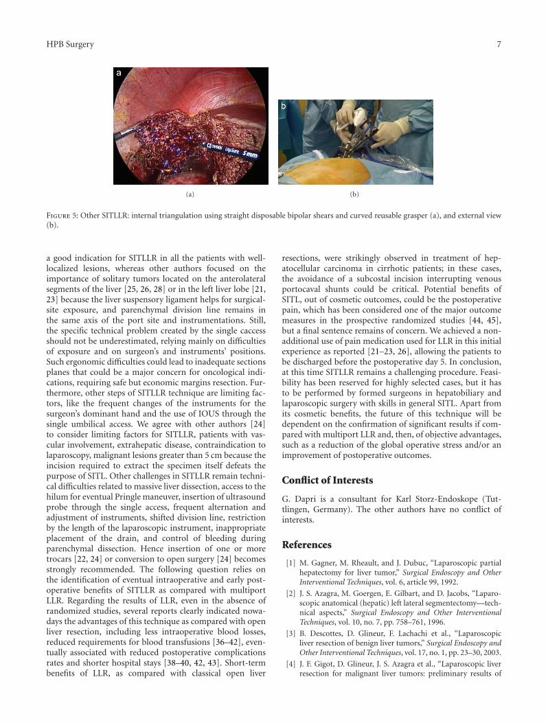
HPB Surgery 7
(a) (b)
Figure 5: Other SITLLR: internal triangulation using straight disposable bipolar shears and curved reusable grasper (a), and external view(b).
a good indication for SITLLR in all the patients with well-localized lesions, whereas other authors focused on theimportance of solitary tumors located on the anterolateralsegments of the liver [25, 26, 28] or in the left liver lobe [21,23] because the liver suspensory ligament helps for surgical-site exposure, and parenchymal division line remains inthe same axis of the port site and instrumentations. Still,the specific technical problem created by the single caccessshould not be underestimated, relying mainly on difficultiesof exposure and on surgeon’s and instruments’ positions.Such ergonomic difficulties could lead to inadequate sectionsplanes that could be a major concern for oncological indi-cations, requiring safe but economic margins resection. Fur-thermore, other steps of SITLLR technique are limiting fac-tors, like the frequent changes of the instruments for thesurgeon’s dominant hand and the use of IOUS through thesingle umbilical access. We agree with other authors [24]to consider limiting factors for SITLLR, patients with vas-cular involvement, extrahepatic disease, contraindication tolaparoscopy, malignant lesions greater than 5 cm because theincision required to extract the specimen itself defeats thepurpose of SITL. Other challenges in SITLLR remain techni-cal difficulties related to massive liver dissection, access to thehilum for eventual Pringle maneuver, insertion of ultrasoundprobe through the single access, frequent alternation andadjustment of instruments, shifted division line, restrictionby the length of the laparoscopic instrument, inappropriateplacement of the drain, and control of bleeding duringparenchymal dissection. Hence insertion of one or moretrocars [22, 24] or conversion to open surgery [24] becomesstrongly recommended. The following question relies onthe identification of eventual intraoperative and early post-operative benefits of SITLLR as compared with multiportLLR. Regarding the results of LLR, even in the absence ofrandomized studies, several reports clearly indicated nowa-days the advantages of this technique as compared with openliver resection, including less intraoperative blood losses,reduced requirements for blood transfusions [36–42], even-tually associated with reduced postoperative complicationsrates and shorter hospital stays [38–40, 42, 43]. Short-termbenefits of LLR, as compared with classical open liver
resections, were strikingly observed in treatment of hep-atocellular carcinoma in cirrhotic patients; in these cases,the avoidance of a subcostal incision interrupting venousportocaval shunts could be critical. Potential benefits ofSITL, out of cosmetic outcomes, could be the postoperativepain, which has been considered one of the major outcomemeasures in the prospective randomized studies [44, 45],but a final sentence remains of concern. We achieved a non-additional use of pain medication used for LLR in this initialexperience as reported [21–23, 26], allowing the patients tobe discharged before the postoperative day 5. In conclusion,at this time SITLLR remains a challenging procedure. Feasi-bility has been reserved for highly selected cases, but it hasto be performed by formed surgeons in hepatobiliary andlaparoscopic surgery with skills in general SITL. Apart fromits cosmetic benefits, the future of this technique will bedependent on the confirmation of significant results if com-pared with multiport LLR and, then, of objective advantages,such as a reduction of the global operative stress and/or animprovement of postoperative outcomes.
Conflict of Interests
G. Dapri is a consultant for Karl Storz-Endoskope (Tut-tlingen, Germany). The other authors have no conflict ofinterests.
References
[1] M. Gagner, M. Rheault, and J. Dubuc, “Laparoscopic partialhepatectomy for liver tumor,” Surgical Endoscopy and OtherInterventional Techniques, vol. 6, article 99, 1992.
[2] J. S. Azagra, M. Goergen, E. Gilbart, and D. Jacobs, “Laparo-scopic anatomical (hepatic) left lateral segmentectomy—tech-nical aspects,” Surgical Endoscopy and Other InterventionalTechniques, vol. 10, no. 7, pp. 758–761, 1996.
[3] B. Descottes, D. Glineur, F. Lachachi et al., “Laparoscopicliver resection of benign liver tumors,” Surgical Endoscopy andOther Interventional Techniques, vol. 17, no. 1, pp. 23–30, 2003.
[4] J. F. Gigot, D. Glineur, J. S. Azagra et al., “Laparoscopic liverresection for malignant liver tumors: preliminary results of

8 HPB Surgery
a multicenter European study,” Annals of Surgery, vol. 236, no.1, pp. 90–97, 2002.
[5] Y. S. Yoon, H. S. Han, J. Y. Cho, and K. S. Ahn, “Total lapa-roscopic liver resection for hepatocellular carcinoma locatedin all segments of the liver,” Surgical Endoscopy and OtherInterventional Techniques, vol. 24, no. 7, pp. 1630–1637, 2010.
[6] D. Cherqui, O. Soubrane, E. Husson et al., “Laparoscopic liv-ing donor hepatectomy for liver transplantation in children,”The Lancet, vol. 359, no. 9304, pp. 392–396, 2002.
[7] P. C. Giulianotti, I. Tzvetanov, H. Jeon et al., “Robot-assistedright lobe donor hepatectomy,” Transplant International, vol.25, pp. e5–e9, 2012.
[8] M. A. Pelosi and M. A. Pelosi III, “Laparoscopic appendectomyusing a single umbilical puncture (minilaparoscopy),” TheJournal of Reproductive Medicine, vol. 37, no. 7, pp. 588–594,1992.
[9] I. Ahmed and P. Paraskeva, “A clinical review of single-incisionlaparoscopic surgery,” Surgeon, vol. 9, pp. 341–351, 2011.
[10] K. Shibao, A. Higure, and K. Yamaguchi, “Case report: laparo-endoscopic single-site fenestration of giant hepatic cyst,” Sur-gical Technology International, vol. 20, pp. 133–136, 2010.
[11] K. Sasaki, G. Watanabe, M. Matsuda, M. Hashimoto, and T.Harano, “Original method of transumbilical single-incisionlaparoscopic deroofing for liver cyst,” Journal of Hepato-Bili-ary-Pancreatic Sciences, vol. 17, no. 5, pp. 733–734, 2010.
[12] S. Kobayashi, H. Nagano, S. Marubashi et al., “A single-incision laparoscopic hepatectomy for hepatocellular carci-noma: initial experience in a Japanese patient,” MinimallyInvasive Therapy and Allied Technologies, vol. 19, no. 6, pp.367–371, 2010.
[13] A. G. Patel, A. P. Belgaumkar, J. James, U. P. Singh, K. A.Carswell, and B. Murgatroyd, “Single-incision laparoscopicleft lateral segmentectomy of colorectal liver metastasis,” Sur-gical Endoscopy and Other Interventional Techniques, vol. 25,no. 2, pp. 649–650, 2011.
[14] L. Aldrighetti, E. Guzzetti, and G. Ferla, “Laparoscopic hepaticleft lateral sectionectomy using the laparoendoscopic singlesite approach: evolution of minimally invasive liver surgery,”Journal of Hepato-Biliary-Pancreatic Sciences, vol. 18, no. 1, pp.103–105, 2011.
[15] S. K. Y. Chang, M. Mayasari, I. S. Ganpathi, V. L. T. Wen,and K. Madhavan, “Single port laparoscopic liver resection forhepatocellular carcinoma: a preliminary report,” InternationalJournal of Hepatology, vol. 2011, pp. 579–203, 2011.
[16] G. Belli, C. Fantini, A. D’Agostino et al., “Laparoendoscopicsingle site liver resection for recurrent hepatocellular carci-noma in cirrhosis: first technical note,” Surgical LaparoscopyEndoscopy & Percutaneous Techniques, vol. 21, pp. 166–168,2011.
[17] H. Kashiwagi, K. Kumagai, and M. Nozue, “Single incisionlaparoscopic surgery for a life-threatening, cyst of liver,” TokaiJournal of Experimental and Clinical Medicine, vol. 36, no. 1,pp. 13–16, 2011.
[18] M. G. Hu, G. D. Zhao, D. B. Xu, and R. Liu, “Transumbilicalsingle-incision laparoscopic hepatectomy: an initial report,”Chinese Medical Journal, vol. 124, no. 5, pp. 787–789, 2011.
[19] U. Barbaros, T. Demirel, O. Gozkun et al., “A new era in min-imally invasive liver resection (MILR) single-incision lapa-roscopic liver resection (SIL-LR): the first two cases,” SurgicalTechnology International, vol. 21, pp. 81–84, 2012.
[20] B. I. Rosok and B. Edwin, “Single-incision laparoscopic liverresection for colorectal metastasis through stoma site at the
time of reversal of diversion ileostomy: a case report,” Mini-mally Invasive Surgery, vol. 2011, Article ID 502176, 3 pages,2011.
[21] S. Gaujoux, T. P. Kingham, W. R. Jarnagin, M. I. D’Angelica, P.J. Allen, and Y. Fong, “Single-incision laparoscopic liver resec-tion,” Surgical Endoscopy and Other Interventional Techniques,vol. 25, no. 5, pp. 1489–1494, 2011.
[22] G. Zhao, M. Hu, R. Liu et al., “Laparoendoscopic single-site liver resection: a preliminary report of 12 cases,” SurgicalEndoscopy and Other Interventional Techniques, vol. 25, pp.3286–3293, 2011.
[23] F. Cipriani, M. Catena, F. Ratti, M. Paganelli, F. Ferla, and L.Aldrighetti, “LESS technique for liver resection: the progress ofthe mini-invasive approach: a single-centre experience,” Mini-mally Invasive Therapy & Allied Technologies, vol. 21, pp. 55–58, 2012.
[24] G. S. Shetty, Y. K. You, H. J. Choi, G. H. Na, T. H. Hong,and D. G. Kim, “Extending the limitations of liver surgery:outcomes of initial human experience in a high-volumecenter performing single-port laparoscopic liver resection forhepatocellular carcinoma,” Surgical Endoscopy and Other Inter-ventional Techniques, vol. 26, pp. 1602–1608, 2012.
[25] M. Aikawa, M. Miyazawa, K. Okamoto, Y. Toshimitsu, K.Okada, and Y. Ueno, “Single-port laparoscopic hepatectomy:technique, safety, and feasibility in a clinical case series,” Sur-gical Endoscopy and Other Interventional Techniques, vol. 26,pp. 1696–1701, 2012.
[26] E. K. Tan, V. T. Lee, S. K. Chang, I. S. Ganpathi, K.Madhavan, and D. Lomanto, “Laparoendoscopic single-siteminor hepatectomy for liver tumors,” Surgical Endoscopyand Other Interventional Techniques, vol. 26, pp. 2086–2091,2012.
[27] W. Cai, J. Xu, M. Zheng, M. Qin, and H. Zhao, “Combinedlaparoendoscopic single-site surgery: initial experience of asingle center,” Hepato-Gastroenterology, vol. 59, pp. 986–989,2012.
[28] M. Pan, Z. Jiang, Y. Cheng et al., “Single-incision laparoscopichepatectomy for benign and malignant hepatopathy: initialexperience in 8 chinese patients,” Surgical Innovation. In press.
[29] S. K. Reddy, A. Tsung, and D. A. Geller, “Laparoscopic liverresection,” World Journal of Surgery, vol. 35, pp. 1478–1486,2011.
[30] J. F. Buell, D. Cherqui, D. A. Geller et al., “The internationalposition on laparoscopic liver surgery: The Louisville State-ment,” Annals of Surgery, vol. 250, pp. 825–830, 2008.
[31] G. B. Hanna, T. Drew, P. Clinch, B. Hunter, and A. Cuschieri,“Computer-controlled endoscopic performance assessmentsystem,” Surgical Endoscopy and Other Interventional Tech-niques, vol. 12, no. 7, pp. 997–1000, 1998.
[32] F. Helgstrand, J. Rosenberg, and T. Bisgaard, “Trocar site her-nia after laparoscopic surgery: a qualitative systematic review,”Hernia, vol. 15, no. 2, pp. 113–121, 2011.
[33] S. A. Antoniou, R. Pointner, and F. A. Granderath, “Single-incision laparoscopic cholecystectomy: a systematic review,”Surgical Endoscopy and Other Interventional Techniques, vol.25, no. 2, pp. 367–377, 2011.
[34] A. Koffron, D. Geller, T. C. Gamblin, and M. Abecassis,“Laparoscopic liver surgery: shifting the management of livertumors,” Hepatology, vol. 44, no. 6, pp. 1694–1700, 2006.
[35] B. Gayet, D. Cavaliere, E. Vibert et al., “Totally laparoscopicright hepatectomy,” American Journal of Surgery, vol. 194, no.5, pp. 685–689, 2007.

HPB Surgery 9
[36] G. Belli, P. Limongelli, C. Fantini et al., “Laparoscopic andopen treatment of hepatocellular carcinoma in patients withcirrhosis,” British Journal of Surgery, vol. 96, no. 9, pp. 1041–1048, 2009.
[37] D. Castaing, E. Vibert, L. Ricca, D. Azoulay, R. Adam, and B.Gayet, “Oncologic results of laparoscopic versus open hepa-tectomy for colorectal liver metastases in two specialized cen-ters,” Annals of Surgery, vol. 250, no. 5, pp. 849–855, 2009.
[38] I. Dagher, N. O’Rourke, D. A. Geller et al., “Laparoscopicmajor hepatectomy: an evolution in standard of care,” Annalsof Surgery, vol. 250, no. 5, pp. 856–860, 2009.
[39] M. Morino, I. Morra, E. Rosso, C. Miglietta, and C. Garrone,“Laparoscopic vs open hepatic resection: a comparative study,”Surgical Endoscopy and Other Interventional Techniques, vol.17, no. 12, pp. 1914–1918, 2003.
[40] B. Topal, S. Fieuws, R. Aerts, H. Vandeweyer, and F. Penninckx,“Laparoscopic versus open liver resection of hepatic neo-plasms: comparative analysis of short-term results,” SurgicalEndoscopy and Other Interventional Techniques, vol. 22, no. 10,pp. 2208–2213, 2008.
[41] F. M. Polignano, A. J. Quyn, R. S. M. de Figueiredo, N. A.Henderson, C. Kulli, and I. S. Tait, “Laparoscopic versus openliver segmentectomy: prospective, case-matched, intention-to-treat analysis of clinical outcomes and cost effectiveness,”Surgical Endoscopy and Other Interventional Techniques, vol.22, no. 12, pp. 2564–2570, 2008.
[42] M. Tsinberg, G. Tellioglu, C. H. Simpfendorfer et al., “Com-parison of laparoscopic versus open liver tumor resection: acase-controlled study,” Surgical Endoscopy and Other Interven-tional Techniques, vol. 23, no. 4, pp. 847–853, 2009.
[43] R. Troisi, R. Montalti, P. Smeets et al., “The value of lapa-roscopic liver surgery for solid benign hepatic tumors,” Sur-gical Endoscopy and Other Interventional Techniques, vol. 22,no. 1, pp. 38–44, 2008.
[44] E. C. Tsimoyiannis, K. E. Tsimogiannis, G. Pappas-Gogoset al., “Different pain scores in single transumbilical inci-sion laparoscopic cholecystectomy versus classic laparoscopiccholecystectomy: a randomized controlled trial,” SurgicalEndoscopy and Other Interventional Techniques, vol. 24, no. 8,pp. 1842–1848, 2010.
[45] P. C. Lee, C. Lo, P. S. Lai et al., “Randomized clinical trialof single-incision laparoscopic cholecystectomy versus mini-laparoscopic cholecystectomy,” British Journal of Surgery, vol.97, no. 7, pp. 1007–1012, 2010.

Hindawi Publishing CorporationHPB SurgeryVolume 2012, Article ID 148387, 7 pagesdoi:10.1155/2012/148387
Review Article
Laparoscopy in Liver Transplantation: The Future Has Arrived
Quirino Lai,1 Rafael S. Pinheiro,2 Giovanni B. Levi Sandri,1 Gabriele Spoletini,1
Fabio Melandro,1 Nicola Guglielmo,1 Marco Di Laudo,1 Fabrizio M. Frattaroli,1
Pasquale B. Berloco,1 and Massimo Rossi1
1 Department of General Surgery and Organ Transplantation, Sapienza University of Rome, Umberto I Policlinic of Rome,Viale del Policlinico 155, 00161 Rome, Italy
2 Department of Liver Transplantation, University of Sao Paulo, 01005 010 Sao Paulo, SP, Brazil
Correspondence should be addressed to Quirino Lai, [email protected]
Received 8 May 2012; Accepted 11 July 2012
Academic Editor: Andrea Lauterio
Copyright © 2012 Quirino Lai et al. This is an open access article distributed under the Creative Commons Attribution License,which permits unrestricted use, distribution, and reproduction in any medium, provided the original work is properly cited.
In the last two decades, laparoscopy has revolutionized the field of surgery. Many procedures previously performed with an openaccess are now routinely carried out with the laparoscopic approach. Several advantages are associated with laparoscopic surgerycompared to open procedures: reduced pain due to smaller incisions and hemorrhaging, shorter hospital length of stay, and a lowerincidence of wound infections. Liver transplantation (LT) brought a radical change in life expectancy of patients with hepatic end-stage disease. Today, LT represents the standard of care for more than fifty hepatic pathologies, with excellent results in termsof survival. Surely, with laparoscopy and LT being one of the most continuously evolving challenges in medicine, their recentcombination has represented an astonishing scientific progress. The intent of the present paper is to underline the current role ofdiagnostic and therapeutic laparoscopy in patients waiting for LT, in the living donor LT and in LT recipients.
1. Introduction
In the last decades, laparoscopy has revolutionized the fieldof surgery. Video laparoscopy was officially born in 1987,when Professor Phillipe Mouret performed the first chole-cystectomy in Lyons, France [1]. Many procedures previouslyperformed with the open technique are now carried out withthe laparoscopic approach. Several advantages are associatedwith laparoscopic surgery compared to open procedures:reduced pain due to smaller incisions and haemorrhaging,shorter hospital length of stay, and a lower incidence ofwound infections are all arguments that gave strength to thewidespread of laparoscopy.
Similarly, liver transplantation (LT) has radically changedthe care for many patients with hepatic end-stage diseases.The first human LT was performed in 1963 by ProfessorThomas Starzl [2] in Denver, United States: however, dueto its initial poor results, LT remained an experimentaltherapy for several years. Only introduction of cyclosporine[3] markedly improved patient outcomes, turning LT to astandard clinical treatment for more than fifty adult andpaediatric liver pathologies and, at the same time, allowingto achieve excellent results in terms of survival.
The intent of the present paper is to underline the currentrole of diagnostic and therapeutic laparoscopy in patientswaiting for LT, in the living donor LT and in LT recipients.
2. Pretransplant Surgery
2.1. Laparoscopic Liver Resection. The first nonanatomicallaparoscopic hepatectomy was performed by Gagner in 1992[4] and the first anatomical one by Azagra in 1996 [5]. Sincethese first experiences, laparoscopic approach for hepaticresection has been adopted in different centres, showing itsfeasibility and safety in well-selected patients [6–10] andalso confirming its prerogatives (shorter operative times, lessbleeding) even in this very complex type of surgery [7, 11].
However, malignant tumors were initially considered acontraindication for mini-invasive approach. Only in thelast years, this risk has been reconsidered, showing nodifference in margin-free resection, port-site recurrence,or tumour seeding rates between open and laparoscopictechniques [12–15]. Consequently, malignant tumors do notrepresent anymore a contraindication for an expert surgeonin choosing a laparoscopic approach [16]. Until now, more

2 HPB Surgery
than 3,000 minimally invasive hepatic resections have beenreported in the literature [17], and-small-to medium sizedprocedures have become commonplace in many centres [18].
Recently, a consensus conference [19] has underlinedthat acceptable indications for laparoscopic liver resectionare (a) solitary lesions with a diameter ≤5 cm, located insegments II–VI; (b) laparoscopic approach to left lateralsectionectomy (LLS) should be considered as a standardpractice; (c) although all types of liver resection can be runlaparoscopically, major liver resections (e.g., right or lefthepatectomies) should be reserved for experienced surgeonsfacile with more advanced laparoscopic hepatic resections.
The main indication for resection in patients waitingfor LT is represented by hepatocellular carcinoma (HCC).Transplant surgeons are well aware that a LT after previoussurgery could represent a real challenge, increasing technicaldifficulty caused by adhesions [20–22]. Basing on theseconsiderations, laparoscopy as bridge to LT may reduce suchproblems [23].
Despite no prospective randomized controlled trialshave been performed yet, some studies based on matchedcomparisons showed similar mortality and even lowermorbidity rates after laparoscopy with respect to open liverresection. After laparoscopy, 3-year patient survival rates of60–93% and 3-year disease-free survivals of 52–64% havebeen reported [8, 24–29].
Advent of robotic surgery has further improved theopportunity of mini-invasive treatment of HCC. The roboticapproach may enable liver resection in patients with cirrho-sis, allowing for technical refinements of laparoscopic liverresection due to 3-dimensional visualization of the operativefield and instruments with wrist-type end-effectors [30–32].
2.2. Laparoscopic Radiofrequency Ablation. Radiofrequency(RF) ablation represents a nonsurgical locoregional treat-ment, used in very well-selected patients with nonresectableHCC waiting for LT. In the last years, laparoscopic orhand-assisted RF has shown promising necrosis and survivalrates [33, 34], providing a substrate for their safe adoption[35]. The main advantage of laparoscopic RF with respectto percutaneous approach is the opportunity to detectpre-operatively undetectable lesions using an intraoperativeultrasound (IOUS) [36, 37]. IOUS remains the most sensitiveimaging modality for HCC, being able to detect new lesionsin 13.1% to 30% of cases [38–42]. In a study comparinglaparoscopic liver resection and RF, Santambrogio et al. [43]identified 15 (20%) of 74 cases with previously undetectedlesions in the RF group. Laparoscopic RF also consents totreat lesions considered inappropriate for percutaneous RFdue to the high risk of injury in the diaphragm, stomach, orbowel [44–46]. Similarly, laparoscopic RF minimizes the riskof complications in patients previously operated in the upperabdominal quadrants [47].
2.3. Laparoscopic Kasai Procedure in Children with BiliaryAtresia. Laparoscopic portoenterostomy, also named Kasaiprocedure, for biliary atresia was first reported by Esteves etal. in 2002 [48]. Besides its safety and feasibility, laparoscopic
Kasai can provide the advantage of a lower hepatic adhesionsrate, which ease the potential “salvage” LT. Martinez-Ferroet al. reported 41 cases of laparoscopic Kasai, with onlyone conversion [49], and encouraging results in terms ofpostoperative bile flow rates. However, the only prospectivestudy comparing open and laparoscopic procedure wasstopped after observing that laparoscopic patients showeda significantly shorter time between Kasai procedure andLT [50]. Therefore, the role of laparoscopic Kasai remainsunclear [51], being reserved to paediatric centres with highspecialization in minimally invasive surgery.
3. Laparoscopic Living Donor-Hepatectomy
The early idea of solid organ transplantation using livingdonors began with kidney transplants; similarly, laparoscopyfor living donation was initially developed for kidneytransplantation, with the intent to offer a less aggressiveprocedure to the donor. In fact, laparoscopic nephrectomyis associated with less postoperative pain, decreased length ofhospital stay, faster return to normal activity, smaller scars,and less morbidity [52]. As a consequence, an increasednumber of kidney transplants using living donors have beenrecently observed in many centres [53, 54].
Living-donor liver transplantation is a complex proce-dure, with major risks of morbidity and mortality withrespect to kidney donation: the reported mortality of thisprocedure has varied from 0.2% to 0.5% [55]. Clearly, donorrisk increases according to the type of hepatectomy (LLS <left hepatectomy < right hepatectomy (RH)).
Typically, a LLS or a left hepatectomy is sufficient in apaediatric living liver donation, while a RH is necessary foran adult-to-adult donation.
However, despite different surgical approaches couldbe adopted, open living donation always requires a largeabdominal incision. This aspect, combined with postoper-ative pain, long hospital stay, and long periods of recovery,represents a barrier to donation, especially in young women[56].
Recently, experimental model has demonstrated thefeasibility of laparoscopic living donation using the availabletechnology [57].
Concurrent improvement in laparoscopic surgery forhepatic tumours enhanced feasibility and safety of morecomplex procedures [18], leading to the establishment oflaparoscopic surgery for liver living donors. Soubrane et al.[58] reflected on the quality of the graft and the morbidityrates in the donor: they reported comparable results in bothconventional and laparoscopic techniques apart from longeroperative times and lower blood losses in the laparoscopygroup.
3.1. Paediatric Donation. Paediatric living donation providessimilar or better short-term graft function and long-termsurvival rates with respect to postmortem donor LT: the firstcase of laparoscopic donation was reported by Cherqui etal. [56]. In 2006, Soubrane et al. [58] reported the safetyof laparoscopic LLS in 16 consecutive live donors compared

HPB Surgery 3
with the conventional LLS. According to the first seriesexperienced, the liver graft typically includes the LLS (i.e.,segments II and III according to Couinaud’s classification),left branch of hepatic artery, left portal branch, left bile duct,and left hepatic vein. After these initial experiences, severalother new series have been reported worldwide [59, 60].
Recently, Kim et al. compared 11 laparoscopic LLS forliving donation with 11 open ones, showing that the laparo-scopic group had significantly shorter hospital stay, whilstduration of operation, blood loss, warm ischemia time,and out-of-pocket medical costs were comparable betweengroups [61].
A similar study from Washington DC compared 15laparoscopic or laparoscopic-assisted left or right hepatec-tomies for liver donation with 15 hepatectomies with openaccess: no substantial differences were observed in terms ofearly graft function, allograft biliary, and vascular complica-tions and survivals (1-year graft and patient survival: 100%versus 93% in laparoscopic and open group, resp.) [62].
3.2. Adult Donation. In 2006, Koffron et al. [63] describedthe first hand-assisted laparoscopic RH for live donation.Kurosaki et al. [64] reported in the same period 13 con-secutive video-assisted adult-to-adult laparoscopic hepate-ctomies (3 RH and 10 left ± segment I hepatectomies):surgical manipulation was obtained via ports or via a 12 cmincision whilst view resulted by a combination of direct andlaparoscopic vision. Reported median operation time was363 ± 33 minutes and a median blood loss of 302 ± 191 mL.No complications were reported, restoration of liver functionwas smooth, and analgesics use was inferior with respect tothe historical control (median: 1.2 versus 3.8 times).
In 2008, the transplant group from Seoul [65, 66]commented on the first series of hand-assisted laparoscopicmodified RH preserving the middle hepatic vein; theauthors reported 2 cases of laparoscopic RH and 7 casesof laparoscopy-assisted RH with a hand-port device. Hilardissection and parenchymal transection were performedunder pneumoperitoneum (n = 2) or through a mini-laparotomy incision (n = 7). The graft was extracted throughthe site of the hand-port device or the minilaparotomy.Operative time was 765 and 898 min in the laparoscopicRH patients, and it ranged from 310 to 575 min for thelaparoscopy-assisted surgery. In one case, a fluid collectionalong the liver resection margin was reported, but it wasresolved after percutaneous drainage.
At the Northwestern University, Baker et al. [67] ret-rospectively compared 33 open versus 33 laparoscopic liv-ing donor RHs, suggesting that laparoscopy could presentequivalent safety, resource utilization, and effectiveness, withseveral adjunctive physical and psychological benefits. Donoroperative times were shorter for the laparoscopic group(265 min versus 316 min). Blood loss and length of stay werecomparable. Additionally, total hospitalization costs wereequivalent. Finally, the group from Seoul compared single-port laparoscopy-assisted donor right hepatectomy (n = 40)with laparoscopy-assisted donor right hepatectomy (n = 20)and open donor right hepatectomy (n = 90); postoperative
complication and reoperation rates revealed no significantdifferences, the single-port group showing the lowest level ofpostoperative pain [68].
Although very limited experiences have been reportedworldwide until now, no mortalities have been encounteredin laparoscopic living donor hepatectomy, whether adult orpaediatric. Larger experience is needed in this field, but onlycentres with a coincident expertise in hepatic mini-invasivesurgery and living donor LT should approach this type ofsurgery.
4. Laparoscopic-AssistedLiver Transplantation
The first nine cases of living donor LT through a short-midline incision combined with hand-assisted laparoscopicsurgery have been reported in Japan [69]. All the patientswere cirrhotic (median MELD score 14). Total hepatec-tomy was carried out through a hand-assisted laparoscopicapproach with an 8 cm upper midline incision. Explantationof the diseased liver was obtained through the upper midlineincision which was extended to 12 to 15 cm. Partial livergrafts were implanted through the upper midline incision.Median surgical time was 741 min, and the median bloodloss was 3,940 g.
This preliminary report of application of laparoscopyduring LT procedure represents an extraordinary innovation,opening new porspectives in this fascinating surgical field.Further evolutions in the use of mini-invasive surgery in LTare expected in the next years.
5. Laparoscopic Posttransplant Surgery
Postoperative laparoscopic management of LT patients is lesscommon with respect to renal transplant recipients: in fact,laparoscopy is easily applied after kidney transplantation,given the fact that in this type of transplant the dissectionis completely extraperitoneal.
In postLT patients, laparoscopy is a useful tool to solve anumber of surgical complications; however, its use is strictlyconnected to surgeon’s experience and versatility.
5.1. Laparoscopic Incisional Hernia Repair. Incisional herniais caused by several aetiologies, many of whose could beconcomitantly observed in LT recipients: advanced age,wound infection, ascites, steroids, diabetes, surgical tech-niques, suture material, retransplantation, bilateral sub-costal incision with midline extension, and, not less impor-tant, surgeon’s experience. The most common site forincisional hernia in LT patients is located at the junction ofthe transverse and upper midline incisions [70]. In literature,the incidence of incisional hernia varies from 5% to 17%[71]. Large incisional and ventral hernias in nontransplantpatients are now routinely repaired using laparoscopictechnique. Laparoscopic ventral hernia repair seems to havea reduced risk of recurrence and infection compared tostandard repair [72]. In LT patients, laparoscopic hernia

4 HPB Surgery
repair is safe and with similar results when compared withopen repair [70, 73].
Andreoni et al. [74] successfully completed 12 out of13 attempted incisional hernia repairs by the laparoscopictechnique in LT patients. Gore-Tex mesh was used. Atthe time of publication, they report no recurrence. Theyconcluded that laparoscopic mesh repair of incisional herniasis practical and safe in patients with a surgical history of LTtransplantation, with a low incidence of infections and norecurrence. However, in a monocentre study [74], a higherrate of postoperative seroma was observed in LT with respectto nontransplanted patients [75]. A study from Germanyanalyzed a population of 29 solid organs recipients: 15 caseswere treated with intraperitoneal onlay mesh repair and 14with conventional hernia repair [76]. Recurrence rate was 6%versus 50%, and complication rate was 33% versus 21% inlaparoscopy and conventional groups, respectively.
A study from Spain described 20 cases of laparoscopicincisional hernia repair in patients after LT, using a BardComposix mesh, showing excellent results and few compli-cations [77]. Observing the excellent results obtained usinglaparoscopy for the treatment of incisional hernia in LTpatients, we can conclude that it could be safely performedalso in this particular type of patients, and it could beconsidered a standard practice, mainly in an expert surgeon’shands.
5.2. Other Indications. In the last years, different uses oflaparoscopy have been attempted in LT recipients. Merendaet al. [78] reported two cases of intestinal occlusion causedby adhesions and three cases of lymphocele, all approachedwith laparoscopic surgery. In all cases but one, the authorswere able to complete the surgery by laparoscopic means;in one of the two occlusions, the procedure was switchedto laparotomy because of a choledochojejunal anastomosislesion.
Gill et al. [79] reported a single case of a right adrenal-ectomy after LT in a 63-year-old female patient with aright adrenal mass and a previous story of left radicalnephrectomy for a renal cell carcinoma and LT for primarybiliary cirrhosis. A laparoscopic right adrenalectomy via theretroperitoneoscopic approach was successfully performed,and the patient was discharged home on the first postopera-tive day.
DeRoover and Sudan [80] documented the case ofa 46-year-old female transplanted for primary sclerosingcholangitis who presented multiple splenic aneurysms andabdominal pain: after a laparoscopic splenectomy, the patientwas discharged on postoperative day 3 free of symptoms. AJapanese experience [81] reported 5 cases of hand-assistedlaparoscopic splenectomy for hypersplenism in living donorLT recipients. On the basis of the excellent results, the authorsconsider it as a possible standard procedure after LT.
Robles et al. [82] commented on 2 cases of biliaryperitonitis after T-tube removal who failed conservativetreatment and subsequently underwent laparoscopy: lysis ofadhesions was carried out in the right upper quadrant, aPenrose drain was placed, and both patients were discharged
home on postoperative day 4. In 2010, Zhu et al. [83]reported the first total laparoscopic hysterectomy after LT.Authors confirmed that no viscera adhesions were observedto the undersurface of the umbilicus.
In 2011, Lee et al. [84] were the first to successfullycomplete a laparoscopic total gastrectomy in a previouslytransplanted 72-year-old patient, showing that laparoscopy isa feasible method for gastric cancer treatment in LT patients.
Finally, the Hannover group reflected on the applicabilityof laparoscopy in the management of posttransplantationlymphoproliferative disorder in a pediatric population: 6 outof 34 (18%) solid organs recipients underwent laparoscopicbiopsies because of the lack of superficial lesions, with a 83%success rate. In one patient, a trocar metastasis was identifiedand treated successfully with chemotherapy [85].
Despite few cases have been reported until now, wecan affirm that several “conceptual” barriers in the fieldof laparoscopy have been overcome: previous LT no longerrepresents an absolute contraindication for laparoscopy; notonly in case of small procedures (biopsies) or submesocolicand pelvic surgery, but also when supramesocolic organs areinvolved. Further experience is needed in this field and onlysurgeons with a high expertise in mini-invasive proceduresmust approach this type of surgery. However, the authors areconfident that in the future laparoscopic or robotic surgerywill substitute open surgery in many cases even in previouslytransplanted patients.
6. Conclusion
Use of laparoscopy in the field of LT is safe and feasible.Mini-invasive approach is commonly adopted in the bridgetreatment of HCC in patients waiting for LT: in case of LLS,laparoscopic procedure is recognized as the gold standardtherapy. In living donor hepatectomy, and, recently, in LT,pure mini-invasive approaches or hybrid forms of laparo-scopic and open surgery have been attempted. However, alimited number of reports are currently available on this sub-ject, and great ability and confidence are recommended forstarting these laparoscopy-assisted programs. Laparoscopyfor abdominal surgery after LT has been demonstrated to befeasible and safe, not only in patients candidates for pelvicsurgery, but also in case of surgery in the upper abdominalquadrants. In the next future, welcome improvement intechnologies will give impulse to further expansion of thissurgical area.
Conflict of Interests
There is no conflict of interests to declare.
Authors’ Contribution
Q. Lai designed the study; Q. Lai, R. S. Pinheiro, G. B. L.Sandri and G. Spoltini wrote the paper; F. Melandro, N.Gugliemo, M. D. Laudo, F. M. Frattaroli, P. B. Berloco andM. Rossi participated in the critical evaluation of the paper.

HPB Surgery 5
References
[1] A. Cuschieri, F. Dubois, J. Mouiel et al., “The European experi-ence with laparoscopic cholecystectomy,” American Journal ofSurgery, vol. 161, no. 3, pp. 385–387, 1991.
[2] T. E. Starzl, T. L. Marchioro, K. N. Vonkaulla, G. Hermann, R.S. Brittain, and W. R. Waddell, “Homotransplantation of theliver in humans,” Surgery, Gynecology & Obstetrics, vol. 117,pp. 659–676, 1963.
[3] R. Y. Calne, S. Thiru, and D. J. G. White, “Cyclosporin A inpatients receiving renal allografts from cadaver donors,” TheLancet, vol. 2, no. 8104, pp. 1323–1327, 1978.
[4] M. Gagner, M. Rheault, and J. Dubuc, “Laparoscopic partialhepatectomy for liver tumor,” Surgical Endoscopy, vol. 6, article99, 1992.
[5] J. S. Azagra, M. Goergen, E. Gilbart, and D. Jacobs, “Laparo-scopic anatomical (hepatic) left lateral segmentectomy—technical aspects,” Surgical Endoscopy, vol. 10, no. 7, pp. 758–761, 1996.
[6] T. Mala, B. Edwin, A. R. Rosseland, I. Gladhaug, E. Fosse,and Ø. Mathisen, “Laparoscopic liver resection: experience of53 procedures at a single center,” Journal of Hepato-Biliary-Pancreatic Surgery, vol. 12, no. 4, pp. 298–303, 2005.
[7] B. Descottes, F. Lachachi, M. Sodji et al., “Early experiencewith laparoscopic approach for solid liver tumors: initial 16cases,” Annals of Surgery, vol. 232, no. 5, pp. 641–645, 2000.
[8] D. Cherqui, A. Laurent, C. Tayar et al., “Laparoscopic liverresection for peripheral hepatocellular carcinoma in patientswith chronic liver disease: midterm results and perspectives,”Annals of Surgery, vol. 243, no. 4, pp. 499–506, 2006.
[9] L. Biertho, A. Waage, and M. Gagner, “Laparoscopic hepate-ctomies,” Annales de Chirurgie, vol. 127, no. 3, pp. 164–170,2002.
[10] I. Dagher, J. M. Proske, A. Carloni, H. Richa, H. Tranchart,and D. Franco, “Laparoscopic liver resection: results for70 patients,” Surgical Endoscopy and Other InterventionalTechniques, vol. 21, no. 4, pp. 619–624, 2007.
[11] C. Simillis, V. A. Constantinides, P. P. Tekkis et al., “Laparo-scopic versus open hepatic resections for benign and malig-nant neoplasms-a meta-analysis,” Surgery, vol. 141, no. 2, pp.203–211, 2007.
[12] K. Ito, H. Ito, C. Are et al., “Laparoscopic versus openliver resection: a matched-pair case control study,” Journal ofGastrointestinal Surgery, vol. 13, no. 12, pp. 2276–2283, 2009.
[13] X. J. Cai, J. Yang, H. Yu et al., “Clinical study of laparoscopicversus open hepatectomy for malignant liver tumors,” SurgicalEndoscopy and Other Interventional Techniques, vol. 22, no. 11,pp. 2350–2356, 2008.
[14] Y. Endo, M. Ohta, A. Sasaki et al., “A comparative study of thelong-term outcomes after laparoscopy-assisted and open leftlateral hepatectomy for hepatocellular carcinoma,” SurgicalLaparoscopy, Endoscopy and Percutaneous Techniques, vol. 19,no. 5, pp. e171–e174, 2009.
[15] B. Topal, S. Fieuws, R. Aerts, H. Vandeweyer, and F. Penninckx,“Laparoscopic versus open liver resection of hepatic neo-plasms: comparative analysis of short-term results,” SurgicalEndoscopy and Other Interventional Techniques, vol. 22, no. 10,pp. 2208–2213, 2008.
[16] I. Dagher, G. Di Giuro, J. Dubrez, P. Lainas, C. Smadja, andD. Franco, “Laparoscopic versus open right hepatectomy: acomparative study,” American Journal of Surgery, vol. 198, no.2, pp. 173–177, 2009.
[17] H. H. Kim, E. K. Park, J. S. Seoung et al., “Liver resectionfor hepatocellular carcinoma: case-matched analysis of laparo-scopic versus open resection,” Journal of the Korean SurgicalSociety, vol. 80, no. 6, pp. 412–419, 2011.
[18] R. I. Troisi, J. Van Huysse, F. Berrevoet et al., “Evolution oflaparoscopic left lateral sectionectomy without the Pringlemaneuver: through resection of benign and malignant tumorsto living liver donation,” Surgical Endoscopy and Other Inter-ventional Techniques, vol. 25, no. 1, pp. 79–87, 2011.
[19] J. F. Buell, D. Cherqui, D. A. Geller et al., “The internationalposition on laparoscopic liver surgery: the Louisville State-ment, 2008,” Annals of Surgery, vol. 250, pp. 825–830, 2009.
[20] R. Adam and D. Azoulay, “Is primary resection and salvagetransplantation for hepatocellular carcinoma a reasonablestrategy?” Annals of Surgery, vol. 241, no. 4, pp. 671–672, 2005.
[21] V. Mazzaferro, S. Todo, A. G. Tzakis, A. C. Stieber, L.Makowka, and T. E. Starzl, “Liver transplantation in patientswith previous portasystemic shunt,” American Journal ofSurgery, vol. 160, no. 1, pp. 111–116, 1990.
[22] A. Steib, G. Freys, C. Lehmann, C. Meyer, and G. Mahoudeau,“Intraoperative blood losses and transfusion requirementsduring adult liver transplantation remain difficult to predict,”Canadian Journal of Anesthesia, vol. 48, no. 11, pp. 1075–1079,2001.
[23] A. Laurent, C. Tayar, M. Andreoletti, J. Y. Lauzet, J. C.Merle, and D. Cherqui, “Laparoscopic liver resection facilitatessalvage liver transplantation for hepatocellular carcinoma,”Journal of Hepato-Biliary-Pancreatic Surgery, vol. 16, no. 3, pp.310–314, 2009.
[24] E. C. H. Lai, C. N. Tang, J. P. Y. Ha, and M. K. W. Li,“Laparoscopic liver resection for hepatocellular carcinomaten-year experience in a single center,” Archives of Surgery, vol.144, no. 2, pp. 143–148, 2009.
[25] I. Dagher, P. Lainas, A. Carloni et al., “Laparoscopic liverresection for hepatocellular carcinoma,” Surgical Endoscopyand Other Interventional Techniques, vol. 22, no. 2, pp. 372–378, 2008.
[26] L. Aldrighetti, E. Guzzetti, C. Pulitano et al., “Case-matchedanalysis of totally laparoscopic versus open liver resectionfor HCC: short and middle term results,” Journal of SurgicalOncology, vol. 102, no. 1, pp. 82–86, 2010.
[27] H. Tranchart, G. Di Giuro, P. Lainas et al., “Laparoscopicresection for hepatocellular carcinoma: a matched-pair com-parative study,” Surgical Endoscopy and Other InterventionalTechniques, vol. 24, no. 5, pp. 1170–1176, 2010.
[28] G. Belli, P. Limongelli, C. Fantini et al., “Laparoscopic andopen treatment of hepatocellular carcinoma in patients withcirrhosis,” British Journal of Surgery, vol. 96, no. 9, pp. 1041–1048, 2009.
[29] B. S. Hu, K. Chen, H. M. Tan et al., “Comparison oflaparoscopic vs open liver lobectomy (segmentectomy) forhepatocellular carcinoma,” World Journal of Gastroenterology,vol. 17, pp. 4725–4728, 2011.
[30] F. Panaro, T. Piardi, M. Cag et al., “Robotic liver resectionas a bridge to liver transplantation,” Journal of the Society ofLaparoendoscopic Surgeons, vol. 15, pp. 86–89, 2011.
[31] E. C. H. Lai, C. N. Tang, G. P. C. Yang, and M. K. W.Li, “Multimodality laparoscopic liver resection for hepaticmalignancy—from conventional total laparoscopic approachto robot-assisted laparoscopic approach,” International Journalof Surgery, vol. 9, no. 4, pp. 324–328, 2011.
[32] S. B. Choi, J. S. Park, J. K. Kim et al., “Early experiences ofrobotic-assisted laparoscopic liver resection,” Yonsei MedicalJournal, vol. 49, no. 4, pp. 632–638, 2008.
6 HPB Surgery
[33] Y. Asahina, H. Nakanishi, and N. Izumi, “Laparoscopicradiofrequency ablation for hepatocellular carcinoma: review,”Digestive Endoscopy, vol. 21, no. 2, pp. 67–72, 2009.
[34] A. Siperstein, A. Garland, K. Engle et al., “Laparoscopicradiofrequency ablation of primary and metastatic livertumors: technical considerations,” Surgical Endoscopy, vol. 14,no. 4, pp. 400–405, 2000.
[35] F. Panaro, T. Piardi, M. Audet et al., “Laparoscopic ultrasound-guided radiofrequency ablation as a bridge to liver trans-plantation for hepatocellular carcinoma: preliminary results,”Transplantation Proceedings, vol. 42, no. 4, pp. 1179–1181,2010.
[36] V. R. Tandan, M. Asch, M. Margolis, A. Page, and S. Gallinger,“Laparoscopic vs. open intraoperative ultrasound examina-tion of the liver: a controlled study,” Journal of GastrointestinalSurgery, vol. 1, no. 2, pp. 146–151, 1997.
[37] M. Montorsi, R. Santambrogio, P. Bianchi et al., “Laparoscopywith laparoscopic ultrasound for pretreatment staging ofhepatocellular carcinoma: a prospective study,” Journal ofGastrointestinal Surgery, vol. 5, no. 3, pp. 312–315, 2001.
[38] N. Kokudo, Y. Bandai, H. Imanishi et al., “Management ofnew hepatic nodules detected by intraoperative ultrasonog-raphy during hepatic resection for hepatocellular carcinoma,”Surgery, vol. 119, no. 6, pp. 634–640, 1996.
[39] Y. Takigawa, Y. Sugawara, J. Yamamoto et al., “New lesionsdetected by intraoperative ultrasound during liver resectionfor hepatocellular carcinoma,” Ultrasound in Medicine andBiology, vol. 27, no. 2, pp. 151–156, 2001.
[40] G. Torzilli and M. Makuuchi, “Intraoperative ultrasonographyin liver cancer,” Surgical Oncology Clinics of North America, vol.12, no. 1, pp. 91–103, 2003.
[41] H. Cerwenka, J. Raith, H. Bacher et al., “Is intraopera-tive ultrasonography during partial hepatectomy still neces-sary in the age of magnetic resonance imaging?” Hepato-Gastroenterology, vol. 50, no. 53, pp. 1539–1541, 2003.
[42] K. Zhang, N. Kokudo, K. Hasegawa et al., “Detection of newtumors by intraoperative ultrasonography during repeatedhepatic resections for hepatocellular carcinoma,” Archives ofSurgery, vol. 142, no. 12, pp. 1170–1176, 2007.
[43] R. Santambrogio, E. Opocher, M. Zuin et al., “Surgical resec-tion versus laparoscopic radiofrequency ablation in patientswith hepatocellular carcinoma and child-pugh class a livercirrhosis,” Annals of Surgical Oncology, vol. 16, no. 12, pp.3289–3298, 2009.
[44] K. K. Tanabe, S. A. Curley, G. D. Dodd, A. E. Siperstein, and S.N. Goldberg, “Radiofrequency ablation: the experts weigh in,”Cancer, vol. 100, no. 3, pp. 641–650, 2004.
[45] T. Livraghi, L. Solbiati, M. F. Meloni, G. S. Gazelle, E. F.Halpern, and S. N. Goldberg, “Treatment of focal liver tumorswith percutaneous radio-frequency ablation: complicationsencountered in a multicenter study,” Radiology, vol. 226, no.2, pp. 441–451, 2003.
[46] A. Casaril, M. Abu Hilal, A. Harb, T. Campagnaro, G.Mansueto, and N. Nicoli, “The safety of radiofrequencythermal ablation in the treatment of liver malignancies,”European Journal of Surgical Oncology, vol. 34, no. 6, pp. 668–672, 2008.
[47] K. C. Yun, H. Rhim, S. A. Yong, Y. K. Mi, and K. L. Hyo,“Percutaneous radiofrequency ablation therapy of hepato-cellular carcinoma using multitined expandable electrodes:comparison of subcapsular and nonsubcapsular tumors,”American Journal of Roentgenology, vol. 186, supplement 5, pp.S269–S274, 2006.
[48] E. Esteves, E. C. Neto, M. O. Neto, J. Devanir, and R. E. Pereira,“Laparoscopic Kasai portoenterostomy for biliary atresia,”Pediatric Surgery International, vol. 18, no. 8, pp. 737–740,2002.
[49] M. Martinez-Ferro, E. Esteves, and P. Laje, “Laparoscopictreatment of biliary atresia and choledochal cyst,” Seminars inPediatric Surgery, vol. 14, no. 4, pp. 206–215, 2005.
[50] B. M. Ure, J. F. Kuebler, N. Schukfeh, C. Engelmann, J.Dingemann, and C. Petersen, “Survival with the native liverafter laparoscopic versus conventional kasai portoenterostomyin infants with biliary atresia: a prospective trial,” Annals ofSurgery, vol. 253, no. 4, pp. 826–830, 2011.
[51] K. W. E. Chan, K. H. Lee, J. W. C. Mou, S. T. G. Cheung, andY. H. P. Tam, “The outcome of laparoscopic portoenterostomyfor biliary atresia in children,” Pediatric Surgery International,vol. 27, no. 7, pp. 671–674, 2011.
[52] J. L. Flowers, S. Jacobs, E. Cho et al., “Comparison of open andlaparoscopic live donor nephrectomy,” Annals of Surgery, vol.226, no. 4, pp. 483–490, 1997.
[53] L. E. Ratner, L. R. Kavoussi, M. Sroka et al., “Laparoscopicassisted live donor nephrectomy—a comparison with the openapproach,” Transplantation, vol. 63, no. 2, pp. 229–233, 1997.
[54] L. E. Ratner, J. Hiller, M. Sroka et al., “Laparoscopic livedonor nephrectomy removes disincentives to live donation,”Transplantation Proceedings, vol. 29, no. 8, pp. 3402–3403,1997.
[55] P. Intaraprasong, A. Sobhonslidsuk, and S. Tongprasert,“Donor outcomes after Living Donor Liver Transplantation(LDLT),” Journal of the Medical Association of Thailand, vol.93, no. 11, pp. 1340–1343, 2010.
[56] D. Cherqui, O. Soubrane, E. Husson et al., “Laparoscopic liv-ing donor hepatectomy for liver transplantation in children,”The Lancet, vol. 359, no. 9304, pp. 392–396, 2002.
[57] E. Lin, R. Gonzalez, K. R. Venkatesh et al., “Can currenttechnology be integrated to facilitate laparoscopic living donorhepatectomy?” Surgical Endoscopy and Other InterventionalTechniques, vol. 17, no. 5, pp. 750–753, 2003.
[58] O. Soubrane, D. Cherqui, O. Scatton et al., “Laparoscopicleft lateral sectionectomy in living donors: safety and repro-ducibility of the technique in a single center,” Annals ofSurgery, vol. 244, no. 5, pp. 815–820, 2006.
[59] R. Troisi, R. Debruyne, and X. Rogiers, “Laparoscopic livingdonor hepatectomy for pediatric liver transplantation,” ActaChirurgica Belgica, vol. 109, no. 4, pp. 559–562, 2009.
[60] J. C. U. Coelho, A. C. T. de Freitas, and J. E. F. Mathias,“Laparoscopic resection of the left lateral segment of theliver in living donor liver transplantation,” Revista do ColegioBrasileiro de Cirurgioes, vol. 36, no. 6, pp. 537–538, 2009.
[61] K. H. Kim, D. H. Jung, K. M. Park et al., “Comparison of openand laparoscopic live donor left lateral sectionectomy,” BritishJournal of Surgery, vol. 98, pp. 1302–1308, 2011.
[62] A. Thenappan, R. C. Jha, T. Fishbein et al., “Liver allograftoutcomes after laparoscopic-assisted and minimal access livedonor hepatectomy for transplantation,” American Journal ofSurgery, vol. 201, no. 4, pp. 450–455, 2011.
[63] A. J. Koffron, R. Kung, T. Baker, J. Fryer, L. Clark, and M.Abecassis, “Laparoscopic-assisted right lobe donor hepatec-tomy,” American Journal of Transplantation, vol. 6, no. 10, pp.2522–2525, 2006.
[64] I. Kurosaki, S. Yamamoto, C. Kitami et al., “Video-assistedliving donor hemihepatectomy through a 12-cm incision foradult-to-adult liver transplantation,” Surgery, vol. 139, no. 5,pp. 695–703, 2006.
HPB Surgery 7
[65] K. S. Suh, N. J. Yi, J. Kim et al., “Laparoscopic hepatectomyfor a modified right graft in adult-to-adult living donor livertransplantation,” Transplantation Proceedings, vol. 40, no. 10,pp. 3529–3531, 2008.
[66] K. S. Suh, N. J. Yi, T. Kim et al., “Laparoscopy-assisted donorright hepatectomy using a hand port system preserving themiddle hepatic vein branches,” World Journal of Surgery, vol.33, no. 3, pp. 526–533, 2009.
[67] T. B. Baker, C. L. Jay, D. P. Ladner et al., “Laparoscopy-assistedand open living donor right hepatectomy: a comparative studyof outcomes,” Surgery, vol. 146, no. 4, pp. 817–825, 2009.
[68] H. J. Choi, Y. K. You, G. H. Na et al., “Single-port laparoscopy-assisted donor right hepatectomy in living donor liver trans-plantation: sensible approach or unnecessary hindrance?”Transplantation Proceedings, vol. 44, pp. 347–352, 2012.
[69] S. Eguchi, M. Takatsuki, A. Soyama et al., “Elective livingdonor liver transplantation by hybrid hand-assisted laparo-scopic surgery and short upper midline laparotomy,” Surgery,vol. 150, pp. 1002–1005, 2011.
[70] A. Kurmann, G. Beldi, S. A. Vorburger, C. A. Seiler, and D.Candinas, “Laparoscopic incisional hernia repair is feasibleand safe after liver transplantation,” Surgical Endoscopy andOther Interventional Techniques, vol. 24, no. 6, pp. 1451–1455,2010.
[71] T. Piardi, M. Audet, F. Panaro et al., “Incisional hernia repairafter liver transplantation: role of the mesh,” TransplantationProceedings, vol. 42, no. 4, pp. 1244–1247, 2010.
[72] B. T. Heniford, A. Park, B. J. Ramshaw, G. Voeller, J. G. Hunter,and R. J. Fitzgibbons, “Laparoscopic repair of ventral hernias:nine years’ experience with 850 consecutive hernias,” Annals ofSurgery, vol. 238, no. 3, pp. 391–400, 2003.
[73] K. Mekeel, D. Mulligan, K. S. Reddy, A. Moss, and K.Harold, “Laparoscopic incisional hernia repair after livertransplantation,” Liver Transplantation, vol. 13, no. 11, pp.1576–1581, 2007.
[74] K. A. Andreoni, H. Lightfoot, D. A. Gerber, M. W. Johnson,and J. H. Fair, “Laparoscopic incisional hernia repair in livertransplant and other immunosuppressed patients,” AmericanJournal of Transplantation, vol. 2, no. 4, pp. 349–354, 2002.
[75] K. Harold, K. Mekeel, J. Spitler et al., “Outcomes analysisof laparoscopic ventral hernia repair in transplant patients,”Surgical Endoscopy and Other Interventional Techniques, vol.23, no. 8, pp. 1835–1838, 2009.
[76] H. Scheuerlein, F. Rauchfuss, A. Gharbi, M. Heise, and U.Settmacher, “Laparoscopic incisional hernia repair after solid-organ transplantation,” Transplantation Proceedings, vol. 43,no. 5, pp. 1783–1789, 2011.
[77] R. Gianchandani, E. Moneva, P. Marrero et al., “Feasibility andeffectiveness of laparoscopic incisional hernia repair after livertransplantation,” Transplantation Proceedings, vol. 43, no. 3,pp. 742–744, 2011.
[78] R. Merenda, G. E. Gerunda, D. Neri et al., “Laparoscopicsurgery after orthotopic liver transplantation,” Liver Trans-plantation, vol. 6, no. 1, pp. 104–107, 2000.
[79] I. S. Gill, A. M. Meraney, J. T. Mayes, and E. L. Bravo,“Laparoscopic right adrenalectomy after liver transplanta-tion,” Transplantation, vol. 71, no. 9, pp. 1350–1351, 2001.
[80] A. DeRoover and D. Sudan, “Treatment of multiple aneurysmsof the splenic artery after liver transplantation by percuta-neous embolization and laparoscopic splenectomy,” Trans-plantation, vol. 72, no. 5, pp. 956–958, 2001.
[81] H. Uehara, H. Kawanaka, T. Akahoshi et al., “The feasibilityand effectiveness of a hand-assisted laparoscopic splenectomy
for hypersplenism in patients after living-donor liver trans-plantation,” Surgical Laparoscopy, Endoscopy and PercutaneousTechniques, vol. 19, no. 6, pp. 484–487, 2009.
[82] R. Robles, P. Parrilla, J. A. Lujan, J. A. Torralba, P. Ramirez,and F. S. Bueno, “Laparoscopic treatment of biliary peritonitisafter T tube removal in patients undergoing orthotopic livertransplantation,” British Journal of Surgery, vol. 84, no. 9, p.1244, 1997.
[83] H. B. Zhu, Y. Jin, S. T. Xu, Y. X. Xia, and L. P. Xie,“Total laparoscopic hysterectomy after liver transplantation,”Hepatobiliary and Pancreatic Diseases International, vol. 9, no.4, pp. 438–440, 2010.
[84] M. S. Lee, E. Y. Kim, J. H. Lee et al., “Laparoscopy-assisted dis-tal gastrectomy for gastric cancer after liver transplantation,”Journal of the Korean Surgical Society, vol. 80, supplement 1,pp. S1–S5, 2011.
[85] M. L. Metzelder, T. Schober, L. Grigull et al., “The roleof laparoscopic techniques in children with suspected post-transplantation lymphoproliferative disorders.,” Journal ofLaparoendoscopic & Advanced Surgical Techniques, vol. 21, pp.767–770, 2011.


