Embed Size (px)
Citation preview

Louisiana State UniversityLSU Digital Commons
LSU Historical Dissertations and Theses Graduate School
1954
Innervation of the Heart and Ductus Arteriosusand Other Features of the Autonomic NervousSystem of the Full Term Human Fetus.Frank D. AllanLouisiana State University and Agricultural & Mechanical College
Follow this and additional works at: https://digitalcommons.lsu.edu/gradschool_disstheses
Part of the Life Sciences Commons
This Dissertation is brought to you for free and open access by the Graduate School at LSU Digital Commons. It has been accepted for inclusion inLSU Historical Dissertations and Theses by an authorized administrator of LSU Digital Commons. For more information, please [email protected].
Recommended CitationAllan, Frank D., "Innervation of the Heart and Ductus Arteriosus and Other Features of the Autonomic Nervous System of the FullTerm Human Fetus." (1954). LSU Historical Dissertations and Theses. 8086.https://digitalcommons.lsu.edu/gradschool_disstheses/8086
THE INNERVATION OF THE HEART AND DUCTUS ARTERIOSUS AND OTHER FEATURES OF THE AUTONOMIC NERVOUS SYSTEM
OF THE FULL TERM HUMAN FETUS
A Dissertation
Submitted to the Graduate Faculty of the Louisiana State University and
Agriculture and Mechanical College in partial fulfillment of the
requirements for the degree of Doctor of Philosophy
in
The Department of Anatomy
School of Medicine
byFrank Duane Allan
B.S., University of Utah, 1947 M.S., University of Utah, 1949
May, 1954

UMl Number: DP69464
All rights reserved
INFORMATION TO ALL USERS The quality of this reproduction is dependent upon the quality of the copy submitted.
In the unlikely event that the author did not send a complete manuscript and there are missing pages, these will be noted. Also, if material had to be removed,
a note will indicate the deletion.
UMlDissertation Publishing
UMl DP69464
Published by ProQuest LLC (2015). Copyright in the Dissertation held by the Author.
Microform Edition © ProQuest LLC.All rights reserved. This work is protected against
unauthorized copying under Title 17, United States Code
ProQuest LLC.789 East Eisenhower Parkway
P.O. Box 1346 Ann Arbor, Ml 48106- 1346
ProQuestQue

MANUSCRIPT THESES
U npublished th e s e s su b m itted f o r th e m aster*s and d o c to rTs
deg rees and d e p o s ite d in th e L ou isiana S ta te U n iv e rs i ty L ib ra ry
a re a v a i la b le f o r in sp e c tio n * Use o f any th e s is i s l im i te d by th e
r i g h t s o f the a ia thor. B ib lio g ra p h ic a l re fe re n c e s may be noted^ b u t
passages may n o t be co p ied u n le ss th e a u th o r has g iven perm ission*
C re d it must be g iv en in subsequent w r i t te n o r p u b lish ed work.
A l ib r a r y w hich borrows th i s th e s is f o r use by i t s c l i e n t e l e
i s expected to make su re th a t th e borrow er i s aware o f th e above
r e s t r i c t i o n s .
LOUISIANA STATE UNIVERSITY LIBRARY
1 1 9 -a

A C K N O W L E D G E M E N T
A complete acknowledgement of those to whom I am indebted
for aid in preparation of this dissertation is impossible. To my wife,
whose patience, understanding, and help have made my efforts possible,
most credit is due. Many of the references used in this study were
obtained by use of the Inter-library loan, and for help in this regard,
1 am grateful to the library staff, especially Miss Ann Hodge, The
photographs, mounting, and labeling were done essentially by the
Art Department to whom I am most grateful. Miss Merle Fowler
typed the final manuscript. To Dr, Charles M, Goss, my major
professor, thanks are given for his advice, and, with Dr. Ruth Miller,
invaluable help in the organisation and correction of the manuscript.
Similarly, gratitude is due the other members of the Anatomy De
partment and my examining committee for their encouragement.

T A B U S O F C O N T E N T S
I. INTRODUCTION *
II. HISTORICAL.Comparative anatomy 2Human anatomy 11Ductus arteriosus 22Intermediate cervical ganglion 25Fetal studies 28
HL MATERIAL AND METHODS 30
IV. OBSERVATIONSSympathetic trunk 34Cervical sympathetic cardiac nerves
Superior cervical sympathetic cardiac nerve 41Middle cervical sympathetic cardiac nerve 43Inferior cervical sympathetic cardiac nerve 45Cardiac nerves of the intermediate ganglion 47
Thoracic sympathetic cardiac nerves 47Intercommunications of sympathetic cardiac nerves 52Accessory Ganglia associated with sympathetic
cardiac nerves 54Cardiac nerves of the vagus
Superior cervical cardiac nerves 56Inferior cervical cardiac nerves 59Thoracic cardiac nerves 60
Communications between sympathetic and vagal cardiac nerves 61Vago-sympathetic communications 62Sympathetico-vagal communications 63
Superficial cardiac plexus 63Deep cardiac plexus 67Nerve supply of the ductus arteriosus 71
V. DISCUSSIONSympathetic trunk 7 9
Superior cervical sympathetic cardiac nerve 83Middle cervical sympathetic cardiac nerve 84Inferior cervical sympathetic cardiac nerve 85Thoracic sympathetic cardiac nerves 8 8Intercommunications of sympathetic cardiac nerves 89Accessory ganglia associated with sympathetic
cardiac nerves 90Superior cervical cardiac nerve of the vagus 9 2Inferior cervical cardiac nerve of the vagus 9 3
i i i

Thoracic cardiac nerves of the vagus 94Vago-sympathetic intercommunications 95Superficial cardiac plexus 96Deep cardiac plexus 99Ductus arteriosus 102
VI. SUMMARY 106
VII. SELECTED BIBLIOGRAPHY 109
vm. VITA 138
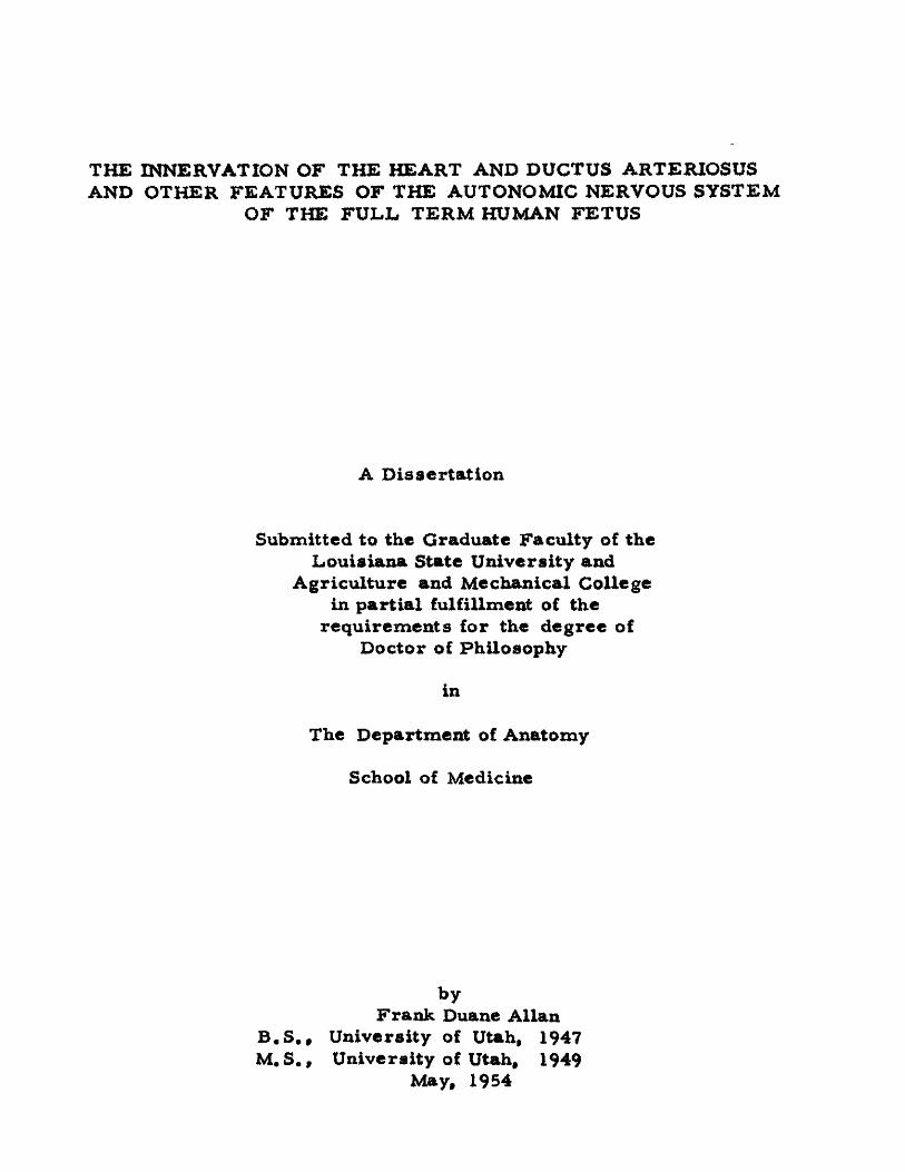
T A B L E S
I. Intermediate Cervical Sympathetic Ganglion 129
II, Superior Cervical Sympathetic Cardiac Nerve(s) 130
III, Middle Cervical Sympathetic Cardiac Nerve(s) 131
IV, Inferior Cervical Sympathetic Cardiac Nerve(s) 132
V. Thoracic Cardiac Sympathetic Nerves 133
VI. Vagal Cardiac Nerves 134
VII, Superficial Cardiac Plexus 135
VIII. Deep Cardiac Plexus 136
IX. Innervation of the Ductus Arteriosus 137
v

FIGURES
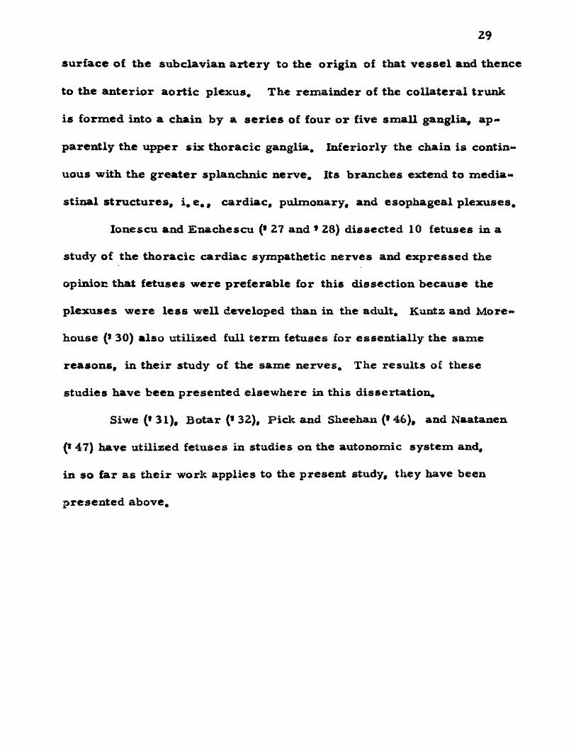
1. Left Sympathetic Trunk and Branches 35
2. Right Sympathetic Trunk and Branches 36
3. Types of Intermediate Cervical Ganglia 3S
4. Sympathetic Cardiac Nerves with an Anomalous Right
Subclavian Artery 48
5. Chart of Thoracic Sympathetic Cardiac Nerves 51
6 . Left Vagus Nerve and Branches 57
7. Right Vagus Nerve and Branches 58
8 . Relations of Cardiac Plexuses to Vessels and Pericardial
Sinuses 6 8
9. Innervation of the Ductus Arteriosus 72
10, Innervation of the Ductus Arteriosus 73
11, Innervation of the Ductus Arteriosus 74
12, Photograph (Dissection 11) 122
13, Photograph (Dissection 8 } 123
14, Photograph (Dissection 15) 124
15, Photograph (Dissection 13, on left) 125
16, Photograph (Dissection 14 before removal of the
Subclavian Artery) 126
17, Photograph (Dissection 14 after removal of the
Subclavian Artery) 127
18, Photograph (Dissection 13, on right) 128
v i

LIST OF ABBREVIATIONS (used in Figures and Tables )
A. arteryAnt, anteriorAo. AortaBr. large branchbr. small branchc. cervicalCard. CardiacCar. CarotidCcrv. cervicalCollai. collateralCom. commonCoron. CoronaryD.# D.A.or Duct. Ductus arteriosusExt. externalG. ganglionICCSN Inferior Cervical Sympathetic Cardiac NerveICG Inferior Cervical GanglionIGTsg Inter ganglionic Trunk near superior cervical ganglionIGT luterganglionic TrunkIGTamg Inter ganglionic Trunk above middle cervical ganglionIn. InnominateInf. InferiorInt. InternalIntcrmed. Inte rmediat elint. C. G. Intermediate Cervieal GanglionD. leftDat. lateral
A. Deft Atrium or AuricleDCCA Deft Common Carotid ArteryD.V. Deft VentricleDaryng. DaryngealMCCSH Middle Cervical Sympathetic Cardiac NerveMCG Middle Cervical GanglionMam. MammaryMed. medialMid. middleN. nerveP or Plex. plexusPhren. N. Phrenic nerveProx. proximalP.T. Pulmonary TrunkPulm. PulmonaryR. right

R.A. Right Atrium or AuricleR. V. Right VentricleRec. N. Recurrent NerveRt. rootsSCCSN Superior Cervical Sympathetic Cardiac NerveSCG Superior Cervical GanglionSl-X Superior Laryngeal Nerve of the VagusSp. spinalStell. StellateSub. SubclavianSup. superiorSup. Plex. Superficial Cardiac PlexusS.V.C. Superior Vena CavaSym. SympatheticT or Tbor. ThoracicTbyr. ThyroidThyrocerv. ThyrocervicalTr. TrunkTrans. TransverseTrans. S. Transverse SinusV. VeinVert. Vertebra or VertebralX Vagus nerve
v i i i

ABSTRACT
Although studies of the autonomic nervous system of the
cervicothoracic region in man have been numerous, certain features
have escaped notation, whereas, others are in need of reexamination
in order that a more accurate knowledge may be obtained. Further
more, most previous work has been done on the adult, with few sys -
tematic studies existing regarding this system in the fetus. Thus, no
detailed consideration of the origin and distribution of the extrinsic
nerves to the ductus arteriosus has taken place.
The technics of this study were simple, but required tedious
and delicate dissection of the neck and upper thorax of the full term
fetus with detailed records, written and graphic, regarding the auto
nomic system in that area.
The study demonstrates that innervation of the ductus arterio
sus is accomplished by the left inferior cervical sympathetic car
diac nerve by way of a plexus on the proximal part of the left sub
clavian artery, and by branches from the inferior cervical cardiac
nerve of the vagus primarily as branches of the superficial cardiac
plexus. The plexus upon the ductus distributes fibers to it and the
pulmonary trunk, continuing to the left coronary plexus.
Sympathetic and vagal cardiac nerves are analyzed in regard
to their frequency, origin, course, termination, communications, and
association with accessory ganglia. These are largely confirmation
of previous studies but present a more detailed knowledge of each.
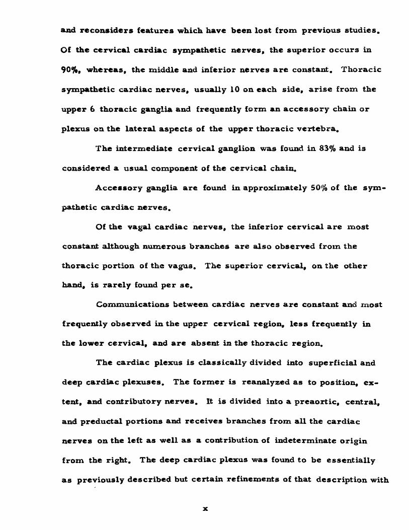
and reconsiders features which have been lost from previous studies.
Of the cervical cardiac sympathetic nerves, the superior occurs in
90%, whereas, the middle and inferior nerves are constant. Thoracic
sympathetic cardiac nerves, usually 10 on each side, arise from the
upper 6 thoracic ganglia and frequently form an accessory chain or
plexus on the lateral aspects of the upper thoracic vertebra.
The intermediate cervical ganglion was found in 83% and is
considered a usual component of the cervical chain.
Accessory ganglia are found in approximately 50% of the sym
pathetic cardiac nerves.
Of the vagal cardiac nerves, the inferior cervical are most
constant although numerous branches are also observed from the
thoracic portion of the vagus. The superior cervical, on the other
hand, is rarely found per se.
Communications between cardiac nerves are constant and most
frequently observed in the upper cervical region, less frequently in
the lower cervical, and are absent in the thoracic region.
The cardiac plexus is classically divided into superficial and
deep cardiac plexuses. The former is reanalyzed as to position, ex
tent, and contributory nerves. It is divided into a preaortic, central,
and preductal portions and receives branches from all the cardiac
nerves on the left as well as a contribution of indeterminate origin
from the right. The deep cardiac plexus was found to be essentially
as previously described but certain refinements of that description with
x

regard to its relationship to the pericardial sinuses and its communi
cations with the superCicial cardiac plexus have been added.

INTRODUCTION
The nerves to the heart have been the subject of numerous
investigations during the past two centuries, but the information
which has been gained still needs extension and clarification. The
autonomic nerves and associated ganglia of the cervical and upper
thoracic regions of full term human fetuses have been studied rela
tively infrequently, and no systematic study of the extrinsic nerves
to the ductus arteriosus of the human fetus has been found in the
literature.
The purpose of the present study is to describe the gross
anatomical features of the extrinsic nerves to the heart and espe
cially to the ductus arteriosus in the full term human fetus.
As an introduction to the problem, a broad review of published
work will be presented including general features of cardiac inner
vation in addition to those which apply specifically to the present
obse rvations #
1
HISTORICAL
Comparative Anatomy
It is of interest that the first studies of cardiac innervation
were made on man and that only during the last century and a half
has much attention been given to lower forms. Comparative studies
were eventually undertaken, however, in order to simplify or clarify
the problem in man.
Although studies on mammals were made as early as 1815
by Weber, using the calf, the impetus which seems to have initiated
these studies was the discovery and demonstration of the depressor
nerve in the rabbit by Cyon and Ludwig (1867). This stimulated a
series of papers concerning that nerve and led physiologists
Gaskell and Gadow (1884) to undertake its study in reptiles and am
phibians. His. Jr. (1891) included embryologicai studies in fishes
as well as higher vertebrates. Schumacher (• 02 a and b) made an
extensive study of cardiac innervation from a comparative anatom
ical standpoint, especially on the depressor nerve of mammals.
Mollard (* 08) considered lower animals, including the invertebrates,
in his work on the cardiac nerves, but apparently this was not the
major part of his material. Perman* s (• 24) extensive work attemp
ted to cover all aspects of the problem including comparative and
embryologicai studies on mammals as well as man. A series of
detailed studies of the cardiac nerves in several mammals and
birds was made by Ssinelnikow (* 28, bird), Anufriew (• 28, cat),
2

3
Schurawlew (• 28, dog), and Wolhynski (* 28, calf) from the same
laboratory. Riegele (* 26) presented a less complete work on the
apes. Mention should also be made of studies by Shaner (* 30) on
the calf, Nonidez (* 35, * 39 and * 41) on puppies, rabbits, cats and
guinea pigs and Saccomanno (* 43) on cats.
The studies referred to above and many others whose main
purpose does not justify their citation here, have established a
general plan for the cardiac nerves throughout the phylogenetic
series. This plan begins in fishes (His, ji r., 1890), with fibers to
the heart derived exclusively from the vagus nerves, apparently
limited to the venous mesocardium. In the amphibians (Gaskell and
Gadow 1884) the sympathetic trunks contribute some fibers to the
heart, but they join the vagus and are distributed with its branches.
In the reptiles (Gaskell and Gadow) the innervation of the
heart is from both vagal and sympathetic sources, but the two sets
of nerves usually follow separate paths to the heart. The vagal
fibers pass along the great arterial trunks, whereas, those from
the sympathetic reach the heart along the venae cavae. There is
some indication that in snakes and lizards (Hirt, *2 1 ) the nerves
from the two sources are fused. A ganglion in the vagus as it enters
the thorax limits the origin of the cardiac nerves superiorly, and
the sympathetic chain is enclosed in the intertransverse canal of the
cervical region, thus limiting the uppermost origin of the sympathetic
nerves to the heart.
4
An isolated but rather complete study of the cardiac nerves
in birds by Ssinelnikow (• 2$) shows that these nerves present certain
characteristics which resemble the reptiles. For example, a gang
lion of the vagus at the superior aperture of the thorax, called the
ganglion of Couvreuri, limits the superior origin of the cardiac
nerves. In addition, the cervical sympathetic chain in birds is also
enclosed in the canal of the intertransverse foramina and, therefore,
the cardiac nerves are limited to an origin from the trunk prior to
its entrance into the canal. Perhaps the most significant observa
tions in the study are that the nerves arising from one side of the
body supply the ipsilateral side of the heart, and that the anterior
plexuses of the heart are mainly supplied with vagal fibers, whereas,
sympathetic fibers from the upper three thoracic ganglia pass to the
posterior plexuses.
As indicated previously, studies on the comparative anatomy
of the cardiac nerves and associated structures have considered
mammals more frequently than other forms, and certain papers
provide a reasonably complete presentation of the cardiac nerves
in this class.
The work of Schumacher (* 02) reviews the pre-existing
papers, and although its coverage of the mammalian series is quite
complete, it loses some of its significance because of the small
number of animals used in certain parts of the study. The depressor
nerve is represented as including all nerves to the heart or great

5
vessels from the vagus between the origins of the superior laryn
geal (and sometimes from this nerve) and the recurrent nerves.
The sympathetic cardiac nerves originate from the middle cervical
down to the 6 th thoracic ganglia, but those from the thoracic gang*
lia are found only in the Artiodactyla. The presence of a superior
cardiac sympathetic nerve is limited to the apes and man. The
depressor nerve ends in the wall of the aorta (and ductus arteriosus)
and is designated, therefore, the aortic nerve of the vagus. Schu
macher describes the sympathetic cardiac nerves as passing to the
ventricles for the most part and, consequently, calls them the ven
tricular nerves; the nerves from the left side pass anterior to the
great vessels and are usually more discrete than those from the
right. He considers the cardiac nerves in mammals as supplying
the ipsilateral side of the heart with an overlap where a portion of
the left ventricle adjacent to the anterior longitudinal sulcus is sup
plied by fibers from the right side.
Per man* s study (f 24), which includes a representative num
ber of mammals, is more complete but agrees in most parts with
Schumacher* s. According to Ferman the depressor nerve, with
roots of origin from the superior laryngeal nerve of the vagus, the
vagus proper, and the sympathetic, exists only in cats and rabbits
and more frequently in the latter. In other mammals this nerve is
usually united with the vagus trunk or with the sympathetic. The
presence of a common vago-sympathicus, i.e ., a fusion between

6
the cervical portion of the sympathetic trunk and the vagus, in the
dog, calf, goat and sheep, usually results in the absence of a discrete
depressor nerve in those animals. The sympathetic cardiac nerves
consist of three or four nerves with origins from the middle cervi
cal ganglion and the trunk above it, the ansa subclavia, and the stell
ate ganglion. As mentioned above, only the Primates, man and the
macaque, have a superior cardiac nerve from the sympathetic.
The Artiodactyla are the only animals observed to have thoracic car
diac nerves.
Perman divides the nerves to the heart into two groups, de
pending upon their relation to the traasver^pericardial sinus.
Nerves which arise at relatively higher levels pass ventral to the
sinus, while those with origins at a lower level pass dorsal to it.
The nerves passing ventral to the sinus follow the great arterial
trunks, whereas those passing dorsal usually follow the remnants
of the ducts of Cuvier to reach the heart. On the left, this pathway
involves the pericardial fold of Marshall, a vestige of the reflec
tion of the pericardium on the old duct of Cuvier. The nerves
which follow the arterial trunks pass primarily to the anterior por
tions of the heart, whereas those with the ducts of Cuvier, pass to
the posterior aspect. Perman, agreeing with Schumacher, notes
both ipsi- and contralateral distribution of the nerves from a single
side, although the contribution is greater from the left.

7
The most detailed accounts of cardiac innervation in an indi
vidual mammal are presented by Schurawlew (• 28), Anufriew (* 28),
and Wolhynski (* 28), These works agree with previous presentations,
but modify and enlarge certain details.
The cardiac nerves of the dog (Schurawlew * 28), distribute
themselves to ipsi* and contralateral sides of the heart. Those nerves
having a relatively high origin from the vagus or the sympathetic
trunk pass anterior to the transverse pericardial sinus and eventually
reach the anterior surface of the ventricles. Those with lower
sources, i. e ., at or below the superior aperture of the thorax, pass
posterior to the sinus and extend to the posterior portions of the heart.
A detailed description of the atrial plexuses and a similarly detailed
mention of the nerves to the pressor receptor areas are the primary
contributions of this work.
According to Anufriew (* 28), as well as Schurawlew, the
majority of fibers arising on the left side from the stellate ganglion
or below reach the heart through the pericardial fold of Marshall and
are distributed to the posterior cardiac plexus.
Certain characteristics of the cardiac nerves in the calf
(Wolhynski, 9 28) are modified by the presence of a brachiocephalic
artery (anterior aorta) but the general plan of the mammalian car
diac nerves is the same and thoracic cardiac nerves from the sym
pathetic trunk are present. These branches follow the left superior
vena cava, which persists in the calf, in order to reach the posterior

8
aspect of the heart.
The history of the thoracic sympathetic cardiac nerves has
been reviewed by Mitchell (• 49) who noted that they had been ob
served in the calf by Weber as early as 1815. Physiological studies
by Cannon and associates (* 26 a and b) provided the stimulus for the
” rediscovery11 of these nerves by Ionescu and Knaschescu (* 2 ? and
*28), and Braeueker (* 27), although these nerves needed no ” redis
covery” in an anatomical sense, since they appeared in papers by
Schumacher (* 02). Mollard (• 08). and Perman (• 24).
The studies of Noniden (* 35. 9 39. and 9 41) extend beyond the
scope of this investigation but it should be mentioned they establish
the location of the pressor receptor areas and determine the nerves
which pass to them. This work was done with puppies, rabbits,
guinea pigs, and cats. Careful analysis must be given to Nonidea9 s
work because his nomenclature is different than that of previous in
vestigators. For example, a superior cardiosympathetic nerve is
described in dogs, whereas, other investigators state that no nerve
homologous to the superior cardiac sympathetic nerve is present.
Furthermore, this description is modified by the presence of a vago
sympathetic fusion in the neck which limits the conclusions which can
be drawn regarding branches from this region.
In summary, certain general trends of the comparative anatomy
of the cardiac nerves may be outlined. In fishes the heart receives
its nerve sypply from the vagus only. There is a rudimentary
9
sympathetic system which lacks any apparent nervous connection
with the heart* This situation prevails in amphibians also where
the primary source of nerves to the heart is the vagus* although
some nerves of sympathetic origin probably reach the heart.
In reptiles* the sympathetic trunk ganglia in the lower cerv
ical and upper thoracic region give a definite contribution to the
heart. In some reptiles the cervical sympathetic trunk enters the
canal formed by the iniertransverse foramina of the cervical verte
brae and is* therefore* buried deep in the cervical region.
In birds and mammals the autonomic nervous system and
its subdivisions have reached their greatest development* probably
in response to* or concommitant with* greater freedom from the
environment. This necessitated the development of homeostatic
mechanisms and their control through an integrating center. The
circulatory system* being one of the chief homeostatic mechanisms*
has come more and more under the influence of the sympathetic
system with a consequent increase in the number and complexity
of its nerves.
In birds the presence of the ganglion of Couvreuri on the
vagus at its entrance into the thorax limits the exit of vagal car
diac nerves superiorly. The origin of sympathetic fibers is limited
superiorly by the fact that the trunk is buried in the intertransverse
canal of the cervical region. Vagal fibers arising from one side of
the body reach and supply the ipsilateral side of the heart. Sympathetic
10
fibers reach the heart along the great veins, whereas, the vagal car-
diac fibers pass along the arteries or veins depending upon whether
they arise from the vagus high or low in its course. Kuxsta (* 1 0 )
believes that the autonomic nervous system in birds followed a sep
arate evolutionary trend, and any comparison of the system between
birds and mammals ought to be qualified with that possibility in mind.
In mammals, members of the same order demonstrate the
same characteristics of origin, course, and distribution of nerves
to the heart. In the Carnivora, nerves from both sides of the body
take part in the formation of the cardiac plexuses and are distributed
to the same and opposite sides of the heart. However, the contribu
tion of nerves from the left side are usually more numerous and
pass anterior to the aortic arch. The postion and innervation of the
pressor receptive areas have been quite weU established inthis order.
More work has been done on cardiac innervation in rabbits
than in any other mammalian form, except man. No basic differ
ences have been shown from other mammals, however, except that
the aortic depressor nerve is an entity.
In the Ungulata, a characteristic feature of the cardiac nerves
is the presence of a well established group of nerves to the heart
from the thoracic sympathetic trunk. Another feature, apparently
limited to this order, is the occasional presence of a group of nerves
which pass to the right of the aortic arch to reach the anterior surface
of the heart.
11
Little work has been done on primates other than man, but
from all indications the similarity which is to be noticed between
members of the same order holds true here. The presence of car
diac sympathetic nerves from the upper four thoracic segments is a
constant feature, and it appears that the superior sympathetic car
diac nerve is present only in this order.
Human Anatomy
Fallopius (1561) apparently was the first to investigate the
nerves of the heart in detail. He recognized a plexus between the
aorta and the pulmonary trunk with contributions from both sympa
thetic and vagus nerves. Prior to this, a single nerve to the heart
was described by anatomists, including Vesalius.
During the next 100 years very little was added. Willis (1667)
made a division of the plexus, but it remained for Senac (1749) to
locate the divisions definitely. He describes the cardiac plexus as
consisting of " le grande plexus anterieur" anterior to the aortic
arch and ft le grande plexus posterieur” located on the deep surface
of the arch. This interpretation was substantiated by Haller (1757)
and Andersch (1791).
Three plexuses were distinguished by Murray (1772), the
first anterior to the aortic arch, the second between the aorta and
(right?) pulmonary artery, and a third on the posterior wall of the
heart. Wrisberg (1786) found a ganglion, which today bears his

12
name, within the cardiac plexus below the arch of the aorta.
The work of Scarpa (1794) is of particular significance, be
cause of the excellent drawings and the first detailed consideration
of the origin and course of the cardiac nerves. Of particular inter
est to the present study are certain features of the extrinsic nerves
of the cardiac plexus, such as the plexus on the proximal portion of
the subclavian artery formed by nerves from the middle and in
ferior cervical ganglia, and the nerve from the inferior cervical
ganglion on the right passing anterior to the aorta to reach the in
terval between the latter and the pulmonary trunk. Also, a large
branch from the plexus in this interval loops anterior and to the left
of the ligamentum arte riosum and then along the lateral aspect of
the pulmonary trunk to the left coronary plexus. Finally, the vagal
cardiac branches are limited in origin to, or below, the level at
which the nerve crosses the subclavian artery, Mayer (1794), also
described the superficial portion of the cardiac plexus as receiv
ing the superior cardiac nerves of both sides.
During the first half of the 19th century not much significant
work appeared. Usually the cardiac plexus was regarded as an en
tity divided into two portions, but some authorities looked upon it as
having further subdivisions. Swan (1&30) separated it into right and
left lateral cardiac plexuses, which communicated with the cardiac
nerves in the neck, the ventricular and atrial plexuses, and the right
and left thoracic plexuses. His is the first description of the thoracic

13
sympathetic cardiac nerves in man, but he was not aware of their
significance,
Valentin (1841) divided the superficial cardiac plexus into
two parts, one anterior to the aortic arch and ext rape ricar dial, the
other intrapericardi&l along the aorta and the pulmonary artery.
The presence of a small ganglion on the superior cardiac nerve was
also noted and called the r* gangliacum cardiac superior s. minus11 ,
Cruveilhier* s (1845) description of the cardiac nerves is
reminiscent of that by Murray cited above. The nerves are divided
into three planes according to their relationship to the aorta. In
one plane, the superior and middle cardiac nerves from the left
side pass ventral to the aortic arch toward the right coronary plexus.
The nerves from the right side pass dorsal to the aorta and join the
two previously mentioned nerves in the concavity of the arch at which
point the ganglion of Wrisberg is located, Cruveilhier associated
the latter ganglion particularly with the nerves passing ventral to
the aortic arch. The second plane of nerves passes from the deep
surface of the aortic arch to the coronary plexus of both sides.
The third plan of nerves passes dorsal to the aortic arch and is
distributed to the atria and posterior surface of the heart.
The work of Ellis (1849) and to a lesser extent that of Swan,
have been used as a basis for most descriptions of the cardiac
nerves in textbooks of anatomy written in English, Ellis followed

14
the lead of earlier investigators in describing two plexuses, a super
ficial and a deep. The former plexus, which is found between the
arch of the aorta and the ligamentum arte rio sum, receives the left
superior cardiac nerve of the sympathetic and the lower cardiac
nerve of the left vagus, as well as a considerable contribution from
the deep cardiac plexus. With regard to the deep plexus, Ellis
makes the classical statement that it receives all the cardiac nerves
except those referred to above, i. e ., the left superior cardiac nerve
of the sympathetic and the lower cardiac nerve of the vagus. Nerves
from the superficial cardiac plexus extend between the aorta and the
pulmonary artery to the anterior coronary plexus, whereas those
from the deep plexus, especially the left half, pass to the posterior
coronary plexus. Confusion has arisen because Ellis refers to the
right as the anterior coronary artery and to the left as the poster
ior coronary artery.
Sappey (1852) holds much the same position in the French
school of anatomists. He identifies the cardiac plexus as the com
munications of cardiac nerves from the vagus and sympathetic in a
space limited to the right and above by the angle of the aortic arch,
on the left by the ligamexxtum arteriosum, interiorly by the right
pulmonary artery, and posteriorly by the tracheal bifurcation. In
the center is the ganglion of Wrisberg, which may consist of sev
eral rather than a single ganglion. Interiorly the plexus divides
into three portions; an anterior, between and superficial to the aorta

15
and pulmonary trunk, continuing as the right coronary plexus; a
middle, anterior to the right pulmonary artery in the interval be
tween the pulmonary trunk and the aorta; and a posterior, behind the
right pulmonary artery, the pulmonary trunk and the auricle. The
anterior portion of the plexus receives branches from the cardiac
nerves on the left. Thoracic cardiac branches are described, but
their contribution is small.
S&ppey recognises the communications between the superior
laryngeal nerve of the vagus and the superior cardiac nerve. The
cardiac nerves of the vagus are divided into superior, middle, and
inferior groups, the first two representing the cervical and the last
the thoracic branches.
Henle (1871) established a plan of cardiac innervation, agree
ing with the earlier data which was used by many German texts.
He described a superficial and a deep cardiac plexus, and fibers
from the latter which reached the atria. He included the ganglion
of Wrisberg in the superficial plexus.
His, Jr. (1891), in describing the development of the cardiac
nerves, gives three plexuses, an arterial plexus, an atrial plexus,
and a plexus of nerves uniting the two. Gegenbauer (* 03) applied
His* designations, with slight modifications, to the adult. Both of
the latter interpretations resemble those of Cruveilhier cited above.
Schumacher (• 02) states that the cardiac plexuses can be
divided logically into superficial and deep portions, with boundaries

16
essentially the same as those described above. He indicates, how
ever, that these plexuses constitute a 11 Grenzstein" or boundary
Cor studies on the innervation of the heart. He considered the gang
lion of Wrisberg as a point to which all the cardiac nerves converge.
He also recognized a small nerve from the right passing to the su
perficial cardiac plexus.
Jacques (9 02) presented essentially the same picture as that
of Sappey, indicating that all of the cardiac nerves on the left con
tribute branches to the superficial cardiac plexus.
The next articles of significance to this study are by Worobiew
(9 17 and 9 26). The nerves intimately associated with the heart are
divided into six plexuses according to the portion of the heart which
they innervate. Although these plexuses may be classed as independ
ent cardiac plexuses, they are actually extensions of the classically
described superficial and deep cardiac plexuses. The divisions are
outlined as follows: 1. Left anterior longitudinal plexus, located on
the left ventricle, extends from the atrioventricular sulcus to the
apex of the heart. II. Eight anterior logitudinal plexus, on the right
ventricle. HI. Right posterior longitudinal plexus, includes fibers
innervating the right atrium (especially the sinus venosus), the post
erior portion of the right ventricle, and the inferior and lateral aspect
of the left ventricle at the apex. IV. Left posterior longitudinal
plexus, encompasses the left atrium and ventricle adjacent to the
sulcus. V. Anterior atrial plexus, includes nerves on the pulmonary

17
veins and radiates over the anterior surface of the atria. VI. Post
erior atrial plexus, includes nerves on the pulmonary veins at their
entrance to the left atrium (Haller9 s plexus).
Jonnesco (9 23), in a monograph on the cervical and thoracic
portions of the sympathetic system, suggests that the cardiac nerves
be numbered, since not infrequently one of them is lacking. He in
dicates that the ganglion of Wrisberg is in the center of the cardiac
plexus and that the latter is separated into three planes, superficial,
middle, and deep, for terminal distribution.
Perman (9 24) was the last investigator to undertake a really
comprehensive study of the cardiac nerves in man. In this study,
which includes comparative and embryological observations as well
as an extensive review of the literature, Perman concludes that a
uniform picture of the cardiac nerves cannot be obtained from re
ports in the literature, not only because of the differences in the
observations of various investigators, but also because of misin
terpretations of the material presented. He points out that the car
diac plexus need not be considered the 11 Grenzstein" (Schumacher,
9 02) for investigation of the innervation of the heart. He traces
nerves from both sides of the body to the heart with some degree
of accuracy, showing that both sides of the heart may receive
nerves from the same and opposite sides of the body.
Perman emphasizes the relation pf the nerves to the trans
verse pericardial sinus in determining the portion of the heart they

18
eventually reach. Thus, the nerves passing along the great arteries
ventral to the sinus go to the coronary plexuses, whereas, those
which pass with the great veins, or the remnants thereof, go to the
atria and posterior aspect of the heart. This is an expression in the
adult of the concept of His, J r., cited above, regarding passage of
nerves through the arterial and venous mesocardia of the embryo.
Perman noted that occasionally the left middle cardiac nerve passes
ventral to the aortic arch and in one instance he observed that all the
cardiac nerves on the left joined and passed ventral to that vessel.
Sometimes the superior cardiac nerve on the left passes dorsal to
the arch, while all others on that side go ventral to it and enter
the superficial cardiac plexus. He also observed that an occasional
small branch from the right reaches the latter plexus. A branch
from the plexus sometimes crosses laterally over the ductus Botali
(ductus arteriosus) and reaches the left pulmonary plexus. Nerves
from the same plexus, in the interval between the aortic arch and
pulmonary trunk, contribute fibers to the ductus Botali as well as to
the pulmonary artery.
Of the nerves which pass to the deep cardiac plexus, Perman
noted that those on the left supply fibers to the ductus Botali and
the pulmonary trunk, and one of the branches extends to the heart
by transversing the pericardial fold of Marshall. The majority of
the nerves on the right pass ventral to the right pulmonary artery, be
tween the pulmonary trunk and the aorta, to reach the right coronary
20
lonescu and Bnaschescu (• 27 &) established the presence of
these nerves arising as low as the 5th thoracic ganglion in human
full term fetuses. The nerves were found bilaterally in 7 of 10
specimens examined and unilaterally in the others, with only one or
two nerves observed on each side, and the most common source
being the third thoracic ganglion. The nerves passed to the heart
either directly or by way of the aortic plexus. Braeucker (* 27)
gave less information, but indicated that the contribution was by
way of the mediastinal branches of the upper 4 or 5 thoracic ganglia,
entering, for the most part, the deep cardiac plexus. Communica
tions with the inferior cervical cardiac nerves were also noted.
Kuntz. and Morehouse (f 30), using fetal and adult human ma
terial, concluded that the nerves which arise from the 2nd, 3rd,
and -4th thoracic ganglia pass to either the deep or the superficial
cardiac plexuses. According to Saccomanno (* 43), in cats and man
the superior cervical cardiac nerve passes posterior to the aortic
arch, whereas, the middle and inferior nerves usually pass anterior
to it. He noted that these nerves become materially reduced in sine
as they traverse the neck and attributes the loss to branches given
off to visceral structures. The thoracic sympathetic cardiac nerves
pass from the 3rd, 4th, and 5th ganglia, numbering 15 to 20 on each
side with a total cross sectional area about twice that of the cervical
cardiac nerves.

19
plexus.
Hovelacque (* 27) presented a complete summary of the ex
trinsic nerves of the heart, describing in detail the inferior sympa
thetic cardiac nerve and the 4 th cardiac nerve, the latter noted as
often crossing anterior to the aorta on the left side.
More recently, Arnulf (* 39, 1 49 and * 50) has described the
nerves passing anterior to the aortic arch as belonging to a pre
sort ic plexus. The nerves are usually 5 in number, two from the
left vagus in the neck, one from the superior cervical and two from
the stellate ganglia on the same side. A portion of this plexus con
tinues upon the ascending aorta, whereas, the remainder joins the
" precardiaque*' plexus, and with fibers from the latter, forms M le
nerf principal da coeur" , which descends on the anterior surface of
the pulmonary trunk. These observations have been substantiated
by Hantz (* 51).
Prior to 1920, cardiac nerves arising from the thoracic
portion of the sympathetic chain had been described in the Artio-
dactyLa (Weber, 1815, Schumacher, • 02) and in man by Swan (1830).
Valentin (1841) made brief notations on these nerves, especially the
highest, but most investigators considered the first thoracic gang
lion as the lowermost boundary for origin of cardiac nerves, phys
iological studies by Cannon and associates (* 26) led to a series of
anatomical investigations of the thoracic cardiac nerves.

21
The demonstration of vagal inhibition of the heart by Webers
(1845) and the demonstration of a depressor nerve by Gyon and Lud
wig (1867) in rabbits, caused an increased interest in the anatomy
of the vagal cardiac nerves in man, Finklestein (1880) describes a
depressor nerve from the vagus high in the neck in two of five human
specimens, Alpiger (1890), on the other hand, considered communi
cations between the vagus and the superior cardiac nerve of the sym
pathetic to be the proper depressor nerve,
Schumacher (* 0Z) considers this nerve in man to be represented
by the upper cardiac branches of the vagus and the cardiac branch of
the superior laryngeal nerve, Perman (f 24) agreed with these findings,
but emphasised that the communications between the latter and the
superior (cervical 7) cardiac nerve presumably correspond to the
depressor nerve, Jonnesco and Ionesco (9 26) reported an isolated
depressor nerve in a patient undergoing sympathectomy, which
caused the classical response when stimulated. In the same paper,
one of the authors (Jonnesco) observed that he had found this nerve
in man in 50% of the cases examined.
The depressor nerve was also studied by Duncan (* 29) who
concluded that its existence as an isolated nerve, 1, c ,, the super
ior cardiac ramus of the vagus, is not tenable, and that fibers rep
resenting this nerve have widespread origin and vary greatly in
size.

22
Ductus Arteriosus
A paucity of information, exists regarding the source of the
nerves to the ductus arteriosus, their course, relationships and the
manner in which they reach their destination.
The presence of nerve fibers in the wall of the ductus arterio
sus, which can be demonstrated histologically, has been reported and
described by numerous investigators; TeUo (* 24), Nonidez (* 35, * 37),
Boyd (*37, v41), Murafori (* 37), Watanabe (* 38), Takino and Watanabe
(* 38), Kennedy and Clark (* 41), Hammond (* 41) and more recently,
Noback and Anderson (* 52).
In contrast to this formidable series of investigations, those
which have attempted to present the gross anatomy of the innerva
tion are rare. Lomakina (* 00) briefly noted nerves passing to the
ductus Botali of the horse, and although the accompanying illustra
tion shows a plexus of considerable size on that vessel, the origin
of its nerves cannot be observed in the illustration nor determined
from the text. In studies which have included mice, rabbits, cats,
and guinea pigs, Tello (f 24), Boyd (* 37, f 41), Nonidez (* 35, f 37),
and Hammond (*41) concur in the observation that the ductus arter
iosus receives fibers from the left aortic depressor nerve. In all
of these papers, save those by Boyd, the descriptions are very brief
statements that the aortic depressor sends fibers to the ductus.
Boyd is more explicit and states that in embryonic and fetal rabbits
nerves can be traced from the aortic depressor beyond its normal

23
area of distribution (the aortic pressor receptor area), to the ductus
arteriosus at its junction with the aorta. Other branches leave the
aortic depressor nerve proximal to those to the pressor receptor
area and pass to the ductus at a point some distance from it s junction
with the aorta. As the vagus crosses the ductus it supplies fibers to
the anterior and inferior surfaces of that vessel, whereas, the branches
mentioned above pass to the superior and posterior aspects. In a few
specimens the left recurrent nerve was observed to send a few bran
ches to the ductus.
Boyd also observed that the aortic nerve was a branch of the
vagus which included fibers from the lower cervical sympathetic trunk.
He further states that some sympathetic fibers reach the ductus ar
teriosus from the lower cervical and upper thoracic portions of the
sympathetic cord, but admits that the serial sections used in this
study as well as the . . . . H experimental results on adult rabbits made
no contribution to this aspect of the problem, the relationship of the
sympathetic fibers to the ductus is left undecided by the material avail
able. M
Boyd (*41) cited a concept held by Koch (• 31) that derivatives
of the primitive aortic arches have specialized pressor receptors in
them. The vagal branches to the ductus arteriosus, a remnant of
the primitive sixth aortic arch, may very well fit into this category.
References to the nerves which supply the ductus arteriosus
of man are rare and incomplete. The first contribution to the
24
literature was made by Schumacher (• 02) who observed that branches
of the aortic depressor nerve of mammals, including man, reach the
ductus arteriosus. Perman (f 24) states that nerves running ventral
to the aorta yield some branches to the ductus Botali, Boyd (* 41)
concluded that the innervation of the ductus in other types of animals
(including man) is similar to that described in the rabbit,
Kennedy and Clark (* 41, * 42) studied the ductus arteriosus in
the guinea pig from anatomical and physiological aspects. Presence
of nervous tissue in the wall of the ductus was affirmed and, as in ear
lier investigations, nervous elements with apparently both sensory
and motor functions were described. The latter type of nerve ap
peared less frequently compared with the sensory elements. On the
physiological side (* 42), doubt was cast on the necessity of a nervous
reflex mechanism mediating closure of the ductus arteriosus. Ex
tirpation of possible sources of nerve fibers to the ductus arteriosus
was undertaken, and included the vagi, stellate ganglia and portions
of the spinal cord, with apparently little or no effect upon the closure
of the ductus. This seemed to indicate that closure of the structure
may be independent of nervous control. Electrical stimulation of
the same structures, i. e ., the vagi and stellate ganglia, could not
be correlated with closure of the ductus.
In contrast to the findings just presented, Barcroft, Kennedy
and Mason (* 38) reported blanching and constriction of the ductus
25
arteriosus when the peripheral end of the cut vagus was electrically
stimulated. The study of Kennedy and Clark however* gives evidence
of being better controlled. Barclay and associates (f 45)* in their
monograph on fetal circulation* have summarised the existing knowl
edge regarding the innervation of the ductus arteriosus in the state
ment* ** It (the ductus) has some innervation* but the full details are
not available*. . . . w .
Intermediate Cervical Ganglion
Jonnesco (*24) was the first investigator to name the small
ganglionic swelling of the cervical sympathetic trunk frequently
found Just above the inferior cervical ganglion in intimate relation
ship with the anterior and medial surface of the vertebral artery.
The intermediate cervical ganglion* as it was so named* is also
closely associated with the inferior and medial surface of the infer
ior thyroid artery. It is linked with the inferior cervical ganglion
by two trunks which embrace the vertebral artery* one passing
anterior and one posterior. This ganglion is joined to the 6 th and
7th cervical spinal nerves and gives branches to the subclavian
artery.
Hovelacque (* 27) observed this structure and agrees to its
designation as the intermediate cervical ganglion. JLanorthes and
Cassan (* 39) observed the integrity with constant occurence of the
ganglion and believe that it should be considered with the inferior

26
cervical and 1st thoracic as the cervicothoracic ganglion. The latter
investigators emphasised the fact that the inter ganglionic trunk splits
to enfold the artery and constitutes the 11 ansae vertebrales*1 . They
also indicated that the constancy of the origin of the ansa subclavia
from this ganglion.
Axford (* 28), on the other hand, concludes that all ganglia of
the cervical trunk between the relatively constant superior and infer
ior ganglia should be considered portions of the middle cervical gang
lion. He further states that the middle cervical ganglion can be
classed as either a high or low type relative to its position. The low
type of the middle cervical ganglion of Axford corresponds, in most
particulars, with the intermediate ganglion of Jonnesco. Axford
based his conclusion on the rami communicante s of the ganglion to
the 5th and 6 th cervical spinal nerves, noting, however, that they
sometimes included the 7th.
Matsui (* 25 a), studying the cervical sympathetic chain in
human new bom, indicated that as many as five ganglia may exist
between the superior and inferior cervical ganglia. The average
mimher of the ganglia was slightly over two, and their usual position
was either opposite the 6 th cervical vertebra, the high type of Axford
(in 41%), or opposite the 7th cervical vertebra (in 50.6%).
Kirgis and Kuntz (* 42} indicate that the intermediate cervical
ganglion seometimes receives a white ramus communicans from the
8 th cervical spinal nerve when the thoracolumbar outflow is prefixed.

27
Saccamonno (# 43) states that the intermediate cervical ganglion was
present in all cases examined, but that the middle cervical ganglion
was often lacking.
Pick and Sheehan (* 46) described the middle cervical ganglion,
using the method of Axford, and observed the upper type ganglion in
about 70% of the cases, regardless of presence or absence of the low
type, whereas the latter was noted in 38%, The lower ganglion usually
communicated with the 6th cervical spinal nerve and sometimes with
the 7th,
Recently Jamieson, Smith and Anson (* 52) have agreed with
Axford, and observed that the middle cervical ganglion was present
in 53% of 100 sides dissected, about equally divided between the high
and low types. Vascular relations were noted in one - fifth of the
dissections, and a middle cervical ganglion of the low type was ob
served anterior to the vertebral artery.
In the treatises on the autonomic nervous system by Mitchell
(* 53) and Kuntz (4th edition, * 53) the intermediate cervical ganglion
is described as a normal component of the cervical sympathetic
trunk. The former author prefers, however, to designate this gang
lion as the vertebral ganglion and to reserve the term intermediate
for inconstant ganglia found on the rami communicantes (see also
Wrete, *35 and *41),

28
Fetal Studies
In the preceding historical survey, most of the human mater-
ial had to do with the adult, Schumacher (f 02) conducted some of
his investigations on the human fetus, although no distinction was
drawn between fetus and adult, Braeucker (• 23), in describing the
nerves to the thyroid and thymus of a 7 month fetus, gave origins
and relationships of the cervical cardiac nerves which agree with
descriptions of these structures in the current literature, Perman
p 24), in his much-cited work, conducted some observations on fe
tuses (25 full term), but incorporated them into the general body of
the work and differences from the adult were not mentioned,
Matsui (* 25 a and b) made a detailed study of the cervical
and thoracic sympathetic trunks in the fetus, but did not consider
their branches. It was pointed out that these structures appeared
more primitive in the newborn (or late fetus) than in the adult,
Kondratjew (* 26) used fetuses and children under two years
of age for observation of accessory nervous structures, essentially
sympathetic, in the thorax. Of greatest interest is the description
of a collateral sympathetic trunk in the upper thoracic region found
in one-third of the cases examined. This trunk begins in a small
ganglion, the ganglion sup re mum, which lies posterior to the sub
clavian artery, is part of the subclavian or suprapleural plexus, and
receives branches from the inferior and first thoracic ganglia and the
ansa subclavia. It sends one large branch interiorly along the anterior
29
surface of the subclavian artery to the origin of that vessel and thence
to the anterior aortic plexus. The remainder of the collateral trunk
is formed into a chain by a series of four or five small ganglia, ap
parently the upper six thoracic ganglia. Inferiorly the chain is contin
uous with the greater splanchnic nerve. Its branches extend to media
stinal structures, i. e., cardiac, pulmonary, and esophageal plexuses.
lonescu and JSnachescu (* 27 and * 28) dissected 10 fetuses in a
study of the thoracic cardiac sympathetic nerves and expressed the
opinion that fetuses were preferable for this dissection because the
plexuses were less well developed than in the adult. Kuntz and More
house (* 30) also utilized full term fetuses for essentially the same
reasons, in their study of the same nerves. The results of these
studies have been presented elsewhere in this dissertation.
Siwe (f 31), Botar (* 32), Pick and Sheehan (9 46), and Naatanen
(* 47} have utilized fetuses in studies on the autonomic system and,
in so far as their work applies to the present study, they have been
presented above.

UI MATERIAL AND METHODS
Full term fetuses, obtained from the Pathology Department
of Charity Hospital of Louisiana at New Orleans, were embalmed
with a mixture of formaldehyde, glycerine, phenol and water through
one of the umbilical arteries or through a femoral artery. Twenty-
four hours of infiltration by gravity usually penetrated the tissues
completely and gave good fixation. If an area was not well fixed
at the end of the infiltration period, additional fluid was injected
directly into that area with a hypodermic syringe. Between the
period of embalming and that of dissection the specimens were
kept immersed in a light-weight paraffin oil to prevent dehydration,
in most instances, at least 6 months.
The fetuses used in the study were selected using size, ap
parent age, and nutritional state as primary criteria. Specimens
which showed signs of venous congestion in the area to be dissected,
made evidence by discoloration, were not used.
The specimens used were negroes with the exception of two
(numbers 7 and 10) which were Caucasian,
During and after dissection, a fetus was wrapped in cloths
and kept moist at all times with a solution consisting of 2% phenol
in water.
The first dissection entailed a thorough study of the cervico-
thoracic region from the base of the skull to the diaphragm and
30

31
including all structures Crum the most superficial to the deep. Con*
stant reference was made to a mounted skeleton of a full term fetus.
During this dissection the technic necessary to preserve the delicate
structures under consideration was developed and differences between
fetal and adult anatomy were recorded, A series of 12 drawings of
this dissection and detailed notations regarding all features of interest
to this study were made,
Dissection was accomplished under a lens which provided mag*
nification of two to three diameters, and instruments used in the
dissections were as follows:
Scalpel - Bard-Parker #4 handle, " 10 and #11
Blades
Forceps - fine tipped and rat toothed
Scissors * fine straight, medium curved, large
curved
Chisel
Syringe * 20 cc with various sizes of needles
In all dissections except the first, as described above, the
skin incisions were (a) a midline incision from the tip of the man*
dible to the xiphoid process, (b) a transverse incision along the
lower border of the mandible, extending to the ear posteriorly, (c)
a transverse one from the sternoclavicular articulation laterally to
the tip of the acromion, then distaily along the lateral aspect of the
arm, (d) a transverse incision midway between b and c extending to

32
the posterior border of the sternocleidomastoideus, (e) an incision
along the lower border of the costal arch. The skin flaps, thus
formed, the pectoral muscles and the clavicle were reflected laterally.
The insertion of the Scalenus anterior was identified, this muscle
reflected and the first rib bisected at that jsoinfc. The ribs were
severed along the mid-axillary line to the eighth or ninth rib and the
whole anterior thoracic wall was then lifted and turned downward.
The thymus gland was removed, thus clearing the area for further
dissection.
In order to gain enough space to observe and dissect the
structures at the base of the skull, i, e ., the superior cervical sym
pathetic ganglion and its branches and the nodose ganglion of the
vagus and its branches, the connective tissue, submaxillary gland
and muscles superficial to the internal carotid artery were removed.
The last mentioned structure was n stripped1* and sectioned at its
entrance to the skull. Frequently it was necessary to sever the bran
ches of the external carotid artery and when this was done, the cor
responding nervous plexuses were preserved by severing the arteries
as far distally as possible.
For the first five dissections, notes were made regarding
each and a mimeographed M basic" drawing was completed of the
structures important to this study (Figs 1, 2, 6 and 7), A separate
semidiagrammatic drawing was made for each of the sympathetic
trunks and each of the vagi, their ganglia and branches. In all later

33
dissections a summarized form of record was employed in which
the desired information was arranged in a form convenient for anal*
ysis, The semidiagraznmatic drawings of each dissections were
continued.

OBSERVATIONS
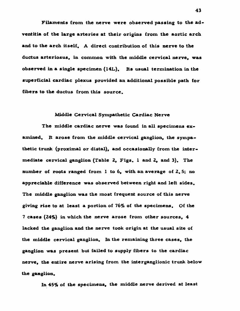
Sympathetic Trunk
A brief summary of the major features of the sympathetic
trunk of the fetus will be presented first to serve as a useful back
ground for later descriptions* In addition* the intermediate cervical
ganglion will be considered in more detail because of its contro
versial significance.
The sympathetic trunk was found to be completely developed
and relatively large (Figs. 1 and 2). In the cervical region the trunk
contained an average of 4 ganglia* with a variation of from 3 to &,
The superior and inferior cervical ganglia were the most constant
with regard to sine* shape* position* and occurence. The inferior
was frequently fused with the 1st thoracic ganglion in which case it
is termed the stellate ganglion. Between the superior and inferior
ganglia* the cervical trunk contained two additional ganglia, the mid
dle and intermediate. Each of the latter ganglia was occasionally
divided into two or three independent enlargements.
The superior cervical sympathetic ganglion* oval or fusiform
in shape* was constantly found* and extended from the base of the
skull to a point approximately opposite the 2nd cervical vertebra
(Figs. 1 and 2). It was intimately associated on its lateral side
with the vagus nerve or the nodose ganglion. It had communications
with the upper 3 or 4 cervical spinal nerves* and sent branches
along both the internal and eternal carotid arteries* forming the
3 4
To S U P LARYNG. N . ~ ‘r
VHNT- C-A ROT I D N's ' - — To p h a r y n g e a l p l e x u s
2 ^ - V - aGUS c o m m u n i c a t i o n s
SUR CERV, G.
S U P CERV. SYM. CARD. N.
MID CERV. SYM CARD. N.-
A C C E S S O R Y G's
SYM. CARD. BRS. ANT.To AORTIC ARCH i_ C Q
jTHYROID
MID CERV, G.
NTERMEDCERV. G C8
ANSA
Contribution AT Ao.ion AT From —
The Right
Sub Cord P lex
Inf. Cerv.
T.G ^r- T 4 loop'^Oof the Vogu^
R.A./
V ERT EBR A L A. SUB. A.
V s U B C L A V I A
- S T E L L A T E G.
PROX. SUB. PL EX.
T_2_THOR. SYM CARD.Ns
_ Plexu s upon T 3 JJuctus
A rter io su s
L. P u lm on a ry A.
N .o f Sup-Plexus :ingD uc tus
-Thor. Sym Card N ’s A ort ic Plex.
F ig u r e I.

TO T H E V A G U S ‘v p C I —
S U R CERV. S YM. C A R D
(with a c c e s s o r y C3,©.) y
TO INT. CAROTID PLEX.
SUP. CERV. G.
E X T .
C A R O T I DP L E X U S E S
M I D . CERV. S YM . G.
S U B . A
S T E L L A T E G.
T H O R A C IC SYM.~ C A R D . N .
MID. CERV. SYM. CARD. N.
ACCESSORY G. IN COMMON CERV. CARD. N.
R IG H T COM. C A R O T I D A .A N S A S U B C L A V I A
INF. CERV. S Y M . CARD. N.
R E C U R R E N T N.
C A R O T I D B I F U R C A T I O N
TH Y R O ID
C O M P O U N D N. TO \ \ T HE S U P E R F I C I A L
CARD. P L E X .
FIGURE 2

sa
corresponding carotid plexuses. Other branches passed from the
ganglion to the pharyngeal plexus, to the larynx, thyroid gland, and
the common carotid artery. A detailed description o£ its cardiac
branch will be given later.
The middle cervical ganglion was present in 8 6 % of the sym
pathetic trunks examined, but varied considerably in size, shape,
and position (Figs. 1. 2 and 8 ). It was often very small and frequently
intimately related to the inferior thyroid artery, usually at the level
of the 6th cervical vertebra. It communicated most frequently with
cervical spinal nerves 4. 5 and 6 . although communications with 3
and 7 were also observed. In one-third of the specimens its lower
pole was joined by the ansa subclavia. The ganglion usually gave
branches to the vessels in close proximity, especially the inferior
thyroid artery. Its contribution to the middle cervical sympathetic
cardiac nerve will be considered later.
The presence of a ganglionic enlargement of the cervical
sympathetic trunk between its classically described middle and in
ferior ganglia was observed in 83% of the specimens (Figs. 1. 2.
3A. B. and C). It was usually located opposite the 7th cervical
vertebra or slightly higher (Table 1). anterior and medial to the
vertebral artery or occasionally laterally on that vessel. It was
designated the intermediate cervical ganglion. In those cases in
which the intermediate ganglion was not observed per se. a portion
of the inferior cervical ganglion presented features which corresponded

P H R E N I C N.
M I D . C E R V. G .
COM. SP. N. C O M M U N I C A T I O N
S T E L L A T E G.
SUP. C E R V . SYM. CARD. N.
M I D . C E R V , CARD. SYM, N.
I N T E R M E D . C E R V . G.
C 7 N F . CERV. S YM . CARD N .
A N S A S U B C L A V I A
S U B C L A V .
SUP. C E RV C A R D . N.
MI D . CE RV . G
p — To THYROID P L E X U S
MID. CERV. C A RD . S Y M . N
IN TERMED . CERV. ^
* JS* 'iF z - \rs V E R T E B R A L A
ANSAE V E R T E B R A L E S
A N S A S U B C L a V i A ;
INF. CERV. CARD S YM. N.
p r o x . subclov, plex
S T E L L A T E G ,
/ H f ' COM. SP N. /■COMMUNICATION
F I G U R E 3
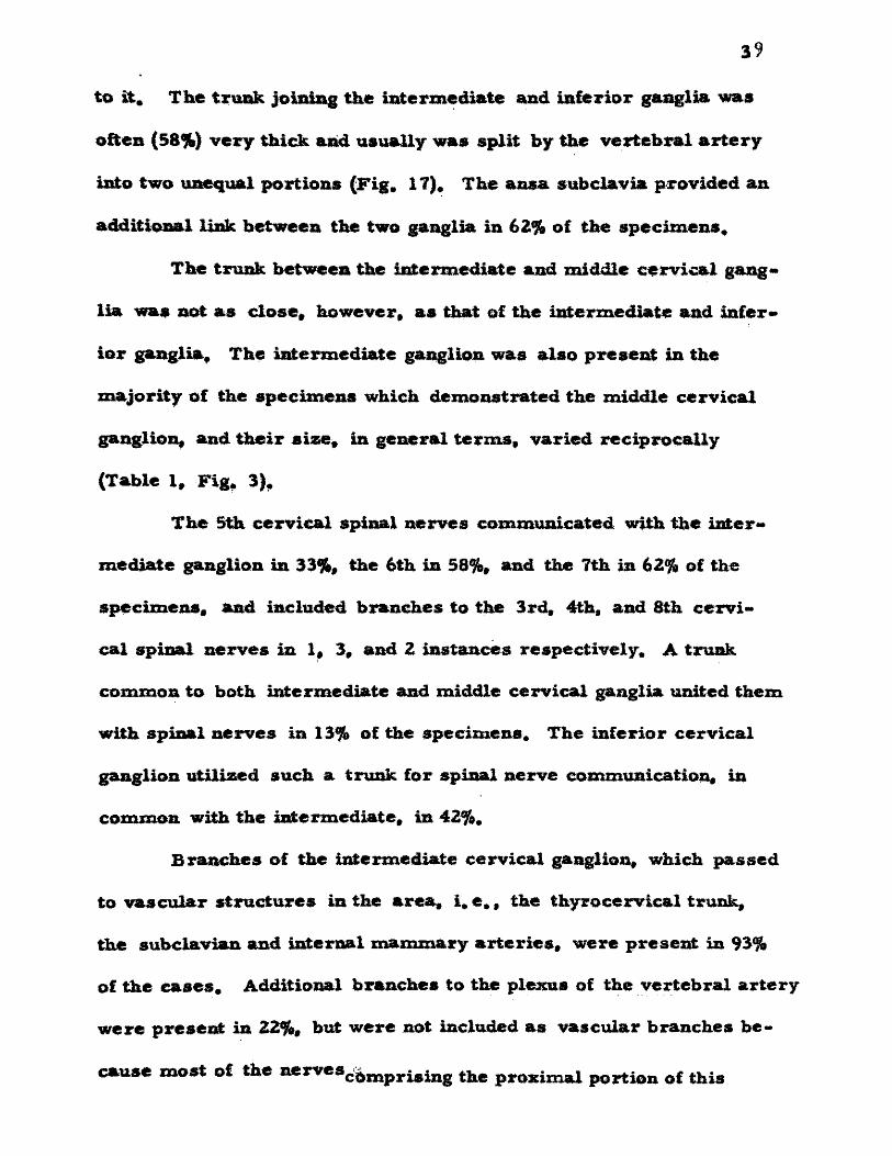
to it. The trunk joining the intermediate and inferior ganglia was
often (58%) very thick and usually was split by the vertebral artery
into two unequal portions (Fig. 17), The ansa subclavia provided an
additional link between the two ganglia in 62% of the specimens.
The trunk between the intermediate and middle cervical gang*
lia was not as dose, however, as that of the intermediate and infer*
ior ganglia. The intermediate ganglion was also present in the
majority of the specimens which demonstrated the middle cervical
ganglion, and their size, in general terms, varied reciprocally
(Table 1, Fig, 3),
The 5th cervical spinal nerves communicated with the inter*
mediate ganglion in 33%, the 6 th in 58%, and the ?th in 62% of the
specimens, and included branches to the 3rd, 4th, and 8th cervi
cal spinal nerves in 1, 3, and 2 instances respectively. A trunk
common to both intermediate and middle cervical ganglia united them
with spinal nerves in 13% of the specimens. The inferior cervical
ganglion utilised such a trunk for spinal nerve communication, in
common with the intermediate, in 42%.
Branches of the intermediate cervical ganglion, which passed
to vascular structures in the area, i. e ., the thyrocervical trunk,
the subclavian and internal mammary arteries, were present in 93%
of the eases. Additional branches to the plexus of the vertebral artery
were present in 22%, but were not included as vascular branches be
cause most of the nervcscomprising the proximal portion of this
m
plexus were given of£ to the cervical spinal nerves within the verte
bral canal. Occasional branches from the ganglion were observed
passing to the esophagus and trachea, and in one instance a branch
was noted which connected with the phrenic nerve.
Dissection 9R was not included in the above data, although
an intermediate cervical ganglion was present with branches corre
sponding to those described above, because of the presence of an
anomalous right subclavian artery.
No essential difference between the two sides in the various
features of the intermediate ganglion was observed.
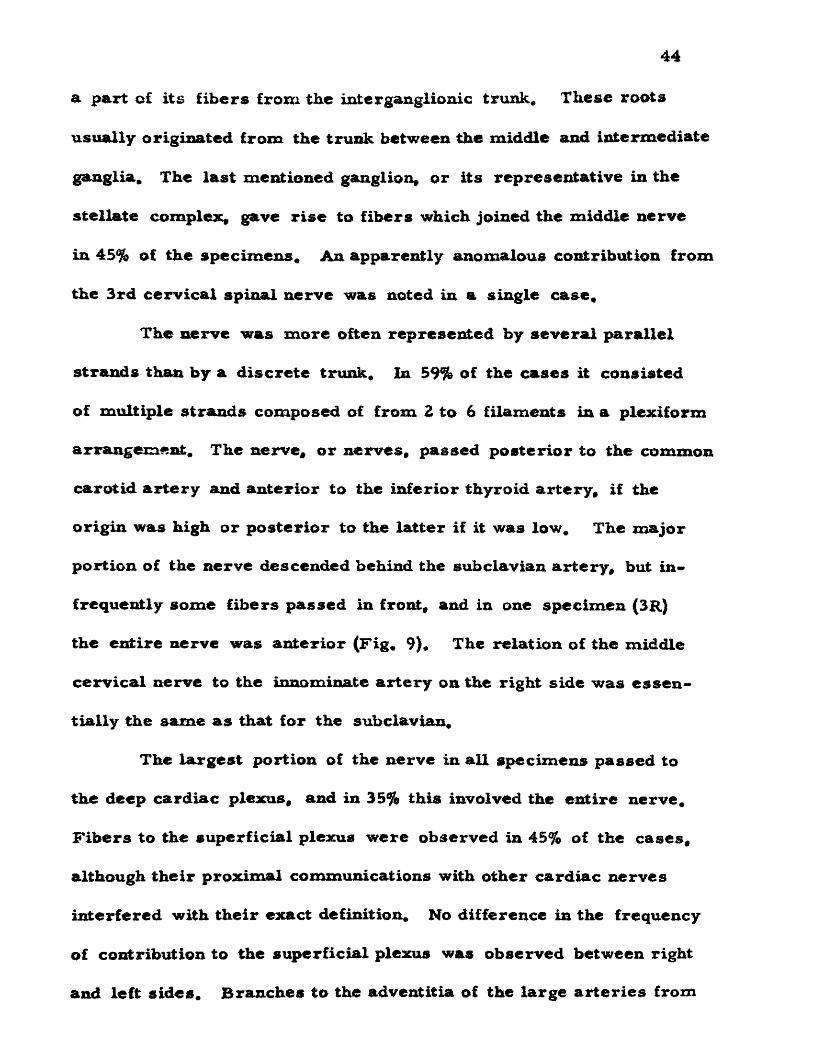
The inferior cervical ganglion was frequently distinguishable
as such, but in most instances only a slight constriction separated
it from the 1st thoracic (Figs, 1, 2, and 3). The large mass thus
formed, the stellate ganglion, also was fused with the intermediate
cervical ganglion and the 2nd thoracic ganglion in some instances
(Figs, 16 and 17), This elongated ganglionic complex was located
on the anterior surface of the head of the 1st rib immediately post
erior to the subclavian artery. Its relatively constant spinal nerve
communications were with the 7th and 8th cervical and 1st thoracic;
additional communications were occasionally observed reaching as
high as the 4th cervical and as low as the 3rd thoracic spinal nerves.
Vascular branches passed to the plexuses about the thyrocervical
trunk, internal mammary artery, and distal part of the subclavian.
Through the latter plexus, a branch occasionally communicated with

41
the phrenic nerve. An additional large branch usually accompanied
the vertebral artery. The ansa subclavia, a nerve looping around the
subclavian artery and coming from the middle or intermediate gang**
lion, joined the interior cervical ganglion on its anterior surface
near the origin of the ramus to the 1st thoracic nerve. The cardiac
nerves from the inferior ganglion will be considered later.
The sympathetic trunk in the upper thoracic region consisted
of a series of ganglia, located between the ribs, and the intergang-
lionic trunks which crossed the anterior surfaces of the heads of the
ribs (Figs. 1, 2, and 14). Adjacent ganglia were sometimes fused,
and when such was the case, the resulting structure was found on
the intervening rib. As mentioned above, the 1st and sometimes the
2nd thoracic ganglia were incorporated in the stellate ganglion. The
ganglia communicated with their corresponding spinal nerves, and
occasionally with those of adjacent levels. Bach ganglion usually gave
rise to branches which passed to the mediastinal viscera, although
such branches were as often derived from the inter ganglionic trunks.
The latter structures were sometimes doubled.
Cervical Sympathetic Cardiac Nerves
Superior Cervical Sympathetic Cardiac Nerve
The superior nerve was present in 8 6 .3% of the specimens
(Table 2). It originated by ope_pr more roots, with an average of
two, from the lower pole <f§ ^ A£upprior cervical ganglion or the

4 2
sympathetic trunk immediately below (Figs. 1 and 2), A root from
the trunk was present in 48%, whereas, the root from the superior
ganglion was observed in all but one case. In this instance, the
entire nerve arose from the trunk 6 . 5 cm. below the superior cervi
cal ganglion. In 20% of the specimens there was an additional root
from the external branch of the superior laryngeal nerve of the vagus.
The superior cardiac nerve descended within the carotid sheath
posterior to the internal and common carotid arteries (Figs. 15, 16,
and 17). It gave numerous small filaments to the arteries, which
were destroyed as the dissection proceeded. The nerve was usually
closely associated with the lateral aspect of the thyroid gland. In
dissection 14R it passed within the connective tissue sheath of the
gland. The nerve descended anterior to the inferior thyroid artery
in every case except one (13L>).
On the left side, all or part of the superior cardiac nerve
passed anterior to the subclavian artery en route to the superficial
cardiac plexus. In a single case, the nerve passed entirely to the
deep cardiac plexus. On the right side the nerve descended either
anterior or posterior to the bifurcation of the innominate artery
and along the lateral side of that vessel. From that point it passed
to the deep cardiac plexus in 9 2 % of the instances, although a few
fibers occasionally reached the superficial cardiac plexus. In dis
section 9R (anomalous) the nerve passed entirely to the superficial
plexus.
43
Filaments from the nerve were observed passing to the ad-
vent it ia of the large arteries at their origins from the aortic arch
and to the arch itself. A direct contribution of this nerve to the
ductus arteriosus, in common with the middle cervical nerve, was
observed in a single specimen (141*), Its usual termination in the
superficial cardiac plexus provided an additional possible path for
fibers to the ductus from this source.
Middle Cervical Sympathetic Cardiac Nerve
The middle cardiac nerve was found in all specimens ex
amined. It arose from the middle cervical ganglion, the sympa
thetic trunk (proximal or distal), and occasionally from the inter
mediate cervical ganglion (Table 2, Figs. 1 and 2, and 3). The
number of roots ranged from 1 to 6 , with an average of 2 .5; no
appreciable difference was observed between right and left sides.
The middle ganglion was the most frequent source of this nerve
giving rise to at least a portion of 76% of the specimens. Of the
7 cases (24%) in which the nerve arose from other sources, 4
lacked the ganglion and the nerve took origin at the usual site of
the middle cervical ganglion. In the remaining three cases, the
ganglion was present but failed to supply fibers to the cardiac
nerve, the entire nerve arising from the interganglionic trunk below
the ganglion.
In 45% of the specimens, the middle nerve derived at least
44
a part of its fibers from the inter ganglionic trunk. These roots
usually originated from the trunk between the middle and intermediate
ganglia. The last mentioned ganglion, or its representative in the
stellate complex, gave rise to fibers which joined the middle nerve
in 45% of the specimens. An apparently anomalous contribution from
the 3rd cervical spinal nerve was noted in a single case.
The nerve was more often represented by several parallel
strands than by a discrete trunk. In 59% of the cases it consisted
of multiple strands composed of from 2 to 6 filaments in a plexiform
arrangement. The nerve, or nerves, passed posterior to the common
carotid artery and anterior to the inferior thyroid artery, if the
origin was high or posterior to the latter if it was low. The major
portion of the nerve descended behind the subclavian artery, but in
frequently some fibers passed in front, and in one specimen (3 R)
the entire nerve was anterior (Fig. 9), The relation of the middle
cervical nerve to the innominate artery on the right side was essen
tially the same as that for the subclavian.
The largest portion of the nerve in all specimens passed to
the deep cardiac plexus, and in 35% this involved the entire nerve.
Fibers to the superficial plexus were observed in 45% of the cases,
although their proximal communications with other cardiac nerves
interfered with their exact definition. No difference in the frequency
of contribution to the superficial plexus was observed between right
and left sides. Branches to the adventitia of the large arteries from
4 5
the aortic arch and to the arch itself were observed in 2 0% of the
specimens and occurred twice as frequently on the left as on the
right.
Inferior Cervical Sympathetic Cardiac Nerve
This nerve was observed in all dissections but was small
on the right side in 33%, It received fibers from the inferior cer
vical or stellate ganglion in 93%, and from the intermediate cervical
ganglion, or the portion of the stellate complex: which represented
it in 72%, with no appreciable difference between the two sides
(Table 4, Figs, I, 2, and 3}, No contribution was given by the ansa
subclavia on the right; if was observed in 28% of the cases on the
left. Branches of the interganglionic trunk sometimes took part in
the formation of this nerve, but because these branches arose im
mediately adjacent to either the intermediate cervical or the stellate
ganglion, they have been recorded as branches of the nearest gang
lion, The number of roofs forming the nerve varied from 1 to 6
and averaged 2, 5 on the right and 3. 2 on the left.
The parallel strands composing the inferior cardiac nerve
descended into the superior mediastinum in more or less intimate
association with the subclavian artery, on the left and the sub
clavian and innominate arteries on the right (Figs, 1, 2, and 3),
A series of loops or communications between the strands of the
nerve formed a plexus along the subclavian artery. The plexus
occurred in 85% of the specimens on the left and in 29% on the

46
right, and was limited, on the left, primarily to that portion of the
artery between its origin and its major branches (Figs. 1, 3, 16,
and 17). The plexus varied considerably in sine and complexity with
the greater portion of the nerves posterior to the subclavian. On
the right, the branches on the anterior and superior aspect were
usually regrouped at the proximal end of the subclavian and then
wound around the innominate artery to gain its lateral side.
From the lower portion of the stellate ganglion a slender
nerve arose, which often dissociated itself from the other strands
of the inferior cervical nerve. It descended along the thoracic duct
on the anterior and lateral aspect of the upper two thoracic vertebrae
to the cardiac plexus (Figs. 10, 14, and 17),
The primary termination of the inferior cardiac nerve on
both sides was the deep cardiac plexus, with 96. 5% of the nerves
contributing at least a portion of their fibers. On the left there
was also a contribution to the ductus arteriosus in all cases, al
though in two the contribution was small. Fibers, varying in sine,
were given to the central portion of the superficial cardiac plexus
by the left nerve in 60% of the specimens. On both sides, branches
passed to the adventitia bf the aortic arch and the large arteries at
their origin from it. The nerve on the right may have contributed
fibers to the superficial cardiac plexus in 35% of the cases by way
of its communications with other cardiac nerves.

47
An unusual course of the nerve on the right was observed in
dissection 9R, in which an anomalous right subclavian artery arose
from the aorta distal to the left subclavian and passed posterior to
the esophagus (Fig* 4), In this specimen* the right superior cervi~
cal sympathetic cardiac nerve ended entirely in the superficial plexus
and the middle went essentially to the deep plexus* although sending a
branch with the inferior cardiac nerve to form a plexus about the
anomalous artery. The inferior nerve arose from the intermediate
and inferior cervical ganglia* and with the branch from the middle
nerve, formed a plexus which followed the anomalous vessel post
erior to the esophagus to its origin from the aorta (Fig. 4). At that
point* the nerves regrouped into two trunks which passed anteriorly
on the medial surface of the descending aorta to the deep cardiac
plexus. Vagal cardiac nerves were unaffected by this anomaly.
Cardiac Nerves of the Intermediate Ganglion
Although the cardiac branches of the intermediate cervical
ganglion formed* in part, the middle or inferior cardiac nerves* as
has been described above, it should be noted that 22% of them passed
directly to either the deep or superficial cardiac plexuses unassociated
with any other cardiac nerves*
Thoracic Sympathetic Cardiac Nerves
A series of visceral branches from the upper portion of the

S u p e r i o r Cferv- icc i
G n f . g l i o r .
S u p e r i o r C e r v i c a l C a r d i a c
S y m p a t h e t i c N e r v e
Middle Cervical Ganglion
Thyrocervical Trunk Interm ed ia te Cervical
Ganglion Inferior Cervical
Ganglion
1st T h orac ic Ganglion
Right Subclav ian
Artery
A sc en d in g Aorta
Middle Cervical C ard iac S ym p a th et ic N e r v e s
Branch of the Vagus Nerve
/ T \ Inferior Cervical « - V Cordiac Sym pa
t h e t ic N erves
To th e D e e p Cordiac Plexus
PulmonaryTrunk
Figure 4
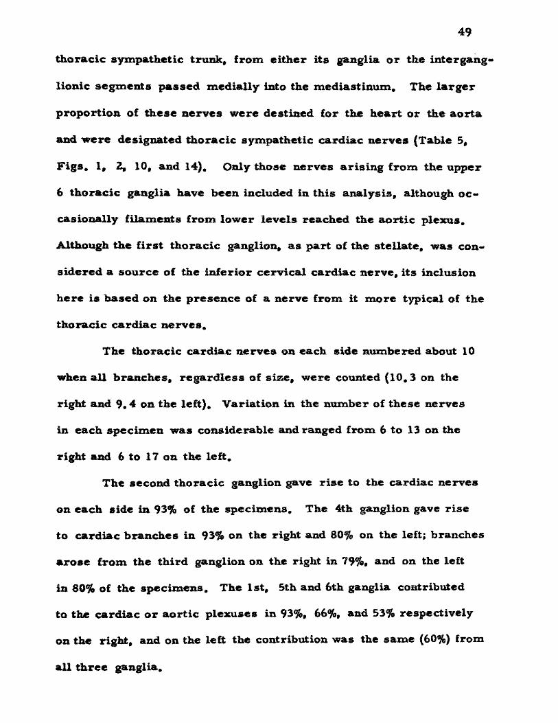
49
thoracic sympathetic trunk, from either its ganglia or the intergang-*
lionic segments passed medially into the mediastinum. The larger
proportion of these nerves were destined Cor the heart or the aorta
and were designated thoracic sympathetic cardiac nerves (Table 5,
Figs. 1, 2, 10, and 14). Only those nerves arising from the upper
6 thoracic ganglia have been included in this analysis, although oc
casionally filaments from lower levels reached the aortic plexus.
Although the first thoracic ganglion, as part of the stellate, was con
sidered a source of the inferior cervical cardiac nerve, its inclusion
here is based on the presence of & nerve from it more typical of the
thoracic cardiac nerves.
The thoracic cardiac nerves on each side numbered about 10
when all branches, regardless of size, were counted (1 0 .3 on the
right and 9,4 on the left). Variation in the number of these nerves
in each specimen was considerable and ranged from 6 to 13 on the
right and 6 to 17 on the left.
The second thoracic ganglion gave rise to the cardiac nerves
on each side in 93% of the specimens. The 4th ganglion gave rise
to cardiac branches in 93% on the right and 80% on the left; branches
arose from the third ganglion on the right in 79%* and on the left
in 80% of the specimens. The 1st, 5th and 6 th ganglia contributed
to the cardiac or aortic plexuses in 93%, 6 6 %, and 53% respectively
on the right, and on the left the contribution was the same (60%) from
all three ganglia.
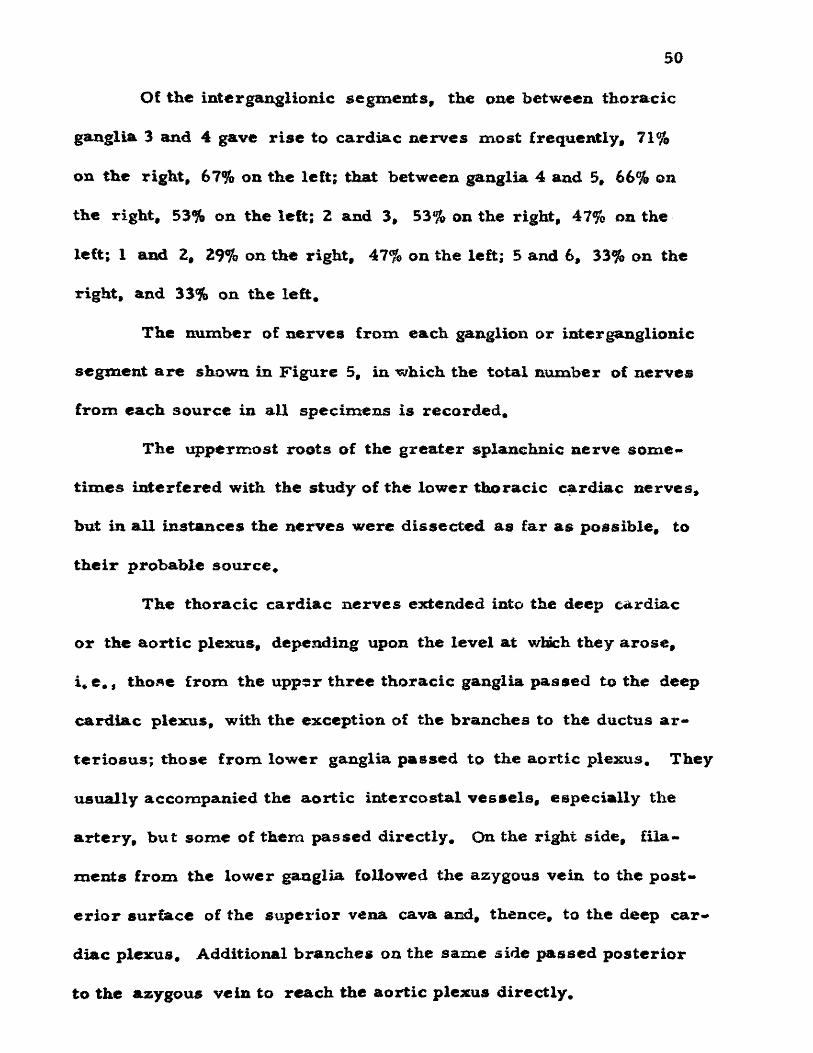
50
Of the inter ganglionic segments, the one between thoracic
ganglia 3 and 4 gave rise to cardiac nerves most frequently, 71%
on the right, 67% on the left; that between ganglia 4 and 5, 6 6 % on
the right, 53% on the left; 2 and 3, 53% on the right, 47% on the
left; 1 and 2, 29% on the right, 47% on the left; 5 and 6 , 33% on the
right, and 33% on the left.
The number of nerves from each ganglion or inter ganglionic
segment are shown in Figure 5, in which the total number of nerves
from each source in all specimens is recorded.
The uppermost roots of the greater splanchnic nerve some
times interfered with the study of the lower thoracic cardiac nerves,
but in all instances the nerves were dissected as far as possible, to
their probable source.
The thoracic cardiac nerves extended into the deep cardiac
or the aortic plexus, depending upon the level at which they arose,
i, e ,, those from the upper three thoracic ganglia passed to the deep
cardiac plexus, with the exception of the branches to the ductus ar
teriosus; those from lower ganglia passed to the aortic plexus. They
usually accompanied the aortic intercostal vessels, especially the
artery, but some of them passed directly. On the right side, fila
ments from the lower ganglia followed the azygous vein to the post
erior surface of the superior vena cava and, thence, to the deep car
diac plexus. Additional branches on the same side passed posterior
to the azygous vein to reach the aortic plexus directly.

j 1
RIGH T SIDE
20-
18 -
L E F T S I D E Fi gur e 5

52
The majority of thoracic cardiac nerves on the right side
passed to the deep cardiac plexus, while those on the left passed
to the deep and the aortic plexuses in approximately equal numbers.
Some o£ the fibers on the left descended anterior to the aorta and
reached the superficial cardiac plexus. The aortic and deep cardiac
plexuses communicated on the medial side of the aorta just below
the arch.
Intercommunications of the Sympathetic Cardiac Nerves
Communications between the various sympathetic cardiac nerves
were frequently observed. These were divided into three main groups:
intercervical, cervicothoracic, and interthoracic.
Communications involving at least two of the cervical car*
diac nerves were present in 93% of the specimens and most frequently
involved the superior and middle nerves. These nerves communi
cated in 8 8 % of the specimens in which both were present. Commun
ications between the middle and inferior cardiac nerves were observed
in 6 6 % of the specimens, and although no appreciable difference in
frequency of communication on right and left sides was noted, the
size of the communication was appreciably smaller on the left. The
superior and inferior nerves communicated (in part) in 16% by a
branch of the former nerve to the proximal subclavian plexus, and
in still other instances by a common cervical cardiac nerve.

53
The occurrence oC a common trunk formed by the fusion of
several or all of the cervical candiac nerves was observed in 35% of
the specimens and always included the middle nerve, The trunk was
present on both sides with equal frequency, but the complex of nerves
usually appeared more compact on the right side. The common trunk,
instead of continuing to the cardiac plexus per se, usually separated
into 2 to 4 terminal branches. The termination of these branches
varied according to the nerves which were incorporated in the trunk,
but most of them ended in the deep cardiac plexus.
The cervicothoracic communications involved portions of the
inferior cervical cardiac nerves, often through branches of the proxi
mal subclavian plexus, and the uppermost thoracic cardiac nerves.
It was usually observed, however, that the portion of the inferior
nerve, if it can be so called, most frequently involved was derived
from the inferior portion of the stellate ganglion corresponding to
the first thoracic ganglion. In 47% of the specimens on the left,
and in 2 1% on the right, the contribution from the inferior cervical
nerve took part in the formation of a well defined collateral chain or
trunk (Fig, 10), In the other cases a simple union of a branch of the
stellate ganglion with a branch of the second thoracic ganglion occurred,
Interthoracic nerve communications were a relatively constant
feature. The degree of complexity varied considerably, being more
marked among the upper thoracic cardiac nerves. There were ex
amples of a simple exchange of fibers between adjacent nerves, arch

54
formation by complete fusion of a series of adjacent nerves, forma
tion of a collateral trunk roughly paralleling the descending; aorta, and
formation of a complex network of nerves along the lateral sides of
the vertebral bodies (Figs. 1, 2, 9, 10, 14, and 17). The nerves
from the 4th thoracic and lower ganglia usually passed directly to
the aortic plexus.
On the right side, the nerves passed directly to the cardiac
or aortic plexuses in 53% of the bodies examined. In 2 0 % of the
remaining specimens, a collateral trunk was observed. A plexiform
arrangement of the nerves was found in an additonal 2 0 %, and a single
example of arch formation occurred. On the left side a collateral
trunk was found in 47%, arch formation in 20%, direct branches in
20%, and plexiforzn networks in 13%.
Accessory Ganglia associated with
Sympathetic Cardiac Nerves
Small ganglionic enlargements of the cardiac nerves were
variable in their frequency of occurrence, position, and size (Figs.
1, 2, 9# 10, and 13). Occasionally the enlargement was found in
the course of a nerve unassociated with other nerves in the vicinity
but, more frequently, the ganglia were found at the union of several
cardiac nerves or their branches. Such ganglia were sometimes
noted at the terminations of large nerves which divided into numerous
smaller branches. Only those ganglia which were completely

55
separated from the sympathetic trunk are considered here, although
the enlargement of a nerve at its origin, as if a small ganglion were
present, was often observed.
Accessory ganglia, which appeared to be confined to the su
perior cardiac nerve were observed in 36% of the specimens. By
union with the middle cardiac nerve the superior was associated
with an additional 5 ganglia on the left. Occasionally two ganglia
were observed in the course of a single nerve. The point in the
superior nerve at which these ganglia appeared was either near its
origin or at the entrance of the nerve into the thorax, with the
majority in the latter position.
Accessory ganglia of the middle cardiac nerve alone were
found in 20% of the specimens on the left and 13% on the right. In
other specimens, 33% on the left and 20% on the right, there were
ganglia common to both superior and middle nerves. A ganglion
was found, therefore, in the latter nerve on the left in 57% of the
specimens examined and on the right in 37%. In this nerve, the
ganglion usually lay opposite the 7th cervical vertebra.
The accessory ganglia associated with the inferior cardiac
nerve comprised those which were found in the nerves of the sub
clavian plexus (41% of the specimens on the left, none on the right)
and those associated with the thoracic cardiac nerves (2 0% on the
left and 13% on the right). The ganglia associated with the nerves
of the subclavian plexus are characteristically small but well

56
defined (Figs. 9 and 13).
Including the ganglia with both inferior cervical and thoracic
nerve contributions, accessory ganglia of the thoracic cardiac nerves
were found in 53% of the specimens on the left and 40% on the right.
These ganglia were observed most frequently in association with the
nerves, from the 1st, 2nd, and 3rd thoracic ganglia. Those speci
mens which had a collateral chain or trunk usually had two or three
ganglia within the chain.
Cardiac Nerves of the Vagus
The cardiac branches arising from the vagus above the 6th
cervical spinal nerve were considered part of the superior cervical
cardiac nerves, whereas, those arising below this point, but above
the lower border of the subclavian artery are included in the inferior
nerves (Table 6 , Figs. 6 and 7).
Superior Cervical Cardiac Nerves
Although small upper cervical branches of the vagus, of prob
able cardiac distribution, were found in 75% of the specimens exam
ined, only 14% formed a relatively independent branch. In all other
cases the vagal representative joined the superior sympathetic car
diac nerve. It varied in frequency and size on the two sides, being
present in 71% on the left and 79% on the right, half the nerves on
both sides being very small.

G L O S S O P H A R Y N G E A L N.
HYPOGLOSSALTO PHARYNGEAL P L E X .
To CAROTID BIFURCATION— &
TO SYMPATHETIC
SUPERIOR LARYNGEAL- SYM.
SUP. CERV. SYM. CARD. N;
VAGUSTRUNK
THYROID
SUBCLAVIAN
INF. CERV. CARD. N. Of VAGUS
LEFT COMMON CAROTID ~1
THOR. CARD. ■Njlf VAGUS
AORTAD.A.
SUP-/CARD. P LEX .
PULMONARY TRUNK (
R.A,
FIGURE 6
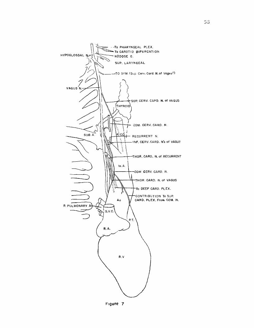
I“* o3 o
>■ - - T o P H A R Y N G E A L P L E X .
:=:=a»‘ To CAROTI D B I F U R C A T I O N
‘ N O D O S E G.H Y P O G L O S S A L
S U P . l a r y n g e a l
-TO SYM. ( S u p . Cerv, Card. N . o f V a g u s ? )
V A G U S N.-
S UP. CERV. CARD. N. o f VAGUS
COM. CERV. CARD. N .
p ::C'C| J— R E C U R R E N T N.
\ - V \ I NF. CERV . CARD. N ’s o f VAGUS
S U B . A.
■THOR. C AR D . N. o f RECURRENT
In. A.
COM CERV. CARD. N.
■THOR. C A R D . N . o f VAGUS
To OE E P CARD. P L E X .
- C O N T R I B U T I O N To SUP. C A R D . P L E X . F rom COM. N .A o
R. P U L M O N A R Y
S. V. C.
P.T.
R . A .
R .V
Figure 7

59
The termination of the fibers belonging to the superior nerve
was difficult to determine because of communications with other
nerves* Branches were traced to the aortic arch, the subclavian
and common carotid arteries at their origins, and the cardiac plex
uses, especially the deep*
Inferior Cervical Cardiac Nerve
An inferior cardiac nerve was observed in 96% of the speci
mens examined* It usually represented the major contribution of the
vagus to the heart and often arose by several roots. The most fre
quent site of origin, on both sides, was opposite the upper border of
the subclavian artery, although other branches arose from the vagus
near the upper and lower limits set for its origin. In two specimens
(?%), in which it arose from the vagus at the upper limit, 1* e ,, op
posite the 6th cervical spinal nerve, no superior cardiac nerve was
found*
The major portion of the inferior cardiac nerve passed an
terior to the subclavian artery on both sides. On the left, it term
inated in the superficial cardiac plexus in all but one instance, in
which it was small and joined the proximal subclavian plexus, leav
ing the superior nerve to contribute to the superficial plexus. In
4 3 % on the left side, some of its branches terminated in the adven
titia of the aorta at the origin of the common carotid artery, and
in 5 5 % in the plexus upon the ductus arteriosus*
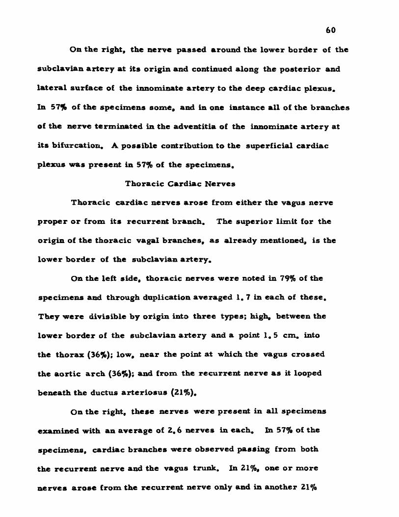
60
On the right, the nerve passed around the lower border of the
subclavian artery at its origin and continued along the posterior and
lateral surface of the innominate artery to the deep cardiac plexus.
In 57% of the specimens some, and in one instance all of the branches
of the nerve terminated in the adventitia of the innominate artery at
its bifurcation, A possible contribution to the superficial cardiac
plexus was present in 57% of the specimens.
Thoracic Cardiac Nerves
Thoracic cardiac nerves arose from either the vagus nerve
proper or from its recurrent branch. The superior limit for the
origin of the thoracic vagal branches, as already mentioned, is the
lower border of the subclavian artery.
On the left side, thoracic nerves were noted in 79% of the
specimens and through duplication averaged 1. 7 in each of these.
They were divisible by origin into three types; high, between the
lower border of the subclavian artery and a point 1.5 cm. into
the thorax (36%); low, near the point at which the vagus crossed
the aortic arch (36%); and from the recurrent nerve as it looped
beneath the ductus arteriosus (2 1 %).
On the right, these nerves were present in all specimens
examined with an average of 2.6 nerves in each. In 57% of the
specimens, cardiac branches were observed passing from both
the recurrent nerve and the vagus trunk. In 2 1 %, one or more
nerves arose from the recurrent nerve only and in another 2 1 %

61
nerves were noted arising from the vagus only.
The terminations of the nerves differed on each side of the
body. On the left side, the nerves which arose high in the thorax
passed solely to the superficial cardiac plexus. Those which arose
from the vagus as it approached the aorta and those from the recur-
rent nerve passed to the deep cardiac plexus by looping beneath the
ductus arteriosus, giving fine branches to its inferior surface. One
nerve, arising from the vagus midway between the high and low types
of origin, passed to the ductus arteriosus where it separated into two
branches, one passing directly to the superficial cardiac plexus, and
the other looping around the ductus to reach the deep plexus.
On the right side the deep cardiac plexus received all of the
cardiac nerves in 43% of the specimens and the major portion of the
nerves in 57%. A relatively small number of the nerves last men
tioned passed to the complex which contributed nerves to both plex
uses.
Communications between Sympathetic
and Vagal Cardiac Nerves
Numerous communications between sympathetic and vagal
cardiac nerves were observed in most specimens; they varied from
complete fusion to simple exchange of small branches. The com
munications comprised those between the vagus, its branches and
the sympathetica, and those between the sympathetic and vagal branches.

6 2
Vago- Sympathetic Communications
The superior cervical cardiac nerve of the vagus was most
frequently (8 6 %) incorporated into the superior sympathetic nerve,
and the remaining nerves (14%) also communicated in part. There
was no appreciable difference between the two aides. This commun-
i cat ion with the superior sympathetic nerve allowed the correspond*
ing branch of the vagus to communicate with the middle sympathetic
nerve also. In addition to these, branches to the middle sympathetic
nerve were observed in 45% of the specimens in which this nerve
was present. The inferior vagal nerve communicated with the
sympathetic less frequently and less completely. On the left side,
it was connected with the superior sympathetic nerve in 54% and,
in these cases, additional communications were noted with the inferior
sympathetic nerve in 38% by way of connections with the proximal
subclavian plexus. On the right, communications with the sympathetic
occurred in 79%f by way of union with the common trunk (essentially
the middle sympathetic nerve) formed in the cervical region. How
ever, in one i stance, the communication was with the superior sym
pathetic nerve alone, and in another specimen there was a small
branch to the inferior sympathetic nerve.
Communications between the thoracic cardiac nerves of the
vagus and the cervical sympathetics varied on the two sides. On
the left they occurred infrequently as small branches to the proximal
subclavian plexus or with branches in or approaching the superficial
63
cardiac plexus. On the right, the communications were found in*
57% of the cases; they consisted of branches to a nerve complex
on the posterior and lateral aspect of the innominate artery. This
complex was derived from the common cervical cardiac trunk.
Sympathetica-Vagal Communications
The superior sympathetic nerve, when present, demonstrated
two types of communication with the vagal cardiac nerves: 1) a
communication high in the neck including all or part of the superior
cervical nerve of the vagus in 34%; and 2} a communication lower
in the neck, opposite the point of crossing of the subclavian artery,
with the inferior vagal nerve in 50% on the left and 17% on the right.
The middle cervical sympathetic cardiac nerve had com
munications with the vagus in 9 0% of the specimens. It gave fibers
to the vagal cardiac branches in 23%, to the left recurrent in 60%,
and to the right recurrent in 36%,
The inferior sympathetic nerve communicated with the cerv
ical cardiac branches of the vagus on the left in 33%, and with the
recurrent nerve in 13%, On the right, the most frequent types of
communication were with the recurrent nerve in 43%, and with the
inferior cardiac nerve of the vagus in 29%,
Superficial Cardiac Plexus
The superficial cardiac plexus was formed by groups of
nerves whose center was found in the concavity of the aortic arch

64
and whose component fibers spread over the superficial surface of
the aorta, pulmonary trunk, and ductus arteriosus. It was divisi-
ble into 1 ) a preaortic portion anterior to the ascending aorta and
the arch, 2) a p re ductal portion laterally (anterior to the ductus
arteriosus and the pulmonary trunk ), and 3 ) a central portion be
tween the aorta and the ductus (Figs, 1, 10, 15, 16, 17),
The nerves to the superficial plexus communicated with each
other as they crossed the anterior surface of the aortic arch, and
in the concavity of the arch formed a network which either dipped
into the cleft between the arch and the ductus arteriosus or, contin
ued upon the anterior surface of the ductus more laterally.
On the left side, in 85% of the dissections in which the super
ior sympathetic cardiac nerve was present, all or a portion of the
nerve reached the superficial plexus, especially the preaortic and
central portions. On the same side, the middle sympathetic nerve
contributed fibers to the plexus in 29%* with possible contributions
occurring in an additional 43%, The inferior sympathetic nerve con
tributed a portion of its fibers to the superficial plexus, especially
to the lateral or preductal portion, in all specimens, although the
contribution was small in two of them. The left thoracic sympa
thetic nerves contributed fibers to the lateral portion of the super
ficial plexus in 47%,
The left inferior cardiac branches of the vagus gave fibers
to the superficial plexus in all cases. In one specimen, however.
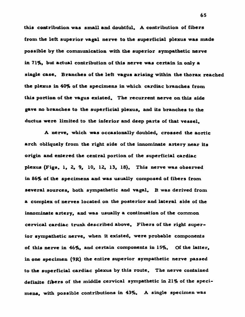
65
this contribution was small and doubtful. A contribution of fibers
from the left superior vagal nerve to the superficial plexus was made
possible by the communication with the superior sympathetic nerve
in 71%, but actual contribution of this nerve was certain in only a
single case. Branches of the left vagus arising within the thorax reached
the plexus in 40% of the specimens in which cardiac branches from
this portion of the vagus existed. The recurrent nerve on this side
gave no branches to the superficial plexus, and its branches to the
ductus were limited to the inferior and deep parts of that vessel.
A nerve, which was occasionally doubled, crossed the aortic
arch obliquely from the right side of the innominate artery near its
origin and entered the central portion of the superficial cardiac
plexus (Figs. I, 2, 9, 10, 12, 13, 18). This nerve was observed
in 8 6 % of the specimens and was usually composed of fibers from
several sources, both sympathetic and vagal. It was derived from
a complex of nerves located on the posterior and lateral side of the
innominate artery, and was usually a continuation of the common
cervical cardiac trunk described above. Fibers of the right super
ior sympathetic nerve, when it existed, were probable components
of this nerve in 46%, and certain components in 15%. Of the latter,
in one specimen (9 R) the entire superior sympathetic nerve passed
to the superficial cardiac plexus by this route. The nerve contained
definite fibers of the middle cervical sympathetic in 2 1 % of the speci
mens, with possible contributions in 43%. A single specimen was

66
found with an obvious contribution from the inferior sympathetic
nerve, and possible contributions from this source were seen in
50%. The inferior cervical cardiac nerves of the vagus gave fibers
to this nerve in 21%, with possible contributions in 45%. The other
branches of the vagus on the right supplied no definite contribution,
although the possibility existed in 23%.
Within the superficial plexus, the contribution from the
right side remained somewhat separate from the other components
of the plexus, passed interiorly, forming a delicate network anterior
to the ascending aorta, and crossed again to the right of that vessel.
Most frequently, the nerves of the lateral portion of the sup
erficial plexus which did not terminate in the adventitia of the ductus
or pulmonary trunk formed a well defined nerve, which descended
along those vessels and passed through the deep cardiac plexus to
end in the left coronary plexus (Figs. 10 and 13). Fibers of the
superficial plexus located on the medial surface of the ductus and
the pulmonary trunk usually joined those fibers from the central por
tion passing to the right coronary plexus. Most of the nerves derived
from the central portion of the plexus also continued toward the
heart between the ascending aorta and the pulmonary trunk to reach
the right coronary plexus. A loop between the right and left coronary
plexuses was often observed passing across the pulmonary trunk at
its origin from the conus arteriosus (Figs. 16 and 17).

6 7
Deep Cardiac Plexus
The deep cardi&c plexus was small and circumscribed; most
of it was situated along the upper border of the right pulmonary artery
(Table 8 , Fig. 8 ), Plexus formation was not evident until the nerves
approached the superior border of the vessel. The center of the
plexus was on the superior border of the right pulmonary arteyy
anterior to the origin of the left bronchus. At this point the plexus
was also posterior to the junction of the ascending aorta and the
arch. The nerves entering into the plexus descended anterior to the
trachea as parallel strands with relatively few intercommunications
below the cervical region. The peripheral limits of the plexus were
difficult to determine because the union and intercommunication of
contributory nerves did not occur at a given point.
The greater part of the deep cardiac plexus was located post~
erior to the transverse pericardial sinus and its upward continuation*
the aortic recess (Fig. 8). This applied particularly to the nerves
on the right. The left extremity of the plexus was in contact with the
posterior surfaces of the aorta, ductus arteriosus, and pulmonary
trunk. The nerves in the latter situation were primarily from the
left side, although a contribution from the right, often large, con-*
stantly reached this area. Frequently, a relatively small nerve, or
group of nerves, reached the posterior surface of the aorta above
the pericardial reflection at the upper extent of the aortic recess and

S u p e r f i c i a l C a r d i a c P l e x u s
s.v.c Asc. Ao. D. A.A o r t i c P l e x u s
Aortic Recess!
Asc. Ao. D. A L.Pul m. A.D eep Cardiac
P lexusS.V.C. Pulm.Tr.
D esc .A o.R.Pulm.
Deep Card. P lexu s
C
S u p e r io r C ard iac P lex u s
A sc. AoS.V.C] Pericard ia l fold of Marshall
T ra n sv e rse .------- ̂ P er id io l S inus
\ R. P u lm .\ Veins
L. Pulm. Veins
Ao.
Deep C ardiac P lexu s
D, A.R.Pulm. A.L. Pulm. A.
B, C ,a n d D c ro ss s e c t io n s
Indicated by l in es on A.PT.R.A.
LA.
A F ig u re 8
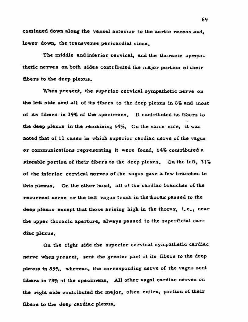
69
continued down along the vessel anterior to the aortic recess and,
lower down, the transverse pericardial sinus.
The middle and inferior cervical* and the thoracic sympa
thetic nerves on both sides contributed the major portion of their
fibers to the deep plexus.
When present, the superior cervical sympathetic nerve on
the left side sent all of its fibers to the deep plexus in 8% and most
of its fibers in 39% of the specimens. It contributed ho fibers to
the deep plexus in the remaining 54%. On the same side, it was
noted that of 11 cases in which superior cardiac nerve of the vagus
or communications representing it were found, 64% contributed a
sizeable portion of their fibers to the deep plexus. On the left, 31%
of the inferior cervical nerves of the vagus gave a few branches to
this plexus. On the other hand, all of the cardiac branches of the
recurrent nerve or the left vagus trunk in the thorax passed to the
deep plexus except that those arising high in the thorax, i. e ., near
the upper thoracic aperture, always passed to the superficial car**
diac plexus.
On the right side the superior cervical sympathetic cardiac
nerve when present, sent the greater part of its fibers to the deep
plexus in 83%, whereas, the corresponding nerve of the vagus sent
fibers in 73% of the specimens. All other vagal cardiac nerves on
the right side contributed the major, often entire, portion of their
fibers to the deep cardiac plexus.

70
The nerves into the right extremity of the plexus, usually
isolated low cervical or thoracic cardiac branches of the right vagus,
passed posterior to the aortic recess and the transverse sinus, di
vided, and continued either anterior or posterior to the right pul
monary artery. Those which passed anterior deviated slightly to
the right and reached the base of the superior vena cava and the
right atrium. The nerves which passed posterior to the right pul
monary artery, still including only those nerves in the right half of
the plexus, extended interiorly to the reflection of pericardium be
tween the transverse and oblique sinuses. Through this septum,
actually the venous mesocardium, the nerves passed to the posterior
surfaces of the atria between the openings of the pulmonary veins.
Nerves from the left half of the deep plexus extended to the
heart anterior and posterior to the right pulmonary artery. Those
anterior to the vessel followed the posterior surface of the aorta to
its base, where they entered into the left coronary plexus. Of those
which passed posterior to the right pulmonary artery, some went
through the venous mesocardium to the atria as described above,
whereas others continued along the posterior surface of the artery
and the pulmonary trunk. Within the pericardial cuff enclosing the
latter vessel and the aorta, the nerves reached the heart where they
entered into the coronary plexuses, especially the left.
In the description of the superficial cardiac plexus, mention
was made of a nerve on the inferior or lateral border of the ductus

71
arteriosus and pulmonary trunk which passed to the left coronary
plexus (Figs, 10 and 13), However, at the upper reflection of the
pericardium of the transverse sinus on the left, a portion of the
nerve passed posterior to the sinus, forming a Slight ridge on its
wall, and penetrated the venous mesocardium to reach the posterior
surface of the left atrium.
Communications between the superficial and deep cardiac
plexuses, although variable, were observed at three points: 1) on
the right side of the aorta, 2) between the ascending aorta, its arch
and the ductus arteriosus or the pulmonary trunk, and 3) on the in*
ferior or lateral surface of the ductus arteriosus.
Nerve Supply of the Buctus Arteriosus
The ductus arteriosus received its sympathetic nerve supply
from cardiac nerves of the lower cervical and upper thoracic: trunk
(Tabled, Figs, 9, 10, 11, 12, 13, 14, 15), Cervical branches of
the left vagus to the superficial cardiac plexus were associated with,
and supplied fibers to the superior and medial surfaces of the ductus,
whereas, thoracic cardiac nerves of the vagus passed to the inferior
and deep portions of the structure,
The most frequent and only constant source of nerves to the
ductus was the inferior cervical sympathetic cardiac nerve (Fig, 11),
In two specimens the nerves from this source were exceedingly fine.
In one of the specimens, dissection 4, the majority of the sympathetic

72
SUP. CERV. SYM . CARD N.
MID. CERV. SYM. CARD. N’S.
ACCESSORY G.
SUP CERV SYM. CARD N,
SUPERFICIAL CARD. PLEX.—
MID. CERV. SYM. G.
INTERMED. CERV. S Y M . G
VERTEBRAL A.
INT. CERV. CARD. N. OF VAGUS
ACCESSORY G.T| G.(STELLATE)
CERV. SYM. CARD. N ( Prox. Subclov. Plex.)
THORACIC SYM. CARD. N'S.
T3<S.PLEXUS OF DUCTUS
ARTERIOSUS
FIGURE 9

73
COROfiRIGHTPLEXUS
THORACIC SYM. CARD. N 't. (Collot. trunk)
PLEXUS upon th tDUCTUS ARTERIOSU8
L . PULMONARY A’t .
LEFT CORONARY PLEXUS
ACCESSORY 0. (Sup. and mid. cerv.
card, n't.)
RECURRENT N.
COM. VAGAL-SYM. N 't.
COMPOUND N. from RIGHT
SUPERFICIAL CARDIAC PLEXUS
THYROtDMLD. CERV. G.
SUP. CERV. SYM. CARD. N.
VAGUS NERVE
THYROID A .INTERMED. CERV. SYM. G. VERT. A.MID. CERV. SYM. CARD. N. INF. CERV. CARD. N. of VAGUS
INF.CERV. SYM. CARD N. (Prox. tubcl.pltx.)
ACCESSORY G.(In 4 t h . c a r d . n . )
Figure 10

74
C o n t r i b u t o r y N e r v e s of t h e
D u c t u s A r t e r i o s u s
p. Cerv. Sympoth. Card. N.
Mid. Cerv. Sympath. Card. N.
■ 3Thor. Collat. Sympath. Tr.
• ‘ n. s \ l
“ IDirect Thor. Sympath. Card. Ns
0 i 2 i 4Black- Pr imary Source Cross Hatched - Strong Clear-Weak
S i 8
OccurrenceF i g u r e I I
10 i 12 i 14

75
fibers to the ductus originated from the common cervical cardiac
nerve on the left, formed essentially from the superior and middle
sympathetic nerves and the cervical cardiac branches of the vagus.
Most of the nerves to the ductus from the common nerve appeared
to be derived from the middle cervical sympathetic cardiac nerve,
but the possibility of contributions from the other components could
not be eliminated. In the second case, dissection 15, the ductus
received most of its innervation from thoracic sources, especially
from the cardiac nerves of the second and third ganglia.
The proximal subclavian plexus, formed by the inferior cerv
ical sympathetic cardiac nerve on the left side, most frequently gave
rise to the nerves -which supplied the ductus arteriosus (Figs. I, 9,
10, 12, 13, 15, 16). Branches on the posterior and lateral surface
of the left subclavian artery near its origin descended to the super
ior border of the aortic arch, crossed its anterior and lateral sur
face, and formed a plexus in the adventitia of the ductus. In dis
section 6, however, a single nerve reached the ductus from the
proximal subclavian plexus on the anterior surface of the left sub
clavian artery (Fig. 9). When the proximal subclavian plexus was
poorly developed, the nerves passed directly to the ductus from
the inferior cervical or stellate ganglion.
The most frequent source of sympathetic fibers to the ductus
next was the upper three thoracic cardiac nerves, most frequently
from the second and third segments of the thoracic trunk, although

76
a branch, from the stellate ganglion, in addition to the inferior cerv
ical cardiac nerve from that ganglion, was also frequently observed.
The nerves followed the intercostal vessels, passing anterior to the
aorta, traversed the aortic plexus, and reached the ductus at its
union with the aorta. These nerves were neither as constant nor as
large as those from the inferior cervical sympathetic cardiac nerve,
Extreme development of the thoracic sympathetic cardiac
nerve attribution to the ductus was observed in dissection IS (Fig,
14), A large branch reached the ductus from the third thoracic
ganglion. Several additional small twigs from the lower portion of
the stellate ganglion, corresponding in this instance, to the first
and second thoracic ganglia descended along intercostal arteries
in the first and second interspaces to the aorta and ductus as de
scribed above.
Occasional filaments passed to the ductus from the super
ior and middle cervical sympathetic cardiac nerves. In one speci
men the superior nerve made a noteworthy contribution through a
communication with the proximal subclavian plexus. In all other
cases, contributions from this nerve were small or doubtful and
reached the ductus through the superficial cardiac plexus.
The branches of the vagus nerve to the ductus arteriosus
were relatively few and small compared to those of the sympathetic.
Vagal branches reached the ductus from the superficial cardiac
plexus. Fibers of the vagus which formed a plexus in the adventitia

77
of the aortic arch occasionally could be traced to the distal portion
of the ductus* Fibers were also encountered passing from the vagus
proper and the recurrent nerve as it passed around the lower border
of the ductus. Occasionally a large nerve from the superficial plexus
looped across the anterior surface of the ductus and at first appeared
to be the major source of its fibers (Figs. 1* 16* and 17). Upon
closer dissection* however* it was found that this loop contributed a
very few small filaments to the ductal plexus.
The nerve branches to the ductus arteriosus formed a plexus
in the adventitia of that vessel at its junction with the aorta* The
contributing nerves either passed anterior to the aortic arch from
the proximal subclavian plexus* crossed anterior to the aorta as
branches of thoracic sympathetic cardiac nerves* or reached the
ductus directly from the adjacent superficial cardiac plexus* vagus*
or recurrent nerves. The smaller fibers of the plexus were* in
general* directed proximally on the ductus to ramify and lose them
selves in its adventitia. Other branches* somewhat larger* con
tinued proximally into a plexus on the pulmonary trunk (Figs. 9* 10*
12* 13* and 14). Occasionally a rather large nerve on the inferior
and lateral borders of the ductus and pulmonary trunk was formed
by a regrouping of the nerves of the plexuses on those vessels.
The larger branch* mentioned above* from the superficial cardiac
plexus* which looped anterior to the ductus* often joined the nerve
just described and together they proceeded to the left coronary plexus.

78
Although these larger nerves gave small filaments to the ductus, they
were less intimately associated with the adventitia of the vessel than
was the intrinsic plexus.
The ductus in two specimens had undergone the initial stages
of anatomical obliteration. The plexus of nerves upon the ductus in
these instances was still visible in the adventitia. Although the size
of the ductus had been materially reduced, the component nerves
supplying it showed no obvious changes except that their arrangement
was mo le compact and had the appearance of being incorporated, for
the most part, into the superficial cardiac plexus.

DISCUSSION
Sympathetic Trunk
The results of the present investigation are in general agree
ment with the classical representation of the sympathetic trunk in
the cervical and upper thoracic regions. Certain slight variations
are probably characteristic of the fetal condition. Although numerous
investigators have used fetal material in similar studies9 few have
made comparisons with adult material, Matsui (* 2 5a and b) and
Naatanen (9 47) have presented systematic studies of the cervical
and thoracic trunk in the newborn. The differences between fetal
and adult sympathetic trunks, pointed out by these investigators and
supported by the present observations, involve the position and rela
tions of the ganglia, such as the short, stout inter ganglionic trunks
in the thoracic region of the fetus.
In agreement with Matsui (• 25b) we found that the superior
cervical ganglion of the fetus was in a relatively high position,
rarely extending below the second cervical vertebra. Its intimate
connection with the nodose ganglion of the vagus and communica
tions with spinal nerves is also confirmed, although no communi
cation with the 6th cervical nerve, mentioned in his report, was
observed.
Our observation of several ganglia between the superior and
inferior cervical ganglia agrees with Matsui who found as many as
5 separate ganglionic enlargements in that position. He termed
79
80
these the intermediate ganglia, but it was apparent that he made this
term synonymous with middle cervical ganglia. They were found
opposite the 6th and Tth cervical vertebrae and were divided into
high and low types respectively. The high type appears to cor res-
pond with oucr middle cervical ganglion with respect to the spinal
nerve communications, but was less frequently noted by Matsui
(61%) than in the present study (86%),
The low type of " intermediate" ganglion of Matsui appears
to be identical with our intermediate ganglion, both as to posi
tion, opposite the 7th cervical vertebra, and frequency, 84% in the
former and 83% in the present study. These observations agree with
the original description of the intermediate cervical ganglion by
Jonnesco (* 23) as well as with Hov&lacque (9 27) and JLaaorthes and
Cassan (* 39). It was not always present as described by the latter,
but might have been represented by a portion of the inferior cervi
cal ganglion,
Lazorthes and Cassan represented the intermediate ganglion
as the perivascular portion of the common cervicothoracic ganglion
with the ansa subclavia and the ansae vertebrates joining the differ
ent portions of the larger ganglion. The present study agrees with
these authors that the intimate relationship between the intermediate
and inferior cervical ganglia is a fairly constant feature, but the
writer believes that the former should be treated a s an entity. It
agrees also that the trunk is constantly split to enclose the vertebral

81
artery. The origin of the ansa subclavia from the intermediate
ganglion occurred in 62% in this study, whereas. Lanorthes and Cassan
considered it to be always present.
This study agrees with Matsui and others (Siwe, * 26. Pick and
Sheehan. * 46. and Jamieson et al. * 52) that the intermediate ganglion
most frequently communicates with the 5th. 6th. and 7th cervical
spinal nerves. The significance of spinal nerve communications
as criteria for identification of sympathetic ganglia in the cervical
region is open to question. It has been observed that the spinal
nerve communications of the classically described cervical ganglia
may overlap as much as two segments. In addition, the vertebral
nerve or plexus, which has its primary origin from the inferior cerv-
ical ganglion, ascends with the vertebral artery and is distributed as
high as to the 4th cervical nerve (Christensen et al. *52). Branches
to the vertebral plexus from the intermediate ganglion were present
in 23% which does not entirely support the contention of L.azorthes
and Cassan {* 39) and Mitchell (* 53) that this is a constant feature of
the ganglion.
Exception is taken to the views of Axford (* 29). Pick and
Sheehan (* 46). and Jamieson et al (* 52) that this ganglion is a dis
placed portion of the middle cervical ganglion. Their interpretations
were based on the spinal nerve communications of the ganglion which,
as pointed out. are unreliable criteria and do not explain the frequent
communi cations between the intermediate ganglion aid the 7th cervical

82
spinal nerve. The intimate association in the inter ganglionic trunk,
the perivascular ansae, and common spinal nerve communications in
dicate that it would be more appropriate to link it with the inferior
than the middle cervical ganglion, although it should be recalled that
in the full term fetus the intermediate cervical ganglion is usually a
well defined, separate ganglion and should be treated as such.
Mitchell (* 53), on the basis of its intimate association with
the vertebral artery, has called this ganglion the vertebral ganglion.
This designation tends to exclude the ganglion from the cervical sym
pathetic chain and overemphasizes its relationship to the vertebral
artery. This term should be reserved for the small ganglia which
are found within the plexus which ascends on that vessel as first de
scribed by Valentin (1841). Finally, if precedence is to be honored,
the term used by Jonnesco should be maintained and confusion with
the small ganglia occasionally occurring in the sympathetic rami
(Wrete, * 35) can be avoided by using the full term, intermediate cerv
ical sympathetic ganglion.
Features of tie inferior cervical ganglion or its representative
in the stellate ganglion were observed to be essentially the same as
classically represented. In the full term fetus, however, the infer
ior ganglion was always separated from the first thoracic ganglion
by a slight constriction (see Matsui, * 25 a).
Our observations on the thoracic sympathetic trunk are in
agreement with those of Matsui (*25 a) and Naatanen (*47). The

83
occasional fusion of adjacent ganglia and splitting of the inter gang**
lionic trunks have been observed in the adult9 but the relatively short,
stout inter ganglionic segments are apparently characteristic of the
fetus, before they are lengthened by growth of the trunk. The spinal
nerve communications of the thoracic sympathetic usually arose from
the ganglia and joined the corresponding spinal nerves, although oc
casionally a ramus arose from the inter ganglionic trunk and passed
to the spinal nerve above or below.
Superior Cervical Sympathetic Cardiac Nerve
The present study confirms, for the most part, previous ac
counts of this nerve and adds statistical data concerning various
features. It is present in 90% of the specimens but only rarely (10%)
is it relatively independent. In the majority of the specimens it is
joined with the middle cervical sympathetic nerve to form a common
trunk, which often consists of several parallel, intercommunicating
nerves.
The site of its termination on the left side in the superficial
cardiac plexus is confirmed. Fibers from this nerve often reached
the deep cardiac plexus, however, and in one instance the entire
nerve terminated in the latter. On the right, the converse was
true, i. e ., the nerve passed to the deep cardiac plexus in all in
stances, although occasional branches to the superficial cardiac plexus
were noted. In a single instance the entire nerve on the right passed

84
to the superficial plexus. The observation of the right nerve passing
to the superficial plexus agrees with Mayer (1794). On the right, it
was noted that this nerve often sends a large number of its fibers to
the innominate artery at its bifurcation. These fibers, many of
them possibly representing the corresponding nerve of the vagus,
which communicates with the sympathetic nerve high in the neck,
are probably concerned with the pressor-receptive function of this
area in lower animals as outlined by Nonidez (* 37) and Hammond
(f 41>.
Although branches of the superior cervical sympathetic car**
diac nerve pass to visceral structures in the neck, the observations
of this study are not in agreement with those of Saccomaono (* 43)
that the cervical sympathetic cardiac branches were materially re*
duced by such branches. Indeed, if the nerve contained an accessory
ganglion, it was often observed to be larger distal to the ganglion.
Middle Cervical Sympathetic Cardiac Nerve
Analysis of the relative frequency of this nerve and its com
munications is an essential contribution in this study. The nerve
was found in all specimens and. in agreement with Scarpa (1794)
and others, it could usually be aptly termed, the great cardiac nerve.
In agreement with earlier writers we observed that the origin of
this nerve was not dependent upon the presence of a middle cervical
ganglion, but arose from the trunk at the usual site of the ganglion

as
when that ganglion was lacking. At its origin and Cor a variable dis
tance beyond, the nerve consisted oC multiple nerves in approximately
60% of the specimens. In 52% a portion of the nerve was derived
from the intermediate cervical ganglion. Such contributions were
relatively small in our material, but Mitchell (* 53) observed that it
occasionally gave rise to the entire nerve.
The classical concept of the distribution of the middle cervi
cal sympathetic cardiac nerve to the deep cardiac plexus was sup
ported by this investigation, but it was observed that a portion of
this nerve, often small, also reached the superficial cardiac plexus
in 45% of the specimens. In 20% of the fetuses, fibers from this
nerve were given to the adventitia of the large arteries at their
origin from the aortic arch, but our impression is that these fibers
are actually of vagal origin.
Inferior Cervical Sympathetic Cardiac Nerve
This nerve was constantly found and was usually represented
by several nerves rather than a single nerve. These nerves fre
quently formed a plexus on the proximal portion of the subclavian
artery, especially on the left. According to Lazorthes and Cassan
(* 39) the inferior sympathetic nerve constantly had its origin from
the intermediate and inferior cervical ganglia, and the ansa sub-
clavia, whereas, in this study only the root from the inferior cervi
cal ganglion was constant. A root from the intermediate cervical
36
ganglion was found in approximately 70% of the specimens and the
root from the ansa in 28%, limited to the left side.
As mentioned9 these roots form the proximal subclavian plexus
more frequently (36%) and better defined on the left than the right
(29%). The plexus has been ignored by most writers on the car**
diac nerves although Hovelacque (? 27) gives a rather complete de
scription of the plexiform nature of the inferior cervical sympathetic
cardiac nerve. It is also represented in the drawings of Scarpa
(1794), although not mentioned in the text, and is also observed in
the work by Kondratjew (• 27). Laz-orthes and Cassan (* 39) empha
sized the fact that the major portion of the inferior sympathetic
nerve was posterior to the subclavian artery, a feature supported
by this investigation, and refer to a " bandelette spirale de
Guillaume1 * with an " external** relationship to the subclavian artery
which apparently corresponds to the portion of the plexus on the lat
eral and inferior aspect of the artery.
Branches of the inferior sympathetic nerve on both sides,
whether part of the proximal subclavian plexus or not, may send
fibers to the superficial cardiac plexus, and all send at least a por
tion of their fibers to the deep cardiac plexus. On the right, the
great majority of the fibers pass to the latter plexus, whereas, on
the left, fibers to the superficial plexus are constantly found in addi
tion to those to the deep cardiac plexus.

87
In a single dissection the presence of an anomalous right sub
clavian artery altered the course of the middle and inferior cardiac
nerves. In this instance the right subclavian artery arose from the
aorta distal to the left subclavian artery, ascended obliquely behind
the esophagus and trachea, and crossed the first rib in an essentially
normal situation. The fact that a portion of the middle and inferior
cardiac nerves in this specimen followed the aberrant vessel to the
deep cardiac plexus may be of particular significance. It suggests
that the sympathetic nerves utilise the large vessels from the aorta
to reach the heart. It also indicates that these elements do not
pass to the heart until after the aortic arches have formed the adult
pattern. If this were not so, the sympathetic nerves would probably
have followed their normal course along the arch prior to its oblit
eration, In the same anomaly, the vagal cardiac nerves were unaf
fected, possibly indicating their earlier extension to the heart. This
agrees with the work of Kuntz (* 20) and Ldcata (’ 54) who note that
the vagal elements are established before the sympathetics, but a
questionable statement is made in the latter paper, that the sym
pathetic elements follow the vagal cardiac nerves. In the same
specimen, the right superior cervical sympathetic cardiac nerve
was isolated and passed entirely to the superficial cardiac plexus,
possibly indicating that if the sympathetic nerves on the right were
segregated, the contribution to the superficial cardiac plexus would be
made by that nerve. This would agree with the observations of Mayer

8 8
(1 7 9 4 ).
The nerve to the heart from the lower portion of the stellate
ganglion, observed in this study, is probably the same as the 4th
cardiac nerve of Valentin (1841) and thenervus cardiacus imus of
Taadler (* 13) and Rauber-Kopsch (* 53). Because of its resemblance
to, and communications with the thoracic cardiac nerves, in the
present study It is considered as the cardiac nerve of the first thor
acic ganglion.
Thoracic Sympathetic Cardiac Nerves
The results of this study agree, for the most part, with the
features of the thoracic sympathetic cardiac nerves which have been
presented in recent years. However, an inherent weakness in the
previous descriptions of these nerves should be pointed out, namely,
the failure to emphasize the presence of visceral branches of the
thoracic sympathetic chain which pass into the mediastinum without
going to the heart or the aorta. The distinction is difficult since the
nerves pass into the mediastinum together, but the nerves which
arise from the medial aspect of the thoracic sympathetic chain,
while in large part destined for the heart or aorta, also contain fibers
which pass to the pulmonic structures and esophagus. If the term
thoracic sympathetic cardiac nerves is reserved for those nerves
which actually reach the cardiac plexus, a picture considerably dif
ferent from the one drawn by Saccomaixno (̂ 43) is seen. Even when

89
all the nerves arising from the upper 6 thoracic ganglia were included
as cardiac nerves, which is essentially true, the total number of these
nerves was considerably less than observed by the author just cited.
In the present study, the number of nerves on each side was observed
to be approximately 10, whereas, Saccoxnanno indicated that I Si to 20
nerves were noted with a cross sectional area twice that of the
cervical sympathetic cardiac nerves. Although no definite measure-
ment of the cross sectional area of the nerves was made in the pres
ent study, after careful examination, the impression gained does not
agree with the above cited findings. The origin of these nerves,
primarily from the third and fourth ganglia, and the fact that the
higher thoracic cardiac branches pass to the cardiac plexus, whereas,
the lower branches first become incorporated in the aortic plexus
are confirmed.
intercommunications of Sympathetic Cardiac Nerves
The observation of numerous communications between adja
cent sympathetic cardiac nerves agrees with virtually all previous
investigations. In contrast, however, it has been possible in many
dissections to trace the elements beyond the communications and to
determine their distribution.
A common cervical cardiac nerve formed by the middle
cervical sympathetic cardiac nerve and the major portion of the
superior cervical sympathetic cardiac nerve was often (35%)

90
observed and agrees with observations of Perman (* 24), Hovelacque
(* 27) and others. The inferior cervical sympathetic cardiac nerve,
especially the left, was observed communicating with the other sym
pathetic cardiac nerves less frequently. When the nerve formed a
plexus on the subclavian artery its only communication consisted of
small branches joining the plexus from the other nerves. The nerve
on the right more frequently joined or gave a portion of its fibers to
the common trunk.
The communications between the thoracic sympathetic cardiac
nerves on the sides of the upper three or four thoracic vertebral
bodies are probably the same as the n akzessorische Nervengebilde*1
of Kondratjew (t 26) who reported a collateral trunk in about one-
third of his specimens. This includes a nerve which descends from
the stellate ganglion (1st thoracic ganglion) and probably represents
the fourth cardiac nerve which was described by Valentin (1841),
Within this plexus or series of arches a collateral trunk is sometimes
distinguished. It usually appears as a continuation of the fourth
cardiac nerve or nervus cardiacus imus (T&ndler *13) and was found
in 40% on the left and 20% of the specimens on the right.
Accessory Ganglia Associated with Sympathetic Cardiac Nerves
A small ganglion such as those described by Valentin (1841)
and Arnold (1851) was observed in the superior cervical sympathetic
cardiac nerve in 40% of our specimens and its greater frequency at

91
the junction of this nerve with other cardiac nerves was also ob
served.
Muller* s (*12) description of the cells making up the gang
lion of Wrisberg, and the junction of the superior cervical sympa
thetic cardiac nerve with the superficial cardiac plexus suggests
the possibility that the small ganglia on this nerve represent small
aggregates of nerve cells which were halted in their migration to
the ganglion. The recent paper by Li cat a (* 54), on the other hand,
suggests that the ganglion of Wrisberg is derived from neuroblastic
tissue which follows, the inferior cervical cardiac branch of the
vagus to the sub-aortic recess. It is probable, however, that the
neuroblastic tissue continues to the heart wall to form the intrinsic
cardiac ganglia, whereas the primordium of the ganglion of Wrisberg
reaches that site by way of the sympathetic nerves.
A small accessory ganglion in the middle cervical cardiac
nerve was noted in approximately one-half the specimens. This
ganglion was found at the junction of several of the cardiac nerves,
often in the common cervical cardiac nerve and probably corresponds
to the one- described by Arnold (1851).
A small ganglion observed in the left proximal subclavian
plexus in 40% but none on the right, was associated with the inferior
cervical sympathetic cardiac nerve.
The small ganglionic enlargements in the collateral trunk
or plexus formed by the upper thoracic nerves in our observations

9 2
support Kondratjew (* 26). The highest of these ganglia was noted at
the junction of the nerve from the stellate ganglion with the collateral
trunk, but probably does not represent the ganglion suprcmum of the
last mentioned investigator, because the latter was described as ap
pearing at the union of a branch from the middle cervical cardiac
serve with the collateral trunk. Additional ganglia of the collateral
chain or plexus were noted in approximately 50% of the specimens
and were located at points of junction of the cardiac nerves with the
chain or with the adjacent cardiac nerves when only a plexus was
observed. This collateral chain may represent an upward exten
sion of the lateral chains of the prevertebral plexus associated with
the abdominal aorta as described by Hartmann-Weinberg (f 26).
The importance of the accessory ganglia mentioned above is
that they represent additional sites for the location of sympathetic
postganglionic neurones which would not be removed when the ordi
nary sympathectomy, experimental or therapeutic, is performed.
Superior Cervical Cardiac Nerve of the Vagus
The cardiac nerves of the vagus were considered along
classical lines with the establishment of a definite level, the 6th
cervical spinal nerve, as a dividing point between its superior and
inferior cervical cardiac branches.
The distinct superior cervical cardiac nerve of the vagus
did not exist in the specimens studied but fibers representing this

93
nerve, including nil branches of the vagus to the heart arising above
the division point mentioned above, joined the superior cervical sym
pathetic cardiac nerve in 64% of the specimens. Only 36% of these
specimens demonstrated fair sined communications and it is more
appropriate to consider the fibers a normal constituent of the super
ior cervical sympathetic nerve. The lack of an isolated superior
cervical cardiac nerve of the vagus supports the previous report of
Duncan (* 30),
Inferior Cervical Cardiac Nerve of the Vagus
Below the 6th cervical spinal nerve the cardiac branches of
the vagus retain greater individuality and are designated the infer
ior cervical cardiac nerves. It was noted that the higher the origin
of the nerve, the more likely was its communication with the sym
pathetic cardiac nerves. Such communications occurred in approxi
mately 50% of the specimens on the left side and in 75% on the right.
The difference in the two sides is probably related to the distribution
of the majority of the nerves on the left to the superficial cardiac
plexus. On the right, the nerve is frequently associated with the
common cervical cardiac nerve which passes to the deep cardiac
plexus.
On both sides filaments to the adventitia of the aortic arch
or the large vessels arising from it may represent fibers of pressor
receptor function supporting in part, contentions by Schumacher (* 02),
perman (t 24) and Duncan (» 30).
94
Thoracic Cardiac Nerves of the Vagus
On the left side, the cardiac nerves were observed arising
from the vagus nerve as it entered the thoracic cavity in 36% of the
specimens, from the nerve as it crossed the aorta in an additional
36%, and from the recurrent nerve in 20% of the specimens. The
first type terminated with the inferior cervical cardiac branches
and probably represents a portion of the latter nerve which is car*
ried into the lower cervical region when growth and extension of
the trunk take place. The lower two types may be grouped together
since their course and termination is the same. Both types from
the vagus proper are ignored in the literature and the statement
is made in most texts that the vagus on the left side does not have
cardiac branches.
On the right, however, cardiac branches from the vagus
within the thorax were constantly found and averaged 2.6 nerves
which were divisible into those which arose from the vagus proper
and those which arose from the recurrent nerve. Although com
munications between the cardiac branches of the recurrent nerve
and the sympathetic occurred, the branches of the vagus proper
usually passed as relatively isolated strands directly to the deep
cardiac plexus. These observations correspond with those classi
cally held regarding the nerves on this side.

95
Vago-Sympathetic Intercommunications
Certain features apparently influence the frequency and degree
of communication between vagal and sympathetic elements which occurs
in virtually all of the specimens. One of these is the distance between
the origin of the nerve and its termination. That is* when a nerve
has an extended course* such as the superior cervical cardiac nerve*
its communications are more frequent. The middle and inferior cervi
cal* and thoracic cardiac nerves of both types show correspondingly
fewer communications as their length decreases. Another feature
is the*intervention of the subclavian artery between vagal and sympa
thetic elements. Thus* those sympathetic cardiac nerves which arise
behind or below the subclavian artery are physically separated from
the vagus or its branches and are less able to effect communications
with it. In the thoracic region* this separation is complete and no
communications between vagal and sympathetic thoracic cardiac nerves
exist.
The presence of the numerous communications between the
various nerves mentioned above conditions any interpretation that
can be made in experimental studies in which those nerves are stimu
lated or sectioned in order to determine their distribution. In a
sense* this study is in agreement with Swan (1830) who considered
the cardiac plexus to extend into the cervical region by virtue of the
numerous communications between the cervical cardiac nerves. On
the other hand* if enough specimens are examined and if careful

96
dissection is made, the individual characteristics of most of the
cardiac nerves can be established.
Superficial Cardiac Plexus
The superficial cardiac plexus was found to conform, for
the most part, with classical descriptions, although certain features
of the plexus may profitably be reanalyzed. The plexus has been de
scribed as occupying the interval between the aortic arch and the
ligamentum arteriosum (ductus arteriosus) and pulmonary trunk.
However, Valentin (1841) included the nerves which crossed anter
ior to the aortic arch (extrapericardial) and those anterior to the as
cending aorta and pulmonary artery (intrape r iea rdial). This, linked
with the presentations of Arnulf (* 39* *49, and * 50) and Hantz {• 51)
describing a preaortic plexus consisting of nerves passing anterior
to the aortic arch, and supported by the findings of the present study,
indicates that the superficial cardiac plexus should be defined as in
cluding all those fibers on the same plane anterior to the aortic arch,
the descending aorta, the pulmonary trunk and the ductus arteriosus.
The plexus so defined is divided into three major portions
according to its relationship to the structures mentioned above. The
portion of the plexus corresponding to the classical description is
designated as the central portion. The nerves anterior to the aorta,
or any portion thereof, are considered the preaortic portion and
97
those upon the ductus arteriosus and the pulmonary artery were con
sidered to be the lateral or preductal portion of the plexus.
In addition to classically described sources, i,eM the left
superior cervical sympathetic cardiac nerve and the left inferior
cervical cardiac nerve of the vagus, the middle and inferior cervi
cal sympathetic cardiac nerves on the left side contributed fibers to
the superficial cardiac plexus, the former through its communica
tions with the superior sympathetic nerve and the latter by its con
tributions to the ductus arteriosus. Scattered references to these
contributions have been made in the literature (Sappey, 1852, Jaques,
* 02, and Arinilf, 1 39).
Is sozne instances, branches of the thoracic sympathetic car
diac nerves reach the superficial plexus by their contribution to the
ductus arteriosus. This supports the report of Kunta and More
house (* 30).
The contributions to the plexus from the vagus, in addition
to the inferior nerve described above, include those branches of the
vagus arising high in the thorax and from the superior cervical nerve
by its communications with the corresponding sympathetic nerve.
The superficial cardiac plexus frequently (86%) receives a
nerve, or nerves from the right side, most frequently of compound
origin and only once, in the specimen with an anomalous right sub
clavian artery, being derived from a single source, the superior
cervical sympathetic cardiac nerve. This feature of the superficial
9 3
cardiac plexus seems to be recognised in most French writings on
this subject but ignored in the English and German.
In contrast to Mitchell* s (* 53) opposition to separation of the
cardiac plexus into superficial and deep portions, this study indicates
that such separation is feasible and that considerable facility is af
forded the descriptions of the cardiac nerve terminations by this di
vision, Certain portions of the latter author* s work would have been
clarified if such a division had been used.
More recently, Licata (* 54) describing the 9 week human
embryo, termed this plexus the franco-conal plexus. This term
seems inadvisable because it includes the nerves on the deep surface
of the aorta and pulmonary trunk previously considered part of the
deep cardiac plexus.
Most of the nerves from the superficial plexus pass to the
heart between the pulmonary trunk and the aorta continuing, at the
base of the latter, as the right coronary plexus. In the enlarged
concept of the plexus, however, the branches on the anterior aspect
of the ascending aorta are occasionally observed joining the right
coronary plexus. On the anterior and lateral aspect of the pulmonary
trunk, some of the fibers constituting the superficial cardiac plexus
pass around the inferior border of that vessel to join the left cor
onary plexus. The nerves which arise high on the left pass to the
right coronary plexus, whereas, those from the left with lower origins
eventually reach the left coronary plexus. Fibers which reach the

99
superficial plexus from the right continue to the right coronary
plexus.
Beep Cardiac Plexus
This study is in accord with earlier investigations but certain
variations or modifications of previous descriptions of this plexus
are suggested. Most of the fibers of the cardiac nerves reach this
plexus with exception of the left superior cervical sympathetic car
diac nerve and the left inferior cervical cardiac nerve of the vagus,
but occasionally these nerves do send branches to this plexus.
Limiting the plexus peripherally is an arbitrary matter and
one which presents certain difficulties. Swan (1830), for example,
considered the plexiform cervical cardiac nerves to be upward con
tinuations of the cardiac plexus. In our study, however, the outer
curvature of the aortic arch was used as its peripheral limit, & is
found on the anterior aspect of the tracheal bifurcation, posterior
to the arch of the aorta, and limited interiorly by the right pulmonary
artery. These observations agree with most previous studies. An
additional feature emphasized by the present study, however, is the
fact that the nerves extending from the cervical region pass behind
the aorta with relatively few communications until they approach the
right pulmonary artery, then forming a noteworthy plexus which is
oriented along the superior border of that vessel.

100
Division of the deep cardiac plexus into two portions has been
suggested by previous investigations (Cruvielhier, 1845, His, 1391*
Gegenbauer, • 03 and Perman, * 24), In the latter study, division is
accomplished by an upward continuation of the transverse pericardial
sinus, the aortic recess. In the light of the observations made in the
present study, however, this statement must be modified to note the
portions axe continuous at the left border of the aortic recess* The
nerves passing anterior to the recess extend toward the heart with
the great arteries and join the coronary plexuses especially the left.
These nerves are more frequently from the left with a branch from
the right usually joining them.
The nerves which extend to the heart posterior to the aortic
recess are from the right in largest part, and, are essentially the
thoracic cardiac branches of the vagus. These branches pass al~
most exclusively to the right atrium at the base of the superior
vena cava. In this situation they are adjacent to the pacemaker
of the heart (sinoatrial node).
The nerves which pass posterior to the transverse sinus
proper include branches from both sides which extend to the post
erior aspect of the atria between the pulmonary veins.
The superficial and deep cardiac plexuses were observed
communicating in three areas. One of these is on the right of the
ascending aorta where occasional communications take place be
tween the fibers on its anterior surface and the plexus passing

101
down its posterior surface. Another, and strongest, is located be
tween the aortic arch and the ductus arteriosus or pulmonary trunk.
The third is at the lateral and inferior border of the ductus arteriosus
and the pulmonary trunk. Although the communication with the central
portion of the superficial cardiac plexus has been emphasised in prev
ious investigations, the other communications have been ignored.
A large nerve crossing the ductus arteriosus or the pulmonary
trunk represents the third type of communication referred to above,
ft was noted to be of variable composition and probably corresponds
to one represented in the plates of Scarpa* s work (1794) which crosses
the ligamentum arteriosum. Arnulf (* 39) described a nerve, derived
from the nerves of the preaortic plexus, which descended on the pul
monary trunk and was designated H le nerf principal du coeur" . ft
winds inferior and lateral to the pulmonary trunk, sends a few branches
to the left coronary plexus and joins the deep cardiac plexus on the
posterior aspect of the pulmonary trunk. Other fibers of this nerve
extend posterior to the transverse pericardial sinus, forming a slight
ridge in the posterior wall of the sinus designated the pericardial fold
of Marshall or the plica nervina atrii sinistra (Worobiew, * 28). The
fibers which pass posterior to the sinus pass to the heart through a
reflection of pericardium representing the venous mesocardium as
suggested by His (1691) and Per man (* 24), and are distributed to the
posterior aspect of the atria and ventricles.

102
Ductus Arteriosus
The observations of this investigation confirm the inference
made by Boyd (* 3? and 1 40) and Kennedy and Clark (• 41 and 9 42) that
the lower cervical and upper thoracic portions of the sympathetic trunk
are the sources for sympathetic nerves to the ductus arteriosus. It
is further shown that the most significant source is the left inferior
cervical sympathetic cardiac nerve which arises from a ganglionic
complex of the sympathetic trunk at the superior aperture of the thorax.
This nerve, which is relatively free from communications with other
cardiac nerves, forms a plexus on the proximal portion of the left
subclavian artery which gives rise to one or two well defined branches
which cross anterior to the aortic arch and ramify on the surface of
the ductus arteriosus. When this nerve or plexus is small, a cor
respondingly small contribution is given to the ductus.
Additional sympathetic fibers to the ductus arise from the
sympathetic trunk adjacent to the major source and vary in strength
inversely to the contribution of the latter. These are most frequency
observed arising from the second and third thoracic ganglia, whence
they traverse the collateral chain, cross the lateral aspect of the
descending aorta, and reach the ductus at its junction with the aorta.
These fibers might correspond to those observed by Kuntn and More
house (*30) as passing to the superficial cardiac plexus. In a single
instance, the primary nerve to the ductus was from a common
superior and middle cervical sympathetic cardiac nerve, and,

103
consequently, may have contained fibers from either or both of these.
Contributions from the latter source, however, are probably insig
nificant.
Most of the vagal fibers pass to the ductus arteriosus either
directly or by way of the superficial cardiac plexus, in either case
being derived from the inferior cervical cardiac nerve. Small bran
ches from the vagus and the recurrent nerve pass to the ductus as they
are applied to its lateral aspect. In lower animals, the principle
source of vagal nerves to the ductus is the aortic depressor nerve which
does not exist per se in man as shown by Duncan (f 30) and confirmed
in these studies. However, if the inferior cervical cardiac nerve is
considered to represent, in part, the aortic depressor nerve of man,
and this is not without justification, the findings of the present and
previous studies are comparable, Licata (* 54) mentioned fibers to
the ductus from the inferior nerve of the vagus in the description of
a heart of a 9 week human embryo. In the same paper the close
association of neuroblastic tissue, thought to give rise to the gang
lion of Wrisberg of the adult, to the ductus was described and the
suggestion made that n it may be concerned in regulation of its (the
ductus) caliber” , No support to this concept is offered from the
present study. Although branches to the ductus sometimes arise
from the central portion of the superficial cardiac plexus, the site
of the neuroblastic tissue, their numbers and size does not indicate
a particularly close nervous connection. In several instances the
104
central portion of the superficial cardiac plexus was entirely disso
ciated from the ductus in so far as nervous communications were
concerned.
The size of the plexus upon the ductus varies* but In all in
stances the component nerves give rise to smaller branches which
can be traced short distances into the wall of the ductus. The
larger bundles of fibers are limited to the more superficial layers
of the adventitia.
Ho interruption of the plexus occurs as the pulmonary trunk
is approached, but regrouping of some of the nerves occurs and
the trunk or trunks thus formed continue proximally on the pulmonary
artery, perhaps representing ** le nerf principal du coeur*1 of Araulf
It is recognized, therefore, that only a portion of the nerves
which form the plexus upon the ductus actually terminate there. It
is observed also that the plexus upon the pulmonary trunk gives small
filaments to that structure.
Following the initial stages of obliteration of the ductus, the
plexus upon It was somewhat compressed and appeared more closely
associated with the superficial cardiac plexus. No evidence of de
generation of the plexus was grossly visible at this stage but the
ductus had not long been closed when these observations were made.
Takino and Watanabe (f 37) found no degeneration of sensory (vagal ?)
elements in the wail of the obliterated ductus (ligamentum arterio-
tum) but no study has considered the motor (sympathetic ?) elements.

105
The fate of the latter nerves upon obliteration of the ductus remains
a question.
This study does not conflict with the experimental findings of
Kennedy and Clark (* 42) which indicate that nervous control is not
required for the closure of the ductus at birth. However, the pres
ence of a well defined sympathetic nerve plexus distributing fibers
to the ductus arteriosus indicates further physiological examination
in order to establish or refute its functional significance.
SUMMARY
The autonomic nervous system of the cervical and upper thor
acic region of 15 full term human fetuses is described.
1. Sympathetic nerves to the ductus arteriosus pass from the
left inferior cervical sympathetic cardiac nerve by way of the plexus
on the proximal portion of the subclavian artery. Additional fibers
are contributed to the ductus by thoracic cardiac nerves, particularly
those from the 2nd and 3rd thoracic ganglia.
2. Vagal nerves to the ductus are derived from the inferior
cervical and thoracic cardiac nerves, some of the fibers passing
directly, others by way of the superficial cardiac plexus.
3. The ductal plexus distributes fibers to the wall of the ductus
and the pulmonary trunk and, winding about the inferior border of the
latter, passes to the left coronary plexus.
4. The superior cervical sympathetic cardiac nerve occurs
in 90%, is rarely independent, with almost constant high vagal com
munications. The middle cervical sympathetic cardiac nerve is
always found, is usually larger, and consists of multiple strands.
The inferior cervical sympathetic cardiac nerve is constant, but varies
in size. 2t is usually composed of multiple nerves which form the
proximal subclavian plexus, especially on the left (85%).
5. The thoracic cardiac sympathetic nerves, usually 10 on
each side, from the upper 6 thoracic ganglia, especially the 2nd,
3rd, and 4th. A nerve from the lower part of the stellate ganglion
104

107
(1st thoracic) probably corresponds to the 4th cardiac nerve of Val
entin. These nerves usually form an accessory chain or plexus on
the lateral surfaces of the upper thoracic vertebrae.
6. An intermediate cervical sympathetic ganglion was observed
in 83%, was usually closely associated with the inferior cervical gang
lion, and may be considered a usual component of the cervical sympa
thetic trunk.
7. Accessory ganglia associated with the sympathetic cardiac
nerves are observed in approximately 50%.
8. The superior cervical cardiac nerve of the vagus usually
exists as a communication with the superior sympathetic cardiac
nerve. The inferior cervical cardiac nerve of the vagus is constant,
the primary source of vagal cardiac fibers, and originates at the level
of the subclavian artery. Thoracic cardiac nerves of the vagus are
more numerous on the right but are frequently found on the left.
f . Communications between vagal and sympathetic cardiac
nerves are most frequently observed in the upper cervical region,
less frequently in the lower cervical, and are absent in the thoracic
region,
10. The cardiac plexus was divided along classical lines into
superficial and deep plexuses. The former consistsof the cardiac
nerves on a plane anterior to the aortic arch, and is separated into
preaortic, central, andpreductal portions. It receives the left super
ior cervical sympathetic and left inferior cervical vagal cardiac nerve.
108
but also receives fiber® from other cardiac nerves, especially the
1eft inferior cervical sympathetic cardiac nerve. A nerve from
the right, of indeterminate origin, is a frequent contributor (86% )
to this plexus. The deep cardiac plexus is small and circumscribed
with its center along the superior border of the right pulmonary
artery. Xt is separated into two portions by the pericardial sinuses,
one portion anterior and small, the other posterior and large.
These freely communicate on the left border of the aortic recess
but are completely separated by the transverse pericardial sinus.
SELECTED BIBLIOGRAPHY
Alpiger, M. 1890 Anatomiache Studien uber das gegenseitige Ver-
haten dcr vagus • un Sympatheticusa&te im Gebiete des
Kehl-Kopfes. Arch. C, Klin* Chir, , 40; 761*
Anders ch, C. 1791 Fragmentum descriptiones nervorum cardi-
acorum. Scriptores neurol. min. Sel. , V. 2.
Anufriew, W. M. 1928 Die Herznerven Der Katze. Ztschr. C. d.
Ges. Anat. u. Entwg., Bd, 86: 639*654.
Argaud, 1912 Nerfs du coeur, Traite dv anat. humaine de Poirer
et charpy. Bd. 2, H.
Arnulf, G. 1949 La resection du plexus preartique dans I9 angine
de poitrine. Arch, des maladies coeur et des vaisseaux,
42: 1191-1197.
Arnulf, G. 1939 De la section du plexus preaortique. La Presse
Medicale, No. 94, V. 2; 1635-1638.
Axford, M. 1927 Some observations on the cervical sympathetic in
man. J. Anat., 62: 301-318.
Barclay, A. E ., J. Bar croft, D. H. Barron, K. J. Franklin and M.
M. L. Prichard 1941 Studies of the foetal circulation
and of certain changes that take place after birth. Am.
J* Anat., 69: 383-406.
Barclay, A. E., K. J. Franklin and M. M. L. Prichard 2945 The
Foetal Circulation. Charles C. Thomas, Springfield,
IH . 275 pp.
109

noBarcroft, J . , J. A, Kennedy and M.F. Mason 1938 The relation
of the vagus to the ductus arteriosus in the guinea pig,
J, Physiol,, 92: 1-2,
Baumann, A, 1934 Observations sur ies premierstades de 1® in
nervation du coeur. Revue Suisse de Zooiogie 41: 235-
261.Botar, J. 1932, Sur les ganglions thor&ciques chea le nouveau-
ne. Ann. d®Anat. Pathol, , 9: 812-818.
Boyd, J. D, 1937 Nerve supply of ductus arteriosus in the rabbit.
J. Anat., 72: 146-147.
Boyd, J. D. 1940 The nerve supply of the mammalian ductus ar
teriosus. J. Anat., 75: 475-468.
Braeucker, W. 1923 Die Nerven der S child ruse und der Epithelkar-
perchen, Anat. Anz., 56: 225-249*
Cannon, W.B., J. T. Lewis and S. W. Britton 1926 A lasting prepa
ration of the denervated heart for detecting internal secre
tion with evidence for accessory accelerator fibers from
the thoracic sympathetic chain. Am. J. Physiol., 77:
326-352.
Christensen, K., E. H. Policy and E. Lewis 1952 The nerves along
the vertebral artery and innervation of the blood vessels
of the hind brain of the cat. J. Comp. Neurol., 96: 71-
92.

I l l
Crave ilhier, J. 1845 Traite d* anatomic descriptive. Paris: Labe
(2nd edition),
Cunningham, D.J. 1951 Cunningham* s textbook of anatomy, 9th
edition, Edited by J. C, Brash, Oxford University Press,
London Pp. 1, 1604,
Cyon, E. de and C,F, Ludwig 186? Die Reflexe eines der sensiblen
Nerven des Herzens auf die motorischen der Blutgefasse.
Arb, a, d. Physiol. Anat. zu Leipzig, I; 128-149.
Duncan, D. 1929 The anatomy of the depressor nerve in man. Arch.
Neurol. Psychiat., 21: 1010-1019*
Ellis, G.V. 1849 Demonstrations of Anatomy. London.
Fallopius, G. 1561 Observationes anatomicae, Venet.
Fizkklestein, A, 1880 Der Nervus depressor beim Menschen, Kanin-
chen, Hunde bei der Ratze und dem Pferd, Arch. f. Anat.
a. Physiol, (anat. sect.), p. 245.
Fukutake, K. 1925 Beit rage zur Histologie und Entwickelungs -
geschichte des Herznervensystems. Ztschr. f. Anat. u.
Enfwg., 76: 592-639.
Gaskell, W.H. and H. Gadow 1884 On the anatomy of the cardiac
nerves in certain cold blooded vertebrates. J. Physiol.,
5: 362-372.
Gegenbauer, C, 1903 Lehrbuch der Anatomic des Menschen, 7.
AuQ. XVIII, 478 pp., x. 658 pp. Leipzig, W. Engelmann.

112
Govaerts, J. 1936 Contribution a 1* etude de 1* innervation sympa-
thique du coeur. Arch. int. Med. exp., 11: 629-707.
Gray, H. (in press) Anatomy of the human body. 26th edition,
Edited by G. M. Goss, Lea and Febiger, Philadelphia.
Haller, A. von 1751 Elementa physiologiae corporis hurnani. Lau
sanne.
Hammond, W. S. 1941 The development of the aortic arch bodies
in the cat. Am. J. Anat., 69: 265-294.
Hants, E. 1951 Contribution a I9 etude anatomique et experimentale
du plexus preaortique pour le traitement le I9 angine de
poitrine. Pidancet: Lyon.
Hartmann-Weinberg 1926 Cited from Kuntz, A. 1953 The Auto
nomic Nervous System. 4th edition Lea and Febiger:
Philadelphia.
Henle, J. 1871 Handbuch der Nervenlehre des Menschen. Braun
schweig: F. Vieweg u. Sohn.
Hirschfeld, L. and J.B. Leveille 1853 Neurologie, ou description
et iconographie du system nerveux et des or genes des
sens de Phorame, avec leur mode de preparation. Paris:
J.B. Bailiierc.
Hirt, A. 1921 Der Grenzstrang des sympathicus bei einigen Saurien.
Ztschr. f. d. Ges. Anat. u. Entwg,, 62: 536-551.
His, W. Jr. 1891-92 Die Entwickelung des Herznervensystems bei
Wirbeltheiren. Abhand. des Sachs Gesellsch., Math-phys.

Classe. 18: pp. 1-64.
His. W. Jr. and E. Romberg 1890 Beitrage aur Herainnervation
Forts chritte dcr Medicin. 8: 374-416.
Hovelacque, A. 1927 Anatomie des nerfs craniens et rachidiens ct
du systems grand sympathique chez 1* homme. Paris:
G. Do in et Cie.
Ionescu, D. and M. Enachescu 1927a Les nerfs cardiaques thor-
aciques chez V homme. C.R. Soc. deBiol.. Paris. 97:
900-982.
Ionesco. D. and M. Enasch.escu 1927b Nerfs cardiaques naissant de
la chaine thoracique du sympathique. au dessous du gang
lion Stella ire. L.es nerfs cardiaques thoraciques chez
quclques mammiferes, C.R. Soc. Biol.. Paris. 97:
977-980.
Ionesco. D., M. Enachescu and A. Te it el-Bernard 1928 Ueber eine
neue Gruppe von symp&thetischen aus dem Brustgrenz-
strang unterhalb des Ganglion stellatum entspringenden
Herznerven (Nervi cardiaci thoracales) Klin. Wchnschr..
7: 991-992.
Ionescu. D. and M. Enachescu 1928 Untersuchungen bie Saugetieren
und beim Menschen uber die aus dem Brustgrenzstrang
des Sympathicus unterhalb hes Ganglion stellatum ents-
pringenden Herznerven. Ztschr. £. Anat. u. Entwg. 9 85:
Jamieson*
Jaques*
Jonnesco*
Jonnesco*
Kennedy*
Kennedy,
Kennedy*
KirgU* H
114
R.W.* D.B. Smith andB.J. Anson 1952 The cervical
sympathetic ganglia. An anatomical study of 100 cervico-
thoracic dissections. Quart. Bull. Northwestern Univ.
Med. School* 26: 219-227.
1902 Nerfs du coeur, Traite d* anat. humaine de Poirer
et Charpy. Bd. 2* II Paris: Masson et Cie.
T. 1923 Lc sympathique cervico-thoracique. Paris:
Masson et Cie.
T. and D. Ionescu 1926 Experimental Untersuchungen
uber die afferenten kardioaertalen Bahnen und uber
die physiologischen nachweis der existenz des depressor
als isolierter Nerv beim Mens chen. Ztschr. f. d. ges.
exp. Med.* 48: 490-515.
J. A. 1942 A new concept of the cause of patency of the
ductus arteriosus. Am. J. Med. Sc.* 204: 570-573.
I. A. * and S. L.. Clark 1941 Observations on the ductus
arteriosus of guinea pig in relation to the method of
closure. Anat. Rec. * 79: 349-371.
J.A. and S.L>. Clark 1942 Observations on the physio
logical reactions of the ductus arteriosus. Am. J.
Physiol.* 136: 140-147.
>D. and A. Kuniz. 1942 Inconstant sympathetic neural path
ways. Their relations to sympathetic denervation of the
upper extremity. Arch. Surg., 44: 95-102.

115
Kondratjew, N.S. 1926 Uber akzessorische Nervengebilde in der
Brusthohle beim Menschen. Anat. Anz.i 61: 69-78.
Kuntz, A. 1910 The development of the sympathetic nervous system
in mammals. J. Comp.\Neurol., 20: 211-258.
Kuntz, A. 1920 The development of the sympathetic nervous system\
in man, J. Comp. NeurOl., 32: 173-320.
Kuntz, A. 1953 The Autonomic Nervous System. 4th Edit^pn
L>ea and Febiger: Philadelphia, 687 Pp.
Kuntz, A. and A. Morehouse 1930 Thoracic sympathetic cardiac
nerves in man. Arch. Surg., 20: 607-613.
Lazorthes, G. 1949 l*e Systeme Neurovasculaire. Masson et Cie:
Paris.
Lazorthes, G. and L. Cassan 1939 Essai de schematisation des
ganglions etoile et intermediaire. C. R. Ass. Anat.
(programme de la reunion de Budapest).
Lepore, A. 1951 Osservazioni suUa morfo-topographia del gangli
intermediario e stellato. Policlinico: Sezione Cherur-
gica (Roma), 58: 266-279.
Lomakina, N. 1900 Ueber Verlauf and Bedeutung der Herznerveh.
Ztschr. f. Biol., 39 (21 in new series): 377-429.
Licata, R. H. 1954 The human embryonic heart in the ninth week.
Am. J. Anat., 94: 73-125.
Marshall,
Matsui, Y
Matsui, Y
Mayer, J.
Mitchell,
Mitchell,
116
J. 1950 On the development of the great anterior veins
in man and mammalia; including an account of certain
remnants of foetal structures found in the adult, a compar
ative view of these great veins in the different mammalia,
flnH an analysis of their occasional peculiarities in the
human subject. Phils. Trans., 141: 133-170.
1925a Beitrage zur Kenntnis der Anatomie des sympa-
thischen Nervensy stems. 1 Mitteilung: Truncus sympa-
thicus cervicales bei Neugebornen und Foten. Acta Scholae
Medicinalis, 8: 397-424.
. 1925 Bertrage zur Kenntnis der Anatomie des Sympa-
thischen Nervensy steins. 1 Mitteilung: Truncus Sympa-
thicus thoracalis bei Neugebornen und Foten. Anhang:
Die Mikroskopische Untersuchung Acta Scholae Midicinalis.
Kioto, 8: 1-24.
C.A. 1794 Beschreibung des ganzen Korpers, mit den
wichtigsten neueren anatomischen Entdeckungen berei-
chert nebst nebst physiologischen Er tauter ungen. Berlin
u. Leipzig.
G.A.G. 1949 Discoverers of the thoracic cardiac nerves.
Edinburgh Med. J . , 56: 156-159.
G.A.G. 1953 The innervation of the heart. Brit. Heart.
J ., 15: 159-171.

117
M itchell, G. A . G. 1953 Anatomy of the Autonomic Nervous System.
E. and S. Livingstone Ltd.: Edinburgh, Pp xvi, 356.
Mollard, J . 1908 Les nerfs du couer. Rev. gen. D* histol., 3
(Fasc. 9): 1-306.
Morris, H. 1953 Morris* Human Anatomy. 11th Edition, edited
by J. P. Schaeffer. Blakiston Co.: New York, Pp xiv,
1704.
Muller, L.R. 1910 Be it rage aur Anatomie, Histologic und Patho-
logie des Nervus vagus, zugleich des Herzens, der
Bronchien und des Magens. Bents ch. Arch. f. Klin.
Med., 101: 421-481.
Muratori, G. 1923 Zona recettrice aortica e tessuto paragangliare.
BoU. d. Soc. ital. di Biol. Sper., 9: 1041-1044.
Murray, A. 1772 Observationes anatomicae, circa infundibulum
cerebri; ossium capitis in foetu structuram alien&m:
partemque nervi intercostalis cervicalem. Upssaliae.
Naatanen, E.K. 1947 Uber die Anatomie des Truncus sympa-
thicus Thoracicus bei Neuegebornen und Feten. Soc-
eitas Medico rum Fennica Duodecim. Acta. Ser. A.,
23; 15-27.
No back, G.J. and F.B. Anderson 1952 On the presence and char
acteristics of nerve tissue in the human ductus arterio
sus in postnatal life. Anat. Rec., 112: Abstract #170,
P. 67.
118
Nonidez, J. F. 1935 Observation on the blood supply and the inner
vation of the aortic paraganglion of the cat. J# Anat.»
70: 215*225.
Nonidez, J«F. 1935 The aortic (depressor) nerve and its associated
epitheloid body, the glomus aorticum. Am, J» Anat.,
57: 259-302.
Nonidez, J. F. 1937 Distribution of the aortic nerve fibers and
epitheloid bodies (supracardial paraganglia) in the dog.
Anat. Rec., 69: 299-317.
Nonidez, J. F. 1939 Studies on the innervation of the heart. Am. J.
Anat., 65: 361-413.
Nonidez, J.F. 1941 Studies on the innervation of the heart. II.
Afferent nerve endings in the large arteries and veins.
Am. J. Anat., 68: 151-178.
Perxnan, £. 1924 Anatomische Untersuchungen uber die Herznerven
bei den hoheren Saugetieren und beim Menschen. Ztschr.
£. Anat. u. Entwg., 71: 382-457.
Pick, J. andD. Sheehan 1946 Sympathetic rami in man. J. Anat.,
86: 12-20.
Rauber-Kopsch, 1950 Lehrbuch und Atlas der Anatomie des Menschen.
17th Edition, edited by Fr. Kopsch, Band III, Leipzig:
Georg Thieme, pp. 569.
Riegele, L. 1926 Ueber die Innervation der Hals- und Brustorgane
bei einigenAffen. Ztschr. f. Anat. u. Entwg., 80: 777-858.

119
Saccomanno, G. 1943 The components of the upper thoracic sym
pathetic nerves. J. Comp. Neurol., 79: 355-378.
Sappey, M. 1852 Traite d* anatomie descriptive. Tome 12, Victor
Masson: Paris.
Scarpa* A. 1794 Tabulae neurologicac. B. Comini; Tic ini.
Schumacher, S. von 1902a Die Herznerven der Saugetiere und
des Menschen. Siizungsberg. d. K. Akad. d. W is sens ch.
Wien* Mathem-naturw. Klasse. Abt. HI, 111: 133-236.
Schumacher, S. von 1902b Zur Frage der Herzinnervation bei den
Saugetieren. Anat. Ana., 21: 1-7.
Schurawlew* A. N. 1928 Die Herznerven des Hundes. Ztschr. f.
d. Ges. Anat. u. Entwg.* 86: 655-697.
Sense* J. B. de 1749 Traite de la structure du coeur* de son action
de ses maladies. J. Vincent: Paris* 504 pp. 17 pi.
Shaner* R. F. 1930 On the development of the nerves to the mam
malian heart. Anat. Rec. * 46: 23-39.
Siwe* S. A. 1931 The cervical part of the ganglionated cord with
special reference to its connections with the spinal nerves
and certain cerebral nerves. Am. J. Anat. * 48: 479-
497.
Ssineinikow* R. 1928 Die Herznerven der Vogel. Ztschr. d. Ges.
Anat. u, Entwg. * 86: 540-562.
Swan* J. 1830 A demonstration of the nerves of the body. London:
Longman* Rees, Qrme* Brown* Green* and Longman.

120
Takino, K, and S. Watanabe 1937 Ueber die Bedeutung dee Liga-
mentum Arteriosum bzw, dee Ductus Botali und der ansa-
tzs telle des s c lb en an. der P ulmo nwand ale B lutd ruck -
zugler bei Verscheidenen Tierarten. Ark, C. Kreislauf-
forsch. , 2: 18-27.
Tandler, J. 1913 Anatomie des Herzens, Handbuch der Anatomie
des Menschen. Von Bardeleben. Gustave Fischer: Jena,
292 pp.
Telia, J. F. 1924 Developpement et terminaisoa dm nerf depresseur.
Travaux du Laborate ire de Kecherches biolog. de
!• Universite de Madrid., 22: 295*303,
Tschernjache wsky, A. 1929 Note sur le developpement du systeme
nerveux du coeur, lecterminaison du nerC depresseur et
U innervation du sinus caretidien. Travaux du JLaboratoire
de Recherch.es biol. de 1* Universite de Madrid, 26: 75-
98.
Valentin, G. G. 1841 Traite de nevrologie. Vol. IV of Encyclopedic
anatomique, J.B. Bailliere: Paris.
Weber, E.F. W. andE.H. Weber 1845 Experiments, quibus probatur
nervos vagos rotations macbinae galvanomagneticae irrita-
tos motum cordis retardare. Ann. Univ. di Med., Milano
3 se r., 20: 227-233.
Weber, E.H. 1815 Dissertatio inauguralis medica de anatomia corn-
parata nervi sympathici. Lipsiae: C. H. Reclam.

121
White. J. C.. R.H. Smithwick and F. A. Simeone 1952 The autonomic
nervous system. 3rd Edition. MacmiUan end Son: New
York. pp. xxii. 569.
Willis. T. 1667 Cerebri anatomie. cui access it nervorum descriptio
et usus, London.
Wolhynski, F. A. 192S Die Herznerven des Kalbes. Ztschr. £. d.
Ges. Anat. u. Entwg.. 86: 579-607.
Worobiew. W. P. 1922 Die Nerven des Herzens beim Menschen.
First Congr. o£ Russ. Zool.. Anat. and Hist.. Petrograd.
15 bis 21, XH.
Worobiew, W. P. 1926 Untersuchungen de Nerven de Herzens. Med.
Klin.. 24: 925-926.
Wrete, M. 1935 Die Entwicklung der intermedia ren Ganglien beim
Menschen. Morph. Jalrb., 75: 229-268.
Wrisberg, H.A. 1786 Observationes anatomico-medicae de nervis
arterias venasque comitanibus. Gottingae.

Com. Card. N. SupCerv. Sym. 6* Inf. Cerv. N.o f Vagus
T rach ea
I n .
— y -------------- - ■ Mid. Cerv. Sym.Card l
- . ___ ----- -■ — Sup .St Mid. Cerv.Sym. Card. N's
« . i — —------ — ■» ■— Ver tebra I A.
>■ Vagus N .
’■»' Ansa Subclavia
---------- Inf. Cerv. Sym.Card. N
— — Prox. Sub. Plex.
Sub. A.
L.C.C.A.
ni 1 " Mid & Inf. Cerv. Sym j - j, Card. N's/ N 's from Prox .
S u b . P le x .
0. A .
P l e x . o n D u c tu s
Sup. Card. Piex. (with contr ibu t ion from the r ig h t )
AO. Cont inuat ion of Plexus upon PuIm.TL . Pulm. A.
R.A. P.T.
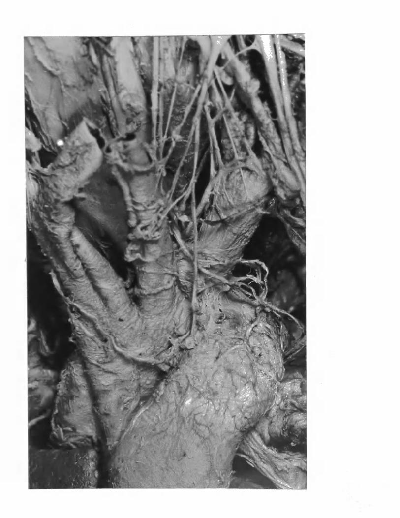

— Mid. Gerv. G.
— Mid. Cerv. Sym. Card, N.— Sup.Cerv. Sym. Card. N.
----------------------------------------------- — Intermed. Cerv. G.
TRACHEA - R ecu rrent N.
V e r te b r a l A.
■ - ----------- In term ed . Cerv. G
— -----------------Inf. Cerv. Sym . Card. N./ 6 br* to Prox.Sub. Plex
In. A. L.C.C. A .
Prox. Sub. P lex . ( w i t h an A c c e s s o r y G.)
Inf. Cerv. Card. N‘s of Vagus
Sym. N's to Sup. Card. Plex.
— N's from Prox . Sub. P lex to D u c tu s
V agus
P l e x . on D u c t u s
D. A.Ao.Sup. Card. P lex . (w i t hC ontr ib u t ion from r i g h t )
C o n t in u a t io n of N's of Plex. on Ductus
Origin of L. P u l m o n a r y A.
R. A. RT.
L. A.


Thyroid
V a g u s N.
Trachea
Ao,
~ y .
t-.A. N------ 5th. Thor G.
Mid. Cerv.Sym. Card. N.
Intermed. Cerv. G.
Inf. Cerv. Sym. Card. N.
S t e l l a t e G.
Inf. Cerv. Card N. of Vagus
Sub. a . Gard.br. of S t e l l a t e G.
— Inf. Cerv. S y m . Card. N.
N------------------------- 2nd. Thor. G.
—----------------— 3rd. Thor. G.P.T. D.A. —̂ ----------------------- Thor. Sym. Card. N's
P l e x .o n Ductus
4 t h Thor. G.
Aortic P lexu s


Sup. Cerv. Sym .C arf l .
Vagus N.
Mid. Cerv. Sym G.
Mid. Cerv. S y m .C a r d . N.
I n term ed . Cerv.G.L.C.C.A
--------------------------------------------------------- R e c u r r e n t N.
Inf. Cerv. Card. N .o f V a g u s
Inf. Cerv. S y m . Card. N.
P r e a o r t i c P l e x u s ( P a r t . )
Sup. Card. P l e x .
P l e x u s on D u c t u s
L . P u l m o n a r y A.
V agu s N. (drawn b a c k )
In.A.
Ao. /
P. T. D.A./
L.A.

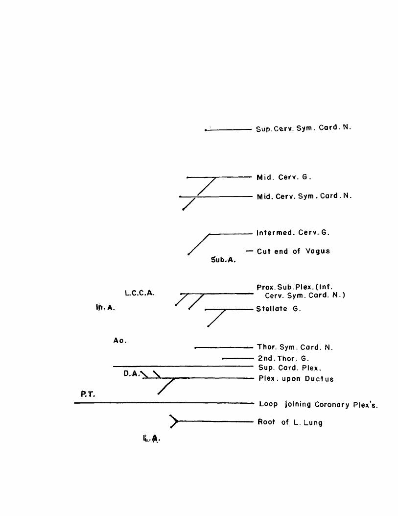
S up.C ^rv . S y m . C ard . N.
Mid. Cerv. G .
Mid. Cerv. Sym . Card . N.
I n te r m e d . C erv . G.
C ut en d o f V a g u s
Prox. Sub. Plex. (Inf .Cerv. S y m . Card. N . )
S t e l l a t e G.
Thor. S y m . Card. N.
2 n d . Thor. G.Sup. Card. P le x .P l e x . upon D u c t u s
Loop jo in ing Coronary Plex*
Root of L. Lung

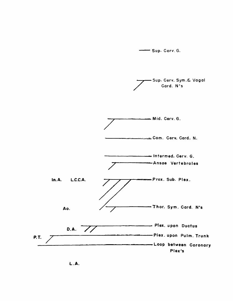
Sup- Cerv. G.
P.T.
Sup. Cerv. Sym.C. Vagal Card. N ' s
Mid. Cerv. G.
Com. Cerv. Card. N.
zIntermed. Cerv. G.
A n s a e V e r t e b r a l e s
In. A. L.C.C.A.
Ao.
P rox . Sub. P l e x .
T h o r . S y m . Card. N*s
D A . / /Plex. upon D u c tu s
P l e x . upon Pulm . Trunk
Loop b e t w e e n C o r o n a r y P l e x ' s
L.A.


Vagus N.
Sub.A
R.C.C.A.
In.A.
7 r
L.C.G.A,
M id Cerv. Sym . 6 Phrenic N.Vago-Sym . br. to I n A. b i f u r c a t i o n
Mid. & Inf. Cerv. Sym. Card. Ns.t
Int. Med. Cerv. Sym. G.
Card. br. of Recurrent N.
Com. Cerv. Card. N .
Thor, Vagal Card, br's
br's to Deep Card. Plex.
br. of Com . Cerv. Card. N
Ao.
P.T.Sup. Card. Plex.
R . A.
R.V.
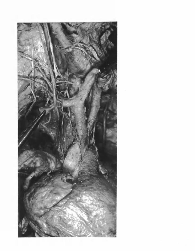
t " w ' ̂ ..

TABLE NO. I . INTERM EDIATE CERVICAL SYMPATHETIC GANGLION
D issec tion Description R e la t i o n s h ip sSp ina l N e rv e
CommunicationsTerm inalB ra n c h e s
No. S id eS jz e
in S hapecm. V e sse lsV ertebral
L e v e lsInf. Cervical
S te lla te G.
M iddleCervicalGanglion C5 C6 C7 c 8
I
L 0* 4 fused
0*1 5 2G-no information
C7intimate,doubled1GT
discrete fair siz-e at C5-6
- - - -2 cardiac N1 s (1 separate & 1 with MCCSN
R0, 2 small x glob-
0 .Z ose
lower C6 upper C7
fused, or thick IGT between
2G., 0.2 cm. above 0, 2cm, dia.
- - lb r - -
2L not recorded
R0, 5x ovoid
0. Z-
lower C6 upper C7
IGT thick between
fair size 1 cm, above
- - 1 lgB r. -
1 cardiac N» s (1 separate & 1 with MCCSN)
3
L°.b
0. 15 sm.G
1GT split to enfold Vert. A.
lower C6 upper C7
IGT split into 4(1 large 3 sm.
separate, IGT split bv Inf , T . A
- lb r. - -2 cardiac N1 s (1 to MCCSN & 1 to ICCSN)
R0,8 union x of 3
0.2 sm.G
inf, to Inf, T , A, t ant. med, Vert,^
lower C6 upper C7
IGT split & enfolds Vert, A.
separate IGT split by Inf, T , A
lb rvithC4 - - -
2 card. N1 s ,b r s . to MCCSN&ICCSN lb r , to Phren. N,
L0. 5x ovoid
o. z
med. to Inf ant, -med. Vert. A.
C7IGT short
br. C.Sp.T1joined by two trunks
combr.
comb r. br. -
1 cardiac b r. via Subclavian Plexus
4R
0. 5x ovoid
0. Z
ant. Vert. A, , med, to nf T. A
lower C6 upper C7
Ansa Sub, & single IGT
sm all G. at C5, IGT single
C4b r.5nlv
- - -2 card, N* s to MCCSN&ICCSN, v ascular b rs .
5
L° - 7 large
0*35 ovoid
ant. Vert. A, , med. to C7
Ansa Sub, ic single IGT.
small G. at upper C6. 2 IGT
com, of th fc C
t r . e se&4
com with Of I
• t r . B r.
;g .
weak card, b r. to MCCSN & b r. to Thvrocerv. T r.
R?0.2 flatX wiHf*
0.4 fi.
?ost.Sub. A. (some lower C7
thick IGT only part, separation
single IGT large MCG
Lbr,lcard . b r . to ICCSN, B rs . to Sub. A. & Vessels
6
L0.4x ovoid
0.2
ant. -med.tc V ert.A . atorigin
C7Ansa Sub. 1 Ig. Asm. 1G- T* s(Vert. A
several (3) G* on Inf.
T .A .- 2
ars.lb r 2 card. N» s(MCO
MSfclCCSN), Vert. >r. b vase. b rs .
R0.2 -0.3 round diam.
ant. -med. to V ert.A .at oriein
Clintimate, double IGT & Ansa Sub
2 weak swellings at Inf. T. A.
lb r.withTCG
- -ICCSN fc MCCSN b r s . to v e r t , .
7L
0.2in round diam.
ant. -la t, to V ert.A .
Clintimate thick IGT Ansa Sub,
elongate several G. fused
- -lbr,wit!ICG
lcard.N , with [CCSN, V ert. &Thvrocerv. A* s.
R0.3x ovoid
0. 15ant. to Vert. A.
Clthick IGT Ansa Sub.
definite G. la t. -sup, to Inf. T.
-lb r.IGTSUD.
lbr,IGT .
small b r , to LCCSN, b r . to rhv rocerv .. T r.
8L none per se, however, superior end of stellate ganglion characteristic
R^ small
0*15 OVOid
ant. - med, o V ert. A.
lower C6 upper C7
stout IGT doubled Ansa Sub.
sm all oval opposite Inf. T .A .
- - -arge b r . to MCCSN, 1 b r . to nl, Mam. A,
L0 .4x ovoid
0.25
ant. -med, to V ert, A. at orisfin
Cldouble IGT (Vert, A .) Ansa Sub.
discre te G, post. -Inf. T .A .
lb r . IBr,IBrviaY.d
3r's. to both MCCSN & ICCSN v ascular b rs .
R*0 . 2 .
rounddiam.
ant, to V ert. A. Cl
thick IGT Loops Vert. A ., Ansa
discre te G. inf. to Inf. T .A .
- -lb rwitlICG
3 b rs . (cardiac?) vascular B rs, anom. Sub. A .)
10L
0 .2in round
ant, to V ert. A.
lower C6 upper Cl
IGT double Ansa Sub.
weak G. inf. to Inf. T .A .
lbr,with lb r. - -
v ascular b rs . [Suh., Int. Mam. ■nivr'ocerv. )- card
R0 .4 2 G. x fused
0.15. oval?
ant. -V ert. A. -origin, med. -Tnf-T
lower C7[GT & Ansa lu b ,, Sp. N . coin' s
strong G. sup. -la t.to Int. T .A .
2b r witl1GQ
. vascular b r s . ,3 cardiac b rs . MZCSN & ICCSN
11L
0.3 2 G. x fused
ant, -med.t V ert.A ., lower C6
upper C7
strong IGT doubled, Sp N. com. Ana
strong G. sup. -la t. to Inf T A
lb rvithu r r
lb r .with -
v ascu lak b rs ., 2 -4 card, b rs . (ICCSN)
R I 5x small 0.1 ovoid
ant. -med, to V ert. A, Cl
double IGT Sp. N. commumca
strong G, sup. -la t. to Inf. T .A .
lbr,vithVffrf
3brslviaICQ
- -vascular b r s , , 1 card, b r, (ICCSN)
12L
0.4 irreg x flat-
0.25 jtened
ant, -med.tc Vert. A. C7 intimate
IGT stout Ansa Sub.
sm all G, sup. to Inf. T .A .
lb r.withICO
lb r,withrcci
-C ard .b r. (ICCSN) of IGT, vascular b r s . . esooh.
R?0.2 weak x swell
0. 1 ing
as the 1CT Loops med. to V ert. A.
C7 thick IGT Loops Vert. A . , Ansa
weak G. at level of Inf. T .A .
- Zbrs - part of ICCSN
13L
0. 25 ;trun- x cated
0. 3 'Dvran
ant, -med.tc Vert.A . at>rigin
lower C6 unner C7
very thick IGT, Ansa doubled
small.G. sup. to Inf. T .A .
- 2bralb r..wit!ICG
most of ICCSN, v ascular b rs .
R0 .2 ! in rounc diam.
Tied, toThyrocerv.Tr.
lower C6 upper C7
intimate IGT, Ansa Sub.
2 sm all G. at level of Inf. TfAf
-b rs.via Vj A
IB r -
v ascular b rs , form s ICCSN Vert. A. Plex.
14L 7
UP ? of
stell.
ant. -med.:o Vert. A, Cl
a portion of Stell. G.
strong G. post, to - IB r lb r -
vascular b rs . V ert. A. Plex. c a rd .b r . (ICCSN)
R0 .2in round diam.
ant. -med. to V ert, A,at oriein
Clthick IGT (2), Ansa
elongate G. post.Inf. T .A .
lb rviaICG
lb rviaICG
1 delicate card, b r.
15L
0.3 ' * ovoid
0 .2
ant. -med.to Vert* A.
lower C6 i^per C7
Intimate slight cons tric t IGT
none weab r.
2B rs lb r -
cardiac b r s . (MCCSN&ICCSN) vascu lar b r s .
R6 .2 x 10 .2 Xround
___ant. - la t . aspect of Vert. A .
upper C7doubled LGT, Ansa n on -ex ist
entlb r lb r lb r -
b r. to MCCSN, 2b rs, to ^grt«Plea vascu lar b r s . 1
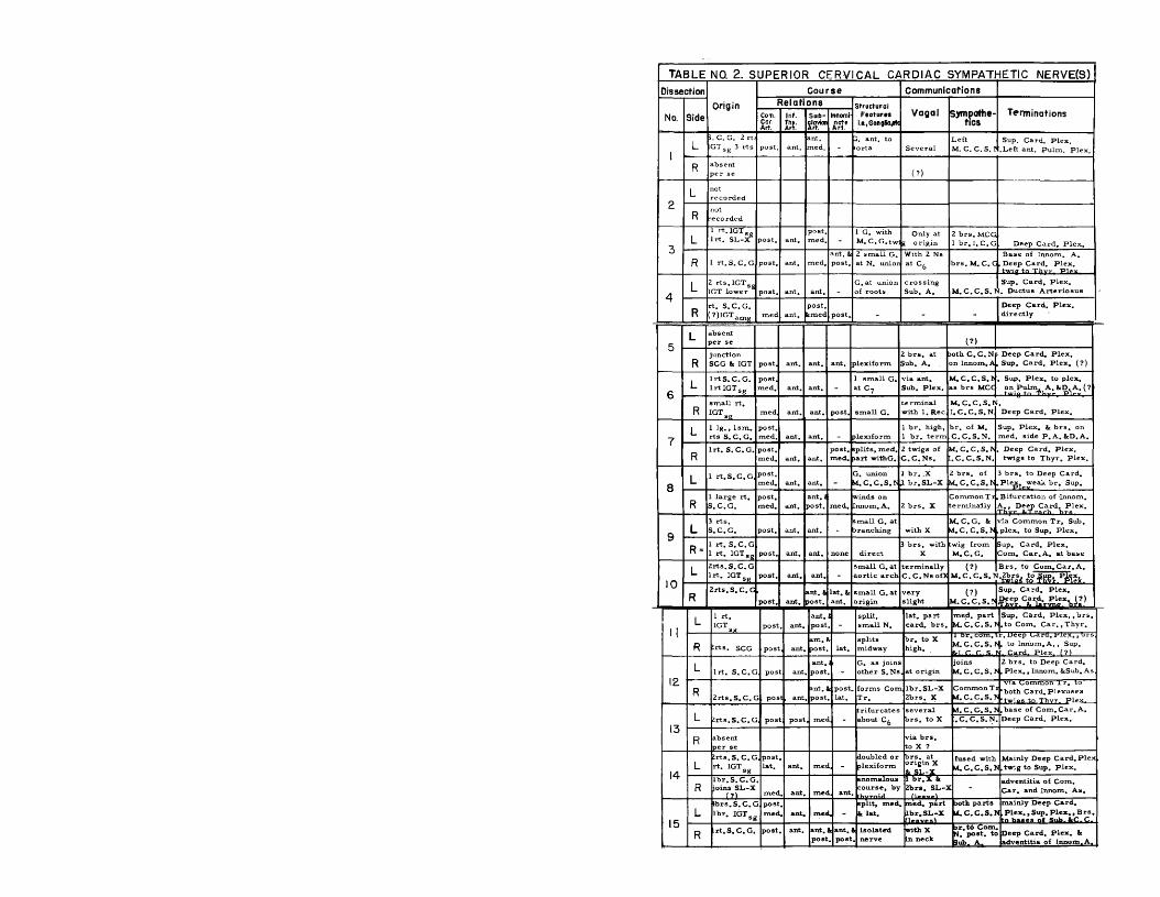
TA B LE NO. 2. S U P E R IO R C E R V IC A L C A R D IA C SYM PATHETIC NERVE(S)
DissectionOrigin
C o u r se C om m unications
No. Side
R e la tio n s StructuralFeatures
i.e.jGongBOjiftV agal Sympathe-
ticsTerm inationsCom.
Car.Art.
Inf.Thy.Art.
Sub-clownArt.
Innomi-
1L
3. C. C. 2 rts GTsg 3 rts post. ant.
ant.med.
1. ant. to lorta Several
LeftM. C. C. S. I
Sup. Card. Plex..Left ant. Pulm. Plex.
R absent per se (?)
2L not
recorded
Rnotrecorded
3L
1 rt. IGTS Ir t. SL->? post. ant.
post,med. -
1 G. with M. C.G.twi
Only atg origin
2 b rs. MCG 1 b r .I .C .G Deep Card. Plex,
R 1 rt.S .C .G post. ant. med.ant. & post.
2 small G. at N. uni or
With 2 Ns at C6 brs.M . C.C
Base of Innom. A. .Deep Card. Plex.
AL 2 rts . IGTSg
GT lower post. ant. ant. _ G. at union of roots
crossing Sub. A. M .C .C .S .I'
Sup. Card. Plex.. Ductus A rteriosus
Rrt. S .C .G . ?)IGTam« med ant.
post.tm ed post. - - -
Deep Card, Plex, directly
L absent »er se (?)
Rjunction SCG & IGT post. ant. ant. ant. plexiform
2 b rs , at Sub. A,
>oth C .C .N on I nnom-, A
Deep Card. Plex. Sup, Card. Plex. (?)
c L Ir tS .C .G .lrtIG T sg
post,med. ant. ant. -
1 small G. at C? Sub. Plex.
M .C .C .S .J as brs MCC
. Sup, Plex. to plex. on Pulm. A .kD .A .(?
Rsmall rt. IGT med ant. ant. post. small G.
term inal with 1. Rec
M .C .C .S.N I. C .C .S .N Deep Card. Plex.
7L 1 lg ,f Ism .
rts S. C.G.post.med. ant. ant. - ilexiform
1 b r, high, 1 b r. term
t>r. of M. C .C .S .N .
Sup. Plex. k b rs . on med. side P . A. kD.A.
RI r t. S .C .G . post.
med. ant. ant.post.med.
splits, med. ja rt withG.
2 twigs of C .C .N s.
M .C .C .S .I'.C .C .S .N .
Deep Card. Plex, twigs to Thyr. Plex.
8L 1 rt.S .C .G , post,
med. ant. ant. -j , union M .C.C.S.b
1 b r. .X X br.SLi-X
2 b rs . of M .C.C.S.N
3 b rs . to Deep Card. Plex. weak b r. Sup.
RL large rt, S .C .G .
post,med. ant.
ant. 1 jost. med.
winds on .nnom. A. 2 b rs . X
Common T i terminally
Bifurcation of Innom. A ., Deep Card. Plex.
9 L3 rts . S .C .G . post. ant. ant. -
small G. at >ranching with X
M .C.G. & M .C.C.S.N
via Common T r, Sub, plex. to Sup. Plex.
R *1 rt. S.C.G 1 rt. IGTSg post. ant. ant. none direct
3 b rs . with X
twig from M .C.G.
Sup. Card. Plex, Com. C ar. A. at base
L 2rts .S .C .G I r t . IGTSfi post. ant. ant. -
small G. at aortic arch
term inally C .C .N s ofX
(?)M .C.C.S.N
B rs. to Com .Car.A .
1 0
R2rts. S. C, G
post. ant.ant, & >ost.
la t. & ant.
Small G. at origin
veryslight
(?) JSup. Card. Plex.M .C .C .S .^ - P C a r d . r P lex ^ (s?)
I I
L 1 rt, IGT sg post ant.
ant.post
split, small N.
la t. part card. b rs .
me<l. part |Sup. Card. P le x ., b rs . M. C. C.S. N. to Com. C a r ., Thyr.
R •rts. SCG post ant.ant. 1 post. lat.
splitsmidway
b r. to X high.
1 br. com .tM. C .C . S.*k l.C .C .S .I
r.u eep Gard. P lex ,, brs |. to Innom.A., Sup,_ Card. Plex. (?)
12L I r t. S.C.G post ant
ant.post -
G, as joins other S.Ns at origin
joinsM .C.C.S.I'
2 b rs . to Deep Card., Plex. , Innom. kSub. As,
R 2rts. S. C, C pos t , anta.nt. 1 post.
t post, lat.
forms Com T r.
lb r . SL-X 2brs, X
CommonTi M. C. C.S. ̂
via Lommon i r . to 'both Card. Plexuses
13L £rts.S . C. G pos post met
trifurcates about C6
several b rs, to X
M .C.C.S.h.C .C .S .N
.base of Com .Car.A . Deep Card. Plex.
R absent per se
via b rs . to X ?
14L
2rts.S .C .G rt. IGTsg
postlat. ant. met
doubled or plexiform
brs, at origin X k SL-X
fused with VLC.C.S.f
Mainly Deep Card. Plex 1, twig to Sup, Plex.
Rlb r .S .C .G joins SL-X
t?) med . ant. met , ant
anomalous course, by
l b r .X k 2brs. SL-J
adventitia of Com. C ar. and Innom, As.
15L
tb rs.S .C .C lb r. IGTsg
postmed • ant. mec
split, med, k lat.
med. part lbr.SL-X
both parts tf.C.C.S.J>
mainly Deep Card.I , Plex. , Sup. Plex. , B rs.
R r t.S .C .G . post. ant. an t.. post
k ant.. post
i isolated . nerve
with X In neck
sr. to Com, N. post, to Sub. A.
Deep Card. Plex, k adventitia of Innom. A.

TABL E N 0.3. MIDDLE CERVICAL CARDIAC SYMPATHETIC NERVE(S)Dissection
OriginC ourse Communications
TerminationsNo. Side
RELATIONS STRUCTURAL FEATURES i.t^gonglio.ets.
Vagal Sympathe-t ic s
Com.Cor.Art.
Inf.Thy.Art.
SubclavianA r t
Innominate
Art.
1L
1 rt. MCG l r t . Int. CG post.
ant.inf.
ant. b post. -
G. at unioi with
s r . r s N
twig to left
SCCSN b ICCSN
essentially in the Deep Caraiac Plexus
Ri rt. MCG lr t . from 3rd CSN po St, ant. post. post.
portion from Co with X.
fibers to X noted terminally Deep Cardiac Plexus
Lnotrecorded -
2R
2 rts MCG 1 rt. Int. CG post. _ post. post.
largenerve
with left recurrent
twigICCSN
term inates in Deep Cardiac Plexus
Lr t . IGTbmg
1 r t . Int, CG post. inf. po st. -G. at unioiwithSCCSN
SL-XCommon tr, SCCSN Deep Cardiac Plexus
3R
2rts. MCG lr t . Int. CG post. ant. ant. lat.
2-3 small G, at unioi of roots
with C.C. b rs . of X, recurrent
twig of ICCSN all te rm .
Deep Cardiac Plexus Sup. Card. Plex. (:?)
L rt. MCG lrt. IGTbmg
post.lat. inf. post. - plexiform
leftrecurrent
SCCSN ICCSN via Sub. P,
essentially to Deep Cardiac Plexus
Rl r t . IGT at site MCG
■ lr t, Hat. CGpost.med. post.
post,lat.
separate N*s, lower with G.
leftrecurrent
(?) fibers ICCSN
Common Carotid A. Deep Cardiac Plexus
5L
2rts. IGT (MCG) lr t . Int. CG post. ant.
post.bant. _
plexiform with sm. G
5 b rs , to leftrecurrent
part of ICCSN
Both Sup. and Deep Cardiac Plexuses
R3-4rts, fror MCG and IGTbmg
l
post. ant.post.bant.
post.lat.
individual b rs . not a single N.
lb r. with CCN1 s of X
2b rs . to SCCSN
adventitia of Subclavian, Com, Carotid b Innom. A*!s , Deep Plex.
6L 3 rt&r, MCG
lr t . int. CGpost.lat.
lb r.ant.
ant.post. _
G* at union of roots b Com. T r.
lb r. to L. recurren t b b r. of X
Com, T r. SCCSN br. of I, -
possibility that both plexuses get b rs .
Rlb r . from Int. C.G, lb r. MCG
post,lat. post.
post.lat.
separate N* s to Cone T r. bL. Rec
B r. of MC • G. to Rec. . lb r . X
uniteswithSCCSN
Deep Cardiac Plexus Sup. Card. Plex. (?)
7 L6rts . from MCG
post.lat.
3rts,ant.
ant.post. -
r ts . form 3 main N*s SCCSN
SCCSN b Sub. Plex,
Deep Cardiac Plexus Sup. Card. Plexus(?)
R 4rts .MCG
post.la t.
Zrts.post.
post.med.
rts . unite in G, with SCCSN
b r. to recurrent from G*
at small G. with SCCSN
Deep Cardiac Plexus twig to Sup. Plex (?)
8L 2 large brs
of MCGpost,lat. inf.
post'.med. _
r ts . unite in G, with SCCSN SCCSN SCCSN
Deep Cardiac Plexus b r. to ant. of Aorta
R2brs. IGT (MCG) b lb r . Int. CG
post.lat.
lb r,ant. no st. folds
Com. T r,withSCCSN
CCN* s of X by Com* T r.
SCCSNbyCom. T r.
Deep Cardiac Plexus
9L
2brs. MCG 2brs.CG
post. 2brs.ant.
post. -G. (?) at union of r ts .
noneCom. T r. b rs . of S, bICCSN
Deep Cardiac Plexus
R l r t . MCG post,med. - post.
trachtoea
simple N. with corn-
with CCN* sr»f TC
see text anomalous course) Deep Cardiac Plexus
10L
l r t . IGTbmg post. - post. - simple N.
twig to left slight Deep Cardiac Plexus
Rlrt.W dG lr t . Int. CG b r .to Rec. po St, ant.
post.ant.
post,lat.
G. in Ig. b r, with SCCSN
MCG to recurrent lb r . of X
SCCSN Deep Cardiac Plexus Sup. Card, plex, (?)
11L
2rts . MCG weak rt. of IGTbmg
post.lat. ant.
post.med.
2 separate nerves
part of other
r.r.SNi
especially Common Carotid A, Deep Card.PIpv
R3rts, of MCG post. ant. post.
post.lat.
b rs . form Com. T r,
CCN* s of X fc L.Recurrent
Com. T r. SCCSN
b r. to Sup. Card. Plex, Deep Cardiac Plexus
12L
l r t . MCG 2rts, IGT- bme post. ant. post. _
Gf at unionwithSCCSN
SCCSNr?>
Com. T r. SCCSN
Deep Cardiac Plexus
Rl r t . MCG post. - aost. post,
ant. Com, T r.via twigs :n SCCSN
Com. T r. SCCSN
Deep Cardiac Plexus Sup. Card, Plex, (?)
13L
l r t . MCG lr t . IGT- post.
ant,post.
sup.med. doubled
2 twigs from X
SCCSN b rs . of I, bTnt. (JC.
Deep Cardiac Plexus adventitia of Com.
RlR t .MCG (small) , lrt. IGTbma
post.- ant. post. post.moderately simple N.
X proper
recurrent
ICCSNfrom Deep Cardiac Plexus
14L
IRt. immet above MCG "post,
lat.loops
med.
strong N. small G. at oripin
via com. with Com. T r.
SCCSN
Deep Cardiac plexus twig to Sup, P lex .,
R2 weak brsMCGelongate post. post. post. very weak none none Deep Cardiac Plexus
15L
2rts. IGT- bmg bint. CG
post.Lat. fold
sup,med.
splits, 4 b rs , G, at oriffin
b r. from X at Sub- clav. A.
Com. T r. SCCSN, br.of ICCSN -
Deep Cardiac Plexus with twig to Sup. Card. Plex.
R2 rts . IGTbmg, l r t . Int. CG
post.Lat,
enfold
int. b post.
post,lat.
doubled twig to recurrent
near te rm .withICCSN
Deep Cardiac Plexus weak filament to
Card* Plex*

TABLE NO. 4. INFERIOR CERVICAL CARDIAC SYMPATHETIC NERVE(S)Dlssectfon
Origin
Course Communications
TerminationNo. SideRELATIONS Structural
Features i.e., ganglfciefc
Vagal Sympathe-tlc s
Subclavian
Art.
InnominateArt.
1L
3rts, from St ell, G.
essentially pos1
roots form 1 N.
with C .C . N*'s of X
MCCSN TCSN» s
Deep Cardiac Plexus Ductus arteriosus
Rlr t , from Stell. G. post.
post. b lat. single N. none
Com, T r, TCSN* s Deep Cardiac Plexus
2L lr t , from
SteR. G. post.quite weak N.
with X proper none
Deep Cardiac Plexus Ductus arteriosus
Rnotrecorded
3L
3rts. Stell. G.
plexusmainlynost.
-plexiform but 1 main none :wig to
Com. T r.Deep Cardiac Plexus Ductus A rteriosus
Rlr t , ICG lr t . Int.CG
post. ?inf.
post. & lat.
essentially 1 N.
CCN* s of X
SCCSNMCCSN
Deep Cardiac Plexus Sup. Card. Plex. (?)
4L
Zrts. Stell, l r t . Ansa Subclavia
post. & inf. -
plexiform 3b rs . Sub, Plex
none MCCSNTCCSN
Deep Cardiac Plexus Ductus A rteriosusVia CwK Dlov
R1 rt, Stell lr t , Int. CG
post. &inf.
>ost. & lat. single N.
twig to recurrent erminal Deep Cardiac Plexus
5L
l r t . Stell l r t . Ansa Subclavia
plexus on prox, portion
-2 main portions
recurrentMCCSN via Sub.
Ductus A rteriosus Deep Cardiac Plexus
R2rts . ICG lr t , Int.CG
post. & sup.
post. b lat.
splits b reforms (?)
twig to Com. T r, Deep Cardiac Plexus
6L
2rts . Int. CG.Zrts.Slell.
plexus on prox, portion -
G. in Sub. plexus
CCN* s of X
:wig from Com* T r. TCSl!{>s
Deep Cardiac Plexus Ductus A rteriosus
R21aree rts, ICGlr t . Int. CG
post. & inf.
)OSt. binf.
discrete N. thenCom, T r.
CCN1 s ofx brecurrent
Com. T r. Deep Cardiac Plexus
7L
ZRts. Stell. lr t . Int. CG post. .
separate N*1 s to term .
nonetwigs to MCCSN bTCSNs
Deep £ardiac plexus weak b r, to Sup, Card. Plex. b Ductus A rt.
Rl r t . Stell lr t.In t. CG
post,loop.
post, it lat.
2 small N* s with
upper N. joined T r, to br. X
SCCSNbifurcation of Innom A. with T r,Deen Cardiac Plexus
8L
3rts. Stell, l r t . Int. C.c . (?)
plexus on prox, portion
-4-6 fibers extensive with 2 G.
CCN* s of X, b r, recurrent
none2brs. to Ductus Art. Deep Cardiac Plexus Sup. Card. Plexus
R2rts. of Stell. G.
post. & inf.
post. b lat.
relatively simple N.
part with C. b r. of recurrent
part to - Com. T r, Deep Cardiac Plexus
9L
1 lg. Rt. of Int. CG lr t . Stell.
post. &plexus -
main trunk with b rs. looping A.
small br. of X into Sub. Plex.
2 twigs to Com. T r.
Deep Cardiac Plexus Ductus A rteriosus
R*3r ts . IGT- inf. Int. G. 2 rts . Stell.
post.sip post. int.
anom. no Inncrr present
all 5 rts . . separate courses
none noneDeep Cardiac Plexus via anomalous A rtery
10L
2rts , IGT- as . or Int. C.G.
smallplexus30St. S.Up,
-large N. with twigs Sub. Plex.
noneoranch to SCCSN
Ductus A rteriosusSup. Card. P le x ., b rs . to
R2 weak rts, Stell. G _RUst_CG.
weakplexus
post. & lat.
weakplexiform none none
weak b rs , in wall of Sub. b Innom. A*'s. Deftp Cardiac Plexus----------
IIL
2rts. Int. C.G.3rts.S tell.
plexusprox. -
1 main T r. yielding b rs . plex.
nonebr. to 1st. TCSN
Deep Cardiac Plexus Sup, Card. Plex, j Ductus A rteriosus
Rlr t . Stell. lr t . Int. C.G.
ant. sup. & DOSt.
lateral 3 small N* s independent
none lb r. to Com. T r.
Deep b Sup. (?) Card. i Plexuses b aaventit.'a ,
12L
1 Rt. Int. C.G.2rts.S tell.
plexus esp.post b sup.
1 mam T r. with b rs. to plexus
none1 b r, to TCSN* s
Both Cardiac Plexuses Ductus A rteriosus
R2rts.S tell.
fnt r. r.post. post,
b lat.
2 N* s not very strong Com. T r.
CCN* s X1 N. to Com. T r.
Deep Cardiac Plexus Sup. Card. Plex. (?)
1 3L
2Rts, Int. C .G ,, Ansc l r t , Stell.
plexusmainlypost. -
3 main T rs. sup. , ant. post.to A.
nonetwig from MCG
Deep Cardiac Plexus Sup. Card. Plex. b Ductus A rteriosus
R2Rts. Int. C.G. clas] Vert. A.
post.& sup.
PO£t. h lat.
2 main brs.elaboratecourses
recurrentlb r . to Com. T r.
Deep Cardiac Plexus Sup. Card. Plex. (?)
14L
1 LargeRt. Int. Cu, rts
AnsabStell.
heavy or dens plexus
-Swollen as ifganglionic
none small twig of MCG
Large contribution to both Card, Plex. b
R5rts. Stell (weak)
post, b inf.
post, b lat.
diffuse group of N* s.
with CN* s ofrecurrent
TCSN* sDeep Cardiac Plexus adventitia on ant. asDect of Innom. A.
15L
lR t. Int. CG, 2-3 r ts . Stell
sup.weak -
1 main br. b several none 1 twig of
MCG
Deep Cardiac Plexus weak b rs , to Sup.
RIRt. Stell. l r t . Stell.
post, b inf.
post, b lat. 1 main br. receives
br. .of recurrent
b rs , to SbMCb TCSN* s
Deep Cardiac Plexus Sup. Card. Plex. (?)
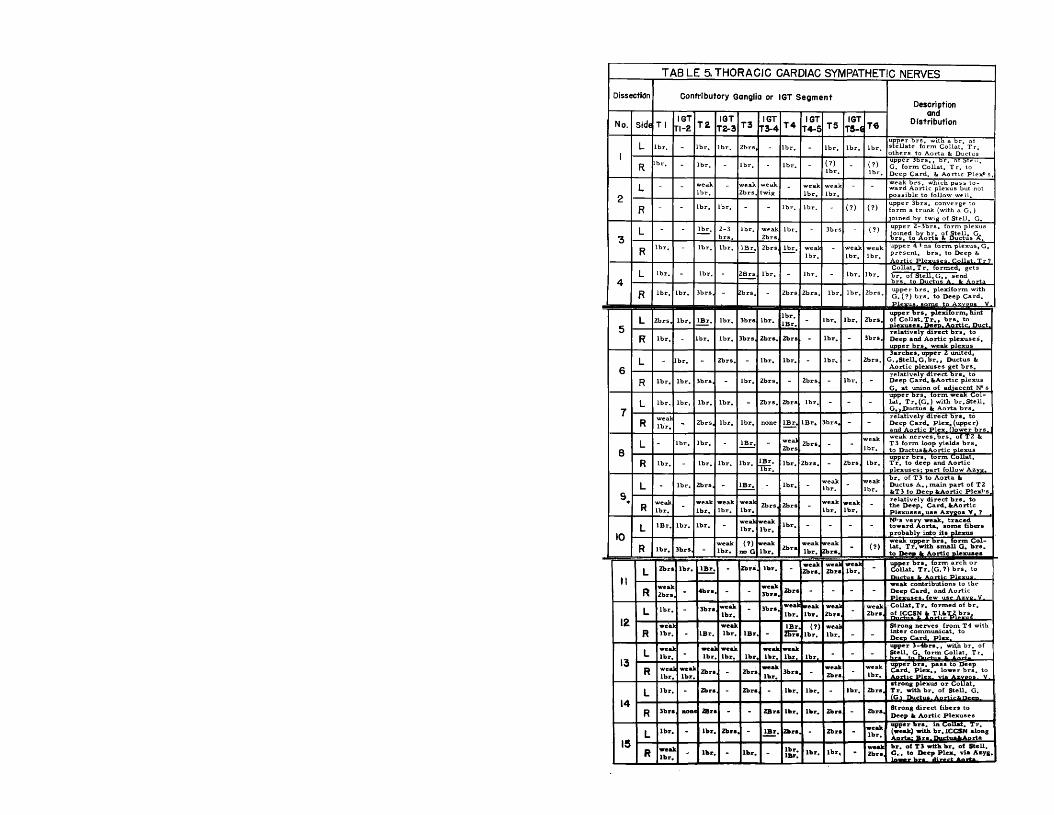
TABLE 5 .THORACIC CARDIAC SYMPATHETIC NERVES
Dissection Contributory Ganglia or IGT S egm en tDescription
No. Side T 1IGT
T l- l T 2IGT
T2-3 T3IGT
T3-4 T 4IGT
T 4-5 T5IGT
T5-C T €and
Distribution
1L Lbr, - lb r. lb r. 2b-rs, - lb r. - lb r. lb r. lb r.
upper b rs, with a br. of stellate form Collat. Tr. others to Aorta & Ductus
R br. - lbr. - lb r. - lb r. - (?)lb r. ' (?)
lb r.
upper Jb rs ., br. of Steii.G. form Collat, T r. to Deep Card. & Aortic Plex* s.
2L - - weak
lb r.weak2brs.
weaktwig - weak
lb r. lb r.- -
weak b rs, which pass toward Aortic plexus but not possible to follow well.
R - - lb r. lbr. - - lbr. lbr. (?) (?)upper 3brs. converge to form a trunk (with a G .) joined by twig of Stell. G.
3L - - lbr. 2-3
brs.lb r. weak
2brslbr. - 3brs - (?)
upper 2-3brs. form plexus joined by br, of Stell. G, b rs , to Aorta & Ductus A,
Rlb r. - lbr. lb r. IBr. 2brs lb r. weak
lbr.- weak
lb r.weaklb r.
upper 4 * ns form plexus, G, present, brs. to Deep fc
A.L lbr. - lbr. - 2Brs lbr. - lb r. - lbr. lb r.
Collat.T r. formed, gets br. of Stell. G ., send
R lb r. lbr. 3brs, - Zbrs. - 2brs 2brs. lb r. lb r. 2brs. upper b rs . plexiform with G. (?) b rs . to Deep Card.
■ -J?l£xus,_aom£ to Azyeo, V.
L 2brs. lb r. IBr. lbr. 3brs lb r.lb r.IBr, - lbr. lb r. 2brs,
upper b rs . plexiform, hint of C o llat.T r., b rs . to nlexuses. Deeo. Aortic. Duct.
oR lb r. - Lbr, lb r. 3brs, 2brs, 2brs - lb r. - 3brs.
relatively direct b rs , to Deep and Aortic plexuses, upper b rs. weak Dlexus
0L - lbr. 2brs. - lbr. lbr. - lbr. - 2brs.
3arches, upper Z united, G .*Stell,G .br,, Ductus & Aortic plexuses get b rs .
R lb r. lb r. 3brs, - lbr. 2brs. - 2brs, - lbr. -relatively d irect b rs . to Deep Card. fcAortic plexus G. at union of adjacent N1 s
7L lb r. lbr. lb r. lb r. - 2brs. 2br& lbr. - - -
upper brs, torm weak Collat. T r. (G.) with b r. Stell. G.».Ductus fc Aorta b rs .
R we at lb r. 2brs, lb r. lb r. none IBr. IB r. 3brs, - -
relatively direct b rs . to Deep Card. Plex. (upper) and Aortic Plex. flower brs.
8L - lb r. lb r. - IBr. - weal
2brs2brs, - - lbr.
weak nerves.brs. of T2 & T3 form loop yields b rs . to Ductus fcAortic plexus
R lb r. - lb r. lbr. lbr. IBr.lb r.
lbr. 2b rs . - 2brs, lbr.upper brs. form ColLat.T r. to deep and Aortic plexuses; part follow Azyg.
9,L lbr. 2brs, - IBr. lb r. weak
Lbr. - weaklb r.
b r. of T3 to Aorta &Ductus A ., main part of T2 fcT3 to Deep fcAortic Plex*'s.
R weaklb r. lb r.
weaklb r. lbr.
2brs, 2brs weakLbr.
weaklb r. - relatively direct b rs , to
the Deep. Card. fcAortic Plexuses, use Azygos V. ?
m L IBr. lb r. lbr. - lbr.weaklb r.
lb r. - - - - H*'s very weak, traced toward Aorta, some fibers probably into its plexus
R lb r. 3br&, -weaklb r.
(?) no G
weaklbr. Zbrs lb r.
weakZbrs. - (?)
weak upper b rs . form Collat. T r. with small G. b rs . to Deep fc Aortic nlexuses
HL Zbrs lb r. IBr. - Zbrs. lb r. - weak
Zbrs.wealZbrs
weaklbr. - upper b rs . form arch or
Collat. T r. (G. ?) b rs. to
Rweak2brs 4brs -
weak3brs. Zbrs - - - -
weak contributions to the Deep Card, and Aortic Plexuses, few use Azve.V.
12L lb r. - 3brs weak
lbr. - 3brs. weaklb r.
weaklb r.
weak2brs
weak 2b rs,
Collat.Tr. formed of br. of ICCSN fc Tl.fcTZ brs.
Rweallbr. IBr.
weaklbr. IBr. -
IBr.Zbrs
(?)lb r.
weaklbr. - -
Strong nerves from T4 with inter communicat, to Deep Card. Plex.
13L wea
lbr.c wea]
lbrweaklbr. lbr.
weaklb r.
weaklbr. lbr. - - -
upper 3-4brs., with br, of Stell. G, form Collat, Tr.
R wealbr.
i wea)lb r.
Zbrs - Zbrsweaklbr.
3brs,weak2b rs - lb r.
upper b rs . pass to Deep Card. P lex ., lower b rs. to
14L lb r. - Zbrs - Zbrs lbr. lb r. - lb r. Zbrs,
strong plexus or Collat, T r, with br. of Stell. G.
R 3brs , non 2&ri - - 2Brs lbr. lbr. Zbrs - Zbrs, Strong direct fibers to Deep fc Aortic plexuses
15L lb r. - lb r. 2b rs - IBr. Zbrs - 2brs -
weaklbr.
upper b rs. in Collat, T r. (weak) with b r. ICCSN along Aorta: B rs. DnrtusfcAorta
R wea]lb r.
t lb r. - lbr. -lb r.IBr. lbr. lb r. -
weakZbrs,
b r. of T3 with b r. of Stell. G ., to Deep Plex. via Asyg.

T A B L E 6. VAG AL CARDIAC NERVESi
Dissection C e r v ic a l T h oracic
N o.Superior Inferior
Origin Coursea TerminationSideOrigin Course a Termination Origin Courses Termination
L2 .b rs . of t proper
communicate at o r igin with SCCSN and included in it - -
lb r , ant, to aortic arch
passes with recu rrent N, lat. to Duc-
R
ant. , ini. to the Sub. A.
masses inf., med.>n lower border Sub. A. t Deep Plex,
1 several1 b rs . of X proper
direct to Deep Cardiac Plexus
L notrecorded
2
Rnotrecorded
LIB r. of S L - X
?IB r. 1cm lat. It post, to
Innom,A., commun3brs. below rec-
all to Deep Card. Plex, strong b rs .
3
R
tiny brs. o f X . go toSCCSN
with Sym, fibers to adventitia Aorta and Deep. Card. Plex
3brs. ant. to Sub. A.
part to Common N. others direct to Sup.Plex.lt Duct. ?
lb r , of X at Rec.
loops beneath and sends twig to Ductus then Deep Plexus
L - -IBr. of X, at Sub. A.
joins SCCSN, b rs , to aortic arch,Sub. A. IcSup. C. Plex -
4
R - -
IB r. ofX at 6th C. vert.
ant. to bifurcat. of Innom.A, b rs . to it, Sup. ItDeep Plex.
IB r. high lb r , low in Thor.
along post, la t, side of Innom. A, to Deeo Card. Plexus
5
Llb r . of X proper, 1 b r. SL-X
both b rs . join Sym. T r. fc yielded as MCCSN?
lb r . ofX ant, to Sub. A.
b rs . to adventitia of aortic a rch ,b rs , to Sup. Plex. t Duct.
lb r , high lb r, low
high b r. to SCCSN fc Sup. Plex* , low hr. fweakVto Ductus
R
3-4 b rs . of X join Sym.
branch formed re joins vagus ( ? )
3brs.sup. fc ant. to Sub. A,
complex communications with 5ym*:s both Plext sit Innom.
lB r.o fX just below Rec.
post, la t, to Innom, A . , joins other N^s, Deep Plexus
6L ( ? ) twigs to Syznt's ( ? )
2brs,sup. It inf, to Sub. A.
joins proxim al Sub, Plex, b rs , to both Plex* s It Ductus
lb r, 1cm, below Sub. A.
joins N* s of X mentioned above by ascending
Rlb r . of X_(G.)
post, to C. C .A . Ic then ant. to Innom. joins complex.
lb r . , IBr, both sup. Sub. A.
lb r, to C .C .A ., IB r. joins plex. of all C.N»s
2brs, of recu rr.
descend post, lat, to Innom. A ,, b rs , to Deeo. Card. Plex
7
Llb r . of X
joins SCCSNfbr, of SCCSN rejoins X)
lb r. at sup. edge of Sub. A,
direct to Sup. Card. Plex.
2small b rs . of De£p Card. Plexus
Ductus A rteriosus
R very small b r( joins SCCSN
2brs, above Sub. A.
1 to SCCStf and 1 to adventitia of C .C .A .
1 large2 small recu rr.
all join and pass directly to Deep
8L
Ifcr.ofet lb r . of SL-X
both join SCCSN which forms loop with X lower
2brs, on sup. It infi
lb r . to Sub, Plex. IB r. direct to
lb r , 1cm, below Sub. A.
to Sup. Card. Plex, Ductus A rteriosus ?
R 2brs, of X
lt>r. to Innom. b ifurcation, lb r . to SCCSN
3brS. sup to
Sub. A.
lb r . to SCCSN, 2brs to Common T r. lower in neck
2brs, of recu rr. lb r . ofX
Deep Card, Plexus
9
Lr f ^ f t r s . of X to
Zbrs. of SCCSN(re- ^oining?) pass to
1 large S r. of X Sub. Ar
communication with Sub. P le x , , to Sup. Plex. It Ductus
lb r . of X 1cm abov
loops beneatk ; Ductus to reach
R(?) lb r. of X joins SCCSN
Zbrs, le a ibove Sub. A.
complex, anomalous Sub, A ,, b rs . to Deep Card. Plex.
3brs. of X (low)
direct to Deep Cardiac Plexus
1 0L .
IB r. sup. lbr* inf. Sub, A.
sup .b r, to Sub. P lex, (Ductus ? ) lb r. to Sup. P lex . .SCCSN
2brs. of X above recu rr.
loops beneath Ductus to reach Deep Card. Plex.
Rlb r . of X midway in neck
joins SCCSNI B r . 1 c m tb o v e ub. A.
b r. to Sym. twig to Sup. Plex. res t t o B e e n P l e x . -------------
2Brs. of recu rr.
IB r, recurrent to Com, T r . other di- r c c t t o B e t a P l e x u s
11
L -J b r s . s u p , i b r s . a n f . o S u b . A .
s u p . b r s . to S C C S N lo w e r b r s , to C . C . A . , S C C S N -S u p . P I .
NZors. of X IG . )
jO .n s S C C S NI B r . a n t . o S u b . A .
jo in C o m m o n T r , w ith S y m 1 s . , b r , t o S u p . It D e e p p l e x .
2 b r s . of
I B r . ofX
b o th t o D e e p C a r d . P l e x . , r e c u r r e n t
!2L
l ^ r . of X ( G . \ 1b ' - o ' S ^
r,d \ id u a l N . to C . C . A . a n d D e e p C a r d . P l e x .
l b r . K m sup ,, l b r , a n t , Sub,^
_nf. b r , to b a s e o f C . C . A . It A o r t a , s u p . b r , - S u p . P l e x ,
2 b r s , o f X a t s u p . a p e r t u r e
both to S u p . C a r d . P l e x u s d i r e c t
RL s m a l l b r . of X (G . )
;oir.s S C C S N2 b r s . a n t . to S -*-b. A..
c o m m u n ic a t e s with C o m m o n T r , (Sym * s to D e e o P l e x .
l b r . of r e c u r r . IBr. of X
b o th to D e e p C a ’-d . P l e x u s
1 3L
lb r. of S L - X jo i n s S C C S N
IB r., lbr ant. to Sub. A.
joins b r. of MCCSN multiple term inal, b rs . , 1 to Sup. Plex.
IBr. of X midway in thor.
direct to Sup. Plex, and loop around Ductus Arteriosus
R - -3 small b r s . , ant. sup.-Sub.
all 3 communicate with Sym* s which . b rs .to Innom.A.
2Brs, of
2Brs.ofX
lb r . of recurrent joins Sym1 s. all to
1 4
Llbr. of X IG.), lbr X proper
unite and then join . SCCSN, definite
contribution
IB r. at sup. edge of Sub. A
brs, to trachea It recurrent, main b r. -Prox. Sub. Plex. -
R
lb r. of SL-X
( ? )
w ithSym .br. on post, lat. Thyroid to C . C . A. & Innom,
2brs. at 1cm, sup •toSub. A.
lb r.to Innom.A. lb r. with Sym1 s to Deep Card.Plex.
lb r, of
Zbrs.-X
all to Deep Cardiac Plexus
15L
see inf. cerv, carc V of X
i1 large B r . mid, of neck
main portion to Sup, Card, Plex, lb r .to base C .C .A .
lb r. of recu rr.
to plexus upon Ductus Arteriosus
Rlb r . of X midway in neck
joins SCCSN (innominate A.)
1 weak br. ant. to Sub. A.
joins plexus at bifurcation of Innominate A.
lb r. of recu rr. 2brs. -X
direct to Deep Cardiac Plexus

TA B LE7 SUPERFICIAL CARDIAC PLEXUS
Dfss<Contributory Nerves
Description of Contribution (s) from th e right
D e sc r ip tio n of P lex u s proper
Sympathetic Vagal
No. Side c ! f tDardn
Mid.Cerv.Gorin
InfCerv.Gantn
ThorOardf.N.
Sup.CentGajdji
Inf.Cerv.Gorin
Rec.
f t*Thor.Cord.N.
L all (?) (?)via?Duct. s e e
5Nabs. - abs. No contribution
from the right noted
of moderate size containing ganglionic m ass. Fairly simple >lexus. C lassical >osition.
1R abs.
L incor plete Duct, - none recordednotation made in re gard to the communications with the >lexus upon Duct, Art.
c .R not i ?cord< d
■aL - - (?)
Duct.Plex.
(?) - inpart - abs. Noteworthy branch
from a Common T r. on right. Bas.es obliquely lat, to Innom. A. to cross ant, to
Classical position communications with
Plexus upon Ductus Art. B r. extends to Conus A rt. B r. loop-R
via Dom, T r. ?
via R Com. T r. 7
via R Com, T r. 1
-via R Com. T r. 7
- -
L all (?)
via ? Duct. Plex.
via ? Duct. Plex.
abs. all abs. abs.Contribution from branch of vagus just above Sub, A. Crosses innominate A, obliquely and ant. to aorta at A*'S oris.
Preaortic portion of moderate size fc continuous. Linear appearing. Definite contribution to Ductus Plexus.
*rR - - - - abs.
7 via ji. APie*-
- abs.
5L abs. part
viaDuct.Pie* f e ?*
_somebrs.
bighbrs. abs.
Very weak branches apparently crossed
Linear plexus along the superior border of the Ductus A rt. P reaortic part essentially a single branch.R
via R, Com.frr.
? -viaR,Com.T r.
? abs.a plexus on the ant, surface of Innom. A.
6L >art
?via5 om.
?viaDuct.Plex.
?via- part - abs.
Moderate sized branch source noted. C rosses Innom. A. obliquely to pass ant, to Aorta at origin of
Weak plexus with a simple trunk riding
on upper border of Ductus to which it gives branches.R 7
?viaCom.T r.
? -viaR. Com. T r, ?
viaR. Com, T r . 7 ' -
7L most ?
weakb r.
via 7 Duct, Plex. - all - -
poor specimen, blood infiltration- Very small branch arches over aorta at la t. angle formed by
Heavy trunk along the superior border of the Ductus A. continuation of fairly complex P re aortic portion.R -
?viasmalltwig -
8L
weakt>r.
?br.ant. part
via ? Duct. Plex. - all part abs.
No contribution noted from the right
Somewhat linear and moderate sized P re aortic portion chiefly vagal, not particularly intimate with Ductus,R -
9*L mo st
?viaDuct.P lex. (?)
viaSCCSN?
most - abs.The SCCSN on the rt. (with a contribution of X) passes ant, to b ifurcation of Innom, follows lat. border
Moderate sized plexus -divisible into a med. and la tera l portion. ‘ Strong communications wi£h Ductus fc Pulm.R
most - - - W ./la S- CCSN
- - abs.
1 0L part - part - abs. part - abs.
5.trong contrib. with source noted, typical
Linear, moderate s ized plexus with contributions to Ductus A rt. Weak P reaortic portion e xcept B r. from rt.R
most?via3r.toic c s r
- -?viaSCCSN
via Com. T r. 7
bighb r.
7-
br* s directed mainly to r t. coronary plex.
11
L part (?)part part DuctPlex
abs. most abs. abs.Moderate contrib. of r t . , source noted, gives rise to br* s to
M>derate sized linear plexus with b rs , loop* xng ductus and form ing trunk on its inf. bo rd er. Ganglion in sup. part.R
?viaComTr.
via
Tr™*
? via .cm . - -
def,b r.
?viaCom.T r.
-
prox. part of Aorta. Passes near ganglion in upper part
12
L - - partvia $ D(itt. Plex.
- part all abs.Strong plexiform con- :rib. o f the r t.
Plexus essentially vagal fibers, weak communication with Ductus Art, Though between latter and Aorta, gap exists.
RTvi'a Com. r r .
?viaCom,T r.
?viaCom.T r.
- - ComT r.
- -»f Aorta as well. Mainly SymP's,
(3L
yiakort,Plex.
viaAort,Plex.
part (?) ? part lb r. absfair contrib. of the r t . splits and sends 1 b r. down rt. side of
Weak plexus on sup. border of the Ductus (as if laterally placed) linear in structure. Ganglion in upper portion.R abs.
viaCom.T r.
Com,T r.
abs.? via b r.to T r.
- -Aorta with another b r, the la tte r at orig. Innom. A rterv
14L twig twig large
crrouz
viaDuct.Plex.
?viaseeSN
7 via Sub. abs. abs.
A very sm all filament could be traced across Aorta from the rt.
P reaortic N*s ganglionic, appearing, pass into a strong linear plexus. Large B r, loops Ductus,R - - ? _ ?
but source indeterminate. The ? marie indicate oossjlpilitv
15L part twig
weakb rs .
viaDuctp i«
abs.mainpart abs. -
Moderate sized individual N. from r t .
Linear plexus with ganglion in upper part. Weak Preaortic . Brs. to medial aspect of Ductus.
Pvlff.Tr.R ? weak
b r.? 7 - ■-
angle formed by origin of innom. A. from that vessel.

TABLE8. DEEP CARDIAC PLEXUS
DisscContributory Nerves
Oescrfptlon TerminationSympathetic Vagal
No. SideSup.Cerv.3ard.n.
Mid.Cerv.Gordn
Inf.Cerv.Canto.
Thor.Cord.N(s)
SunCerv.Cantn
Inf.Cerv.
Carijn
Rec.Card.N(*l
Thor.
1L - most part part - abs. abs. all
center of plexus is post, to Aorta (arch & asc. portions) but ext, curvature of arch approx, limit, com- mun. with Sup. Plex.
splits into plexiform brs. ant. and post, to rt. Pulm, A. ant, b rs, around Aorta to both Coron. Plex* s ■Dost. brs. -to atriumR abs. all all all abs. all abs. all
L rec rec most most not not notrec. rec
incompletely recorded somewhat more compact plexus seen,Thor. Sym. Card, brs. of both sides to rt. of esoph. to Deep P.
apparently muscH the same as dissection #1
£R rec all
notail not
rec.not
re tnotrec.
3 L - all part panall^weal
partabs. abs.
diffuse plexus, esp, on rt, where brs. separate, most post, to Trans. Sin., commun. with Sup. Plexus, weak Thor. Sym1 s-lft.
most brs. post, to Trans, Sin., split a- round rt. Pulm. A„ both to atria, brs. ant, to Sin, along Aorta to Coron. P1 sR all most all all most most abs. all
4L all all part
(?)most mo st part abs. all
essentially a group of parallel strands most post, to Trans Sin,, those on left commun. with Sup. P. & those on right direct
brs. post, to Trans, Sin. enfold Pulm. A. to atria, Br, to left of Sin, meet Sup. P. fc extend to Coron. P. brs,an t.to Sin. -Coron,R most most most all ill
weakpart abs. all
5L abs. part part part t i l - abs. low
contrib. N* s form plexus as they pass
b rs. along Aorta to Coron, Plext-s, few ant to Pulm .^ , to base of Sup.V.C,, brs. post, to Pulm. A. to areaR most part all all i n part all I?)
br. along Aorta, brs. post, to Pulm, A. are
6L - part part part
(?)part abs.
(?)most
N1 s pass mostly post, to Aortic R ec., mainly parallel It form a plexus oh sup. bord. of rt. Pulm. A. which has a trans. arrang.
b rs. passing ant. to Pulm.A. to sup. V.C. at base, those post, to atria between Pulm. Vs, brs. left of Rec. to Coron. P* sR all most all all part most all abs.
7L - most most part - most abs.
N* s essentially direct to sup. bord. rt, Pulm. A. to form plex, these brs. post, to Aortic R ec., brs. on
brs. from rt. mainly post, to Aortic Rec. to atria as above, b rs. from left mainly along post, aspect ofR most most most all Sym. most all all
8L most most part part most - abs. -
1 large N. paralleled by several smaller passed post, to Aortic Rec, ant. to pulm. A. ( r t .) deviated left, other b rs. tvnical
main br, passed to Coron, Plex* s fc post, aspect of atria betweei Pulm, V* s, br. on rt. (X] to base Sup, V.C,R most all all all all .a all all
9L - all part most - - abs. all
N* s numerous but direct R can be traced individ., bear same relationships as above b rs, of each side limited to it. mostly
same relat. to Aortic Rec, as above, brs. to rt. atrium at base of Sup, V ,C ., brs. of left & post, to Sin. to post, atria.R - all all all - all abs. all
10L - all most all abs. (?) abs. aU
N* s extend to sup, bord, of rt, Pulm. A, to there form a trans.
b rs. post, to Aortic Rec. with typical distribution, br, from rt, to left essentially along Aorta to Coron. Plex* s. with 1ft. brs.R (?)
partmost most all
(?)Svm.
most all allarranged plex. post, to Aortic R ec,, largely br.to left from rt.-----
1 1L - part part part (?) abs. abs.
a series of parallel nerves upon tracheal bifurcation, most of
3 main groups, 1 ant. to Trans, Sin. to atrium at base of V. C ., 1 post, to Sin. between atria,' I to left of Sin,-Coron, P* sR most most mos all
most
Sym.part most all
these post, to Trans. Sin., commun. with Sup. Plex. to its left
12L most all part most
most
Sym. - abs. -main portion ot Plex, ant. to left bronch. rather dense, most
brs. ant. to Pulm. A. on rt. direct, on left split to enfold it, those ant. along Aorta those post, to post, aspect atria, Pulm. V.
R part most mos allmost
Sym. most all anpost, to Aortic Rec. br. of X on rt. direct to base Sup. V.C.
13L most most part most
most
Sym.part abs. part
more plexiform than others, b rs. of xt, still direct but br. deviates to left It with left brs. forms dense plex. - Pulm, A,*
brs. essentially as described above, those on left part of plex. essentially to left Coron. Plex, fc post, atria between Pulm. V* i
R abs. all mos all abs. part all all
14L most mo st part part most (?) abs. abs.
numerous commun. between component N* s prior to formation of trans. plex. post, to Aort, Rec. fc sup. to rt. Pulm. A.
strong brs. ant. to Pulm. A. on rt. to base Sup,V. C ., brs. on left ant. to Pulm.A. pass along Pulm .Tr. to 1ft, Coron. P lex ,, brs.. toR - all mo st all - all all all
15L mo st most most part abs. - abs.
1 large N, of rt. to ant, aspect of left
some brs. ant. to Rec. along post, aspect of Aorta, others post.to Rec. to sup. bord, of rt, Pulm, A ., to term* s as noted above
R most most most all - part all allof rt. Pulm. A. where it is joined by brs. of left, other

9 . INNERVATION OF THE DUCTUS ARTERIOSUSSource Of Contributory Nerves Descri jtlon
Dissect.Sympathetic Nerves V agal Nerves S iz e
aDistribution
CommunicationsSuperior Cerv.
C ard ia cMiddle Cerv.
CordiocInferiorCerv.
C ard iacT h o ra c icC ardiac
CervicalC a rd ia c
T horacicC ard iac
1 ?) Via >up. Plex,
(?) Via Sup. Plex,
1 Br. direct from I. C. G.
1 b r, TG3, 1 b r. TG4, 1 b r. TG2 with 1 br. of IGT2-3
(?) V;a Sup. Plex.
1 b r. of Left Rec.
Mode rateextendsalongPulm. T r,
Sup. Plex. and Aortic Plexus
2?) Via up. Plex,
(?) Via Sup. Plex.
b r . to X
again to ductus
None(?) Via Sup. Plex.
(?)Left Rec.
Weak andindefiniteplexus
with Sup. Plex.
3 None None
3r. along subclav. a rt. "(Sub. Plex. ?)
(?) Via A.ortic plex
(?) Via Sup. Plex.
T iny Twig Erom left Rec.
Well definedplexus(I.C .C .S .N .
withSup. Plex.
4
5
Via .Common N. M .C.C.S.F :.C .N . of 3
■ 1
Via , Common
C) Nerve
Via twig or Sup. Plex,
Via. collat, T . trunk (T2, 3 and 4
ViaCommonNerve
(?)
3 parts, 1 common n, on sup. , T. n, inf. I .C .C .S .N .
Sup. Plex, and collat. T. trunk
Mot present >er se
(?) Via Sup. Plex.
Via B r. of Sub. Plex.
2 twigs via slexus of >rs, from rG l,2 , 3fc4
Via5up. Plex,
Tiny Twig :rom C ardiac N. of Vagus
moderate sized plex, mainly L C .C .S .N .
Intimate Sup. Plex. connect.
6
Jr. from >lexus •nt. to iubclav, rt.
tone (?)
fia Sub. >lex. andtr. tohoracictlexus
►rs. of up- »er 3 T. lang. form tlexus, brs, 0 ductus
?) Via Sub. »lex. with i.C .C .S .N .
NoneWell defined, extends on >ulm, trunk
•up. Plex,
7large
tr. Via up. Plex.
None
/ia twig ;rom Sub. Plex. and >r, to T. :ard. n* s.
Jpper‘3 r .C .N .» > s 7ia plexus vith b r. . C . C . S . N ,
?) Via Sup, Plex.
2 small trs , of -.eft Rec.
Poorspecimen Plexus infiltrated with blood
--
8 None NoneVia well defined Sub, Plex.
Twigs from 2nd and 3rd Thor. G.
ViaSub. Plex.
orSup, Plex,
1 b r, of Vagus
P rim arily 1 N. of Sub. Plex. with Br.J'S Pulm . T r.
(?)Sup. Plex.
and Aor. Plex.
9?) Via
Sup. Plex.(?) Via Sup. Plex.
2 B rs. well iefined of Sub. Plex. rh is N.
(?) F air sized N* s fromTG2 and 3
2 b rs . loop luctus part >f Sup. Plex.
None
F a ir sized with loop of Vagal N*'s, source identified
communicationsslight
10
TiaCommon N* irtth all lossible
tone (?) ilight im m un isation with
Tia commoi N* from weak
tone T.N* s
fia . b r , mites with j.C_C.S-N.
Twig from left Rec.
1 B r. loops Ductus Yields b r s . ,1 b r . to dist.
Sup.' Plex,
I ISlight if any
Slight b r. from N. looping Ductus
1 B r. from ll B r. from Sub. Plex. Icollat. t r .1 b r. joins [formed by Thor. rTG b rs , 2, contrib. |3 and 4.
(?) Via Sup. Plex. None
Essentially 2-3 b rs , extends to Pulm, T r.
few com- munica.makt source obvious
12 Vone None
2 B rs, fron Sub. Plex. (I B r. loop Ductus)
Weak b r. pf collat, kr. made >f b rs , of
ViaSup. Plex.
Via Sup. Plex. left Rec. ( ? )
Essentially 1 large N. descends on Ductus
Sup, Plex, (nearly all vagal)
I J? ) Via
tup. Plex.
[? ) Weak btriaSub. Plex.
. 1 large Bt f small b r .fromSub, Plex.
Fine b rs , Collat. t r . o r med by >r, of ICG b Tr.l
None[see b r.o f K In thorax
L B r. of X in thorax splits to enclose It
Mainly 1 b r, along left side, give twigs to
Sup. Plex,
14 lone (?) tone (?)2 large Brs 2 small brs tromtub- Plex.
I -2 b rs . of collat. t r .>£ T.S.N.»«
Via twig to sub. Plex.
Twig from Left Rec.
Extensive 3-4 b rs . of Sub. Plex. 1-2 b rs . of
Sup. Plex.
Collat. t r . of T .S .N .I s
15 lone (?) None
1 twig from Sub. Plex. ind twig fia anom.
Derived via collat, t r . formed by b rs . of
ViaSup. Plex,
Twig from Vagus asit X"*s
Marked with nerve along inf, border essentially
Sup. Plex.Collat. t r . of T .S .N .1**

VITA
The candidate was born in Sait Lake City, Utah on May 19,
1925, He attended the public schools of that city and graduate from
South High School in June of 1943. He then enrolled at the University
of Utah and graduated from that institution, gaining a Bachelor of
Science degree, in June 1947. The candidate then entered graduate
work in the Department of Anatomy, School of Medicine of the Univ
ersity of Utah. In August, 1949 he was awarded a Master of Science
degree, having written on the neurocytological and electrophysiological
effects of Metrazol and ethyl alcohol on the albino rat. He entered
the graduate school of Louisiana State University in September, 1949,
working in the Department of Anatomy of the Medical School.

EXAMINATION AND THESIS REPORT
Candidate:
Major Field: * .Anatcsgy
Title of Thesis. x m a M m & iQ n o f ths Heart and the Suetua Arteriosus and OtherFeatures of the Autonando {ferrous System of the FullTerm Human fetus*. .Approved:
Date of Examination:
Major Professor and Chairman
Dean of^the Graduate School
EXAMINING COMMITTEE:
Hir la , 195fc