Embed Size (px)
Citation preview

Injectable Bone Applied for RidgeAugmentation and Dental Implant
Placement: Human Progress StudyMinoru Ueda, DDS, PhD,*† Yoichi Yamada, DDS, PhD,‡ Hideaki Kagami, DDS, PhD,§ and Hideharu Hibi, DDS, PhD‡
In the field of implant surgery, boneavailability is the key to successfulplacement of endosseous implants in
the posterior maxilla and mandible.When the thickness of the bone betweenthe sinus and alveolar crest is less than 5mm, increasing the thickness of the al-veolar sinus floor through grafting isnecessary to support the required lengthof implants. On the other hand, the dis-tance from the mandibular canal is acritical condition to avoid serious nerveinjury during implant installation. In acase with insufficient alveolar bone, ver-tical ridge augmentation through onlaygrafting is needed to increase the alveo-lar bone height.
Dahlin et al1 reported an experi-mental study on rabbits involving theformation of new bone around titaniumimplants using the membrane technique.In addition, various bone grafting mate-rials have been used for augmentationincluding autogenous grafts, freeze-dried bone allograft, hydroxyapatite,and xenografts.2,3 Although the resultsof these investigations indicate that aug-mentation is clinically successful forvarious graft materials, it is questionablewhether these materials, except for au-togenous bone, have osteogenic poten-tial and biomechanical properties.4,5 Onthe other hand, autogenous bone, which
currently remains the material of choice,is available for bone reconstructive pro-cedures.6 However, its use is limited dueto donor site morbidity and limitedamounts of graft material available forharvesting. Recently, zygoma implanthas been used for the treatment of se-verely resorbed maxilla. If zygoma im-plant is used, onlay bone grafting orsinus grafting may not be necessary, be-cause the zygoma implant can be placedfrom the alveolar crest and pass throughthe sinus cavity close to the crest ofzygomatic bone. A zygoma implant canoffer the patient shorter treatment timewithout grafting. However, there aresome problems with application of zy-goma implant such as its invasiveness tothe patient for installation of a long fixture.
Because of these circumstances,we attempted to regenerate bone in asignificant osseous defect with minimalinvasiveness and good plasticity, and toprovide a clinical alternative to the pre-viously mentioned graft materials. Thenew technology that we developed is
called “injectable bone,”7,8 and involvesthe morphogenesis of new tissue usingconstructs formed from isolated cellswith biocompatible scaffolds andgrowth factors, which had been estab-lished by means of a tissue engineeringconcept.9 Preliminarily, we have re-ported about a few clinical cases that theinjectable bone induces excellent boneregeneration and promotes osseointegra-tion between implant and regeneratedbone.10 However, implants placed inconjunction with a new material shouldbe evaluated on a long-term basis withrespect to the success of the implantsand the stability of the regenerated bone.The aim of this study was to clinicallyevaluate, after functional loading, peri-implant tissues of titanium fixtures thathad been placed in regions augmentedusing the injectable bone.
MATERIALS AND METHODSCell Preparation
One month before the operation,mesenchymal stem cells (MSCs) were
*Professor and Chairman, Department of Oral and MaxillofacialSurgery, Nagoya University, Graduate School of Medicine,Nagoya, Japan.†Professor, The Stem Cell Engineering (Tooth Regeneration),Institute of Medical Science, University of Tokyo, Tokyo,Japan.‡Assistant Professor, Center for Genetic and RegenerativeMedicine, Nagoya University School of Medicine, Nagoya,Japan.§Associate Professor, Department of Tissue Engineering,Nagoya University School of Medicine, Nagoya, Japan.
ISSN 1056-6163/08/01701-082Implant DentistryVolume 17 • Number 1Copyright © 2008 by Lippincott Williams & Wilkins
DOI: 10.1097/ID.0b013e31815cd591
Purpose: The aim of this studywas to clinically evaluate the successof implants placed in conjunction witha new material, tissue-engineeredbone, and the stability of the regener-ated bone after functional loading ona long-term basis.
Methods: The tissue-engineeredbone was applied to 14 cases, in which6 patients were with partially or to-tally edentulous arches scheduled forsinus floor grafting and 8 patients un-derwent concurrent onlay plasty.
Results: This study showed thatthe injectable bone formation induced
bone in this anatomical site in 100%of the patients. The results also indi-cate that it might be possible toachieve the osseointegration of simul-taneous implant placements with thegrafts.
Conclusions: It may be possiblethat injectable bone can shorten theperiod of implant treatment and re-duce the patient’s burden and expectgood long-term prognosis. (ImplantDent 2008;17:82–90)Key Words: human study, injectabletissue-engineered bone, mesenchy-mal stem cells, dental implant
82 INJECTABLE BONE APPLIED FOR RIDGE AUGMENTATION AND DENTAL IMPLANT PLACEMENT

isolated from the patient’s iliac crestmarrow aspirates (10 mL) accordingto the reported method.11 Briefly, thebasal medium, low-glucose Dulbec-cdo’s Modified Eagle’s Medium, andgrowth supplements (50 mL of serum,10 mL of 200 mM L-glutamine, and 0.5mL of penicillin–streptomycin mixturecontaining 25 units of penicillin and 25�g of streptomycin) were purchasedfrom Cambrex Inc. (Walkersville, MD).Three supplements, dexamethasone, so-dium �-glycerophosphate, and L-ascorbicacid 2-phosphate,for inducing osteogene-sis were purchased from Sigma ChemicalCo. (St. Louis, MO). The cells wereincubated at 37°C in a humidified atmo-sphere containing 95% air and 5% CO2.The MSCs were replated at densities of3.1 � 103 cells/cm2 in 0.2 mL/cm2 ofcontrol medium. The differentiatedMSCs were confirmed by detecting al-kaline phosphatase activity using p-nitrophenylphosphatase as a substrate.In culture, MSCs were trypsinized andused for implanting. For the safety ofcultured cell, the culture media were ex-amined for contaminations of bacte-rium, fungus, and mycoplasma beforetransplantation.
Platelet-Rich Plasma Preparation
Preoperative hematological as-sessments included a complete bloodcount with platelet levels. The result-ing pellet of platelets (PRP) was ex-tracted 1 day before surgery. The PRPwas isolated in a 200-mL collectionbag containing the anticoagulant ci-trate under a sterilized condition at theblood transfusion service departmentof Nagoya University Hospital, Japan.Briefly, the blood was first centrifugedfor 10 minutes at 350g. Subsequently,the yellow plasma containing thebuffy coat, which contained the plate-lets and leukocytes, was removed. Asecond centrifugation at 3500g for 10minutes was performed to combine theplatelets into a single pellet and theplasma supernatant, which was platelet-poor plasma and contained relatively fewcells, was removed. The buffy coat/plasma fraction (PRP) was resus-pended in 20 mL of residual plasmaand used in the platelet gel.
Injectable Bone Preparation
The PRP was stored at 22°C in aconventional shaker until used. Hu-
man thrombin in a powder form (5000units) was dissolved in 5 mL of 10%calcium chloride in a separate sterilecup. Next, 3.5 mL of PRP, MSCs(1.0 � 107 cell/mL), and air were as-pirated into a 5-mL sterile syringe. Ina second 2.5 mL syringe, 500 �L ofthe thrombin/calcium chloride mixturewas aspirated. The cells were resus-pended directly into the PRP. The 2syringes were connected with a T con-nector and the plungers of the syringeswere alternatively pushed and pulledallowing the air bubble to transversethe 2 syringes. Within 5 to 30 seconds,the contents assumed a gel-like con-sistency as the thrombin affected thepolymerization of the fibrin to producean insoluble gel.
Patient Selection
There were 14 cases aged from 44to 74 years (mean age 54.6 years). Sixpatients with partially or totally eden-tulous ridges were scheduled for sinusfloor grafting and 8 patients underwentconcurrent onlay plasty. All patientshad conventional denture retentionproblems because of severe anterior orposterior alveolar ridge atrophy. Incases of the maxilla, patients had aresidual sinus floor of less than 5 mmin height, to such an extent that thesinus graft and implant would haveresolved the problem (Table 1); in theother patients, a large part of the re-sidual alveolar arch was atrophied in
the horizontal and sagittal directions(Table 1).
After routine oral and physical ex-aminations, patients were selected andinjectable bone grafting was plannedbecause the patients preferred not toundergo any surgery for harvesting ofthe autogenous bone. In all cases, thereconstruction included sinus floorgrafting and onlay plasty in the ante-rior or part of the posterior maxilla andmandible with simultaneous implantreplacement. All patients were healthyand free from any disease that mayhave influenced the treatment outcome(e.g., diabetes, immunosuppressivechemotherapy, chronic sinus inflam-mation, rheumatoid arthritis). The pa-tients were informed extensively aboutthe procedures, including the surgery,graft material, implants, and uncertain-ties of using a new bone-regenerativemethod. They were asked for their co-operation during treatment, and the re-search protocol was approved by theuniversity ethics committee (Fig. 1).
Surgical Technique
Sinus augmentations. In all 6 pa-tients, surgery was carried out undergeneral anesthesia. The sinus graftingprocedure followed Tatum’s classicaldescription.12 In brief, after the eleva-tion of a mucoperiosteal flap, a doorwas created with a round hollow bur inthe lateral maxillary sinus wall. Aftermobilization, the door was reflected
Table 1. Patient Data
Age (y) Sex Location Operation Number of Implants
1 51 F 7 6 6 7 Maxillary sinus lift 6
2 60 F 56 7 Maxillary sinus lift 3
3 44 F 7 6 Maxillary sinus lift 2
4 54 F 765 567 Maxillary sinus lift 6
5 50 F 654 Maxillary sinus lift 3
6 56 F 56 7 Maxillary sinus lift 3
7 52 F 7 6 Onlay graft 3
8 74 M 7 6 5 4 Onlay graft 4
9 54 F 7 6 Onlay graft 3
10 54 M 32 Onlay graft 2
11 54 F 32 Onlay graft 2
12 58 F 7 6 5 4 Onlay graft 4
13 52 F 5 - 2 2 - 5 Onlay graft 8
14 52 F 5 - 1 1 - 5 Onlay graft 8
IMPLANT DENTISTRY / VOLUME 17, NUMBER 1 2008 83

inward. The space created by this pro-cedure was filled with 1.5 to 5.8 g oftissue-engineered injectable bone, andsimultaneous implant placement wasperformed. Care was taken to keep theinner epithelial lining intact to avoidspilling the grafting material. The mu-coperiosteal flap was repositioned andsutured in the usual manner.
Alveolar ride augmentation. Theregular titanium fixtures were placedinto the atrophied maxilla or mandibleat a depth of at least 5 mm, with thecoronal part of the fixture exposed.The injectable bone was appliedaround the implant to completelycover the exposed threads. After coag-ulation of the tissue-engineered bone,the grafted area was covered by colla-gen membrane lined with titaniumplate (W.L. Gore & Associates, Inc.,Tokyo) to protect the flap compres-sion. The membrane was fixed withcover screws, and/or microscrews orpin. Finally, the buccal and labial peri-
osteum was extended in the customarymanner, and the wound was closedtension-free.
The patients were instructed not towear any removable prosthesis for 14days in all cases and not to blow theirnoses for 7 days in cases of sinus graft.Second stage surgeries were performedapproximately 4 to 8 months later.
RESULTSClinical and Radiographic Observations
The MSCs were trypsinized at day7 and used for the implants at a con-centration of 1.0 � 107 cells/mL. ThePRP mean platelet count was 972,269(range 524,480–2,033,000). The val-ues confirmed the platelet sequestra-tion ability of the process, whichshowed that the mean concentrationwas 446% above the baseline plateletcounts. None of the patients had post-operative problems besides normalswelling and inflammation at the sur-
gical sites. The main complicationsduring surgery were sinus membraneperforation and wound separation.Perforation of the sinus mucosa wasrecorded in 4 procedures and resultedin only minor postoperative nasalbleeding without severe inflammatorysign in maxillary region during totalobservation period. In case of sinusfloor augmentation, evaluation wasdone from 2 to 5 years after the firstsurgery. Twenty-three fixtures were in-stalled with injectable bone. The clinicalobservation was carried out on thegrafted area. Cumulative survival andsuccess rates for fixtures placed in con-junction injectable bone were 100%.Postoperative radiographic findingswere consistent with integration be-tween the implant and the regeneratedbone (no bone loss or peri-implant ra-diolucency). Pre- and postoperativeradiographic evaluations showed thatthe increasing in mineralized tissuewas 8.7 mm.
Table 1 also describes the verticalridge augmentation procedure for eachpatient and the survival data for im-plants available at re-examination.Also the clinical conditions associatedwith the 34 remaining fixtures placedin conjunction with ridge augmenta-tion using injectable bone are pre-sented in the table.
At the second surgery, which wasperformed after a mean healing periodof 4.8 months, the mucosal flap waselevated to observe the grafted site.
In all cases of vertical ridge argu-mentation, the spaces around the tita-nium fixtures were filled with newlyformed tissue, which seemed to be cal-cified tissue. In 2 of 8 cases withwound separation, the bone regenera-tion was not enough. Average increas-ing of bone height was 5.0 mm. At 6months after loading, as tested afterremoval of the prosthetic reconstruc-tion, all implants maintained stability.Marginal bone resorption at 6 monthsafter loading did not exceed 1.5 mm.
CASE REPORTSCase 1
A 44-year-old woman (patient 3in Table 1) presented with an edentu-lous right maxilla. She complained ofinability to wear her maxillary dentureand comfortably chew hard food. Her
Fig. 1. Protocol of tissue engineered bone.
84 INJECTABLE BONE APPLIED FOR RIDGE AUGMENTATION AND DENTAL IMPLANT PLACEMENT
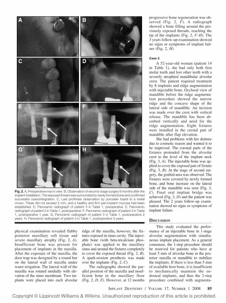
physical examination revealed flabbyposterior maxillary soft tissue andsevere maxillary atrophy (Fig. 2, A).Insufficient bone was present forplacement of implants in the maxilla.After the exposure of the maxilla, thedoor trap was designed by a round buron the lateral wall of maxilla underwater irrigation. The lateral wall of themaxilla was rotated medially with ele-vation of the sinus membrane. Two im-plants were placed into each alveolar
ridge of the maxilla, however, the fix-tures exposed in sinus cavity. The inject-able bone (with beta-tricalcium phos-phate) was applied in the maxillarysinus and around the fixtures completelyto cover the exposed thread (Fig. 2, B).A spark-erosion prosthesis was madeover the implants (Fig. 2, C).
The radiographs showed the par-allel position of the maxilla and insuf-ficient bone in the maxillary floor(Fig. 2, D, E). However, at 12 months
progressive bone regeneration was ob-served (Fig. 2, F). A radiographshowed a bone filling around the pre-viously exposed threads, reaching thetip of the implants (Fig. 2, F–H). The3 years follow-up examination showedno signs or symptoms of implant fail-ure (Fig. 2, H).
Case 2
A 52-year-old woman (patient 14in Table 1), she had only both firstmolar teeth and lost other teeth with aseverely atrophied mandibular alveolarcrest. The patient required treatmentby 8 implants and ridge augmentationwith injectable bone. Occlusal view ofmandible before the ridge augmenta-tion procedure showed the narrowridge and the concave shape of thelateral side of mandible. An incisionwas made over the crest with verticalrelease. The mandible has been ab-sorbed vertically and need for theridge augmentation. Eight fixtureswere installed in the crestal part ofmandible after flap elevation.
She had problems with her denturedue to cosmetic reason and wanted it tobe improved. The coronal parts of thefixtures protruded from the alveolarcrest to the level of the implant neck(Fig. 3, A). The injectable bone was ap-plied to cover the exposed part of fixture(Fig. 3, B). At the stage of second sur-gery, the grafted area was observed. Thefixtures were covered by newly formedbone, and bone increase on the lateralside of the mandible was seen (Fig. 3,C). Final oral implant bridge wasachieved (Fig. 3, D) and the patient waspleased. The 2 years follow-up exami-nation showed no signs or symptoms ofimplant failure.
DISCUSSION
This study evaluated the perfor-mance of an injectable bone in 1-stagealveolar augmentation with simulta-neous implant placement. As a generalconsensus, the 1-step procedure shouldbe reserved for patients who have atleast 5 mm of alveolar bone in the pos-terior maxilla or mandible to stabilizethe implants. If there is less than 5 mmof available host bone, it is insufficientto mechanically maintain the en-dosteal implants, and thus the 2-stepprocedure combined with augmenta-
Fig. 2. A, Preoperative macro view. B, Observation of second-stage surgery 6 months after theimplant installation. The exposed thread was surrounded by newly formed bone and confirmedsuccessful osseointegration. C, Last prothesis observation by porcelain fused to a metalcrown. These did not exceed 2 mm, and a healthy and firm peri-implant mucosa had beenestablished. D, Panoramic radiograph of patient 3 in Table 1, preoperative. E, Panoramicradiograph of patient 3 in Table 1, postoperative. F, Panoramic radiograph of patient 3 in Table1, postoperative 1 year. G, Panoramic radiograph of patient 3 in Table 1, postoperative 2years. H, Panoramic radiograph of patient 3 in Table 1, postoperative 3 years.
IMPLANT DENTISTRY / VOLUME 17, NUMBER 1 2008 85

tion procedure is recommended forthese patients.13–15 On the other hand,the 1-step procedure offers the advan-tages of less surgical treatment for thepatient and coordinated consolidationof the graft around the implants duringhealing, thus reducing the surgical andhealing times for the patient. Anotheradvantage is that it not only eliminatesthe need to harvest autogenous bonevia its inherent morbidity, but also de-creases the surgical recovery time.16 Inthis study, all cases of posterior maxillahad more than 5 mm in the sinus floorand in the mandible. The patients under-went the 1-step augmentation procedurewith injectable bone application and si-multaneous implant placement. Themacro findings showed that injectablebone induced bone regeneration and thatthe dental implant thread was not ex-posed. Thus, these results indicate thatridge augmentation caused by injectablebone and that simultaneous implantationis possible.
The results of this study provideevidence of the safety and technicalfeasibility of injectable bone for max-illary sinus floor augmentation andvertical ridge augmentation in agree-ment with those from earlier animalstudies that have indicated that treat-ment with injectable bone dose not
result in toxicity, significant immuno-logic reactions, or other serious ad-verse effects.17–20 Adverse experiences(e.g., pain, swelling after operation)observed with injectable bone wereconsistent with the usual morbidityobserved in the maxillary sinus flooraugmentation procedure and verticalridge augmentation.
Radiographic assessments indicatedthat injectable bone induced new bonegrowth in the maxillary sinus floor in100% of the patients treated, and showed8.7 mm mean increase in mineralized tis-sue. In the meantime, in clinical humantesting, protruding into the sinus cavitystimulated reactive bone regeneration byhuman bone morphogenetic protein-2 thatis limited to 8.51 mm in height.21 This isalmost the same as that regenerated byinjectable bone in this study. Furthermore,in the case of vertical ridge augmentationthe mean increase of mineralized tissuewas 5 mm, which was affected by thestability of the grafted area. These effectsmight be dependent on MSCs and PRP.The MSCs in the bone marrow are in-duced into cells with osteogenic capacity,the MSCs are considered to be more fea-sible for this tissue engineering becausethe former proliferates faster because of alower degree of differentiation. In addi-tion, the PRP contains not only fibrinogen
that forms a fibrin network acting as amatrix but also cytokinetic substancessuch as platelet-derived growth factor,transforming growth factor beta, and fi-broblast growth factor. These growth fac-tors contribute to cellular proliferation,matrix formation, collagen synthesis, os-teoid production, and other processes thataccelerate tissue regeneration.
CONCLUSION
This study showed that injectablebone induced bone in the anatomicalsite in 100% of the patients. The re-sults also indicate that it might be pos-sible to achieve osseointegration ofsimultaneous implant placements withinjectable bone grafts. It may be pos-sible that the injectable bone canshorten the period of implant treat-ment and reduce the patient’s burdenphysically and mentally. The potentialfor injectable bone in general and inparticular is exciting for both patientsand dental practitioners. The releasefor general clinical use seems to bevery near, although it has not yet beenapproved for use by the Japanese Foodand Drug Administration.
Disclosure
The authors claim to have no finan-cial interest in any company or any ofthe products mentioned in this article.
ACKNOWLEDGMENTS
The authors wish to thank to Kenji Ito,and the members of the Department ofOral and Maxillofacial Surgery,Nagoya University Graduate Schoolof Medicine for help, encouragementand contributions to the completion ofthis study. This work was partly sup-ported by the Ministry of Education,Culture, Sports, Science and Technol-ogy (MEXT) on Grant-in-Aid foryoung scientists (B) (15791163) andby Japan Society for the Promotion ofScience (JSPS) on Grant-in-Aid forScientific Research (B) (16390583).
REFERENCES
1. Dahlin C, Sennerby L, Lekholm U,et al. Generation of new bone around tita-nium implants using a membranetechnique: An experimental study in rab-bits. Int J Oral Maxillofac Implants.1989;4:19-25.
Fig. 3. A, Macro view of fixtures insertion into a prepared implant site. The coronal part offixtures was protruded from the alveolar crest to level the implant neck with next natural teeth.B, Macro view of a tissue-engineered bone application. C, Observation of second-stagesurgery 4 months after the implant installation. The exposed thread was surrounded by newlyformed bone and confirmed successful osseointegration. D, Final prosthesis observation.
86 INJECTABLE BONE APPLIED FOR RIDGE AUGMENTATION AND DENTAL IMPLANT PLACEMENT

2. Hersh JM, Ericsson I. Maxillary sinusaugmentation using mandibular bone graftsand simultaneous installation of implants.Clin Oral Implants Res. 1991;2:91-96.
3. Smiler DG, Johnson PW, LozadaJL, et al. Sinus lift grafts and endosseousimplants. Treatment of the atrophic poste-rior maxilla. Dent Clin North Am. 1992;36:151-186.
4. Wheeler SL, Holmes RE, CalhounCJ. Six-year clinical and histologic study ofsinus-lift grafts. Int J Oral Maxillofac Im-plants. 1996;11:26-34.
5. Moy PK, Lundgren S, Holmes RE.Maxillary sinus floor augmentation. IntJ Oral Maxillofac Implants. 1993;51:857-862.
6. Wood RM, Moore DL. Grafting ofthe maxillary sinus with intraorally har-vested autogenous bone prior to implantplacement. Int J Oral Maxillofac Implants.1988;3:209-214.
7. Yamada Y, Boo JS, Ozawa R, et al.Bone regeneration following injection ofmesenchymal stem cells and fibrin gluewith a biodegradable scaffold. J Crani-omaxillofac Surg. 2003;31:27-33.
8. Yamada Y, Ueda M, Naiki T, et al.Autogenous injectable bone for regenera-tion with mesenchymal stem cells andplatelet-rich plasma: Tissue-engineeredbone regeneration. Tissue Eng. 2004;10:955-964.
9. Langer R, Vacanti JP. Tissue engi-neering. Science. 1993;260:920-926.
10. Ueda M, Yamada Y, Ozawa R, et
al. A clinical report of injectable tissue-engineered bone applied for alveolar aug-mentation with simultaneous implantplacement. Int J Periodontics RestorativeDent. 2005;25:129-137.
11. Pittenger MF, Mackay AM, BeckCB, et al. Multilineage potential of adult hu-man mesenchymal stem cells. Science.1999;284:143-147.
12. Tatum H. Maxillary and sinus im-plant reconstructions. Dent Clin North Am.1986;30:207-230.
13. Jensen J, Simonsen EK, Sindet-pedersen S. Reconstruction of the se-verely resorbed maxilla with bone graftingand osseointegrated implants: A prelimi-nary report. J Oral Maxillofac Surg. 1990;48:27-32.
14. Marx RE. Clinical application ofbone biology to mandibular and maxillaryreconstruction. Clin Plast Surg. 1994;21:377-392.
15. Raghoebar GM, Brouwer TJ, Re-intsema H, et al. Augmentation of the max-illary sinus floor with autogenous bone forplacement of endosseous implants: A pre-liminary report. J Oral Maxillofac Surg.1993;51:1198-1203.
16. Marx RE, Morales MJ. Morbidityfrom bone harvested in major jaw recon-struction. J Oral Maxillofac Surg. 1988;48:196-203.
17. Smith JL. Osseous regeneration inpreclinical models using bioabsorbabledelivery technology for recombinant hu-man bone morphogenetic protein 2
(rhBMP-2). J Control Release. 1995;36:183-195.
18. Gerhart TN, Kirker-Head CA, KrizMJ, et al. Healing segmental femoral de-fects in sheep using recombinant humanbone morphogenetic protein. Clin OrthopRelat Res. 1993;293:317-326.
19. Toriumi DM, Kotler HS, LuxenbergDP, et al. Mandibular reconstruction with arecombinant bone-inducing factor: Func-tional, histologic, and biomechanical eval-uation. Arch Otolarnygol Head Neck Surg.1991;117:1101-1112.
20. Sigurdsson TJ, Lee MB, Kubota K,et al. Periodontal repair in dogs: Recombi-nant human bone morphogenetic protein-2significantly enhances periodontal regenera-tion. J Periodontol. 1995;66:131-138.
21. Boyne P, Marx RE, Nevins M, et al.A feasibility study evaluation rhBMP-2/absorbable collagen sponge for maxillarysinus floor augmentation. Int J Periodon-tics Restorative Dent. 1997;17:11-25.
Reprint requests and correspondence to:Minoru Ueda, DDS, PhDDepartment or Oral and Maxillofacial SurgeryNagoya University, Graduate Schoolof Medicine65 Tsurumai-choShowa-ku, Nagoya 466-8560JapanPhone: (81) 52-744-2345Fax: (81) 52-744-2352E-mail: [email protected]
Abstract Translations
GERMAN / DEUTSCHAUTOR(EN): Minoru Ueda, DDS, PhD, Yoichi Yamada,DDS, PhD, Hideaki Kagami, DDS, PhD, sowie HideharuHibi, DDS, PhD. Schriftverkehr: Professor Minoru Ueda,Abteilung fur Gesichts- und Kieferchirurgie, Nagoya Univer-sitat, medizinisches Graduiertenkolleg, 65 Tsurumai-cho,Showa-ku, Nagoya 466-8560, Japan. Telefon: �81-52-744-2345, Fax: �81-52-744-2352, eMail: [email protected] Knochengewebe in Anwendung beim Aufbauder Kieferleiste sowie bei der Einpflanzung von Implan-taten: eine Fortschrittsstudie am Menschen
ZUSAMMENFASSUNG: Zielsetzung: Die vorliegendeStudie zielte darauf ab, den Erfolg einer Implantierungsbe-handlung zu bewerten, sofern diese in Verbindung mit einemneuen Material, einem dem naturlichen Gewebe nachempfun-denen Knochen, angewendet wird. Außerdem sollte die Sta-bilitat des regenerierten Knochengewebes nach funktionaler
Belastung auf lange Sicht beurteilt werden. Methoden: Derdem naturlichen Gewebe nachempfundene Knochen fand ininsgesamt 14 Fallen Anwendung. Dabei wurde bei 6 derPatienten mit teilweise oder komplett zahnlosem Bogen eineTransplantierung am Sinusboden vorgesehen und bei 8 Pati-enten wurde gleichzeitig eine Onlay-Plastik eingesetzt.Ergebnisse: Die Studie konnte zeigen, dass das injizierbareKnochengewebe bei 100% der Patienten zu einer Bildungvon Knochengewebe an der Versuchsstelle fuhrte. Die Ergeb-nisse weisen außerdem aus, dass mit diesen Transplantatenunter Umstanden eine Knochengewebsintegration dergleichzeitig erfolgenden Implantierungsbehandlungen erzieltwerden konnte. Schlussfolgerungen: Moglicherweise kanndas injizierbare Knochengwebe die Implantierungsbehand-lungszeit verkurzen und die Belastungen des Patienten ver-ringern sowie fur eine gute langfristige Prognose sorgen.
SCHLUSSELWORTER: Studie am Menschen, injizierbares,dem naturlichen Gewebe nachempfundenes Knochengewebe,Mesenchym-Stammzellen, Zahnimplantat
IMPLANT DENTISTRY / VOLUME 17, NUMBER 1 2008 87

SPANISH / ESPAÑOLAUTOR(ES): Minoru Ueda, DDS, PhD, Yoichi Yamada,DDS, PhD, Hideaki Kagami, DDS, PhD, y Hideharu Hibi,DDS, PhD. Correspondencia a: Professor Minoru Ueda,Department of Oral and Maxillofacial Surgery, Nagoya Uni-versity, Graduate School of Medicine, 65 Tsurumai-cho,Showa-ku, Nagoya 466-8560, Japan. Telefono: �81-52-744-2345, Fax: �81-52-744-2352. Correo electronico:[email protected] inyectable aplicado para el aumento de la cresta y lacolocacion de un implante dental: Estudio sobre el progresohumano
ABSTRACTO: Proposito: El objetivo de este estudio fueevaluar clınicamente el exito de los implantes colocados juntocon un nuevo material, hueso con tejido sintetico, y la esta-bilidad a largo plazo del hueso regenerado luego de la cargafuncional. Metodos: El hueso con tejido sintetico se aplico a14 casos, que fueron 6 pacientes con arcos parciales o total-mente edentulosos programados para un injerto del piso delseno y 8 pacientes recibieron a la vez una plastia de restau-racion de la cara oclusal. Resultados: Este estudio demostroque la formacion de hueso inyectable indujo hueso en estelugar anatomico en un 100% de los pacientes. Los resultadostambien indican que podrıa ser posible lograr la oseointegra-cion de la colocacion simultanea de implantes con los injer-tos. Conclusiones: Podrıa ser posible que el hueso inyectablepuede acortar el perıodo de tratamiento con el implante yreducir los problemas para el paciente y esperar un buenpronostico a largo plazo.
PALABRAS CLAVES: estudio humano, hueso inyectablecon tejido inyectable, celulas madres mesenquimaticas, im-plantes dentales
PORTUGUESE / PORTUGUÊSAUTOR(ES): Minoru Ueda, Cirurgiao-Dentista, PhD, YoichiYamada, Cirurgiao-Dentista, PhD, Hideaki Kagami,Cirurgiao-Dentista, PhD, and Hideharu Hibi, Cirurgiao-Dentista, PhD. Correspondencia para: Professor MinoruUeda, Department or Oral and Maxillofacial Surgery,Nagoya University, graduate school of Medicine, 65Tsurumai-cho, Showa-ku, Nagoya 466-8560, Japan. Tele-fone: �81-52-744-2345, Fax: �81-52-744-2352, e-Mail:[email protected] Injetavel Aplicado para Aumento do Rebordo e Colo-cacao de Implante Dentario: Estudo do Progresso Humano
RESUMO: Objetivo: O objetivo deste estudo era avaliarclinicamente o sucesso de implantes colocados em conjuntocom osso de material novo, trabalhado pelo tecido, e a esta-bilidade do osso regenerado apos carga funcional em regimede longo prazo. Metodos: O osso trabalhado pelo tecido foiaplicado a 14 casos, os quais eram 6 pacientes com arcadasparcial ou totalmente desdentadas designados para fazer enx-erto da superfıcie da cavidade e 8 pacientes que se subme-
teram a onlay-plastia simultanea. Resultados: Este estudomostrou que a formacao de osso injetavel induziu osso nestelocal anatomico em 100% dos pacientes. Os resultados tam-bem indicam que poderia ser possıvel obter a osseointegracaode colocacoes de implante simultaneo com os enxertos. Con-clusoes: Talvez seja possıvel que o osso injetavel possaencurtar o perıodo de tratamento do implante e reduzir ofardo do paciente e aguardar bom prognostico de longo prazo.
PALAVRAS-CHAVE: estudo humano, osso trabalhado portecido injetavel, celulas-tronco mesenquimais, implantedentario
RUSSIAN /������: Minoru Ueda, ������ ���������� �����-������, ������ ���������, Yoichi Yamada, ���������������� �����������, ������ ���������,Hideaki Kagami, ������ ���������� �����������,������ ��������� � Hideharu Hibi, ���������������� �����������, ������ ���������.����� ��� ���� � �������������: Professor Mi-noru Ueda, Department or Oral and Maxillofacial Surgery,Nagoya University, graduate school of Medicine, 65Tsurumai-cho, Showa-ku, Nagoya 466-8560, Japan.�������: �81–52-744–2345, �� �: �81–52-744–2352,����� ��� ������ ���: [email protected]������� � ������� ���� ��� ���������������������� �������� ������ ��������������������� !�"�� #��������:#����������� ���������� ��������
�$%&'$: ���� ������ ������������ – ������� ����������� �������� ������������� �����������,������������� ��������� � ������, ����������� �� ����� ��������� ����� – ��������������������, � ���� ������ �������������� ��-��� ����� ������������� ������� �������������� ������. �����: �����, ������������������� ������������, ����������� � 14 ������,�� ������� 6 ��������� � ������ ��� ����������������������� ������ ����� ���� ���������������������� ��������� ������, � 8 ������������������ ����������� �������� ��������.������ �: ������ ������������ ��������, �������������� ���������������� �������� ����� ��������� ������������ ����� � 100 % ���������. ���� ���, ���������� ��������� �� ������������������������� ������������ ������������� ��-��������� � �������������������� �������.����: ��������, �� ������������ ����� �������������� ������ ��������������� ������,������� ����� �������� � ���� ������� �� ��-��!� ��������� ���������.
()&*$��$ +)���: ������������ �������,������������ ������� ����� – �������
88 INJECTABLE BONE APPLIED FOR RIDGE AUGMENTATION AND DENTAL IMPLANT PLACEMENT

������������, ����������� ��������� ������,������������� ����� ���������.
TURKISH / TURKCEYAZARLAR: Prof. Dis Hekimi Minoru Ueda, Prof. DisHekimi Yoichi Yamada, Prof. Dis Hekimi Hideaki Kagami,ve Prof. Dis Hekimi Hideharu Hibi. Yazyþma icin: Prof.Minoru Ueda, Department of Oral and Maxillofacial Sur-gery, Nagoya University, Graduate School of Medicine, 65Tsurumai-cho, Showa-ku, Nagoya 466-8560, Japonya. Tele-fon: �81–52-744–2345, Faks: �81–52-744–2352, E-posta:[email protected] Augmentasyonu ve Dental Ymplant YerleþtirilmesindeUygulanan Enjektabl Kemik: Ynsan Ylerleme Calyþmasy
OZET: Amac: Bu calısmanın amacı, yeni bir materyal olup,doku muhendisliginden elde edilmis kemik ile birlikte yerle-
stirilen implantların basarı oranının yanı sıra fonksiyonelyukleme sonrasında rejenere olmus kemigin stabilitesini uzunvadede klinik olarak degerlendirmekti. Yontem: Doku mu-hendisligi yoluyla elde edilen kemik, 14 olguda uygulandı.Bunlardan altısı kısmen veya tamamen dissiz olan arkınasinus zemini grefti yapılacak olan hastalardı. Sekiz hastaya daaynı zamanda onlay plasti uygulandı. Bulgular: Bu calısma,enjektabl kemik formasyonunun hastaların %100�unde buanatomik yerde kemik gelisimine neden oldugunu gostermi-stir. Bulgular ayrıca, es zamanlı implantlar ile greftlerarasında osseointegrasyonun mumkun olabilecegine isaret et-mistir. Sonuc: Enjektabl kemigin, implant tedavisinin sures-ini kısaltması, hastaya zahmeti azaltması ve uzun vadede iyiprognoz saglaması olası gorulmektedir.
ANAHTAR KELYMELER: insan calısması, doku muhend-isligi ile gelistirilen enjektabl kemik, mesenkimal kok hucre-leri, dental implant
JAPANESE /
IMPLANT DENTISTRY / VOLUME 17, NUMBER 1 2008 89

CHINESE /
KOREAN /
90 INJECTABLE BONE APPLIED FOR RIDGE AUGMENTATION AND DENTAL IMPLANT PLACEMENT