Embed Size (px)
Citation preview
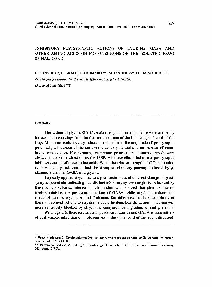
Brain Research, 100 (1975) 327-341 327 © Elsevier Scientific Publishing Company, Amsterdam - Printed in The Netherlands
INHIBITORY POSTSYNAPTIC ACTIONS OF TAURINE, GABA AND OTHER AMINO ACIDS ON MOTONEURONS OF THE ISOLATED F R O G SPINAL CORD
U. SONNHOF*, P. GRAFE, J. KRUMNIKL**, M. LINDER AND LUCIA SCHINDLER
Physiologisches lnstitut der Universitiit Miinchen, 8 Munich 2 (G.F.R.)
(Accepted June 9th, 1975)
SUMMARY
The actions of glycine, GABA, a-alanine, fl-alanine and taurine were studied by intracellular recordings from lumbar motoneurons of the isolated spinal cord of the frog. All amino acids tested produced a reduction in the amplitude of postsynaptic potentials, a blockade of the antidromic action potential and an increase of mem- brane conductance. Furthermore, membrane polarizations occurred, which were always in the same direction as the IPSP. All these effects indicate a postsynaptic inhibitory action of these amino acids. When the relative strength of different amino acids was compared, taurine had the strongest inhibitory potency, followed by fl- alanine, a-alanine, GABA and glycine.
Topically applied strychnine and picrotoxin induced different changes of post- synaptic potentials, indicating that distinct inhibitory systems might be influenced by these two convulsants. Interactions with amino acids showed that picrotoxin selec- tively diminished the postsynaptic actions of GABA, while strychnine reduced the effects of taurine, glycine, a- and fl-alanine. But differences in the susceptibility of these amino acid actions to strychnine could be detected: the action of taurine was more sensitively blocked by strychnine compared with glycine, a- and fl-alanine.
With regard to these results the importance of taurine and GABA as transmitters of postsynaptic inhibition on motoneurons in the spinal cord of the frog is discussed.
* Present address: I. Physiologisches Institut der Universit/it Heidelberg, 69 Heidelberg, Im Neuen- heimer Feld 326, G.F.R. ** Permanent address: Abteilung ftir Toxikologie, Gesellschaft fiir Strahlen- und Umweltforschung, Mtinchen, G.F.R.

328
I N T R O D U C T I O N
Recordings from ventral and dorsal roots of the isolated spinal cord of the frog have been used in several laboratories to analyze pre- and postsynaptic actions of transmitter candidates3,16-1s,39,42,45, 46. With such measurements the inhibitory
actions of some amino acids and their interactions with presumed amino acid block- ers, like picrotoxin, bicuculline and stiychnine3,1v, ls,48 have been studied by means of a direct drug application to the bathing solution. The conclusive demonstration of postsynaptic actions of transmitter candidates, such as conductance increase of the membrane, and identity of reversal potentials 4s depends on intracellular recordings. Intracellular measurements in the spinal cord and the brain stem of the cat have provided good evidence for postsynaptic actions of GABA, glycine, fl-alanine and taurine, when these substances were applied electrophoreticallyl,V,9,14,31,~, 4°,47.
With electrophoretic application techniques, test substances can be released within the immediate vicinity of single neurons. However, this technique is coupled with many artefacts and gives little knowledge about the concentration of test sub- stances. On the other hand test substances can be most easily applied to an isolated preparation, in controlled concentrations, by bathing the whole organ. Therefore it was considered valuable for pharmacological investigations to develop a technique which allows a quick exchange of the bathing solution without displacement of the intracellular recording electrode. With this technique in particular the actions of GABA, glycine, a- and fl-alanine and taurine were investigated, because pronounced activities of these substances can be seen in extracellular recordings from spinal roots of the frog and characteristic actions can be seen in mammals (for review see ref. 15).
A comparison of the effects of amino acids either released iontophoretically or applied by bathing the cord was an additional interesting possibility offered by the isolated spinal cord preparation.
Furthermore, intracellular records from spinal motoneurons of the frog were used to study the effects of the convulsants strychnine and picrotoxin on both amino acid actions and postsynaptic potentials. In the spinal cord and in the brain stem of mammals strychnine can block the actions of glycine and glycine-like 13 amino acids, e.g. a- and fl-alanine and taurine 5,11-a4,2°,28,29,38, while picrotoxin can influence the actions of GABA 8,11,19,~5. The possibility of adding amino acids to the bath in defined concentrations was used to investigate quantitative differences in the strychnine sensi- tivity of a-alanine, fl-alanine, glycine and taurine.
A preliminary report has been already published 44.
METHODS
Experiments were performed with Rana esculenta (80-120 g body wt.). After decapitation a ventral laminectomy was performed in cooled Ringer solution. The spinal cord, including dorsal and ventral roots of the lumbar segments 8-10 (see ref. 24), was removed and placed in a recording chamber. The chamber consisted of a perspex block with a small groove (volume 1.5 ml), which was continuously super-

329
fused with Ringer solution by means of a roller pump (2-3 ml/min). The temperature of the perfusion fluid was adjusted to 16 °C by a Peltier element and monitored by a thermistor placed in the chamber. Ventral and dorsal roots of the lumbar segments of one side were placed on silver wire electrodes for stimulation, and covered with a mixture of paraffin and vaseline. Test solutions were stored in vessels which could be connected to the chamber by switching the tap of a distributor system. Solutions were gassed with a mixture of 02 and CO2 and were pH-stabilized at 7.4 with Tris-(hydroxy- methyl)-methyl-2-aminoethane-sulfonic acid. pH was continuously measured with pH-electrodes. For insertion of microelectrodes, both the pia and the dura mater between ventral and dorsal roots were carefully removed. Recording electrodes (tip size below one #m) were filled either with a mixture of potassium citrate (3 M, 90 ~) and KC1 (3 M, 10 70) or only with potassium citrate (3 M). (Resistances were measured in Ringer solution 30-60 MfL) Stable intracellular records from motoneurons could commonly be achieved for 4-8 h.
Two types of applications of test substances were used. During intracellular recording from a motoneuron the cord was either superfused with Ringer solution containing amino acids, or the amino acids were applied iontophoretically. For ionto- phoresis a concentric 6-barreled iontophoresis electrode, surrounding a central re- cording electrode, was used 4a. Tip distances between electrophoresis and recording orifices varied between 30 and 50 #m. These electrodes were connected to a 6-channel constant current generator, producing electrophoresis currents variable from 10 to 500 nA. For iontophoretic application, 0.5 M solutions of GABA, glycine and fl-ala- nine were adjusted to a pH of 3.5-4 by addition of HCI. Signals were displayed on oscilloscopes and photographed with a camera. In many cases postsynaptic poten- tials were averaged with a laboratory computer. The membrane potential was moni- tored continuously by a compensation writer (maximum frequency 1.5 Hz, linearity 4- 0.5700).
RESULTS
(1) Synaptic potentials
Stimulation of dorsal roots with low strength only evoked depolarizing post- synaptic potentials, which are most likely EPSPs, since they could not be inverted by current injection. The latency for the onset of the depolarizing deflection (EPSP) 26 measured from the beginning of the shock artefact ranged between 2.5 and 3 msec (temp. 16 °C).
Hyperpolarizing deflections did not appear unless the stimulus strength was increased significantly above threshold. Fig. 1A illustrates the appearance and in- crease in amplitude of an IPSpa4, 36 with graded stimulus strength, cutting the rising phase of the EPSP.
This hyperpolarizing component of the PSP, however, in most cases reversed spontaneously within the first minutes after penetration of the cell, even when potassium citrate electrodes were used (Fig. IB). Only in some cells hyperpolarizing

330
A C Depo[ ~ 8nA 5mY [ 6nA
0mV[ B / .\ 0 nA
2mY [L._j ~ ~ . 4nA Hyperpol.~ ~ 6nA
Sins ~ ' 10ms Fig. I. Examples of postsynaptic potentials (PSPs) evoked by dorsal root (DR) stimulation in lumbar motoneurons of the isolated spinal cord of the frog. A: PSPs obtained with increasing strength of stimulation applied to DR : signal 1 at a low strength (0,8 V, T :0.5 V) could not be inverted by current injection. With increasing stimulus strength (l.0; 1.2; 1.5 V) additional hyperpolarizing deflections appeared (signals 24). Each signal in A, B was averaged from 4 single traces; and in C each specimen consists of 4 superimposed traces. (Duration of the test pulses 5 msec.) B: spontaneous inversion of the hyperpolarizing IPSP: 4 superimposed averaged signals recorded successively during the first 2 rain after penetration. Lower trace was recorded first, the hyperpolarizing deflection reversed gradu- ally. C: changes in direction and amplitude of PSPs during current injection: without current (0 hA) a depolarizing potential was recordable. Hyperpolarizing current (4-6 hA) increased the amplitude of PSPs (observe notch in the rising phase). As a result &depolarizing current (4 8 hA) a hyperpolarizi ng PSP-component appeared.
IPSPs remained stable for longer per iods (10 m i n - I h, see Fig. 2A). But in the major i ty
of cells immedia te ly after pene t ra t ion only depolar iz ing PSPs were observed. Injec-
t ions o f depolar iz ing currents revealed a mixed pa t te rn consist ing of an early depolar iza-
t ion (EPSP), fol lowed by a hyperpola r iza t ion (1PSP) (see Fig. I C). Ka tz and Miledi s4
suggest that an extrusion of chlor ide ions f rom the electrode could be responsible for
the invers ion of the IPSP. Since the hyperpola r iz ing signal inversed even when potas-
sium ci t ra te pipet tes were used, they assumed that this might be explained ei ther by a
leakage o f NaCI into the cell a round the po in t of impalement , or tha t there might be
less d iscr iminat ion between ci t ra te and chlor ide in the f rog than in mammal i an neu-
roils.
(2) Postsynaptic (ffects of amino acids
Dur ing in t racel lu lar recording the spinal cord was superfused for a few minutes
with Ringer solut ion conta in ing amino acids. Act ions of amino acids were seen on
membrane potent ia l , pos tsynapt ic potent ials , ac t ion potent ials , and membrane con-
ductance. The effects were quant i ta t ive ly dependent upon the amino acid concentra-
t ion in the test solution. F o r compar i son o f the inh ib i tory s t rength of different amino
acids, concent ra t ions of 8 x 10 -3 M were used, at which clear effects of all substances

331
could be observed. A n app l i ca t ion t ime o f 3 min p roved to be long enough to reach a
s teady state o f act ion, which could no t be increased by ex tended appl ica t ions . The
de lay in onse t of the ac t ion was main ly due to the t r a n s p o r t t ime in the tube system
(1.5 min). The t ime be tween the ent rance of the test so lu t ion into the recording cham-
ber and the first de tec table effect ranged between 20 and 25 sec.
(a) Actions on membrane potential and PSPs Appl i ca t i on o f amino acids led to a l t e ra t ions in m e m b r a n e potent ia l , the direc-
t ion o f which co r re sponded always to the d i rec t ion o f the IPSPs recorded at the same
t ime. Somet imes recordings o f hyperpo la r iz ing IPSPs could be ob ta ined for a b o u t 1
h, in which case amino acid p roduced a m e m b r a n e hype rpo la r i za t ion (Fig. 2A). The
D C record ing o f the m e m b r a n e po ten t ia l o f Fig. 2 is super imposed with synapt ic
A o,oe, c
- GLYC 8 X10"l"l I ~ ~ 5"~S GLYC BxlO'~l 1 " " ~ " " ~ , . . / -'- mV :
10 ms 20 ms 60
3min 223 min Taurine 3 min' GABA 8x10"3H 10-3 i
GABA 8 x I(T'H ~ I - ' ~ D .jL.~
. . . .
2 . L____ 20 I_2k lOms lOs~
Fig. 2. Postsynaptic actions of amino acids. A: records from a cell with a stable hyperpolarizing IPSP. During amino acid application a hyperpolarization of the membrane potential develops (DC-re- cording of membrane potential is superimposed by synaptic potentials evoked by DR-stimulation with 0.2/sec). Changes of individual PSPs corresponding to each DC-recording are shown below (control 1, maximal effect 2). B: amino acid actions on the same cell after the hyperpolarizing IPSP had inversed spontaneously. Membrane polarizations due to amino acids are now depolarizing. Time of amino acid applications marked by bars (BALA = fl-alanine). To the left of each record the time after penetration of the cell is noted. C" upper trace, electrotonic potential followed by a PSP after DR-stimulation; lower trace, DC-recording of membrane potential superimposed by DR-PSPs and a hyperpolarizing electrotonic potential (2 nA). Control: signal 1 ; signals 2-4 are taken during amino acid action. Each signal of the upper plot has been averaged during the time intervals marked by bars (1-4). The electrotonic potential which is much faster in rise time than the PSP in the DC-recording appears with a smaller amplitude because of the low frequency response of the compensation writer. Taurine reduces the membrane resistance synchronously with the amplitudes of the PSPs. D: during electrophoretic application of 48 nA glycine the SD-component of the antidromic AP is blocked and the IS-component is decreased. Membrane potential (registration from top downwards to the right) shows a depolarization during the glycine action (resting membrane potential 45 mV; iontophoresis current 48 nA).

332
potentials following rhythmic dorsal root (DR) stimulation (0.2/sec). The synaptic potential, shown on a fast time base in the insets of Fig. 2A, consists of an EPSP followed by a hyperpolarizing IPSP. In addition to the amino acid-induced hyper- polarization (referring to the thick line lying in between the inversion points of the deflections), there is a reduction of the amplitude of the IPSP (compare the averaged traces 1 with 2) evoked by all 3 amino acids tested in this experiment and an additional reduction of the EPSP during the strong action of fl-alanine. More than 2 h later, the IPSPs had reversed into depolarizing transients (Fig. 2B, averaged traces). During this state of the cell, glycine, GABA, and fl-alanine depolarized the cell membrane. The reduction in amplitude of the compound PSP occurring during amino acid application (averaged traces 2) appears to be of a magnitude similar to that seen in A. The parallel behavior of the amino acid polarization and the compound IPSP illustrated in Fig. 2A and B may indicate that identical equilibrium potentials, i.e. identical ionic processes, are involved in both events. Since IPSPs commonly reversed shortly after penetration of the cell, in the majority of neurons examined, amino acid applications caused a depolarization of the membrane potential.
(b) Actions on membrane resistance and PSPs
The membrane resistance, as indicated by the amplitude of an electrotonic potential produced by constant current pulses applied through the intracellular electrode, decreased during the application of GABA, glycine, a- and fl-alanine and taurine. In Fig. 2C this is exemplified by the action of taurine. At a concentration of 10 -3 M, taurine decreases the membrane resistance about 50 ~o. Changes of membrane resistance were always correlated to a synchronous decrease of the amplitude of PSPs of the same relative strength. Therefore the changes of PSP amplitudes during amino acids application were used for a quantification of the inhibitory strength of different amino acids (see section 3).
(c) Actions on antidromic action potentials The antidromic spike invasion of the SD membrane was blocked by all amino
acids tested. This is exemplified by the action of glycine applied iontophoreticalty (Fig. 2D). Two sec after the injection of 48 nA glycine the soma dendritic component is blocked, while a depolarization of the membrane potential is developing. The re- maining initial segment spike is decreased in amplitude during the action of glycine; probably due to the accompanying increase in membrane conductance 4v. After switch- ing offthe iontophoresis current, the control situation is regained within about 10 sec.
(3) A quantitative compar&on of the actions of amino acids
(a) GABA, glycine, fl-alanine and taurine The same order in the relative strength of action was obtained, when GABA,
glycine and fl-alanine were applied to the bath or by iontophoresis (numbers of ionto- phoretic measurements in parenthesis). In 52 experiments stable records for a com- parison of amino acid actions were obtained in 118 (31) cells. In 68 (19) of 72 (20) cells
333
GABA reduced the amplitude of PSPs more than did glycine; in 4 (1) cells the actions of both amino acids were equal. In 40 (12) cells fl-alanine was much more potent in diminishing PSP amplitudes than GABA. fl-Alanine always produced a stronger polarization of membrane potential than GABA, and its action endured much longer. In 26 (10) cells fl-alanine and glycine effects were compared, fl-Alanine always showed much stronger and longer lasting actions.
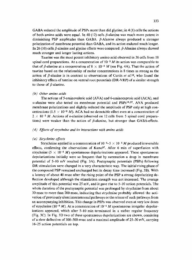
Taurine was the most potent inhibitory amino acid observed in 36 cells from 18 spinal cord preparations. At a concentration of 10 -3 M its action was comparable to that of fl-alanine at a concentration of 8 × 10 -a M (see Fig. 4A). That the action of taurine based on the relationship of molar concentrations is 8 times as strong as the action of fl-alanine is in contrast to observations of Curtis et al. 16, who found the inhibitory effects of taurine on ventral root potentials (DR-VRP) of a similar strength to those of fl-alanine.
(b) Other amino acids The actions of 5-aminovaleric acid (AVA) and 6-aminocaproic acid (ACA), and
a-alanine were also tested on membrane potential and PSPs16, 27. AVA produced membrane polarizations and slightly reduced the amplitude of PSP only at high con- centrations (1.5 × 10 -2 M). ACA had no detectable effect even at a concentration of 2 × 10 -2 M. Actions of a-alanine (observed on 12 cells from 5 spinal cord prepara- tions) were weaker than the action of fl-alanine, but stronger than GABA-effects.
(4) Effects of strychnine and its interactions with amino acids
(a) Strychnine effects Strychnine applied in a concentration of 10-6-5 × 10 -4 M produced irreversible
effects, confirming the observations of Kuno 37. After 6 min of superfusion with strychnine (5 × 10 -4 M) spontaneous depolarizations appeared. These spontaneous depolarizations initially were so frequent that by summation a drop in membrane potential of 5-10 mV resulted (Fig. 3A). Postsynaptic potentials (PSPs) following DR stimulation were changed in a very characteristic way. The initial rising phase of the compound PSP remained unchanged but its decay time increased (Fig. 3B). With a latency of about 40 msec after the rising point of the PSP a strong depolarizing de- flection developed although the stimulation strength was not increased. The average amplitude of this potential was 25 mV, and it gave rise to 5-10 action potentials. The whole duration of the postsynaptic potential was prolonged by strychnine from about 30 msec to more than 200 msec, indicating that strychnine probably allowed the acti- vation of previously silent interneuronal pathways or the release of such pathways from an accompanying inhibition. This change in PSPs was observed even at very low doses of strychnine (10 -6 M). At a concentration of 10 -4 M spontaneous irregular depolar- izations appeared, which after 5-10 min terminated in a rather regular frequency (Fig. 3C). In Fig. 3D two of these spontaneous depolarizations are shown, consisting of a slow deflection of 300-500 msec and a maximal amplitude of 25-30 mV, carrying 16-25 action potentials on top.
334
5.10 '
L 4mJn
tl
STaY .
v 5r~V
F 60
D 7O
rnV
8 .10 t t4 ..r" 8.10 ~NI
53 m,n after Strychnine ( 5 • 10 )o f f
BALA GABA GLYC 8.10 - I 8~10 -~ 8~10 -~
::;.- . . . . . . . ) a > L
~ffer Strychn*~ 18gOre s E lOOms
8x lO 'JN
BALA , ,
8~10 ~ Lmln
J, j , - - . i
i
J ~
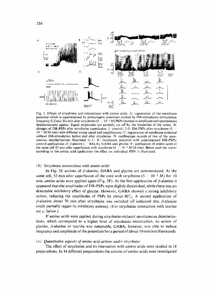
Fig. 3. Effects of strychnine and interactions with amino acids. A: registration of the membrane potential which is superimposed by postsynaptic potentials evoked by DR-stimulation (stimulation frequency 0.2/sec). Six rain after strychnine (5 ~ 10 4 M)PSPs increase in amplitude and spontaneous depolarizations appear. Signal amplitudes are partially cut off by the borderline of the writer. B: changes of DR-PSPs after strychnine superfusion, I : control; 2-5 : DR-PSPs after strychnine (5 . 10 4 M/IO rain) with different sweep speed and amplification. C: registration of membrane potential without DR-stimulation before and after strychnine. D: oscilloscope records of two of the spon- taneous depolarizations illustrated in C. E: membrane potential with superimposed DR-PSPs: control applications of/l-alanine ( B A L A ) , GABA and glycine. F: application of amino acids at the same cell 53 rain after superfusion with strychnine (5 -: 10 '~ M/IO rain). Below each bar corre- sponding to the amino acid application the effect on individual PSPs is illustrated.
(b) Strychnine interactions with amino acids In Fig. 3E act ions of /%alan ine , G A B A and glycine are demons t ra ted . At the
same cell, 53 min after superfusion of the cord with s t rychnine (5 ;< 10 -4 M ) for 10
rain, amino acids were appl ied again (Fig. 3F). At the first appl ica t ion of /3-a lanine it
appeared tha t the ampl i tudes o f DR-PSPs were sl ightly diminished, while there was no
detec table inh ib i tory effect o f glycine. However , G A B A showed a s t rong inh ib i tory
act ion, reducing the ampl i tudes o f PSPs by abou t 80°o. A second appl ica t ion o f
/4-alanine abou t 70 rain after s t rychnine was switched off indica ted that /%alanine
could par t ia l ly regain its inhibi tory potency. (Fo r s t rychnine interact ion with taur ine
see c, below.)
I f amino acids were appl ied dur ing s t rychnine- induced spontaneous depolar iza-
tions, which cor respond to a higher level o f s t rychnine intoxicat ion, no ac t ion o f
glycine, f l-alanine or taur ine was detectable, G A B A , however, was able to reduce
frequency and ampl i tude o f the potent ia ls for a per iod o f abou t 10 min (not i l lustrated).
( c) Quantitative aspects of amino acid actions under strychnine The effect o f s trychnine and its in terac t ion with amino acids were s tudied in 18
prepara t ions . In 14 different p repara t ions the act ions o f amino acids were investigated
335
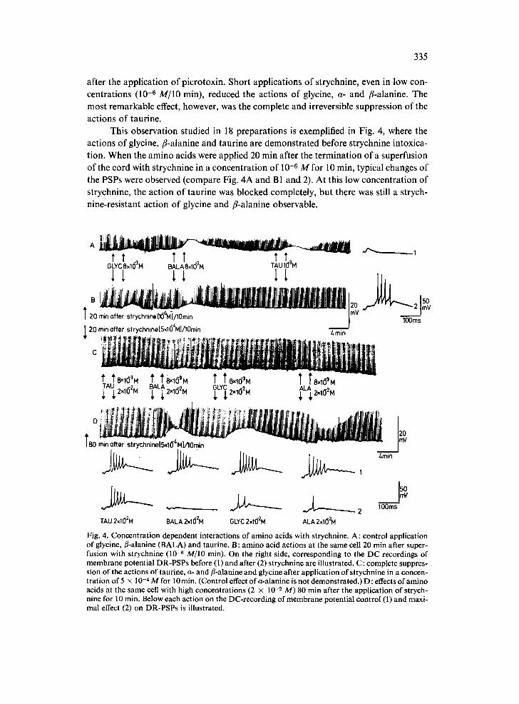
after the application of picrotoxin. Short applications of strychnine, even in low con- centrations (10 -6 M/IO min), reduced the actions of glycine, a- and fl-alanine. The most remarkable effect, however, was the complete and irreversible suppression of the actions of taurine.
This observation studied in 18 preparations is exemplified in Fig. 4, where the actions of glycine, fl-alanine and taurine are demonstrated before strychnine intoxica- tion. When the amino acids were applied 20 min after the termination o fa superfusion of the cord with strychnine in a concentration of 10 -6 M for 10 min, typical changes of the PSPs were observed (compare Fig. 4A and B1 and 2). At this low concentration of strychnine, the action of taurine was blocked completely, but there was still a strych- nine-resistant action of glycine and fl-alanine observable.
A ~ . _ 7~ ' , ' - - " ~ .r.~__._.__ 1 t t ~ TT ~ TT~
G L Y C 8 x 1 ( ~ M B A L A 8x1(~ M T A U 1( ] M
T 20 rain after strychnine[l(]~vt-]/lOmln j20 # 2 JSm~ lOOms ~ 20 rain after strychninelS~164MlDOmin Amin
!,!}1 I i i I i ! . . . . !1 , ! I ! I! ! ' ' ~ ,
_. i.'l'.i~. ..t,ili, l i t l I. lllill!l Ilt<. t .. t iliitiliiiil!l,, Iiil tlt!ttllttltilltlll , III lilillll ii','~ llilll,.. ,, i t ~ ,,~,! ,, ',L I ' , i t l =~ 7~= ~ ' i , I t 1 1 ~ 1 1 l~,,,~l,,i,~t.ltti~tliUlkilllllt"~lil,l~hllllilttll!,lu: . . . . - - " ! ~ ; _ ' - "ilht . . . . ,ll . . . . . . ill
l ! ~ ! ~ f i ~ ' ' . . . . ~ ' ' I ,
T82min offer strychnine[Sxlgl'MlA0rnin ~1~ 3 <-'o
TAU 2x10²M BALA 2x10²M OLYC 2xl(]2M ALA 2xlO2M Fig. 4. Concentration dependent interactions of amino acids with strychnine. A: control application of glycine, fl-alanine (BALA) and taurine. B: amino acid actions at the same cell 20 min after super- fusion with strychnine (10 -8 M/IO min). On the right side, corresponding to the DC recordings of membrane potential DR-PSPs before (1) and after (2) strychnine are illustrated. C: complete suppres- sion of the actions of taurine, a- and fl-alanine and glycine after application of strychnine in a concen- tration of 5 x 10 -4 M for 10 min. (Control effect of a-alanine is not demonstrated.) D : effects of amino acids at the same cell with high concentrations (2 × 10 _2 M) 80 min after the application of strych- nine for 10 min. Below each action on the DC-recording of membrane potential control (1) and maxi- mal effect (2) on DR-PSPs is illustrated.

336
Superfusion with strychnine in a concentration of 5 x 10 -'~ M for 10 min sup- pressed all amino acid (8 × 10 -3 M) actions (Fig. 4C). If the concentration of these amino acids was increased to 2 × 10 -2 M, a-alanine, fl-alanine and glycine showed depolarizations of the membrane potential and reduced the amplitude of DR-PSPs as well as the number of action potentials elicited (Fig. 4Dl,z). Taurine, however, did not produce any inhibitory effects. The order of the relative strength of action of a-alanine, fl-alanine, and glycine applied in high concentrations after strychnine was the same as in low concentrations without strychnine. In contrast, taurine which had the strong- est inhibitory potency under normal conditions was completely ineffective in the pres- ence of strychnine even when applied in this high concentration. On the same cell as illustrated in Fig. 4, taurine when applied at a concentration of 5 x 10 -2 M produced a weak reduction of PSP amplitudes (not illustrated).
(5) Effects of picrotoxin and interactions with amino acids
(a) Picrotoxin effects When the spinal cord was superfused with picrotoxin (5 x 10 -4 M), the ampli-
tudes of PSPs increased, and spontaneous long-lasting depolarizations appeared after about 5-10 min. Spontaneous depolarizations induced by picrotoxin appeared rather irregularly compared to those observed under strychnine (Fig. 5A). Two of these depolarizations photographed from the oscilloscope are shown in Fig. 5B. On their rising phase 8-10 action potentials were commonly generated and the whole duration of the signal varied between 20 and 30 sec being thus much longer than under strych- nine. In Fig. 5C the compound DR-PSP before and after picrotoxin application (5 × 10 -3 M) is illustrated. The rising phase as well as the duration were increased with picrotoxin. Stimulation at the same strength now produced several action potentials. In contrast to strychnine, where the rising phase of the PSP remained unchanged and action potentials were elicited only during a late depolarizing deflection, under picro- toxin the rising phase of the PSP increased remarkably.
The spontaneous inversion of the hyperpolarizing IPSP in motoneurons of the frog ~4 introduced difficulties when the influences of strychnine and picrotoxin upon the IPSP were to be determined. However, the pronounced differences in the change of the compound DR-PSP (E + IPSP inversed) after strychnine or after picrotoxin indicate that different inhibitory systems are blocked by these two convulsants.
(b) Picrotoxin and its interaction with amino acids' Picrotoxin diminished the effect of GABA selectively, since no interaction with
/%alanine, glycine or taurine was observed. After control applications of glycine, GABA and/3-alanine (Fig. 5D) the spinal cord was superfused with picrotoxin (5 × 10 -4 M) for 20 min. Although spontaneous picrotoxin induced depolarizations occur- red during GABA and/3-alanine actions, it seems clear that glycine and/%alanine still caused a reduction of the amplitude of PSPs and a membrane depolarization, while the action of GABA was completely blocked (Fig. 5E).
In all cases studied, picrotoxin (5 x 10 -4 to 5 x 10 -3 M) selectively influenced

337
s I i mv[ , • I A
I , I
PICRO 5x10⁻³M
8 2s T~
, . . j - . . . .~ C
PICRO "
o 10m s
mV oo[ 65
70
~t PI£~^ 20f
5xll
mV oo[ 65
70
xlo- M x,o-iM
D
t~AL~t
j 15rnV 4rain
Fig. 5. Effects of picrotoxin on membrane potential, postsynaptic potentials and interactions with amino acids. A: registration of membrane potential. During the application of picrotoxin typical spontane- ous depolarizations develop. B: oscilloscope records of two of these long-lasting spontaneous depolar- izations. C: changes of DR-PSPs after picrotoxin (5 × 10 -3 M[15 min; from another experiment). D: membrane potential with superimposed DR-PSPs; control applications of glycine, GABA and fl-alanine (BALA). E: amino acid applications at the same cell after 20 min superfusing the cord with picrotoxin (5 × 10 -a M).
the G A B A action (8 x 10 -3 M). In some experiments, however, and in contrast to
Fig. 5 the action o f G A B A was only diminished. Taurine was also applied during picrotoxin intoxication, but in no case was the taurine action found to be influenced (not illustrated).
DISCUSSION
Electrical stimulation o f ventral and dorsal roots or descending pathways o f the frog spinal cord can evoke complex responses which are recordable in adjacent ventral or dorsal roots and which have been assumed to correspond to pre- and postsynaptic events4,0,22, 23. Many investigators have studied the action o f inhibitory amino acids

338
on these signals 3,16-18,~9,42,4,~,46. GABA, glycine, and /3-alanine produce polariza- tions of the ventral roots, which can be taken as an indication of postsynaptic inhibi- tion~, 16,46. These polarizations, however, were either hyper- or depolarizing and show- ed a great variability TM. Therefore analysis of the underlying processes on the moto- neuron's membrane was rather difficult. For taurine Curtis e t al. 1~ described a depres- sion of ventral root reflexes, indicating that this amino acid may have a postsynaptic action. Our results show that GABA, glycine, a-alanine, /%alanine and taurine act postsynaptically on the motoneuron's membrane. Additional actions of amino acids on interneurons or presynaptic sites cannot be excluded with the technique of intra- cellular recording. Therefore nothing can be said about the role of GABA 3,17,18,39,46, taurine or/%alanine ~9 mediating presynaptic inhibition. The weak inhibitory action of glycine compared with taurine, fl-alanine and GABA is in correspondence with extra- cellular measurements in the spinal cord of amphibians of Curtis e t al. 16 and Fukuya2L In contrast, in the spinal cord of the cat, gJycine shows much stronger actions than GABA and/%alanine 7,13,14, while taurine produces only weak depression 13. Chemical analysis has shown that glycine is present in the lumbar enlargement of the spinal cord of the bullfrog 2. Collins 10 has recently compared the concentrations of free amino acids in the whole spinal cord. He found almost twice as much GABA as glycine and more than twice as much taurine than glycine. The relatively low concen- tration of glycine, coupled with its comparatively weak electrophysiological effects may indicate that glycine has a less important role as a transmitter in the spinal cord of the frog than GABA and taurine. However, further evidence for GABA and taurine as transmitters depend on an analysis of the distribution of these amino acids in different areas of the spinal cord. The amount of ~L-alanine in the spinal cord of the frog is about one-fifth that of glycinO °, and for/#alanine concerning the frog no data are described. However, in the CNS of other vertebrates its concentration is near or below the level of detection 16,33. The strong postsynaptic inhibitory effects of these amino acids cannot be considered as a proof of their importance as inhibitory trans- mitters in the spinal cord of the frog, since their low concentration in the tissue is contradictory to any physiological meaning.
Certain types of postsynaptic inhibition in the spinal cord of the cat can be blocked by strychnine (for review see ref. 15), while other postsynaptic inhibitory processes are not sensitive to strychnine but can be suppressed by picrotoxin ~5. The defined changes of the PSP under picrotoxin indicate an interference with inhibitory systems distinct from those which are affected by strychnine. As on spinal neurons of mammals8,11,19,25, picrotoxin blocks the postsynaptic actions of GABA on spinal motoneurons of the frog. The actions of taurine, ~L- and fl-alanine and glycine were never influenced. However, in many cases even under high concentrations of picro- toxin the action of GABA could only be diminished. Therefore picrotoxin must be considered as a specific, but weak blocker of the postsynaptic actions of GABA 3°. In summary, the postsynaptic inhibitory action of GABA on spinal motoneurons and its high concentration in the tissuO 0 can be regarded as an indication that this amino acid is of importance as a transmitter in a picrotoxin-sensitive postsynaptic inhibition.
As described in spinal neurons of the catS, 11-14,~°,28,~9,38 strychnine blocked the

339
actions of taurine, a- and fl-alanine and glycine, while GABA actions were not affected. Taurine, which before strychnine produced the strongest inhibitory effects of all amino acids tested, was the most sensitive. This observation supports the idea that the increase of excitability observed undel strychnine may derive mainly from a blockade of postsynaptic receptors on which taurine may act as a natural transmitter. The relatively high concentration of this amino acid in the spinal cord of the frog 1° also supports this hypothesis. The blockade of 'glycine receptors', as is postulated for spinal motoneurons of the cat 1~, can hardly account for the powerful, irreversible change of excitation, considering the relatively weak inhibitory potency of glycine, its relatively low concentration in the tissue 1° and its lower susceptibility to strychnine compared with taurine. Also in the Mauthner cell of the goldfish a strychnine sensitive inhibition is described, on which glycine cannot be the natural transmitterm, 41. There- fore the possibility cannot be ruled out that on spinal motoneurons of the frog glycine has not such an important role as a transmitter as in the cat spinal cord. Our results in- dicate that in the spinal cord of the frog taurine may be a more important transmitter of the strychnine-sensitive postsynaptic inhibition than glycine.
ACKNOWLEDGEMENTS
The authors thank Dr. G. ten Bruggencate, Dr. D. Richter and Dr. H. Seller for critical review of the manuscript and Dr. D. Blick and Dr. D. Tracy for valuable support in correcting the English. The preparation of the figures by Mrs. C. Kottmair, Mrs. G. Froelich and Miss A. Zimmermann is grateful acknowledged, as well as the writing of the manuscript by Miss K. Issbrficker.
This work was supported by the Deutsche Forschungsgemeinschaft.
REFERENCES
1 ALTMANN, H., BRUGGENCATE, G. TEN, SONNHOF, U., AND STEINBERG, R., Action of T-aminobutyric acid and glycine on red nucleus neurons, Pfliigers Arch. ges. Physiol., 342 (1973) 283-288.
2 APRISON, M. H., SHANK, R. P., AND DAVIDOFF, R. A., A comparison of the concentration of glycine, a transmitter suspect, in different areas of the brain and spinal cord in seven different vertebrates, Comp. Biochem. Physiol., 28 (1969) 1345-1355.
3 BARKER, J. L., AND NICOLL, R. A., The pharmacology and ionic dependency of amino acid re- sponses in the frog spinal cord, J. Physiol. (Lond.), 228 (1973) 259-277.
4 BARRON, D. H., AND MATTHEWS, B. H. C., The interpretation of potential changes in the spinal cord, J. Physiol. (Lond.), 92 (1938) 276-321.
5 BISCOE, T. J., DUGGAN, A. W., AND LODGE, D., Some actions of bicuculline and strychnine on neurones of the spinal cord and cerebral cortex of the rat, J. Physiol. (Lond.), 222 (1972) 141-142.
6 BROOKHART, J. M., MACHNE, X., AND FADIGA, E., Patterns of motor neuron discharge in the frog, Arch. itaL Biol., 97 (1959) 53-67.
7 BRUGGENCATE, G. TEN, AND ENGBERG, I., Analysis of glycine actions on spinal interneurones by intercellular recording, Brain Research, 11 (1968) 446-450.
8 BRUGGENCATE, G. TEN, AND ENGBERG, I., IS picrotoxin a blocker of the postsynaptic inhibition induced by GABA (~,-aminobutyric acid), Pfliigers Arch. ges. Physiol., 312 0969) 121.
9 BRUGGENCATE, G. TEN, AND ENGBERG, I., Iontophoretic studies in Deiters' nucleus of the inhibi- tory actions of GABA and related amino acids and the interactions of strychnine and picrotoxin, Brain Research, 25 (1971) 431~1-48.

340
10 COLLINS, G. G. S., The spontaneous and electrically evoked release of [aH]GABA from the isolat- ed hemisected frog spinal cord, Brain Research, 66 (1974) 121-137.
11 CURTIS, D. R., DUGGAN, A. W., AND JOHNSTON, G. A. R., Glycine, strychnine, picrotoxin and spinal inhibition, Brain Research, 14 (1969) 759-762.
12 CURTIS, D. R., DUGGAN, A. W., AND JOHNSTON, G. A. R., The specificity of strychnine as a glycine antagonist in the mammalian spinal cord, Exp. Brain Res., 12 (1971) 547-565.
13 CURTIS, D. R., HOSLI, L., AND JOHNSTON, G. A. R., A pharmacological study of the depression of spinal neurones by glycine and related amino acids, Exp. Brain Res., 6 (1968) 1-18.
14 CURTIS, D. R., H6SLL L., JOHNSTON, G. A. R., AND JOHNSTON, ]. H., The hyperpolarization of spinal motoneurones by glycine and related amino acids, Exp. Brain Res., 5 (1968) 235-258.
15 CURTIS, D. R., AND JOHNSTON, G. A. R., Amino acid transmitters in the mammalian central nervous system, Rev. Physiol., 69 (1974) 98-188.
16 CURTIS, D. R., PHILLIS, J. W., AND WATKINS, J. C., Actions of amino acids on the isolated hemi- sected spinal cord of the toad, Brit. J. Pharmaeol., 16 (1961) 262-283.
17 DAVIDOFV, R. A., Gamma-aminobutyric acid antagonism and presynaptic inhibition in the frog spinal cord, Science, 175 (1972) 331-333.
18 DAVIDOFF, R. A., The effects of bicuculline on the isolated spinal cord of the frog, Exp. Neurol., 35 (1972) 179-193.
19 DAVIDOFF, R. A., AND APRISON, M. H., Picrotoxin antagonism of the inhibition ofinterneurons by glycine, Life Sci., 8 (1969) 107-112.
20 DAVIDOFF, R. A., APRISON, M. H., AND WERMAN, R., The effects of strychnine on the inhibition of interneurones by glycine and ~'-aminobutyric acid, Int. J. Neuropharmacol., 8 (1969) 191-194.
21 DIAMOND, J., ROPER, S., AND YASARGIL, G. M., The membrane effects, and sensitivity to strych- nine of neural inhibition of the Mauthner cell, and its inhibition by glycine and GABA, J. Physiol. (Lond.), 232 (1973) 87-111.
22 ECCLES, J. C., Synaptic potentials of motoneurones, J. Neurophysiol., 9 (1946) 87-120. 23 ECCLES, J. C., AND MALCOLM, J. L., Dorsal root potentials of the spinal cord, J. Neurophysiol., 9
(1946) 139-160. 24 ECKER, A., WIEDERSHEIM, R., AND GAUPP, E., Anatomic des Frosches, Zweite Abteilung, 2. Auf-
lage, Vieweg Braunschweig, 1899. 25 ENGBERG, I., AND THALLER, A., On the interaction of picrotoxin with GABA and glycine in the
spinal cord, Brain Research, 19 (1970) 151-154. 26 FADIGA, E., AND BROOKHART, J. M., Interactions of excitatory postsynaptic potentials generated at
different sites on the frog motoneuron, J. Neurophysiol., 25 (1962) 790-804. 27 FUKUYA, M., Studies on some physiological properties of 7-aminobutyric acid and related com-
pounds, Jap. J. Physiol., l l (1961) 126-146. 28 GROAT, W. C. DE, The effects of glycine, GABA and strychnine on sacral parasympathetic pre-
ganglionic neurones, Brain Research, 18 0970) 542-544. 29 HAAS, H. L., AND HOSLr, L., The depression of brain stem neurones by taurine and its interaction
with strychnine and bicuculline, Brain Research, 52 (1973) 399-402. 30 HILL, R. G., SIMMONDS, M. A., AND STRAUGHAN, D. H., A comparative study of some convulsant
substances as y-aminobutyric acid antagonists in the feline cerebral cortex, Brit. J. Pharmacol., 49 (1973) 37-51.
31 H/SSLI, L., AND HAAS, H. L., The hyperpolarization of neurones of the medulla oblongata by glycine, Experientia (Basel), 28 (1972) 1057.
32 H0SL~, L., HAAS, H. L., AND HOSLI, E., Taurine - - a possible transmitter in the mammalian central nervous system, Experientia (Basel), 29 (1973) 743-744.
33 JOHNSTON, G. A. R., The intraspinal distribution of some depressant amino acids, J. Neuroehem.. 15 (1968) 1013-1017.
34 KATZ, B., AND MILEDt, R., A study of spontaneous miniature potentials in spinal motoneurones, J. Physiol. (Lond.), 168 (1963) 389-422.
35 KELLERT:4, J. O., ANO SZUMSKI, A. J., Effects of picrotoxin on stretch-activated post-synaptic in- hibitions in spinal motoneurones, Acta physiol, seand., 66 (1966) 146-156.
36 KUBOTA, K., AND BROOKHART, J. M., Inhibitory synaptic potential of the frog motoneurones, Amer. J. Physiol., 204 (1963) 660-666.
37 KUNO, M., Effects of strychnine on the intracellular potentials of spinal motoneurons of the toad, Jap. J. Physiol., 7 (1957) 42-50.
38 LARSON, M. D., An analysis of the action of strychnine on the recurrent I PSP and amino acid in- duced inhibitions in the cat spinal cord, Brain Research, 15 (1969) 185-200.
341
39 NICOLL, R. A., AND BARKER, J. L., Effects of strychnine on dorsal root potentials and amino acid responses in frog spinal cord, Nature New Biol., 246 (1973) 224-225.
40 OBATA, K., ITO, M., OCHI, R., AND SATO, N., Pharmacological properties of the postsynaptic inhibition by Purkinje cell axons and the action of 7-aminobutyric acid on Deiters' neurones, Exp. Brain Res., 4 (1967) 43-57.
41 ROPER, S., AND DIAMOND, J., DOeS strychnine block inhibition postsynaptically, Nature (Lond.), 223 (1969) 1168-1169.
42 SCHMIDT, R. F., Pharmacological studies on the primary afferent depolarization of the toad spinal cord, Pfliigers Arch. ges. Physiol., 277 (1963) 325-346.
43 SONNHOF, U., A multi-barrelled coaxial electrode for iontophoresis and intracellular recording with a gold shield of the central pipette for capacitance neutralization, Pfliigers' Arch. ges. Physiol., 341 (1973) 351-358.
44 SONNHOF, O., GRAFE, P., KRUMNIKL, G., LINDER, i . , AND SCHINDLER, L., Postsynaptic actions of GABA, glycine and glutamate on motoneurons of the isolated spinal cord of the frog, Pfliigers Arch. ges. PhysioL, 343, Suppl. R136 (1973).
45 TEBECIS, A. K., AND PHILLIS, J., The effects of topically applied biogenic monoamines on the iso- lated toad spinal cord, Comp. Biochem. Physiol., 23 (1967) 553-563.
46 TEBEClS, A. K., AND PHILLIS, J. W., The use of convulsants in studying possible functions of amino acids in the toad spinal cord, Comp. Biochem. Physiol., 28 (1969) 1303-1315.
47 WERMAN, R., DAVIDOFF, R. A., AND APRISON, M. H., Inhibitory action of glycine on spinal neu- rons in the cat, J. Neurophysiol., 31 (1968) 81-95.
48 WERMAN, R., Criteria for identification of a central nervous system transmitter, Cornp. Biochem PhysioL, 18 (1966) 745-766.