Embed Size (px)
Citation preview

© Copyright The Korean Academy of Asthma, Allergy and Clinical Immunology • The Korean Academy of Pediatric Allergy and Respiratory Disease http://e-aair.org 25
INTRODUCTION
Asthma is a chronic airway disease characterized by airway remodeling. The related structural changes consist of subepi-thelial fibrosis, myofibroblast and myocyte hyperplasia, and an increase in smooth muscle fibers.1,2 Airway remodeling results in lung function decline and is associated with a lack of respon-siveness to treatment. Recently, the traditional concept of air-way remodeling, as a consequence of long-standing airway in-flammation, has shifted. Instead, airway remodeling is now thought to proceed in parallel with disease development.3-6 At the same time, the treatment strategy for asthma is changing because of the limited ability of inhaled corticosteroids to re-verse airway remodeling.3,7-9
Epithelial-mesenchymal transition (EMT) describes the dif-ferentiation of a polarized epithelium, attached to the basal membrane, into fibroblast-type mesenchymal cells.10 EMT is a physiological process that plays a role in embryogenesis, heal-
ing, and repair. However, EMT is also involved in pathologic conditions, such as cancer and abnormal fibrosis seen in idio-pathic pulmonary fibrosis. Moreover, studies of the bronchial epithelium have suggested that abnormal EMT is involved in bronchial asthma.2,11,12
Resveratrol (trans-3,4,5-trihydroxystilbene) is a natural poly-
Inhibitory Effects of Resveratrol on Airway Remodeling by Transforming Growth Factor-β/Smad Signaling Pathway in Chronic Asthma ModelHwa Young Lee, In Kyoung Kim, Hyoung Kyu Yoon, Soon Suk Kwon, Chin Kook Rhee,* Sook Young Lee*
Division of Allergy, Pulmonary and Critical Care Medicine, Department of Internal Medicine, The Catholic University of Korea, Seoul, Korea
This is an Open Access article distributed under the terms of the Creative Commons Attribution Non-Commercial License (http://creativecommons.org/licenses/by-nc/4.0/) which permits unrestricted non-commercial use, distribution, and reproduction in any medium, provided the original work is properly cited.
Purpose: Asthma is a chronic airway disease characterized by airway remodeling, leading to a progressive decline in lung function. Therapeutic agents that attenuate airway remodeling can complement the limited effects of traditional glucocorticoids. In this study, we investigated the effect of resveratrol on allergic airway inflammation and remodeling in a murine model of chronic bronchial asthma. Methods: Peribronchial smooth mus-cle thickening that developed in mice challenged with a 3-month repeated exposure to ovalbumin (OVA) was used to study airway remodeling. Oral resveratrol was administered daily during the OVA challenge. The expression of TGF-β1/Smad signaling proteins and downstream mesenchymal markers in the presence or absence of resveratrol was examined in bronchial epithelial cells. Results: OVA sensitization and chronic challenge in-creased airway hyperresponsiveness, inflammation, goblet cell hyperplasia, α-smooth muscle actin (SMA), and collagen deposition. Resveratrol ef-fectively suppressed OVA-induced airway inflammation and remodeling. The expression of TGF-β1/phosphorylated Smad2/3 was increased in the lung tissues of OVA-challenged mice but effectively inhibited by resveratrol. In bronchial epithelial cells, the TGF-β1-induced expression of the mes-enchymal markers snail, slug, vimentin, and α-SMA was suppressed by resveratrol treatment. Conclusions: Resveratrol effectively ameliorated both airway inflammation and airway structural changes in a mouse model of bronchial asthma. These effects were mediated by decreased TGF-β1 expression, in turn suppressing TGF-β1/Smad signaling and the epithelial-mesenchymal transition process. Our results demonstrate the potential benefits of resveratrol for the treatment of airway remodeling associated with bronchial asthma.
Key Words: Resveratrol; TGF-β1; airway remodeling
Correspondence to: Chin Kook Rhee, MD, PhD, Division of Allergy, Pulmonary and Critical Care Medicine, Department of Internal Medicine, Seoul St. Mary’s Hospital, College of Medicine, The Catholic University of Korea, 222, Banpo-daero, Seocho-gu, Seoul 06591, Korea. Tel: +82-2-2258-6067; Fax: +82-2-599-3589; E-mail: [email protected]
Co-correspondence to: Sook Young Lee, MD, PhD, Division of Allergy, Pulmonary and Critical Care Medicine, Department of Internal Medicine, Seoul St. Mary’s Hospital, College of Medicine, The Catholic University of Korea, 222, Banpo-daero, Seocho-gu, Seoul 06591, Korea.Tel: +82-2-2258-6061; Fax: +82-2-599-3589; E-mail: [email protected]: January 17, 2016; Revised: May 25, 2016; Accepted: June 08, 2016•There are no financial or other issues that might lead to conflict of interest.
Original ArticleAllergy Asthma Immunol Res. 2017 January;9(1):25-34.
https://doi.org/10.4168/aair.2017.9.1.25pISSN 2092-7355 • eISSN 2092-7363

Lee et al.
Allergy Asthma Immunol Res. 2017 January;9(1):25-34. https://doi.org/10.4168/aair.2017.9.1.25
Volume 9, Number 1, January 2017
26 http://e-aair.org
phenol found in various fruits and vegetables, and in abun-dance in grapes. In addition to its anti-cancer, anti-inflammato-ry, and anti-oxidant effects,13 resveratrol has been shown to protect against airway inflammation and remodeling in a mouse model of asthma.14,15 These effects of resveratrol have been mainly explained by its inhibition of the cyclooxygenase pathway and nuclear factor kappa B mediated by the inhibition of IκB kinase.13 Moreover, evidence that resveratrol inhibiting the transforming growth factor (TGF)-β1 signaling pathway leads to EMT induction is emerging. For example, resveratrol has been shown to suppress the invasion and metastasis of lung cancer cells and colorectal cancer cells by inhibiting TGF-β1 induced EMT.16,17 In renal tubular epithelial cells, resve-ratrol attenuates renal injury and fibrosis by inhibiting TGF-β1-induced EMT signaling and metalloproteinase 7.18 In addition, the suppressive effects of natural compounds on airway re-modeling in an animal model of bronchial asthma have been described. These compounds include kaempferol, triptolide, praeruptorin, and sesamin, which act as inhibitors of TGF-β1/Smad signaling.19-21
In this study, we investigated the effects of resveratrol on air-way inflammation and remodeling in a murine model of chron-ic bronchial asthma based on repeated ovalbumin (OVA) chal-lenge and in a bronchial epithelial cell line. Specifically, we as-sessed whether resveratrol could inhibit TGF-β1/Smad signal-ing and EMT.
MATERIALS AND METHODS
Animals and experimental designFemale 7-week-old BALB/c mice (Dae-Han Experimental An-
imal Center, Daejon, Korea) were used in this study. The mice were randomly assigned to 1 of the 4 following: (1) control, (2) OVA challenge, (3) OVA challenge plus 10 mg resveratrol/kg ad-ministered orally, and (4) OVA challenge plus 50 mg resvera-trol/kg administered orally.
Sensitization and antigen challenge protocolThe mice were immunized by subcutaneously injecting with
25 µg of OVA (grade V; Sigma-Aldrich, St. Louis, MO, USA) ad-sorbed to 1 mg of aluminum hydroxide (Aldrich, Milwaukee, WI, USA) in 200 µL of phosphate-buffered saline (PBS). The in-jections were administered on days 0, 7, 14, and 21. Intranasal challenge with 20 μg OVA/50 μL PBS was administered on days 27, 29, and 31 to mice under isoflurane (Vedco, St. Joseph, MO, USA) anesthesia. The intranasal OVA challenges were then re-peated twice a week for 3 months. Age- and sex- matched con-trol mice were treated identically but with PBS alone. All of the mice were euthanized 24 hours after the final OVA challenge, at which time bronchoalveolar lavage (BAL) fluid and lung tissues were obtained. All of the animal research procedures were con-ducted in accordance with the Laboratory Animals Welfare Act,
the Guide for the Care and Use of Laboratory Animals, and the Guidelines and Policies for Rodent Experiments mandated by the Institutional Animal Care and Use Committee of the School of Medicine, The Catholic University of Korea.
Resveratrol administrationResveratrol (Sigma, R5010) was prepared as a 100 mM stock
solution in ethanol and stored at -20°C. Allergy-sensitized mice were treated by oral gavage with 0.2 mg of saline containing res-veratrol (10 or 50 mg/kg) once a day starting on day 35 for 3 months. Control mice were treated with normal saline in the same way.
Measurement of airway hyperresponsiveness Airway hyperresponsiveness (AHR) to methacholine (Mch;
Sigma-Aldrich) was measured 24 hours after the final OVA in-halation using the flexiVent system (SCIREQ, Montreal, Que-bec, Canada) as previously described.22 Briefly, the mice were anesthetized with an intraperitoneal injection of a 1:4 mixture of Rompun and Zoletil. After exposure and cannulation of the trachea, the mice were connected to a computer-controlled small-animal ventilator and ventilated with a tidal volume of 10 mL/kg at a frequency of 150 breaths/min and a positive end-expiratory pressure of 2 cm H2O, to achieve a mean lung vol-ume close to that during spontaneous breathing. Each mouse was administered PBS, followed by increasing concentrations of Mch (6.25, 12.5, 25, 50 mg/mL) in PBS. The peak airway re-sponse to the inhaled Mch was recorded.
Bronchoalveolar lavage Immediately after the AHR measurement, BAL was per-
formed by instillation into the lung of 1 mL of sterile PBS through the trachea. The total number of cells in the BAL fluid was counted using a hemacytometer. The BAL fluid was cytos-pun (7 minutes at 2,000 rpm), placed onto microscope slides, and stained with Diff-Quick (Sysmax, Kobe, Japan). The per-centages of macrophages, eosinophils, lymphocytes, and neu-trophils in the BAL fluid were determined by counting 400 leu-kocytes in randomly selected areas of the slide using light mi-croscopy. The supernatants were stored at -70°C.
Real-time polymerase chain reactionQuantitative real-time reverse-transcription polymerase
chain reaction (qRT-PCR) analysis of TGF-β1 gene expression was carried out using total RNA, isolated from lung homoge-nates, and TRIzol reagent (Invitrogen, Carlsbad, CA, USA). The primer sequences for the TGF-β1 gene were: forward 5´-GGAC TCTCCACCTGCAAGAC-3´, reverse 5´-GACTGGCGAGCCTTA GTTTG-3´. Total RNA was extracted from the cells using the Trizol reagent in accordance with the manufacturer’s protocol. One microgram of RNA was then reverse-transcribed directly using a PrimeScriptTM first-strand cDNA synthesis kit (RR037A,

Resveratrol Suppressed Airway Remodeling
Allergy Asthma Immunol Res. 2017 January;9(1):25-34. https://doi.org/10.4168/aair.2017.9.1.25
AAIR
http://e-aair.org 27
TaKaRa, Tokyo, Japan) according to the manufacturer’s proto-col. The RT samples were incubated at 37°C for 15 minutes, in-activated at 85°C for 5 seconds, and then cooled at 4°C. There-after, qRT-PCR was performed using a SYBR® FAST qPCR kit Master Mix (2×) Universal (KR0389, KAPA Biosystems, Boston, MA, USA) with the forward and reverse primers for E-cadherin, snail, slug, vimentin, and GAPDH. PCRs were carried out using a real-time PCR apparatus (CFX96 Touch; Bio-Rad, Hercules, CA, USA). The primer sequences were: snail, forward 5´-TCG-GAAGCCTAACTACAGCGA-3´, reverse 5´-AGATGAGCATTG-GCAGCGAG-3´; slug, forward 5´-AAGCA TTTCAACGCCTC-CAAA-3´, reverse 5´-CCAGAGGGAGTGAA TCCAGATTA-3´; and GAPDH, forward 5´-TGTGTCCGTCGTGGATCTGA-3´, re-verse 5´-CCTGCTTCACCACCTTCTTGAT-3’. The samples were incubated at 95°C for 3 minutes, followed by 40 cycles at 95°C for 5 seconds, and then at 60°C for 30 seconds. Differences in expression were determined in a 2 delta-delta Ct relative quan-titative analysis. The Ct is the fraction cycle number at which the fluorescence generated by the reporter dye exceeds a set level above baseline. When indicated, the target signal was nor-malized against the relative quantity of GAPDH and expressed as ΔCt=CtTarget-CtGAPDH. The change in the target signal relative to the total amount of genomic DNA was expressed as Δ ΔCt=ΔCttreatment-ΔCtcontrol. Relative changes were then calculated as 2-ΔΔCt.
Enzyme-linked immunosorbent assay (ELISA)The levels of interleukin (IL)-4, IL-5, and IL-13 were measured
in the supernatant of BAL fluid using an ELISA kit (R&D Sys-tems, Minneapolis, MN, USA) according to the manufacturer’s instructions.
Western blottingDissected lung tissues frozen in liquid nitrogen were disrupt-
ed using a Polytron homogenizer (Tissue TearorTM, Biospec Products Inc., Bartlesville, OK, USA) and then centrifuged. The proteins were purified from the supernatant, and their concen-trations were assessed using the Bradford method.23 SDS-PAGE was carried out on an 8% acrylamide gel to separate the pro-teins, which were then transferred to a polyvinylidene difluo-ride membrane (Amersham Pharmacia Biotech, Little Chal-font, UK). After a blocking step using 10% skimmed milk (BD Difco, Franklin Lakes, NJ, USA), the membrane was incubated overnight with antibodies against TGF-β1, phosphorylated (p)-Smad2, Smad2, p-Smad3 (1:1,000; Cell Signaling Technology, Danvers, MA, USA), Smad4, Smad7, and β-actin (1:1,000; Santa Cruz, CA, USA), washed 3 times with PBS, and incubated with secondary antibody for 2 hours. For the in vitro analyses, the cultured cells were lysed using radio-immunoprecipitation as-say (RIPA) cell lysis buffer containing a mixture of protease in-hibitors (GenDEPOT, San Jose, CA, USA) and centrifuged at 13,000 rpm for 30 minutes at 4°C. Equal amounts of extracted
protein (40 µg/sample) were separated by SDS-PAGE using a 10% polyacrylamide gel in Tris-glycine-SDS buffer and trans-ferred to 0.45-μm PVDF membranes. The membranes were blocked with 5% skim milk prepared in Tris-buffered saline containing 0.05% Tween 20 and incubated overnight with anti-bodies against snail and slug (1:1,000; Cell Signaling Technolo-gy) and α-smooth muscle actin (SMA) (1 µg/mL; Abcam, Cam-bridge, UK) at 4°C. The membranes were then washed and in-cubated with a horseradish peroxidase-conjugated secondary antibody. Bands were detected using the western blotting lumi-nol reagent (ELIPIS Biotech., Inc., Daejeon, Korea).
Lung tissue histopathologyFollowing the collection of BAL fluid, the mouse lungs were
inflated, fixed in 4% paraformaldehyde for 24 hours, and then embedded in paraffin. Sections (4-µm thick) cut using a micro-tome were stained with hematoxylin and eosin (H&E) using standard histological techniques. The paraffin-embedded tis-sues were also sectioned (5- to 6-µm thick) and stained with periodic acid-Schiff (PAS) to identify goblet cells in the epitheli-um. Goblet cell hyperplasia was quantified using the method described by Padrid et al.24 The pathological changes were eval-uated according to a modified five-point scoring system (grades 0-4) based on the percentage of goblet cells in the epithelium: grade 0 (no goblet cells); grade 1 (<25%); grade 2 (25%-50%); grade 3 (51%-75%); and grade 4 (>75%). The mean goblet cell hyperplasia score was then calculated for each mouse.
Measurement of smooth muscle areasAs previously described, α-SMA was immunohistochemically
detected.25 The area in each paraffin-embedded lung immu-nostained by α-SMA was outlined and quantified using a light microscope attached to an image analysis system (BX50; Olym-pus, Tokyo, Japan). The results were expressed as the immu-nostained area of the bronchiolar basement membrane (inter-nal diameter 150-200 μm). At least 10 bronchioles were count-ed in each slide.
Hydroxyproline analysisThe hydroxyproline assay was carried out using 60 mg of ho-
mogenized lung tissue from each mouse. The samples were in-cubated in 250 μL of 12 N HCl for 16 hours at 110°C. After cen-trifugation, 25 μL of supernatant was incubated for 20 minutes in 25 μL of citrate/acetate buffer (5% citric acid, 7.2% sodium acetate, 3.4% sodium hydroxide, and 1.2% glacial acetic acid) and 500 μL of chloramine T solution (1.41 g of chloramine T, 26 mL of n-propanol, 20.7 mL of distilled water, and 53.3 mL of ci-trate/acetate buffer), after which 500 μL of Ehrlich’s solution (4.5 g of p-dimerthylaminobenzaldehyde, 18.6 mL of n-propa-nol, and 7.8 mL of 70% perchloric acid) was added. These sam-ples were incubated for 15 minutes at 65°C, cooled, and then analyzed in a spectrophotometer at 550 nm. Hydroxyproline

Lee et al.
Allergy Asthma Immunol Res. 2017 January;9(1):25-34. https://doi.org/10.4168/aair.2017.9.1.25
Volume 9, Number 1, January 2017
28 http://e-aair.org
concentrations were calculated from a standard curve of hy-droxyproline.
Cell culture and resveratrol and TGF-β1 treatmentThe human bronchial epithelial cell line BEAS-2B was ob-
tained from the American Type Culture Collection (CRL-9609; ATCC, Rockville, MD, USA). The cells were cultured in DMEM/F-12 (1:1) medium (WelGENE Inc., Daegu, Korea) supplement-ed with 10% fetal bovine serum and 1% penicillin/streptomycin (10,000 units/mL and 10 mg/mL, respectively) at 37°C under 5% CO2 in air.
Resveratrol was prepared as described above and then dilut-ed in cell culture medium to obtain working concentrations. The maximum final concentration of ethanol was less than 0.025% for each treatment. Recombinant human TGF-β1 (00-21-10; Peprotech, Rocky Hill, NJ, USA) was prepared as a stock solution at a concentration of 0.1 mg/mL in 10 mM citric acid and stored at -20°C. Cells that had reached 70%-80% conflu-ence were maintained in serum-free DMEM/F-12 for 24 hours and then incubated for 48 hours with 5 µM TGF-β1 in the pres-ence or absence of different concentrations of resveratrol.
Data analysisThe results from each group were compared by ANOVA and
the nonparametric Kruskal-Wallis test, followed by post hoc testing with Dunn’s multiple comparison of means. All statisti-cal analyses were performed using Graph-Pad Prism for Win-dows software (ver. 5.00; GraphPad Software, San Diego, CA, USA). A P value of <0.05 was considered to indicate statistical significance. Results are expressed as means±SEM.
RESULTS
Inhibitory effects of resveratrol on airway inflammation and remodeling
Mice in the OVA group showed increased AHR after Mch challenge compared to mice in the control group. In the pres-ence of resveratrol, however, the increased AHR was sup-pressed (Fig. 1). After a 3-month OVA challenge, the number of inflammatory cells in BAL fluid was higher than in the control (Fig. 2) but reduced by resveratrol treatment. Total cells and eo-sinophil counts in BAL fluid were significantly lower in the res-veratrol-treated group than in the OVA group (P<0.05), as were the levels of IL-4, -5, and -13 (P<0.05) (Fig. 3).
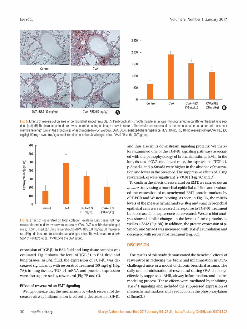
Histopathologic staining using H&E showed that compared to the OVA group, resveratrol effectively suppressed the infiltra-tion of peribronchial inflammatory cells (Fig. 4A). On the PAS-stained sections, the number of goblet cells were decreased with resveratrol treatment (Fig. 4B), and the pathological-change scores were significantly lower (Fig. 4C). Immunostain-ing of peribronchial α-SMA (Fig. 5) showed that a repetitive 3-month challenge with OVA resulted in an immunostained area of α-SMA that was significantly larger than in the control, an effect that was reduced by resveratrol (P<0.05). A hydroxy-proline assay of total lung collagen showed that resveratrol (50 mg/kg) suppressed OVA-challenge-induced hydroxyproline levels (Fig. 6).
Effects of resveratrol on the expression of TGF-β1 in BAL fluid and lung tissues
To evaluate the effect of resveratrol in TGF-β1 signaling, the
Airw
ay re
sista
nce
(cm
H 2O/
mL)
PBS 6.25 12.5 25 50
Methacholine (mg/mL)
10
9
8
7
6
5
4
3
2
1
0
ControlOVAOVA+RES (10 mg/kg)OVA+RES (50 mg/kg)
Fig. 1. Effect of resveratrol on airway hyperresponsiveness (AHR) to methacho-line (Mch). AHR was measured using the flexiVent system 24 hours after the fi-nal ovalbumin (OVA) challenge. The concentration of Mch was increased from 6.25 to 50 mg/mL. OVA, OVA-sensitized/challenged mice; RES (10 mg/kg), 10 mg resveratrol/kg+OVA; RES (50 mg/kg), 50 mg resveratrol/kg administered to sen-sitized/challenged mice. The values are means±SEM (n=5/group). *P<0.05 vs the OVA group.
*
Cells
in B
AL fl
uid
(104 /m
L)
Control OVA OVA+RES OVA+RES (10 mg/kg) (50 mg/kg)
180
160
140
120
100
80
60
40
20
0
Total cells Macrophages Eosinophils Lymphocytes Neutrophils
Fig. 2. Effect of resveratrol on total and differential cell counts in bronchoalveo-lar lavage (BAL) fluid. BAL was performed immediately after the measurement of AHR. OVA, OVA-sensitized/challenged mice; RES (10 mg/kg), 10 mg resvera-trol/kg+OVA; RES (50 mg/kg), 50 mg resveratrol/kg administered to sensitized/challenged mice. The values are means±SEM (n=8-12/group). *P<0.05 vs the OVA group.
*
*
*
*

Resveratrol Suppressed Airway Remodeling
Allergy Asthma Immunol Res. 2017 January;9(1):25-34. https://doi.org/10.4168/aair.2017.9.1.25
AAIR
http://e-aair.org 29
pg/m
Lpg
/mL
pg/m
L
Control OVA OVA+RES OVA+RES (10 mg/kg) (50 mg/kg)
Control OVA OVA+RES OVA+RES (10 mg/kg) (50 mg/kg)
Control OVA OVA+RES OVA+RES (10 mg/kg) (50 mg/kg)
IL-4
IL-13
IL-5200
150
100
50
0
100
80
60
40
20
0
100
80
60
40
20
0
Fig. 3. Effects of resveratrol on cytokine levels in BAL fluid. Interleukin (IL)-4, IL-5, and IL-13 levels in the supernatant of BAL fluid of OVA-sensitized/challenged mice were measured using an ELISA. RES (10 mg/kg), 10 mg resveratrol/kg+OVA; RES (50 mg/kg), 50 mg resveratrol/kg administered to sensitized/chal-lenged mice. The values are means±SEM (n=8-12/group). *P<0.05 vs the OVA group.
* *
*
**
Fig. 4. Effects of resveratrol on peribronchial inflammation in lung tissue. (A) The fixed tissues were embedded in paraffin, sectioned (4-µm thick), and then stained with H&E (×200). (B) The effects of resveratrol on goblet cell hyperpla-sia in lung tissue. Paraffin-embedded tissues were sectioned (5- to 6-µm thick) and then stained with periodic acid-Schiff (PAS). (C) Goblet cell hyperplasia was quantified based on the percentage of goblet cells in the epithelium, scored as follows: grade 0 (no goblet cells); grade 1 (<25%); grade 2 (25%-50%); grade 3 (51%-75%); grade 4 (>75%). The mean goblet cell hyperplasia score was cal-culated for each mouse (n=8-12/group). OVA, OVA-sensitized/challenged mice; RES (10 mg/kg), 10 mg resveratrol/kg+OVA; RES (50 mg/kg), 50 mg resveratrol/kg administered to sensitized/challenged mice. *P<0.05 vs the OVA group.
Control OVA OVA+RES OVA+RES (10 mg/kg) (50 mg/kg)
PAS point scoring
0 : No goblet cell 1 : <-25% 2 : <25%-50% 3 : <50%-75% 4 : <75%-100%
3.0
2.5
2.0
1.5
1.0
0.5
0
C
*
*Control
Control
OVA+RES (10 mg/kg)
OVA+RES (10 mg/kg)
OVA
OVA
OVA+RES (50 mg/kg)
OVA+RES (50 mg/kg)
A
B
Lee et al.
Allergy Asthma Immunol Res. 2017 January;9(1):25-34. https://doi.org/10.4168/aair.2017.9.1.25
Volume 9, Number 1, January 2017
30 http://e-aair.org
expression of TGF-β1 in BAL fluid and lung tissue samples was evaluated. Fig. 7 shows the level of TGF-β1 in BAL fluid and lung tissues. In BAL fluid, the expression of TGF-β1 was de-creased significantly with resveratrol treatment (50 mg/kg) (Fig. 7A); in lung tissues, TGF-β1 mRNA and proteins expression were also suppressed by resveratrol (Fig. 7B and C).
Effect of resveratrol on EMT signaling We hypothesize that the mechanism by which resveratrol de-
creases airway inflammation involved a decrease in TGF-β1
and thus also in its downstream signaling proteins. We there-fore examined one of the TGF-β1 signaling pathways associat-ed with the pathophysiology of bronchial asthma, EMT. In the lung tissues of OVA-challenged mice, the expression of TGF-β1, p-Smad2, and p-Smad3 were higher in the absence of reserva-tion and lower in the presence. The suppressive effects of 50 mg resveratrol/kg were significant (P<0.01) (Fig. 7C and D).
To confirm the effects of resveratrol on EMT, we carried out an in vitro study using a bronchial epithelial cell line and evaluat-ed the expression of mesenchymal EMT protein markers by qRT-PCR and Western blotting. As seen in Fig. 8A, the mRNA levels of the mesenchymal markers slug and snail in bronchial epithelial cells were increased in response to TGF-β1 treatment but decreased in the presence of resveratrol. Western blot anal-ysis showed similar changes in the levels of these proteins as well as α-SMA (Fig. 8B). In addition, the protein expression of p-Smad2 and Smad4 was increased with TGF-β1 stimulation and decreased with resveratrol treatment (Fig. 8C).
DISCUSSION
The results of this study demonstrated the beneficial effects of resveratrol in reducing the bronchial inflammation in OVA-challenged mice in a model of chronic bronchial asthma. The daily oral administration of resveratrol during OVA challenge effectively suppressed AHR, airway inflammation, and the re-modeling process. These effects were mediated by inhibiting TGF-β1 signaling and included the suppressed expression of mesenchymal markers and a reduction in the phosphorylation of Smad2/3.
Control
OVA+RES (10 mg/kg)
OVA
OVA+RES (50 mg/kg) A
Control OVA OVA+RES OVA+RES (10 mg/kg) (50 mg/kg)
2,500
2,000
1,500
1,000
500
0
B
*
*
Fig. 5. Effects of resveratrol on area of peribronchial smooth muscle. (A) Peribronchial α-smooth muscle actin was immunostained in paraffin-embedded lung sec-tions (red). (B) The immunostained area was quantified using an image analysis system. The results are expressed as the immunostained area per unit basement membrane length (µm) in the bronchioles of each mouse (n=8-12/group). OVA, OVA-sensitized/challenged mice; RES (10 mg/kg), 10 mg resveratrol/kg+OVA; RES (50 mg/kg), 50 mg resveratrol/kg administered to sensitized/challenged mice. *P<0.05 vs the OVA group.
Hydr
oxyp
rolin
e(μg
/lung
)
Control OVA OVA+RES OVA+RES (10 mg/kg) (50 mg/kg)
700
600
500
400
300
200
100
0
*
Fig. 6. Effect of resveratrol on total collagen levels in lung tissue (60 mg/mouse) determined by hydroxyproline assay. OVA, OVA-sensitized/challenged mice; RES (10 mg/kg), 10 mg resveratrol/kg+OVA; RES (50 mg/kg), 50 mg resve-ratrol/kg administered to sensitized/challenged mice. The values are means±SEM (n=8-12/group). *P<0.05 vs the OVA group.

Resveratrol Suppressed Airway Remodeling
Allergy Asthma Immunol Res. 2017 January;9(1):25-34. https://doi.org/10.4168/aair.2017.9.1.25
AAIR
http://e-aair.org 31
In our search for therapeutic agents targeting airway remodel-ing, we referred to our published reports demonstrating the suppressive effects of tyrosine kinase inhibitors, such as ima-tinib and nilotinib, in the OVA-challenged mouse model of chronic bronchial asthma.25,26 Resveratrol was investigated as an extension of previous reports of a significant inhibition of goblet cell hyperplasia, smooth muscle proliferation, and total lung collagen levels. In this work, we additionally showed that AHR and airway inflammation, both of which contribute to the remodeling process, are effectively ameliorated by resveratrol treatment. Airway remodeling consists of structural changes in the airway wall, including epithelial injury, subepithelial thick-ening, airway smooth muscle hyperplasia, goblet cell hyperpla-sia, and hypertrophy.3 Although these processes result in a pro-gressive and irreversible loss of lung function, current asthma therapies, such as corticosteroids, are ineffective in suppressing airway remodeling.27 The results of our study suggest the use of resveratrol as an additional therapeutic agent that targets not only allergic AHR and inflammation, but also the accompany-ing, irreversible structural changes in the airways.
Previous reports have described the beneficial effects of resve-ratrol in different animal models of bronchial asthma. Lee et al.15 showed that resveratrol effectively suppressed AHR, eosin-ophilia, and mucus hypersecretion in a mouse model of acute asthma. In that study, the efficacy of resveratrol was similar to that of glucocorticoid. Those authors also examined the effect of resveratrol produced in BAL fluid, both in a histopathology study and by measuring cytokine levels; their results were con-sistent with ours. In a murine model of chronic asthma induced by OVA challenge, Royce et al.14 evaluated the effects of resvera-trol with respect to subepithelial collagen deposition, goblet cell hyperplasia, and TGF-β1 immunohistochemistry staining in lung specimens. However, 6-week OVA challenge was insuf-ficient to allow assessment of peribronchial smooth muscle cell proliferation in the lung tissues of control, OVA-challenged, and OVA-challenged, resveratrol-treated mice. This is an important advantage of the asthma model used in our study compared to other animal models of chronic asthma. Other animal models could not demonstrate the changes airway smooth muscle area and total lung collagen level quantitatively. Specifically, follow-
TGF-β 1
(pg/
mL)
Rela
tive
mRN
A ex
pres
sion
Prot
ein
expr
essio
n le
vel
(nor
mal
ized
to β
-act
in)
Control OVA OVA+RES OVA+RES (10 mg/kg) (50 mg/kg)
OVA - + + + Resveratrol (mg/kg) - - 10 50
Control OVA OVA+RES OVA+RES (10 mg/kg) (50 mg/kg)
Control OVA OVA+RES OVA+RES (10 mg/kg) (50 mg/kg)
TGF-β1
3,500
3,000
2,500
2,000
1,500
1,000
500
0
3
2
1
0
5
4
3
2
1
0
A C
B D
*
*
Fig. 7. Effect of resveratrol on the expression of TGF-β1 and phosphorylated (p)-Smad in BAL fluid and lung tissues. (A) TGF-β1 expression was measured in BAL flu-id by ELISA. *P<0.05 vs the OVA group. (B) Quantitative real-time reverse-transcription polymerase change reaction (qRT-PCR) analysis of TGF-β1 gene in homoge-nized lung tissues. *P<0.05 vs the OVA group. (C and D) Expression of TGF-β1, p-Smad2, and p-Smad3 proteins in lung tissues as determined by western blotting. OVA, OVA-sensitized/challenged mice; RES (10 mg/kg), 10 mg resveratrol/kg+OVA; RES (50 mg/kg), 50 mg resveratrol/kg administered to sensitized/challenged mice. The values are means±SEM (n=8-12/group). *P<0.05 and **P<0.01 vs the control group, #P<0.05 and ##P<0.01 vs the OVA group, $$P<0.01 vs the OVA+RES10 group.
**
*
*
#
$$##
p-smad2 p-smad3 TGFb
TGF-β1
p-smad2
p-smad3
β-actin

Lee et al.
Allergy Asthma Immunol Res. 2017 January;9(1):25-34. https://doi.org/10.4168/aair.2017.9.1.25
Volume 9, Number 1, January 2017
32 http://e-aair.org
ing a 12-week OVA challenge, the area of α-SMA coverage was significantly greater than in the control group. This difference was minimized by resveratrol treatment, which effectively sup-pressed peribronchial smooth muscle proliferation (P<0.05). Our 12-week OVA challenge model is superior to previous re-ports to explain inhibitory effects of resveratrol on airway re-modeling process, more clinically relevant. Moreover, as far as we know, the role of resveratrol in TGF-β1/Smad signaling in bronchial asthma animal model has not been published yet.
TGF-β1 signaling plays a pivotal role in the airway remodeling that characterizes chronic asthma and has been implicated in most of the cellular processes involved, including subepithelial fibrosis, airway smooth muscle remodeling, and epithelial and microvascular changes. The mechanism by which TGF-β1 in-duces structural changes in bronchial asthma includes the TGF-β1/Smad pathway and TGF-β1-induced EMT. Under stim-ulation by TGF-β1, Smad proteins transport signals from the cell membrane to the nucleus, targeting genes that regulate cel-lular proliferation, transformation, and synthesis. It has been known that Smad2 and Smad3 are receptor regulated Smads (R-Smad) which are phosphorylated by the activation of TGF-β
signaling. Smad4 is a common pathway Smad, which cooper-ates to phosphorylate Smad2/3. Smad6 and Smad7 are inhibi-tory Smads (I-Smad) which down-regulate TGF- β signaling.28 Smad2 and Smad3 must be phosphorylated to be active, whereas Smad7 inhibits their phosphorylation.29,30 In vivo evi-dence of a role for the TGF-β1/Smad pathway in bronchial asthma includes increased levels of TGF-β1 and p-Smad2/3, and a decrease in Smad7 in the lung tissues of OVA-sensitized mice.19,20 In our study, we examined the balance of Smad sig-naling evoked by OVA with or without resveratrol. We observed the expression of Smad2, 3, and 4 mRNA and proteins in BEAS-2B cells as well as mouse lung tissues. Increased levels of phos-pho-Smad2 by OVA and Smad4 by TGF-β were decreased by resveratrol treatment. However, expression of inhibitory Smad7 protein was not changed with resveratrol treatment compared to ovalbumin control (data not shown). Based on these data, we found that resveratrol could effect on OVA-induced mice model through the TGF-β/Smad activation pathway. The con-tribution of EMT to airway fibrosis and remodeling in epithelial cells has been demonstrated by in vitro and in vivo studies. The EMT process can be followed by measuring the expression of
Fig. 8. Effects of resveratrol on the expression of epithelial-mesenchymal transition (EMT) protein markers and Smad proteins in BEAS-2B cells treated for 48 hours with 5 µM TGF-β1 with or without 10 or 20 µM of resveratrol. (A) The expression of snail and slug mRNA was measured by quantitative reverse transcription (qRT)-PCR. (B) Mesenchymal markers (slug, snail and α-smooth muscle actin) were examined by western blot analysis. (C) The expression of Smad2, p-Smad2 and Smad4 proteins were determined by western blotting. CON, control; TGFβ, TGF-β1 (5 µM) treated only; Res10, treated with 10 µM resveratrol; Res20, 20 µM resveratrol; TGFβ+Res10, TGF-β1 and resveratrol 10 µM; TGFβ+Res20, TGF-β1 and resveratrol 20 µM. *P<0.05 and **P<0.01 vs the TGF-β1-treated cells. #P<0.05 vs TGF-β+resveratrol (10 µM)-treated cells.
Rela
tive
mRN
A ex
pres
sion
Rela
tive
mRN
A ex
pres
sion
CON TGFβ Res10 Res20 TGFβ TGFβ +Res10 +Res20
CON TGFβ Res10 Res20 TGFβ TGFβ +Res10 +Res20
Slug Snail
TGF-β (5ng/mL) - + - - + + Resveratrol (μM) - - 10 20 10 20
- - - + + : TGF-β - 10 20 - 10 : RES (μM)
5
4
3
2
1
0
3
2
1
0
A
B C
*
#*
Slug
Snail
α-SMA
β-actin
p-Smad2
Smad2
Smad4
β-actin
* ** **
#*

Resveratrol Suppressed Airway Remodeling
Allergy Asthma Immunol Res. 2017 January;9(1):25-34. https://doi.org/10.4168/aair.2017.9.1.25
AAIR
http://e-aair.org 33
epithelial or mesenchymal markers. In an in vitro study carried out Yang et al.,31 TGF-β1-stimulated EMT was monitored in hu-man bronchial epithelial cells. TGF-β1 treatment decreased the expression of the epithelial marker E-cadherin and increased that of the mesenchymal markers snail, vimentin, and α-SMA. The artificial down-regulation of snail attenuated the TGF-β1-induced EMT phenotype. Gong et al.32 reported the inhibitory effect of kaempferol on EMT in OVA-sensitized mice. They showed that in the mouse trachea, expression of TGF-β1 and the mesenchymal marker α-SMA were increased in OVA-sensi-tized mice but decreased in mice treated with kaempferol. Our results correspond well with studies reporting elevated TGF-β1 expression in OVA-challenged mice and TGF-β1-mediated stimulation of the mesenchymal transition in bronchial epithe-lial cells. Additionally, we found that both TGF-β1 expression and Smad2/3 phosphorylation were decreased significantly in the lung tissues of OVA-challenged mice administered resvera-trol intraorally. Moreover, the TGF-β1-induced stimulation of the mesenchymal markers slug, snail, and α-SMA as well as Smad2/4 proteins in bronchial epithelial cells were effectively suppressed by resveratrol treatment.
Since the EMT is a dynamic process, Western blots of epitheli-al or mesenchymal markers provide only a “snapshot.” The re-verse phenomenon, i.e., mesenchymal-epithelial transition, can occur simultaneously and the 2 processes may influence each other.11 Thus, experimental methods able to track both are needed.
Taken together, in this study the potential therapeutic effect of resveratrol in the treatment of bronchial asthma was investigat-ed. The results showed that the oral administration of resvera-trol effectively suppressed allergic airway inflammation and re-modeling in lung tissues. These effects of resveratrol were me-diated by suppressing of TGF-β1 expression, downstream Smad activation in lung tissues, and mesenchymal transition in bron-chial epithelial cells. Therefore, by targeting the airway remod-eling process resveratrol may be an effective therapeutic agent against bronchial asthma.
REFERENCES
1. Elias JA, Lee CG, Zheng T, Ma B, Homer RJ, Zhu Z. New insights into the pathogenesis of asthma. J Clin Invest 2003;111:291-7.
2. Pain M, Bermudez O, Lacoste P, Royer PJ, Botturi K, Tissot A, et al. Tissue remodelling in chronic bronchial diseases: from the epithe-lial to mesenchymal phenotype. Eur Respir Rev 2014;23:118-30.
3. Durrani SR, Viswanathan RK, Busse WW. What effect does asthma treatment have on airway remodeling? Current perspectives. J Al-lergy Clin Immunol 2011;128:439-48.
4. Manuyakorn W, Howarth PH, Holgate ST. Airway remodelling in asthma and novel therapy. Asian Pac J Allergy Immunol 2013;31:3-10.
5. Usmani OS. Small airways dysfunction in asthma: evaluation and management to improve asthma control. Allergy Asthma Immu-nol Res 2014;6:376-88.
6. Sposato B, Scalese M, Migliorini MG, Di Tomassi M, Scala R. Small airway impairment and bronchial hyperresponsiveness in asthma onset. Allergy Asthma Immunol Res 2014;6:242-51.
7. Makino S, Sagara H. Evolution of asthma concept and effect of cur-rent asthma management guidelines. Allergy Asthma Immunol Res 2010;2:172-6.
8. Chang WS, Kim EJ, Lim YM, Yoon D, Son JY, Park JW, et al. Age-re-lated changes in immunological factors and their relevance in al-lergic disease development during childhood. Allergy Asthma Im-munol Res 2016;8:338-45.
9. Chen Y, Wong GW, Li J. Environmental exposure and genetic pre-disposition as risk factors for asthma in China. Allergy Asthma Im-munol Res 2016;8:92-100.
10. Hay ED. The mesenchymal cell, its role in the embryo, and the re-markable signaling mechanisms that create it. Dev Dyn 2005;233: 706-20.
11. Bartis D, Mise N, Mahida RY, Eickelberg O, Thickett DR. Epithelial-mesenchymal transition in lung development and disease: does it exist and is it important? Thorax 2014;69:760-5.
12. Hackett TL. Epithelial-mesenchymal transition in the pathophysi-ology of airway remodelling in asthma. Curr Opin Allergy Clin Im-munol 2012;12:53-9.
13. Wood LG, Wark PA, Garg ML. Antioxidant and anti-inflammatory effects of resveratrol in airway disease. Antioxid Redox Signal 2010; 13:1535-48.
14. Royce SG, Dang W, Yuan G, Tran J, El Osta A, Karagiannis TC, et al. Resveratrol has protective effects against airway remodeling and airway hyperreactivity in a murine model of allergic airways dis-ease. Pathobiol Aging Age Relat Dis 2011;1:7134.
15. Lee M, Kim S, Kwon OK, Oh SR, Lee HK, Ahn K. Anti-inflammato-ry and anti-asthmatic effects of resveratrol, a polyphenolic stilbene, in a mouse model of allergic asthma. Int Immunopharmacol 2009; 9:418-24.
16. Wang H, Zhang H, Tang L, Chen H, Wu C, Zhao M, et al. Resvera-trol inhibits TGF-beta1-induced epithelial-to-mesenchymal tran-sition and suppresses lung cancer invasion and metastasis. Toxi-cology 2013;303:139-46.
17. Ji Q, Liu X, Han Z, Zhou L, Sui H, Yan L, et al. Resveratrol suppress-es epithelial-to-mesenchymal transition in colorectal cancer through TGF-beta1/Smads signaling pathway mediated Snail/E-cadherin expression. BMC Cancer 2015;15:97.
18. Xiao Z, Chen C, Meng T, Zhang W, Zhou Q. Resveratrol attenuates renal injury and fibrosis by inhibiting transforming growth factor-beta pathway on matrix metalloproteinase 7. Exp Biol Med (May-wood) 2016;241:140-6.
19. Chen M, Lv Z, Jiang S. The effects of triptolide on airway remodel-ling and transforming growth factor-beta(1)/Smad signalling path-way in ovalbumin-sensitized mice. Immunology 2011;132:376-84.
20. Xiong YY, Wang JS, Wu FH, Li J, Kong LY. The effects of (+/-)-Prae-ruptorin A on airway inflammation, remodeling and transforming growth factor-beta1/Smad signaling pathway in a murine model of allergic asthma. Int Immunopharmacol 2012;14:392-400.
21. Lin CH, Shen ML, Kao ST, Wu DC. The effect of sesamin on airway fibrosis in vitro and in vivo. Int Immunopharmacol 2014;22:141-50.
22. Tarkowski M, Vanoirbeek JA, Vanhooren HM, De Vooght V, Merci-er CM, Ceuppens J, et al. Immunological determinants of ventila-tory changes induced in mice by dermal sensitization and respira-tory challenge with toluene diisocyanate. Am J Physiol Lung Cell Mol Physiol 2007;292:L207-14.

Lee et al.
Allergy Asthma Immunol Res. 2017 January;9(1):25-34. https://doi.org/10.4168/aair.2017.9.1.25
Volume 9, Number 1, January 2017
34 http://e-aair.org
23. Kruger NJ. The Bradford method for protein quantitation. Methods Mol Biol 1994;32:9-15.
24. Padrid P, Snook S, Finucane T, Shiue P, Cozzi P, Solway J, et al. Per-sistent airway hyperresponsiveness and histologic alterations after chronic antigen challenge in cats. Am J Respir Crit Care Med 1995; 151:184-93.
25. Rhee CK, Kim JW, Park CK, Kim JS, Kang JY, Kim SJ, et al. Effect of imatinib on airway smooth muscle thickening in a murine model of chronic asthma. Int Arch Allergy Immunol 2011;155:243-51.
26. Rhee CK, Kang JY, Park CK, Lee SY, Kwon SS, Kim YK, et al. Effect of nilotinib on airway remodeling in a murine model of chronic asth-ma. Exp Lung Res 2014;40:199-210.
27. Royce SG, Tang ML. The effects of current therapies on airway re-modeling in asthma and new possibilities for treatment and pre-vention. Curr Mol Pharmacol 2009;2:169-81.
28. Groneberg DA, Witt H, Adcock IM, Hansen G, Springer J. Smads as
intracellular mediators of airway inflammation. Exp Lung Res 2004;30:223-50.
29. Derynck R, Zhang YE. Smad-dependent and Smad-independent pathways in TGF-beta family signalling. Nature 2003;425:577-84.
30. Qu ZH, Yang ZC, Chen L, Lv ZD, Yi MJ, Ran N. Inhibition airway re-modeling and transforming growth factor-beta1/Smad signaling pathway by astragalus extract in asthmatic mice. Int J Mol Med 2012;29:564-8.
31. Yang ZC, Yi MJ, Ran N, Wang C, Fu P, Feng XY, et al. Transforming growth factorbeta1 induces bronchial epithelial cells to mesenchy-mal transition by activating the Snail pathway and promotes air-way remodeling in asthma. Mol Med Rep 2013;8:1663-8.
32. Gong JH, Cho IH, Shin D, Han SY, Park SH, Kang YH. Inhibition of airway epithelial-to-mesenchymal transition and fibrosis by kaempferol in endotoxin-induced epithelial cells and ovalbumin-sensitized mice. Lab Invest 2014;94:297-308.


![Protective effects of sildenafil and resveratrol on … · spectrophotometric method of Ellman [22]. Results were reported as nanomoles per gram of wet tissue. ... Protective effects](https://img.dokumen.tips/doc/110x75/5b91f06909d3f204338cd195/protective-effects-of-sildenafil-and-resveratrol-on-spectrophotometric-method.jpg)