Embed Size (px)
Citation preview

INFORMATION SECTION
© 2004. The materials are copyrighted and duplication in any manner is prohibited.
415m IDENTIFY AND DESCRIBE THE PRIMARY AND MIXED DENTITIONS
LEARNING OBJECTIVES After you have learned the information and
satisfactorily completed the Tasks section, you will be able to:
¬ Describe the primary and mixed dentition. ¬ Identify when mixed dentition is present.
¬ Describe the anatomy and appearance of primary versus permanent teeth, in terms
of size, morphology, and root form. ¬ Discuss the eruption sequence of the
primary and permanent dentitions.
KEY VOCABULARY
Deciduous teeth
Developmental spacing
Eruption
Eruption sequence
Exfoliation
Mixed dentition
Primary dentition
Root resorption
Space loss
Succedaneous
OVERVIEW It is important to recognize the primary and
permanent teeth for correct charting and treatment. The primary teeth are different from the
permanent teeth. Some of the differences are the number, color, anatomy, and eruption sequence.
The primary teeth play a vital role in reserving space for and in guiding their permanent
replacements into proper position. Sometimes the parent or patient will ask questions about primary
and permanent teeth. The information in this module will enable you to discuss characteristics
of the primary teeth and eruption of the teeth with the patient and parent.
CONCEPTS & PRINCIPLES
The first set of teeth is referred to as the PRIMARY DENTITION (Figure 1). The teeth of the
primary dentition are called primary teeth. Because they are shed, the primary teeth were
formally referred to as DECIDUOUS TEETH. The lay population may refer to primary teeth as "baby
teeth" or "milk teeth". These terms minimize the importance of the primary teeth and imply that
they are only useful for a short period of time. The health of primary teeth is very important; they are
needed for many years of growth and physical development.
As the term “deciduous” implies, primary teeth are shed and replaced by their permanent
successors, permanent or succedaneous teeth. SUCCEDANEOUS teeth are the permanent teeth
that take the place of the primary teeth after they have been shed. The process of shedding, or
naturally losing, a tooth is referred to as EXFOLIATION.
Figure 1. The primary dentition.
Primary teeth are different from the permanent teeth in number, size, morphology, color, root
form, and time of eruption.
2
NUMBER AND TYPES OF PRIMARY TEETH To accommodate the small size of the dental
arches during early child development, there are fewer teeth in the primary dentition than in the
permanent dentition. There are 20 teeth in the primary dentition, 10 in each arch and five in each
quadrant (Figure 2). They are classified as: - Incisors (8);
- Canines (4); and
- Molars (8).
There are no premolars or third molars in the
primary dentition.
Figure 2. The primary dentition. Each quadrant contains two incisors, a canine and two molars. Note the absence of premolars.
There are five primary teeth in each quadrant; they are named as:
- Central incisor;
- Lateral incisor;
- Canine (cuspid);
- First molar; and
- Second molar.
Using the Universal numbering system, the
first 20 capital letters of the alphabet are used to identify the primary teeth (Figure 3).
If some primary teeth are missing, the teeth have the same letters they would have if all the
teeth were present. For example, if the patient is missing both the primary maxillary central incisors,
E and F, and the mandibular left first molar, L; the remaining teeth would be identified as shown in
Figure 4.
Figure 3. The Universal system for identifying specific teeth in the primary dentition.
Figure 4. Example of identifying teeth in the primary dentition using the Universal system.
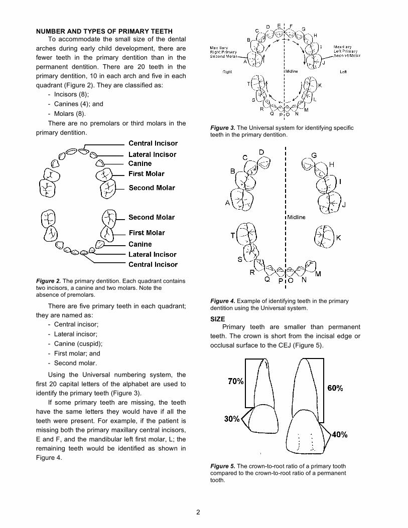
SIZE Primary teeth are smaller than permanent
teeth. The crown is short from the incisal edge or
occlusal surface to the CEJ (Figure 5).
Figure 5. The crown-to-root ratio of a primary tooth compared to the crown-to-root ratio of a permanent tooth.

3
The crown of a primary tooth is about 30% of
the length of the whole tooth. The crown of a permanent tooth is about 40% of the length of the
whole tooth. The ratio of the crown width to its length makes a primary tooth appear to be more
“short and fat” when compared to the same type of permanent tooth.
MORPHOLOGY All of the primary teeth are more constricted at
the cervix than their permanent counterparts. The primary anterior teeth have large cervical ridges
on the labial and lingual surfaces. They also have a large cervical ridge on the buccal of the posterior
teeth (Figure 6). The permanent posterior teeth have a buccal cervical ridge, but it is not as large.
Figure 6. Arrows indicate the cervical ridges of primary and permanent teeth.
All primary teeth are more convex at the CEJ than permanent teeth (Figure 7). The cervix of a
primary tooth is narrower than the cervix on a permanent tooth (Figure 8).
Figure 7. The convexity of the CEJ differs between a primary and permanent molar.
The buccal and lingual surfaces of primary molars slope toward each other more than the
same surfaces of the permanent teeth. Therefore, the occlusal surface is narrower bucco-lingually for
primary molars than for permanent molars.
Figure 8. The cervix of primary and permanent teeth.
The occlusal anatomy of primary teeth is not as well defined as the occlusal anatomy of the
permanent teeth. The occlusal ridges and cusps are shallow.
The proximal contact areas on primary teeth are broader and flatter than their permanent
counterparts. The internal morphology of primary teeth is
also different from the permanent teeth. The size and shape of the pulp in primary teeth is different.
If you cut a tooth in half from top to bottom, you can see some differences between primary and
permanent teeth (Figure 9). These differences in the primary teeth are:
- the pulp chamber is larger in proportion to the hard tissue;
- the pulp horns are closer to the occlusal surface;
- the dentin is thinner; and
- the enamel is thinner. In permanent teeth the enamel is not the same thickness everywhere. In primary teeth the enamel has a more even thickness.
Figure 9. The internal morphology of a primary tooth when compared to a permanent tooth.

4
COLOR The crowns of primary teeth are light in color
compared to permanent teeth. You will see the difference in color in a mixed dentition. The
primary teeth look whiter compared to the color of the permanent teeth because there is less dentin.
ROOT FORM The roots of primary posterior teeth are long, flat
and thin compared to the permanent teeth. The roots spread apart more than the roots of permanent teeth.
The spread of the roots allows space for the developing permanent premolar (Figure 10).
Figure 10. Root form of the primary posterior tooth allows space for the developing permanent tooth.
Figure 11. Root trunks of primary and permanent dentition.
The roots of the primary molars begin to divide
just apical to the cervix. The root trunks of the primary teeth are shorter than the root trunks of
the permanent teeth (Figure 11).
When you use instruments on the primary
teeth, remember: - labial and lingual surfaces of the anterior
teeth and buccal surfaces of posterior teeth have large cervical ridges;
- the cervix is narrow;
- the roots begin to divide at the cervix and there is a very short root trunk;
- the crowns are short and the occlusal anatomy is shallow;
- the roots are long, thin and flat; and
- the roots spread wider than permanent roots.
ANATOMICAL FEATURES OF THE PRIMARY TEETH
Primary Maxillary Central Incisor (Figure 12) The specific anatomical features of the
maxillary central incisor include:
• From the facial, the crown of the maxillary central incisor is wider mesiodistally than it is long incisocervically (giving the tooth a “squatty” appearance). This is the only anterior tooth in either dentition to have a shorter crown height than width.
• The facial surface is smooth and convex and no mamelons are present. None of the primary incisors have mamelons.
• The crown is convex in the mesio-distal dimension.
• The disto-incisal edge is more rounded than the mesio-incisal edge.
• On the lingual, the crown has well developed mesial and distal marginal ridges, a lingual fossa and a cingulum. The marginal ridges and cingulum are more prominent and the lingual fossa is deeper than on the permanent successor.
• The root is conical-shaped and evenly tapered from the cervix to the apex.
Figure 12. Views of the primary maxillary central incisor.

5
Figure 13. Views of the primary maxillary lateral incisor.
Primary Maxillary Lateral Incisor (Figure 13) The specific anatomical features of the
maxillary lateral incisor include:
• The crown is smaller both mesiodistally and incisocervically than the central incisor.
• In contrast to the central incisor, the crown has a greater dimension incisocervically than mesio-distally.
• On the lingual, the crown has well developed mesial and distal marginal ridges and cingulum.
• The root is longer than the maxillary central incisor.
Primary Maxillary Canine (Figure 14) The specific anatomical features of the
maxillary canine include:
• The canine is bulkier than the incisors in every aspect.
• The crown is larger incisocervically than mesiodistally.
• The crown is markedly constricted at the cervix.
• The crown has a relatively long and sharp cusp. The cusp tip is located slightly distal to the center of the tooth, creating a greater slope of the mesio-incisal cusp ridge than the disto-incisal.
• On the lingual, the marginal ridges, incisal ridges and cingulum are all very pronounced. There is a lingual ridge dividing the lingual surface into mesio-lingual and distolingual fossae.
• The root is similar to the maxillary incisors, except longer.
Figure 14. Views of the primary maxillary canine.
Primary Maxillary First Molar (Figure 15) The specific anatomical features of the
maxillary first molar include:
• The crown width is greater mesiodistally than the incisocervical height.
• The occlusal anatomy of the maxillary first molar does not resemble any other crown of either dentition. The crown is triangular in shape. There may be four cusps, (mesiobuccal, mesiolingual, distobuccal, and distolingual) but frequently has only three (the distolingual cusp may be absent).
• There is a prominent oblique ridge connecting the mesio-buccal and lingual cusps.
• There are three roots, which are thinner and have a greater flare than the permanent first molar. The lingual root is longest and most divergent.
Figure 15. Views of the primary maxillary first molar.

6
Primary Maxillary Second Molar (Figure 16) The specific anatomical features of the
maxillary second molar include:
• The primary mandibular second molar is larger than the primary first molar.
• The form of this tooth closely resembles the maxillary first permanent molar that erupts distal to it.
• The tooth has five cusps: - mesiofacial;
- mesiolingual;
- distofacial;
- distolingual; and
- cusp of Carabelli.
• An oblique ridge extends from the distofacial to the mesio-lingual cusp.
• It has the same groove and fossa pattern as the first permanent molar.
• There is a facial groove and a lingual groove.
• Like the first molar, there are three roots which are widely spaced.
Figure 16. Views of the primary maxillary second molar.
Primary Mandibular Central Incisor (Figure 17) The specific anatomical features of the
mandibular central incisor include:
• The mandibular central incisor is smaller than the maxillary central incisor and is not as constricted at the CEJ.
• It is extremely symmetric.
• The lingual surface is smooth, making the marginal ridges and cingulum less pronounced and the lingual fossa more shallow than the primary maxillary incisors.
• The single root is slender and long.
Figure 17. Views of the primary mandibular central incisor.
Primary Mandibular Lateral Incisor (Figure 18) The specific anatomical features of the
mandibular lateral incisor include:
• The crown is similar in form to the central incisor but is wider and longer.
• On the lingual, the marginal ridges are more convex than those of the mandibular central incisor, making the lingual fossa deeper.
• The single root is longer and thicker than the mandibular central incisor and may have a distal curvature in the apical third.
Figure 18. Views of the primary mandibular lateral incisor.

7
Primary Mandibular Canine (Figure 19) The specific anatomically features of the
mandibular canine include:
• The form resembles the primary maxillary canine, although some dimensions are different. It is smaller mesiodistally and labiolingually.
• The distal cusp slope is longer than the mesial cusp slope; this is the opposite of the maxillary canine.
• The lingual features, (marginal ridges, cingulum and lingual fossa) are less distinct than those of the maxillary canine.
• The root is long and narrow and is almost twice the length of the crown. However, it is shorter than the root of a primary maxillary canine.
Figure 19. Views of the primary mandibular canine.Primary
Mandibular First Molar (Figure 20) The crown of the primary mandibular first molar is unlike any other tooth of either dentition. The specific
anatomical features of the mandibular first molar include:
• The occlusal surface is much longer mesiodistally than faciolingually. The narrow faciolingual dimension is a result of the extreme occlusal convergence of the facial surface.
• The tooth has four cusps: - mesiofacial;
- mesiolingual;
- distofacial; and
- distolingual.
• There is a distinct transverse ridge, which runs across the occlusal surface between the mesio-facial and mesio-lingual cusps.
• The tooth has three pits: - mesial;
- central; and
- distal.
• The facial view of the mandibular first molar is unusual. Note the occlusal-cervical length of the tooth at the mesial versus the distal, this unique cervical line is a result of the cemento-enamel junction being positioned more apically.
• The buccal surface has a very prominent facial bulge at the cervix; a characteristic unique to this tooth. This prominent buccal cervical ridge can be easily seen from a proximal (mesial and distal) view.
• Like the permanent mandibular molars, the mandibular first primary molar has two roots; a mesial and a distal. The mesial and distal roots are narrow and convex in the mesio-distal directions (note the buccal and lingual views) but broad and flat facial-lingually (note the mesial and distal views)
Figure 20. Views of the primary mandibular first molar.

8
Primary Mandibular Second Molar (Figure 21) The primary second primary molar closely
resembles the permanent mandibular second molar. The specific anatomical features of the
mandibular second molar include:
• The tooth has five cusps: - mesiofacial;
- esiolingual;
- distofacial;
- distolingual; and
- distal.
• All five cusps can be seen from the lingual.
• The second molar has five major grooves: - mesial;
- mesiofacial;
- distofacial;
- lingual; and
- distal.
• The tooth has three pits: - mesial;
- central; and
- distal.
• The mandibular second molar has two roots, a mesial and a distal. The two roots are larger than the roots of the mandibular first primary molar.
• As with all primary molars the roots are flared to provide space for the developing permanent premolar tooth bud to develop.
Figure 21. Views of the primary mandibular second molar.
ERUPTION The teeth develop in the alveolar bone of the
maxilla and mandible. The primary and permanent teeth begin to develop before the baby is born. The
primary teeth begin formation at 14 weeks in utero (within the uterus). Eruption begins before the root of
the tooth is completely formed. The tooth pushes through the gingiva and moves into occlusion with a
tooth in the opposite arch. This process is called ERUPTION.
Primary Tooth Eruption The teeth erupt into the oral cavity at different
times. The primary teeth begin to erupt into the mouth at about age six months. The first primary
teeth to erupt are usually the mandibular central incisors and the last primary teeth to erupt are the
maxillary second molars. ERUPTION SEQUENCE is the order that the teeth erupt into the oral cavity.

9
The usual order of appearance of the primary
teeth is:
1. Central incisors
2. Lateral incisors
3. First molars
4. Canines
5. Second molars
The Figure 22 gives a summary of the
eruption sequence of the primary dentition. The numbers show the approximate age in months.
It should be emphasized that these are approximate dates; the age at which teeth erupt
can vary widely. The general rule for the eruption sequence is that, like the permanent teeth, the
primary mandibular teeth usually erupt before the maxillary teeth and the teeth in both arches erupt
in pairs, one left and one right. The sequence of eruption is more important than the timing.
The primary dentition is in complete occlusion and all of the roots of the primary teeth are fully
developed by the time the child is three years old.
Figure 22. Average age of eruption for primary teeth.
Developmental Spacing As the jaw of the child continues to grow,
spaces develop between some of the teeth creating open contacts. This is particularly true for
the primary anterior teeth. The open contacts created in the primary dentition as a result of jaw
growth is referred to as DEVELOPMENTAL SPACING. Developmental spacing is a natural
process of providing the necessary space for the larger permanent teeth to erupt.
Resorption The primary teeth are lost through root
resorption. ROOT RESORPTION is the process of loosing root structure. When permanent teeth
begin to erupt, they put pressure on the roots of the primary teeth. This pressure causes root
resorbtion. The resorption process begins at the root apex and continues toward the crown. Root
resorption helps to guide the eruption of the permanent tooth replacement. The permanent tooth follows the resorbing root through the bone.
Exfoliation As the resorption of the root continues, the
primary tooth becomes loose and exfoliates due to
lack of root anchorage. The permanent tooth erupts in the space left by the primary tooth.
Mixed Dentition When permanent teeth begin to erupt, the
child begins a period called the mixed dentition
stage. The MIXED DENTITION is the transitional period when both primary and permanent teeth
are present in the oral cavity (Figure 23). This period begins around the age of 6 when the
permanent first mandibular molars erupts and ends when the last primary tooth is shed
(exfoliated) at about age 12.
Figure 23. A periapical view of the mandibular posterior teeth on a patient at age eight. Notice that tooth #30 has erupted behind tooth T. The premolars (teeth #28 and 29) are positioned under the roots of the primary molars (S and T).

10
All primary teeth are normally replaced by
permanent teeth. Specific permanent teeth replace specific primary teeth. Table 1 identifies which
permanent teeth replace the primary teeth.
Table 1. Tooth replacement.
Permanent Teeth Primary Teeth central incisor central incisor lateral incisor lateral incisor canine canine first premolar first molar second premolar second molar first molar none second molar none third molar none
The permanent incisors develop lingual to the
primary incisors that they replace. The permanent canine replaces the primary canine. The
permanent premolars replace the primary molars. The permanent molars do NOT replace any
primary teeth. Therefore, the permanent molars are nonsuccedaneous teeth. The permanent
molars erupt in the space distal to the primary molars (see Figures 23, 24 and 25). The
permanent first molar will erupt behind the primary second molar.
Teeth Present At Age 8 (Figure 24) When the patient is eight years old he will
usually have:
- permanent central incisors
- permanent lateral incisors
- primary canines
- primary first and second molars
- permanent first molars
Figure 24. The dentition of an eight year old.
Teeth Present At Age 10 At age 10, the patient will usually have:
- permanent central and lateral incisors
- primary maxillary canines
- permanent mandibular canines
- permanent maxillary first premolars
- primary maxillary second molars
- primary mandibular first and second molars
- permanent first molars
Figure 25. The dentition of a ten year old.
Permanent Dentition The permanent teeth also erupt in a
sequence. Figure 26 is a summary of the eruption pattern.
Figure 26. Average eruption of permanent teeth.

11
Teeth Present At Age 12
Figure 27. An average twelve-year-old child’s dentition.
By 12 years of age, all of the primary teeth are usually exfoliated. All of the permanent teeth
should be present except the third molars (Figure 27). The permanent dentition continues to erupt
until the third molars appear in the mouth at 17 to 25 years.
IMPORTANCE OF PRIMARY TEETH Primary teeth are needed for: - normal appearance;
- chewing food;
- clear speech; and
- saving space for the permanent teeth.
The primary teeth are crucial to the child's normal facial appearance and the formulation of
clear speech. A child may not be able to chew hard foods or speak clearly if he has missing or
badly decayed teeth. Missing or decayed baby teeth often cause children to reject foods that are
difficult to chew.
Figure 28. Space loss formed during eruption.
Maintaining the health of the primary teeth is
important to the proper development and eruption of the permanent teeth. Primary teeth with dental
caries should be restored. Dental caries is a bacterial infection. The infection can spread to
permanent teeth. Restoring the primary teeth will stop the infection from spreading to permanent
teeth. The primary teeth play a vital role in reserving
space for the succedaneous teeth. The primary teeth hold the space for and guide the eruption of
the permanent teeth. Primary teeth that are lost before the permanent teeth are ready to erupt will
cause space loss. SPACE LOSS is the condition when adjacent teeth drift into the areas of lost
teeth. Space loss can cause crowding of the permanent teeth when they erupt (Figure 28).
SUMMARY The first tooth to erupt into the mouth is
normally a primary mandibular central incisor at about six months of age. Although there is a wide
range of normalcy in the eruption dates of the primary teeth, the general rule is that the individual
mandibular teeth usually precede the maxillary teeth and the teeth in both arches generally erupt
in pairs (one on the right and one on the left). Eruption in girls occurs slightly earlier than in boys. At around the age 6, the permanent first molars
erupt into the mouth, beginning a form of dentition referred to as mixed dentition. All of the primary
teeth are exfoliated to allow for the eruption of the permanent tooth replacements. The size, shape
and appearance of the teeth in the primary dentition differ greatly from the permanent teeth
replacing them. The function of the primary teeth is to aid in mastication and speech, allow for
normal jaw growth and guide the erupting permanent teeth into proper position.

12
TASKS
DIRECTIONS: Answer the questions listed below. Write your answers on the lines after each question. You may use the Concepts and Principles section if you need help.
1. List the ways the primary teeth are different from the permanent teeth.
____________________________________
____________________________________
____________________________________
____________________________________
____________________________________
____________________________________
2. When does the primary dentition normally begin to erupt? _______________________
3. List the eruption dates for the maxillary and mandibular primary teeth.
a. central incisors ___________________
b. lateral incisors ____________________
c. canines _________________________
d. first molars ______________________
e. second molar ____________________
4. List the four differences between the internal structures of the primary and the permanent
teeth. _______________________________
____________________________________
____________________________________
____________________________________
____________________________________
____________________________________
____________________________________
____________________________________
____________________________________
5. Which type of tooth is not present in the primary dentition?
a. incisor b. canine
c. premolar d. molar
6. List one thing related to size that makes the primary teeth different from the permanent
teeth. _______________________________
____________________________________
____________________________________
____________________________________
____________________________________
____________________________________
____________________________________
7. List two things related to shape that make the
primary teeth different from the permanent teeth. _______________________________
____________________________________
____________________________________
____________________________________
____________________________________
8. List two things related to root form that make the primary teeth different from the
permanent teeth. _____________________
____________________________________
____________________________________
____________________________________
____________________________________

13
9. Tommy Jones, age 4-1/2, has come into the office for a 6-month recall appointment. Mrs.
Jones is concerned because Tommy has very large spaces (4 - 5 mm) between his
teeth, particularly in the area of the maxillary canines. Should Mrs. Jones be concerned?
and why?
10. How many teeth are present in the primary dentition? ___________________________
11. What teeth are present in the permanent dentition that are not present in the primary
dentition?
12. What letter is used to identify for the maxillary
left primary first molar using the Universal system?
13. What is the difference between primary and permanent teeth at the CEJ?
14. What is the difference between primary and permanent pulp chambers?
____________________________________
____________________________________
15. What is the difference in color between primary and permanent teeth?
____________________________________
____________________________________
16. Why do the roots of primary molars curve apart?
17. Where does the permanent first molar erupt?
18. Write two reasons for restoring primary teeth.
19. What is the mixed dentition stage?
20. Dr. Smith has asked you to take a periapical radiograph of tooth L. Identify this tooth.

14
TASKS – Answers
1. number eruption time
size morphology
color root form
2. about six months of age
3. a. 6 - 7 1/2 months
b. 7 - 9 months
c. 16 - 18 months
d. 12 - 14 months
e. 20 - 24 months
4. the pulp chamber is larger; the pulp horns are closer to the occlusal
surface; the dentin is thinner; the enamel is thinner and layered more
evenly.
5. C. Premolar
6. primary teeth are smaller than the permanent teeth;
crowns of the primary teeth are shorter;
primary crowns are relatively wider.
7. large cervical ridges on the labial and lingual surfaces of anterior teeth and buccal
surfaces of posterior teeth;
cervix is narrower;
occlusal surface is more narrow buccolingually.
8. primary roots are thin, long and flat
primary roots are spread wide
9. No, the open contacts are created because the jaw is growing. It is a perfectly normal
process that provides the space that will be necessary when the larger permanent teeth
begin to erupt.
10. 20
11. first and second premolars; third molars
12. I
13. Primary teeth are more convex at the CEJ.
14. Primary pulp chambers are larger and pulp
horns are higher.
15. Primary teeth are a lighter color than
permanent teeth.
16. To make room for the developing tooth bud.
17. behind the primary second molar
18. prevent space loss; chew food; maintain
space; stop the infection from spreading to permanent teeth
19. when both primary and permanent teeth are in the mouth
20. mandibular left primary first molar