Embed Size (px)
Citation preview

JOURNAL OF CLINICAL MICROBIOLOGY, May 1983, p. 909-917 Vol. 17, No. 50095-1137/83/050909-09$02.00/0Copyright C 1983, American Society for Microbiology
Factors Influencing the Reactivity of Legionella Antigens inImmunofluorescence Tests
ROBERT F. BENSON,1 GEORGIA B. MALCOLM,2 LEO PINE,l* AND W. KNOX HARRELL1Biological Products Program1 and Division of Bacterial Diseases,2 Center for Infectious Diseases, Centers
for Disease Control, Atlanta, Georgia 30333
Received 12 November 1982/Accepted 7 February 1983
We examined several factors for their effects on the serological reactivity ofLegionella antigens used for direct or indirect fluorescent-antibody tests. Thesefactors included media, methods of killing, strain differences, and the nature of thereactivity with diverse human sera. The maximum serological reactivities wereobtained with charcoal-yeast extract agar; the relative antigenicity of cells grownon a chemically defined medium could be fourfold less than those grown on thecharcoal-yeast extract agar. Cells grown at 25°C showed only small antigenicdifferences from those grown at 35°C but had better morphological and stainingcharacteristics. Cells killed by 1% Formalin or 37% Formalin vapors showed a20% less relative antigenicity than those killed by heat, but their cell walls stainedmore clearly and they had fewer aberrations. As tested with several human sera,cells of Philadelphia 1 showed great variation in relative antigenicity with changesin media or methods of preparation; Bellingham 1 was quite stable under thesesame conditions. The data suggest that Bellingham 1 had serogroup 1-specificantigens, reactive with human sera, which were not present in Philadelphia 1.
Currently, two types of whole-cell antigensare distributed by the Biological Products Pro-gram, Center for Infectious Diseases (CID),Centers for Disease Control, Atlanta, Ga., forthe diagnosis of legionellosis. One antigen is aFormalin-killed suspension used as a controlantigen for the direct fluorescent-antibody(DFA) examination of clinical materials andisolated cultures (4). The second is a suspensionof heat-killed cells in 0.5% normal egg yolk sacused for serological diagnosis of legionellosis bythe indirect fluorescent-antibody (IFA) proce-dure (25). More recently, a third type of antigenwas required to investigate a solid-phase immu-nofluorometric determination of antibody re-sponses to the disease (2). The latter procedurerequired a lyophilized antigen having high sero-logical reactivity, a morphologically homoge-neous population of single small cells, and astable antigenic composition resistant to repeti-tive washing.
Considerable variation has been observed inthe serological reactivity of cells from variousproduction lots. One strain, Philadelphia 1, com-pletely lost reactivity in the IFA test whengrown on a synthetic liquid medium. Because ofthese observations of the variability of antigenic-ity by this strain, we tested the effects of variousmedia and cultural conditions on the serologicalreactivity of Legionella species in the DFA andIFA tests and by immunofluorometric assay(IFMA).
MATERIALS AND METHODSBacterial strains. The following strains were ob-
tained from R. M. McKinney, Division of BacterialDiseases, CID, Centers for Disease Control: Legion-ella pneumophila strains Philadelphia 1, Philadelphia4, Bellingham 1, Knoxville 1 (all serogroup 1), Togus 1(serogroup 2), Bloomington 2 (serogroup 3), Los An-geles 1 (serogroup 4), Dallas 1E, Dallas 2E, Cambridge2 (all serogroup 5), Chicago 2, and Houston 2 (bothserogroup 6), L. micdadei (Tatlock); L. gormanii (LS13); L. dumoffii (New York 23); L. bozemanii(WIGA); L. longbeachae (Longbeach 4, serogroup 1);and L. longbeachae (Tucker 1, serogroup 2). Thestrains were transferred at 2-week intervals on char-coal-yeast extract (CYE) agar slants (8) and incubatedat 35°C in air with 5% carbon dioxide.The strain of Philadelphia 1, above, was designated
strain Philadelphia 1 (R.B.) at approximately the 40thtransfer after its receipt. Another strain of Philadelphia1 (designated F.G.), obtained from W. B. Cherry,Division of Bacterial Diseases, CID, in 1979, wascultured on Feeley-Gorman (F-G) agar (9) and incubat-ed as above; the cells were suspended in water andstored in ampoules at -70°C (18) during a 3-yearperiod. A third strain of Philadelphia 1 (designatedGPSp) and a strain of L. pneumophila, Vermont, wereobtained from G. W. Gorman, Division of BacterialDiseases, CID, as frozen guinea pig spleen fromanimals infected with egg yolk sac of the primaryisolation from human patients. These were stored at-700C.
Inocula. Inocula for diverse experiments weregrown on CYE agar for 48 to 72 h; cells were washedfrom the agar with 1 ml of distilled water per 10 ml ofmedia, and 0.1 ml was used to inoculate experimental
909
on October 7, 2018 by guest
http://jcm.asm
.org/D
ownloaded from

910 BENSON ET AL.
media. Inocula for Philadelphia 1 (F.G.) were takendirectly from the frozen suspension; inocula of Phila-delphia 1 GPSp and Vermont were grown for a singletransfer on CYE slants before passage to CYE platesfor production of cells.
Media. The media tested were CYE agar, bufferedCYE agar (17), F-G agar, chemically defined L. pneu-mophila (CDLp) broth (18) with or without agar orNorit charcoal, chemically defined broth with 2-(N-morpholino)-ethanesulfonic acid buffer and adjustedmetals (CDM) (20), and chemically defined broth withcholine and rhamnose (Ristroph) (21). To determinethe effects of media and methods of killing on antigenproduction, cells of the first 18 strains cultured onCYE slants were transferred, in duplicate, to 125-mlErlenmeyer flasks containing 25 ml of each syntheticbroth and incubated with shaking at 35°C for 72 h.Three passages were made in each of the syntheticbroths. Transfers were then made to CYE slants, to F-G slants, or from CYE slants to F-G slants sinceseveral cultures failed to grow when they were trans-ferred from synthetic broth to F-G media. Samples ofcell suspensions from each culture were made andkilled by heating in a boiling-water bath for 15 min orby suspension in 1% Formalin in phosphate-bufferedsaline (0.01 M, pH 7.2) for 24 h at 5°C. Killed cellswere washed twice with buffered saline and thenadjusted to an optical density (OD) of 0.1 (Beckmanmodel B spectrophotometer, 660 nm, 1.8 cm) forevaluation of relative antigenicity.
Antisera and control antigens. Antisera for use in thedirect immunofluorescence test were prepared in rab-bits against the respective serogroup or species,using a schedule and procedure described previously(15). Strain Knoxville 1 was used as the vaccine strainfor L. pneumophila serogroup 1. Immunoglobulin wasisolated by precipitation with 50% ammonium sulfateand was purified by DEAE-Sephadex A-50 (PharmaciaFine Chemicals, Inc., Piscataway, N.J.) chromatogra-phy and elution with 0.1 M (pH 8.4) Tris-hydrochlo-ride-0.11 M NaCl buffer. The fluorescein isothiocyan-ate (FITC) conjugates of the isolated immunoglobulinswere adjusted to contain 10 mg of immunoglobulin perml; the fluorescein/protein ratio of the conjugate wasapproximately 25 ,ug of fluorescein per mg of protein.A human serum (A) was obtained from a patientinfected with Legionella as diagnosed by seroconver-sion (64 to 1,024) with the IFA test, using Philadelphia1 antigen. An FITC conjugate of this serum wasprepared as described above. Human serum B wasobtained from a patient with legionellosis diagnosed byseroconversion (1,024 to 4,096), using Philadelphia 1antigen; this case was from a Vermont outbreak (3).Human serum 001 was from a patient with legionello-sis diagnosed by the isolation of strain Detroit 1 fromlung tissue obtained by needle biopsy (15). Humansera to serogroups 2, 3, and 4 were reference seraobtained from the Biological Products Program. Rab-bit antihuman immunoglobulin conjugate and controlantigens for the DFA and IFA tests were those of theBiological Products Program, CID, Centers for Dis-ease Control.
Procedures for killing cells. Cells were brought to10% (vol/vol) in distilled water. To kill with Formalin,we added 37% Formalin to the suspension to bring thefinal concentration of Formalin to 1%, and the suspen-sion was incubated at 5°C for 24 h. Cells were also
FIG. 1. Cells of L. pneumophila strain Philadelphia1 (F.G.) grown on CYE agar; IFA test. Cells werestandard lyophilized preparations. x1,000.
killed by placing the suspension in a boiling-water bathfor 15 min or by adding ether (25). For comparison ofcells killed by heat, 1% Formalin, or 37% Formalinvapors, 12 CYE agar plates inoculated with Philadel-phia 1 (F.G.) frozen inoculum were incubated at 25°Cfor 5 days. Cells of nine plates were pooled in distilledwater to give a 10% suspension. Cells of one samplewere heat killed, cells of a second were washed threetimes with distilled water and then heat killed, and thecells of a third sample were killed in 1% Formalin. Thelids of the three remaining plates were raised and heldpartially open with masking tape; the plates were thenexposed in a closed jar to the vapors of 37% Formalinfor 48 h at room temperature. The cells were removedand washed three times with distilled water. The fourlots of cells were adjusted to an OD of 1.00 (lcm, 660nm), and the suspensions were diluted 1/20 for use inthe IFMA.
Determination of antigenicity and RA. Antigenicityor antigenic reactivity was determined by the DFAprocedure (4), the IFA procedure (24), and two typesof IFMA procedures for antigen, indirect (I-IFMA)and direct (D-IFMA) (2, 22; L. Pine and R. F. Benson,Curr. Microbiol., in press). Slides from DFA and IFAtests were examined by microscope at x 1,000 magnifi-cation. Brightness of cells obtained with the DFAprocedure, using FITC conjugates, were recordedsubjectively from negative to 4+. The IFA analyses,using human sera B and 001, were recorded similarly,but the sera were serially diluted and the endpointtiters, taken as 1 + fluorescence (25), were determined.The I-IFMA and D-IFMA procedures were used todetermine relative antigenicity (RA), i.e., the RA ofone antigen as compared with another. The I-IFMAprocedure was identical to that used for evaluation ofhuman legionellosis sera (2) except the logarithm (In)of fluorescence (y value) versus In antibody dilution (xvalue) was used instead of a logit conversion (Pine andBenson, in press). The D-IFMA data were treatedsimilarly. The slope of the line segment subjectively
J. CLIN. MICROBIOL.
on October 7, 2018 by guest
http://jcm.asm
.org/D
ownloaded from
SEROLOGICAL REACTIVITY OF LEGIONELLA ANTIGENS 911
TABLE 1. IFA titration of a human serum by diverse whole-cell antigens of L. pneumophila, serogroup 1
Relative fluorescence at reciprocal antibody dilution' of:Strain Mediuma
16 32 64 128 256 512 1,024 2,048 4,096 8,192
Philadelphia-1 CYE 4 3 3 2 2 2 1 1 +Ristroph 1 1 1 1 1 1 1CDLp 4 3 3 2 2 2 1 + - -
Knoxville-1 Ristroph 2 1 1 1 + -
CDLp 4 3 3 2 1 2 2 + - -CDM 4 4 4 3 3 3 2 1 1 +
Bellingham-1 Ristroph 4 4 3 3 3 3 2 -
CDLp 3 3 2 2 2 2 1 1 + -CDM 4 4 4 3 3 3 1 1 1 1
a See text for composition of media.b Underlined values of antibody dilution were taken as the endpoint titer; serum was B. Cells were heat killed.
determined to be in the linear portion of the curve foreach sample was determined from the least-squares fitto that segment. For computation of the RA (10), theslopes of the standard and test samples were compared(t-test) and, when shown not to be significantly differ-ent, combined to form a common slope for the parallelline analysis. RA was calculated as the antilogarithmof the distance between the parallel lines on the In x
axis for any value of In y. Inference about RA,therefore, was restricted to those dilutions corre-
sponding to the linear portions of the curves that wereused in the analysis. RA values are given as decimalparts of one antigen as compared with the standardantigen given a value of 1.00.
Preparation of standard lyophilized Legionella anti-gen. Standard inocula of 0.1 ml of cell suspension were
streaked over the surface of CYE agar plates, and theplates were incubated at 25°C in candle jars until itappeared that maximum growth had been attained (7to 12 days). The plates were then removed, and thelids were raised slightly with masking tape; the plateswere then placed in sealed jars containing an approxi-mately 0.25 in. (ca. 0.64-cm) level of 37% Formalinand incubated at room temperature for 48 h. The plateswere removed, the cells were removed from the sur-face with distilled water, and the cells were washedthree times with distilled water by centrifugation at2,700 x g. Cells were resuspended in 0.01 M phos-phate-buffered saline (pH 8.0) containing 0.25% bo-vine serum albumin and 0.1% sodium azide, adjustingto an OD of 1.0 (660 nm, 1.0 cm). The suspension was
then distributed in 1.0-ml volumes per 3-ml vial andlyophilized.
Absorption of serum. Eight vials of Philadelphia 1and Bellingham 1 were rehydrated with 1.0 ml ofdistilled water each. All of the vials of each antigenwere pooled and centrifuged at 10,000 x g for 30 min,the supernatant fluid was discarded, and 0.8 ml ofserum 001 was added to each pellet (0.1 ml). Themixed suspensions were incubated at 37°C for 3 h andovernight at 4°C. The suspensions were centrifuged at10,000 x g for 45 min; the supernatant fluid wasremoved and filtered through a 0.45-,utm filter (Milli-pore Corp., Bedford, Mass.). Absorbed and unab-
sorbed sera were tested against strains Philadelphia 1and Bellingham 1.
Additional procedures. Photography was done byJ. D. Howard, Laboratory Training and ConsultationDivision, Laboratory Improvement Program, Centersfor Disease Control. Photographs of comparative fluo-rescence were prepared by using the identical lightexposure for all antigens except Philadelphia 1, forwhich an increased exposure was required to demon-strate the cells, as given below.
Protein was determined with the Folin phenol rea-gent (14); growth characteristics on agar or in liquidmedia were determined with a Beckman model Bspectrophotometer, as described earlier (18).
RESULTSGeneral observations. Initially, our basic crite-
rion for evaluating a Legionella antigen was itsrelative ability to titrate standard sera to areproducible endpoint in the IFA test. However,diverse reactions were observed with the IFAtest. First, it was immediately obvious thatmany lots of cells were not homogeneous in theirstaining reactivities and that it was possible forfluorescent brightness of the cells in any givenfield to be rated from a 4+ to a negative (Fig. 1).Second, it was apparent from the tittation of aserum that certain populations of the cells wouldlose their 4+ fluorescence in a stepwise mannerto an endpoint titer, whereas another antigenpreparation would start at a lower, 2+ or 1+,fluorescence but would maintain this level ofstaining throughout most of the dilution series togive the same titer as its more brilliant counter-part (Table 1). In some cases, marked differ-ences in serum titer were actual reflections oflosses of antigenicity by certain lots of cells thatstained poorly, if at all (Fig. 2). In all cases it wasdifficult to quantitate the antigenic differencesby the IFA procedure, whereas the I-IFMA test
VOL. 17, 1983
on October 7, 2018 by guest
http://jcm.asm
.org/D
ownloaded from

912 BENSON ET AL.
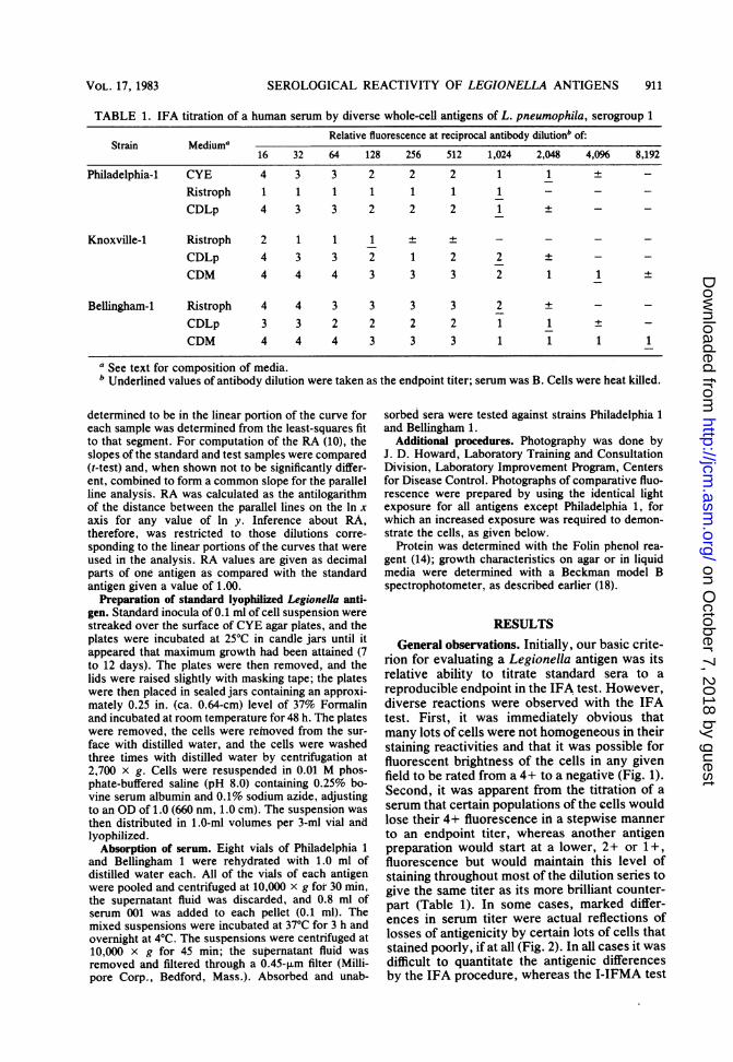
FIG. 2. Comparative IFA serological reactivity ofwhole-cell antigens of L. pneumophila, serogroup 1(x1,000). All photographs were taken and developedunder identical conditions except (D), which requiredadditional exposure for development of the print. (A)Strain Bellingham 1, grown on CDM broth, killed with1% Formalin; (B) strain Bellingham 1, grown on theCDM broth, heat killed; (C) strain Bellingham 1,grown on CYE agar, heat killed; (D) strain Philadel-phia 1 (F.G.), grown on CDM broth, heat killed.
provided a quantitative result. The I-IFMA testpermits a precise measure of the minimum pla-teau fluorescence and the maximum plateaufluorescence, calculation of the RA, and, insome cases, antigenic differences as shown bydifferences of the slope of the response curves.Although the IFMA procedure permitted an
evaluation of several quantitative aspects of theantigenic reactivity, it still measured the averagefluorescence of the cell population. It did notevaluate the morphological aspects of homoge-neity of the cell population necessary for goodIFA antigens. Thus, all products were first ex-amined by the DFA and IFA procedures andthen by I-IFMA and D-IFMA procedures forparticular preparations.
Effects of media, cultural conditions, andgrowth phase on antigenicity of Legionellastrains. When transferred sequentially throughthe diverse media (see Materials and Methods),all cultures grew equally well. No one mediumseriously affected the DFA straining of the vari-ous strains consistently. All rabbit FITC conju-gates were adjusted to the maximum dilution,which gave 4+ staining of the reference antigens(15). Several cultures (Philadelphia 1, Togus 1,Chicago 2, Houston 2, Dallas 2E, and Dallas 1E)showed sporadic decreases in staining reactivitywhen grown on synthetic liquid media. The
original antigenicity was regained when thestrain was transferred to CYE agar.Due to limited human sera, only L. pneumo-
phila serogroups 1 through 4 were tested by theIFA test. Serious losses in the ability to titratethe test serum were observed in several culturesgrown on synthetic liquid media, but the resultswere sporadic and no definitive effect of any onesynthetic broth was noted (Table 1). On one ormore of the synthetic broths, strains Knoxville1, Philadelphia 1 (Fig. 2), Togus 1, Bloomington2, and Los Angeles 1 showed moderately toseverely decreased staining by IFA, althoughthe cells continued to show 3+ and 4+ stainingin the DFA test. The variability in the IFAstaining observed for Philadelphia 1 on chemi-cally defined broths was not observed withBellingham 1 (Fig. 2; Table 1).
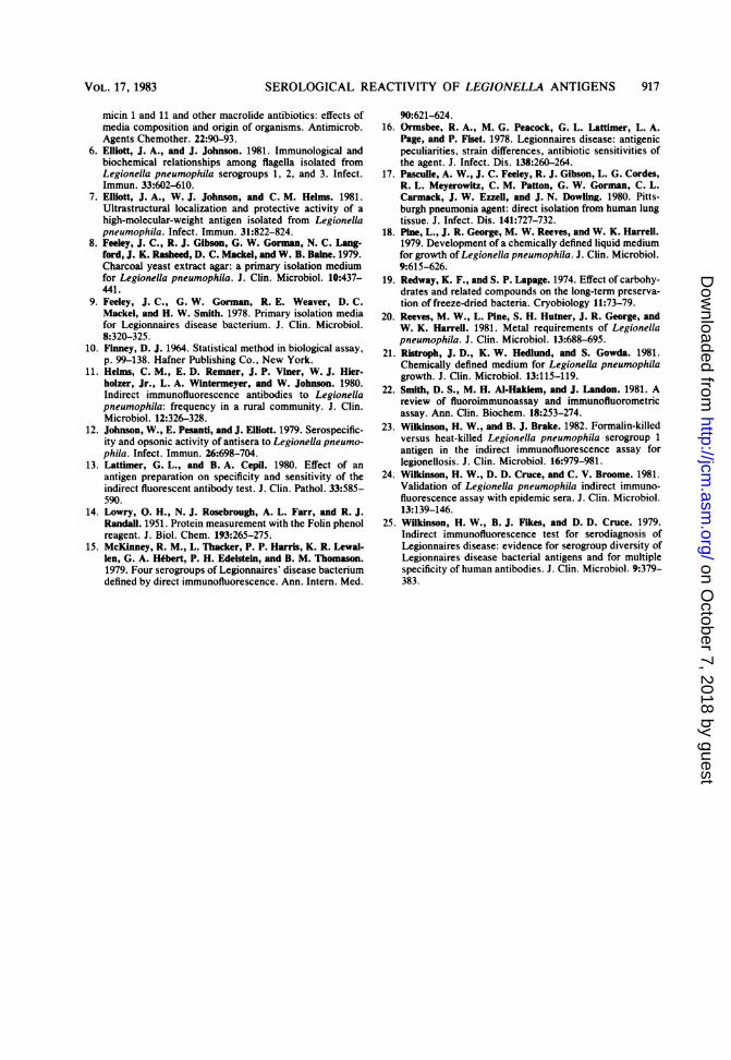
Effects of different agar media, phase ofgrowth, and temperature of incubation on theRA of Philadelphia 1 were determined (Fig. 3and 4). Each set of plates harvested at the timesshown (Fig. 3) were killed with 1% Formalin andanalyzed by the I-IFMA test (Fig. 4; Table 2).Cells grown on the CYE agar had an RA two tofour times that obtained on the CDLp agar, withor without activated charcoal (Table 2, experi-ment 5). As judged by a decreased slope andlower maximum fluorescence, cells grown on F-G agar showed less antigenicity than CYE-grown cells (Fig. 4; Table 2). Examination of thecells at different phases of growth on each of thedifferent media showed no major fluctuations ofRA, nor was there any suggestion that anyparticular phase of growth on any of the mediahad cells of a markedly different antigenicity(Table 2). In general, cells entering the station-ary phase appeared to have maximum antigenic-ity.
Recently, a CYE agar buffered with N-(2-acetamido)-2-aminoethanesulfonic acid was de-scribed (17). It has been our experience, and thatof others (5), that buffered medium gives a muchmore rapid growth of most strains than CYEagar; these two media were compared for anti-gen production. The RA ratios (buffered CYE/CYE) obtained with serum 001 and standardantigens of Philadelphia 1, Philadelphia 4, Bel-lingham 1, and Knoxville 1 were 0.90, 1.04, 1.52,and 1.02, respectively, indicating no major dif-ferences in RA. Both media are therefore recom-mended for antigen production.There was no clear demonstration in these
experiments (Table 2, experiment 1) or in ancil-lary IFA tests that cells grown at either 25 or35°C had differences in antigenicity. However,at 35°C, the CDLp broth and, to a lesser degree,the F-G agar caused abundant filament forma-tion by several strains; fewer filaments wereformed on CYE agar. However, remarkably
J. CLIN. MICROBIOL.
on October 7, 2018 by guest
http://jcm.asm
.org/D
ownloaded from

SEROLOGICAL REACTIVITY OF LEGIONELLA ANTIGENS 913
1.00.9 - CDLp+
.~0.8-0 Norit, 25 C0.6D
0.5
0.4-
0.3-
0.2-
0.11 2 3 4 5 6 7 8 9 10 11 12
DaysFIG. 3. Comparative growth of L. pneumophila
Philadelphia 1 (F.G.) on different agar media at 25 or350C. CYE agar was prepared with 1.5% agar, whereasthe CDLp agar, with or without Norit, contained 1%ion agar. Three or four plates were harvested at eachtime, and the cells from each plate were suspended in10 ml of distilled water. The average OD (660 nm, 1.8cm) is given for each point.
homogeneous suspensions of small single ordouble cells or both were obtained at 250C withall media.
All antigen preparations were adjusted to anOD of 1.00 before RA determinations, althoughwe suspected that a closer correlation mightexist between RA and protein content. Howev-er, determinations of protein relative to RAshowed no obvious correlations, although somecompared products differed by as much as 40%in their relative protein content (Table 2, experi-ments 1 and 5).The effects of growth in air, in air plus carbon
dioxide, and in candle jars on the antigenicity ofPhiladelphia 1 cells were determined with theIFA test. No differences in antigenicity wereobserved under any particular atmospheric con-dition.On the basis of the above results, we used
CYE agar at 25°C for experiments relative to theproduction of a standard lyophilized antigen;growth in a candle jar was chosen arbitrarily.
Effects of killing procedures on RA. For use asantigens, the cells are generally killed with heat
0-
-1.0-
, -2.0-c
0
c -3.0-c-i
-4.0-
,ACYE/
., CDLp + norit' ,:CDLp
/ X F-G
'p
// /
/ / p/
//y ,". 1/ U
x" f.,//7 /
-9.89 -8.79 -7.69 -6.59 -5.49 -4.39 -3.30Ln serum dilution
FIG. 4. Relative I-IFMA fluorescence of L. pneu-mophila cells Philadelphia 1 (F.G.), grown on differentmedia at 25°C. Cell suspensions were those of Fig. 3and Table 2 (experiment 5 and experiment 1, 9 days).
(25) or Formalin (11, 13) and then washed one ormore times. In several experiments, we com-pared the relative reactivity of cells in the IFAtest when killed by heat, different concentra-tions of Formalin, Formalin vapors, or diethylether. Ether-killed cells showed high back-ground fluorescence and many morphologicalaberrations such as that reported previously(25). Experiments comparing heat-killed versusFormalin-killed cells showed no marked differ-ences in reactivity in the IFA or I-IFMA test. Inan I-IFMA test comparing heat-killed cells, cellswashed three times and heat killed, cells killedwith 1% Formalin, and those killed with 37%Formalin vapors, the RA values were 1.24, 1.26,0.92, and 1.00, respectively. However, cellskilled by 1% Formalin or by Formalin vaporsshowed a more clearly defined cell withoutsurface debris or irregularities (Fig. 2). Washingeither heat-killed or Formalin-killed cells withdistilled water did not alter the relative fluores-cence of the cells, although large amounts ofbackground staining material previously ob-served in the IFA test were removed. In theseexperiments, little differences were seen in therelative reactivity of live cells that were heatfixed on the slide, heat-killed cells, cells killedby 1% Formalin, or cells killed by Formalinvapors. However, in one experiment with theF.G., R.B., and GPSp strains of Philadelphia 1,large numbers of flagella were seen on those
VOL. 17, 1983
on October 7, 2018 by guest
http://jcm.asm
.org/D
ownloaded from

TABLE 2. Effect of temperature, growth phase, and agar medium on the RA of L. pneumophila strainPhiladelphia 1 (F.G.)a
Expt Mediumb Temp Days Protein Fluorescence Slope (LB, UB)d RA'(OC) grown (mg/ml)c maximum
1 F-G 35 1.0 320 0.396 0.419 (0.357-0.481) 0.801.8 335 0.410 0.492 (0.460-0.525) 1.172.8 392 0.414 0.474 (0.440-0.507) 1.00*
F-G 25 7.0 240 0.374 0.464 (0.384-0.544) 1.388.0 238 0.397 0.437 (0.407-0.467) 1.619.0 268 0.411 0.394 (0.369-0.418) 1.41
2 CYE 25 6.0 262 0.543 0.636 (0.591-0.682) 0.757.0 267 0.789 0.692 (0.654-0.730) 1.00*9.0 260 0.759 0.694 (0.646-0.741) 0.6810.0 280 0.746 0.730 (0.685-0.776) 0.77
3 CDLp 25 5.0 275 0.410 0.837 (0.732-0.942) 0.996.0 280 0.564 0.757 (0.696-0.818) 1.00*7.0 295 0.549 0.718 (0.626-0.811) 0.899.0 295 0.473 0.750 (0.713-0.786) 0.77
4 CDLp + Norit 25 5.0 295 0.570 0.644 (0.577-0.710) 1.086.0 295 0.564 0.631 (0.566-0.696) 1.00*7.0 270 0.563 0.541 (0.467-0.615) 1.039.0 257 0.539 0.588 (0.546-0.631) 1.04
5 CYE 25 7.0 267 0.789 0.713 (0.664-0.762) 1.00*CDLp 6.0 280 0.546 0.753 (0.693-0.813) 0.37CDLp + Norit 6.0 295 0.564 0.626 (0.556-0.696) 0.43
a Culture history and basic fluorometric data are given in Fig. 3 and 4. Cells were killed with 1% Formalin.b See text for composition of media.Protein per milliliter of bacterial suspension (OD = 1.0).
d LB, UB, Lower and upper 95% confidence bounds of the slope. Experiments 1 and 2 were done at threefolddilution of 1/81 to 1/6,561 dilutions of serum 001; dilutions for experiments 3 to 5 were made at 1/81 to 1/2,187,inclusively. Cells killed with 1% Formalin were washed three times with water, adjusted to an OD of 1.0 (1.0cm,660 nm), and diluted 1/24. CoeffiCients of correlation for all experiments ranged from 0.961 to 0.998.
Standard sample has asterisk.
cells of the GPSp strain that were killed byFormalin vapors. No other preparations showedflagella. The flagella were maintained, althoughin decreasing numbers, through three wash cy-cles. Because of the potential of Formalin va-pors to fix surface antigens in situ, this proce-dure was the one used routinely to preparelyophilized cells for use in the I-IFMA proce-dure.
Lyophilization of whole-cell antigens. Initialtests with Formalin-killed cells (Philadelphia 1)and cells killed with Formalin vapors showedthat these cells, when lyophilized in water orphosphate-buffered saline, could not be recon-stituted with distilled water, 0.1% glycine, or0.015% Tween 20 to give a good suspension;large flat sheets of cells were formed that did notgive homogeneous suspensions even with strongagitation. On the basis of the results of Redwayand Lepage (19), we tested suspending media of4% mannitol, inositol, glycerol, and solublestarch as media for lyophilization, but without
success. Colored products were formed withinositol and mannitol, and the antigenic reactiv-ity (IFA test) was reduced in the presence ofglycerol, inositol, and starch. Additional testswith 0.05 and 0.25% bovine serum albumin,0.2% glycine, or glutamic acid as the suspendingmedium for lyophilization gave cells that werereadily brought into suspension with water.However, small reductions in brilliance (IFAtest) were observed in all lyophilized prepara-tions. A suspending medium of 0.25% bovineserum albumin with 0.1% NaN3, in phosphate-buffered saline (0.01 M, pH 8.0) was chosen forfreeze-drying standard antigens. With this sus-pending medium we have observed no loss inantigenicity of freeze-dried preparations storedfor 1 to 2 years at 5°C or in cells reconstitutedwith water and stored at 5°C for several weeks.
Strain differences exposed by diverse sera. Dur-ing these studies it rapidly became apparent thatvariation of RA with changes in procedure wasprimarily strain dependent. Whereas Philadel-
914 BENSON ET AL. J. CLIN. MICROBIOL.
on October 7, 2018 by guest
http://jcm.asm
.org/D
ownloaded from

SEROLOGICAL REACTIVITY OF LEGIONELLA ANTIGENS 915
0.0o
-1.0*
' -2.0
0
-3.0c
J
-4.0-
-8.79 -7.69 -6.59 -5.49 -4.39 -3.3Ln antibody dilution
FIG. 5. Response curves of L. pneum
gens, serogroups 1 and 2 (I-IFMA test),serum 001.
phia 1 showed marked variation withmedia and growth conditions, Bellingnot (Fig. 2; Table 1). These resultstained with sera 001, A, and B. Altho001 came from a patient infected witha Bellingham-like organism (15), this awas not true for the other sera.The relative antigenicity of strain!
phia 1 (F.G.) and Bellingham 1 werefurther, using FITC-conjugated rabbiglobulin and conjugated and nonconjiman sera in the DFA, D-IFMA, IfIFMA tests. Philadelphia 1, as descreacted much more strongly than Be(D-IFMA) with the conjugated rabbiglobulin (RA = 17). However, withrum 001, A, or B, the RA of BellinPhiladelphia 1 ranged from 3.72 to fgroup 2 antigen was essentially unrezthese human sera at dilutions >1/2Although absorption of sera 001 andshown) by Bellingham 1 greatly deplsera of reactive antibody, absorptiondelphia 1 left antibody highly reactiNlingham 1 (RA = 19.24) (Table 3).
DISCUSSIONAlthough we observed small red
brilliance with the IFA test after thelyophilized, the antigens Philadelphand R.B.) were comparable to theinventory antigen of the Biologica
Program. For production of a standard antigen,our results emphasized the need to choose astrain that was consistent in its responses toproduction procedures. The Formalin-killed, ly-ophilized cells showed no surface or morpholog-
(Serogroup 1) ical aberrations when reconstituted with waterand were antigenically stable in suspension forseveral weeks at 50C. Cells of Legionella grownon the CYE agar for 9 to 12 days at 250C weresmall, of a homogeneous population of single ordouble cells, and without filaments. All of thesecharacteristics were basically essential for the I-IFMA test. As stated earlier, the I-IFMA testused for serum analysis eliminates the use of theegg yolk sac membrane, permits a precise quan-titative fluorescent readout, automatically rec-ords an average fluorescence of the suspendedcell population, and permits a precise calcula-tion of antibody potency (2, 22; Pine and Ben-son, in press). Its potential for use with a pooledantigen remains to be explored (25).
10 -2.20 Although a wide variety of media and culturalconditions have been used for growth of Legion-
ophila anti- ella, the effects of media on the production ofto human antigen that reacts with fluorescein conjugates
are not clearly defined. In general, chemicallydefined liquid media lead to poorer antigen pro-duction by Philadelphia 1 and sporadic losses in
changes in other strains. Initial results obtained with cells;ham 1 did grown on F-G and CYE agars led to the impres-were ob- sion that growth on F-G agar, although poorer
iugh serum than that on CYE agar, gave cells of greater IFADetroit 1, brilliance. This impression, however, was notissociation supported by several tests that used the I-IFMA
procedure, and in the range of serum dilutionss Philadel- where the responses were linear, CYE-growncompared cells were superior. The three chemically de-
it immuno- fined media differed radically in their relativeugated hu- amounts of amino acids, trace element composi-A, and 1- tion, and presence or absence of vitamins.ribed (15), Growth was equally good in all of these media.llingham 1 Although sporadic losses of antigen occurred,it immuno- there was no suggestion that a compound of anyhuman se- one medium regulated production of the IFAigham 1 to reacting antigen; little variation among the di-4.38; sero- verse strains was observed during growthictive with through several transfers in the three chemically7 (Fig. 5). defined media and back to the complex organicB (data not media.leted these We considered the possibility that loss ofwith Phila- antigen by Philadelphia 1, when grown on chem-ve for Bel- ically defined media, was due to curing of a
plasmid-mediating gene. However, at present,plasmids have not been observed in this strain(1). Strain Bellingham 1, however, did not show
luctions in fluctuation of surface antigens and appeared tocells were maintain one or more serogroup-specific anti-ia 1 (F.G. gens not observed on Philadelphia 1 with humanLegionella sera. Other workers have reported large antigen-I Products ic differences among strains of the same sero-
VOL. 17, 1983
on October 7, 2018 by guest
http://jcm.asm
.org/D
ownloaded from
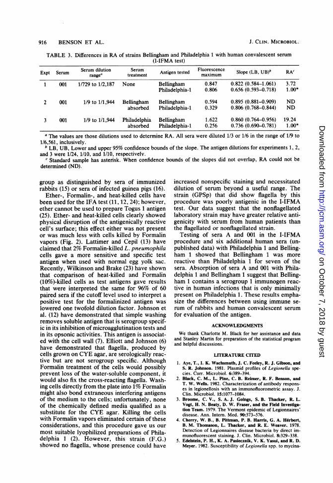
TABLE 3. Differences in RA of strains Bellingham and Philadelphia 1 with human convalescent serum(I-IFMA test)
Expt Serum rangea trSerum Antigen tested maximum Slope (LB, UB)b RAc
1 001 1/729 to 1/2,187 None Bellingham 0.847 0.822 (0.584-1.061) 3.72Philadelphia-1 0.806 0.656 (0.593-0.718) 1.00*
2 001 1/9 to 1/1,944 Bellingham Bellingham 0.594 0.895 (0.881-0.909) NDabsorbed Philadelphia-1 0.329 0.806 (0.768-0.844) ND
3 001 1/9 to 1/1,944 Philadelphia Bellingham 1.622 0.860 (0.764-0.956) 19.24absorbed Philadelphia-1 0.256 0.736 (0.690-0.781) 1.00*
a The values are those dilutions used to determine RA. All sera were diluted 1/3 or 1/6 in the range of 1/9 to1/6,561, inclusively.
b LB, UB, Lower and upper 95% confidence bounds of the slope. The antigen dilutions for experiments 1, 2,and 3 were 1/24, 1/10, and 1/10, respectivelv.
c Standard sample has asterisk. When confidence bounds of the slopes did not overlap, RA could not bedetermined (ND).
group as distinguished by sera of immunizedrabbits (15) or sera of infected guinea pigs (16).
Ether-, Formalin-, and heat-killed cells havebeen used for the IFA test (11, 12, 24); however,ether cannot be used to prepare Togus 1 antigen(25). Ether- and heat-killed cells clearly showedphysical disruption of the antigenically reactivecell's surface; this effect either was not presentor was much less with cells killed by Formalinvapors (Fig. 2). Lattimer and Cepil (13) haveclaimed that 2% Formalin-killed L. pneumophilacells gave a more sensitive and specific testantigen when used with normal egg yolk sac.Recently, Wilkinson and Brake (23) have shownthat comparison of heat-killed and Formalin(10%)-killed cells as test antigens gave resultsthat were interpreted the same for 96% of 60paired sera if the cutoff level used to interpret apositive test for the formalinized antigen waslowered one twofold dilution factor. Johnson etal. (12) have demonstrated that simple washingremoves soluble antigen that is serogroup specif-ic in its inhibition of microagglutination tests andin its opsonic activities. This antigen is associat-ed with the cell wall (7). Elliott and Johnson (6)have demonstrated that flagella, produced bycells grown on CYE agar, are serologically reac-tive but are not serogroup specific. AlthoughFormalin treatment of the cells would possiblyprevent loss of the water-soluble component, itwould also fix the cross-reacting flagella. Wash-ing cells directly from the plate into 1% Formalinmight also bond extraneous interfering antigensof the medium to the cells; unfortunately, noneof the chemically defined media qualified as asubstitute for the CYE agar. Killing the cellswith Formalin vapors eliminated certain of theseconsiderations, and this procedure gave us ourmost suitable lyophilized preparations of Phila-delphia 1 (2). However, this strain (F.G.)showed no flagella, whose presence could have
increased nonspecific staining and necessitateddilution of serum beyond a useful range. Thestrain (GPSp) that did show flagella by thisprocedure was poorly antigenic in the I-IFMAtest. Our data suggest that the nonflagellatedlaboratory strain may have greater relative anti-genicity with serum from human patients thanthe flagellated or nonflagellated strain.
Testing of sera A and 001 in the I-IFMAprocedure and six additional human sera (un-published data) with Philadelphia 1 and Belling-ham 1 showed that Bellingham 1 was morereactive than Philadelphia 1 for seven of thesera. Absorption of sera A and 001 with Phila-delphia 1 and Bellingham 1 suggest that Belling-ham 1 contains a serogroup 1 immunogen reac-tive in human infections that is only minimallypresent on Philadelphia 1. These results empha-size the differences between using immune se-rum of rabbits and human convalescent serumfor evaluation of the antigen.
ACKNOWLEDGMENTS
We thank Charlotte M. Black for her assistance and dataand Stanley Martin for preparation of the statistical programand helpful discussions.
LITERATURE CITED
1. Aye, T., I. K. Wachsmuth, J. C. Feeley, R. J. Gibson, andS. R. Johnson. 1981. Plasmid profiles of Legionella spe-cies. Curr. Microbiol. 6:389-394.
2. Black, C. M., L. Pine, C. B. Reimer, R. F. Benson, andT. W. Wells. 1982. Characterization of antibody respons-es in legionellosis with an immunofluorometric assay. J.Clin. Microbiol. 15:1077-1084.
3. Broome, C. V., S. A. J. Goings, S. B. Thacker, R. L.Vogt, H. N. Beaty, D. W. Fraser, and the Field Investiga-tion Team. 1979. The Vermont epidemic of Legionnaires'disease. Ann. Intern. Med. 90:573-576.
4. Cherry, W. B., B. Pittman, P. B. Harris, G. A. Herbert,B. M. Thomason, L. Thacker, and R. E. Weaver. 1978.Detection of Legionnaires disease bacteria by direct im-munofluorescent staining. J. Clin. Microbiol. 8:329-338.
5. Edelstein, P. H., K. A. Pasiecznik, V. K. Yasul, and R. D.Meyer. 1982. Susceptibility of Legionella spp. to mycina-
916 BENSON ET AL. J. CLIN. MICROBIOL.
on October 7, 2018 by guest
http://jcm.asm
.org/D
ownloaded from
SEROLOGICAL REACTIVITY OF LEGIONELLA ANTIGENS 917
micin 1 and 11 and other macrolide antibiotics: effects ofmedia composition and origin of organisms. Antimicrob.Agents Chemother. 22:90-93.
6. Elliott, J. A., and J. Johnson. 1981. Immunological andbiochemical relationships among flagella isolated fromLegionella pneumophila serogroups 1, 2, and 3. Infect.Immun. 33:602-610.
7. Elliott, J. A., W. J. Johnson, and C. M. Helms. 1981.Ultrastructural localization and protective activity of ahigh-molecular-weight antigen isolated from Legionellapneumophila. Infect. Immun. 31:822-824.
8. Feeley, J. C., R. J. Gibson, G. W. Gorman, N. C. Lang-ford, J. K. Rasheed, D. C. Mackel, and W. B. Baine. 1979.Charcoal yeast extract agar: a primary isolation mediumfor Legionella pneumophila. J. Clin. Microbiol. 10:437-441.
9. Feeley, J. C., G. W. Gorman, R. E. Weaver, D. C.Mackel, and H. W. Smith. 1978. Primary isolation mediafor Legionnaires disease bacterium. J. Clin. Microbiol.8:320-325.
10. Finney, D. J. 1964. Statistical method in biological assay,p. 99-138. Hafner Publishing Co., New York.
11. Helms, C. M., E. D. Remner, J. P. Viner, W. J. Hier-holzer, Jr., L. A. Wintermeyer, and W. Johnson. 1980.Indirect immunofluorescence antibodies to Legionellapneumophila: frequency in a rural community. J. Clin.Microbiol. 12:326-328.
12. Johnson, W., E. Pesanti, and J. Elliott. 1979. Serospecific-ity and opsonic activity of antisera to Legionella pneumo-phila. Infect. Immun. 26:698-704.
13. Lattimer, G. L., and B. A. Cepil. 1980. Effect of anantigen preparation on specificity and sensitivity of theindirect fluorescent antibody test. J. Clin. Pathol. 33:585-590.
14. Lowry, 0. H., N. J. Rosebrough, A. L. Farr, and R. J.Randall. 1951. Protein measurement with the Folin phenolreagent. J. Biol. Chem. 193:265-275.
15. McKinney, R. M., L. Thacker, P. P. Harris, K. R. Lewal-len, G. A. Hebert, P. H. Edelstein, and B. M. Thomason.1979. Four serogroups of Legionnaires' disease bacteriumdefined by direct immunofluorescence. Ann. Intern. Med.
90:621-624.16. Ormsbee, R. A., M. G. Peacock, G. L. Lattimer, L. A.
Page, and P. Fiset. 1978. Legionnaires disease: antigenicpeculiarities, strain differences, antibiotic sensitivities ofthe agent. J. Infect. Dis. 138:260-264.
17. Pasculle, A. W., J. C. Feeley, R. J. Gibson, L. G. Cordes,R. L. Meyerowitz, C. M. Patton, G. W. Gorman, C. L.Carmack, J. W. Ezzeil, and J. N. Dowling. 1980. Pitts-burgh pneumonia agent: direct isolation from human lungtissue. J. Infect. Dis. 141:727-732.
18. Pine, L., J. R. George, M. W. Reeves, and W. K. Harrell.1979. Development of a chemically defined liquid mediumfor growth of Legionella pneumophila. J. Clin. Microbiol.9:615-626.
19. Redway, K. F., and S. P. Lapage. 1974. Effect of carbohy-drates and related compounds on the long-term preserva-tion of freeze-dried bacteria. Cryobiology 11:73-79.
20. Reeves, M. W., L. Pine, S. H. Hutner, J. R. George, andW. K. Harrell. 1981. Metal requirements of Legionellapneumophila. J. Clin. Microbiol. 13:688-695.
21. Ristroph, J. D., K. W. Hedlund, and S. Gowda. 1981.Chemically defined medium for Legionella pneumophilagrowth. J. Clin. Microbiol. 13:115-119.
22. Smith, D. S., M. H. Al-Hakiem, and J. Landon. 1981. Areview of fluoroimmunoassay and immunofluorometricassay. Ann. Clin. Biochem. 18:253-274.
23. Wilkinson, H. W., and B. J. Brake. 1982. Formalin-killedversus heat-killed Legionella pneumophila serogroup 1antigen in the indirect immunofluorescence assay forlegionellosis. J. Clin. Microbiol. 16:979-981.
24. Wilkinson, H. W., D. D. Cruce, and C. V. Broome. 1981.Validation of Legionella pneumophila indirect immuno-fluorescence assay with epidemic sera. J. Clin. Microbiol.13:139-146.
25. Wilkinson, H. W., B. J. Fikes, and D. D. Cruce. 1979.Indirect immunofluorescence test for serodiagnosis ofLegionnaires disease: evidence for serogroup diversity ofLegionnaires disease bacterial antigens and for multiplespecificity of human antibodies. J. Clin. Microbiol. 9:379-383.
VOL. 17, 1983
on October 7, 2018 by guest
http://jcm.asm
.org/D
ownloaded from