Embed Size (px)
Citation preview

City University of New York (CUNY) City University of New York (CUNY)
CUNY Academic Works CUNY Academic Works
All Dissertations, Theses, and Capstone Projects Dissertations, Theses, and Capstone Projects
6-2014
Influences of Motor Control Instruction and Taping on Center of Influences of Motor Control Instruction and Taping on Center of
Pressure and Scapulothoracic Kinematics During Reaching for Pressure and Scapulothoracic Kinematics During Reaching for
Individuals with Hemiparesis Individuals with Hemiparesis
Michelle De Guzman Graduate Center, City University of New York
Tatyana Farber Graduate Center, City University of New York
Anna Kochanova Graduate Center, City University of New York
Jonathan Lazarus Graduate Center, City University of New York
How does access to this work benefit you? Let us know!
More information about this work at: https://academicworks.cuny.edu/gc_etds/808
Discover additional works at: https://academicworks.cuny.edu
This work is made publicly available by the City University of New York (CUNY). Contact: [email protected]

INFLUENCES OF MOTOR CONTROL INSTRUCTION AND TAPING ON CENTER OF
PRESSURE AND SCAPULOTHORACIC KINEMATICS DURING REACHING FOR
INDIVIDUALS WITH HEMIPARESIS
by
MICHELLE DE GUZMAN
TATYANA FARBER
ANNA KOCHANOVA
JONATHAN LAZARUS
A capstone project submitted to the Graduate Faculty in Physical Therapy in partial fulfillment
of the requirements for the degree of Doctor of Physical Therapy,
The City University of New York
2014

REACHING
ii
This manuscript has been read and accepted for the
Graduate Faculty in Physical Therapy in satisfaction of
the capstone project requirement for the degree of DPT.
Suzanne R. Babyar, PT, PhD
______________________ _____________________________________________
Date Chair of Examining Committee
Jeffrey Rothman, PT, EdD
______________________ _____________________________________________
Date Executive Officer
REACHING
iii
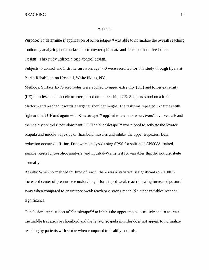
Abstract
Purpose: To determine if application of Kinesiotape™ was able to normalize the overall reaching
motion by analyzing both surface electromyographic data and force platform feedback.
Design: This study utilizes a case-control design.
Subjects: 5 control and 5 stroke survivors age >40 were recruited for this study through flyers at
Burke Rehabilitation Hospital, White Plains, NY.
Methods: Surface EMG electrodes were applied to upper extremity (UE) and lower extremity
(LE) muscles and an accelerometer placed on the reaching UE. Subjects stood on a force
platform and reached towards a target at shoulder height. The task was repeated 5-7 times with
right and left UE and again with Kinesiotape™ applied to the stroke survivors’ involved UE and
the healthy controls’ non-dominant UE. The Kinesiotape™ was placed to activate the levator
scapula and middle trapezius or rhomboid muscles and inhibit the upper trapezius. Data
reduction occurred off-line. Data were analyzed using SPSS for split-half ANOVA, paired
sample t-tests for post-hoc analysis, and Kruskal-Wallis test for variables that did not distribute
normally.
Results: When normalized for time of reach, there was a statistically significant (p <0 .001)
increased center of pressure excursion/length for a taped weak reach showing increased postural
sway when compared to an untaped weak reach or a strong reach. No other variables reached
significance.
Conclusion: Application of Kinesiotape™ to inhibit the upper trapezius muscle and to activate
the middle trapezius or rhomboid and the levator scapula muscles does not appear to normalize
reaching by patients with stroke when compared to healthy controls.

REACHING
iv
Table of Contents
Approval Page ----------------------------------------------------------------------------------------------- ii
Abstract ------------------------------------------------------------------------------------------------------ iii
List of Tables ------------------------------------------------------------------------------------------------- v
List of Figures ----------------------------------------------------------------------------------------------- vi
Introduction -------------------------------------------------------------------------------------------------- 1
Purpose ------------------------------------------------------------------------------------------------------ 12
Methods ----------------------------------------------------------------------------------------------------- 13
Results ------------------------------------------------------------------------------------------------------- 19
Discussion --------------------------------------------------------------------------------------------------- 35
Limitations -------------------------------------------------------------------------------------------------- 37
Conclusion -------------------------------------------------------------------------------------------------- 39
References -------------------------------------------------------------------------------------------------- 40
REACHING
v
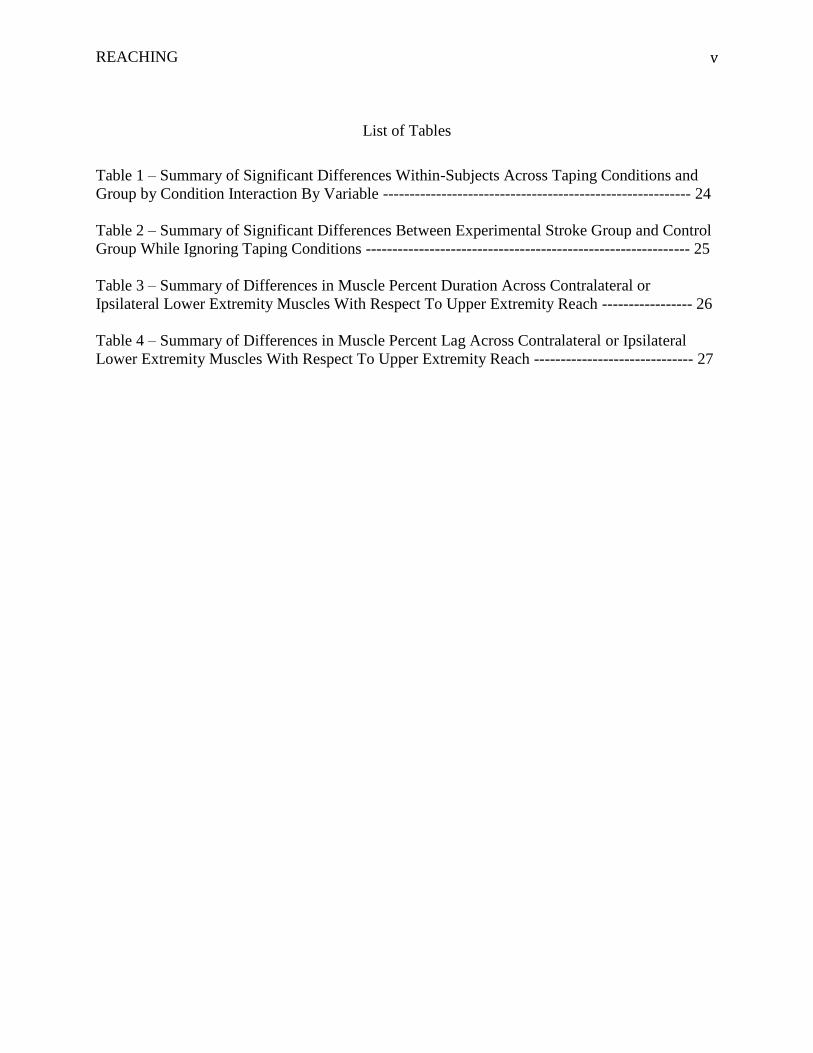
List of Tables
Table 1 – Summary of Significant Differences Within-Subjects Across Taping Conditions and
Group by Condition Interaction By Variable ---------------------------------------------------------- 24
Table 2 – Summary of Significant Differences Between Experimental Stroke Group and Control
Group While Ignoring Taping Conditions ------------------------------------------------------------- 25
Table 3 – Summary of Differences in Muscle Percent Duration Across Contralateral or
Ipsilateral Lower Extremity Muscles With Respect To Upper Extremity Reach ----------------- 26
Table 4 – Summary of Differences in Muscle Percent Lag Across Contralateral or Ipsilateral
Lower Extremity Muscles With Respect To Upper Extremity Reach ------------------------------ 27

REACHING
vi
List of Figures
Figure 1 – Boxplot showing distribution of Center of Pressure in the x plane values across all
taping conditions between the stroke survivor test group and healthy control group. ------------ 28
Figure 2 – Boxplot showing distribution of Center of Pressure in the y plane values across all
taping conditions between the stroke survivor test group and healthy control group. ------------ 29
Figure 3 – Boxplot showing distribution of the Center of Pressure-length normalized for the
duration of the reach across all taping conditions between the stroke survivor test group and
healthy control group. -------------------------------------------------------------------------------------- 30
Figure 4 – Boxplot showing distribution of the percent lag values of the deltoid muscle across all
taping conditions between the stroke survivor test group and healthy control group. ------------ 31
Figure 5 – Boxplot showing distribution of the percent lag values of the upper trapezius muscle
across all taping conditions between the stroke survivor test group and healthy control group. -32
Figure 6 – Boxplot showing distribution of the deltoid muscle’s percent duration of reach across
all taping conditions between the stroke survivor test group and healthy control group. -------- 33
Figure 7 – Boxplot showing distribution of the upper trapezius muscle’s percent duration of
reach across all taping conditions between the stroke survivor test group and healthy control
group. -------------------------------------------------------------------------------------------------------- 34

REACHING
1
Introduction
Stroke is the fourth leading cause of death in the United States, killing over 137,000
people each year; and stroke is a leading cause of serious, long-term adult disability (National
Stroke Association, 2014). On average, every 40 seconds, someone in the United States has a
stroke (National Stroke Association, 2014). Each year, approximately 795,000 Americans will
suffer from a new or recurrent stroke (National Stroke Association, 2014). Current statistics
indicate that there are over 7,000,000 people in the United States who have survived a stroke and
are living with the after-effects (National Stroke Association, 2014) and this number is expected
to increase. Projections made by the American Heart Association’s 2014 report state that by
2030, an additional 4 million people will have had a stroke, a 24.9% increase in prevalence from
2010 (National Stroke Association, 2014). As stroke prevalence increases, the stroke mortality
rates decline. From 1998 to 2008, the annual stroke death rate decreased by 34.8%, and the
actual number of stroke deaths declined 19.4% (National Stroke Association, 2014). As stroke
mortality rates decline, more stroke survivors are likely to have lasting impairments that will
affect their daily living (Harris & Eng, 2007).
Balance Deficits
Stroke can cause different types of disabilities depending on which part of the brain is
damaged and on the extent of the damage itself. Some common complications seen in post-
stroke patients are muscle weakness and spasticity, decreased postural and voluntary control,
sensory loss, poor body alignment and shoulder pain (Harris & Eng, 2007; Jaraczewska & Long,
2006). Approximately 80% of stroke survivors have chronic motor deficits, with hemiparesis
being the most common (National Stroke Association, 2014; Harris & Eng, 2007; Mercer,

REACHING
2
Freburger, Chang, & Purser, 2009). One of the major effects of hemiparesis, or muscle
weakness affecting one side of the body, is the disruption in balance.
Balance can be described as the ability to maintain or move within a weight-bearing
posture without falling. It can be broken down into the following three components: steadiness,
or the ability to stay in a posture with minimal external movement or sway; symmetry, or equal
weight distribution between the weight-bearing components; and dynamic stability, or the ability
to move within a posture without loss of balance (Nichols, 1997). Functional standing balance is
achieved when an individual can maintain standing position in static circumstances and while
experiencing internally and externally produced perturbations linked with movements of their
extremities (Srivastava, Taly, Gupta, Kumar, & Murali, 2009). After stroke, all of the
aforementioned balance parameters are affected to a certain degree. Balance problems have been
linked to the poor recovery of activities of daily living (ADL) and mobility as well as an
increased risk of falls (Tyson, Hanley, Chillala, Selley, & Tallis, 2006; Barclay-Goddard,
Stevenson, Poluha, Moffratt, & Taback, 2009). Individuals with hemiparesis secondary to stroke
have more postural sway during static stance, asymmetric weight distribution by putting more
weight on the non-paretic leg, impaired weight shifting ability and decreased stance capability
(Srivastava et al., 2009; Nichols, 1997). Hemiparetic individuals show difficulty bearing weight
or “loading” the paretic lower extremity (LE), with them bearing between 61-80% of their body
weight through the non-paretic leg (Mercer et al., 2009; Geiger et al., 2001). Furthermore,
overall stance instability has been observed in hemiplegic stroke patients as well as standing
balance deficits when compared to healthy controls (Das et al., 2011).
REACHING
3
Center of Pressure and Postural Control
The small movements of the center of mass (COM) from heel to toe and side-to-side
during regular standing balance are referred to as postural sway. Analysis of postural sway can
be performed using a force plate to measure the ground reaction forces of these movements
under the feet. These pressures under the feet are also referred to as the center of pressure
(COP). In stroke patients, analysis of postural sway, COP fluctuations and weight bearing
tendencies can be performed using force platform technology.
Force platform technology provides a method for measuring and training an
individual's ability to meet balance parameters. The three measures most commonly used by
force platform systems are postural sway, symmetry, and limits of stability (Nichols, 1997). The
amount of sway in a person’s stance can be gathered by the measurement of three force
components along the X, Y and Z axes as well as moments about those axes. These systems also
provide visual or auditory feedback to the patients regarding their center of force (COF) or center
of pressure (COP). Barclay-Goddard et al. (2009) conducted a Cochrane Review of seven
studies and found that feedback from force platforms leads to improved stance symmetry, but not
balance during functional activities or overall independence (Srivastava et al., 2009; Barclay-
Goddard et al., 2009). However most of these studies were done in acute stages after stroke.
One recent study done in the later phases of stroke has shown improvements in balance and
functional ability both at the end of the training as well as at a three-month follow-up (Srivastava
et al., 2009).
Postural sway of stroke survivors has been reported to be twice that of the age matched
healthy individuals (Tyson et al., 2006; Nichols, 1997; Geiger, Allen, O’Keefe, & Hicks,
2001). Moment is the tendency for the body to create rotation around a joint. Peurala et al.
REACHING
4
found that stroke victims demonstrated greater than four times the mean velocity moment than
healthy controls. The same study also found that stroke victims displace their COP twice as
quickly in the medial-lateral and anterior-posterior planes (2007). Mansfield et al. (2011)
demonstrated that while maintaining quiet standing, stroke patients showed reduced between-
limb synchronization during COP movements. These observations were related to the increased
medio-lateral postural sway as well as asymmetric weight bearing shown by stroke patients as
compared to healthy controls.
In addition to force plate and COP deficits observed during quiet standing, patients with
stroke have also shown differences during functional reaching activities. During normal
reaching, there are necessary muscle contractions around the ankle joint which help maintain an
upright posture and compensate for the shift in COP during the reaching activity. Hsu et al.
(2005) studied EMG activity of ankle joint muscles during reaching tasks in hemiparetic stroke
patients and concluded that the hemiparetic population showed significantly different EMG
activity in both the tibialis anterior and the soleus of the affected ankle when compared to
accepted norms. Furthermore, they found the ankle musculature of the stroke patients could not
adequately compensate quickly or efficiently enough for the reaching activity (2005). These
lower extremity deficits can cause inefficient and unsafe standing and reaching conditions for
patients with hemiparesis.
Motor Deficits of Upper Extremity
Incidence of stroke may cause impairment to the upper extremity, causing motor
deficiencies in both acute and chronic cases, by 80% and 40%, respectively, leading to
limitations in activity levels, participation, and functional motor competence (Murphy, Willen, &

REACHING
5
Sunnerhagen, 2011; Nakayama, Jorgensen, Raaschou, & Olsen, 1994; Parker, Wade, & Hewer,
1986; Broeks, Lankhorst, Rumping, & Prevo, 1999). Neuronal injury produced by a stroke can
manifest clinically with hemiparesis and changes in the generation of muscle tone causing
serious insult to upper limb motor control. (Massie, Malcolm, Greene, & Browning, 2012;
Gracies, 2005a and 2005b) Coupled with transformations in musculature such as atrophy that
occurs following stroke, these changes may cause muscle weakness. Furthermore, it may impact
the ability of the affected upper extremity to be actively involved in the execution of many
complex functional tasks included in activities of daily living (Massie et al., 2012; Cirstea &
Levin, 2000). Coordination, the basis of proper motor function, results from the collaborative
interaction of efferent and afferent feedback signals between goals or intention, the nervous
system, and the musculoskeletal system. Synergists are prime examples of such complex
interaction that work to produce smooth and accurate movements across multiple joints (Kisiel-
Sajewicz et al., 2011; Cirstea, Mitnitski, Feldman, & Levin, 2003). Thus, following stroke,
proper initiation, maintenance, and coordination of upper limb motor performance becomes
compromised, such that compensatory strategies are employed during common functional tasks
like reaching (Kisiel-Sajewicz et al., 2011; Massie et al., 2012; Cirstea & Levin, 2000). Aside
from obvious causes stated earlier, other reasons behind poor motor coordination after stroke
may be considered, some of which include sensory deficits, spasticity, weakened cortical signal
coupling between synergists and/or brain and effector (Fang, Daly, Hrovat, Sahgal, & Yue,
2009), unusual patterns of co-contraction/activation and muscle recruitment (Beer, Dewald, &
Rymer, 2000) and distorted corticospinal pathway arrangements post-stroke. (Yao, Chen,
Carmona, & Dewald, 2009).

REACHING
6
One of the most fundamental components of activities of daily living (ADL) involves
the reaching movement, which is commonly affected by hemiparesis secondary to stroke.
Kisiel-Sajewicz et al. (2011) found that during forward reaching movements in hemiparetic
stroke patients, poor motor coordination was exhibited when compared with healthy
controls. This was based on the demonstration of significantly greater lateral deviations away
from the straight trajectory of the arm reaching movement, suggesting impairment to shoulder
adduction and general movement control. (Kisiel-Sajewicz et al., 2011; Cirstea & Levin,
2000) In addition, Kisiel-Sajewicz et al. (2011) also demonstrated that impairments to motor
planning and the proper functioning of muscle synergists occurred while reaching. This was
exemplified through the reading of lower electromyographic (EMG) coherence signals,
specifically between the anterior deltoid (shoulder flexor) and triceps brachii (elbow extensor)
during reaching. In other words, two prime muscle movers, or synergists, of the reaching
movement exhibited a lesser degree of synchronous muscle activity between each other in
hemiparetic patients versus the same in healthy controls. EMG results demonstrated a delay in
muscle activation onset of the triceps brachii compared with the anterior deltoid, and an earlier
onset of the anterior deltoid compared to the triceps brachii. Since these results were exclusive
to the triceps brachii and anterior deltoid, and were not exhibited by other muscle pairs studied,
an interesting explanation was suggested by Kisiel-Sajewicz et al. (2011), but not proven. While
normally, the corticospinal system directs fine motor control of the extremities and digits, an
insult to this pathway may resort to the body’s compensatory use of surviving descending spinal
tract pathways such as the rubrospinal pathway (Latash, 1993). Increased utilization of other
parallel tracts may then result in irregular muscle activation patterns among normally active
muscles or abnormal co-activation patterns between atypical muscle pairings, especially since
REACHING
7
these tracts have substantial amounts of spinal level branching (Kisiel-Sajewicz et al., 2011;
Wagner, Dromerick, Sahrmann, & Lang, 2007). These results suggest that stroke worsens motor
performance, especially in reaching, through abnormal muscle activity because of disruption to
the motor pathways. In addition, taxing efforts are also required merely for the initiation of a
reaching movement in those with hemiparesis secondary to stroke (Kisiel-Sajewicz et al., 2011;
Chae, Yang, Park, & Labatia, 2002).
As already mentioned, kinematics in post-stroke patients are affected such that
compensatory strategies or substitution movement patterns develop, as discussed widely by
current research. While performing forward reaching tasks, stroke patients were found to use
excessive forward trunk movements and reduced elbow extension (Massie et al., 2012; Cirstea et
al., 2003) and shoulder flexion (Cirstea et al., 2003) when compared with healthy
controls. These patterns were performed in order to supplement the achievement of end range
motion in the presence of limited elbow extension and elbow/shoulder interjoint coordination
(Mackey, Walt, Stott, 2006; Levin, Michaelsen, Cirstea, & Roby-Brami, 2002). Recent studies
also suggested that larger compensatory displacements may often times delineate severity of
stroke impairments, from milder ones (Murphy, et al., 2011; Subramanian, Yamanaka,
Chilingaryan, & Levin, 2010). Patients with hemiparesis designated with moderate to severe
disability, according to the Fugl-Meyer scoring scale, used supplemental means to perform tasks
in order to compensate for their motor deficits. Patients designated with mild disability tended to
perform upper limb motor tasks comparable to those without impairments and followed more of
the movement pattern norms (Cirstea & Levin, 2000). Robertson and Roby-Bramy (2011) found
that in seated multi-directional reaching tasks performed by hemiparetic and healthy subjects,
trunk flexion and trunk torsion movements were greater in the hemiparetic group. Trunk flexion
REACHING
8
alone, was greater when arm extension impairments were present, but was relative to the distance,
direction, and height, (or 3D position), of the reaching targets. This was consistent with current
research supporting that trunk flexion during reach was correlated with impairments to elbow
extension, shoulder flexion and adduction, and was thus regarded as a compensatory strategy
(Cirstea & Levin, 2000; Roby-Brami et al., 2003). Trunk torsion, or trunk rotation, however,
varied with the target of reach distance in healthy subjects, while in hemiparetic subjects, they
were more affected by the direction of reach. These results could not clarify whether the trunk
torsion exhibited was related to actual impairment from muscle synergy dysfunction or whether
it was a true example of compensatory strategy, as in trunk flexion (Robertson & Roby-Brami,
2011).
Kinesiotape™
Kinesiotape™ (KT) is an adhesive, pliable taping material that is highly used for
treating athletic injuries and a number of musculoskeletal disorders. Kinesiotape™ resembles
human skin; it is almost as thick as epidermis and is very stretchable. The adhesive part of the
Kinesiotape™ is 100% acrylic and heat activated. The 100% cotton fibers permit evaporation
and speedy drying which allows it to be worn in the shower. The prescribed wear time is usually
3 to 4 days (Thelen, Dauber, & Stoneman, 2008). Application involves the use of Kinesiotape™
applied directly to the skin over a particular muscle or a group of muscles in order to facilitate or
inhibit muscle function, decrease pain and to attain and maintain favored body alignment by
providing proprioceptive feedback (Jaraczewska & Long, 2006). Although it is unclear if
Kinesiotape™ application can achieve all of the above, many studies have shown that taping
improves function immediately and significantly decreases level of pain in various upper

REACHING
9
extremity dysfunctions (Thelen et al, 2008; Jaraczewska et al, 2006; Peters & Lee, 2003;
Yasukawa, Patel, & Sisung, 2006; Host, 1995; Garcia-Muro, Rodriguez-Fernandez, & Herrero-
de-Lucas, 2010).
The research has shown that taping with Kinesiotape™ may be beneficial in restoring the
UE function in post stroke patients (Jaraczewska et al, 2006; Peters et al, 2003; Yasukawa et al,
2006). Trunk and scapular misalignment of post-stroke patients seems to be one of the main
reasons of decreased upper extremity function (Jaraczewska et al, 2006; Peters et al, 2003). Host
(1995) believes that by holding the scapula in a more proper alignment, Kinesiotape™ provides a
safer environment where the patient can move his/her shoulder without further stressing the
impinged tendons. Moreover, taping offers a feedback mechanism allowing the patient to feel
“normal” alignment and positioning of the shoulder (Host, 1995). In other words, Kinesiotape™
has an ability to regulate muscle activity in a way that allows a patient to distinguish the position
of a limb in space and to recognize limb motion (Aydin, Yildiz, Yanmis, Yildiz & Kalyon, 2001).
According to many research findings, shoulder pain seems to be another very important
factor in reduced functional activity of UE in hemiplegic patients. Jaraczewska et al. (2006)
described the etiology of hemiplegic shoulder pain to be an outcome of UE weakness leading to
glenohumeral subluxation, and then pain from the resulting impingements. Conversely,
Zorowitz, Hughes, Idank, Ikai, & Johnson (1996) found that shoulder pain in stroke patients was
not related to subluxation, rather it was correlated with limitations in range of motion combined
with weakness in external rotation. Regardless of the correct etiology, because Kinesiotape™
has an ability to activate muscles, it seems that an early taping intervention may reduce UE
impairment in post stroke patients (Jaraczewska et al, 2006; Peters et al, 2003).
REACHING
10
Some investigators attempted to look into the effects of scapular taping in healthy
individuals using electromyography (EMG). Lin, Hung, & Yang (2011) found that scapular
taping affects the muscle activity of upper trapezius (UT), anterior deltoid (AD) and serratus
anterior (SA) but no change in muscular activity in lower trapezius (LT) was identified. Based
on these results, Lin and his colleagues concluded that the effects of taping might be explained
by neuromuscular control and proprioceptive feedback factors. On the other side, Alexander,
Stynes, Thomas, Lewis, & Harrison (2003) found decreased amplitude of the LT H-reflex which
suggested an inhibitory property of taping, whereas Cools, Witvrouw, Danneels & Cambier
(2002) found no change in the activity in UT, LT and SA.
EMG has demonstrated that Kinesiotape™ has an ability to activate certain groups of
muscles while inhibiting others. Consequently, by applying Kinesiotape™ and activating
affected muscles of a post-stroke patient and inhibiting compensatory muscles, structural
impairment may be reduced. Peter’s et al. (2003) case study of a patient with right hemiplegia 3
months after CVA indicated that taping has increased range of motion and significantly reduced
pain. Patient reported 8/10 pain level (using the following pain rating: 0 no pain at all and 10
worst pain imaginable) at the beginning of a treatment and 1/10 after the 12th session. Range of
motion of shoulder flexion and abduction and elbow extension improved as a result of taping
with Kinesiotape™. Such daily activities as getting off and on the toilet, bathing and dressing
have improved as well, requiring moderate assistance at the beginning of the treatment and only
minimal assistance after the treatment (Peters, 2003).
Yasukawa et al. (2006) investigated the effects of taping in an acute pediatric
rehabilitation setting. The results of the study showed a significant improvement in control and
function of an upper extremity before and after the taping (F(1, 14) = 18.9; p < .02). Fifteen

REACHING
11
children with different musculoskeletal diseases including right and left hemiparesis due to CVA
participated in this study. All the subjects presented with similar causes for functional loss –
muscle weakness and imbalance. Yasukawa and his colleagues (2006) concluded that
application of Kinesiotape™ provided the proper body alignment to allow performance of reach,
grasp, release and manipulation tasks.
A number of studies have shown that the use of Kinesiotape™ may be beneficial in
treating various shoulder problems. Garcı´a-Muro et al. (2010) demonstrated a taping technique
used to treat myofascial shoulder pain. The subject had an intense pain in her right shoulder for
a period of two days and was previously diagnosed with rotator cuff pathology. The
investigators used Kinesiotape™ application over the deltoid muscle to treat the myofascial
pain. The patient showed increased range of motion in shoulder abduction and flexion after two
days of treatment at the tape removal. A telephone follow-up was conducted after 9 days. The
patient stated that she experienced no pain and her shoulder movement came back to almost
normal.
Another case study has shown that taping using Kinesiotape™ proved to be valuable in
the treatment of the shoulder impingement (Host, 1995). The subject of this study experienced 8
months of pain due to anterior shoulder impingement. Immediately after taping, the subject
demonstrated improvement in range of motion and instantaneous pain relief during abduction
and flexion of the humerus. In the telephone follow-up 3 months later, the patient reported no
pain and his successful return to such physical activities as playing tennis three times a
week. Host (1995) concluded that successful treatment of a shoulder impingement may be
achieved by refining the biomechanics of the scapulohumeral and scapulothoracic joints.
REACHING
12
Thelen et al. (2008) suggested that Kinesiotape™ taping may be beneficial in
improving pain – free active range of motion right after its application for patients with shoulder
problems. Forty-two subjects clinically diagnosed with rotator cuff tendonitis/impingement
participated in this randomized, double-blinded, clinical trial. Following taping, the subjects
displayed an instant improvement in pain-free shoulder abduction.
All of the above studies have shown that the use of Kinesiotape™ may be successful in
reducing shoulder pain and restoring UE function due to various shoulder dysfunctions. The
positive results of the studies mentioned suggest that Kinesiotape™ may provide the
proprioceptive feedback to achieve proper body alignment. What the studies have failed to show,
however, is an association between improvements related to the shoulder girdle and
improvements in balance and/or center of pressure. The use of taping with Kinesiotape™ in post
stroke patients may allow for these improvements in addition to improving upper body alignment
and shoulder function.
Purpose
The above studies related to Kinesiotape™ were conducted at acute stages of shoulder
dysfunction. The results showed Kinesiotape™ to be beneficial to UE function. There have been
no studies thus far on the effects of Kinesiotape™ in later, more chronic stages of shoulder
dysfunction. In our study, we analyzed the effects of scapular taping in chronic stages of post
stroke patients to see if the benefits of Kinesiotape™ were able to normalize the overall reaching
motion by analyzing both surface electromyographic data and force platform feedback.
We hypothesize that Kinesio taping around the shoulder girdle musculature will
improve reaching movement patterns in chronic post-stroke patients by normalizing scapulo-
REACHING
13
thoracic movements, improving balance, improving postural sway, and improving weight-
shifting and sensory awareness while reaching.
Methods
Participants and Design
This study utilized a case-control design as outlined by a pilot study performed at Burke
Rehabilitation Hospital (Babyar et al., 2011) to test if Kinesiotape™ would influence both arm
kinematics as well as weight shifting kinetics. Specifically in this study, we examined the effects
of how a taping intervention would influence the reaching kinematics and postural kinetics of a
“weak-armed reach” when compared to the subjects “strong-armed reach” when applied to an
experimental group of stroke survivors and in a control group of healthy subjects. Thus, the
dependent variables included: the percent duration of muscle activation relative to the duration
of the entire reach as per sEMG output for selected upper and lower extremity muscles; the
percent lag (or delay of onset muscle activation or timing of sEMG output from the start of the
reach) relative to the duration of the entire reach for selected UE and LE muscles; forceplate
measurements of the displacement of the COP in the (x) axis direction (medial-lateral
displacement) and (y) axis (anterior-posterior displacements); and COP length normalized for
the duration of the reaching task. Selected muscles included the upper trapezius, deltoid,
gastrocnemius, and tibialis anterior.
Five subjects who were stroke survivors (S) and five healthy controls (C) were recruited
via flyers describing the study that were placed at the front desk of the Adult Fitness Center of
Burke Rehabilitation Hospital. Of the five healthy controls, four were female, the other male,
and of the five test subjects, two were female, and three were male. Additionally, for the

REACHING
14
recruitment of test subjects with hemiparesis, the occupational therapist that treated patients with
hemiparesis at Burke Rehabilitation Hospital added the flyer to the discharge packet of
information. In each case, the potential subject or control participant proactively initiated
contact with the research team. Anyone who chose to participate in the study attended a single,
90-minute session at Burke Rehabilitation Hospital, which consisted of the reaching test
preceded by screening tests of range of motion, strength and sensation. All of the screening tests
were performed by a licensed physical therapist.
Inclusion criteria included: the ability to converse in English, ability to stand
independently without an assistive device, ability to reach forward to 90 degrees of flexion at the
shoulder joint, ability to provide in-person informed consent at the test site, and having no
known allergies to athletic tape, underwrap, hospital-grade skin tape, etc. For the experimental
group, individuals with hemiparesis were tested, using three subscales of the Modified Motor
Assessment Scale, by a licensed physical therapist. Only individuals who achieved a score of
"5" or above on the Upper Limb Function Subscale (Sitting, patient lifts extended arm in forward
flexion to 90 degrees, maintains elevation for 10 seconds, and then lowers it) and a score of "4"
or above on the Sitting to Standing Subscale (ascends to standing and remains standing for 5
seconds with hips and knees extended, without uneven weight distribution) and "3" or above on
the Walking Subscale (Walks 3 meters alone or uses any aid but with no stand-by help) were
allowed to participate in the study.
Exclusion criteria included: significantly decreased shoulder range of motion and/or
impaired standing balance; age under 40 years; major cognitive problems; the need for assistance
to walk function in the community; known allergies to adhesive tape, bandages, or other medical
tape; and, medical conditions that prohibit prolonged standing. Additional exclusion criteria for

REACHING
15
the healthy controls included: history of stroke or current neurological, significant cardiac, or
vascular health issues.
Protocol
Participants were first screened for possible exclusion criteria via standard physical
therapy testing, including assessment of sensation to light touch, gross muscle strength, active
range of motion, and gross motor status. Participants who met the inclusion criteria and
provided signed consent were tested on the same day. Surface EMG (sEMG) electrodes were
secured over subjects' deltoid, upper trapezius, gastrocnemius and anterior tibialis muscles. The
muscles were located by a licensed physical therapist by asking the subject to perform a
sustained muscle contraction (with manual resistance, as needed) and then palpating for the belly
of the muscle. After gently abrading and cleaning the skin with an alcohol wipe, the electrodes
were placed over the muscle bellies, while the ground electrode was placed on a bony
prominence on the lower extremity where it would not impede movement. All the electrodes
were secured with clear medical tape. To limit the interference, Velcro was used to secure the
accelerometer to the upper arms of the subjects so that the cords were less likely to interfere with
the natural reaching motion.
The subjects were asked to stand in the center of the force platform in their preferred,
comfortable stance and to reach to a target set at shoulder height which was suspended from a
microphone stand approximately 40cm away. They repeated this task 5-7 times with first their
right arm and afterwards, with their left arm for another 5-7 trials, while online collection and
recording of sEMG and force platform data took place. After a brief seated rest, Kinesiotape™
was placed on each subject’s “weak arm” (the affected shoulder girdle of the patients with

REACHING
16
hemiparesis and on the non-dominant shoulder girdle of healthy control subjects). Kinesiotape™
was applied on the non-dominant and unaffected sides of participants with a “Y” cut technique in
order to activate the levator scapulae muscle, an “X” cut technique in order to activate the middle
trapezius and/or rhomboid muscles, and an “I” strip over and across these two tapes in order to
inhibit the upper trapezius. Another 5-7 trials of the reaching task were then performed and
recorded with the taped arm while the investigator gave simple motor control instruction such as
“go ahead and reach for the target.” Data reduction occurred off-line and was performed by
members of the research team. All sEMG and forceplate data were reduced to include only four
trials per reach; an untaped strong reach, an untaped weak reach, a taped weak reach, for a total
of 12 reaches per subject.
Instrumentation
The instruments and settings in this study were similar to those used in the pilot study
performed at Burke Rehabilitation Hospital (Babyar et al., 2011). The Advanced Medical
Technology Inc. (AMTI) OR6-6 force plate instrument was used to record movement of the
COP. NetForce™ and BioAnalysis™ AMTI acquisition and analysis software (Watertown,
MA) were used by members of the research team in order to compile and analyze the force plate
data. The sEMG system was manufactured by BIOPAC Systems, Inc., Goleta, CA. Recordings
were made using silver/silver chloride surface electrode/preamplifiers. A Dell computer, using
AcqKnowledge® for Windows (version 4.1) software by BIOPAC Systems and BIOPAC MP
150 hardware (BIOPAC Systems), processed raw sEMG signals (sampled at 200 Hz) by
integrating (averaging more than 100 samples), rectifying (full wave), and conditioning with a
bandpass digital filter (infinite impulse response) of 50 to 100 Hz (Q = .707) (Babyar et al.,
2011). The accelerometer used was the Accelerometer 5G, (BIOPAC Systems, Inc., Goleta, CA)

REACHING
17
and served as the inclinometer. To record the reaching movement, signals from the
accelerometer were transmitted to one channel of HLA-1000 module and MP 150 module
(BIOPAC Systems) and were converted to degrees by the AcqKnowledge® software.
Investigators analyzed the start and end times for the duration of each reaching task from
the sEMG and the accelerometer data. The threshold of muscle activity was calculated to
determine the onset and duration of each muscle contraction by taking the baseline mean sEMG
amplitude and adding it to twice its standard deviation. The “threshold” function in the
AcqKnowledge® software demarcated points in the sEMG signal that exceeded the threshold,
and those points were considered to be activity relevant to the reaching task. Start and end times
for each muscle’s activation pattern were derived and recorded from these sEMG suprathreshold
data points during the reaching task and were recorded as the respective muscle’s duration.
Percent duration of muscle activation was then calculated as a percentage, relative to the total
duration of the reaching task, for each of the selected muscles. Muscle lag duration was
calculated as the difference between the start time of the selected muscle’s activation (onset) and
the start time of the reaching task. This was then converted to represent a percentage relative to
the duration of the reaching task (muscle percent lag). Since dominance among controls and
hemiparetic side among stroke survivors of the study differed between left and right, sEMG and
COP data relative to the participant’s reach were assigned a “weak” or “strong” side prior to
performing data analysis in SPSS™ (IBM, version 20.0). Additionally, since weight shifting
kinetics would be influenced by the side of involvement in stroke survivor test subjects, COP
displacement measurements were converted to absolute values for all participants in terms of
anterior-posterior and medial-lateral displacements, (by converting them to the absolute value of

REACHING
18
the coefficient of variation), as we were more concerned with the magnitude of the weight shift,
and not necessarily the direction.
Statistical Analysis
Split-half ANOVA compared variability of upper extremity muscle activity and COP
excursion between-subjects (Stroke vs. Control groups), and within-subjects (across all 3 test
conditions) during subjects’ four trials of each condition. Conditions included a strong reach
(dominant reach in Controls or non-involved reach in stroke subjects), untaped weak reach (or
non-dominant reach in controls or involved reach in Stroke subjects without Kinesiotape™), and
lastly taped weak reach of the non-dominant side of controls or involved side of subjects with
stroke. Post-hoc analyses using paired sample t-tests determined pairwise differences among the
three conditions.
Lower extremity sEMG data were examined by comparing the relationship of muscle
activity differences in the LE muscles (gastrocnemius and anterior tibialis) based on whether
they were ipsilateral or contralateral to the reaching UE limb. Visual inspection showed that
lower extremity muscles were contracting during reaching but split-half ANOVA could not be
performed for lower extremity sEMG data because these muscles did not consistently meet our
cut-off criteria (having an amplitude of contraction that was 2 standard deviations above baseline
amplitude). In addition, repeated measures non-parametrics could not be used because subjects
did not consistently use these muscles across the 3 conditions. Thus, non-parametric Kruskal-
Wallis Tests (for independent sample) were used to compare conditions (ignoring groups). No
significant differences across conditions were found in any lower extremity muscles. All
statistical analyses were performed using SPSS™ (IBM, version 20.0). The statistical
significance value for all analyses was set at p ≤ 0.05.

REACHING
19
Results
Demographics
Our recruited sample did not match for gender between test and control groups. The
control group consisted of 4 females and 1 male; however, the test group included 2 females and
3 males. A Chi-Square test showed that, despite this mismatch, the frequency distribution was
not statistically significantly different (2
= 1.667, df=1, p < .197). Groups were similar in age:
the Mann Whitney U-test determined that age was not significantly different between control and
test groups (p < 0.059). As part of the qualification/screening process for subject participation in
the study, participants were screened for range of motion limitations, sensation, reflexes, and
strength. Only 2 subjects with stroke and 1 healthy subject had ROM limitations but these did
not preclude them from being in the study. Four out of five healthy controls and two out of five
subjects with stroke had intact sensation. Of those with impaired sensation, sensation was either
present with paresthesia or slightly diminished in the lower extremities. For muscle strength,
most subjects were able to score a 4 or 5, out of 5, for major muscle group strength testing, with
the exception of one stroke survivor who scored a 2 out of 5 for hip and ankle strength. Using
Daniels and Worthingham’s Muscle Testing Principles, a 2/5 denotes “Poor” strength where full
range of motion is achievable only in gravity eliminated positions and is not sustainable against
any resistance, a 4/5 denotes “Good” strength where a person is able to hold a muscle contraction
in an against-gravity test position against moderate resistance, and a 5/5 is “Normal” strength
where a muscle contraction is held in an against-gravity test position against maximal resistance
(Hislop, Avers, & Brown, 2013). Reflex testing among the participants revealed reflexes were
present; most were normal or diminished. One stroke survivor, however, exhibited quadriceps
hyper-reflexia. The five recruited subjects who were stroke survivors varied in the chronicity or

REACHING
20
time since they incurred a stroke. Two survivors incurred a stroke within 2 years, whereas the
remaining 3 subjects incurred a stroke between 4.5 and 7 years prior to being tested.
Force Plate Data - Postural Sway
COP-x. Using Split-half ANOVA, a significant difference was found among the 3
conditions ( = .811, F(2, 37) = 4.301, p < .021) for the absolute value of the coefficient of
variation of COP in the x-direction. The interaction of conditions and group, however, was not
statistically significant ( = .952, F(2, 37) = .932, p < .403). No significant differences in COP-
x between the control and stroke survivor group was found (F =.640, df =1, p < .429). Post-hoc
analysis of COP-x within-subjects did not reveal significance across conditions, with or without
group effect. However, we see that significance is approached but not reached, when
specifically comparing a weak untaped vs. a strong reach (p < .062). (See Table 1 & 2 and
Figure 1)
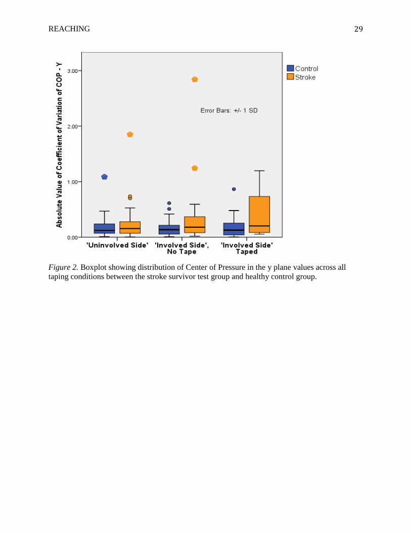
COP-y. Split-half ANOVA found no significant differences in the absolute value of the
CV of COP in the y-direction across conditions and found no interaction of condition and group
( = .974, F(2, 37) = .499, p < .611; = .967, F(2, 37) = .635, p < .536, respectively). Between-
group ANOVA revealed a statistically significant main effect of group, ignoring conditions (F =
4.112, df =1, p < .05). While little variance is seen within the control group and across the 3
conditions (strong, untaped weak, taped weak reach), stroke test subjects appear to have higher
COP-y means with an untaped weak reach when compared to a strong reach, or to a taped weak
reach. A taped weak reach seems to bring COP-y means closer to a strong reach, however, since
no significant group by condition interaction was found, it cannot be assumed that this is due to
the taping intervention. (See Table 1 & 2 and Figure 2)
REACHING
21
COP length normalized for duration of reach. Split-half ANOVA found statistical
significance among conditions and a significant condition and group interaction ( = .628, F(2,
37) = 10.964, p < .001; = .614, F(2, 37) = .635, p< .001, respectively). Additionally,
significant differences were found between subjects for groups (ignoring conditions) (F = 33.737,
df = 1, p < .001). Post-hoc analysis revealed significant differences within subjects across all
conditions with and without group effects (p < .001). Within the control group, little variance in
means for COP percent length is seen across the three conditions (strong, weak untaped, and
weak taped), however within the stroke group, differences are seen where a weak untaped reach
and a weak taped reach have higher means than that of their strong reach. In other words, it
appears that there is increased postural sway or COP excursion/length when normalized for time,
for a weak reach whether it is taped or untaped, when compared to a strong reach. Moreover, it
appears that a taped weak reach did not improve postural sway; in fact, it increased postural
sway when comparing it to an untaped weak reach, or a strong reach. (See Table 1 & 2 and
Figure 3)
sEMG Muscle Activity
Deltoid percent lag. Split-half ANOVA revealed no significant differences across all
conditions and no significant interaction of condition and group for deltoid percent lag ( = .978,
F(2, 37) = .409, p < .667; = .968, F(2, 37) = .616, p < .546, respectively). Between-subjects
(control vs. stroke group) ANOVA, ignoring conditions, revealed group difference, which
approached significance, however, was not reached (F = 3.945, df =1, p < .054). (See Table 1 &
2 and Figure 4)
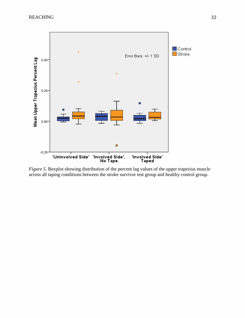
Upper Trapezius percent lag. Split-half ANOVA revealed no significant differences
across conditions and the interaction of condition and group for upper trapezius percent lag was

REACHING
22
not found to be statistically significant ( = .900, F(2, 31) = .409, p < .196; = .949, F(2, 31)
= .616, p < .447, respectively). Between-group ANOVA did not reveal significant group effect
(F = 2.068, df = 1, p < .160). (See Table 1 & 2 and Figure 5)
Deltoid percent duration of reach. Split-half ANOVA revealed a significant difference
across conditions, ignoring groups, but that the group by condition interaction was not
statistically significant ( = .820, F(2, 36) = 3.962, p < .028; = 0.964, F(2, 36) = .676, p
< .515, respectively). Post-hoc analysis within-subjects across conditions (ignoring group effect)
showed statistically significant differences only when comparing a strong reach and a taped
weak reach (p < .048). Between-group ANOVA revealed statistically significant differences
between the control and stroke group, when ignoring conditions (F = 6.636, df = 1, p < .014).
When examining means across conditions within groups, it appears that the stroke test group
experienced shorter deltoid percent durations, more so on their involved UE, however, since no
group by condition interaction was found, the differences in duration of the reach are not affected
by the taping intervention. (See Table 1 & 2 and Figure 6)
Upper Trapezius percent duration of reach. Split-half ANOVA revealed no
significant differences across conditions. In addition, the interaction of condition and group for
upper trapezius percent duration was not found to be statistically significant ( = .874, F(2, 31) =
2.234, p < .196; = .951, F(2, 31) = .796, p < .460, respectively). Between-group ANOVA did
not reveal significant group effect (F = .306, df = 1, p < .584). (See Table 1 & 2 and Figure 7)
Lower Extremity sEMG Data Analysis Relative to the Reaching Limb
Gastrocnemius and Tibialis Anterior percent lag. Kruskal-Wallis Tests determined
that no significant differences existed across conditions (strong UE reach, weak untaped UE
reach, weak taped UE reach) when consolidating both groups for muscle percent lag, regardless
REACHING
23
of whether the gastrocnemius or anterior tibialis was contralateral or ipsilateral to the reaching
limb. (See Table 3)
Gastrocnemius and Tibialis Anterior percent duration of reach. Kruskal-Wallis
Tests, again, determined that no significant differences existed across conditions (strong UE
reach, weak untaped UE reach, weak taped UE reach) when consolidating both groups for
muscle percent duration, regardless of whether the gastrocnemius or anterior tibialis was
contralateral or ipsilateral to the reaching limb. (See Table 4)

REACHING
24
Table 1
Summary of Significant Differences Within-Subjects Across Taping Conditions and Group
by Condition Interaction By Variable
df
Variable Hypothesis Error
F p
COP-x
Across 3 Conditions 2 37
4.301 0.811 0.021
Group by Condition Interaction 2 37
0.932 0.952 0.403
COP-y
Across 3 Conditions 2 37
0.499 0.974 0.611
Group by Condition Interaction 2 37
0.635 0.967 0.536
COP- Length %
Across 3 Conditions 2 37
10.964 0.628 0.001
Group by Condition Interaction 2 37
0.635 0.967 0.001
Deltoid % Lag
Across 3 Conditions 2 37
0.409 0.978 0.667
Group by Condition Interaction 2 37
0.616 0.968 0.546
Upper Trapezius % Lag
Across 3 Conditions 2 37
0.409 0.900 0.196
Group by Condition Interaction 2 37
0.616 0.949 0.447
Deltoid % Duration
Across 3 Conditions 2 36
3.962 0.820 0.028
Group by Condition Interaction 2 36
0.676 0.964 0.515
Upper Trapezius % Duration
Across 3 Conditions 2 31
2.234 0.874 0.124
Group by Condition Interaction 2 31
0.796 0.951 0.460
Note. Split-Half ANOVA: Using Wilks’ Lambda Multivariate Test to report significant differences within
subjects; across conditions when ignoring Groups and Group x Condition interaction. Conditions include a
strong reach untaped (dominant reach in the control group and the uninvolved reach in the experimental stroke
survivor group), and a weak reach (non-dominant reach in the control group and the involved reach of the
experimental stroke group) taped and untaped. COP = Center of Pressure

REACHING
25
Table 2
Summary of Significant Differences Between Experimental Stroke Group and Control
Group While Ignoring Taping Conditions
Variable Type III SS df F p
COP-x 0.108 1 0.640 0.429
COP-y 0.707 1 4.112 0.050
COP- Length % 3.952 1 33.737 0.000
Deltoid % Lag 0.015 1 3.945 0.054
Upper Trapezius % Lag 0.005 1 2.068 0.160
Deltoid % Duration 0.100 1 6.636 0.014
Upper Trapezius % Duration 0.005 1 0.306 0.584
Note. Split-half ANOVA: Using Standard ANOVA to report significant differences between-subjects
(the Control group and Stroke Survivor Experimental Group) when ignoring taping conditions. COP =
Center of Pressure

REACHING
26
Table 3
Summary of Differences in Muscle Percent Duration Across Contralateral or Ipsilateral
Lower Extremity Muscles With Respect To Upper Extremity Reach
Ipsilateral to Reaching Limb
Contralateral to Reaching Limb
Parameter Gastrocnemius
Tibialis
Anterior
Gastrocnemius
Tibialis
Anterior
2 0.037 0.105
0.883 0.05
df 2 2
2 2
p 0.981 0.949
0.643 0.975
Note. Non-parametric Kruskal-Wallis Tests (for independent sample) were used to compare lower extremity
muscle percent duration activity (ignoring groups) with respect to whether it was contralateral or ipsilateral to
the reaching limb. Split-half ANOVA could not be used to compare lower extremity data because LE muscle
contractions did not consistently meet baseline criteria (EMG muscle activity 2 standard deviations above
baseline) across all groups and conditions, despite contractions being visible on examination of sEMG tracing.

REACHING
27
Table 4
Summary of Differences in Muscle Percent Lag Across Contralateral or Ipsilateral Lower
Extremity Muscles With Respect To Upper Extremity Reach
Ipsilateral to Reaching Limb
Contralateral to Reaching Limb
Parameter Gastrocnemius
Tibialis
Anterior
Gastrocnemius
Tibialis
Anterior
2 0.485 1.829
2.106 3.371
df 2 2
2 2
p 0.785 0.401
0.349 0.185
Note. Non-parametric Kruskal-Wallis Tests (for independent sample) were used to compare lower extremity
muscle percent duration activity (ignoring groups) with respect to whether it was contralateral or ipsilateral to
the reaching limb. Split-half ANOVA could not be used to compare lower extremity data since LE muscle
percent lag did not consistently meet baseline criteria (EMG muscle activity 2 SD’s above baseline) across all
groups and conditions, despite contractions being visible on examination of sEMG tracing

REACHING
28
Figure 1. Boxplot showing distribution of Center of Pressure in the x plane values across all
taping conditions between the stroke survivor test group and healthy control group.
REACHING
29
Figure 2. Boxplot showing distribution of Center of Pressure in the y plane values across all
taping conditions between the stroke survivor test group and healthy control group.

REACHING
30
Figure 3. Boxplot showing distribution of the Center of Pressure-length normalized for the
duration of the reach across all taping conditions between the stroke survivor test group and
healthy control group.

REACHING
31
Figure 4. Boxplot showing distribution of the percent lag values of the deltoid muscle across all
taping conditions between the stroke survivor test group and healthy control group.
REACHING
32
Figure 5. Boxplot showing distribution of the percent lag values of the upper trapezius muscle
across all taping conditions between the stroke survivor test group and healthy control group.

REACHING
33
Figure 6. Boxplot showing distribution of the deltoid muscle’s percent duration of reach across
all taping conditions between the stroke survivor test group and healthy control group.

REACHING
34
Figure 7. Boxplot showing distribution of the upper trapezius muscle’s percent duration of reach
across all taping conditions between the stroke survivor test group and healthy control group.

REACHING
35
Discussion
This study did not reveal any statistically significant effects of Kinesiotape™ on the COP
displacement in either the anterior-posterior or medial-lateral directions, though the taped reach
condition did approach significance in the medial-lateral direction. When normalized for time,
however, the results showed a statistically significant increase in COP excursion/length for a
taped weak reach, indicating increased postural sway when compared to an untaped weak reach
or a strong reach. This last finding was the opposite of what was anticipated in the hypothesis,
namely that the Kinesiotape™ would show an improvement in COP excursion/length to
resemble that of a strong reach.
It is not clear at this time whether there was an actual adverse effect of the Kinesiotape™
or if there is another reason why there was an increase in COP excursion/length after the
application of Kinesiotape™. Nam et al. (2013) found that after fatiguing the gastrocnemius
muscle, the medio-lateral length of COP movement was increased, among other parameters,
during single leg stance in the elderly population. This might suggest an influence of fatigue on
the subjects with stroke may therefore have influenced the increase in the COP excursion/length.
Additionally, there may have been an increase in COP excursion/length associated with a
potentially newfound scapular control brought about by the Kinesiotape.™ When new learning
takes place, movement is more variable during the practice phase and becomes less variable as
the task becomes more automatic. Perhaps after waiting for the completion of a learning curve,
the time COP excursion/length would have normalized to levels similar to that of the ‘strong’
side.
The sEMG data collected suggests that the taping intervention did not show any
statistically significant effects. Percent duration of reach for the deltoid muscle seems to indicate

REACHING
36
that the stroke test group experienced shorter deltoid percent durations, more so on their involved
UE. It can’t, however, be concluded that this was due to the taping intervention because no
group by condition interaction was found. The data collected for the sEMG signals for the
gastrocnemius and anterior tibialis muscles showed muscle activation at low levels, which did
not meet the pre-determined criterion of significant and sustained muscle activation.
The results of this study differed from prior studies, which demonstrated improvements
in shoulder function with use of Kinesiotape.™ Yasukawa et al. (2006) showed that use of
Kinesiotape™ provided the proper body alignment to allow performance of reach, grasp, release
and manipulation tasks in children in the acute care setting. This study, however, was quite
different than our study in many ways. Demographically, the current study looked at subjects
over the age of 40, as opposed to pediatric subjects. Additionally, the study performed by
Yasukawa et al. looked at subjects with various neurological diagnoses including Cerebral Palsy,
Traumatic Brain Injury and Spinal Cord Injury and utilized a customized taping protocol for each
child. The current study focused on subjects with stroke and utilized a standardized protocol for
all subjects. Finally, the current study based its results on COP and EMG data while Yasukawa
et al. based their outcomes on the Melbourne Assessment, a pediatric assessment tool of
unilateral upper extremity function.
Renner et al. (2012) studied the ability of Kinesiotape™ to improve shoulder range of
motion. Their results showed a significant difference between control and Kinesiotape™
treatment groups for shoulder internal and external ROM arc in healthy females, but did not
show a difference for males. This difference was only significant after the tape had remained in
place for 4 continuous days, not before. In our study, however, the reaching trials were
REACHING
37
conducted during the application of the Kinesiotape™ and no follow-up trials were performed on
subsequent days.
Studies performed by Host, (1995), Peters et al. (2003), Thelen et al. (2008) and Garcı´a-
Muro et al. (2010) all showed benefits of Kinesiotape™ application with regards to pain
reduction. This study did not utilize pain measures and focused instead on changes in reaching
parameters.
Jaraczewska et al, (2006) suggested specific Kinesiotape™ protocols for the hemiparetic
shoulder with application instructions to be followed based on individual patient needs. The
taping protocol utilized in the current study was different than those suggested by Jaraczewska et
al. because a Certified Kinesio Taping Practitioner (CKTP) created a customized protocol for the
current study, which allowed for a standardization of taping across all subjects.
Limitations
One limitation to our study is fatigue the subjects with stroke experienced due to a
prolonged standing time necessary to gather the necessary data. The protocol of our study
included the subjects reaching first with the ‘strong’ arm 5-7 times followed by the reach of the
‘weak’ arm 5-7 times. After this Kinesiotape™ was applied and a brief seated rest was allowed
before further testing of the ‘weak’ arm. Once the subjects were ready, they performed between
10-15 practice trials before a 5-repetition retest. Overall the subjects had to stand on the force
platform approximately 20 minutes before the trial with Kinesiotape™ was performed. That is a
significant amount of standing time for subjects with stroke. Houdik et al. (2010) showed that
patients with stroke expend 125% more energy during various standing balance conditions than
controls and impaired balance control puts an extra demand on the energy expenditure during
REACHING
38
motor activities. While Gribble et al. (2004) showed that LE muscle fatigue leads to significant
postural control impairments in young, healthy subjects.
The second limitation to our study was the demographics. The major demographics
limitation was the small sample size influencing the power of the tests. The experimental and
control had only 5 subjects each. This was partially due to our strict inclusion criteria, which
required patients with hemiparesis to have 90° of shoulder flexion and the ability to stand for a
full half-hour. Another demographics limitation was disparity of years post-stroke. Two patients
were less than 2 years post-stroke, and 3 subjects were between 4.5 and 7 years post-stroke. So
overall the years ranged from less than 2 to 7 years, a significant range difference that could lead
to major differences in recovery and motor habits learned during the recovery period. Our
recruited sample also did not match for gender or age between test and control groups. The
experimental group had 2 females and 3 males, while the control group had 4 females and 1 male.
And age ranged from 64 to 75 for controls and from 57 to 69 for experimental group.
The third significant limitation to our study was the application of Kinesiotape™.
Although exact instructions from a Certified Kinesio Taping Practitioner (CKTP) were followed,
none of the researchers who applied the tape were CKTPs. Additionally, the taping was slightly
altered from the video instruction to make room for the upper trapezius EMG electrode.
Fratocchi et al. (2013) showed that proper taping technique is important; when KT is applied
over the biceps brachii, it increases concentric elbow peak muscle activity compared with a
placebo taping.

REACHING
39
Conclusion
Application of Kinesiotape™ to inhibit the upper trapezius muscle and to activate the
middle trapezius or rhomboid and the levator scapula muscles does not appear to normalize
reaching in patients with stroke when compared to healthy controls. Our results suggest that this
application of Kinesiotape™ seems to show an adverse effect on COP length normalized for
duration of reach. However, due to multiple limitations in the study, it is impossible to draw
concrete conclusions at this time. Future research is needed to explore the effects of
Kinesiotape™ application on reaching parameters in patients with stroke.

REACHING
40
References
Alexander, C. M., Stynes, S., Thomas, A., Lewis, J., & Harrison, P. J. (2003). Does tape
facilitate or inhibit the lower fibres of trapezius?. Manual therapy, 8(1), 37-41.
Aydin, T., Yildiz, Y., Yanmis, İ., Yildiz, C., & Kalyon, T. A. (2001). Shoulder proprioception: a
comparison between the shoulder joint in healthy and surgically repaired shoulders. Archives of
orthopaedic and trauma surgery, 121(7), 422-425. Babyar, S., Gerkhardt, L., Lagattuta, J., O’Connor, M. (2011) The Influences of Motor Control
Instruction and Taping on Center of Pressure and Scapulothoracic Kinematics during Reaching
for Individuals with Hemiparesis (Unpublished doctoral dissertation). Hunter College, New York,
New York.
Barclay-Goddard, R., Stevenson, T., Poluha, W., Moffatt, M., & Taback, S. (2004). Force
platform feedback for standing balance training after stroke. Cochrane Database Of Systematic
Reviews, 2004(4).
Beer, R., Dewald, J., & Rymer, W. (2000). Deficits in the coordination of multijoint arm
movements in patients with hemiparesis: evidence for disturbed control of limb dynamics.
Experimental Brain Research, 131(3), 305-319.
Broeks, J. G., Lankhorst, G. J., Rumping, K. K., & Prevo, A. H. (1999). The long-term outcome
of arm function after stroke: results of a follow-up study. Disability & Rehabilitation, 21(8), 357-
364. doi:10.1080/096382899297459
Chae, J., Yang, G., Park, B. K., & Labatia, I. (2002). Muscle weakness and cocontraction in
upper limb hemiparesis: relationship to motor impairment and physical disability.
Neurorehabilitation and neural repair, 16(3), 241-248.
Cirstea, M., Mitnitski, A., Feldman, A., & Levin, M. (2003). Interjoint coordination dynamics
during reaching in stroke. Experimental Brain Research, 151(3), 289-300.
Cirstea, M., & Levin, M. (2000). Compensatory strategies for reaching in stroke. Brain: A
Journal Of Neurology, 123(part 5), 940-953.
Cools, A. M., Witvrouw, E. E., Danneels, L. A., & Cambier, D. C. (2002). Does taping influence
electromyographic muscle activity in the scapular rotators in healthy shoulders?. Manual therapy,
7(3), 154-162.
Das, S., & Tibarewala, D. N. (2011). Stabilometric Postural Steadiness Analysis of Post-Stroke
Hemiplegic Patients. International Journal of Engineering Science & Technology, 3(6).
Fang, Y., Daly, J. J., Sun, J., Hvorat, K., Fredrickson, E., Pundik, S., Sahgal, V., & Yue, G. H.
(2009). Functional corticomuscular connection during reaching is weakened following stroke.
Clinical Neurophysiology, 120(5), 994-1002. doi:10.1016/j.clinph.2009.02.173
REACHING
41
Fratocchi, G., Di Mattia, F., Rossi, R., Mangone, M., Santilli, V., & Paoloni, M. (2013).
Influence of Kinesio Taping applied over biceps brachii on isokinetic elbow peak torque. A
placebo controlled study in a population of young healthy subjects. Journal Of Science And
Medicine In Sport / Sports Medicine Australia, 16(3), 245-249. doi:10.1016/j.jsams.2012.06.003
García-Muro, F., Rodríguez-Fernández, A., & Herrero-de-Lucas, A. (2010). Treatment of
myofascial pain in the shoulder with Kinesio Taping. A case report. Manual Therapy, 15(3),
292-295. doi:10.1016/j.math.2009.09.002
Geiger, R., Allen, J., O'Keefe, J., & Hicks, R. (2001). Balance and mobility following stroke:
effects of physical therapy interventions with and without biofeedback/forceplate training.
Physical Therapy, 81(4), 995-1005.
Gracies, J. M. (2005). Pathophysiology of spastic paresis. I: Paresis and soft tissue changes.
Muscle & nerve, 31(5), 535-551.
Gracies, J. M. (2005). Pathophysiology of spastic paresis. II: Emergence of muscle overactivity.
Muscle & nerve, 31(5), 552-571.
Gribble, P., & Hertel, J. (2004). Effect of lower-extremity muscle fatigue on postural control.
Archives Of Physical Medicine And Rehabilitation, 85(4), 589-592.
Harris, J., & Eng, J. (2007). Paretic upper-limb strength best explains arm activity in people with
stroke. Physical Therapy, 87(1), 88-97. doi:10.2522/ptj.20060065
Hislop, H., Avers, D., & Brown, M. (2013). Daniels and Worthingham's Muscle Testing:
Techniques of Manual Examination and Performance Testing. Elsevier Health Sciences.
Host, H.H. (1995). Scapular taping in the treatment of anterior shoulder impingement. Physical
Therapy 75, 803-812.
Houdijk, H., ter Hoeve, N., Nooijen, C., Rijntjes, D., Tolsma, M., & Lamoth, C. (2010). Energy
expenditure of stroke patients during postural control tasks. Gait & Posture, 32(3), 321-326.
doi:10.1016/j.gaitpost.2010.05.016
Hsu, W., Yang, Y., Hong, C., & Wang, R. (2005). Ankle muscle activation during functional
reach in hemiparetic and healthy subjects. American Journal Of Physical Medicine &
Rehabilitation, 84(10), 749-755.
Jaraczewska, E., & Long, C. (2006). Kinesio taping in stroke: improving functional use of the
upper extremity in hemiplegia. Topics In Stroke Rehabilitation, 13(3), 31-42.
Jørgensen, H., Nakayama, H., Raaschou, H. O., & Olsen, T. S. (1994). Stroke in patients with
diabetes. The Copenhagen Stroke Study. Stroke, 25(10), 1977-1984.

REACHING
42
Kisiel-Sajewicz, K., Fang, Y., Hrovat, K., Yue, G., H., Siemionow, V., Sun, C., et al. (2011).
Weakening of synergist muscle coupling during reaching movement in stroke patients.
Neurorehabilitation & Neural Repair, 25(4), 359-368.
Latash ML. Control of Human Movement. Champaign, IL: Human Kinetics; 1993.
Levin, M. F., Michaelsen, S. M., Cirstea, C. M., & Roby-Brami, A. (2002). Use of the trunk for
reaching targets placed within and beyond the reach in adult hemiparesis. Experimental Brain
Research.Experimentelle Hirnforschung.Expérimentation Cérébrale, 143(2), 171-180.
Lin, J. J., Hung, C. J., & Yang, P. L. (2011). The effects of scapular taping on electromyographic
muscle activity and proprioception feedback in healthy shoulders. Journal of Orthopaedic
Research : Official Publication of the Orthopaedic Research Society, 29(1), 53-57.
Mackey, A. H., Walt, S. E., & Stott, N. S. (2006). Deficits in upper-limb task performance in
children with hemiplegic cerebral palsy as defined by 3-dimensional kinematics. Archives of
physical medicine and rehabilitation, 87(2), 207-215.
Mansfield, A., Danells, C. J., Inness, E., Mochizuki, G., & McIlroy, W. E. (2011). Between-limb
synchronization for control of standing balance in individuals with stroke. Clinical Biomechanics,
26(3), 312-317. doi:10.1016/j.clinbiomech.2010.10.001
Massie, C. L., Malcolm, M. P., Greene, D. P., & Browning, R. C. (2012). Kinematic motion
analysis and muscle activation patterns of continuous reaching in survivors of stroke. Journal of
Motor Behavior, 44(3), 213-222.
Mercer, V., Freburger, J., Chang, S., & Purser, J. (2009). Measurement of paretic--lower-
extremity loading and weight transfer after stroke. Physical Therapy, 89(7), 653-664.
doi:10.2522/ptj.20080230
Murphy, M., Alt, Willén, C., & Sunnerhagen, K., S. (2011). Kinematic variables quantifying
upper-extremity performance after stroke during reaching and drinking from a glass.
Neurorehabilitation & Neural Repair, 25(1), 71-80.
Nam, H., Park, D., Kim, D., Kang, H., Lee, D., Lee, S., & ... Choi, S. (2013). The relationship
between muscle fatigue and balance in the elderly. Annals Of Rehabilitation Medicine, 37(3),
389-395. doi:10.5535/arm.2013.37.3.389
National Stroke Association, (2014, March 5). Stroke Fact Sheet. Retrieved from
http://www.stroke.org/site/DocServer/STROKE101_2009.pdf?docID=4541.
Nichols, D. (1997). Balance retraining after stroke using force platform biofeedback. Physical
Therapy, 77(5), 553-558.
Parker, V. M., Wade, D. T., & Hewer, R. L. (1986). Loss of arm function after stroke:
measurement, frequency, and recovery. Disability & Rehabilitation, 8(2), 69-73.
REACHING
43
Peters, S. B. & Lee, G. P. (2003). Functional impact of shoulder taping in the hemiplegic upper
extremity. Occupational Therapy in Health Care, 17(2), 35-46.
Renner, C. M. (2012). Kinesio Tape And Its Effects On Internal And External Range Of Motion
Of The Shoulder (Doctoral dissertation). Retrieved from Indiana State University.
(http://hdl.handle.net/10484/3989)
Robertson, J. V. G., & Roby-Brami, A. (2011). The trunk as a part of the kinematic chain for
reaching movements in healthy subjects and hemiparetic patients. Brain Research, 1382(0), 137-
146.
Roby-Brami, A., Feydy, A., Combeaud, M., Biryukova, E., Bussel, B., & Levin, M. (2003).
Motor compensation and recovery for reaching in stroke patients. Acta Neurologica
Scandinavica, 107(5), 369-381.
Subramanian, S. K., Yamanaka, J., Chilingaryan, G., & Levin, M. F. (2010). Validity of
movement pattern kinematics as measures of arm motor impairment poststroke. Stroke, 41(10),
2303-2308.
Srivastava, A., Taly, A., Gupta, A., Kumar, S., & Murali, T. (2009). Post-stroke balance training:
role of force platform with visual feedback technique. Journal Of The Neurological Sciences,
287(1-2), 89-93. doi:10.1016/j.jns.2009.08.051
Tyson, S., Hanley, M., Chillala, J., A., & Tallis, R. Balance Disability After Stroke. (2006).
Physical Therapy, 86 (1), 30-38.
Thelen, M. D., Dauber, J. A., & Stoneman, P. D. (2008). The clinical efficacy of kinesio tape for
shoulder pain: A randomized, double-blinded, clinical trial. Journal of Orthopaedic & Sports
Physical Therapy, 38(7), 389-395.
Wagner Joanne M., Lang Catherine E., & Sahrmann Shirley A. (2007). Sensorimotor
impairments and reaching performance in subjects with poststroke hemiparesis during the first
few months of recovery. Physical Therapy, 87(6), 751-765.
Yao, J., Chen, A., Carmona, C., & Dewald, J. (2009). Cortical overlap of joint representations
contributes to the loss of independent joint control following stroke. Neuroimage, 45(2), 490-499.
doi:10.1016/j.neuroimage.2008.12.002
Yasukawa, A., Patel, P., & Sisung, C. (2006). Pilot study: investigating the effects of Kinesio
Taping in an acute pediatric rehabilitation setting. The American Journal Of Occupational
Therapy: Official Publication Of The American Occupational Therapy Association, 60(1), 104-
110.
Zorowitz, R. D., Hughes, M. B., Idank, D., Ikai, T., & Johnston, M. V. (1996). Shoulder Pain
and Subluxation After Stroke: Correlation or Coincidence?. American Journal Of Occupational
Therapy, 50(3), 194-201.