Embed Size (px)
Citation preview

Int. J. Electrochem. Sci., 7 (2012) 11655 - 11677
International Journal of
ELECTROCHEMICAL SCIENCE
www.electrochemsci.org
Influence of Surface Oxide Characteristics and Speciation on
Corrosion, Electrochemical Properties and Metal Release of
Atomized 316L Stainless Steel Powders
Y. Hedberg1,*
, M. Norell2, P. Linhardt
3, H. Bergqvist
4, I. Odnevall Wallinder
1
1 KTH Royal Institute of Technology, Div. Surface and Corrosion Science, Dept. Chemistry,
Drottning Kristinas väg 51, SE-10044 Stockholm, Sweden 2 Materials and Manufacturing Technology, Chalmers University of Technology,
SE-412 96 Gothenburg, Sweden 3
Institute for Chemical Technologies and Analytics (CTA), Vienna University of Technology,
Getreidemarkt 9/164, A-1060 Vienna, Austria 4 KTH Royal Institute of Technology, Div. Functional Materials, Dept. Material Physics,
Electrum 229, SE-16440 Kista, Sweden *E-mail: [email protected]
Received: 22 September 2012 / Accepted: 4 November 2012 / Published: 1 December 2012
Surface oxide characteristics of powder particles are important to consider for any toxicological risk
assessment based on in-vitro or in-vivo tests. This study focuses on a multi-analytical approach (X-ray
photoelectron spectroscopy, Auger electron spectroscopy, scanning- and transmission electron
microscopy, and different electrochemical techniques) for in-depth characterization of surface oxides
of inert-gas-atomized (GA) AISI 316L stainless steel powder, compared with massive sheet and a
water-atomized (WA) 316L powder. Implications of differences in surface oxide phases and their
surface distribution on corrosion, electrochemical properties and metal release are systematically
discussed. Cr was enriched in an inner surface layer for both GA powders, with Mn and S enriched in
the outermost surface oxide. The surface oxide was 2-5 nm thick for both GA powder size fractions,
amorphous for the GA powder sized <4 µm and partially crystalline for the powder sized <45 µm. A
strong ennoblement, i.e. positive shift in open circuit potential, of up to 800 mV, depending on
solution, was observed for the GA powders. This ennoblement was induced by catalytic oxygen
reduction properties of tri- or tetravalent Mn-oxides, not present on the massive sheet or WA powder.
In contrast to the predominant presence of a trivalent Cr-oxide in the surface oxide of the GA powder
particles, the WA<45µm powder revealed oxidized Cr, most probably present in its hexavalent state
(not chromate), within a silicate-rich surface oxide. This study clearly shows that the surface oxide
composition and speciation of differently sized GA and WA powders are unique (strongly connected
to the atomization process) and of large importance for their pitting corrosion and metal release
properties. For the GA<45µm powder, Mn-rich oxide nanoparticles were proposed to account for its
higher pitting corrosion susceptibility, a more stable surface ennoblement, and a shift of the MnO2
oxidation/reduction peaks in the cyclic voltammogram, compared with the GA particles sized <4µm.

Int. J. Electrochem. Sci., Vol. 7, 2012
11656
The thermodynamically unstable ferritic structure of the small sized particle fraction (GA <4µm),
despite an austenitic composition, revealed a higher pitting corrosion susceptibility and higher nickel
release compared with the austenitic particle fraction of the GA <45 µm powder.
Keywords: surface oxide, speciation, characterization, manganese dioxide, oxide nanoparticles
1. INTRODUCTION
Within risk assessments of materials, the toxicity of a ”substance” is often classified according
to standardized in-vitro or in-vivo tests. REACH (Registration, Evaluation, Authorisation and
Restriction of Chemicals) [1-2] obligates all producers within the EU (European Union), or exporters
into EU, to show that their substances or products are classified and safe from both an environmental
and health perspective, “No data no market”. The responsibility lies hence on the industry to compile
and/or generate data if lacking. The lack of metal release data and its possible correlation to surface
characteristics and toxic response resulted recently in the generation of a substantial set of data within
this area as a complement to findings in the scientific literature that enabled classification and risk
assessment of ferrochromium alloys including stainless steel [3-11]. However, most recognized
standard in vitro tests are based on studies of powders instead of massive surfaces, assuming the
powder shape to mimic the behavior of massive sheet [6, 12]. Except for the fact that the production
route of metal powders is very different, with variations in surface characteristics as a consequence, the
generation of powders from massive sheet (commonly done) may furthermore not be representative for
their intended use and also change the material/surface characteristics. Reproducible surfaces of
powder particles are difficult to accomplish since they cannot be easily polished or prepared [13-15].
Previous findings have shown significant differences in the metal release behavior of differently sized
powder particles of stainless steel, an effect partly connected to differences in surface oxide
characteristics [13].
Surface oxide characterization is commonly made by using different analytical techniques
including XPS (X-ray Photoelectron Spectroscopy), AES (Auger Electron Spectroscopy), SEM/TEM
(scanning/transmission electron microscopy), EBSD (electron backscattered diffraction) and XRD (X-
ray Diffraction). These techniques are limited in terms of either spatial or lateral resolution or chemical
information of phases, and hence often combined. Reported findings show that the surface oxide
(composition, phases/inclusions and their distribution, thickness etc.) of metal powders is connected to
the cooling rate during atomization [16], the particle size and secondary dendrite arm spacing [16-18],
the type of atomization (e.g., water-, gas-atomization, or rotating electrode process) [18], and the
oxygen availability [19]. Minor elements with higher oxygen affinity, such as Mn, Si and Cr are often
enriched at the surface, either as “oxide particles” or “islands” within or on a more continuous surface
oxide of different composition (“matrix oxide”). This surface enrichment increase with reduced
particle size [17-21]. Cr2O3 with MnO and Fe2O3 phases in the outermost surface layer [21], possibly
combined with MnCr2O4, FeCr2O4, or (Fe,Cr)2O3 as particles or small local surface phases [22], have
been reported for nitrogen atomized AISI 304L powders. Austenitic stainless steel powders may also
Int. J. Electrochem. Sci., Vol. 7, 2012
11657
show a ferritic structure and/or solidify as single crystals if the cooling rate is rapid enough [23-26], as
previously observed for the inert-gas-atomized 316L powder particles sized <4 µm of this study [14].
Electrochemical investigations are another way to identify and characterize oxide phases
within, or on, powder particles. Oxide phases on micron-sized particles immobilized on paraffin-
impregnated electrodes (PIGE) have been characterized using voltammetry [27-29]. Another method,
that in addition is able to analyze dissolved ions, is the carbon paste electrode (CPE) with higher
sensitivity than PIGE. CPE has been widely used to identify different metal oxide/hydroxide phases
[30-35]. The benefit of electrochemical analysis of oxide phases, compared with surface analytical
methods, is the direct information on their reduction/oxidation behavior, important information for
considerations of corrosion and/or metal release mechanisms. It was recently shown that the oxide
properties of inert-gas-atomized AISI 316L particles (sized <45 µm, the same as investigated in this
paper) were crucial for their corrosion behavior, when compared to polished particles and massive
sheet (part II of this paper series, [36]).
The aim of this paper is to employ a multitude of analytical techniques (XPS, AES, SEM,
TEM) combined with different electrochemical methods (Open circuit potential measurements-OCP,
Potentiodynamic polarization and cyclic voltammetry-CV) to characterize differences in surface oxide
characteristics and corrosion properties of inert-gas-atomized AISI 316L stainless steel powders
compared with massive sheet and a water-atomized 316L powder. The inert-gas-atomized AISI 316L
powders have previously been investigated at in-vitro [4-5, 8-9, 13, 37] and in-vivo [15] conditions.
The influence of surface oxide characteristics and composition on the corrosion properties, extent of
metal release and the electrochemical (catalytic) properties are discussed from a mechanistic
perspective.
2. MATERIALS AND METHODS
2.1. Materials
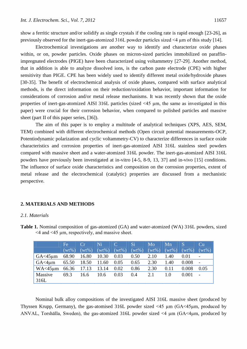
Table 1. Nominal composition of gas-atomized (GA) and water-atomized (WA) 316L powders, sized
<4 and <45 µm, respectively, and massive sheet.
Fe
(wt%)
Cr
(wt%)
Ni
(wt%)
C
(wt%)
Si
(wt%)
Mo
(wt%)
Mn
(wt%)
S
(wt%)
Cu
(wt%)
GA<45µm 68.90 16.80 10.30 0.03 0.50 2.10 1.40 0.01 -
GA<4µm 65.50 18.50 11.60 0.05 0.65 2.30 1.40 0.008 -
WA<45µm 66.36 17.13 13.14 0.02 0.86 2.30 0.11 0.008 0.05
Massive
316L
69.3 16.6 10.6 0.03 0.4 2.1 1.0 0.001 -
Nominal bulk alloy compositions of the investigated AISI 316L massive sheet (produced by
Thyssen Krupp, Germany), the gas-atomised 316L powder sized <45 µm (GA<45µm, produced by
ANVAL, Torshälla, Sweden), the gas-atomized 316L powder sized <4 µm (GA<4µm, produced by

Int. J. Electrochem. Sci., Vol. 7, 2012
11658
Sandvik Osprey Limited, UK), and the water-atomized 316L powder sized < 45 µm (WA<45µm,
produced by Höganäs AB, Sweden) are compiled in Table 1. The test materials were supplied via the
European Confederation of Iron and Steel Industries and International Stainless Steel Forum, Belgium.
Information on the specific surface area, measured by means of the BET method (nitrogen
adsorption at cryogenic conditions) has been described previously [4] and is summarized in Table 2.
Table 2. Specific surface areas, measured by means of the BET method, of the different 316L powders
(GA: gas-atomized, WA: water atomized).
Stainless steel 316L
powders
BET specific surface area
(m2/g)
GA<4µm 0.700
GA<45µm 0.069
WA<45µm 0.087
Particle size distribution values of the three powders in phosphate buffered saline (PBS, 8.77
g/L NaCl, 1.28 g/L Na2HPO4 and 1.36 g/L KH2PO4, pH 7.2-7.4) measured by means of low angle laser
light scattering (LALLS, using a Malvern Mastersizer 2000 equipment with a Hydro SM dispersion
unit, experimental details in [4]) is shown in Table 3.
Table 3. Particle size distribution of the stainless steel powder particles in phosphate buffered saline
(PBS) measured by means of LALLS. d0.5 is the median particle diameter and d0.1 and d0.9 the
10 % and 90 % size distribution cut off points by volume (mass) or number, respectively.
Unit GA316L
<4µm
GA316L
<45µm
WA316L
<45µm
Volume / diameter -
µm
d0.1 2.25 ± 0.05 10.61 ± 0.24 18.46 ± 0.02
d0.5 3.91 ± 0.09 21.37 ± 0.82 35.47 ± 0.12
d0.9 6.63 ± 0.17 38.95 ± 1.52 64.76 ±0.24
Number /
diameter - µm
d0.1 1.58 ± 0.03 2.04 ± 0.19 11.08 ± 0.04
d0.5 2.35 ± 0.04 3.74 ± 0.70 17.61 ± 0.03
d0.9 3.97 ± 0.08 14.22 ± 0.61 32.42 ± 0.04
2.2. Analytical techniques
2.2.1. X-ray photoelectron spectroscopy (XPS)
Compositional analysis of surface oxides of the powders was performed by means of XPS
(UltraDLD spectrometer from Kratos Analytical) using a monochromatic Al x-ray source (150 W) on

Int. J. Electrochem. Sci., Vol. 7, 2012
11659
areas approximately sized 700 x 300 µm. Wide spectra were run to detect elements present in the
outermost surface oxide (information depth of a few nm) at five different locations, and detailed high
resolution spectra (20 eV pass energy) were acquired for the main compositional elements. The
binding energy of the C 1s (C-C, C-H) signal (285.0 eV) was used as internal reference. The Fe 2p
peak was separated according to its metallic (706.9±0.25 eV) and oxidized (711.9±1.2 eV) states. No
metallic chromium, only oxidized (576.8±0.71 eV) chromium was detected. Oxidized manganese was
detected at binding energies of 641.6±0.2 and 644.0±1.0 (GA<4µm) and 641.4±0.05 and 643.1±0.01
eV (GA<45µm). No metallic or oxidized nickel was detected. Oxidized silicon was only detected in
the case of the WA powder at a binding energy of 102.6±0.2 eV (as silicate [38]). Results obtained by
means of XPS are reported in [39], but included for comparative reasons.
2.2.2. Auger electron spectroscopy (AES)
The composition and thickness of the oxide on individual GA powder particles were
determined by means of AES depth profiling (MicroLab 350 from Thermo Scientific). Sensitivity
factors from the manufacturer were used for apparent quantification. The powder was mounted by
gently pressing a quantity between pure Al plates. The powder analyzed has not been in contact with
the Al plates. The rate of Ar+-etching was calibrated on Ta2O5 films of known thickness. This rate
varies with the angle of ion incidence and this was considered as described elsewhere [40]. In short,
the spherical shape of the particle is used to geometrically determine an area of analysis where the
angle of ion incidence gives a maximal etch rate as calibrated. The width of this area is approximately
15% of the particle radius. If the area of analysis deviates from the ideal position, or the particle is not
totally spherical, the etch rate will be slower than that calibrated and the oxide thickness hence
overestimated. This is of importance for the very small particles studied in this study. On GA<45µm,
the area of analysis was about 1 µm in square. On GA<4µm, point analyses were done with a lateral
resolution of approximately 20 nm. The ideal etch position on the typically 1 µm large particles
analyzed was about 75 nm wide. Therefore, any drift in the SEM image during profiling may have
resulted in an overestimated oxide thickness on some particles. Thus, the thinnest oxide thickness
determined is the most reliable.
2.2.3. Transmission electron microscopy (TEM)
The GA<4µm powder was prepared by first mixing the powder particles in an electron beam
compatible epoxy resin (Gatan Inc. US). This mixture was cast within brass tubes (outer Ø - 3 mm).
The tube was cut to 0.6 mm thick slices. These slices were ground on silicon carbide paper with final
grinding on grade 4000 mesh down to a thickness of 0.1 mm. The thin slices were dimpled by using a
dimple grinder (Gatan Inc., US model 656) using diamond paste, and at the last step in a SiO2 water
slurry. In order to minimize the time for the ion polishing, it was essential to calculate the final
minimum thickness which should be as thin as possible. Finally, thinning to electron transparency was
performed by using a precision ion polishing system (PIPS, Gatan Inc. US) and by using a

Int. J. Electrochem. Sci., Vol. 7, 2012
11660
successively lower acceleration voltage ranging from 3.5 kV down to 1.5 kV. The GA<45µm powder
was prepared by using a focused ion beam, FIB (FEI model QUANTA 3D FEG). The powder particles
were attached to a double-sided adhesive carbon based disc. One representative particle was sputtered
with platinum for protection on an area sized approx. 25 x 5 µm2, and cut with a Ga-ion beam down to
a thickness corresponding to electron transparency. Finally, the sliced foil was cut and attached on a
TEM-grid.
The sliced foils were examined by means of TEM (JEOL 2100F (HR)) equipped with scanning
transmission electron microscopy (STEM), energy dispersive x-ray spectrometry (EDXS) and electron
energy loss spectroscopy (EELS). The surfaces of the particles were imaged in high magnification with
lattice resolution in order to characterize the crystal structure and the amorphous degree of the surface
oxide. EDXS analyses were performed to measure the concentration of different elements in the
surface oxide. Point analyses (a few nm resolution) at different locations and line scans were
performed. By using this method a detection limit of 0.1% of different elements was possible to
achieve.
2.2.4. Field emission gun scanning electron microscopy (FEG-SEM)
High resolution morphology studies were performed by means of field emission gun scanning
electron microscopy (FEG-SEM Leo1530 upgraded to Zeiss Supra 55) up to 100,000 times
magnification using secondary electrons, a working distance of 5.7 mm, and a voltage of 10 or 15 kV.
The powders were fixed on carbon tape.
2.3. Electrochemical techniques
2.3.1. Open circuit potential (OCP) measurements
OCP was measured as a function of pH, solution, and time. Both massive sheet and powders
were investigated. The massive sheet was abraded to 1200 SiC grit, consecutively and ultrasonically
cleaned with acetone for 7 min and isopropyl alcohol for 7 min, prior to storage for 24 h ± 1 h in a
desiccator. The powder (approx. 4 mg) was immobilized on a paraffin-impregnated graphite electrode
(PIGE), with an area of approx. 0.5 cm2, which was abraded and shortly heated prior to pressing it into
the powder for fixation. The PIGE background current has previously been found to be significantly
lower compared to the current of the PIGE and of the investigated particles [36, 41]. The massive
sheet, of which approximately 1 cm² was immersed in solution, was fixed using a clamp (not
immersed). The counter electrode was a platinum wire and Ag/AgCl 3M KCl was used as the
reference electrode. The pH dependent OCP measurements were performed for massive sheet, and
powders of GA<45µm, GA<4µm, and MnO2 (specific surface area (BET) of 0.054 m2/g, Sigma
Aldrich, Lot no. MKBC2119V) as reference. The manganese concentration of the buffer solution was
adjusted to 1 mg/L Mn2+
(as this is well above the expected release of manganese for the investigated
time period [13]) which however precipitated to some extent in the case of pH 10.1. The buffer
solutions (from [42]) for the different pH values were prepared as follows: 0.1 M CH3COOH and 0.1

Int. J. Electrochem. Sci., Vol. 7, 2012
11661
M NaCH3COO (pH 4.7), 50 mL 0.1M KH2PO4 and 5.4 mL 0.1 M NaOH in 100 mL (pH 6.0), 50 mL
0.1 KH2PO4 and 32 mL in 100 mL (pH 7.0), 50 mL 0.1 M KH2PO4 and 46.1 mL 0.1 M NaOH in 100
mL (pH 7.8), and 50 mL 0.05 M NaHCO3 and 10.7 mL 0.1 M NaOH in 100 mL (pH 10.1). All
measurements were performed by allowing the OCP to stabilize for 1 h, or after a change in potential
being less than 10 µV/s, before the value was recorded.
The influence of solution on OCP was investigated for massive sheet, GA<45µm, GA<4µm,
and WA<45µm powders, in solutions relevant for metal release studies into synthetic body fluids, i.e.
gastric fluid (GST, pH 1.5-1.7, 4 g/L 25% HCl), artificial lyosomal fluid (ALF, pH 4.5, 20.8 g/L
C6H8O7, 6.00 g/L NaOH, 3.21 g/L NaCl, 0.050 g/L MgCl2, 0.071 g/L Na2HPO4, 0.039 g/L Na2SO4,
0.128 g/L CaCl2·2H2O, 0.077 g/L C6H5Na3O7·2H2O, 0.059 g/L H2NCH2COOH, 0.090 g/L
C4H4O6Na2·2H2O, 0.085 g/L C3H5NaO3, 0.086 g/L C3H3O3Na, 1.7 mL/L 50% NaOH), 3 g/L NaCl
(not pH adjusted, pH approximately 5.4-7), and 8 M NaOH (pH 13.1-13.3). The influence of time was
investigated in GST for up to 48 h, for massive sheet, GA<45µm, GA<4µm, and WA<45µm. All
measurements were made at aerated conditions, with the exception of half of the measurements
investigating the pH dependence, where nitrogen gas was purged through the solution prior to the
measurement and blown on the solution during the measurement.
2.3.2. Potentiodynamic polarization
Anodic polarization curves, starting at OCP (after stabilization) and ending at 1.2 V vs.
Ag/AgCl 3 M KCl with a scan rate of 0.2 mV/s were run for the powders of GA<45µm, GA<4µm, and
WA<45µm in 0.03 M HCl (5 g/L 25% HCl). This solution has previously found to be sufficiently
aggressive to enable observations of differences between the different powders investigated [41]. The
GA<45µm powder was in addition separated into a magnetic (ferritic) and a non-magnetic (austenitic)
particle fraction and investigated using anodic polarization.
2.3.3. Cyclic voltammetry (CV)
Cyclic voltammetry starting at OCP, polarizing cathodically to approx. -1.2 V and
consecutively polarizing anodically to approx. +0.4 V, in 8 M NaOH, was performed to identify the
presence of any oxidized metal phases. Powders of GA<45µm, GA<4µm, WA<45µm, and abraded
particles (abraded from a massive sheet of 316L using a clean diamond file) stored for at least 24 h at
ambient conditions (“massive” particles) were investigated using a carbon (graphite) paste electrode
(CPE) and a reference electrode of Hg/HgO in 8 M NaOH, as previously described [43]. The scan rate
was set to 0.5 mV/s. Powder concentrations in the graphite paste are given in Fig. 8. Different
concentrations were investigated as the powder concentration has previously been shown to
significantly influence the spectrum [43]. In order to obtain representative spectra, differences in BET
area were taken into account.

Int. J. Electrochem. Sci., Vol. 7, 2012
11662
3. RESULTS AND DISCUSSION
3.1. Surface oxide characteristics and composition based on a multi-analytical approach
3.1.1. XPS, outermost surface oxide composition
The surface elemental composition of the gas- and water atomized 316L powders based on
XPS is compiled in Table 4. As previously reported [39], no accurate assignment of di- and tri-valent
Fe oxidized species was possible due to overlapping binding energies. In accordance with literature
findings [16-17], oxidized Si (most probably present as silicates [38]) was strongly enriched at the
surface on the water-atomized particles, in addition with oxidized Mn (indicative of tri- and/or tetra-
valent Mn-species, possibly MnO2) [44] in the case of the gas-atomized powder. In both cases, these
enrichments increased with reduced particle size [18]. There was a significant increase of the binding
energy and peak width of oxidized Cr for the WA-powder compared to the GA-powders (trivalent Cr-
species only) from 576.6±0.35 eV to 577.4±0.09 eV, which suggests a possible higher oxidation state.
However, due to overlapping peaks between hexa- and tri-valent Cr-species, any accurate assignment
was not possible. More speciation-sensitive techniques are required.
Table 4. Surface oxide composition (wt%) of gas-atomized (GA) and water-atomized (WA) 316L
powder particles sized <4 and <45 µm, and massive sheet, measured by means of XPS.
(Relative oxidized metal components only, the presence of carbon and oxygen is not taken into
account).
Mn Fe Cr Si
GA<4µm 47±5 40±4 14±1 <LOD
GA<45µm 13±1 72±7 15±2 <LOD
WA<45µm <LOD 24±2 17±2 59±6
Massive
sheet
<LOD 75±8 25±3 <LOD
<LOD – below limit of detection
3.1.2. AES depth profiles and FEG-SEM investigations
Depth profiles by means of AES (for GA<4µm and GA<45µm) and high resolution images by
means of FEG-SEM (all powders) are displayed in Figs. 1-3. Similar to XPS findings, carbon was
present on the utmost surface but was disregarded as adventitious carbon contamination. The oxide
thickness was estimated to approximately 2 nm for both powders, estimated from half the decay of the
oxygen content. This was the thinnest oxide thickness recorded/estimated for each powder and also the
most reliable measure (c.f. experimental section). Variations in thickness up to 3 nm were recorded for
both powders. Selected depth profiles are shown in Fig. 1. In the case of the GA<45µm particles, other
replicate measurements showed similar depth profiles, but slightly different oxide thicknesses. In the
case of the GA<4µm particles, most profiles were similar to Fig. 1. Some particles revealed
significantly different depth profiles, as discussed below.

Int. J. Electrochem. Sci., Vol. 7, 2012
11663
Mn was clearly enriched at the utmost surface, Fig. 1, to a higher extent for the smallest
particles, findings in agreement with the literature [18-19, 21]. In the case of the GA<4µm particles,
the relative atomic percentage of Mn (total elements) ranged from 4, Fig. 1c, to 7 at% at the utmost
surface, as compared to 1-2 at% for the larger powder, GA <45 µm. The absolute values of the AES
depth profiles should though be interpreted with caution since the sensitivity factors are not exactly
known. S was also enriched at the utmost surface and present up to a few nm into the surface oxide.
This enrichment was more pronounced in the case of the GA<45µm particles compared with GA<4µm
particles. The S depth profile did however not follow the same trend as observed for other elements
such as Mn, which may suggest that it is not solely or necessarily present as MnS.
Figure 1. AES depth profiles (total elements, carbon excluded, left; cationic profile, right) for GA
316L particles sized <45µm (a, b) and <4µm (c, d).
In accordance with previous investigations of stainless steel powders [18, 21] oxides of Fe were
the predominant components of the utmost surface whereas oxidized Cr only appeared further into the
layer reaching a maximum concentration at the depth corresponding to the oxide thickness and always
connected with a minimum in the iron content intensity. These observations were interpreted as an

Int. J. Electrochem. Sci., Vol. 7, 2012
11664
inner Cr-rich oxide, in agreement with the literature [18] and an adjacent outer Fe-rich oxide. The
maximum Cr concentration was more pronounced for the GA<45µm particles compared with the
GA<4µm particles. For one particle out of ten GA<4µm particles analyzed, 6 at% Cr and 5 at% Ni
were detected at the outermost surface (0 nm depth, data not shown). It was not possible to determine
if Ni was in its oxidized or metallic state. However, no Ni was detected within the XPS measurements,
providing average data for a large surface area and number of particles. These observations are in
concordance with literature findings on gas-atomized 304L powders where Ni was not detected by
XPS but still present in small amounts at the surface [21]. AES measurements also revealed the
presence of small amounts of Cu (~2 at%) enriched at the utmost surface of two out of ten investigated
GA<4µm particles (data not shown). Some of the GA<4µm particles mostly contained Ni and Cr (2
out of 10 particles).
Figure 2. High resolution FEG-SEM images of inert-gas-atomized (GA) 316L <45 µm powder
particles, showing their morphology, surface microstructure, grain boundaries and presence of
surface oxide particles. Magnifications: 5,000 and 100,000 x.
Figure 3. High resolution FEG-SEM images of water-atomized (WA) 316L <45 µm particles.
Magnifications: 650 and 20,000 x.
The presence of such particles within the same GA<4µm powder has previously been observed
by means of EDS (36 wt% Ni, 50 wt% Cr, 7.4 wt% Mo and 3.8 wt% Fe) [14]. The origin of these

Int. J. Electrochem. Sci., Vol. 7, 2012
11665
particles is at present unknown, however possibly formed during the atomization process or impurities.
Previous findings propose the initial formation of oxide particles with strong oxide formers such as
Mn, Cr and Si [19-21, 45], and the subsequent formation of a Fe oxide [19, 21, 45] and/or Cr oxide
[21] layer (and in the case of WA-particles an oxidized Si layer [46]) during atomization and handling.
Oxide particles rich in Mn, Fe, and Si (based on EDS findings), have been reported for water-atomized
particles [20]. Surface particles were observed also in this study for the powders sized <45µm, Fig. 2-
3. These surface particles, approximately sized 20 nm up to a maximum size of 100 nm, were
relatively homogeneously distributed on GA powder particles. For the WA powder, less abundant
surface particles were observed, c.f. Fig. 3, compared to the GA powder, suggesting that most of the
oxidized silicon (59 wt%, Table 1) was present within the continuous surface oxide. In the case of the
GA<45µm powder, these oxide nanoparticles were investigated by means of AES point analysis, and
compared with the matrix oxide as reported in [39]. The oxide nanoparticles were enriched in Mn, with
approximately an equal amount of Fe and small amounts of S. These elements were not only present in
these surface nanoparticles but also at further etching depth, suggesting relatively homogeneous oxide
nanoparticles. In contrast, the outermost surface oxide surrounding these particles (matrix oxide)
consisted only of Fe and O, with approximate ratio of 2:3, suggesting Fe2O3 [39]. No Mn or S was
detected there [39].
3.1.3. TEM/EDXS investigations of surface oxide morphology, thickness and crystallinity
TEM (Fig. 4) combined with EDXS investigations of the GA<4µm and GA<45µm powders
confirmed in general the oxide thickness estimation made by means of AES and the elemental
distribution within the surface oxide, with the exception of the Mn content. Significantly lower relative
amounts of Mn were determined with EDXS compared with both AES and XPS. However, this
discrepancy was attributed overlapping peaks for Fe Lα and Mn Lα. In agreement with AES, Ni and Cr
were less abundant at the utmost surface compared with Fe. Based on TEM findings, the oxide
thickness varied at different investigated locations from approximately 2-3 nm to 5 nm for the
GA<45µm particles, and was <5 nm for the GA<4µm particles, Fig. 4. The GA<4µm particles
revealed an amorphous surface oxide without any surface oxide nanoparticles, Fig. 4a. In agreement
with FEG-SEM and AES analyses, the GA<45µm powder revealed in contrast many differently sized
surface nanoparticles (Fig. 4b). The surface oxide revealed both crystalline and amorphous areas, Fig.
4b.
The absence of surface oxide nanoparticles on the GA<4µm powder may be explained by their
high cooling rate, during atomization, hindering their formation. A very rapid cooling rate for this
powder is indicated by previous findings with a bulk structure of a frozen-in ferritic structure, a
cellular microstructure (in contrast to dendrites for the GA<45µm powder) and many single-crystalline
particles [14].

Int. J. Electrochem. Sci., Vol. 7, 2012
11666
Figure 4. TEM images of the surface oxide of a) gas-atomized 316L powder <4µm and b) gas-
atomized 316L powder <45µm. The light grey areas are the background, the slightly darker
regions the oxide and the dark areas the metal (a), oxide nanoparticles and/or metal (b). The
pattern in the metal region in (a) is caused by dislocations.
3.2. OCP measurements - Importance of oxidized manganese in the surface oxide
In parallel studies (part II of this paper series [36]), the GA<45µm powder showed a very high
OCP prior to any applied current, being substantially higher (up to 800 mV) compared with the OCP
after applying small reductive currents. This effect was only observed for non-polished powder
particles, not for polished particles [36], which indicates the importance of the surface oxide formed
already during the inert-gas atomization process. Based on XPS, AES, TEM and Raman [39]
measurements, oxidized Mn-phases are the only possible constituents that may influence the OCP in
this strong way. MnO2 deposits on stainless steel have previously been reported to induce a similar
ennoblement [47-51], i.e. a substantial shift of the OCP to more positive potentials.
To investigate if MnO2 or any other trivalent or tetravalent Mn oxide/hydroxide species could
explain the observed OCP shift for the GA<45µm powder, a series of investigations was initiated
including studies of effects of pH, solution, and time on OCP, and cyclic voltammetry measurements
to possibly identify any oxidized Mn phases. The pH dependence of the OCP of powders of MnO2 (as
reference), GA<4µm, and GA<45µm, and massive sheet is presented in Fig. 5. The massive sheet was
abraded 24 h prior to the measurement and revealed typical OCP values at these conditions [49-50]. At
pH 7, the following reaction is expected for MnO2 [49]:
The Nernst’s equation at pH 7 and a Mn(II) concentration (cMn(II)) of 1 mg/L, predicts a
potential (E) of +341 mVAg/AgCl, and the theoretical pH dependence is -118 mV/pH at neutral and
acidic pH. The measured OCP for the MnO2 powder was 312.5±3.2 mV and a pH dependence of
103.5±3.5 mV/pH (both at aerated and deaerated conditions) was determined experimentally between
pH 4.7 and 7.8 (Fig. 5). Between pH 7.8 and 10.1, a decreased pH dependence (67.5±3.5 mV/pH) was
observed for the MnO2 powder (both at aerated and deaerated conditions, Fig. 5). This is in accordance

Int. J. Electrochem. Sci., Vol. 7, 2012
11667
with the theoretical shift to the one-electron transition between MnO2 and MnOOH at higher pH values
[49]. Observed OCP values of the GA powders were in-between observed figures for massive sheet
and the MnO2 powder, Fig. 5. The GA<45µm and GA<4µm powders revealed similar OCP values at
neutral and alkaline pH, however, not at pH 4.7, where the OCP of GA<45µm was significantly higher
compared to GA<4µm. The pH dependence of massive sheet of 78.5±0.5 mV/pH between pH 4.7 and
7.8 and 52.5±2.5 mV/pH was lower compared with the MnO2 powder. The GA<45µm powder
revealed a very similar pH dependence as MnO2: 134.5±7.5 mV/pH between pH 4.7 and 7.8, and the
GA<4µm powder somewhat lower values (74.5±16.5 mV/pH), close to the pH-dependence for
massive sheet (78.5±0.5 mV/pH). No significant differences between deaerated and aerated conditions
were observed for any of the powders or surfaces, in agreement with previous investigations of
massive stainless steel and MnO2 coated stainless steel [49]. Based on these pH dependent
measurements, it can be concluded that significant positive shifts in OCP of the inert-gas-atomized
powder occurred compared with the massive sheet, and were most probably caused by tri- or tetra-
valent Mn-species. A reduced OCP for GA<4µm particles compared with GA<45µm particles at pH
4.7 indicated in addition that these species may be dissolved to different extent at these conditions.
Figure 5. OCP values of massive 316L, inert-gas-atomized powders - GA 316L < 4 (GA<4µm) and <
45 µm (GA<45µm), and MnO2 powder, in different buffer solutions containing 1 mg/L Mn(II),
at both aerated (filled symbols) and deaerated (unfilled symbols) exposure conditions. The
error bars represent the standard deviation between 2 and 5 independent measurements.
The effect of solution on OCP was investigated for the GA powders compared with massive
sheet and the WA powder (without any oxidized Mn surface species). Studies were conducted in
gastric fluid (GST) of low pH (1.5), artificial lysosomal fluid (ALF)-a highly complexing solution (pH
4.5) [13], 3 g/L NaCl (pH 5.4-7), and 8 M NaOH (pH 13.1-13.3), Fig. 6. In contrast to the influence of

Int. J. Electrochem. Sci., Vol. 7, 2012
11668
pH, Fig. 5, the GA<45µm powder revealed a significantly more positive OCP compared to the
GA<4µm powder in all solutions of a pH between 1.5 and 7. This may be attributed to the dissolution
of surface-active Mn-species in the case of GA<4µm powder. Previous metal release studies in ALF
revealed a significant enhancement of released metals from the GA<4µm powder compared with the
GA<45µm powder. However, the opposite scenario was the case in low aggressive solutions [13]. The
difference in OCP between the <4 µm and the <45 µm GA powders was 800 mV in the highly
complexing ALF solution of low pH, Fig. 6. The same difference in OCP has previously been
observed after applying small cathodic currents on the GA<45µm powder in 0.1 M HCl [36]. The
oxidized Mn-phase on the GA<45µm powder particles seemed hence to be easily reducible when a
cathodic current was applied, but not in any of the solutions at acidic or neutral pH within the
investigated time-frame.
Figure 6. OCP values (after stabilization) of gas atomized GA 316L powders (< 4 and < 45µm), water
atomized WA 316L powder <45µm (only in GST, NaCl, and NaOH), and massive 316L (only
in GST and NaCl) in different solutions. The asterisks indicate significant differences within
one solution, calculated by means of a student t-test (unpaired data with unequal variance): *
for p<0.05, ** for p<0.01, and *** for p<0.001.
To investigate if the shift in OCP, induced by the presence of oxidized Mn-phases, was time-
dependent, the OCP was recorded over time in GST (pH 1.7), Fig. 7. For both massive sheet and the
WA<45µm powder (in two of three replicate samples, respectively), a “breakdown” was observed
from initial OCP values of about 200 mV (Ag/AgCl) to values between -100 and -200 mV, Figs. 7a
and b. This breakdown occurred earlier (after 0-50 s) for massive sheet compared to WA<45 powder
(up to 1000 s), indicating somewhat more protective properties of the surface oxide of WA<45µm
powder particles compared with the abraded (24 h prior to measurements) massive sheet. The
GA<4µm powder revealed a similar final OCP as determined for massive sheet and the WA<45µm
powder (Figs. 7c and 6), however with a different time dependence. No “breakdown” was observed,
rather a continuous decay of the OCP level. This may be caused by the gradual dissolution of oxidized
Mn- phase(s). The GA<45µm powder showed in contrast a completely different OCP-time dependence

Int. J. Electrochem. Sci., Vol. 7, 2012
11669
in GST compared with the other powders and the massive sheet, Fig. 7d. The OCP started at extremely
high values, between 600 and 900 mV, indicative of the presence of oxidizing surface species and/or
catalysis of the cathodic reaction, as will be discussed later. In addition, “spikes” of rapidly decreasing
OCP values that immediately returned to the original value were noticeable for the GA<45µm powder.
These metastable breakdowns decreased in number with prolonged time after which the OCP
stabilized at approximately 450 to 500 mV. A similar fluctuation in OCP has been observed for iron
exposed in a 2% NaCl solution and was referred to as metastable pitting [52]. The higher stability of
the Mn-phase(s), which induced a significant ennoblement in the case of the GA<45µm powder, may
be related to its presence in surface oxide nanoparticles within an Fe-oxide-rich matrix, not present for
the GA<4µm powder (Figs. 1-3, [39]).
Figure 7. OCP measurements over time in gastric fluid, GST (pH 1.7): for a) massive 316L (3
replicate measurements, denoted “1”, “2”, and “3”), b) water atomized, WA powder particles
(<45 µm, 3 replicate measurements), c) gas atomized GA powder particles (<4 µm, 2 replicate
measurements), and d) GA powder particles (<45 µm, 3 replicate measurements) – inset:
longer (48 h) time measurements (GA<45µm 3).

Int. J. Electrochem. Sci., Vol. 7, 2012
11670
3.3. Cyclic voltammetry measurements – electrochemical surface oxide characterization
In order to obtain more detailed information on the speciation/oxidation states of metals within
the surface oxide, cyclic voltammetry measurements were conducted by using CPE, Fig. 8. Detailed
results have recently been reported for the GA<4µm, the GA<45µm, and the WA<45µm powders [39],
but not in relation to powder particles manually generated via surface abrasion of massive sheet, used
as a reference. As previously discussed [39], the first cathodic peak (C1) was assigned to adsorbed
oxygen [35]. Tetravalent Mn was clearly observed in the case of the GA<4µm powder, with a peak at
-0.41 VHg/HgO (C3), and a corresponding anodic peak at -0.11 V (A5), as described in the literature [35,
43]. The GA<45µm powder revealed slightly shifted peaks occurring at -0.44 V and -0.01 V (C4/A6),
respectively, which indicate the presence of a different Mn-containing phase more difficult to reduce.
This may be due to the presence of the Mn-rich oxide nanoparticles within a protective Fe-oxide
matrix, Figs. 1-3 [39]. Trivalent Fe was undoubtedly present on all powders, as judged from the
cathodic peak at -1.12 V (C10), a corresponding anodic peak at about -0.9 V (A1), and possibly also
the small peaks (GA<4µm powder) at -0.78 V (A2) and about -0.7 V [35, 43, 53]. The oxidation of
trivalent to hexavalent Cr was most probably reflected in the strong anodic peak at 0.2 V (A7) [54], a
peak observed on all powders except the WA<45µm powder. As previously discussed [39], several
Mn-oxides of tri- or tetravalent oxidation states were assigned for the GA<4µm powder based on
literature data [35] including β-MnO2 with cathodic peaks at -0.54 V (C5) and -0.98 V (C8), and
corresponding anodic peaks at -0.33 and -0.11 V (A4 and A5) [35]. In addition to β-MnO2, either
Mn3O4 (C8, A4, and A5) [35], or Mn2O3 (C3, C8, A4, and A5) [35], or both were present on the
surface. The presence of γ-MnO2 and MnOOH was however not possible based on literature data [35,
55]. The distinct peak C7 (-0.68 V) observed for the WA<45µm powder (not for the GA powders) was
not reflected in any anodic peak. This indicates that the reduction of this phase resulted in the
formation of another phase not possible to oxidize at given conditions. Considering the
oxidation/reduction possibilities of the surface products at pH 13.1-13.3, only the presence of
hexavalent Cr, reduced to Cr(OH)4- (aq) remained a possible explanation. This observation and the
complete absence of the peak corresponding to trivalent Cr-oxide (A7) suggested that Cr was
predominantly present as hexavalent Cr in the oxide of the WA<45µm powder. Also the estimation of
the transformed mass, based on the peak area and an assumed 3-electron transition (hexavalent
Crtrivalent Cr), as presented in [39], suggested in addition that hexavalent Cr in contrast to e.g. Mn
or Fe remained the only possible explanation. This observation was furthermore supported by higher
XPS binding energies for the Cr-peak of the WA<45µm powder compared to the GA powders lacking
this CV oxidation peak. The distinct cathodic peak C6 (-0.58 V) observed for abraded “massive”
particles was not reflected in any anodic peaks and is at present unknown, however, any contribution
of contaminants from the diamond file cannot be excluded. When analyzing an abraded 316L massive
sheet as reference, this peak was not observed, only the peaks C10, A1, and A7, corresponding to
trivalent Fe and Cr, respectively.

Int. J. Electrochem. Sci., Vol. 7, 2012
11671
Figure 8. Gas-atomized 316L particles sized GA<4µm and GA<45µm, water-atomized 316L particles
WA<45µm, and “massive” particles, measured by means of the graphite paste electrode (CPE)
in 8 M NaOH. The particle concentrations in the graphite paste were 3.2 mg (“massive”), 11.6
mg (WA<45µm), 6 mg (GA<4µm), and 20 mg (GA<45µm), added to 100 mg graphite powder,
respectively. The spectra are off-set for clarity.
Since the presence of MnO2 and/or other tri-or tetravalent Mn-phases was not easily explained
from a thermodynamic perspective [39], it was concluded that its formation during the atomization
process must be of kinetic nature [39]. The strong increased OCP value (a significant ennoblement)
could be explained by the presence of tri- and/or tetravalent Mn-species. Beside numerous reports of
MnO2-induced ennoblement of stainless steel in freshwater [47-51], MnO2 is known to catalyze the
oxygen reduction reaction and thereby increase the OCP [56-57]. This catalytic property was proposed
to be caused by chemical oxidation of trivalent surface Mn-ions generated by the discharge (i.e.,
reduction) of MnO2 [56].
Previous surface oxide characterization by means of confocal Raman microscopy of the
GA<4µm, GA<45µm, and WA<45µm powder particles [39], suggested the following surface oxide
constituents of the inert-gas-atomized powders: Cr2O3, α-Fe2O3, and possibly Fe3O4, Mn3O4, MnO2,
MnFe2O4, MnCr2O4, FeCr2O4, and δ-FeOOH. Except for possibly Fe3O4 or Mn3O4, these components
were not constituents of the surface oxide of the WA<45µm powder predominantly composed of
silicate-rich phases. No chromate-species were observed in any of the powders, suggesting that the
hexavalent Cr-species observed in the case of the WA<45µm powder was incorporated in the silicate
phase.
3.4. Implications of the surface oxide characteristics for the corrosion resistance
From the above discussed influence of trivalent and/or tetravalent Mn-oxide surface species on
the OCP of the particles followed the question whether this ennoblement would influence the overall
corrosion properties of the powders. Previous findings have shown that such an ennoblement may

Int. J. Electrochem. Sci., Vol. 7, 2012
11672
induce pitting corrosion if the OCP exceeds a certain pitting potential [47]. However, the stainless steel
grade 316L (molybdenum-containing) of this study is considered as a pitting corrosion resistant
material [58].
To investigate the influence of ennoblement on corrosion, potentiodynamic anodic polarization
curves were recorded for all powders immobilized on the PIGE, in different solutions. 0.03 M HCl was
found sufficiently aggressive to elucidate differences in corrosion properties between the powders, Fig.
9. It should be underlined that the exact area of the powders actually in electrical contact with the
PIGE is unknown. The current height should hence not be interpreted, only the curve shapes and
features. The results clearly show that OCP was not directly correlated to the corrosion properties
determined from the polarization curves, Fig. 9. The GA<4µm powder particles were relatively
unaffected with no features corresponding to metastable pitting or stable pitting. Instead the typical
oxidation peak between 1 and 1.2 V (Ag/AgCl) occurred as observed in the case of no pitting
corrosion for particles [36] and assigned to the thickening of the inner trivalent Fe and Cr oxide layer
[59]. The WA<45µm powder particles presented a higher pitting corrosion susceptibility, indicated by
numerous metastable pitting features throughout the entire potential range (OCP - +1.2 V). The
GA<45µm particles revealed furthermore strong corrosion upon initial polarization, however, with
only few features at high potentials (0.9-1.2 V), indicative of the activation and dissolution of some
more active particles upon polarization. Previous findings have shown a predominantly ferritic
structure of the GA<4µm powder, i.e. with ferro-magnetic properties, despite its austenitic
composition, whereas the GA<45µm powder could be divided into a larger non-magnetic (austenitic)
and a small magnetic (ferritic) particle fraction. The average particle size of this magnetic fraction was
hence smaller (approx. <10 µm) than the entire fraction of the GA<45 µm powder [14]. To investigate
any influence of the structure, the magnetic and non-magnetic particle fraction of the GA<45µm
powder were investigated separately to display potential differences in corrosion properties, Fig. 9.
The results clearly showed that the polarization curves of the GA<45µm particles primarily were
governed by the magnetic (i.e., smaller) particle fraction, whereas the non-magnetic (i.e. larger and
austenitic) particle fraction revealed a significantly smoother polarization curve with a similar
oxidation peak as observed for the GA<4µm powder. It has previously been suggested that this
metastable ferritic structure of the undercooled GA stainless steel powder particles deteriorates its
corrosion resistance [13, 41].
It should be underlined that the smaller sized (magnetic) fraction most probably revealed a
higher amount of Mn in the surface oxide (not determined), but showed a lower OCP value compared
with the austenitic particle fraction. This observation, in addition to the fact that the GA<4µm powder
particles with the highest amounts of Mn-oxides in the surface oxide displayed a lower OCP value in
most aggressive solutions, emphasizes the importance of the stability of the oxidized Mn-phase for the
OCP level, rather than its relative surface quantity. Previous findings show that a MnO2 surface
coverage of only 0.1-0.2% is sufficient to increase the OCP values to similar values as a full surface
coverage [48]. The presence of Mn-rich oxide nanoparticles, also enriched in S, observed for the
GA<45µm particles [39] may explain their higher pitting corrosion susceptibility compared with the
smaller sized ferritic GA<4µm particles without such oxide nanoparticles.

Int. J. Electrochem. Sci., Vol. 7, 2012
11673
Figure 9. Anodic polarization curves in 0.03 M HCl (5 g/L 25% HCl) for gas-atomized particles
(GA<4µm and GA<45µm), gas-atomized particles < 45 µm separated into non-magnetic
(GA<45µm non-magn.) and magnetic particles (GA<45µm magn.), and water-atomized
particles (WA<45µm). Average curves are shown for 2 replicate measurements and 4 replicate
measurements (WA<45µm), in the range between the lowest OCP value among the replicate
measurements and 1200 mV.
3.5. Implications of the surface oxide characteristics on the metal release process
It is difficult to predict any metal release at OCP conditions from electrochemical
investigations alone, since also chemical (and/or physical) dissolution processes contribute to the metal
release process [13]. The release of Fe, Cr, Ni, and Mn, has previously been investigated for all
powders of this study into different synthetic body fluids and reference solutions using atomic
absorption spectroscopy. Generally, all elements are increasingly released with increasing time,
solution acidity and solution complexation capacity [8, 13, 60]. Also, proteins induce a significantly
enhanced metal release, however, less enhanced for powders compared with massive sheet of 316L
[60]. Normalized on the surface area of the particles, similar amounts of Fe, Cr, and Ni were released
into PBS or PBS containing proteins (bovine serum albumin or lysozyme) [60]. This indicates that the
proposed difference in oxidation state of Cr in the surface oxide of the WA<45µm particles compared
with the GA particles (c.f. discussion above) was not reflected in the released amount of Cr. The
presence of hexavalent chromium (not as chromate based on Raman measurements [39]) is
theoretically possible within a spinel phase, as reported for the cation-doped spinel of LiMn2O4 [61].
Moreover, it has been shown that hexavalent Cr can occur in a gaseous form at high (>600°C)
temperatures, but only in the presence of water vapor, not in dry gas [62]. This may explain the
observed difference in chromium speciation between the gas- and water-atomized powders.
Mn was only released from the GA powders [60]. The GA powders showed in a previous study
that the release of metals was strongly dependent on the solution acidity and its complexation capacity
[13]. The GA<4µm powder released significantly less amounts of metals in non-aggressive (low
Int. J. Electrochem. Sci., Vol. 7, 2012
11674
acidity and complexation capacity) solutions, but significantly more in aggressive solutions, compared
with the GA<45µm powder [13]. The release of Fe was by far more dependent on the complexation
capacity and the acidity of the solution compared with Cr and Ni, whereas the release of Mn was more
dependent on the solution pH [13]. This dependence was significantly more pronounced for the
GA<4µm powder, indicative of a less stable surface oxide in complexing or acidic environments. This
observation is in agreement with the investigation of the effect of solution and pH on the OCP level
(Figs. 5-6), findings that showed that the ennoblement induced by MnO2 (and/or tri- or tetravalent Mn)
for the GA<4µm powder only was valid in non-aggressive and neutral/alkaline solutions. On the other
hand, no indication of any correlation between the pitting corrosion properties of the GA<4µm powder
(Fig. 9) and the extent of metal release was evident. This indicates a partial dissolution of the surface
oxide that does not result in pitting breakdown. Enhanced metal release in complexing solutions of the
GA<4µm powder compared with the GA<45µm powder may be explained by the amorphous surface
oxide, c.f. TEM measurements, which enables stronger complexation to ligands and detachment of
complexes [63]. For both GA powders, a complete dissolution of Mn in the surface oxide was evident
after immersion in highly complexing (citric acid, pH 4.5) solutions for 24 h. This was coupled with a
local oxidation of the trivalent Cr-oxide to a stable (insoluble) hexavalent Cr-phase [13]. The presence
of small amounts of Ni (determined by means of AES) at the utmost surface oxide (Fig. 1) may explain
previously observed nickel release without strong indications of pitting corrosion (Fig. 9) [13].
4. SUMMARY
The focus of this study has been to employ a multi-analytical investigation combining bulk and
surface analytical techniques with electrochemical methods to characterize the surface oxide of inert-
gas- and water-atomized stainless steel AISI 316L powder and thereby enable an in-depth
understanding of its importance for metal release and corrosion processes as well as on the
ennoblement (positive shift in potential) induced by catalytic properties of MnO2 and/or other tri- or
tetravalent manganese oxides present in the surface oxide.
Oxidized Mn was strongly enriched in the outermost surface oxide of the inert gas-atomised
(GA) powders, an effect increasing with decreasing particle size. Oxidized Si (as silicate) was strongly
enriched at the surface of the water-atomised (WA) powder (<45 µm). Mn was predominantly present
in its tetravalent oxidation state in the gas-atomized powders. Mn-rich oxide nanoparticles (20-100
nm) with equal amounts of Fe and small amounts of S were present at the utmost surface of the
GA<45µm powder particles predominantly composed of Fe2O3. No oxide nanoparticles were observed
at the GA<4µm particle surface, most probably as a result of rapid cooling rates during atomization.
AES depth profiles revealed in addition the enrichment of Cr in an inner layer of the oxide for
both GA powders, whereas Mn and S were enriched in the Fe2O3-rich utmost surface oxide. AES
depth profiles and TEM measurements revealed an oxide thickness of approximately 2 nm (by means
of AES, slightly thicker observed by means of TEM) for both GA powders (<4 and <45 µm). The
oxide was amorphous in the case of the GA<4µm powder, and partially crystalline in the case of the
GA<45µm powder.

Int. J. Electrochem. Sci., Vol. 7, 2012
11675
A strong ennoblement of up to 800 mV, depending on solution, was observed for the GA
powder particles and was induced by the presence of tri- and/or tetravalent Mn oxide(s) due to their
catalytic oxygen reduction properties. The ennoblement was not correlated to the Mn content in the
surface oxide, but to its phase stability. In the case of the GA<45µm powder particles, for which Mn
was enriched in oxide nanoparticles at the surface, the induced ennoblement by the Mn-oxide phase
was significantly more stable over time in aggressive solutions compared with observations for the
GA<4µm powder, despite the lower total Mn content in the outermost particle surface.
The total amount of Mn on the surface did not significantly influence the pitting corrosion
behavior in contrast to the bulk structure (austenite or ferrite). However, it is proposed that the Mn-rich
oxide nanoparticles in the case of the GA<45µm powder resulted in higher pitting corrosion. The metal
release at OCP conditions in different solutions was strongly dependent on the oxide properties, and
coupled to the oxidation of Cr induced by tri- or tetravalent Mn in complexing media in the case of the
GA powders.
The WA<45µm powder differed in many aspects from the GA powders. In contrast to only
trivalent Cr-oxide in the surface oxide of the GA powders (confirmed by several techniques), the WA
powder revealed oxidized Cr to be in the hexavalent state (confirmed not as chromate), most probably
within a silicate-rich surface oxide. The WA<45µm powder revealed a higher corrosion susceptibility,
no OCP ennoblement (due to the absence of tri- and/or tetravalent Mn-species in the surface oxide),
and similar amounts of released Fe , Cr and Ni at OCP conditions as observed for the GA powders at
given conditions.
It was clearly shown that the surface oxide composition and speciation of differently sized
inert-gas-atomized and water-atomized 316L powder particles are unique and strongly connected to
the atomization process. The bulk composition alone is hence a too rough estimation of metal release
and/or corrosion properties of stainless steel powders.
ACKNOWLEDGEMENTS
Experimental help from Maria-Elisa Karlsson, Dagny Ullmann, Claudio Baldizzone, Dr. Wubashet
Sahle (Functional Materials Division, KTH), and Oskar Karlsson (Swerea KIMAB, Sweden), travel
grants from Jernkontoret, Sweden, and Björn foundation, Sweden, and financial help by Cusanuswerk,
Germany (for Yolanda Hedberg), are highly acknowledged. Yolanda Hedberg and Inger Odnevall
Wallinder are members of the Stockholm Particle Group, an operative network between three
universities in Stockholm: Karolinska Institutet, Royal Institute of Technology and Stockholm
University, supported by the Swedish Research Councils VR and Formas.
References
1. EU, REACH in brief. 2007, European Commission Environment Directorate General:
ec.europa.eu/environment/chemicals/reach/pdf/2007_02_reach_in_brief.pdf.
2. EC, Regulation (EC) No 1907/2006 of the European Parliament and of the Council of 18
December 2006 concerning the Registration, Evaluation, Authorisation and Restriction of
Chemicals (REACH), establishing a European Chemicals Agency, amending Directive 1999/45/EC
and repealing Council Regulation (EEC) No 793/93 and Commission Regulation (EC) No 1488/94
as well as Council Directive 76/769/EEC and Commission Directives 91/155/EEC, 93/67/EEC,

Int. J. Electrochem. Sci., Vol. 7, 2012
11676
93/105/EC and 2000/21/EC, E. Council, Editor. 2006: Official Journal of the European Union. p.
L396:L136/133.
3. Y. Hedberg: Environmental and health aspects of corrosion–importance of chemical speciation,
PhD thesis, Royal Institute of Technology (KTH), Stockholm, Sweden, 2010.
4. Y. Hedberg, J. Gustafsson, H. L. Karlsson, L. Möller, and I. Odnevall Wallinder, Part. Fibre
Toxicol., 7:23 (2010).
5. Y. Hedberg, K. Midander, and I. Odnevall Wallinder, Integrat. Environ. Assess. Manag., 6(3)
(2010) 456.
6. Y. Hedberg and I. Odnevall Wallinder, Mater. Corros., 63(6) (2012) 481.
7. K. Midander: Metal Particles-Hazard Or Risk?: Elaboration and Implementation of a Research
Strategy from a Surface and Corrosion Perspective, PhD thesis, Royal Institute of Technology
(KTH), Stockholm, Sweden, 2009.
8. K. Midander, A. de Frutos, Y. Hedberg, G. Darrie, and I. Odnevall Wallinder, Integrat. Environ.
Assess. Manag., 6(3) (2010) 441.
9. K. Midander, J. Pan, I. Odnevall Wallinder, and C. Leygraf, J. Environ. Monitor., 9 (2007) 74.
10. T. Santonen, H. Stockmann-Juvala, I. Odnevall Wallinder, G. Darrie, and A. Zitting: Use of read-
across in the health risk assessment of ferro-chromium alloys under REACH, The Twelth
International Ferro Alloy Congress (INFACON XII), Helsinki (FI), 6-9 June 2010, 2010.
11. T. Santonen, H. Stockmann-Juvala, and A. Zitting: REVIEW ON TOXICITY OF STAINLESS
STEEL, Finnish Institute of Occupational Health, Helsinki, Finland (2010),
http://www.ttl.fi/en/publications/Electronic_publications/Documents/Stainless_steel.pdf.
12. UN, The Globally Harmonized System of Classification and Labelling of Chemicals, Annex 10,
Guidance on transformation/dissolution of metals and metal compounds in aqueous media. 2009.
13. Y. Hedberg, J. Hedberg, Y. Liu, and I. Odnevall Wallinder, BioMetals, 24(6) (2011) 1099.
14. Y. Hedberg, O. Karlsson, P. Szakalos, and I. Odnevall Wallinder, Mater. Lett., 65(14) (2011) 2089.
15. H. Stockmann-Juvala, Y. Hedberg, N. K. Dhinsa, D. R. Griffiths, P. N. Brooks, A. Zitting, I.
Odnevall Wallinder, and T. Santonen, (submitted manuscript).
16. T. Tunberg and L. Nyborg, Powder Metall., 38(2) (1995) 120.
17. L. Nyborg, T. Tunberg, and P. X. Wang, Metal Powder Report, 45(11) (1990) 750.
18. M. Norell, L. Nyborg, and I. Olefjord: 'Reactions during atomization of martensitic stainless steel',
San Francisco, CA, USA, 1992, Publ by Metal Powder Industries Federation, 41.
19. L. Nyborg and I. Olefjord, Key Eng. Mat., 29-31 (1989) 9.
20. E. Hryha, C. Gierl, L. Nyborg, H. Danninger, and E. Dudrova, Appl. Surf. Sci., 256(12) (2010)
3946.
21. P. Bracconi and G. Gasc, Metall. Mater. Trans. A, 25(3) (1994) 509.
22. P. Bracconi and L. Nyborg, Appl. Surf. Sci., 133(1–2) (1998) 129.
23. T. Kelly, M. Cohen, and J. van der Sande, Metall. Mater. Trans. A, 15(5) (1984) 819.
24. T. Volkmann, D. Herlach, and W. Löser, Metall. Mater. Trans. A, 28(2) (1997) 461.
25. W. Löser, T. Volkmann, and D. M. Herlach, Mat. Sci. Eng. A, 178(1-2) (1994) 163.
26. W. Löser and D. Herlach, Metall. Mater. Trans. A, 23(5) (1992) 1585.
27. A. Doménech, M. T. Doménech‐Carbó, T. Pasies, and M. C. Bouzas, Electroanal., 23(12) (2011)
2803.
28. F. Scholz, U. Schröder, and R. Gulaboski: Electrochemistry of immobilized particles and droplets,
Springer Verlag, (2005).
29. T. Grygar, P. Bezdicka, D. Hradil, A. Domenech-Carbo, F. Marken, L. Pikna, and G. Cepria,
Analyst, 127(8) (2002) 1100.
30. M. Mouhandess, F. Chassagneux, B. Durand, Z. Sharara, and O. Vittori, J. Mater. Sci., 20(9) (1985)
3289.
31. M. T. Mouhandess, F. Chassagneux, and O. Vittori, J. Electroanal. Chem., 131(0) (1982) 367.

Int. J. Electrochem. Sci., Vol. 7, 2012
11677
32. M. T. Mouhandess, F. Chassagneux, O. Vittori, A. Accary, and R. M. Reeves, J. Electroanal.
Chem., 181(1–2) (1984) 93.
33. P. Encinas, L. Lorenzo, M. L. Tascón, M. D. Vázquez, and P. Sánchez-Batanero, J. Electroanal.
Chem., 371(1–2) (1994) 161.
34. P. Encinas Bachiller, M. L. Tascon Garcia, M. D. Vazquez Barbado, and P. Sanchez-Batanero, J.
Electroanal. Chem., 367(1–2) (1994) 99.
35. F. Chouaib, O. Cauquil, and M. Lamache, Electrochim. Acta, 26(3) (1981) 325.
36. Y. Hedberg, S. Virtanen, and I. Odnevall Wallinder, Int. J. Electrochem. Sci., in press (this issue,
following article).
37. K. Midander, J. Pan, and C. Leygraf, Corros. Sci., 48(9) (2006) 2855.
38. P. Anderson and W. Swartz Jr, Inorg. Chem., 13(9) (1974) 2293.
39. Y. Hedberg, P. Linhardt, M. Norell, J. Hedberg, P. Szakálos, and I. Odnevall Wallinder, Powder
Metall., in press.
40. M. Norell, L. Nyborg, T. Tunberg, and I. Olefjord, Surf. Interf. Anal., 19(1-12) (1992) 71.
41. C. Baldizzone: 'Investigations of passivity and reactivity of alloy particles, effects of sonication and
particle size', PhD thesis, Royal Institute of Technology (KTH), Stockholm, 2010.
42. D. R. Lide, ed. CRC handbook of chemistry and physics, 1973, Boca Raton, USA, CRC Press Inc.
43. P. Linhardt, Mater. Sci. For., 289 (1998) 1267.
44. H. Nesbitt and D. Banerjee, Am. Mineral., 83(3-4) (1998) 305.
45. L. Nyborg and I. Olefjord, Powder Metall. Int., 20(2) (1988) 11.
46. J. J. Dunkley: Progress in Powder Metallurgy, 39, Metal Powders Industries Federation, Princeton,
NJ (1982).
47. W. H. Dickinson, F. Caccavo Jr, and Z. Lewandowski, Corros. Sci., 38(8) (1996) 1407.
48. P. Linhardt, Biodegradation, 8(3) (1997) 201.
49. P. Linhardt, Mater. Corros., 55(3) (2004) 158.
50. P. Linhardt, Mater. Corros., 61 (2010) 1034.
51. C. Marconnet, C. Dagbert, M. Roy, and D. Féron, Corros. Sci., 50(8) (2008) 2342.
52. M. Hashimoto, S. Miyajima, and T. Murata, Corros. Sci., 33(6) (1992) 885.
53. H. Neugebauer, G. Nauer, N. Brinda-Konopik, and G. Gidaly, J. Electroanal. Chem., 122(0) (1981)
381.
54. T. Grygar and P. Bezdička, J. Solid State Chem., 3(1) (1998) 31.
55. J. McBreen, Electrochim. Acta, 20(3) (1975) 221.
56. Y. Cao, H. Yang, X. Ai, and L. Xiao, J. Electroanal. Chem., 557 (2003) 127.
57. F. H. B. Lima, M. L. Calegaro, and E. A. Ticianelli, J. Electroanal. Chem., 590(2) (2006) 152.
58. A. J. Sedriks: Corrosion of Stainless Steels, John Wiley & Sons, Inc., New York (1996).
59. V. Maurice, W. P. Yang, and P. Marcus, J. Electrochem. Soc., 145(3) (1998) 909.
60. N. Mazinanian: 'Influence of microstructure and proteins on the metal release of micron-sized
stainless steel particles', PhD thesis, Royal Institute of Technology, Stockholm, 2012.
61. C. Wu, Z. Wang, F. Wu, L. Chen, and X. Huang, Solid State Ionics, 144(3) (2001) 277.
62. H. Asteman, K. Segerdahl, J. E. Svensson, and L. G. Johansson, Mater. Sci. For., 369-372 (2001)
277.
63. R. F. Carbonaro, B. N. Gray, C. F. Whitehead, and A. T. Stone, Geochim. Cosmochim. Acta, 72(13)
(2008) 3241.
© 2012 by ESG (www.electrochemsci.org)


![V. SPECIATION A. Allopatric Speciation B. Parapatric Speciation (aka Local or Progenitor - Derivative) C. Adaptive Radiation D. Sympatric Speciation [Polyploidy]](https://img.dokumen.tips/doc/110x75/56649d3f5503460f94a186e2/v-speciation-a-allopatric-speciation-b-parapatric-speciation-aka-local.jpg)