Embed Size (px)
Citation preview
Available online at www.sciencedirect.com
Bioorganic & Medicinal Chemistry 16 (2008) 2114–2130
Influence of sulfur oxidation state and steric bulkupon trifluoromethyl ketone (TFK) binding kinetics to
carboxylesterases and fatty acid amide hydrolase (FAAH)
Craig E. Wheelock,a,b Kosuke Nishi,a Andy Ying,a Paul D. Jones,a Michael E. Colvin,c
Marilyn M. Olmsteadd and Bruce D. Hammocka,*
aDepartment of Entomology and Cancer Research Center, University of California, Davis, CA 95616, USAbDivision of Physiological Chemistry II, Department of Medical Biochemistry and Biophysics, Karolinska Institutet,
Scheeles vag 2 SE-171 77 Stockholm, SwedencSchool of Natural Sciences, University of California, Merced, CA 95344, USA
dDepartment of Chemistry, University of California, Davis, CA 95616, USA
Received 6 August 2007; revised 12 October 2007; accepted 23 October 2007
Available online 26 November 2007
Abstract—Carboxylesterases metabolize numerous exogenous and endogenous ester-containing compounds including the chemo-therapeutic agent CPT-11, anti-influenza viral agent oseltamivir, and many agrochemicals. Trifluoromethyl ketone (TFK)-contain-ing compounds with a sulfur atom b to the ketone moiety are some of the most potent carboxylesterase and amidase inhibitorsidentified to date. This study examined the effects of alkyl chain length (i.e., steric effects) and sulfur oxidation state upon TFKinhibitor potency (IC50) and binding kinetics (ki). The selective carboxylesterase inhibitor benzil was used as a non-TFK containingcontrol. These effects were examined using two commercial esterases (porcine and rabbit liver esterase) and two human recombinantesterases (hCE-1 and hCE-2) as well as human recombinant fatty acid amide hydrolase (FAAH). In addition, the inhibition mech-anism was examined using a combination of 1H NMR, X-ray crystallography, and ab initio calculations. Overall, the data show thatwhile sulfur oxidation state profoundly affects both inhibitor potency and binding kinetics, the steric effects dominate and overridethe contributions of sulfur oxidation. In addition, the data suggest that inclusion of a sulfur atom b to the ketone contributes anincrease (�5-fold) in inhibitor potency due to effects upon ketone hydration and/or intramolecular hydrogen bond formation. Theseresults provide further information on the nature of the TFK binding interaction and will be useful in increasing our understandingof this basic biochemical process.� 2007 Elsevier Ltd. All rights reserved.
1. Introduction
Carboxylesterases (CaEs) are members of the a/b hydro-lase-fold family of enzymes that play a role in a broadrange of biological processes.1–3 They are responsiblefor the hydrolysis of numerous exogenous and endoge-nous ester-containing compounds4,5 and have becomeof increased interest due to their utility in the activationof prodrugs and metabolism of softdrugs,6 including thechemotherapeutic agent CPT-117 and the anti-influenzaviral agent oseltamivir.8 CaEs are also important in
0968-0896/$ - see front matter � 2007 Elsevier Ltd. All rights reserved.
doi:10.1016/j.bmc.2007.10.081
Keywords: Carboxylesterase; Esterase; Fatty acid amide hydrolase;
FAAH; Trifluoromethyl ketone; TFK; Inhibitor; Kinetics; Sulfur;
Benzil; Ab initio calculations; Intramolecular hydrogen bond.* Corresponding author. Tel.: +1 530 752 8465; fax: +1 530 752
1537; e-mail: [email protected]
agrochemical research because they detoxify pyrethroid,organophosphate, and carbamate insecticides.9,10
Some of the most potent CaE inhibitors identified todate include the trifluoromethyl ketones (TFKs).11 Thismoiety was first reported by Brodbeck et al. for use inthe inhibition of acetylcholinesterase with potentialapplication as anti-personnel agents.12 This moiety hassince been successfully used to inhibit a range of otherenzymes including proteases,13 phospholipases,14 fattyacid amide hydrolase (FAAH)15 as well as CaEs11,16,17
(see Abeles and Alston18 for a review of enzyme inhibi-tion by fluorine-containing compounds). These enzymesall share a common inhibition mechanism via nucleo-philic attack at the electron deficient carbonyl carbonatom in the TFK moiety. The fluorine atom has beenextensively employed as a substituent in the synthesis
C. E. Wheelock et al. / Bioorg. Med. Chem. 16 (2008) 2114–2130 2115
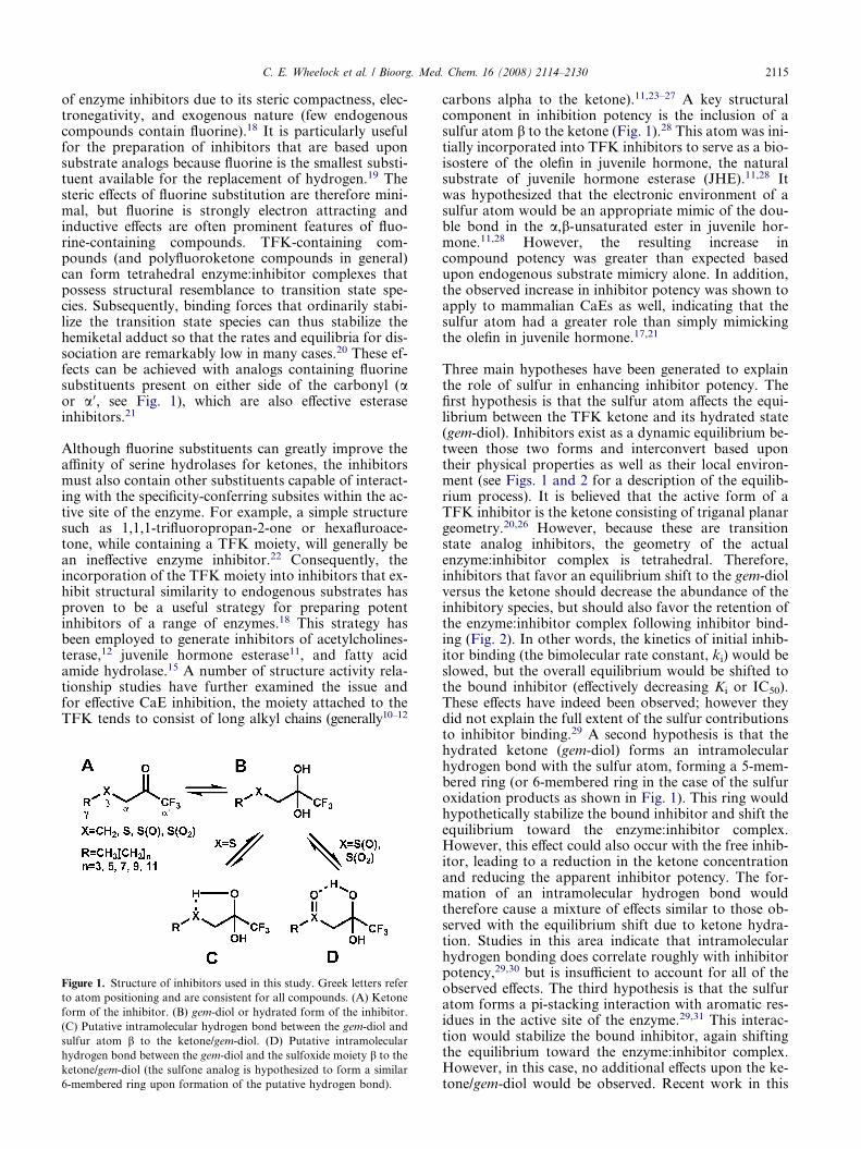
of enzyme inhibitors due to its steric compactness, elec-tronegativity, and exogenous nature (few endogenouscompounds contain fluorine).18 It is particularly usefulfor the preparation of inhibitors that are based uponsubstrate analogs because fluorine is the smallest substi-tuent available for the replacement of hydrogen.19 Thesteric effects of fluorine substitution are therefore mini-mal, but fluorine is strongly electron attracting andinductive effects are often prominent features of fluo-rine-containing compounds. TFK-containing com-pounds (and polyfluoroketone compounds in general)can form tetrahedral enzyme:inhibitor complexes thatpossess structural resemblance to transition state spe-cies. Subsequently, binding forces that ordinarily stabi-lize the transition state species can thus stabilize thehemiketal adduct so that the rates and equilibria for dis-sociation are remarkably low in many cases.20 These ef-fects can be achieved with analogs containing fluorinesubstituents present on either side of the carbonyl (aor a 0, see Fig. 1), which are also effective esteraseinhibitors.21
Although fluorine substituents can greatly improve theaffinity of serine hydrolases for ketones, the inhibitorsmust also contain other substituents capable of interact-ing with the specificity-conferring subsites within the ac-tive site of the enzyme. For example, a simple structuresuch as 1,1,1-trifluoropropan-2-one or hexafluroace-tone, while containing a TFK moiety, will generally bean ineffective enzyme inhibitor.22 Consequently, theincorporation of the TFK moiety into inhibitors that ex-hibit structural similarity to endogenous substrates hasproven to be a useful strategy for preparing potentinhibitors of a range of enzymes.18 This strategy hasbeen employed to generate inhibitors of acetylcholines-terase,12 juvenile hormone esterase11, and fatty acidamide hydrolase.15 A number of structure activity rela-tionship studies have further examined the issue andfor effective CaE inhibition, the moiety attached to theTFK tends to consist of long alkyl chains (generally10–12
Figure 1. Structure of inhibitors used in this study. Greek letters refer
to atom positioning and are consistent for all compounds. (A) Ketone
form of the inhibitor. (B) gem-diol or hydrated form of the inhibitor.
(C) Putative intramolecular hydrogen bond between the gem-diol and
sulfur atom b to the ketone/gem-diol. (D) Putative intramolecular
hydrogen bond between the gem-diol and the sulfoxide moiety b to the
ketone/gem-diol (the sulfone analog is hypothesized to form a similar
6-membered ring upon formation of the putative hydrogen bond).
carbons alpha to the ketone).11,23–27 A key structuralcomponent in inhibition potency is the inclusion of asulfur atom b to the ketone (Fig. 1).28 This atom was ini-tially incorporated into TFK inhibitors to serve as a bio-isostere of the olefin in juvenile hormone, the naturalsubstrate of juvenile hormone esterase (JHE).11,28 Itwas hypothesized that the electronic environment of asulfur atom would be an appropriate mimic of the dou-ble bond in the a,b-unsaturated ester in juvenile hor-mone.11,28 However, the resulting increase incompound potency was greater than expected basedupon endogenous substrate mimicry alone. In addition,the observed increase in inhibitor potency was shown toapply to mammalian CaEs as well, indicating that thesulfur atom had a greater role than simply mimickingthe olefin in juvenile hormone.17,21
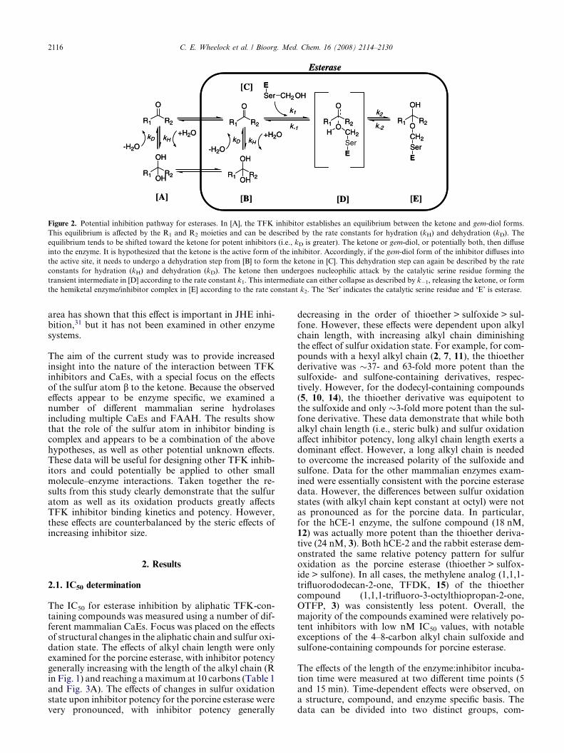
Three main hypotheses have been generated to explainthe role of sulfur in enhancing inhibitor potency. Thefirst hypothesis is that the sulfur atom affects the equi-librium between the TFK ketone and its hydrated state(gem-diol). Inhibitors exist as a dynamic equilibrium be-tween those two forms and interconvert based upontheir physical properties as well as their local environ-ment (see Figs. 1 and 2 for a description of the equilib-rium process). It is believed that the active form of aTFK inhibitor is the ketone consisting of triganal planargeometry.20,26 However, because these are transitionstate analog inhibitors, the geometry of the actualenzyme:inhibitor complex is tetrahedral. Therefore,inhibitors that favor an equilibrium shift to the gem-diolversus the ketone should decrease the abundance of theinhibitory species, but should also favor the retention ofthe enzyme:inhibitor complex following inhibitor bind-ing (Fig. 2). In other words, the kinetics of initial inhib-itor binding (the bimolecular rate constant, ki) would beslowed, but the overall equilibrium would be shifted tothe bound inhibitor (effectively decreasing Ki or IC50).These effects have indeed been observed; however theydid not explain the full extent of the sulfur contributionsto inhibitor binding.29 A second hypothesis is that thehydrated ketone (gem-diol) forms an intramolecularhydrogen bond with the sulfur atom, forming a 5-mem-bered ring (or 6-membered ring in the case of the sulfuroxidation products as shown in Fig. 1). This ring wouldhypothetically stabilize the bound inhibitor and shift theequilibrium toward the enzyme:inhibitor complex.However, this effect could also occur with the free inhib-itor, leading to a reduction in the ketone concentrationand reducing the apparent inhibitor potency. The for-mation of an intramolecular hydrogen bond wouldtherefore cause a mixture of effects similar to those ob-served with the equilibrium shift due to ketone hydra-tion. Studies in this area indicate that intramolecularhydrogen bonding does correlate roughly with inhibitorpotency,29,30 but is insufficient to account for all of theobserved effects. The third hypothesis is that the sulfuratom forms a pi-stacking interaction with aromatic res-idues in the active site of the enzyme.29,31 This interac-tion would stabilize the bound inhibitor, again shiftingthe equilibrium toward the enzyme:inhibitor complex.However, in this case, no additional effects upon the ke-tone/gem-diol would be observed. Recent work in this
OH
R1 R2OH
O
R1 R2
O
R1 R2
OH
R1 R2OH
O
R1 R2OH CH2
Ser
E
OH
R1 R2O
CH2
Ser
E
k2
k-2
k1
k-1
ESer CH2 OH
kD kH-H2O
+H2OkD kH
-H2O
+H2O
[A] [B] [E]
[C]
[D]
Esterase
OH
R1 R2OH
O
R1 R2
O
R1 R2
OH
R1 R2OH
O
R1 R2OH CH2
E
OH
R1 R2O
CH2
Ser
E
k2
k-2
k1
k-1
ESer CH2 OH
kD kH-H2O
+H2OkD kH
-H2O
+H2O
[A] [B] [E]
[C]
[D]
Esterase
Figure 2. Potential inhibition pathway for esterases. In [A], the TFK inhibitor establishes an equilibrium between the ketone and gem-diol forms.
This equilibrium is affected by the R1 and R2 moieties and can be described by the rate constants for hydration (kH) and dehydration (kD). The
equilibrium tends to be shifted toward the ketone for potent inhibitors (i.e., kD is greater). The ketone or gem-diol, or potentially both, then diffuse
into the enzyme. It is hypothesized that the ketone is the active form of the inhibitor. Accordingly, if the gem-diol form of the inhibitor diffuses into
the active site, it needs to undergo a dehydration step from [B] to form the ketone in [C]. This dehydration step can again be described by the rate
constants for hydration (kH) and dehydration (kD). The ketone then undergoes nucleophilic attack by the catalytic serine residue forming the
transient intermediate in [D] according to the rate constant k1. This intermediate can either collapse as described by k�1, releasing the ketone, or form
the hemiketal enzyme/inhibitor complex in [E] according to the rate constant k2. The ‘Ser’ indicates the catalytic serine residue and ‘E’ is esterase.
2116 C. E. Wheelock et al. / Bioorg. Med. Chem. 16 (2008) 2114–2130
area has shown that this effect is important in JHE inhi-bition,31 but it has not been examined in other enzymesystems.
The aim of the current study was to provide increasedinsight into the nature of the interaction between TFKinhibitors and CaEs, with a special focus on the effectsof the sulfur atom b to the ketone. Because the observedeffects appear to be enzyme specific, we examined anumber of different mammalian serine hydrolasesincluding multiple CaEs and FAAH. The results showthat the role of the sulfur atom in inhibitor binding iscomplex and appears to be a combination of the abovehypotheses, as well as other potential unknown effects.These data will be useful for designing other TFK inhib-itors and could potentially be applied to other smallmolecule–enzyme interactions. Taken together the re-sults from this study clearly demonstrate that the sulfuratom as well as its oxidation products greatly affectsTFK inhibitor binding kinetics and potency. However,these effects are counterbalanced by the steric effects ofincreasing inhibitor size.
2. Results
2.1. IC50 determination
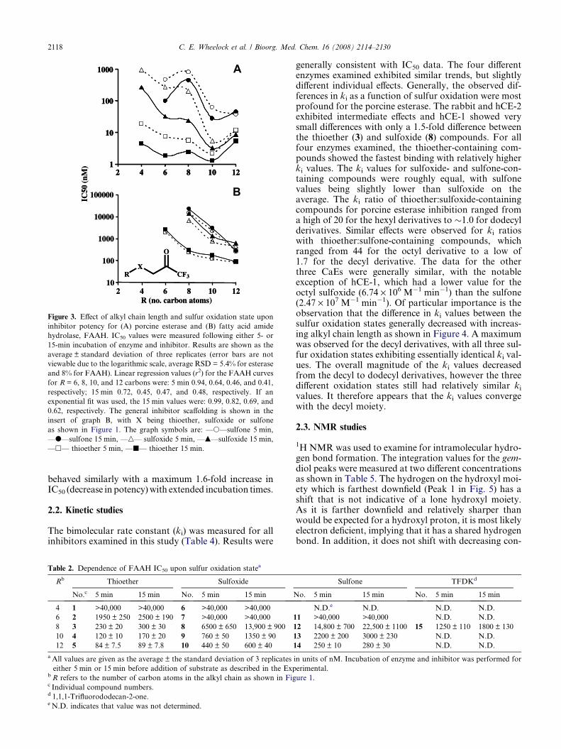
The IC50 for esterase inhibition by aliphatic TFK-con-taining compounds was measured using a number of dif-ferent mammalian CaEs. Focus was placed on the effectsof structural changes in the aliphatic chain and sulfur oxi-dation state. The effects of alkyl chain length were onlyexamined for the porcine esterase, with inhibitor potencygenerally increasing with the length of the alkyl chain (Rin Fig. 1) and reaching a maximum at 10 carbons (Table 1and Fig. 3A). The effects of changes in sulfur oxidationstate upon inhibitor potency for the porcine esterase werevery pronounced, with inhibitor potency generally
decreasing in the order of thioether > sulfoxide > sul-fone. However, these effects were dependent upon alkylchain length, with increasing alkyl chain diminishingthe effect of sulfur oxidation state. For example, for com-pounds with a hexyl alkyl chain (2, 7, 11), the thioetherderivative was �37- and 63-fold more potent than thesulfoxide- and sulfone-containing derivatives, respec-tively. However, for the dodecyl-containing compounds(5, 10, 14), the thioether derivative was equipotent tothe sulfoxide and only �3-fold more potent than the sul-fone derivative. These data demonstrate that while bothalkyl chain length (i.e., steric bulk) and sulfur oxidationaffect inhibitor potency, long alkyl chain length exerts adominant effect. However, a long alkyl chain is neededto overcome the increased polarity of the sulfoxide andsulfone. Data for the other mammalian enzymes exam-ined were essentially consistent with the porcine esterasedata. However, the differences between sulfur oxidationstates (with alkyl chain kept constant at octyl) were notas pronounced as for the porcine data. In particular,for the hCE-1 enzyme, the sulfone compound (18 nM,12) was actually more potent than the thioether deriva-tive (24 nM, 3). Both hCE-2 and the rabbit esterase dem-onstrated the same relative potency pattern for sulfuroxidation as the porcine esterase (thioether > sulfox-ide > sulfone). In all cases, the methylene analog (1,1,1-trifluorododecan-2-one, TFDK, 15) of the thioethercompound (1,1,1-trifluoro-3-octylthiopropan-2-one,OTFP, 3) was consistently less potent. Overall, themajority of the compounds examined were relatively po-tent inhibitors with low nM IC50 values, with notableexceptions of the 4–8-carbon alkyl chain sulfoxide andsulfone-containing compounds for porcine esterase.
The effects of the length of the enzyme:inhibitor incuba-tion time were measured at two different time points (5and 15 min). Time-dependent effects were observed, ona structure, compound, and enzyme specific basis. Thedata can be divided into two distinct groups, com-

Table 1. Dependence of esterase IC50 upon sulfur oxidation statea
Rb Thioether Sulfoxide Sulfone TFDKd
No.c 5 min 15 min No. 5 min 15 min No. 5 min 15 min No. 5 min 15 min
Porcine esterase
4 1 20 ± 1.4 4.5 ± 0.2 6 970 ± 70 270 ± 9 N.D.e N.D. N.D. N.D.
6 2 7.5 ± 0.3 1.8 ± 0.1 7 280 ± 19 33 ± 0.2 11 470 ± 59 100 ± 10 N.D. N.D.
8 3 5.6 ± 0.2 2.4 ± 0.1 8 200 ± 5 23 ± 2 12 830 ± 48 460 ± 44 15 25 ± 2 9.3 ± 0.7
10 4 2.2 ± 0.1 1.3 ± 0.1 9 5.5 ± 0.3 3.2 ± 0.2 13 63 ± 8 28 ± 0.4 N.D. N.D.
12 5 12 ± 0.8 5.3 ± 0.2 10 12 ± 0.2 8.6 ± 0.2 14 37 ± 0.7 46 ± 0.7 N.D. N.D.
hCE-1
8 3 24 ± 2 13 ± 1 8 30 ± 1 5.5 ± 0.5 12 18 ± 1 5.4 ± 0.8 15 65 ± 5 30 ± 2
hCE-2
8 3 8.5 ± 0.2 5.8 ± 0.5 8 11 ± 1 7.4 ± 0.2 12 26 ± 1 21 ± 2 15 36 ± 4 20 ± 1
Rabbit esterase
8 3 6.7 ± 0.2 1.9 ± 0.1 8 16 ± 1 5.0 ± 0.1 12 55 ± 1 35 ± 1 15 42 ± 1 14 ± 1
MsJHE wild typef
8 3 22 ± 3 N.D. N.D. 15 530 ± 32
MsJHE F259I mutantg
8 3 330 ± 57 N.D. N.D. 15 1600 ± 150
a All values are given as the average ± the standard deviation of 3 replicates in units of nM. Incubation of enzyme and inhibitor was performed for
either 5 min or 15 min before addition of substrate as described in the Experimental.b R refers to the number of carbon atoms in the alkyl chain as shown in Figure 1.c Individual compound numbers.d 1,1,1-Trifluorododecan-2-one.e N.D. indicates that value was not determined.f IC50 values were measured for Manduca sexta juvenile hormone esterase (JHE) using juvenile hormone as the substrate. Inhibitor and enzyme were
incubated for 30 min prior to addition of substrate (14C juvenile hormone). Data are from Wogulis et al.31
g IC50 values were measured for a M. sexta mutant JHE in which the 259 phenylalanine had been mutated to a isoleucine. Inhibitor and enzyme were
incubated for 30 min prior to addition of substrate (14C juvenile hormone). Data are from Wogulis et al.31
C. E. Wheelock et al. / Bioorg. Med. Chem. 16 (2008) 2114–2130 2117
pounds with alkyl groups of 4 or 6 carbons and thosewith 8, 10 or 12 carbons (the only exception is com-pound 8 with the sulfoxide). Shorter chain compoundsdemonstrated a much larger time-dependent effect thanlonger chain derivatives. The most extreme examplewas the sulfoxide-containing compounds, which demon-strated the greatest time dependence with an �8.5-foldincrease in inhibitor potency between 5 and 15 min incu-bations for 6- and 8-carbon containing compounds.These effects were dramatically reduced with longer al-kyl chains, with the dodecyl analog exhibiting only a1.4-fold change. The methylene analog (TFDK, 15) ofthe 8-carbon alkyl chain thioether-containing inhibitor(OTFP, 3) evidenced some slight time-dependent inhibi-tion effects being �4.5- and 4-fold less potent at 5 and15 min, respectively. Together these data demonstratethat (1) TFKs are slow tight-binding inhibitors that re-quire significant enzyme:inhibitor incubation intervalsand (2) the observed effects of sulfur oxidation stateupon inhibitor potency are time dependent.
The above-described effects were less pronounced forFAAH (Table 2). Overall, TFKs were weaker inhibitorsof FAAH relative to CaEs, with the most potent FAAHinhibitor exhibiting an IC50 of 84 nM (5) as opposed to1.3 nM (4) for porcine esterase. For all compoundsexamined, inhibitor potency increased with alkyl chainlength, with no maximum observed (Table 2). The in-crease in potency exhibited a linear correlation withthe length of the alkyl chain (with r2 values ranging fromthioether: 0.82, sulfoxide: 0.89, sulfone: 0.92; Fig. 3B);
however it is not appropriate to make any significantconclusions with only three compounds. No differencesin the trend were observed between 5- and 15-min incu-bation times. The effect of sulfur oxidation state wassimilar to the esterase studies in that inhibition orderwas thioether > sulfoxide > sulfone for octyl- or decyl-containing compounds, but it differed for dodecyl-con-taining compounds with thioether > sulfone > sulfoxide.The methylene analog (TFDK, 15) was �5- to 6-foldless potent than the corresponding thioether compound(OTFP, 3) independent of the enzyme/inhibitor incuba-tion time. The observed time-dependent effects wereopposite those of the esterases, with IC50 values increas-ing (becoming less potent) with time (Table 2). However,this effect was not very dramatic, ranging from 1.1- to2.1-fold difference between 5 and 15 min for all theinhibitors examined.
Inhibition studies with benzil exhibited results substan-tially different from that of TFK-mediated inhibition.Benzil was a potent inhibitor for all CaEs examined, how-ever the inhibition was not as potent as the TFK-mediatedinhibition (Table 3). Benzil did not inhibit FAAH at anyconcentration examined. The inhibition kinetics showedvery little time dependence, with essentially full inhibitionoccurring within 1 min following incubation of enzymeand inhibitor (data not shown). As opposed to TFK-med-iated inhibition, benzil’s IC50 values slightly increased withextended enzyme:inhibitor incubation times. However,these increases consisted of a maximum 2.2-fold increasefor porcine esterase, while the other three enzymes
10
100
1000
10000
100000
2 4 6 8 10 12R (no. carbon atoms)
IC50
(nM
) 1
10
100
1000
2 4 6 8 10 12
RX
CF3
O
10
100
1000
10000
100000
2 4 6 8 10 12R (no. carbon atoms)
IC50
(nM
) 1
10
100
1000
2 4 6 8 10 121
10
100
1000
2 4 6 8 10 12
RX
CF3
O
A
B
Figure 3. Effect of alkyl chain length and sulfur oxidation state upon
inhibitor potency for (A) porcine esterase and (B) fatty acid amide
hydrolase, FAAH. IC50 values were measured following either 5- or
15-min incubation of enzyme and inhibitor. Results are shown as the
average ± standard deviation of three replicates (error bars are not
viewable due to the logarithmic scale, average RSD = 5.4% for esterase
and 8% for FAAH). Linear regression values (r2) for the FAAH curves
for R = 6, 8, 10, and 12 carbons were: 5 min 0.94, 0.64, 0.46, and 0.41,
respectively; 15 min 0.72, 0.45, 0.47, and 0.48, respectively. If an
exponential fit was used, the 15 min values were: 0.99, 0.82, 0.69, and
0.62, respectively. The general inhibitor scaffolding is shown in the
insert of graph B, with X being thioether, sulfoxide or sulfone
as shown in Figure 1. The graph symbols are: —s—sulfone 5 min,
—d—sulfone 15 min, —n— sulfoxide 5 min, —m—sulfoxide 15 min,
—h— thioether 5 min, —j— thioether 15 min.
2118 C. E. Wheelock et al. / Bioorg. Med. Chem. 16 (2008) 2114–2130
behaved similarly with a maximum 1.6-fold increase inIC50 (decrease in potency) with extended incubation times.
2.2. Kinetic studies
The bimolecular rate constant (ki) was measured for allinhibitors examined in this study (Table 4). Results were
Table 2. Dependence of FAAH IC50 upon sulfur oxidation statea
Rb Thioether Sulfoxide
No.c 5 min 15 min No. 5 min 15 min N
4 1 >40,000 >40,000 6 >40,000 >40,000
6 2 1950 ± 250 2500 ± 190 7 >40,000 >40,000 1
8 3 230 ± 20 300 ± 30 8 6500 ± 650 13,900 ± 900 1
10 4 120 ± 10 170 ± 20 9 760 ± 50 1350 ± 90 1
12 5 84 ± 7.5 89 ± 7.8 10 440 ± 50 600 ± 40 1
a All values are given as the average ± the standard deviation of 3 replicates
either 5 min or 15 min before addition of substrate as described in the Expb R refers to the number of carbon atoms in the alkyl chain as shown in Figc Individual compound numbers.d 1,1,1-Trifluorododecan-2-one.e N.D. indicates that value was not determined.
generally consistent with IC50 data. The four differentenzymes examined exhibited similar trends, but slightlydifferent individual effects. Generally, the observed dif-ferences in ki as a function of sulfur oxidation were mostprofound for the porcine esterase. The rabbit and hCE-2exhibited intermediate effects and hCE-1 showed verysmall differences with only a 1.5-fold difference betweenthe thioether (3) and sulfoxide (8) compounds. For allfour enzymes examined, the thioether-containing com-pounds showed the fastest binding with relatively higherki values. The ki values for sulfoxide- and sulfone-con-taining compounds were roughly equal, with sulfonevalues being slightly lower than sulfoxide on theaverage. The ki ratio of thioether:sulfoxide-containingcompounds for porcine esterase inhibition ranged froma high of 20 for the hexyl derivatives to �1.0 for dodecylderivatives. Similar effects were observed for ki ratioswith thioether:sulfone-containing compounds, whichranged from 44 for the octyl derivative to a low of1.7 for the decyl derivative. The data for the otherthree CaEs were generally similar, with the notableexception of hCE-1, which had a lower value for theoctyl sulfoxide (6.74 · 106 M�1 min�1) than the sulfone(2.47 · 107 M�1 min�1). Of particular importance is theobservation that the difference in ki values between thesulfur oxidation states generally decreased with increas-ing alkyl chain length as shown in Figure 4. A maximumwas observed for the decyl derivatives, with all three sul-fur oxidation states exhibiting essentially identical ki val-ues. The overall magnitude of the ki values decreasedfrom the decyl to dodecyl derivatives, however the threedifferent oxidation states still had relatively similar ki
values. It therefore appears that the ki values convergewith the decyl moiety.
2.3. NMR studies
1H NMR was used to examine for intramolecular hydro-gen bond formation. The integration values for the gem-diol peaks were measured at two different concentrationsas shown in Table 5. The hydrogen on the hydroxyl moi-ety which is farthest downfield (Peak 1 in Fig. 5) has ashift that is not indicative of a lone hydroxyl moiety.As it is farther downfield and relatively sharper thanwould be expected for a hydroxyl proton, it is most likelyelectron deficient, implying that it has a shared hydrogenbond. In addition, it does not shift with decreasing con-
Sulfone TFDKd
o. 5 min 15 min No. 5 min 15 min
N.D.e N.D. N.D. N.D.
1 >40,000 >40,000 N.D. N.D.
2 14,800 ± 700 22,500 ± 1100 15 1250 ± 110 1800 ± 130
3 2200 ± 200 3000 ± 230 N.D. N.D.
4 250 ± 10 280 ± 30 N.D. N.D.
in units of nM. Incubation of enzyme and inhibitor was performed for
erimental.
ure 1.
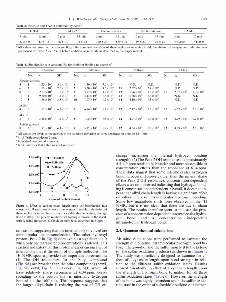
Table 3. Esterase and FAAH inhibition by benzila
hCE-1 hCE-2 Porcine esterase Rabbit esterase FAAH
5 min 15 min 5 min 15 min 5 min 15 min 5 min 15 min 5 min 15 min
27 ± 1.0 43 ± 1.3 38 ± 1.0 44 ± 1.1 370 ± 26 830 ± 34 19 ± 2.0 29 ± 1.7 >40,000 >40,000
a All values are given as the average IC50 ± the standard deviation of three replicates in units of nM. Incubation of enzyme and inhibitor was
performed for either 5 or 15 min before addition of substrate as described in the Experimental.
Table 4. Bimolecular rate constant (ki) for inhibitor binding to esterasesa
R Thioether Sulfoxide Sulfone TFDKb
No.c ki SD No. ki SD No. ki SD No. ki SD
Porcine esterase
4 1 1.53 · 107 3.0 · 105 6 1.30 · 106 5.0 · 104 N.D.d N.D. N.D.c N.D.
6 2 1.05 · 107 7.5 · 105 7 5.20 · 105 3.5 · 104 11 2.67 · 105 2.4 · 104 N.D. N.D.
8 3 2.53 · 107 1.6 · 106 8 5.72 · 106 3.2 · 105 12 5.76 · 105 3.5 · 104 15 2.07 · 107 1.2 · 106
10 4 9.80 · 108 1.4 · 107 9 5.44 · 108 1.8 · 107 13 5.88 · 108 5.6 · 107 N.D. N.D.
12 5 1.86 · 107 3.8 · 105 10 1.97 · 107 1.2 · 106 14 4.10 · 106 1.3 · 105 N.D. N.D.
hCE-1
8 3 1.02 · 107 6.2 · 105 8 6.74 · 106 1.7 · 105 12 2.47 · 107 7.5 · 105 15 6.67 · 106 2.0 · 105
hCE-2
8 3 3.48 · 107 1.9 · 106 8 1.08 · 107 7.8 · 105 12 4.27 · 106 1.4 · 105 15 2.29 · 107 1.2 · 106
Rabbit esterase
8 3 1.71 · 107 6.1 · 105 8 5.12 · 106 1.7 · 105 12 4.04 · 106 1.2 · 105 15 4.39 · 106 2.2 · 105
a All values are given as the average ± the standard deviation of three replicates in units of M�1 min�1.b 1,1,1-Trifluorododecan-2-one.c Individual compound numbers.d N.D. indicates that value was not measured.
1.0E+05
1.0E+06
1.0E+07
1.0E+08
1.0E+09
4 6 8 10 12
R
R (no. of carbon atoms)
Thioether
Sulfoxide
Sulfone
X
O
CF3
Figure 4. Effect of carbon chain length upon the bimolecular rate
constant ki. Results are shown as the average ± standard deviation of
three replicates (error bars are not viewable due to scaling, average
RSD 6 10%). The general inhibitor scaffolding is shown in the insert,
with X being thioether, sulfoxide or sulfone as described in Figure 1.
C. E. Wheelock et al. / Bioorg. Med. Chem. 16 (2008) 2114–2130 2119
centration, suggesting that the interaction(s) involved areunimolecular, or intramolecular. The other hydroxylproton (Peak 2 in Fig. 5) does exhibit a significant shiftwhen only one parameter (concentration) is altered. Thisreaction indicates that this proton is experiencing a set ofinteractions that is the result of multiple molecules. The1H NMR spectra provide two important observations:(1) The OH resonances for the butyl compound(Fig. 5A) are broader than the other compounds (hexyl,Fig. 5B; octyl, Fig. 5C; and decyl, Fig. 5D), which allhave relatively sharp resonances at 6.34 ppm, corre-sponding to the proton that is internally hydrogenbonded to the sulfoxide. This response suggests thatthe longer alkyl chain is reducing the rate of OH ex-
change (increasing the internal hydrogen bondingstrength). (2) The Peak 2 OH resonance at approximately4.5–4.9 ppm tends to be broader and more susceptible toconcentration effects than the resonance at 6.34 ppm.These data suggest that some intermolecular hydrogenbonding occurs. However, other than the general shapeof the Peak 2 OH resonance, concentration-dependenteffects were not observed indicating that hydrogen bond-ing is concentration independent. Overall, it does not ap-pear that alkyl chain length is having a significant effecton either inter- or intramolecular hydrogen bonding.Some low magnitude shifts were observed in the 1HNMR, but it is not clear that these are due to chainlength. The results therefore seem to indicate the pres-ence of a concentration dependent intermolecular hydro-gen bond and a concentration independentintramolecular hydrogen bond.
2.4. Quantum chemical calculations
Ab initio calculations were performed to estimate thestrength of a putative intramolecular hydrogen bond be-tween the gem-diol and the sulfur moiety b to the ketone(or the sulfur oxidation products) as shown in Figure 1.The study was specifically designed to examine for ef-fects of alkyl chain length upon bond strength in rela-tion to the different sulfur oxidation states. Resultsshowed essentially no effect of alkyl chain length uponthe strength of hydrogen bond formation for all threesulfur oxidation states (Table 6). However, the strengthof the bond was highly dependent upon the sulfur oxida-tion state in the order of sulfoxide > sulfone > thioether.
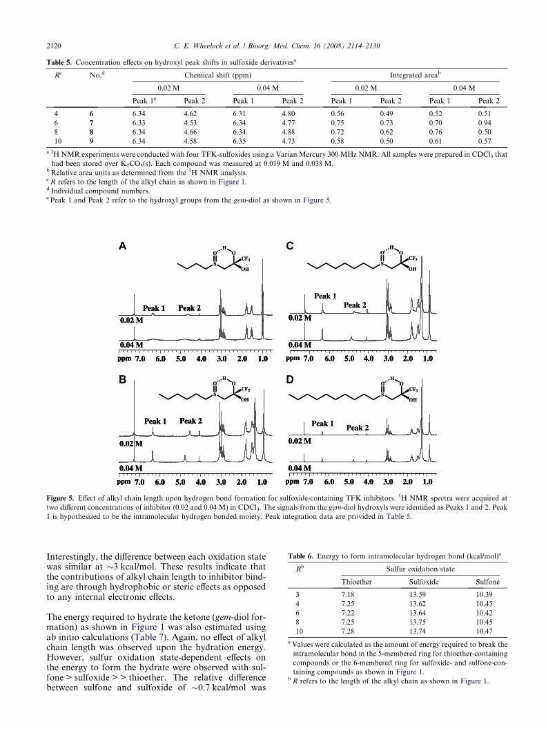
Table 5. Concentration effects on hydroxyl peak shifts in sulfoxide derivativesa
Rc No.d Chemical shift (ppm) Integrated areab
0.02 M 0.04 M 0.02 M 0.04 M
Peak 1e Peak 2 Peak 1 Peak 2 Peak 1 Peak 2 Peak 1 Peak 2
4 6 6.34 4.62 6.31 4.80 0.56 0.49 0.52 0.51
6 7 6.33 4.53 6.34 4.77 0.75 0.73 0.70 0.94
8 8 6.34 4.66 6.34 4.88 0.72 0.62 0.76 0.50
10 9 6.34 4.58 6.35 4.73 0.58 0.50 0.61 0.57
a 1H NMR experiments were conducted with four TFK-sulfoxides using a Varian Mercury 300 MHz NMR. All samples were prepared in CDCl3 that
had been stored over K2CO3(s). Each compound was measured at 0.019 M and 0.038 M.b Relative area units as determined from the 1H NMR analysis.c R refers to the length of the alkyl chain as shown in Figure 1.d Individual compound numbers.e Peak 1 and Peak 2 refer to the hydroxyl groups from the gem-diol as shown in Figure 5.
0.02 M
ppm 1.02.03.04.05.06.07.0
0.04 M
0.02 M
ppm 1.02.03.04.05.06.07.0
0.04 M
0.02 M
ppm 1.02.03.04.05.06.07.0
0.04 M
0.02 M
ppm 1.02.03.04.05.06.07.0
0.04 M
Peak 1 Peak 2
Peak 1Peak 2
Peak 1 Peak 2 Peak 1 Peak 2
S
OH
CF3
OH
O
S
OH
CF3
OH
O
S
OH
CF3
OH
O
S
OH
CF3
OH
O
0.02 M
ppm 1.02.03.04.05.06.07.0
0.04 M
0.02 M
ppm 1.02.03.04.05.06.07.0 1.02.03.04.05.06.07.0
0.04 M
0.02 M
ppm 1.02.03.04.05.06.07.0
0.04 M
0.02 M
ppm 1.02.03.04.05.06.07.0 1.02.03.04.05.06.07.0
0.04 M
0.02 M
ppm 1.02.03.04.05.06.07.0
0.04 M
0.02 M
ppm 1.02.03.04.05.06.07.0
0.04 M
0.02 M
ppm 1.02.03.04.05.06.07.0
0.04 M
0.02 M
ppm 1.02.03.04.05.06.07.0
0.04 M
Peak 1 Peak 2Peak 1 Peak 2
Peak 1Peak 2
Peak 1 Peak 2Peak 1 Peak 2 Peak 1 Peak 2
S
OH
CF3
OH
O
S
OH
CF3
OH
O
S
OH
CF3
OH
O
S
OH
CF3
OH
O
A
B
C
D
Figure 5. Effect of alkyl chain length upon hydrogen bond formation for sulfoxide-containing TFK inhibitors. 1H NMR spectra were acquired at
two different concentrations of inhibitor (0.02 and 0.04 M) in CDCl3. The signals from the gem-diol hydroxyls were identified as Peaks 1 and 2. Peak
1 is hypothesized to be the intramolecular hydrogen bonded moiety. Peak integration data are provided in Table 5.
Table 6. Energy to form intramolecular hydrogen bond (kcal/mol)a
Rb Sulfur oxidation state
Thioether Sulfoxide Sulfone
3 7.18 13.59 10.39
4 7.25 13.62 10.45
6 7.22 13.64 10.42
8 7.25 13.75 10.45
10 7.28 13.74 10.47
a Values were calculated as the amount of energy required to break the
intramolecular bond in the 5-membered ring for thioether-containing
compounds or the 6-membered ring for sulfoxide- and sulfone-con-
taining compounds as shown in Figure 1.b R refers to the length of the alkyl chain as shown in Figure 1.
2120 C. E. Wheelock et al. / Bioorg. Med. Chem. 16 (2008) 2114–2130
Interestingly, the difference between each oxidation statewas similar at �3 kcal/mol. These results indicate thatthe contributions of alkyl chain length to inhibitor bind-ing are through hydrophobic or steric effects as opposedto any internal electronic effects.
The energy required to hydrate the ketone (gem-diol for-mation) as shown in Figure 1 was also estimated usingab initio calculations (Table 7). Again, no effect of alkylchain length was observed upon the hydration energy.However, sulfur oxidation state-dependent effects onthe energy to form the hydrate were observed with sul-fone > sulfoxide > > thioether. The relative differencebetween sulfone and sulfoxide of �0.7 kcal/mol was

Table 7. Energy to hydrate ketone (DE kcal/mol)a
Rb Sulfur oxidation state
Thioether Sulfoxide Sulfone
3 �12.29 �15.74 �16.51
4 �12.23 �15.67 �16.44
6 �12.24 �15.67 �16.42
a Values were calculated for the amount of energy required to hydrate
the ketone (form the gem-diol) as shown in Figure 1.b R refers to the length of the alkyl chain as shown in Figure 1.
C. E. Wheelock et al. / Bioorg. Med. Chem. 16 (2008) 2114–2130 2121
small compared to �3.5 kcal/mol for the sulfoxide andthioether. These data suggest that sulfone- and sulfox-ide-containing compounds hydrate to a greater extentthan the thioether derivatives.
2.5. X-ray structure studies
The crystal structures of four different sulfoxide-con-taining inhibitors (6, 7, 8, 9) were solved to examinefor hydrogen bond formation (Fig. 6). The structureswere characterized by linear chain hydrocarbon stackingof rod-shaped molecules with lateral intermolecularhydrogen bonding interactions between gem-diols andsulfoxide groups. However, no intramolecular hydrogenbonds were observed in any of the compounds. Meltingpoints for these crystalline solids were very similar rang-ing from 79 to 82 �C for 6, 89 to 91 �C for 7, 87 to 90 �Cfor 8, and 90 to 95 �C for 9.
3. Discussion
The inhibition mechanism of TFKs has been examinedby a number of research groups and there is a fairamount of consensus in the field regarding how theTFK moiety interacts with the enzyme. The main out-standing issues revolve around the hydration state ofthe ‘active’ form of the inhibitor and the secondaryinteractions that occur within the active site of the en-zyme that can affect inhibitor binding (mainly van derWaals interactions and electronic interactions such aspi-stacking). The issue of the geometry of the TFK inits active form (i.e., inhibitory form) has not been di-rectly addressed in CaEs, but it has been shown for capt-hepsin B,32 acetylcholinesterase20 and chymotrypsin33
that the ketone is the inhibitory species. These findingswere further supported for CaEs by Roe et al., who pos-tulated that the ketone is the inhibitory species for juve-nile hormone esterase (JHE) inhibition,34 which hasbeen supported by 3-D QSAR studies.26 Therefore, itis probable that the ketone is also the inhibitory speciesin TFK-mediated CaE inhibition, although conflictingresults suggesting that the gem-diol was the active formof the inhibitor have been published.30
The current study was designed to explore some of theremaining questions of the physicochemical propertiesaffecting TFK hydration and examine their correlationwith inhibitor potency. It is well accepted that TFKsform a hydrate in aqueous solutions, with more potentinhibitors tending to exhibit a greater degree of hydra-tion.11,29 In addition, work has shown that the extent
of hydration correlates with inhibition potency and thatthe substituents surrounding the TFK moiety contributeto the ketone hydration.29,35 However, it is still uncer-tain to what extent the substituents affect the bindingkinetics. Potent TFKs are considered to be slow tight-binding inhibitors, with equilibrium times of the orderof hours to days.27 However, the rate of binding (ki)was reported to vary with the substituent b to the ke-tone.29 We therefore measured ki and IC50 values for arange of TFKs containing a sulfur atom of varying oxi-dation state in the b position (Fig. 1). TFK-mediatedinhibition of a non-esterase enzyme, the serine hydrolaseFAAH, was also examined in order to test for esterase-specific effects. These data combined with a number ofstudies on the potential effects of intramolecular hydro-gen bonding in the hydrated ketone serve to expand ourknowledge of the mechanism of TFK-mediated inhibi-tion of CaEs.
3.1. IC50 determinations
The observed IC50 values in this study were in agreementwith previously published work.27,29,36 Generally, a 10-carbon (decyl moiety) chain appears to be the globalmaximum for optimal inhibitor potency with all of thecompounds examined in this study. The only exceptionwas the 5-min sulfone decyl derivative (13), but the 15-min derivative held the same pattern (Table 1). Thesedata are consistent with earlier work for mammalianesterases, which typically exhibit an inhibition maxi-mum with alkyl chains of 8–10 carbon atoms beyondthe thioether.27,36 The time-dependent nature of theinhibition process was observed by measuring the IC50
at 5 and 15 min. An important point is that the 15 minvalues are most likely not in equilibrium either, but be-cause TFK-mediated inhibition of CaEs appears to ex-hibit asymptotic behavior,29 these values are probablyrelatively close to the equilibrium values. However, asdemonstrated by Wadkins et al., the TFK binding pro-cess can be extremely slow.27 Rosell et al. also reportedthat extended incubation times (0 min vs 10 min) in-creased the observed potency of TFK inhibitors.37 Pre-vious work comparing the IC50 values of compounds 2and 7 (thioether and sulfoxide) showed a strong time-dependent effect. A 30-s enzyme:inhibitor incubation re-sulted in 7 being >100-fold less potent than 2, whereas a3-h incubation resulted in 7 being only 5-fold less po-tent.29 Similar results were observed here, with a 5-minincubation resulting in a 37-fold difference as opposedto a 15-min incubation giving an 18-fold difference.These data further demonstrate the point that if trueequilibrium is necessary for experimental design, thenmuch greater incubation times may be required. Themajority of the TFK literature published to date, withthe exception of work by Wadkins et al.,27 generallyused incubation times of 30 min or less. Accordingly,as the length of incubation can affect both the absoluteas well as relative inhibitor potency,27,29 it is importantthat studies on TFKs are aware of the assumptions/lim-itations in assay formats.
An important observation from the IC50 data is thecomparison between the thioether-containing derivative
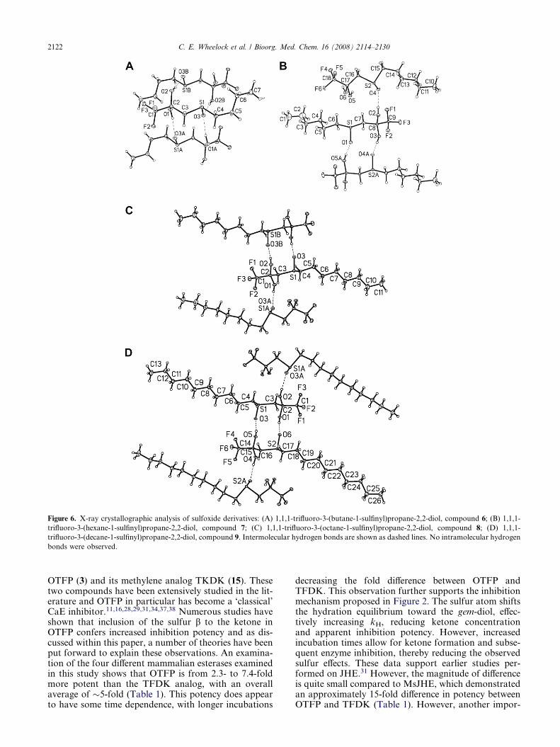
Figure 6. X-ray crystallographic analysis of sulfoxide derivatives: (A) 1,1,1-trifluoro-3-(butane-1-sulfinyl)propane-2,2-diol, compound 6; (B) 1,1,1-
trifluoro-3-(hexane-1-sulfinyl)propane-2,2-diol, compound 7; (C) 1,1,1-trifluoro-3-(octane-1-sulfinyl)propane-2,2-diol, compound 8; (D) 1,1,1-
trifluoro-3-(decane-1-sulfinyl)propane-2,2-diol, compound 9. Intermolecular hydrogen bonds are shown as dashed lines. No intramolecular hydrogen
bonds were observed.
2122 C. E. Wheelock et al. / Bioorg. Med. Chem. 16 (2008) 2114–2130
OTFP (3) and its methylene analog TKDK (15). Thesetwo compounds have been extensively studied in the lit-erature and OTFP in particular has become a ‘classical’CaE inhibitor.11,16,28,29,31,34,37,38 Numerous studies haveshown that inclusion of the sulfur b to the ketone inOTFP confers increased inhibition potency and as dis-cussed within this paper, a number of theories have beenput forward to explain these observations. An examina-tion of the four different mammalian esterases examinedin this study shows that OTFP is from 2.3- to 7.4-foldmore potent than the TFDK analog, with an overallaverage of �5-fold (Table 1). This potency does appearto have some time dependence, with longer incubations
decreasing the fold difference between OTFP andTFDK. This observation further supports the inhibitionmechanism proposed in Figure 2. The sulfur atom shiftsthe hydration equilibrium toward the gem-diol, effec-tively increasing kH, reducing ketone concentrationand apparent inhibition potency. However, increasedincubation times allow for ketone formation and subse-quent enzyme inhibition, thereby reducing the observedsulfur effects. These data support earlier studies per-formed on JHE.31 However, the magnitude of differenceis quite small compared to MsJHE, which demonstratedan approximately 15-fold difference in potency betweenOTFP and TFDK (Table 1). However, another impor-

C. E. Wheelock et al. / Bioorg. Med. Chem. 16 (2008) 2114–2130 2123
tant aspect of the binding process is the interactions ofthe inhibitor with non-catalytic residues. Wogulis et al.demonstrated via mutation studies that the sulfur atomb to the ketone exhibited its effect by forming a pi-stack-ing interaction with a phenylalanine in the active site ofJHE.31 However, even the F259I mutant still evidencedthat OTFP was 5-fold more potent than TDFK. Thesedata therefore suggest that the other physicochemicalinteractions examined in this study such as contributionsto ketone hydration and intramolecular hydrogen, aswell as other potential interactions, also play a role ininhibitor binding—all leading to an �5-fold increase ininhibition potency. A comparison of the sequences forthe different enzymes examined suggests that the mam-malian esterases do not possess the key aromatic residueto form the pi-stacking interactions (Phe262, Fig. 7),potentially explaining the observed differences in sulfurcontribution to inhibitor potency between MsJHE andthe esterases examined in this study. However, theseobservations need to be followed up with structuralstudies for confirmation. Accordingly, the effects of theinclusion of a sulfur atom b to the ketone upon inhibitorpotency are multifaceted and complex and ultimatelyare at least in part enzyme dependent.
Inhibition of FAAH was examined to test for esterase-specific effects. Previous work had shown that TFK-con-taining compounds were effective inhibitors of FAAHactivity.15 However, work has shown that inhibitors ofFAAH can also inhibit CaE activity, highlighting the is-sue of inhibitor selectivity for serine hydrolases.39
FAAH is a mammalian integral membrane enzyme thatdegrades the fatty acid amide family of endogenous sig-naling lipids.40 This class of compounds includes theendogenous cannabinoid anandamide,41 the sleep-inducing oleamide,42 and the anorexigenic compoundoleoylethanolamide.43 FAAH is a potential pharmaceu-tical target in the treatment of anxiety, depression, andother nervous system disorders.44 An advantage ofworking with FAAH is that the endogenous substrateis well understood as opposed to CaEs, whose endoge-nous role is still in question. The inhibition data wereconsistent in that for all three sulfur oxidation states,the 12-carbon chain derivative (dodecyl) exhibited thegreatest potency. The lack of an observed inhibitionmaximum suggests that even longer chain aliphaticgroups would be potent FAAH inhibitors. However,data reported by Boger et al. for similar derivatives sug-gest that the inhibition maximum is in fact around 10–12carbons.15 While the exactly same compounds were nottested in both studies, compound 15 (1,1,1-trifluorodod-ecan-2-one, IC50 = 1.25 lM) in the present study can becompared with 1,1,1-trifluoroundecan-2-one and 1,1,1-trifluorotridecan-2-one from the Boger et al. study,which found these compounds to only be moderately ac-tive inhibitors with IC50 values of 0.60 and 0.72 lM,respectively. Compounds with longer alkyl chains suchas 1,1,1-trifluoropentadecan-2-one exhibited decreasedinhibition potency (IC50 = 2.3 lM).15 The inhibitiondata reported by Boger et al. were similar to the data re-ported here (an �2-fold difference), which can easily beexplained by differences in the assay formats. Bogeret al. used 14C oleamide as the substrate as opposed to
the fluorescent substrate N-(6-methoxypyridin-3-yl)octanamide used in this study. In addition, this studyused recombinant human enzyme as opposed to homog-enized rat liver. Interestingly, the thioether-containingderivative (OTFP, 3) was an �5-fold more potent inhib-itor of FAAH activity then the methylene analog(TFDK, 15). These data agree very nicely with the ester-ase data, suggesting that contributions of sulfur toinhibitor binding are similar for different enzymes.FAAH is evolutionarily distinct from the esterases; how-ever it performs a similar hydrolysis reaction (withamides as the substrate) and is inhibited by similar com-pounds. Alignment of FAAH with the esterases showsthe low homology between the two divergent groups(Figure S1), making direct sequence-based conclusionsdifficult. However, the data suggest that the sulfur atomis not undergoing direct pi-stacking interactions as withMsJHE. Accordingly, it is likely that the sulfur atom isaffecting inhibitor potency by contributions to ketonehydration and/or intramolecular hydrogen bonding.
Data from the FAAH inhibition studies were interestingin that they demonstrated the opposite trend of the CaEdata, with IC50 values increasing (decreased potency)from the 5 to the 15 min incubation. The mechanism be-hind the differences in the time dependency of the TFKinhibition is unclear. The catalytic mechanisms of thetwo enzymes differ because FAAH deviates from thenormal Ser-His-Glu/Asp catalytic triad of many serinehydrolases.5 Instead, FAAH uses a Ser-Ser-Lys triad,where the Lys serves as the base to activate the Sernucleophile.45 The Lys residue is believed to stronglyactivate the catalytic Ser, resulting in a constitutively ac-tive nucleophile, enabling FAAH to hydrolyze amidesand esters with equal efficiency. It is therefore possiblethat differences in the architecture of the catalytic triadand/or in the composition of the enzyme active sitecould account for the differences in binding kinetics. Inany case, this work has demonstrated that aliphaticTFK-containing compounds are potent inhibitors ofboth FAAH and CaEs. It is therefore important that at-tempts to develop inhibitors for these two divergentpharmaceutical targets ensure selectivity. For example,benzil is a selective CaE inhibitor because no FAAHinhibition was observed at either time point (Table 3).Future FAAH-based studies on inhibitor developmentneed to verify that compounds do not affect CaEactivity.
Inhibition of CaE activity by benzil generally agreedwith previously published values by Wadkins et al.46
Interestingly, studies of benzil-mediated CaE inhibitiongave similar results to the FAAH TFK studies, in thatfor all four esterases examined the 15-min incubation re-sulted in increased IC50 values (decreased potency) rela-tive to the 5-min incubation. However, the mechanism ismost likely much different. Work by Fleming et al.examined the nature of benzil binding to hCE-1 via crys-tallography.47 They postulated that benzil was actually avery poor esterase substrate and that the benzil inhibi-tion mechanism involved repeated cycling within the ac-tive site of the enzyme. This cyclic interaction involvesnucleophilic attack by the catalytic Ser221 residue

Figure 7. Alignment of esterases examined in this study using BioEdit v. 7.0.9 (ClustalW alignment61 using a BLOSUM62 matrix displayed with an
80% threshold). hCE-1, human carboxylesterase 1 (NM_001025194); hCE-2, human carboxylesterase 2 (NM_003869); pCE, porcine esterase
(NM_214246); rCE, rabbit esterase (AF036930); MsJHE, juvenile hormone esterase from Manduca sextra (AF327882). The catalytic triad is marked
with a black box (Ser229, Glu364, and His481; His484 for MsJHE) and the glycines involved in oxyanion hole formation are shown in a blue box
(Gly 148 and Gly 149). The putative conserved second serine involved in the catalytic mechanism is also shown in a black box (Ser255).62 The residue
involved in pi-stacking interactions in MsJHE is marked with a red box (Phe262) and the ER retention sequence is shown in a green box (HXEL).
2124 C. E. Wheelock et al. / Bioorg. Med. Chem. 16 (2008) 2114–2130
(Ser229 in Fig. 7) on one of the benzil carbonyl carbons,forming a covalent intermediate that can reverse to gen-erate benzil and the free enzyme. This process could be
described by the k1 and k�1 equilibrium shown in Figure2. Accordingly, it is possible that esterases turn overbenzil during the incubation process, which would
C. E. Wheelock et al. / Bioorg. Med. Chem. 16 (2008) 2114–2130 2125
explain differences in IC50 values between 5 and 15 min.The time-dependent inhibition for hCE-1, hCE-2 andthe rabbit esterase was fairly minimal and can be ex-plained by inhibitor hydrolysis. However, the porcineesterase exhibited a �2.5-fold reduction in inhibition po-tency from a 5- to 15-min incubation. This decrease isfairly large and unexpected if benzil is acting as a verypoor substrate. It is therefore possible that porcine ester-ase turns over benzil with greater efficiency than theother esterases examined in this study. This point ispotentially very interesting and should be further pur-sued. It would be useful to conduct studies where arange of esterase:benzil incubations were performedfrom 1 min to >24 h similar to work done by Wadkinset al. for TFKs.27 It would be beneficial to couple theseactivity assay studies with quantification of the benzoicacid and/or benzaldehyde hydrolysis products in orderto confirm inhibitor turnover. Taken together, thesetypes of studies would provide a great deal of informa-tion on inhibitor binding processes for CaEs.
3.2. Kinetic studies
These studies focused on examining the electronic andphysical effects of TFK inhibitor structure uponenzyme binding kinetics. Earlier work with porcineesterase had demonstrated that sulfur oxidation statestrongly affected ki, but did not examine the effectsof alkyl chain length.29 The reported differences be-tween the thioether (compound 2) and the sulfoxide(compound 7) were quite large at 3.2 · 107 M�1 min�1
and 9.5 · 105 M�1 min�1, respectively.29 These dataagree relatively well with those reported in thisstudy (1.05 · 107 M�1 min�1 and 5.2 · 105 M�1 min�1,respectively; Table 4), clearly demonstrating that sul-fur oxidation state affects inhibitor binding kinetics.However, data provided in the current study showthat the effects of sulfur oxidation are less importantthan the steric effects of increased alkyl chain length.Accordingly, inhibitors with long aliphatic chains (de-cyl and dodecyl) exhibited essentially identical ki val-ues for all sulfur oxidation states. However, a clearmaximum was observed at 10-carbon atoms (decyl),which is consistent with the IC50 data (Table 1). Theseeffects could not be directly examined with the otherenzymes in this study because ki values were onlymeasured for the octyl derivatives. However, the thio-ether (3) and sulfoxide (8) data for hCE-1, hCE-2 andrabbit esterase are similar to the porcine esterase data.Conversely, the sulfone (12) demonstrated a differentpattern, with the porcine esterase having a muchslower ki than the other esterases. There was no over-all pattern observed in terms of ordering the enzymeson the basis of ki, however hCE-2 was generally thefastest (except for the sulfone, in which case it wasthe second fastest).
Taken together, these data provide insight into theTFK binding mechanism shown in Figure 2. As dis-cussed above, the active form of the inhibitor is stillin dispute, but is hypothesized to be the ketone. Theinhibition process can be postulated to occur basedupon the four distinct steps shown in Figure 2. Step
A shows the equilibrium process between ketone andthe gem-diol. It has been solidly demonstrated that po-tent TFK inhibitors favor the gem-diol state in aque-ous solution.11,29 It is therefore expected that the kH
value will be high relative to kD. Work by Roeet al.34 as well as Rosell et al.37 has measured KH val-ues and reported that they are greater for potent inhib-itors (i.e., greater degree of hydration). Accordingly, itis expected that the gem-diol form of the inhibitor pre-dominates at this step. However, either form couldpotentially diffuse into the enzyme as shown in Figure2. If the inhibitor is still in the gem-diol form as shownin step B, which is the hypothesized mechanism, theinhibitor then needs to undergo a dehydration step tothe ketone in step C. This step is most likely the ratelimiting step of the reaction, which would agree withdata published by Brady and Abeles for chymotrypsininhibition.33 This mechanism would also explain thetime dependence of both the kinetic and IC50 data ob-served in this study as well as others.27,29 For inhibitorssuch as the sulfoxide and sulfone, which demonstratesignificantly slower binding kinetics (i.e., ki values) rel-ative to the thioether, the dehydration reaction is thekey step. Inhibitor potency then becomes a questionof significant incubation time. If a potent inhibitor ishydrated to the extent that ketone concentrations arevanishingly small, then kD will become the limiting stepto enzyme inhibition. Indeed, some research groups‘correct’ Ki values based upon the assumption of actualketone concentrations relative to total inhibitor con-centrations (i.e., the sum of gem-diol and ketone lev-els). This type of correction can subsequently resultin Ki values in the low femtomolar range.48 In a sense,it may therefore be possible to have an inhibitor that is‘too hydrated’ to be effective over a useful time interval(i.e., <24 h). These observations will be of particularrelevance if TFK-containing inhibitors are developedfor in vivo applications.
3.3. Intramolecular hydrogen bonding studies
The IC50 and ki data demonstrated that sulfur oxidationstate had a strong effect upon inhibitor potency and bind-ing kinetics. Previous work had shown that ketone hydra-tion explained a significant amount, but not all, ofinhibition potency.29,31 It was therefore of interest todetermine the mechanism behind these effects. One ofthe predominant theories regarding the binding mecha-nism of TFK-containing inhibitors centers around theformation of an intramolecular hydrogen bond as shownin Figure 1.11,29,30,36,49 In particular, Filizola et al. con-ducted an ab initio study on the relative strength of theintramolecular hydrogen bond, reporting that the bondstrength inversely correlated with inhibitor potency.30
This study therefore attempted to further examine this is-sue using a number of different methods including 1HNMR, X-ray crystallography, and ab initio calculations.The 1H NMR and crystallography studies focused on thesulfoxide-containing compounds because these were pre-dicted to form the strongest intramolecular hydrogenbond due to the dipole formed by the sulfoxide moiety.29
Because this intramolecular bond was not observed in theX-ray structures of any of the sulfoxide derivatives (see

2126 C. E. Wheelock et al. / Bioorg. Med. Chem. 16 (2008) 2114–2130
below), its inferred presence by 1H NMR is very interest-ing. Alkyl chain length appears to affect the intermolecu-lar bond, but not the intramolecular bond (Fig. 5).However, a limitation in drawing conclusions from thisexperiment is the fact that spectra were collected in CDCl3as opposed to water (or D2O), which would be more rep-resentative of the biological environment. The conforma-tions and hydrogen bonding behavior will be different inan aqueous environment, which is the most biologicallyrelevant to this project. However, this approach appearspromising and future studies should pursue it to elucidatethe formation and strength of the putative intra- andintermolecular hydrogen bonds.
The crystal structure analyses of the sulfoxide-contain-ing inhibitors did not show any intramolecular hydro-gen bonding, only intermolecular (Fig. 6). Thisobservation is interesting, especially considering thattwo other sulfur-containing TFK crystal structures,1,1,1-trifluoro-3-(octane-1-sulfonyl)propane-2,2-diol29
and 1,1,1-trifluoro-5-phenyl-4-thiapentane-2,2-diol,50
did contain an intramolecular hydrogen bond. In addi-tion, two sulfur-containing heterocyclic compoundsgave mixed results with 3-(2-pyridylthio)-1,1,1-tri-fluoro-2-propanone showing intramolecular hydrogenbonding and 3-(4-pyridylthio)-1,1,1-trifluoro-2-propa-none showing only intermolecular.51 However, theintramolecular hydrogen bond in 3-(2-pyridylthio)-1,1,1-trifluoro-2-propanone was with the nitrogen inthe pyridyl ring and not with the sulfur. One possibleexplanation is that there are potentially multiple crystalmorphologies associated with these compounds.Accordingly, both the crystalline and structural datamay depend in part on the conditions of crystallization.It is difficult to interpret the biological significance ofthese hydrogen bonds, especially considering that bondformation within a crystalline lattice may not bereflective of bond formation in an aqueous system or aprotein microenvironment.
Ab initio analysis of the strength of the purportedintramolecular hydrogen bond confirmed earlier studiesreporting the strength of the intramolecular hydrogenbond between the hydrate and the heteroatom b tothe carbonyl inversely correlated with inhibitor po-tency.29,30 The data in Table 6 show a bond energy ofsulfoxide > sulfone > thioether as opposed to the por-cine IC50 data which are in the order of thioe-ther > sulfoxide > sulfone. There is therefore not adirect correlation between calculated intramolecularhydrogen bond strength and IC50. However, a majorcaveat to this observation is the extreme time-depen-dent nature of the sulfoxide TFK binding process.The data in Table 1 are for a maximum 15-min incuba-tion process. However, it has been reported that sulfox-ide TFKs may require hours to achieve full binding.29
An examination of the hCE-1 enzyme shows a differenteffect, agreeing with the data from Table 6. In additiondata from Wadkins et al., who compared 5 min and24 h enzyme/inhibitor incubations, showed the sametrend for hCE-1 of sulfoxide = sulfone > thioether,27
thus making it difficult to draw any general conclusionsbased upon these data. Some of the data suggest that
an intramolecular hydrogen bond does form as shownin Figure 1; however, this putative bond is most likelyquite weak. It therefore appears that this effect doescontribute to inhibitor binding and/or potency, but isrelatively minor compared to other physicochemicalinteractions.
An interesting correlation was observed between the en-ergy to hydrate the ketone in Table 7 and the IC50 datain Table 1. An examination of the correlation for com-pounds showed an inverse relationship between alkylchain length and r2 value, suggesting that compoundswith shorter alkyl chains exhibit more sulfur oxidationstate related effects as opposed to longer chain inhibitorsthat are predominantly steric. These data support the ki-netic studies which showed that ki values for compoundswith short alkyl chains (octyl or shorter) varied on a sul-fur oxidation state-dependent basis.
Earlier work by Wheelock et al.29 calculated the energyto form an intramolecular hydrogen bond of a trun-cated TFK-containing compound (the alkyl groupwas a methyl moiety). In comparison to the currentstudy, the absolute value of the numbers is quite differ-ent (4.1 and 7.8 kcal/mol for the thioether and sulfonederivatives, respectively). However, the relative differ-ences agree fairly well with a �3.2 kcal/mol differencebetween thioether and sulfone in the current study asopposed to 3.7 kcal/mol from Wheelock et al.29 Asthe data in Table 6 demonstrate that alkyl chain doesnot significantly affect the strength of the putativebond, the discrepancies are most likely due to the cal-culation methods employed. Similar differences wereobserved with the ketone hydration energy calculationsin Table 7, with earlier published work showing aslightly different trend in the data. Those data reporteda trend of sulfoxide > thioether > sulfone (�25.06 > �19.51 > � 17.42 kcal/mol, respectively).29 The majordifference between the ab initio calculations performedin these two studies was the choice of basis set. Earlierwork used a 6-31G* basis set as opposed to an aug-cc-pVDZ set for the current study. The double-zeta basisset is preferred because it treats each orbital separatelywhen conducting the Hartree–Fock calculation as op-posed to the split-valence basis set, which only calcu-lates a double-zeta for the valence orbital (inner-shellelectrons are described with a single Slater Orbital).It is therefore expected that the double-zeta calcula-tions will provide a more accurate representation ofeach orbital. The asterisk (*) notation on the 6-31G*basis set indicates that p-orbital polarization was takeninto account for the calculation. As a check of thedata, we re-ran the original data set from the Wheelocket al.29 study under the reported conditions with the 6-31G* basis set and reproduced the same numbers.Accordingly, the discrepancy between the numbersfrom the two studies arises from two issues—first, theuse of the larger basis set, and second slightly differentminimum energy configurations were used. The latterissue is a problem with molecules that have a largenumber of degrees of freedom. A comparison of thecurrent configurations versus those in the Wheelocket al.29 study confirmed that the current structures
C. E. Wheelock et al. / Bioorg. Med. Chem. 16 (2008) 2114–2130 2127
are of lower energy. It is therefore expected that thecurrent energies are more reliable than those publishedin earlier work. These observations demonstrate thepoint that caution should be used when examiningthe results of these types of ab initio calculations,and it is often most appropriate to compare relativeversus absolute values.
4. Summary
Further work on TFKs should attempt to conclusivelyaddress the question of the geometry of the ‘active’ formof the inhibitor. Work performed by the group of Abeleson the active form of peptide-based TFK inhibitorswould most likely be an appropriate model.20,33 In addi-tion, the potential role of the putative intramolecularhydrogen bond needs to be addressed in greater detail.To date, most studies have attempted to infer the pres-ence and/or role of the bond in inhibitor potency via abinitio calculations29,30 or crystallography.29,51 Unfortu-nately, neither of these approaches represents an appro-priate study of the potential interactions within abiological system and as such, these studies can only pro-vide suggestive conclusions. The 1H NMR studies pro-duced in this work are a promising start, but are stillinconclusive. Further work needs to examine a range ofTFK inhibitors consisting of structural variation at theb moiety (oxygen, sulfur, sulfoxide, and sulfone) as wellas multiple degrees of fluorination. It would also be ofinterest to move the heteroatom to the gamma position.23
A study of this magnitude may be able to conclusively ad-dress the issue of intramolecular hydrogen bond forma-tion. Enzyme crystallography studies would also beuseful. However, the only esterase structure solved todate with a TFK inhibitor did not show evidence of anintramolecular hydrogen bond.31 However, these resultswere for JHE, and no TFK-containing mammalian struc-tures have been solved to date. One could envision astudy in which a mammalian esterase was solved co-crys-tallized with a number of different heteroatom-contain-ing TFK inhibitors. These data would be very useful inaddressing the issue of intramolecular hydrogen bondformation, but could still potentially be criticized as notbeing reflective of the dynamic environment of a non-crystallized enzyme. It is most likely that with increasingNMR capability, a solution-based esterase structure willeventually be produced. This structure could be probedwith a series of TFK inhibitors and given the unique pos-sibility of using a fluorine specific probe, conclusive infor-mation regarding the potentially hydrogen bondedinhibitor may be obtainable. In conclusion, this studyhas shown that (1) inclusion of a sulfur atom b to the car-bonyl increases inhibitor potency by �5-fold, (2) sulfuroxidation state affects inhibitor binding kinetics, but thatsteric interactions dominate, (3) the effects of sulfur aswell as its oxidation products on inhibitor potency occurthrough a combination of contributions to ketone hydra-tion and intramolecular hydrogen bonding (as well asother potential effects), and (4) secondary interactions,such as those observed with MsJHE, can profoundly af-fect inhibition potency, but are enzyme specific. It is clearthat in the nearly 30 years since the first report of a TFK-
mediated inhibitor of acetylcholinesterase, a great deal ofinformation has been collected regarding the mechanismof these compounds. However, there are still many ques-tions that remain to be answered.
5. Experimental
5.1. Chemicals
All commercial chemicals were purchased from eitherSigma Chemical (Saint Louis, MI) or Fisher Scientific(Pittsburgh, PA) and used without further purification.Inhibitors were synthesized according to previously pub-lished procedures. Alkyl thioethers (compounds 1–5)and sulfones (compounds 11–14) were synthesized asreported previously36 and sulfoxides (compounds 6–10)were prepared as reported by Wadkins et al.27 1,1,1-Trifluorododecan-2-one (TFDK, compound 15) wasprepared according to Wogulis et al.31 All recrystalliza-tions were performed in dichloromethane/hexane vapordiffusion systems. Compound structure and purity wereverified using thin-layer chromatography (TLC) on10 cm F254 silica plates (250 lm thickness, EM Science;Gibbstown, NJ) visualized with either phosphomolybdicacid and heating or 2,4-dinitrophenylhydrazine, GC/MS(HP 6890 GC interfaced with an HP 5973 mass spec-trometer, Agilent Technologies; Engelwood, CO) and1H NMR (Mercury 300, Varian; Palo Alto, CA) as wellas melting point when appropriate (Thomas-Hooverapparatus, A.H. Thomas Co.; Philadelphia, PA). In allcases, compounds were greater than 97% pure and struc-tural data agreed with previously published values andare therefore not provided here.
Attempts to purify the butyl sulfone derivative wereunsuccessful and a number of different synthesis effortswere attempted. The thioether starting material wasmixed for 4 days with 3 equiv of the peracid. Analysisof the crude reaction mixture showed formation of thesulfone product and excess acid/peracid, with minimalside reactions. The reaction mixture was then dividedinto two portions, with one portion stored over KFfor 2 h. However, both 1H and 13C NMR of the filtrateshowed 100% decomposition of the product. Flash chro-matography of the second portion successfully removedall remaining m-CPBA, giving the desired product andm-CBA acid. However, washing of an aliquot of thismixture with sodium bicarbonate (sat) resulted indecomposition and complete loss of product. Attemptsat recrystallizing the product were unsuccessful. Subse-quently, no enzyme kinetic measurements were per-formed with the butyl sulfone derivative.
5.2. Preparation of recombinant human carboxylesterases
Recombinant human CaEs hCE-1 and hCE-2 were pro-duced in a baculovirus expression system as shown pre-viously.52 In brief, High Five cells derived fromTrichoplusia ni (1 · 106cells/mL) were inoculated withhigh titer of the recombinant baculoviruses harboringthe hCE-1 or hCE-2 gene. At 72 h post-infection, the in-fected cells were harvested by centrifugation (2000g,

2128 C. E. Wheelock et al. / Bioorg. Med. Chem. 16 (2008) 2114–2130
20 min, 4 �C) and homogenized using a Polytronhomogenizer. After ultracentrifugation (100,000g,60 min, 4 �C), the supernatant was loaded onto a DEAEanion-exchange chromatography column. The elutedCaE was further purified by preparative isoelectricfocusing using a Rotofor apparatus (Bio-Rad Laborato-ries; Hercules, CA). Protein concentration was deter-mined according to Bradford with bovine serumalbumin as the standard.53
5.3. Preparation of recombinant human FAAH
Transgenic expression of the human FAAH in baculovi-rus system was performed using methods previously de-scribed.54 Briefly, the cDNA encoding the humanFAAH (GenBank Accession No. # NM_001441) wascloned into pAcUW21 and co-transfected into Spodop-tera frugiperda-derived Sf21 cells with Bsu36I-cleavedBacPAK6 viral DNAs (Clontech Laboratories; Moun-tain view, CA) to produce recombinant baculovirusesharboring the human FAAH gene, Ac-hFAAH.55
T. ni-derived High Five cells (1 · 106cells/mL) were inoc-ulated with high titer of Ac-hFAAH. At 72 h post-infec-tion, the infected cells were harvested by centrifugationat 2000g for 20 min at 4 �C and suspended in 50 mMTris–HCl (pH 8.0) containing 150 mM NaCl, 1 mMEDTA, 1 lM pepstatin, 100 lM leupeptin, and 0.1 mg/mL aprotinin. The cell suspension was then homoge-nized using a Polytron homogenizer and centrifuged at10,000g for 20 min at 4 �C. The microsomal fractionwas collected by ultracentrifugation of the supernatantat 100,000g for 60 min at 4 �C. The pellet was resus-pended in 20 mM Tris–HCl (pH 8.0) containing 10%(w/v) glycerol and 1% (w/v) Triton X-100 and storedat �80 �C until use.
5.4. Bimolecular rate constant (ki) assays
Kinetic analyses of porcine esterase (E.C. 3.1.1.1) wereconducted using a porcine liver preparation (SigmaChemical; Lot no. 102K7062; 184 U/mg protein and10 mg protein/mL). Working solutions of enzyme wereprepared by diluting enzyme in sodium phosphate buffer(pH 7.4, 0.1 M) to obtain an activity of 200–300 mAU/min (�10 lL of esterase solution into 100–150 lL ofbuffer). The amount of buffer added was varied in orderto maintain the enzyme activity in the range of 200–300 mAU/min, therefore the exact concentration of pro-tein added varied from assay to assay, but was constantover a given range of activity.
Inhibitors were dissolved in ethanol to the desired con-centration. All assays were conducted in 96-well polysty-rene flat-bottom microtiter plates (Dynex Technologies,Inc.; Chantilly, VA). Assays were initiated by adding1 lL of the inhibitor solution to each well and mixingwith 140 lL of sodium phosphate buffer (pH 7.4,0.1 M). Inhibitor concentrations were fixed for all da-tum points of each compound and never exceeded morethan 1% of the total assay volume. Controls were runwith solvent blanks and no significant solvent effectswere observed. Microtiter plates were incubated untilinternal temperature reached 37 �C, after which 20 lL
of enzyme diluted in buffer was added to each well.Plates were then incubated at 37 �C for the designatedtime interval, followed by substrate addition (final con-centration 0.5 mM, p-nitrophenyl acetate, PNPA).Absorbance was monitored for 2 min at 405 nm usinga SpectraMax 340PC384 Microplate Spectrophotometer(Molecular Devices; Sunnyvale, CA).
The natural log of the mean residual activity was re-corded for each time interval. Each time point was per-formed in triplicate. A linear regression function of atleast five datum points and a linear coefficient greaterthan 98% were used for each ki determination, whichwas calculated using the equation ln(t0/t) = ki[I]t, whereln(t0/t) is given by the slope, t is time in s, and [I] is theinhibitor concentration.
5.5. IC50 determination
Assays were performed as previously described.36
Briefly, working solutions of porcine esterase (SigmaChemical; Lot no. 102K7062) were prepared by dissolv-ing 10 lL of esterase in 80 lL of 0.1 M sodium phos-phate buffer (pH 7.4, 0.1 M). Inhibitors were dissolvedin ethanol to the desired concentration. Assays wererun as described above, except 2 lL of inhibitor solutionwas added to 140 lL of sodium phosphate buffer (pH7.4, 0.1 M), then serially diluted 2-fold. A total of20 lL of enzyme diluted in sodium phosphate bufferwas added to each standard well. Control wells received20 lL of sodium phosphate buffer only. Plates wereincubated for 5 or 15 min at 37 �C. A linear regressionfunction with at least 98% linear coefficient was ob-tained by plotting the enzyme residual activity againstthe log of inhibitor concentration. The linear functionconsists of at least five datum points with a minimumof two datum points above and below 50% residualactivity. Each datum point consists of three individualanalyses.
Determinations of IC50’s for FAAH were performed in96-well microtiter plates using methods described byHuang et al.54 Briefly, 180 lL of 0.1 M sodium phos-phate buffer (pH 7.4) containing 1.6 lg of recombinanthuman FAAH microsomal preparation was added toeach well, followed by 1 lL of inhibitor dissolved in eth-anol. After 5 or 15 min of pre-incubation, 20 lL of thefluorescent substrate N-(6-methoxypyridin-3-yl)octana-mide (final concentration 50 lM) was added to each welland the production of 5-amino-2-methoxypyridine wasmonitored for 5 min at an excitation wavelength of302 nm, an emission wavelength of 396 nm, and a cutoffwavelength of 325 nm on a Spectra Max M2 microplatereader. The assay was formatted such that the linearfunction consisted of at least five datum points, with aminimum of two datum points above and below 50%residual activity. Each datum point consisted of threeindividual analyses.
5.6. Intramolecular hydrogen bonding studies
1H NMR experiments were conducted with four TFK-sulfoxides. Each compound was assayed using a Varian
C. E. Wheelock et al. / Bioorg. Med. Chem. 16 (2008) 2114–2130 2129
Mercury 300 MHz NMR as described above. All sam-ples were prepared in CDCl3 that had been stored overK2CO3(s). Each compound was prepared as a 0.038 and0.019 M solution (0.50 mL sample volume) and spectrawere processed using the MestreC NMR package(http://www.mestrec.com). A polynomial baseline cor-rection was used during the processing of each individ-ual Free Induction decay (FID).
5.7. Ab initio calculations
Ab initio quantum chemical simulations were per-formed on the hydrated gem-diol forms of the sulfox-ide, sulfone or thioether compounds with alkyl R-groups with 3, 4, 6, 8, and 10 carbons. Additionallythe dehydrated (ketone) forms of the compounds wereoptimized with alkyl R-groups of 3, 4, and 6 carbons.The alkane chains were built in the fully extended all-trans conformation, consistent with recent experimen-tal data for aqueous-phase saturated alkanes.56 Allstructures were optimized using Density Function The-ory with the Becke three-parameter hybrid exchangefunctional57 and Lee-Yang-Parr gradient correctedelectron correlation functional58 (B3LYP) using a dou-ble-zeta correlation consistent basis set with diffuse ba-sis functions (aug-cc-pVDZ) (for overview of basis sets,see Cramer59). All quantum chemical calculations wereperformed using Gaussian 03 Rev. C.02. (Gaussian,Inc., Wallingford, CT).60
The strength of an intramolecular hydrogen bond be-tween one of the gem-diol hydroxyl groups and thesulfoxide, sulfone or thioether sulfur atoms was calcu-lated. These compounds were reoptimized includinginternal constraints on the rotations of the gem-diolhydroxyl groups to prohibit hydrogen bonding to thesulfur atoms. The intramolecular hydrogen bond ener-gies were then estimated as the difference in electronicenergies between the constrained and unconstrainedstructures. The hydration energies were calculated asthe reaction energy to add water to the ketone formof the compounds. In all cases the energies reportedare the total electronic energies without enthalpy orfree energy corrections.
5.8. X-ray crystal structure determinations
X-ray single crystal diffraction experiments were carriedout on a Bruker SMART 1000 with the use of MoKaradiation (k = 0.71073 A). Solution and refinement soft-ware were SAINT for data reduction, SHELXS97 forsolution, and SHELXL97 for refinement. Crystallo-graphic data for these structures have been depositedwith the Cambridge Crystallographic Data Centre assupplementary publication numbers CCDC 639660–639663. Copies of the data can be obtained, free ofcharge, on application to CCDC, 12 Union Road, Cam-bridge CB2 1EZ, UK (fax: +44 1223 336033 or e-mail:[email protected]).
C7H13F3O3S, 6, M = 234.23, monoclinic, a = 5.8201(4)A, b = 22.2114(16) A, c = 7.8434(6) A, b = 107.574(2)�,V = 1009.66(13) A3, space group P21/n, Z = 4,
T =90(2) K, Dcalcd = 1.541 Mg m�3, l(MoKa) = 0.346mm�1, 11,955 reflections measured, 2514 unique(Rint = 0.021) used in all calculations. The final wR2
was 0.0749 (all data) and R1(2289I > 2r(I)) = 0.0261.Residual electron density was 0.601 and �0.237 e A�3.
C9H17F3O3S, 7, M = 262.29, orthorhombic, a = 10.828(3)A, b = 7.865(2) A, c = 28.828(8) A, V = 2455.3(11) A3,space group Pca21, Z = 8, T = 90(2) K, Dcalcd = 1.419Mg m�3, l(MoKa) = 0.293 mm�1, 28,516 reflectionsmeasured, 7107 unique (Rint = 0.048) used in all calcula-tions. The final wR2 was 0.1671 (all data) and R1
(6583I > 2r(I)) = 0.0519. Residual electron density was1.214 and �0.627 e A�3.
C11H21F3O3S, 8, M = 290.34, monoclinic, a = 16.776(2)A, b = 7.8174(11) A, c = 10.8575(16) A, b = 93.920(4)�,V = 1420.6(3) A3, space group P21/c, Z = 4, T = 90(2) K, Dcalcd = 1.358 Mg m�3, l(MoKa) = 0.260 mm�1,10,608 reflections measured, 3246 unique (Rint = 0.054)used in all calculations. The final wR2 was 0.1024 (alldata) and R1 (3246I > 2r(I)) = 0.0399. Residual electrondensity was 0.360 and �0.336 e A�3.
C13H25F3O3S, 9, M = 318.39, monoclinic, a = 10.840(3)A, b = 7.781(2) A, c = 37.836(11) A, b = 90.271(7)�, V =3191.5(16) A3, space group P21/n, Z = 8, T = 90(2) K,Dcalcd = 1.325 Mg m�3, l(MoKa) = 0.238 mm�1, 33,814reflections measured, 7320 unique (Rint = 0.056) usedin all calculations. The final wR2 was 0.1573 (all data)and R1(5292I > 2r(I)) = 0.0639. Residual electron den-sity was 0.825 and �1.203 e A�3.
Acknowledgments
The authors are grateful to two anonymous reviewerswho provided valuable critique of the manuscript and toAsa Wheelock for extensive assistance with figure genera-tion. C.E.W. was supported by an EU Sixth FrameworkProgramme (FP6) Marie Curie International IncomingFellowship (IIF). P.D.J. was supported by an NIH/NHLBI Ruth L. Kirchstein NRSA Grant (F32HL078096). This work was supported in part by NIEHSGrant R37 ES02710, NIEHS Superfund Grant P42ES04699, USDA Grant 35607 17830, and NIEHS Centerfor Environmental Health Sciences Grant P30 ES05707.
Supplementary data
Supplementary data associated with this article can befound, in the online version, at doi:10.1016/j.bmc.2007.10.081.
Supplementary Figure S1 provides a version of Figure 7that includes FAAH in the alignment.
References and notes
1. Ollis, D. L. et al. Protein Eng. 1992, 5, 197.2. Cygler, M. et al. Protein Sci. 1993, 2, 366.
2130 C. E. Wheelock et al. / Bioorg. Med. Chem. 16 (2008) 2114–2130
3. Heikinheimo, P. et al. Struct. Fold Des. 1999, 7, R141.4. Satoh, T. et al. Annu. Rev. Pharmacol. Toxicol 1998, 38,
257.5. Redinbo, M. R. et al. Drug Discov. Today 2005, 10, 313.6. Bodor, N. et al. Med. Res. Rev. 2000, 20, 58.7. Bencharit, S. et al. Nat. Struct. Biol. 2002, 9, 337.8. Shi, D. et al. J. Pharmacol. Exp. Ther. 2006, 319, 1477.9. Sogorb, M. A. et al. Toxicol. Lett. 2002, 128, 215.
10. Wheelock, C. E. et al. J. Pestic. Sci. 2005, 30, 75.11. Szekacs, A. et al. In Rational Approaches to Structure,
Activity, and Ecotoxicology of Agrochemicals; Draber, W.,Fujita, T., Eds.; CRC Press: Boca Raton, Florida, 1992; p219.
12. Brodbeck, U. et al. Biochim. Biophys. Acta 1979, 567, 357.13. Brady, K. et al. Biochemistry 1990, 29, 7600.14. Street, I. P. et al. Biochemistry 1993, 32, 5935.15. Boger, D. L. et al. Bioorg. Med. Chem. Lett. 1999, 9, 265.16. Abdel-Aal, Y. A. I. et al. Science 1986, 233, 1073.17. Ashour, M. B. A. et al. Biochem. Pharmacol. 1987, 36, 1869.18. Abeles, R. H. et al. J. Biol. Chem. 1990, 265, 16705.19. Pauling, L. The Nature of the Chemical Bond; Cornell
University Press: Ithaca, New York, 1960.20. Allen, K. N. et al. Biochemistry 1989, 28, 8466.21. Quero, C. et al. Bioorg. Med. Chem. 2003, 11, 1047.22. Guthrie, J. P. Can. J. Chem. 1975, 53, 898.23. Linderman, R. J. et al. Pestic. Biochem. Physiol. 1987, 29,
266.24. Roe, R. M. et al. J. Agric. Food Chem. 1990, 38, 1274.25. Huang, T. L. et al. Pharm. Res. 1996, 13, 1495.26. Wheelock, C. E. et al. Bioorg. Med. Chem. 2003, 11, 5101.27. Wadkins, R. M. et al. Mol. Pharmacol. 2007, 71, 713.28. Hammock, B. D. et al. Pestic. Biochem. Phys. 1984, 22,
209.29. Wheelock, C. E. et al. J. Med. Chem. 2002, 45, 5576.30. Filizola, M. et al. Quant. Struct.-Act. Relat. 1998, 17, 205.31. Wogulis, M. et al. Biochemistry 2006, 45, 4045.32. Smith, R. A. et al. Biochemistry 1988, 27, 6568.33. Brady, K. et al. Biochemistry 1990, 29, 7608.34. Roe, R. M. et al. Arch. Insect Biochem. Physiol. 1997, 36,
165.
35. Linderman, R. J. et al.. In Reviews in Pesticide Toxicol-ogy; Hodgson, E., Roe, R. M., Motoyama, N., Eds.;North Carolina State University: Raleigh, 1991; Vol. 1, p261.
36. Wheelock, C. E. et al. Chem. Res. Toxicol. 2001, 14, 1563.37. Rosell, G. et al. Biochem. Biophys. Res. Commun. 1996,
226, 287.38. Hammock, B. D. et al. Pestic. Biochem. Physiol. 1982, 17,
76.39. Zhang, D. et al. Neuropharmacology 2007.40. McKinney, M. K. et al. Annu. Rev. Biochem. 2005, 74,
411.41. Cravatt, B. F. et al. Nature 1996, 384, 83.42. Cravatt, B. F. et al. Science 1995, 268, 1506.43. Rodriguez de Fonseca, F. et al. Nature 2001, 414, 209.44. Cravatt, B. F. et al. Curr. Opin. Chem. Biol. 2003, 7,
469.45. McKinney, M. K. et al. J. Biol. Chem. 2003, 278, 37393.46. Wadkins, R. M. et al. J. Med. Chem. 2005, 48, 2906.47. Fleming, C. D. et al. J. Mol. Biol. 2005, 352, 165.48. Nair, H. K. et al. J. Am. Chem. Soc. 1993, 115, 9939.49. Linderman, R. J. et al. Pestic. Biochem. Physiol. 1989, 35,
291.50. Olmstead, M. M. et al. Acta Crystallogr. 1987, C43, 1726.51. Szekacs, A. et al. Chem. Res. Toxicol. 1990, 3, 325.52. Nishi, K. et al. Arch. Biochem. Biophys. 2006, 445, 115.53. Bradford, M. B. Anal. Biochem. 1976, 72, 248.54. Huang, H. et al. Anal. Biochem. 2006.55. Lopez-Ferber, M. et al.. In Baculovirus Expression Pro-
tocols; Richardson, C. D., Ed.; Humana Press: Totowa,NJ, 1995; Vol. 25,.
56. Tolls, J. et al. J. Phys. Chem. A 2002, 106, 2760.57. Becke, A. D. J. Chem. Phys. 1993, 98, 5648.58. Lee, C. et al. Phys. Rev. B 1988, 37, 785.59. Cramer, C. J. Essentials of Computational Chemistry; John
Wiley & Sons: Chichester, 2004.60. Frisch, M. J., et al.; Gaussian Inc.: Pittsburg, PA, 1998.61. Thompson, J. D. et al. Nucleic Acids Res. 1994, 22, 4673.62. Stok, J. E. et al. Arch. Biochem. Biophys. 2004, 430, 247.63. Bracey, M. H. et al. Science 2002, 298, 1793.


Supplementary Figure S1. Alignment of enzymes examined in this study using BioEdit v. 7.0.9 (ClustalW alignment using a BLOSUM62 matrixdisplayed with an 80% threshold). hCE-1, human carboxylesterase 1 (NM_001025194); hCE-2, human carboxylesterase 2 (NM_003869); pCE,porcine esterase (NM_214246); rCE, rabbit esterase (AF036930); MsJHE, juvenile hormone esterase from Manduca sextra (AF327882); FAAH, fattyacid amide hydrolase (NM_001441). The catalytic triad is marked with a black box (Ser229, Glu364, and His481; His484 for MsJHE) and theglycines involved in oxyanion hole formation are shown in a blue box (Gly 148 and Gly 149). The putative second conserved serine involved in thecatalytic mechanism is also shown in a black box (Ser255).62 The residue involved in pi-stacking interactions in MsJHE is marked with a red box(Phe262) and the ER retention sequence is shown in a green box (HXEL). The FAAH catalytic triad is marked with red asterisks: Lys154, Ser232,and Ser256 (corresponding to Lys142, Ser217, and Ser241 in the original sequence).63


![Index [application.wiley-vch.de] · alkyl trifluoromethyl (or perfluoroalkyl) sulfoximines 681 alkyl trifluoromethyl sulfoxides [(Alk) S(O)CF 3] applications 491–492 preparation](https://img.dokumen.tips/doc/110x75/6064b9f559fa9f231e4ca32d/index-alkyl-trifluoromethyl-or-perfluoroalkyl-sulfoximines-681-alkyl-trifluoromethyl.jpg)
methyl ... · 2-{[2,8-Bis(trifluoromethyl)quinolin-4-yl](hydroxy)methyl}piperidin-1-ium 3-amino-5-nitrobenzoate sesquihydrate Marcus](https://img.dokumen.tips/doc/110x75/60a506270581565a335bb2fb/2-28-bistrifluoromethylquinolin-4-ylhydroxymethyl-2-28-bistrifluoromethylquinolin-4-ylhydroxymethylpiperidin-1-ium.jpg)


![Identification of 2-[2-nitro-4-(trifluoromethyl)benzoyl ... · Regular paper Identification of 2-[2-nitro-4-(trifluoromethyl)benzoyl]- cyclohexane-1,3-dione metabolites in urine of](https://img.dokumen.tips/doc/110x75/5ad096377f8b9ae2138dec36/identification-of-2-2-nitro-4-trifluoromethylbenzoyl-paper-identification.jpg)