Embed Size (px)
Citation preview

Page 1
Title: Lipoteichoic acid is important in innate immune responses to Gram-positive bacteria
Running Title: Importance of LTA in innate immunity to bacteria.
5
Author
Ho Seong Seo1,2
, Suzanne M. Michalek2, Moon H. Nahm
1,2
Department of Pathology1 and Microbiology
2, University of Alabama at Birmingham,
Birmingham, Alabama
10
Corresponding author
Moon H. Nahm, M.D.
Department of Pathology
Univeristy of Alabama at Birmingham 15
845 19th
Street, South (BBRB-614)
Birmingham AL 35294
Phone: 205-934-0163
FAX: 205-975-2149 20
E mail: [email protected]
25
ACCEPTED
Copyright © 2007, American Society for Microbiology and/or the Listed Authors/Institutions. All Rights Reserved.Infect. Immun. doi:10.1128/IAI.01140-07 IAI Accepts, published online ahead of print on 22 October 2007
on July 8, 2018 by guesthttp://iai.asm
.org/D
ownloaded from

Page 2
Abstract
To define the role of lipoteichoic acid (LTA) in innate immunity to Gram-positive (Gr+)
bacteria, we investigated the production of TNF-α by macrophages stimulated with Gr+ bacterial
culture supernatants (GPCSs) after their LTA was removed or inactivated. GPCSs were obtained
from three Gr+ species (pneumococci, staphylococci, and group B streptococci) during the 30
exponential growth phase (labeled early GPCS) or at the senescent stage (labeled late GPCS).
LTA was removed using an anti-LTA antibody or was inactivated by alkaline hydrolysis or
platelet activating factor-acetylhydrolase (PAF-AH) treatment. Both early and late GPCS from
the three Gr+ bacteria stimulated macrophages to produce TNF-α primarily via TLR2 although
late pneumococcal supernatant could stimulate via TLR4 as well. Following both LTA 35
inactivation methods, early GPCS lost about 85-100% of its activity and late GPCS lost about
50-90%. Both early and late culture supernatants from Escherichia coli could be inactivated by
alkali hydrolysis, but not by PAF-AH. In addition, removal of LTA from an early
staphylococcal culture supernatant with a monoclonal antibody reduced about 70-85% of its
potency. Reconstitution of inactivated early GPCS with a highly purified LTA restored its 40
inflammatory activity but the restored GPCS had higher activity than the pure LTA alone. These
findings indicate that LTA is the primary TLR2 ligand in the early phase of Gr+ bacterial
infection and remains a major ligand in the late phase when other TLR2 and TLR4 ligand(s)
appear. In addition, our findings suggest that other Gr+ bacterial factor(s) synergize with LTA in
inducing inflammatory responses. 45
ACCEPTED
on July 8, 2018 by guesthttp://iai.asm
.org/D
ownloaded from

Page 3
Introduction
Innate immunity to bacteria provides the first line of defense against bacterial invasion by
triggering the host’s initial inflammatory responses (7, 11, 40). Central to innate immunity are
Toll-like receptors (TLRs), which recognize the conserved pathogen-associated molecular 50
patterns (PAMPs) and trigger the innate immune response (1, 2, 53). In the case of Gram-
negative (Gr-) bacteria, TLR4 is the primary receptor and mainly senses LPS, which is a major
component of the outer membrane (27, 30, 48). For Gram-positive (Gr+) bacteria, TLR2 is the
primary host receptor involved in inflammatory responses to the bacteria (14, 34, 47), but the
nature of TLR2 ligands is unclear. 55
Several components of Gr+ bacterial cell wall have been proposed to be TLR2 ligands.
Although there are controversies (17, 28, 31, 51), peptidoglycan (PGN) may be a TLR2 ligand
(13). Many studies suggested that lipoteichoic acid (LTA) is the key TLR2 ligand (12, 42, 43,
52). Indeed, highly purified LTA as well as chemically synthesized LTA analogs can stimulate
TLR2 (39). Despite this body of evidence, a recent study reported evidence that a contaminating 60
lipoprotein, not LTA, may be the TLR2 ligand even when LTA was purified using an updated
method (21, 23). This controversy arises primarily because of difficulties in obtaining LTA
without structural damage and/or biologically active contaminants although methods of purifying
LTA have been greatly improved (15, 16, 33).
Therefore, to evaluate the role of LTA in innate immune responses to Gr+ bacteria, we 65
investigated the effects of inactivating LTA on the inflammatory properties of bacterial culture
supernatants, which contain molecules that are shed or released from bacteria (24, 26, 32, 46, 54,
57). Selective inactivation was possible since we have shown that platelet activating factor-
acetylhydrolase (PAF-AH), a phospholipase A2 (PLA2), selectively inactivates staphylococcal
ACCEPTED
on July 8, 2018 by guesthttp://iai.asm
.org/D
ownloaded from

Page 4
and pneumococcal LTAs by removing their acyl-2 chain (44), but does not affect other acylated 70
bacterial molecules such as LPS and phosphatidylcholine (44). We now report the effects of
LTA inactivation on the inflammatory properties of Gr+ bacterial culture supernatants (GPCS).
ACCEPTED
on July 8, 2018 by guesthttp://iai.asm
.org/D
ownloaded from

Page 5
Materials and Methods
Reagents
Recombinant human plasma PAF-AH was kindly provided by ICOS Corporation (Bothell, WA). 75
This enzyme was prepared by expressing the full-length cDNA of human plasma PAF-AH in
Escherichia coli (50), which is as active as native plasma PAF-AH enzyme, and has been used in
clinical studies (25, 29). Pefabloc SC (a serine protease inhibitor), PGN of Staphylococcus
aureus and anti-phosphorylcholine (PC) antibody (TEPC-15) were obtained from Sigma-Aldrich
(St. Louis, MO). Mouse anti-LTA monoclonal IgG1 antibody (BD1701) and rabbit anti-80
staphylococcal LTA polyclonal antibody were obtained from BioDesign (Saco, ME). A
monoclonal antibody (mAb) to pneumolysin was purchased from NovoCastra (Newcastle, UK).
mAb to pneumococcal surface protein A (PspA) (clone Xir126) (10) and rabbit polyclonal
antibody to pneumococcal surface protein C (PspC) (9) were obtained, respectively, from Dr. D.
Briles (University of Alabama at Birmingham, Birmingham, AL) and Dr. L. McDaniel 85
(University of Mississippi Medical Center, Jackson, MS). A synthetic triacylated lipoprotein
(Pam3CSK4) and synthetic muramyl dipeptide (MDP) were obtained from Calbiochem (San
Diego, CA). The TLR ligands synthetic dsRNA (Poly (I:C)), a synthetic diacylated lipoprotein
(FSL-1), an oligonucleotide with unmethylated CpG (ODN1826), and a TLR7/8 ligand
(Imiquimod) were purchased from Invivogen (San Diego, CA). Protein-G conjugated agarose 90
was purchased from Santa Cruz Biotechnology (Santa Cruz, CA).
Cells and culture conditions
The mouse macrophage cell line RAW264.7 (ATCC TIB-71) was obtained from American Type
Culture Collection (Manassas, VA) and was cultured in Dulbecco’s modified Eagle’s medium
ACCEPTED
on July 8, 2018 by guesthttp://iai.asm
.org/D
ownloaded from

Page 6
(DMEM, Cellgro Mediatech, Herndon, VA) supplemented with 10% defined FBS (HyClone, 95
Logan, UT), 2 mM L-glutamine, 100 unit/ml penicillin, and 100 µg/ml streptomycin at 37ºC in a
5% CO2 humidified incubator. Two Chinese hamster ovary (CHO) cell lines 3E10-TLR2 or
3E10-TLR4, which constitutively express human CD14 and human TLR2 or TLR4 respectively,
were obtained from Dr. D. Golenbock (Boston Medical Center, Boston, MA) (37). All the cell
lines can express CD25 upon stimulation since they have the gene encoding membrane CD25 100
with the human E-selectin (ELAM-1) promoter, which has NF-κB binding sites. The cells were
grown in Ham’s F-12 medium (GIBCO-BRL, Rockville, MD) supplemented with 10% defined
FBS (HyClone, Logan, Utah), 1 mg/ml of G418 (Calbiochem, La Jolla, CA), and 400 U/ml of
hygromycin B (Calbiochem, La Jolla, CA) at 37ºC in a 5% CO2 humidified incubator. To
determine the responses of these CHO cell lines to bacterial products, CHO cells (1 × 10⁵/ml) 105
were incubated with various bacterial culture supernatants for 16 h, harvested using 10 mM
EDTA, stained for CD25 expression with PE-labeled anti-CD25 (Becton Dickinson, San Diego,
CA), and analyzed by flow cytometry.
Bacterial strains and the generation of bacterial supernatants
S. pneumoniae strain R36A (ATCC 12214), S. aureus (ATCC 6538), group B streptococci 110
(GBS) (strain COH1), and E. coli (ER2357) were cultured in 100 ml DMEM with 5% FBS, and
supernatants were harvested at mid-log phase (OD600=0.4~0.47) and late-stationary phase culture
(at 16 h).
Neutralizing LTA activity in bacterial culture supernatants
Three different approaches were used to inactivate or remove LTA in bacterial culture 115
supernatants. In the first approach, 9 volumes of bacterial supernatants were mixed with 1
ACCEPTED
on July 8, 2018 by guesthttp://iai.asm
.org/D
ownloaded from

Page 7
volume of 2 N NaOH, incubated for 2 h at 37ºC, and neutralized with 6 N HCl to a pH of 6.5-
7.5. In the second approach, the supernatants were incubated at 37oC for 2 h with varying
concentrations of PAF-AH and then mixed with Pefabloc SC (100 µM final concentration), as
we have described previously (44). In the third approach, LTA was removed from 120
staphylococcal GPCS with an anti-LTA mAb (BD1701). Briefly, the mAb was first
immobilized to protein G-coupled agarose beads by incubating the two reagents together (final
concentrations: 40 % beads and 10 µg/ml of mAb) with gentle shaking for 6 h at 4ºC. After
washing the agarose beads twice with PBS, the agarose bead pellets were mixed with the same
volume of staphylococcal GPCS and the mixture was gently shaken for 2 h at 4ºC. LTA-125
depleted GPCS was obtained by removing agarose beads with centrifugation.
Purification of lipoteichoic acid
Pneumococcal and staphylococcal LTAs were prepared using organic solvent extraction, octyl-
Sepharose, and an ion-exchange chromatography method, as previously described (15, 16, 33).
The level of endotoxin contamination was determined using the QCL-1000 quantitative 130
chromogenic Limulus amoebocyte lysate assay, according to the manufacturer’s directions (Bio-
Whittaker, Walkersville, MD). The resulting LTA preparations contained less than 10 pg of
endotoxin per µg of LTA.
TNF-α measurement
RAW264.7 cells (2×10⁵ cells/well) and mouse peritoneal macrophages were cultured in 96-well 135
plates (Costar, Corning, NY), and stimulated with bacterial culture supernatants for 24 h. The
amount of mouse TNF-α in the culture supernatant was determined with a commercially
ACCEPTED
on July 8, 2018 by guesthttp://iai.asm
.org/D
ownloaded from

Page 8
available sandwich-type ELISA (eBioscience, San Diego, CA), using the manufacturer’s
protocol.
Western blotting analysis 140
Bacterial culture supernatants and purified staphylococcal LTAs were loaded onto a 15% native
PAGE gel. After 100 min of electrophoresis at 60-100 v/cm, the gel was blotted onto a
nitrocellulose membrane. The membrane was blocked with 1.5% BSA in PBS (0.05% Tween20)
and incubated with an anti-LTA mAb BD1701 (1:5000), TEPC-15 (1:5,000), anti-pneumolysin
mAb (1:4000), anti-PspA hybridoma culture supernatant (1:50), or polyclonal anti-PspC rabbit 145
antibodies (1:8000). The membrane was then washed and incubated with a 1:5,000 dilution of
peroxidase conjugates of a rabbit anti-mouse immunoglobulin (Southern Biotech, Birmingham,
AL) or a goat anti-rabbit immunoglobulin (Southern Biotech) reagent. After washing, the
membrane-bound peroxidase was detected with Lumiglo Reagent (Cell Signaling, Danvers,
MA). 150
Data analysis
All of the experiments in this study were conducted at least three times. The data shown are
representative results. Experimental values are given as means ± standard deviations (SD). The
statistical significance of differences between two means was evaluated with Student’s unpaired t
test. 155
ACCEPTED
on July 8, 2018 by guesthttp://iai.asm
.org/D
ownloaded from
Page 9
Results
1) Early and late GPCS differ in inflammatory properties and molecular compositions.
To establish whether the GPCS primarily stimulates cells via TLR2, we determined if early or
late GPCS could activate the CHO cell lines 3E10-TLR2 and 3E10-TLR4. All early GPCSs
induced an up-regulation in CD25 expression on 3E10-TLR2, but not on 3E10-TLR4 (Fig. 1A). 160
In contrast, the early and late culture supernatant of E. coli (EC) activated both 3E10-TLR2 and
3E10-TLR4. The response with the 3E10-TLR2 cell line was not unexpected since it is known
that the CHO cells express hamster TLR4 (19, 42).
When we examined late GPCS for its ability to stimulate the CHO cell lines expressing human
TLRs, we found that staphylococcal (SA) and group B streptococcal (GBS) GPCSs stimulated 165
only 3E10-TLR2 and did not stimulate 3E10-TLR4 (Fig. 1A). In contrast, late pneumococcal
(SP) GPCS stimulated both 3E10-TLR2 and 3E10-TLR4 (Fig. 1A). Late pneumococcal GPCS
should have pneumolysin that is a TLR4 ligand (36) and is released from dead pneumococci (6).
Thus, we examined a late culture supernatant of a pneumococcal strain without a pneumolysin
gene and found that the supernatant did not stimulate 3E10-TLR4 (Fig. 1B), although it 170
stimulated 3E10-TLR2. These results suggest that the main inflammatory molecules in late
GPCSs are likely TLR2 ligands, although pneumolysin in late pneumococcal GPCS may activate
cells via TLR4 as well.
To determine if pneumolysin is present only in the late pneumococcal culture supernatants, we
directly examined for the presence of LTA and pneumolysin in GPCS harvested at various times. 175
Pneumolysin, as well as other proteins like PspA were easily detected in late culture
supernatants; however, pneumolysin was not detected in early pneumococcal culture
ACCEPTED
on July 8, 2018 by guesthttp://iai.asm
.org/D
ownloaded from

Page 10
supernatants (Fig. 1C). In contrast, LTA was detected in both early and late GPCS (Fig. 1D).
When pneumococcal LTA was visualized with a mAb (clone TEPC15) in Western blots, the
bands were found in both early and late pneumococcal culture supernatants. Similarly, all 180
staphylococcal and GBS supernatants showed LTA bands, which were visualized as 10 Kb bands
binding to an LTA-specific mAb (clone BD1701) (20, 58) (Fig. 1D). No LTA was detected in
the E. coli culture supernatants with either TEPC15 or BD1701 mAb (data not shown). These
Western blot analyses suggested the presence of about 0.2-1 µg/ml of LTA in early GPCS and
about 1-5 µg/ml of LTA in late GPCS (Fig. 1D). Since 0.1 µg/ml of staphylococcal LTA is 185
known to be inflammatory (39, 42), our results indicate that both the early and late GPCS contain
biologically relevant amounts of LTA. Furthermore, the molecular compositions of early and
late culture supernatants are different and consistent with their inflammatory properties.
2) The inflammatory capacity of GPCS is significantly reduced (almost lost) by LTA
inactivation. 190
We have previously reported (44) that PAF-AH can monodeacylate pneumococcal and
staphylococcal LTAs, which makes them inactive in stimulating RAW264.7 cells. Although it is
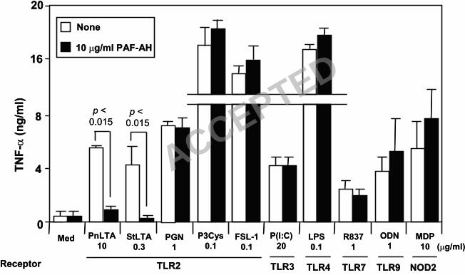
unlikely that PAF-AH, a phospholipase A2, will affect other PAMP, we next investigated the
effect of PAF-AH on other TLR ligands including Poly (I:C) (a TLR3 ligand), LPS (a TLR4
ligand), R837 (a TLR7 or TLR8 ligand), ODN1826 (a TLR9 ligand), and MDP (a NOD2 ligand) 195
(Fig. 2). We found that the treatment did inactivate LTA, as we previously reported (44), but all
other ligands were unaffected by treatment with 10 µg/ml of PAF-AH (Fig. 2). Included in the
ligands tested, were peptidoglycan (PGN) and two synthetic lipoproteins, Pam3CSK4 (a TLR2/1
ligand) and FSC-1 (a TLR2/6 ligand) (Fig. 2), which represent other TLR2 ligands of Gr+
bacteria (13, 23). Therefore, PAF-AH is a highly specific LTA inhibitor. 200
ACCEPTED
on July 8, 2018 by guesthttp://iai.asm
.org/D
ownloaded from
Page 11
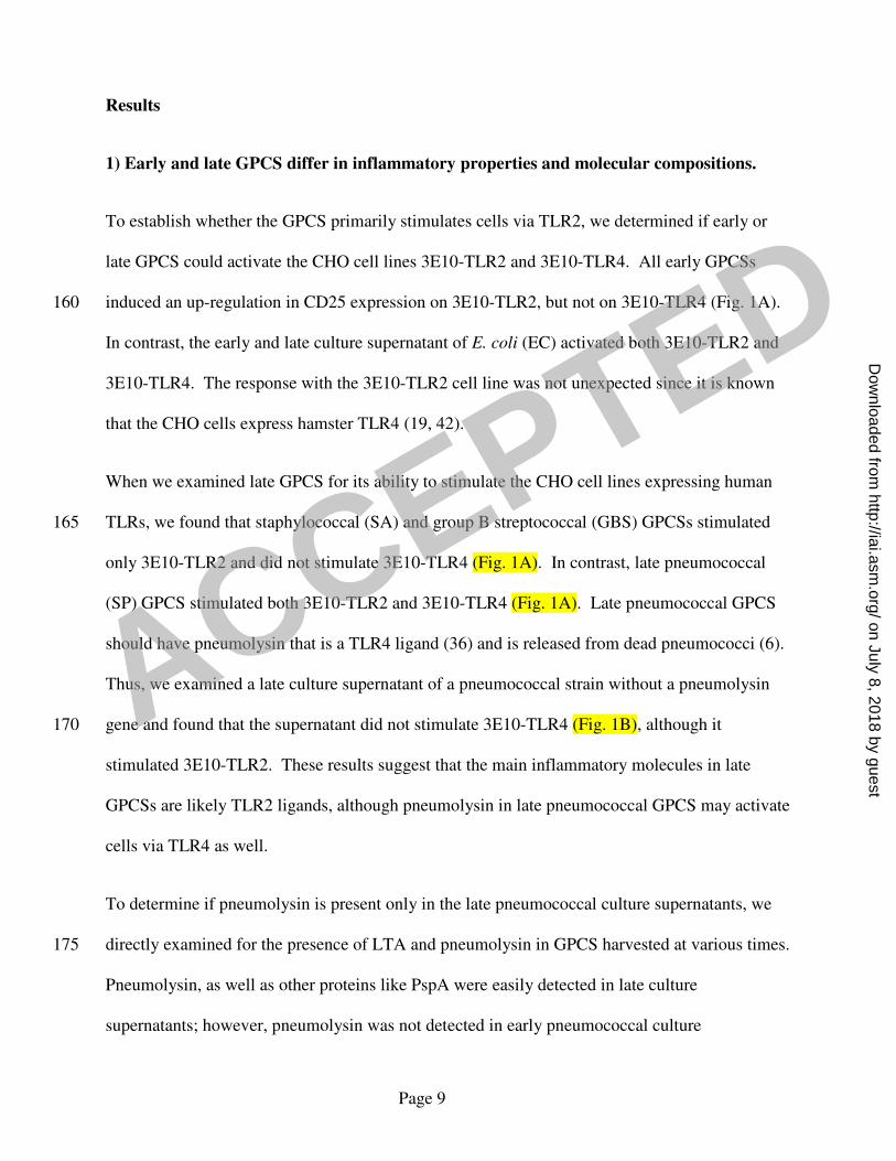
To determine the role of LTA in early GPCS, we next investigated the inflammatory properties
of early (Fig. 3A) and late (Fig. 3B) GPCSs after treatment with PAF-AH or alkaline hydrolysis
(0.2 N NaOH). Based on preliminary studies, we used 10% of early- and 5% of late GPCS.
Untreated early and late culture supernatants dramatically increased TNF-α production (Fig. 3A
and Fig. 3B). When culture supernatants were treated with alkaline hydrolysis and neutralized 205
with HCl, early culture supernatants from the three Gr+ bacteria species as well as the E. coli
retained less than 10% of the original activity (Fig. 3A). The hydrolysis also reduced activity of
late culture supernatants by 50~90% (Fig. 3B). Alkaline hydrolysis can inactivate LTA,
synthetic lipopeptides, and LPS, but does not inactivate PGN or MDP (data not shown). Thus,
LTA, lipoprotein or LPS may be important inflammatory factors in these culture supernatants. 210
To selectively inactivate LTA, bacterial culture supernatants were treated with PAF-AH. When
early supernatants of pneumococci and staphylococci were treated with PAF-AH, 1 µg/ml PAH-
AH reduced TNF-α production by 70-90% and 10 µg/ml of PAF-AH reduced TNF-α production
almost completely (>90%) (Fig. 3A). When the late culture supernatants were investigated, the
PAF-AH treatment (10 µg/ml) reduced their ability to induce TNF-α production by 50% and 215
70%, respectively (Fig. 3B). The inactivation procedure itself did not affect GPCS activity since
inactivation with 0 µg/ml of PAF-AH did not alter GPCS activity (Fig. 3A and 3B), and the
addition of 10 µg/ml of PAF-AH alone did not affect the activities of RAW264.7 cells (Data not
shown) (44). In contrast to GPCS, treatment of early and late E. coli culture supernatants with
10 µg/ml of PAF-AH had no effect on its inflammatory activity (Fig. 3A and 3B). These results 220
strongly suggest that LTA is the dominant inflammatory factor in early GPCSs and is still a
major factor in late GPCSs.
ACCEPTED
on July 8, 2018 by guesthttp://iai.asm
.org/D
ownloaded from

Page 12
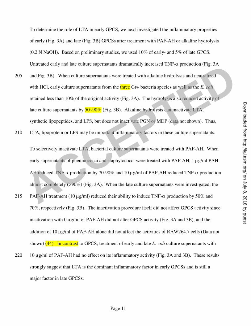
To further confirm the inflammatory role of LTA in GPCS, we depleted LTA in the
staphylococcal GPCS with anti-LTA antibody and examined the antibody-treated GPCS for its
ability to induce RAW264.7 cells to produce TNF-α (Fig. 4). Treatment with mAb BD1701 225
reduced the inflammatory properties of early staphylococcal supernatants by 80%, but reduced
late supernatants by only 65%. The control mouse IgG1 antibody had no effect on the
inflammatory properties of either GPCS (Fig. 4). Also, mAb BD1701 did not reduce the
inflammatory capacity of E. coli culture supernatant and LPS (Fig. 4). Thus, all three
independent approaches to removing LTA support the contention that LTA is essential to the 230
ability of early GPCS to stimulate RAW264.7 cells and is important to the parallel ability of late
GPCS.
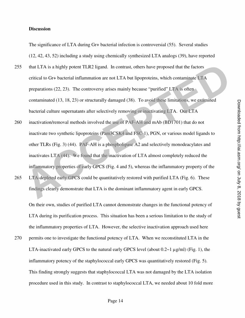
3) Addition of purified LTA restores the inflammatory capacity of LTA-inactivated early
GPCS.
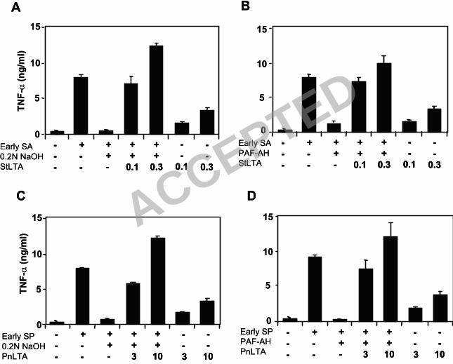
To investigate whether the treatments used above to inactivate LTA also removed other critical 235
molecules other than LTA, we examined the effect of restoring deactivated early GPCS with
purified LTA. Ten percent of alkali-inactivated early staphylococcal supernatants, which
originally contained about 0.02-0.1 µg/ml LTA prior to inactivation (Fig. 1D), regained its
original activity when 0.1 µg/ml of staphylococcal LTA was added (Fig. 5A). In contrast, LTA,
by itself, induced several times less TNF-α than the same amount of LTA mixed with inactivated 240
early GPCS (Fig. 5A). PAF-AH-inactivated staphylococcal supernatants showed almost
identical results (Fig. 5B). These findings demonstrate the inflammatory activity of LTA and
suggest that early GPCS contains a factor(s) synergizing with LTA for inflammatory activity.
ACCEPTED
on July 8, 2018 by guesthttp://iai.asm
.org/D
ownloaded from

Page 13
When the pneumococcal culture supernatants were similarly investigated, we found that
pneumococcal culture supernatants could also be reconstituted with purified LTA. However, the 245
restoration required about 3-10 µg/ml of LTA (Fig. 5C and D), whereas 10 % of early
pneumococcal supernatant has only about 0.02-0.1 µg/ml. Thus, the restoration of inactivated
culture supernatants required about 10 times more purified LTA. In addition, the activity of
purified pneumococcal LTA was enhanced several folds with the inactivated pneumococcal
culture supernatants (Fig. 5C and D). This suggests that pneumococcal culture supernatants also 250
contain factors that synergize with LTA for inflammatory activity.
ACCEPTED
on July 8, 2018 by guesthttp://iai.asm
.org/D
ownloaded from
Page 14
Discussion
The significance of LTA during Gr+ bacterial infection is controversial (55). Several studies
(12, 42, 43, 52) including a study using chemically synthesized LTA analogs (39), have reported
that LTA is a highly potent TLR2 ligand. In contrast, others have proposed that the factors 255
critical to Gr+ bacterial inflammation are not LTA but lipoproteins, which contaminate LTA
preparations (22, 23). The controversy arises mainly because “purified” LTA is often
contaminated (13, 18, 23) or structurally damaged (38). To avoid these limitations, we examined
bacterial culture supernatants after selectively removing or inactivating LTA. Our LTA
inactivation/removal methods involved the use of PAF-AH and mAb (BD1701) that do not 260
inactivate two synthetic lipoproteins (Pam3CSK4 and FSC-1), PGN, or various model ligands to
other TLRs (Fig. 3) (44). PAF-AH is a phospholipase A2 and selectively monodeacylates and
inactivates LTA (44). We found that the inactivation of LTA almost completely reduced the
inflammatory properties of early GPCS (Fig. 4 and 5), whereas the inflammatory property of the
LTA-depleted early GPCS could be quantitatively restored with purified LTA (Fig. 6). These 265
findings clearly demonstrate that LTA is the dominant inflammatory agent in early GPCS.
On their own, studies of purified LTA cannot demonstrate changes in the functional potency of
LTA during its purification process. This situation has been a serious limitation to the study of
the inflammatory properties of LTA. However, the selective inactivation approach used here
permits one to investigate the functional potency of LTA. When we reconstituted LTA in the 270
LTA-inactivated early GPCS to the natural early GPCS level (about 0.2~1 µg/ml) (Fig. 1), the
inflammatory potency of the staphylococcal early GPCS was quantitatively restored (Fig. 5).
This finding strongly suggests that staphylococcal LTA was not damaged by the LTA isolation
procedure used in this study. In contrast to staphylococcal LTA, we needed about 10 fold more
ACCEPTED
on July 8, 2018 by guesthttp://iai.asm
.org/D
ownloaded from

Page 15
pneumococcal LTA to regain the inflammatory capacity of pneumococcal early GPCS (Fig. 5). 275
One possible explanation for this finding is that the widely used procedure for purifying
pneumococcal LTA yields pneumococcal LTA with reduced activity. Consistent with our
conclusion, Draing et al. reported, during the preparation of this manuscript, that the classical
purification procedure removes the alanyl group from pneumococcal LTA and thus reduces its
inflammatory potency (12). 280
LTA inactivation also made the late GPCSs of several Gr+ bacteria less inflammatory (Fig. 4 and
5). However, unlike the early-phase supernatants, LTA inactivation left the late GPCSs with
significant amounts of residual inflammatory activity. Thus, late GPCS must have a TLR2
ligand(s) other than LTA, which may be PGN or a lipoprotein. Lipoproteins should be present in
late GPCS because many bacteria begin to die in the late phase of culture and because 285
lipoprotein(s) account for the majority of inflammatory properties of dead Gr+ bacteria (4, 35,
41, 45). Thus, our data suggest that the dominant TLR2 ligands may vary during the stages of
infection: LTA may be a critically important TLR2 ligand in the early phase of infection, but
other molecules such as lipoproteins may become significant as TLR2 ligands in the late phase
of infection. 290
There is current debate as to whether lipoprotein or LTA is the dominant inflammatory molecule
of Gr+ bacteria or their lysates. Staphylococci with a ∆lgtA mutation cannot produce
lipoproteins, and their lysates are less stimulatory than the wild-type staphylococcal lysates (45).
Yet, a chemically synthesized staphylococcal LTA analog is strongly stimulatory (39). There is
also disagreement concerning the ability of LTA extracted from ∆lgtA-mutant bacteria to 295
stimulate human blood cells (55 ). Since we did not investigate Gr+ bacteria themselves, our
study cannot directly address these debates. However, our data does show that LTA is the
ACCEPTED
on July 8, 2018 by guesthttp://iai.asm
.org/D
ownloaded from

Page 16
dominant TLR2 ligand in early GPCSs and is still a significant ligand in late GPCSs. Thus, LTA
should be considered as an important TLR2 ligand. LTA is readily released into and is abundant
in culture supernatants (32, 57), whereas lipoproteins are primarily associated with bacteria and 300
are not released into supernatants (45). In fact, consistent with our conclusions, is the finding
that the culture supernatant of the lipoprotein-deficient bacteria (∆lgtA) is as inflammatory as
wild-type bacterial culture supernatant (45).
In the present study, we provide evidence that the role of LTA in innate immunity changes with
bacterial culture stages. For instance, LTA is responsible for almost all the inflammatory activity 305
of early Gr+ culture supernatants, but late culture supernatants have other important
inflammatory molecules, including pneumococcal pneumolysin (Fig. 1). This suggests that the
role of LTA during infections should be assessed in the context of a specific pathophysiological
process. For instance, pneumolysin may be more prominent than LTA in pneumococcal
pneumonia where a large number of dead pneumococci are present. Indeed, deletion or 310
neutralization of pneumolysin makes pneumococci less effective in causing pneumonia, but does
not significantly affect their ability to mediate sepsis (3, 8). In contrast, LTA would be relatively
more important than other inflammatory molecules when the pneumococcal density is low, such
as in nasopharyngeal carriage or at the beginning of a pathologic process. Such a bacteria-
limiting step may be seen during the invasion of the endothelial barrier by a single live bacterium 315
or early sepsis. Consistent with this, anti-LTA antibodies were shown to protect animals in a
sepsis model (49, 56). Also, a recent study on pneumococcal invasion of endothelium showed
that LTA could replicate the morphologic changes of the endothelium that are observed with
pneumococci (5).
ACCEPTED
on July 8, 2018 by guesthttp://iai.asm
.org/D
ownloaded from

Page 17
Acknowledgment 320
We thank Drs. W. Welch and M. Deeley at ICOS for providing us with PAF-AH. The work was
supported by NIH funding; AI-69695 and AI-30021 to MHN and AI-56460 to SM.
ACCEPTED
on July 8, 2018 by guesthttp://iai.asm
.org/D
ownloaded from

Page 18
References
1. Abreu, M. T., and M. Arditi. 2004. Innate immunity and Toll-like receptors: clinical 325
implications of basic science research. J Pediatr 144:421-9.
2. Aderem, A., and R. J. Ulevitch. 2000. Toll-like receptors in the induction of the innate
immune response. Nature 406:782-787.
3. Alexander, J. E., A. M. Berry, J. C. Paton, J. B. Rubins, P. W. Andrew, and T. J.
Mitchell. 1998. Amino acid changes affecting the activity of pneumolysin alter the 330
behaviour of pneumococci in pneumonia. Microb Pathog 24:167-74.
4. Baumgartner, M., U. Karst, B. Gerstel, M. Loessner, J. Wehland, and L. Jansch. 2007.
Inactivation of Lgt allows systematic characterization of lipoproteins from Listeria
monocytogenes. J Bacteriol 189:313-24.
5. Beisswenger, C., C. B. Coyne, M. Shchepetov, and J. N. Weiser. 2007. Role of p38 MAP 335
kinase and TGF-beta signaling in transepithelial migration of invasive bacterial
pathogens. J Biol Chem 282:28700-8.
6. Benton, K., J. Paton, and D. Briles. 1997. Differences in virulence for mice among
Streptococcus pneumoniae strains of capsular types 2, 3, 4, 5, and 6 are not attributable to
differences in pneumolysin production. Infect. Immun. 65:1237-1244. 340
7. Beutler, B. 2004. Innate immunity: an overview. Mol Immunol 40:845-59.
8. Briles, D. E., S. K. Hollingshead, J. C. Paton, E. W. Ades, L. Novak, F. W. van Ginkel,
and W. H. Benjamin, Jr. 2003. Immunizations with pneumococcal surface protein A and
pneumolysin are protective against pneumonia in a murine model of pulmonary infection
with Streptococcus pneumoniae. J Infect Dis 188:339-48. 345
9. Brooks-Walter, A., D. E. Briles, and S. K. Hollingshead. 1999. The pspC gene of
Streptococcus pneumoniae encodes a polymorphic protein, PspC, which elicits cross-
ACCEPTED
on July 8, 2018 by guesthttp://iai.asm
.org/D
ownloaded from

Page 19
reactive antibodies to PspA and provides immunity to pneumococcal bacteremia. Infect
Immun 67:6533-42.
10. Crain, M. J., W. D. Waltman II, J. S. Turner, J. Yother, D. F. Talkington, L. S. McDaniel, 350
B. M. Gray, and D. E. Briles. 1990. Pneumococcal surface protein A (PspA) is
serologically highly variable and is expressed by all clinically important capsular
serotypes of Streptococcus pneumoniae. Infect Immun 58:3293-3299.
11. Doherty, T. M., and M. Arditi. 2005. Innate immunity, Toll-like receptors and host
response to infection. Pediatr Infect Dis J 24:643-4. 355
12. Draing, C., M. Pfitzenmaier, S. Zummo, G. Mancuso, A. Geyer, T. Hartung, and S. von
Aulock. 2006. Comparison of lipoteichoic acid from different serotypes of Streptococcus
pneumoniae. J. Biol. Chem. 281:33849-59.
13. Dziarski, R., and D. Gupta. 2005. Staphylococcus aureus peptidoglycan is a Toll-like
receptor 2 activator: a reevaluation. Infect Immun 73:5212-6. 360
14. Echchannaoui, H., K. Frei, C. Schnell, S. L. Leib, W. Zimmerli, and R. Landmann. 2002.
Toll-like receptor 2-deficient mice are highly susceptible to Streptococcus pneumoniae
meningitis because of reduced bacterial clearing and enhanced inflammation. J Infect Dis
186:798-806.
15. Ellingsen, E., S. Morath, T. Flo, A. Schromm, T. Hartung, C. Thiemermann, T. Espevik, 365
D. Golenbock, D. Foster, R. Solberg, A. Aasen, and J. Wang. 2002. Induction of cytokine
production in human T cells and monocytes by highly purified lipoteichoic acid:
involvement of Toll-like receptors and CD14. Med Sci Monit 8:BR149-56.
16. Fischer, W., H. U. Koch, and R. Haas. 1983. Improved preparation of lipoteichoic acids.
Eur J Biochem 133:523-30. 370
ACCEPTED
on July 8, 2018 by guesthttp://iai.asm
.org/D
ownloaded from

Page 20
17. Fujimoto, Y., S. Inamura, A. Kawasaki, Z. Shiokawa, A. Shimoyama, T. Hashimoto, S.
Kusumoto, and K. Fukase. 2007. IEIIS Meeting minireview: Chemical synthesis of
peptidoglycan fragments for elucidation of the immunostimulating mechanism. J
Endotoxin Res 13:189-96.
18. Gao, J. J., Q. Xue, E. G. Zuvanich, K. R. Haghi, and D. C. Morrison. 2001. Commercial 375
preparations of lipoteichoic acid contain endotoxin that contributes to activation of mouse
macrophages in vitro. Infect Immun 69:751-757.
19. Golenbock, D. T., Y. Liu, F. H. Millham, M. W. Freeman, and R. A. Zoeller. 1993.
Surface expression of human CD14 in Chinese hamster ovary fibroblasts imparts
macrophage-like responsiveness to bacterial endotoxin. J Biol Chem 268:22055-9. 380
20. Grundling, A., and O. Schneewind. 2006. Cross-linked peptidoglycan mediates
lysostaphin binding to the cell wall envelope of Staphylococcus aureus. J. Bacteriol.
188:2463-2472.
21. Hashimoto, M., M. Furuyashiki, and Y. Suda. 2007. Response to comment on "Not
lipoteichoic acid but lipoproteins appear to be the dominant immunobiologically active 385
compounds in Staphylococcus aureus." J. Immunol. 178:2610-2611.
22. Hashimoto, M., K. Tawaratsumida, H. Kariya, K. Aoyama, T. Tamura, and Y. Suda.
2005. Lipoprotein is a predominant Toll-like receptor 2 ligand in Staphylococcus aureus
cell wall components. Int Immunol 18:355-362.
23. Hashimoto, M., K. Tawaratsumida, H. Kariya, A. Kiyohara, Y. Suda, F. Krikae, T. 390
Kirikae, and F. Gotz. 2006. Not lipoteichoic acid but lipoproteins appear to be the
dominant immunobiologically active compounds in Staphylococcus aureus. J Immunol
177:3162-3169.
ACCEPTED
on July 8, 2018 by guesthttp://iai.asm
.org/D
ownloaded from

Page 21
24. Heer, C., K. Stuertz, R. R. Reinert, M. Mader, and R. Nau. 2000. Release of teichoic and
lipoteichoic acids from 30 different strains of Streptococcus pneumoniae during exposure 395
to ceftriaxone, meropenem, quinupristin/dalfopristin, rifampicin and trovafloxacin.
Infection 28:13-20.
25. Henderson, W. R., Jr., J. Lu, K. M. Poole, G. N. Dietsch, and E. Y. Chi. 2000.
Recombinant human platelet-activating factor-acetylhydrolase inhibits airway
inflammation and hyperreactivity in mouse asthma model. J Immunol 164:3360-7. 400
26. Henneke, P., O. Takeuchi, R. Malley, E. Lien, R. R. Ingalls, M. W. Freeman, T.
Mayadas, V. Nizet, S. Akira, D. L. Kasper, and D. T. Golenbock. 2002. Cellular
activation, phagocytosis, and bactericidal activity against group B streptococcus involve
parallel myeloid differentiation factor 88-dependent and independent signaling pathways.
J Immunol 169:3970-7. 405
27. Hirschfeld, M., Y. Ma, J. H. Weis, S. N. Vogel, and J. J. Weis. 2000. Cutting edge:
repurification of lipopolysaccharide eliminates signaling through both human and murine
Toll-like receptor 2. J Immunol 165:618-22.
28. Hoebe, K., P. Georgel, S. Rutschmann, X. Du, S. Mudd, K. Crozat, S. Sovath, L. Shamel,
T. Hartung, U. Zahringer, and B. Beutler. 2005. CD36 is a sensor of diacylglycerides. 410
Nature 433:523-7.
29. Hofbauer, B., A. K. Saluja, M. Bhatia, J. L. Frossard, H. S. Lee, L. Bhagat, and M. L.
Steer. 1998. Effect of recombinant platelet-activating factor acetylhydrolase on two
models of experimental acute pancreatitis. Gastroenterology 115:1238-47.
30. Hoshino, K., O. Takeuchi, T. Kawai, H. Sanjo, T. Ogawa, Y. Takeda, K. Takeda, and S. 415
Akira. 1999. Cutting edge: Toll-like receptor 4 (TLR4)-deficient mice are
ACCEPTED
on July 8, 2018 by guesthttp://iai.asm
.org/D
ownloaded from

Page 22
hyporesponsive to lipopolysaccharide: evidence for TLR4 as the Lps gene product. J
Immunol 162:3749-52.
31. Inamura, S., Y. Fujimoto, A. Kawasaki, Z. Shiokawa, E. Woelk, H. Heine, B. Lindner, N.
Inohara, S. Kusumoto, and K. Fukase. 2006. Synthesis of peptidoglycan fragments and 420
evaluation of their biological activity. Org Biomol Chem 4:232-42.
32. Jones, K. J., A. D. Perris, A. B. Vernallis, T. Worthington, P. A. Lambert, and T. S.
Elliott. 2005. Induction of inflammatory cytokines and nitric oxide in J774.2 cells and
murine macrophages by lipoteichoic acid and related cell wall antigens from
Staphylococcus epidermidis. J Med Microbiol 54:315-21. 425
33. Kim, J. H., H. Seo, S. H. Han, J. Lin, M. K. Park, U. B. Sorensen, and M. H. Nahm.
2005. Monoacyl lipoteichoic Acid from pneumococci stimulates human cells but not
mouse cells. Infect Immun 73:834-40.
34. Knapp, S., C. W. Wieland, C. van 't Veer, O. Takeuchi, S. Akira, S. Florquin, and T. van
der Poll. 2004. Toll-like receptor 2 plays a role in the early inflammatory response to 430
murine pneumococcal pneumonia but does not contribute to antibacterial defense. J
Immunol 172:3132-8.
35. Leskela, S., E. Wahlstrom, V. P. Kontinen, and M. Sarvas. 1999. Lipid modification of
prelipoproteins is dispensable for growth but essential for efficient protein secretion in
Bacillus subtilis: characterization of the Lgt gene. Mol Microbiol 31:1075-85. 435
36. Malley, R., P. Henneke, S. C. Morse, M. J. Cieslewicz, M. Lipsitch, C. M. Thompson, E.
Kurt-Jones, J. C. Paton, M. R. Wessels, and D. T. Golenbock. 2003. Recognition of
pneumolysin by Toll-like receptor 4 confers resistance to pneumococcal infection. Proc
Natl Acad Sci U S A 100:1966-71.
ACCEPTED
on July 8, 2018 by guesthttp://iai.asm
.org/D
ownloaded from

Page 23
37. Medvedev, A. E., P. Henneke, A. Schromm, E. Lien, R. Ingalls, M. J. Fenton, D. T. 440
Golenbock, and S. N. Vogel. 2001. Induction of tolerance to lipopolysaccharide and
mycobacterial components in Chinese hamster Ovary/CD14 cells is not affected by
overexpression of Toll-like receptors 2 or 4. J Immunol 167:2257-2267.
38. Morath, S., A. Geyer, I. Spreitzer, C. Hermann, and T. Hartung. 2002. Structural
decomposition and heterogeneity of commercial lipoteichoic acid preparations. Infect 445
Immun 70:938-44.
39. Morath, S., A. Stadelmaier, A. Geyer, R. R. Schmidt, and T. Hartung. 2002. Synthetic
lipoteichoic acid from Staphylococcus aureus is a potent stimulus of cytokine release. J
Exp Med 195:1635-40.
40. Paterson, G. K., and T. J. Mitchell. 2006. Innate immunity and the pneumococcus. 450
Microbiology 152:285-93.
41. Petit, C. M., J. R. Brown, K. Ingraham, A. P. Bryant, and D. J. Holmes. 2001. Lipid
modification of prelipoproteins is dispensable for growth in vitro but essential for
virulence in Streptococcus pneumoniae. FEMS Microbiol Lett 200:229-33.
42. Schroder, N. W., S. Morath, C. Alexander, L. Hamann, T. Hartung, U. Zahringer, U. B. 455
Gobel, J. R. Weber, and R. R. Schumann. 2003. Lipoteichoic acid (LTA) of
Streptococcus pneumoniae and Staphylococcus aureus activates immune cells via Toll-
like receptor (TLR)-2, lipopolysaccharide-binding protein (LBP), and CD14, whereas
TLR-4 and MD-2 are not involved. J Biol Chem 278:15587-94.
43. Schwandner, R., R. Dziarski, H. Wesche, M. Rothe, and C. J. Kirschning. 1999. 460
Peptidoglycan- and lipoteichoic acid-induced cell activation is mediated by Toll-like
receptor 2. J Biol Chem 274:17406-9.
ACCEPTED
on July 8, 2018 by guesthttp://iai.asm
.org/D
ownloaded from

Page 24
44. Seo, H. S., J. H. Kim, and M. H. Nahm. 2006. Platelet-activating factor-acetylhydrolase
can monodeacylate and inactivate lipoteichoic acid. Clin Vaccine Immunol 13:452-8.
45. Stoll, H., J. Dengjel, C. Nerz, and F. Gotz. 2005. Staphylococcus aureus deficient in 465
lipidation of prelipoproteins is attenuated in growth and immune activation. Infect.
Immun. 73:2411-2423.
46. Stuertz, K., H. Schmidt, H. Eiffert, P. Schwartz, M. Mader, and R. Nau. 1998.
Differential release of lipoteichoic and teichoic acids from Streptococcus pneumoniae as
a result of exposure to beta-lactam antibiotics, rifamycins, trovafloxacin, and 470
quinupristin-dalfopristin. Antimicrob Agents Chemother 42:277-81.
47. Takeuchi, O., K. Hoshino, and S. Akira. 2000. Cutting edge: TLR2-deficient and
MyD88-deficient mice are highly susceptible to Staphylococcus aureus infection. J
Immunol 165:5392-6.
48. Tapping, R. I., S. Akashi, K. Miyake, P. J. Godowski, and P. S. Tobias. 2000. Toll-like 475
receptor 4, but not Toll-like receptor 2, is a signaling receptor for Escherichia and
Salmonella lipopolysaccharides. J Immunol 165:5780-7.
49. Theilacker, C., Z. Kaczynski, A. Kropec, F. Fabretti, T. Sange, O. Holst, and J. Huebner.
2006. Opsonic antibodies to Enterococcus faecalis strain 12030 are directed against
lipoteichoic acid. Infect Immun 74:5703-12. 480
50. Tjoelker, L. W., C. Eberhardt, J. Unger, H. L. Trong, G. A. Zimmerman, T. M. McIntyre,
D. M. Stafforini, S. M. Prescott, and P. W. Gray. 1995. Plasma platelet-activating factor
acetylhydrolase is a secreted phospholipase A2 with a catalytic triad. J Biol Chem
270:25481-7.
ACCEPTED
on July 8, 2018 by guesthttp://iai.asm
.org/D
ownloaded from
Page 25
51. Travassos, L. H., S. E. Girardin, D. J. Philpott, D. Blanot, M. A. Nahori, C. Werts, and I. 485
G. Boneca. 2004. Toll-like receptor 2-dependent bacterial sensing does not occur via
peptidoglycan recognition. EMBO Rep 5:1000-6.
52. Triantafilou, M., M. Manukyan, A. Mackie, S. Morath, T. Hartung, H. Heine, and K.
Triantafilou. 2004. Lipoteichoic acid and Toll-like receptor 2 internalization and targeting
to the Golgi is lipid raft dependent. J Biol Chem 279:40882-9. 490
53. Van Amersfoort, E. S., T. J. Van Berkel, and J. Kuiper. 2003. Receptors, mediators, and
mechanisms involved in bacterial sepsis and septic shock. Clin Microbiol Rev 16:379-
414.
54. van Langevelde, P., E. Ravensbergen, P. Grashoff, H. Beekhuizen, P. H. Groeneveld, and
J. T. van Dissel. 1999. Antibiotic-induced cell wall fragments of Staphylococcus aureus 495
increase endothelial chemokine secretion and adhesiveness for granulocytes. Antimicrob
Agents Chemother 43:2984-9.
55. von Aulock, S., T. Hartung, and C. Hermann. 2007. Comment on "Not lipoteichoic acid
but lipoproteins appear to be the dominant immunobiologically active compounds in
Staphylococcus aureus". J Immunol 178:2610. 500
56. Walsh, S., J. Kokai-Kun, A. Shah, and J. Mond. 2004. Extended nasal residence time of
lysostaphin and an anti-staphylococcal monoclonal antibody by delivery in semisolid or
polymeric carriers. Pharm Res 21:1770-5.
57. Wicken, A. J., and K. W. Knox. 1975. Lipoteichoic acids: A new class of bacterial
antigen. Science 187:1161-1167. 505
58. Zhang, Q., N. Mousdicas, Q. Yi, M. Al-Hassani, S. D. Billings, S. M. Perkins, K. M.
Howard, S. Ishii, T. Shimizu, and J. B. Travers. 2005. Staphylococcal lipoteichoic acid
ACCEPTED
on July 8, 2018 by guesthttp://iai.asm
.org/D
ownloaded from

Page 26
inhibits delayed-type hypersensitivity reactions via the platelet-activating factor receptor.
J Clin Invest 115:2855-2861.
510
ACCEPTED
on July 8, 2018 by guesthttp://iai.asm
.org/D
ownloaded from

Page 27
Figure Legends
FIG. 1. Panel A shows percentage of CD25+ 3E10-TLR2 (open bar) or 3E10-TLR4 (black bar)
cells in response to early and late bacteria culture supernatants. Cells were stimulated with the
bacterial culture medium alone (labeled Med), pneumococcal GPCS (labeled SP), staphylococcal
GPCS (labeled SA), Group B streptococcal GPCS (labeled GBS), or E. coli culture supernatant 515
(labeled EC). Based on preliminary studies, early-GPCSs were used at 10%, late-GPCS were at
5%, early E. coli supernatant was at 0.1%, and late E. coli supernatant was at 0.01%. Panel B
shows percentage of CD25+ 3E10-TLR4 cells in response to indicated concentrations of early
and late culture supernatants collected from S. pneumoniae strain D39 (labeled WT) and its
pneumolysin-deficient isogenic mutant (labeled PLN-A). Panel C shows the presence of 520
pneumolysin (Ply)(53 KDa), PspA (90 KDa), and PspC (92KDa) in various pneumococcal
culture supernatants. Truncated PspC (85 KDa) was detected only in the late supernatant (O/N).
The time of harvest for the culture supernatants is shown (in hrs) at the top of each lane. O/N
indicates an overnight culture. The left two lanes were loaded with lysates of 108 and 10
7 CFU
of a pneumococcal strain, R36A. Panel D shows LTA in GPCS of S. pneumoniae (labeled SP), 525
S. aureus (labeled SA), and Group B streptococci (labeled GBS). 20 µl of purified LTA were
loaded in the left three lanes, of early GPCS in the middle three lanes, and of late GPCS in the
right three lanes. Concentrations of purified LTA and GPCS are indicated at the top of each
lane. The molecules in the samples were separated by PAGE and visualized with anti-
phosphorylcholine antibody (TEPC-15) to pneumococcal LTA or with a mouse anti-LTA mAb 530
(BD1701) for the others.
FIG. 2. TNF-α production by RAW264.7 cells in response to stimulation by pneumococcal
LTA (PnLTA), staphylococcal LTA (StLTA), staphylococcal PGN (PGN), Pam3CSK4 (P3Cys),
ACCEPTED
on July 8, 2018 by guesthttp://iai.asm
.org/D
ownloaded from

Page 28
FSL-1, Poly (I:C) [P(I:C)], LPS, imiquimod (R837), ODN1826 (ODN), or MDP before (open
bar) or after (black bar) 10 µg/ml PAF-AH treatment. The ligand concentrations (in µg/ml) and 535
receptor specificities are identified at the bottom of the figure. Bars indicate the mean of results
of triplicate wells of a representative experiment. Error bars indicate SD. The p values are
indicated in the figure.
FIG. 3. TNF-α production by RAW264.7 cells in response to bacterial culture supernatants of S.
pneumonia, S. aureus, GBS, or E. coli. The two bars in the left indicate controls: un-stimulated 540
(first bars) or stimulated with untreated bacterial culture supernatants (second bars). The third
bars indicate the cells stimulated with culture supernatant treated with alkali hydrolysis and the
remaining bars (4th
to 7th
) indicate the cells stimulated with supernatants treated with PAF-AH at
indicated concentrations (in µg/ml). PAF-AH was inactivated with 100 µM Pefabloc SC after
the reaction. Early-GPCSs were used at 10%, late-GPCSs were at 5%, early E. coli supernatant 545
was at 0.1%, and late E. coli supernatant was at 0.01%. Bars indicate the mean of results of
triplicate wells of a representative experiment. Error bars indicate SD. The p values are
indicated in the figure.
FIG. 4. TNF-α production by RAW264.7 cells in response to bacterial culture medium (labeled
Med), staphylococcal early GPCS (labeled Early), staphylococcal late GPCS (labeled Late), 550
purified staphylococcal LTA (labeled LTA), E. coli early culture supernatant (labeled Early, E.
coli), or E. coli LPS (labeled LPS). Each stimulant was used unabsorbed (black bars), after
absorption with an irrelevant isotype-matched control mAb (open bars), or after absorption with
an anti-LTA mAb (hatched bars). Bars indicate the mean of results of triplicate wells of a
representative experiment. Error bars indicate SD. The p values are indicated in the figure. 555
ACCEPTED
on July 8, 2018 by guesthttp://iai.asm
.org/D
ownloaded from

Page 29
FIG. 5. TNF-α production by RAW264.7 cells in response to early GPCS from staphylococci
(Panels A and B, Early SA) or from pneumococci (Panels C and D, Early SP). Early GPCS was
inactivated by alkali hydrolysis (labeled as 0.2 N NaOH) or with 10 µg/ml PAF-AH (labeled as
PAF-AH). Then an indicated concentration (µg/L) of purified pneumococcal LTA (PnLTA) or
staphylococcal LTA (StLTA) was added to respective inactivated culture supernatants (10%). 560
Bars indicate the mean of results of triplicate wells of a representative experiment. Error bars
indicate SD.
ACCEPTED
on July 8, 2018 by guesthttp://iai.asm
.org/D
ownloaded from

Med SP SA GBS EC
0
20
40
60
80
CD
25
+cell
s (
%)
A
Early
D LTA (µg/ml) Early Late
SP
SA
GBS
1.0
0.2
0.0
4
4 %
20
%
100
%
4 %
100
%
20
%
C
Ply
(53KDa)
PspA
(90KDa)
108 107 0.0 0.2 0.5 1.0 1.3 (OD600)
0 2.5 2.5 3.0 3.5 O/N (hrs)
R36A Pneumococcal GPCS
PspC
(92KDa)
0
20
40
60
80
100
Med SP SA GBS EC
3E10-TLR2
3E10-TLR4
Late
B
3E10-TLR2
3E10-TLR4
p < 0.001 p < 0.001
p < 0.001p < 0.001
p < 0.001
(85KDa)
0
20
40
60
Med
PLN-A
3%
10%
30%
Early Late Early Late
D39
3E10-TLR4
CD
25
+cell
s (
%)
Med SP SA GBS EC
0
20
40
60
80
CD
25
+cell
s (
%)
A
Early
D LTA (µg/ml) Early Late
SP
SA
GBS
1.0
0.2
0.0
4
4 %
20
%
100
%
4 %
100
%
20
%
C
Ply
(53KDa)
PspA
(90KDa)
108 107 0.0 0.2 0.5 1.0 1.3 (OD600)
0 2.5 2.5 3.0 3.5 O/N (hrs)
R36A Pneumococcal GPCS
PspC
(92KDa)
0
20
40
60
80
100
Med SP SA GBS EC
3E10-TLR2
3E10-TLR4
Late
B
3E10-TLR2
3E10-TLR4
p < 0.001 p < 0.001
p < 0.001p < 0.001
p < 0.001
(85KDa)
0
20
40
60
Med
PLN-A
3%
10%
30%
Early Late Early Late
D39
3E10-TLR4
CD
25
+cell
s (
%)
ACCEPTED on July 8, 2018 by guest
http://iai.asm.org/
Dow
nloaded from
MDP
10 (µg/ml)Med PnLTA
10StLTA
0.3
P3Cys
0.1FSL-1
0.1
P(I:C)
20LPS
0.1
R837
1
ODN
1
TN
F-α
(ng
/ml)
None
0
4
8
16
20
10 µg/ml PAF-AH
p <
0.015
p <
0.015
TLR2 Receptor TLR3 TLR4 TLR7 TLR9 NOD2
PGN
1
MDP
10 (µg/ml)Med PnLTA
10StLTA
0.3
P3Cys
0.1FSL-1
0.1
P(I:C)
20LPS
0.1
R837
1
ODN
1
TN
F-α
(ng
/ml)
None
0
4
8
16
20
10 µg/ml PAF-AH
p <
0.015
p <
0.015
TLR2 Receptor TLR3 TLR4 TLR7 TLR9 NOD2
PGN
1
ACCEPTED
on July 8, 2018 by guesthttp://iai.asm
.org/D
ownloaded from

A. Early
0
2
4
6
TN
F-α
(ng
/ml)
0
1
2
3
4
S. aureus early GPCS (10%)
0
4
8
12
16
E. coli early Supernatant (0.1%)
S. pneumoniae early GPCS (10%)
PAF-AH
0.2N NaOH
p <
0.002 p <
0.001
p <
0.041
p <
0.007
p <
0.001
p <
0.025
p <
0.002
p <
0.004
p <
0.004
- - - 0 0.1 1 10
- - + - - - -
- - - 0 0.1 1 10
- - + - - - -
- - - 0 0.1 1 10
- - + - - - -
0
1
2
3
GBS early GPCS (10%)
p <
0.005
p <
0.025
p <
0.005
p <
0.005
- - - 0 0.1 1 10
- - + - - - -
TN
F-α
(ng
/ml)
TN
F-α
(ng
/ml)
TN
F-α
(ng
/ml)
B. Late
0
4
8
12
0
4
8
12
E. coli late Supernatant (0.01%)
S. aureus late GPCS (5%)
S. pneumoniae late GPCS (5%)
p <
0.003
p <
0.004
p <
0.062p <
0.059
0
2
4
6
8
p <
0.005
p <
0.004
p <
0.032
- - - 0 0.1 1 10
- - + - - - -
- - - 0 0.1 1 10
- - + - - - -
- - - 0 0.1 1 10
- - + - - - -
0
0.4
0.8
1.2
1.6
p <
0.001
p <
0.003
p <
0.008
GBS late GPCS (5%)
- - - 0 0.1 1 10
- - + - - - -
PAF-AH
0.2N NaOH
PAF-AH
0.2N NaOH
PAF-AH
0.2N NaOH
TN
F-α
(ng
/ml)
PAF-AH
0.2N NaOH
TN
F-α
(ng
/ml)
TN
F-α
(ng
/ml)
TN
F-α
(ng
/ml)
PAF-AH
0.2N NaOH
PAF-AH
0.2N NaOH
PAF-AH
0.2N NaOH
A. Early
0
2
4
6
TN
F-α
(ng
/ml)
0
1
2
3
4
S. aureus early GPCS (10%)
0
4
8
12
16
E. coli early Supernatant (0.1%)
S. pneumoniae early GPCS (10%)
PAF-AH
0.2N NaOH
p <
0.002 p <
0.001
p <
0.041
p <
0.007
p <
0.001
p <
0.025
p <
0.002
p <
0.004
p <
0.004
- - - 0 0.1 1 10
- - + - - - -
- - - 0 0.1 1 10
- - + - - - -
- - - 0 0.1 1 10
- - + - - - -
- - - 0 0.1 1 10
- - + - - - -
- - - 0 0.1 1 10
- - + - - - -
0
1
2
3
GBS early GPCS (10%)
p <
0.005
p <
0.025
p <
0.005
p <
0.005
- - - 0 0.1 1 10
- - + - - - -
TN
F-α
(ng
/ml)
TN
F-α
(ng
/ml)
TN
F-α
(ng
/ml)
B. Late
0
4
8
12
0
4
8
12
E. coli late Supernatant (0.01%)
S. aureus late GPCS (5%)
S. pneumoniae late GPCS (5%)
p <
0.003
p <
0.004
p <
0.062p <
0.059
0
2
4
6
8
p <
0.005
p <
0.004
p <
0.032
- - - 0 0.1 1 10
- - + - - - -
- - - 0 0.1 1 10
- - + - - - -
- - - 0 0.1 1 10
- - + - - - -
- - - 0 0.1 1 10
- - + - - - -
- - - 0 0.1 1 10
- - + - - - -
0
0.4
0.8
1.2
1.6
p <
0.001
p <
0.003
p <
0.008
GBS late GPCS (5%)
- - - 0 0.1 1 10
- - + - - - -
PAF-AH
0.2N NaOH
PAF-AH
0.2N NaOH
PAF-AH
0.2N NaOH
TN
F-α
(ng
/ml)
PAF-AH
0.2N NaOH
TN
F-α
(ng
/ml)
TN
F-α
(ng
/ml)
TN
F-α
(ng
/ml)
PAF-AH
0.2N NaOH
PAF-AH
0.2N NaOH
PAF-AH
0.2N NaOH
ACCEPTED on July 8, 2018 by guesthttp://iai.asm
.org/D
ownloaded from

0
2
4
6
8
10
12
None
mIgG1
α-LTA
TN
F-α
(ng
/ml)
Med Early (10%) Late (5%) LTA
(0.3 µg/ml)S. aureus
Early (0.1%)
E. coli
LPS
(0.01 µg/ml)
p < 0.006
p < 0.003
p < 0.001
Med0
2
4
6
8
10
12
None
mIgG1
α-LTA
TN
F-α
(ng
/ml)
Med Early (10%) Late (5%) LTA
(0.3 µg/ml)S. aureus
Early (0.1%)
E. coli
LPS
(0.01 µg/ml)
p < 0.006
p < 0.003
p < 0.001
Med
ACCEPTED
on July 8, 2018 by guesthttp://iai.asm
.org/D
ownloaded from
B
0
5
10
15
0
5
10
15TNF-α(ng/ml)
A
Early SA
PAF-AH
StLTA
Early SA
0.2N NaOH
StLTA
-
-
-
+
-
-
+
+
-
+
+
0.1
+
+
0.3
-
-
0.1
-
-
0.3
-
-
-
+
-
-
+
+
-
+
+
0.1
+
+
0.3
-
-
0.1
-
-
0.3
0Early SP
PAF-AH
PnLTA
0
DC
5
10
15
5
10
15
TNF-α(ng/ml)
Early SP
0.2N NaOH
PnLTA
-
-
-
+
-
-
+
+
-
+
+
3
+
+
10
-
-
3
-
-
10
-
-
-
+
-
-
+
+
-
+
+
3
+
+
10
-
-
3
-
-
10
B
0
5
10
15
0
5
10
15TNF-α(ng/ml)
A
Early SA
PAF-AH
StLTA
Early SA
0.2N NaOH
StLTA
-
-
-
+
-
-
+
+
-
+
+
0.1
+
+
0.3
-
-
0.1
-
-
0.3
-
-
-
+
-
-
+
+
-
+
+
0.1
+
+
0.3
-
-
0.1
-
-
0.3
-
-
-
+
-
-
+
+
-
+
+
0.1
+
+
0.3
-
-
0.1
-
-
0.3
-
-
-
+
-
-
+
+
-
+
+
0.1
+
+
0.3
-
-
0.1
-
-
0.3
0Early SP
PAF-AH
PnLTA
0
DC
5
10
15
5
10
15
TNF-α(ng/ml)
Early SP
0.2N NaOH
PnLTA
-
-
-
+
-
-
+
+
-
+
+
3
+
+
10
-
-
3
-
-
10
-
-
-
+
-
-
+
+
-
+
+
3
+
+
10
-
-
3
-
-
10
-
-
-
+
-
-
+
+
-
+
+
3
+
+
10
-
-
3
-
-
10
-
-
-
+
-
-
+
+
-
+
+
3
+
+
10
-
-
3
-
-
10
ACCEPTED
on July 8, 2018 by guesthttp://iai.asm
.org/D
ownloaded from