Embed Size (px)
Citation preview

Inelastic soft x-ray scattering, fluorescence and elastic radiationWhat happens to the emission (or fluorescence) when the energy of the exciting photons changes?The emission spectra (can) change.One can usually distinguish easily the following contributions to the spectra:
140 145 150 155 160 1650
1000
2000
3000
4000
5000
6000
XAS
calc.
TEYRIXS
PFY
155.2 eV
153.1 eV
Cou
nts
Emission Energy [eV]
150 155 160 165
• Elastic contribution (same energy as incoming photons).
• Fluorescence features (at fixed energy).
• Inelastic features (energy changes and tracks excitation energy).
Note:When approaching the Fluorescence threshold, the inelastic scattering features evolve into fluorescence.
The spectra in the graph are offset in y-direction for clarity.

Emission Energy axis versus Energy loss axis
3d10 4f 0 → 3d9 4f 1 excitations for LaAlO3 (single crystal):
-70 -60 -50 -40 -30 -20 -10 00
1000
2000
3000
4000
823.9 eV
828.9 eV
833.9 eV
838.9 eV
843.9 eV
846.4 eV
848.9 eV
853.9 eV
858.9 eV
7.5 eV
16.3 eV
Cou
nts
Energy loss [eV]760 780 800 820 840 860 880 9000
1000
2000
3000
4000
826 eV
831 eV
836 eV
841 eV
846 eV
848.5 eV
851 eV
856 eV
861 eV
866 eV871 eV
866 eV
834.85 eV851.6 eV
819.4 eV
813.1 eV
Cou
nts
Emission energy [eV]
Note that on the new energy loss scale:
Electron loss scale:hνloss=hνexc- hνemission
• Energy losses appear at fixed energy.
• Fluorescence features track energy.
• Elastic features appear at 0 eV.

From emission (XES) to Resonant Inelastic X-ray Scattering (RIXS)
Advantages of RIXS:• charge-neutral probing• low-energy excitations • site selectivity• bulk and buried structures• band dispersion• no core state lifetime broadening• ultra-fast dynamics
F(ω,ω´) = Σ Σ δ(Εg+ hω − Εf −hω´)f m
<f D m><m D g>Eg + hω - Em - iΓm
2
Formalism of scattering as a one-step process is described by Kramers-Heisenberg formula:
hω´= hω - ∆EPhoton out
EF
Valence band
Conduction band
hω
Photon in
∆E
Corelevel
Any possible transition that can be excited directly can also occur as an energy loss ∆E. The transitions are only governed by (1) Selection rules.(2) Energy and momentum conservation.

Resonant Inelastic Scattering of Dy2O3
• Strong and resonant loss features.• Intensity depends on selected intermediate state (Kramers-Heisenberg formula).• 4d10 5p6 4f 9 (6H15/2)→4d9 5p6 4f10→4d10 5p6 (4f 9)* Net transition 4f 9 → (4f 9)*
Moewes et al., PRB 60, 15728 (2000)
140 145 150 155 160 1650
5x10³
1x10⁴
calc.
TEYXES
PFY
165.4 eV
161.7 eV
155.2 eV
153.1 eV
hνexc = 150.8 eV
Cou
nts
Emission Energy [eV]
150 155 160 165
9 8 7 6 5 4 3 2 1 0 -10
1x10⁴
2x10⁴
Energy loss [eV]
4I15/2; 4M17/2;
4L17/2
hνexc = 150.8 eV
161.7 eV6H13/2
6I15/2; 4M17/2
6K17/2
155.2 eV
153.1 eV
Cou
nts
21962106210 654554443... sdfpsdpsdElectron configuration Dy:
Dy core levels Dy bands

Bandmapping in Graphite with XES
Soft x-ray emission spectroscopy can map the band structure (for light elements).
-20
-15
-10
-5
0
5
10
15Eexc
292.7291.9291.3
288.0
285.6285.0
Graphite
AΓKM LΓ
Ener
gy (e
V)
265 270 275 280 285
299292.7
291.9
291.3
288.0
285.6
Excita
tion E
nerg
y (eV
)
285.0
284.3
C Kα XES
Emission Energy (eV)
Cou
nts (
arb.
uni
ts)280 285 290 295 300
0.2
0.4
0.6
XASabso
rptio
n
Calculation
Experiment
Emission
(Relaxation)
Absorption(Excitation)

One example of hard X-ray fluorescenceFluorescence spectroscopy (10 keV) of impurities on a Si wafer.• From energy and intensity of the
spectrum the type (element) and concentration of impurities can be determined (Ni: 10 fg).
• Due to Ifluo~Z4 this is less feasible for lighter elements.
• In the graph Si Kα (1740 eV) is not in the detector window.
• For energetically not too deep core holes the exact energy of the core hole depends on the exact chemical environment. In this case even more details about the chemical structure can be obtained (like the chemical valence of the elements).

Photo Electron Spectroscopy (PES)
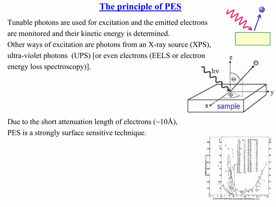
The principle of PES
Tunable photons are used for excitation and the emitted electrons are monitored and their kinetic energy is determined. Other ways of excitation are photons from an X-ray source (XPS), ultra-violet photons (UPS) [or even electrons (EELS or electron energy loss spectroscopy)].
Due to the short attenuation length of electrons (~10Å), PES is a strongly surface sensitive technique.

Reminder: Electron processes are dominating (for lighter elements)
• Radiative transitions are competing with non-radiative transitions (Auger and Autoionization).
• In soft x-ray range radiationless transitions (Auger) are dominant.

PhotoElectron Spectr. (PES) or X-ray Photoelectr. Spectr. (XPS)
T. Greber, 4th PSI Summer School, 2005

The principle of PES
T. Greber, 4th PSI Summer School, 2005


Auger versus photoelectrons
How can one distinguishAuger electrons and Photoelectrons?
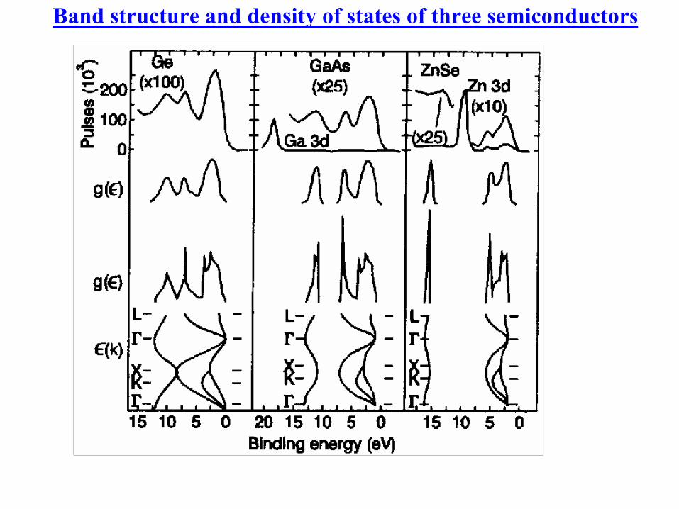
Band structure and density of states of three semiconductors


![High resolution inelastic X-ray scattering from thermal collective … · 1 Note that resonant X-ray scattering can have very strong magnetic contributions [14], and inelastic experiments](https://img.dokumen.tips/doc/110x75/60a9022064d640760449217f/high-resolution-inelastic-x-ray-scattering-from-thermal-collective-1-note-that-resonant.jpg)