Embed Size (px)
Citation preview
Induction of terminal differentiationby constitutive activation of p38 MAPkinase in human rhabdomyosarcoma cellsPier Lorenzo Puri,1,6,7 Zhenguo Wu,2,7 Peilin Zhang,3 Lauren D. Wood,1 Kunjan S. Bhakta,1
Jiahuai Han,5 James R. Feramisco,4 Michael Karin,2,4 and Jean Y.J. Wang1,4,8
Departments of 1Biology, 2Pharmacology, 3Pathology, and 4Cancer Center, University of California at San Diego, La Jolla,California 92093 USA; 5Department of Immunology, Scripps Clinic and Research Foundation, La Jolla, California 92037USA; 6Laboratory of Gene Expression, Fondazione A. Cesalpino, University of Rome La Sapienza, 00161, Rome, Italy
MyoD inhibits cell proliferation and promotes muscle differentiation. A paradoxical feature ofrhabdomyosarcoma (RMS), a tumor arising from muscle precursors, is the block of the differentiation programand the deregulated proliferation despite MyoD expression. A deficiency in RMS of a factor required for MyoDactivity has been implicated by previous studies. We report here that p38 MAP kinase (MAPK) activation,which is essential for muscle differentiation, is deficient in RMS cells. Enforced induction of p38 MAPK by anactivated MAPK kinase 6 (MKK6EE) restored MyoD function and enhanced MEF2 activity in RMS deficientfor p38 MAPK activation, leading to growth arrest and terminal differentiation. Stress and cytokines couldactivate the p38 MAPK in RMS cells, however, these stimuli did not promote differentiation, possibly becausethey activated p38 MAPK only transiently and they also activated JNK, which could antagonizedifferentiation. Thus, the selective and sustained p38 MAPK activation, which is distinct from thestress-activated response, is required for differentiation and can be disrupted in human tumors.
[Key Words: p38 MAPK, MKK6; muscle regulatory factors; RMS; differentiation]
Received December 20, 1999; revised version accepted January 28, 2000.
The capability of both escaping the differentiation pro-gram and undergoing deregulated proliferation is a hall-mark of tumor cells. This may be achieved by severalmechanisms, including disruption of cell growth regula-tory pathways and functional inactivation of proteinsthat stimulate the differentiation program. For instance,rhabdomyosarcoma (RMS), one of the most commonsolid tumors of childhood, arises from muscle precursorcells and fails to complete the differentiation programdespite the expression of the muscle regulatory proteinMyoD (Dias et al. 1992; Tapscott et al. 1993). The abilityof MyoD to arrest cell proliferation and to activate themyogenic program is repressed in RMS (Tapscott et al.1993; Otten et al. 1997) but can be rescued by hetero-karyon fusion with fibroblasts, suggesting that a positiveregulatory pathway toward MyoD may be deficient inthese tumor cells (Tapscott et al. 1993). Thus, inactiva-tion of a pathway required for MyoD-dependent growtharrest and muscle-specific gene expression may underlieRMS formation.
Mitogen-activated protein kinase (MAPK) cascades
transduce extracellular stimuli into intracellular path-ways to regulate a number of cellular functions, such asproliferation, differentiation, and stress response (Hilland Treisman 1995; Marshal 1995; Minden and Karin1997; Robinson and Cobb 1997). Three major MAPKgroups have been defined, including extracellular signal-regulated kinases (ERK) and the stress-activated proteinkinases, Jun N-terminal kinases (JNK), and p38 kinases(p38 MAPK). Whereas JNK and p38 MAPK are simulta-neously activated by either proinflammatory cytokines,such as tumor necrosis factor a (TNFa), or stress, such asexposure to UV and osmotic agents (Minden and Karin1997; Robinson and Cobb 1997), an independent activa-tion of p38 occurs at the onset of differentiation in vari-ous cell types (Engelman et al. 1998; Cuenda and Cohen1999; Zetser et al. 1999; Z. Wu, P.J. Woodring, K. Bhakta,K. Tamura, F. Wen, J. Feramisco, M. Karin, J.Y.J. Wang,and P.L. Puri, in prep.). In muscle cells p38 MAPK isactivated upon serum withdrawal and is required for theexpression of muscle-specific genes and myotube forma-tion (Cuenda and Cohen 1999; Zetser et al. 1999; Z. Wu,P.J. Woodring, K. Bhakta, K. Tamura, F. Wen, J. Feram-isco, M. Karin, J.Y.J. Wang, and P.L. Puri, in prep.). Al-though the transcriptional activity of two myogenicregulators, MEF2A and MEF2C, has been shown to be
7These authors contributed equally to this work.8Corresponding author.E-MAIL [email protected]; FAX (858) 534-2821.
574 GENES & DEVELOPMENT 14:574–584 © 2000 by Cold Spring Harbor Laboratory Press ISSN 0890-9369/00 $5.00; www.genesdev.org
Cold Spring Harbor Laboratory Press on January 2, 2020 - Published by genesdev.cshlp.orgDownloaded from
stimulated by p38 MAPK through direct phosphoryla-tion (Han et al. 1997; Yang et al. 1999; Zhao et al. 1999),a number of observations also implicate MyoD activa-tion under the control of p38 MAPK. For instance,MyoD-dependent myogenic conversion of fibroblastsand the expression of the cell cycle inhibitor p21, whichis specifically stimulated by MyoD in muscle cells, bothrequire the activity of p38 MAPK (Zetser et al. 1999; Z.Wu, P.J. Woodring, K. Bhakta, K. Tamura, F. Wen, J. Fe-ramisco, M. Karin, J.Y.J. Wang, and P.L. Puri, in prep.).Thus, p38 MAPK is a potential positive regulator ofMyoD.
We report here that p38 MAPK is indeed an essentialactivator of MyoD during myogenic differentiation andthat the p38 MAPK pathway is inactivated in RMS cellsinduced to differentiate. Both MyoD and p38 MAPK areinhibited by serum in proliferating myoblasts. However,the enforced activation of p38 MAPK by an activatedform of its upstream kinase MKK6 (MKK6EE) (Han et al.1996; Raingeaud et al. 1996) is sufficient to activateMyoD-dependent cell cycle arrest and muscle-specifictranscription in the presence of serum. Similar to prolif-erating myoblasts, the lack of p38 MAPK activation in asubgroup of RMS cell types correlates with the repres-sion of MyoD function. Strikingly, the function of thelatent MyoD in RMS cells deficient for p38 MAPK acti-vation could be restored by the ectopic expression ofMKK6EE, leading to suppression of tumor cell growthand induction of terminal differentiation. MKK6EE ex-pression also enhanced MEF2-dependent transcription inRMS cells. Finally, activation of p38 MAPK by extracel-lular stress or cytokines fails to restore the differentia-tion program in RMS, suggesting that a selective andpersistent activation of p38 MAPK pathway is necessaryto arrest cell growth and to induce terminal differentia-tion in RMS.
Results
Activation of p38 MAPK pathway by constitutiveactive MKK6 overrides serum-dependent repressionof MyoD functions
Growth factors contained in the serum (20% FBS) repressthe transcriptional activity of MyoD and this allows un-differentiated myoblasts to escape the differentiationprogram and to proliferate (for review, see Olson 1992;Alema and Tato 1994; Lassar et al. 1994; Florini et al.1996; Walsh and Perlman 1997; Arnold and Winter1998). Serum removal stimulates p38 MAPK activity andthis correlates with the induction of MyoD function(Zetser et al. 1999; Z. Wu, P.J. Woodring, K. Bhakta, K.Tamura, F. Wen, J. Feramisco, M. Karin, J.Y.J. Wang, andP.L. Puri, in prep.). To establish a functional link be-tween p38 MAPK activation and the induction of MyoDfunction, we tested whether deliberate activation of p38MAPK by the ectopic expression of the constitutive ac-tive form of MKK6EE can bypass the inhibitory effect ofserum on MyoD.
The ability of MKK6EE to stimulate the activity of
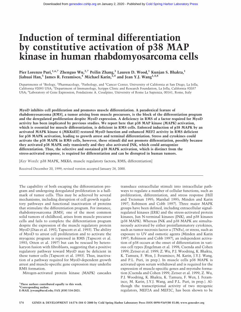
MyoD in the presence of serum can be demonstrated inmouse 10T1/2 fibroblasts growing in 20% FBS [growthmedium (GM)] (Fig. 1). Under these culture conditions,ectopic expression of either MKK6EE or MyoD alone didnot significantly activate the artificial promoter contain-ing four MyoD-binding sites, 4RE Luc (Fig. 1A), or theMyoD-dependent p21Cip1 promoter (p21 luc) (data notshown) and failed to promote efficient myogenic conver-sion (Table 1) and cell cycle arrest (Table 2). Coexpress-ing MyoD with MKK6EE synergistically activated
Figure 1. An activated mutant of MKK6EE stimulates thefunction of MyoD. (A) Activation of the MyoD-dependent pro-moter 4RE luc (four repeated MyoD-binding E-box sites) wasdetermined by cotransfection of 10T1/2 fibroblasts with theindicated expression plasmids. Cells were transfected in GMcontaining 20% FBS and harvested 36 hr later for luciferaseassay. Where indicated, the p38 MAPK inhibitor SB203580 (5µM) was added to the transfected cells. The luciferase activitywas normalized for the expression levels of MyoD in eachsample, and the fold activation was relative to vector-trans-fected sample. The means and standard deviations from threeindependent experiments are shown. (B) MKK6EE stimulatedthe activity of MyoD–Gal4 fusion proteins. Plasmids expressingthe indicated MyoD–Gal4 fusion proteins were cotransfectedwith a gal4–luc reporter gene plus a plasmid expressingMKK6EE (stippled and hatched bars) into 10T1/2 fibroblastsgrowing in 20% FBS. MKK6EE cotransfected cells were treatedwith 5 µM SB203580 (hatched bars). Cells were harvested 36 hrafter transfection for LUC assay. The luciferase activity wasnormalized for the expression levels of the transfected Gal4fusion proteins. The data presented are representative of fourindependent experiments.
Deficient activation of p38 MAPK in rhabdomyosarcoma cells
GENES & DEVELOPMENT 575
Cold Spring Harbor Laboratory Press on January 2, 2020 - Published by genesdev.cshlp.orgDownloaded from
MyoD-dependent promoters (Fig. 1A; data not shown)and stimulated both MyoD-dependent myogenic conver-sion (Table 1) and cell growth arrest (Table 2). Additionof a specific inhibitor of p38 MAPK SB203580 (SB), butnot the MEK1 inhibitor PD98059 (PD), eliminated thestimulatory effect of MKK6EE (Fig. 1; Tables 1 and 2),showing that p38 kinase is a necessary component in theactivation of MyoD function.
By coexpression of MKK6EE with Gal4–MyoD fusionmutants (Fig. 1B), we found that the basic region ofMyoD was dispensable for the MKK6EE-dependent tran-scriptional stimulation. Because the basic region ofMyoD is required for functional synergism with MEF2C(Molkentin et al. 1995; Black et al. 1998), which is di-rectly activated by p38 MAPK (Han et al. 1997; Yang etal. 1999; Zhao et al. 1999), this result suggested thatMKK6EE could stimulate MyoD function independentof MEF2C binding. In addition, MKK6EE could stimulatethe transactivation function of a Gal4–MyoD chimera,in which only the amino-terminal domain of MyoD wasfused to the Gal4 DNA-binding domain (Fig. 1B). As acontrol, a Gal4–E2F1 fusion chimera was not activatedsignificantly (less than twofold) by MKK6EE. This obser-vation suggests that the amino-terminal domain ofMyoD is one target, either direct or indirect, of the p38MAPK pathway. Taken together, these results show thatMKK6EE can activate the function of MyoD and suchactivation can occur in the presence of mitogens, whichare known to inhibit the differentiation function ofMyoD.
Differentiation-programmed activation of p38 MAPKpathway is deficient in RMS cells
Given its ability to stimulate MyoD in the presence ofmitogenic cues, we reasoned that MKK6EE might be able
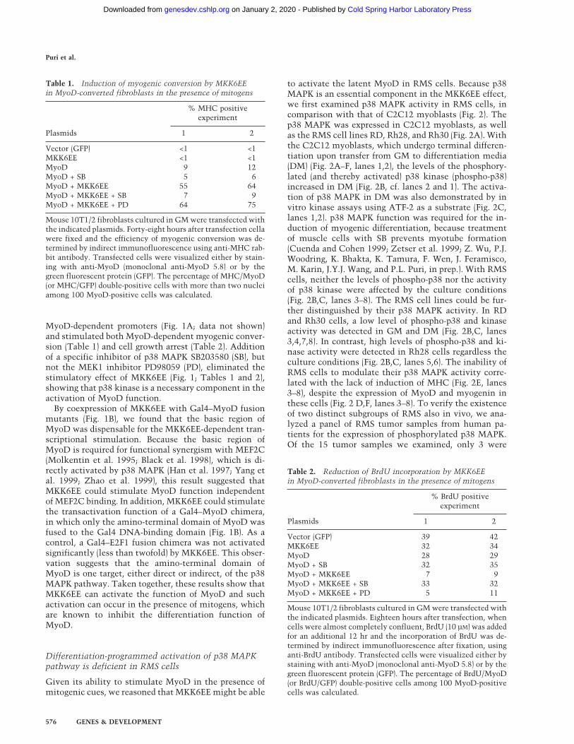
to activate the latent MyoD in RMS cells. Because p38MAPK is an essential component in the MKK6EE effect,we first examined p38 MAPK activity in RMS cells, incomparison with that of C2C12 myoblasts (Fig. 2). Thep38 MAPK was expressed in C2C12 myoblasts, as wellas the RMS cell lines RD, Rh28, and Rh30 (Fig. 2A). Withthe C2C12 myoblasts, which undergo terminal differen-tiation upon transfer from GM to differentiation media(DM) (Fig. 2A–F, lanes 1,2), the levels of the phosphory-lated (and thereby activated) p38 kinase (phospho-p38)increased in DM (Fig. 2B, cf. lanes 2 and 1). The activa-tion of p38 MAPK in DM was also demonstrated by invitro kinase assays using ATF-2 as a substrate (Fig. 2C,lanes 1,2). p38 MAPK function was required for the in-duction of myogenic differentiation, because treatmentof muscle cells with SB prevents myotube formation(Cuenda and Cohen 1999; Zetser et al. 1999; Z. Wu, P.J.Woodring, K. Bhakta, K. Tamura, F. Wen, J. Feramisco,M. Karin, J.Y.J. Wang, and P.L. Puri, in prep.). With RMScells, neither the levels of phospho-p38 nor the activityof p38 kinase were affected by the culture conditions(Fig. 2B,C, lanes 3–8). The RMS cell lines could be fur-ther distinguished by their p38 MAPK activity. In RDand Rh30 cells, a low level of phospho-p38 and kinaseactivity was detected in GM and DM (Fig. 2B,C, lanes3,4,7,8). In contrast, high levels of phospho-p38 and ki-nase activity were detected in Rh28 cells regardless theculture conditions (Fig. 2B,C, lanes 5,6). The inability ofRMS cells to modulate their p38 MAPK activity corre-lated with the lack of induction of MHC (Fig. 2E, lanes3–8), despite the expression of MyoD and myogenin inthese cells (Fig. 2 D,F, lanes 3–8). To verify the existenceof two distinct subgroups of RMS also in vivo, we ana-lyzed a panel of RMS tumor samples from human pa-tients for the expression of phosphorylated p38 MAPK.Of the 15 tumor samples we examined, only 3 were
Table 2. Reduction of BrdU incorporation by MKK6EEin MyoD-converted fibroblasts in the presence of mitogens
Plasmids
% BrdU positiveexperiment
1 2
Vector (GFP) 39 42MKK6EE 32 34MyoD 28 29MyoD + SB 32 35MyoD + MKK6EE 7 9MyoD + MKK6EE + SB 33 32MyoD + MKK6EE + PD 5 11
Mouse 10T1/2 fibroblasts cultured in GM were transfected withthe indicated plasmids. Eighteen hours after transfection, whencells were almost completely confluent, BrdU (10 µM) was addedfor an additional 12 hr and the incorporation of BrdU was de-termined by indirect immunofluorescence after fixation, usinganti-BrdU antibody. Transfected cells were visualized either bystaining with anti-MyoD (monoclonal anti-MyoD 5.8) or by thegreen fluorescent protein (GFP). The percentage of BrdU/MyoD(or BrdU/GFP) double-positive cells among 100 MyoD-positivecells was calculated.
Table 1. Induction of myogenic conversion by MKK6EEin MyoD-converted fibroblasts in the presence of mitogens
Plasmids
% MHC positiveexperiment
1 2
Vector (GFP) <1 <1MKK6EE <1 <1MyoD 9 12MyoD + SB 5 6MyoD + MKK6EE 55 64MyoD + MKK6EE + SB 7 9MyoD + MKK6EE + PD 64 75
Mouse 10T1/2 fibroblasts cultured in GM were transfected withthe indicated plasmids. Forty-eight hours after transfection cellawere fixed and the efficiency of myogenic conversion was de-termined by indirect immunofluorescence using anti-MHC rab-bit antibody. Transfected cells were visualized either by stain-ing with anti-MyoD (monoclonal anti-MyoD 5.8) or by thegreen fluorescent protein (GFP). The percentage of MHC/MyoD(or MHC/GFP) double-positive cells with more than two nucleiamong 100 MyoD-positive cells was calculated.
Puri et al.
576 GENES & DEVELOPMENT
Cold Spring Harbor Laboratory Press on January 2, 2020 - Published by genesdev.cshlp.orgDownloaded from

stained positive for phospho-p38, despite the normal ex-pression of total p38 in all the samples (P. Zhang and P.L.Puri, unpubl.), supporting the existence also in vivo oftwo distinct classes of RMS: either with or without ac-tivated p38 MAPK.
Previous studies have demonstrated that fusion of RDcells with 10T1/2 fibroblasts can rescue MyoD function(Tapscott et al. 1993). To determine whether the activa-tion of p38 MAPK is important for that rescue, hetero-karyons were prepared in the presence of SB (Fig. 2H).Similar to normal muscle cells (Cuenda and Cohen 1999;Zetser et al. 1999; Z. Wu, P.J. Woodring, K. Bhakta, K.Tamura, F. Wen, J. Feramisco, M. Karin, J.Y.J. Wang, andP.L. Puri, in prep.), the inactivation of p38 by SB reducedthe number of MHC-positive heterokarions. This inhibi-tory effect was not observed with vehicle (DMSO) orPD98059, which inhibits MEK1 (Fig. 2H). Thus, the p38MAPK activity was required for the rescue of MyoDfunction. A reduction in MHC-positive heterokaryonswas also observed by the ectopic expression of kinase-defective forms of MKK6 or MKK3 (another upstreamactivator of p38 MAPK) in 10T1/2 fibroblasts prior thefusion with RD cells (Fig. 2H). These results further sup-ported the notion that the provision of a functionalMKK3/6 to p38 MAPK pathway by fibroblasts is a criti-
cal event in the reactivation of the myogenic programin RMS on heterokaryon fusion.
Enforced activation of p38 MAPK pathway by MKK6overrides the differentiation block in RMS cells
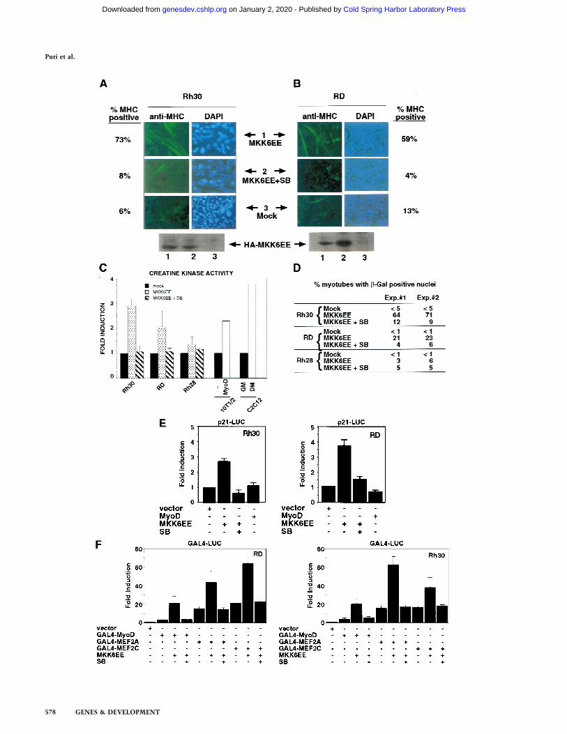
To activate p38 MAPK in RMS cells, we infected themwith an adenovirus expressing a HA-tagged MKK6EEprotein (Fig. 3A,B) (Huang et al. 1997). After infection,cells were cultured in DM and then examined for theinduction of MHC. Infection of Rh28 cells withMKK6EE adenovirus did not lead to an induction ofMHC expression (data not shown). Because the Rh28cells already contained a high level of phospho-p38 (Fig.2B, lanes 5,6), its differentiation defect was likely to beeither downstream to p38 MAPK activation or in an-other pathway. Hence, the resistance of Rh28 cells toMKK6EE was not unreasonable. In contrast, infection ofeither RD or Rh30 cells with MKK6EE adenoviruscaused several changes that were indicative of terminaldifferentiation (Fig. 3A,B). With Rh30 cells, infectionwith MKK6EE adenovirus followed by culture in DMaltered the morphology of the cells, which became largerand flatter, and induced the formation of multinucleatedcells, which expressed MHC (Fig. 3A). After four days of
Figure 2. Human RMS cell lines express p38 MAPK but do not modulate its activity under differentiation conditions. The levels ofp38 MAPK (A) and phosphorylated p38 MAPK (B) were determined by immunoblotting with the appropriate antibodies (see Materialsand Methods) in total lysates of mouse C2C12 (lanes 1,2) and three human RMS cell lines: RD (lanes 3,4), Rh28 (lanes 5,6), and Rh30(lanes 7,8). (GM) Growth medium; (DM) differentiation medium. (C) p38 MAPK activity was measured by an immunocomplex kinaseassay using GST-ATF2 fusion protein as a substrate. The levels of myogenin (D), MHC (E), MyoD (F), and actin (G) were alsodetermined by immunoblotting with the appropriate antibodies. The formation of heterokaryons of RD and 10T/12 fibroblasts (H) wasperformed as described previously (Tapscott et al. 1993). The heterokaryons were transferred into DM with (+SB) or without (−SB)SB203580 (5 µM) to inhibit p38 MAPK. DMSO and PD98059 were also used as negative controls. After 36 hr in DM, heterokaryonswere fixed and stained for the expression of MHC (top) and for the visualization of nuclei with DAPI (bottom). The different patternof nuclear staining–punctate for mouse nuclei in 10T1/2 cells and uniform staining for human nuclei in RD cells—indicates theformation of heterokaryons. The number of MHC-positive heterokaryons/field was scored and the results of two experiments aresummarized. In the case of MKK3KD and MKK6KD overexpression, these dominant-negative forms were expressed by transienttransfection in 10T1/2 prior the fusion with RD. A GFP-encoding plasmid was cotransfected to localize successfully transfected cells.The number of GFP/MHC-positive heterokaryons/field was scored and the results of two experiments are summarized.
Deficient activation of p38 MAPK in rhabdomyosarcoma cells
GENES & DEVELOPMENT 577
Cold Spring Harbor Laboratory Press on January 2, 2020 - Published by genesdev.cshlp.orgDownloaded from
Puri et al.
578 GENES & DEVELOPMENT
Cold Spring Harbor Laboratory Press on January 2, 2020 - Published by genesdev.cshlp.orgDownloaded from
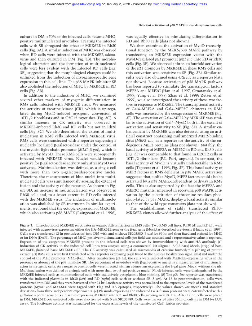
culture in DM, >70% of the infected cells became MHC-positive multinucleated myotubes. Treating the infectedcells with SB abrogated the effect of MKK6EE in Rh30cells (Fig. 3A). A similar induction of MHC was observedwhen RD cells were infected with the MKK6EE adeno-virus and then cultured in DM (Fig. 3B). The morpho-logical alteration and the formation of multinucleatedcells were less evident with the infected RD cells (Fig.3B), suggesting that the morphological changes could beunlinked from the induction of myogenic-specific geneexpression in this cell line. The p38 MAPK inhibitor SBalso abolished the induction of MHC by MKK6EE in RDcells (Fig. 3B).
In addition to the induction of MHC, we examinedseveral other markers of myogenic differentiation inRMS cells infected with MKK6EE virus. We measuredthe activity of creatine kinase (CK), which is up-regu-lated during MyoD-induced myogenic conversion of10T1/2 fibroblasts and in C2C12 myotubes (Fig. 3C). Asimilar increase in CK activity was observed inMKK6EE-infected Rh30 and RD cells but not in Rh28cells (Fig. 3C). We also determined the extent of multi-nucleation in RMS cells infected with MKK6EE virus.RMS cells were transfected with a reporter expressing anuclearly localized b-galactosidase under the control ofthe myosin light chain promoter (MLC–b-gal), which isactivated by MyoD. These RMS cells were subsequentlyinfected with MKK6EE virus. Nuclei would becomepositive for b-galactosidase activity only after MyoD wasactivated. Multinucleation was defined as a single cellwith more than two b-galactosidase-positive nuclei.Therefore, the measurement of blue nuclei into multi-nucleated cells was simultaneously scoring the extent offusion and the activity of the reporter. As shown in Fig-ure 3D, an increase in multinucleation was observed inRh30 cells and, to a lesser extent, in RD cells infectedwith the MKK6EE virus. The induction of multinucle-ation was abolished by SB treatment. In similar experi-ments, we found that the ectopic expression of MKK3EE,which also activates p38 MAPK (Raingeaud et al. 1996),
was equally effective in stimulating differentiation inRD and Rh30 cells (data not shown).
We then examined the activation of MyoD transcrip-tional function by the MKK6/p38 MAPK pathway bytransfecting an MKK6EE expression vector with theMyoD-regulated p21 promoter (p21 luc) into RD or Rh30cells (Fig. 3E). We observed a three- to fourfold activationof the p21 promoter by MKK6EE in these RMS cells andthis activation was sensitive to SB (Fig. 3E). Similar re-sults were also obtained using 4RE luc as a reporter (datanot shown). Because activation of p38 MAPK pathwayhas been reported to stimulate the transcription factorsMEF2A and MEF2C (Han et al. 1997; Ornatansky et al.1999; Yang et al 1999; Zhao et al. 1999; Zetser et al.1999), we also investigated the activity of these two fac-tors in response to MKK6EE. The transcriptional activityof Gal4–MEF2A and Gal4–MEF2C chimeras in RMScells was increased by the coexpression of MKK6EE (Fig.3F). The activation of Gal4–MEF2 by MKK6EE was simi-lar to the activation of Gal4–MyoD both in the extent ofactivation and sensitivity to SB (Fig. 3F). A similar en-hancement by MKK6EE was also detected using an arti-ficial construct containing multimerized MEF2-bindingsites (MEF2–luc) as a reporter for the activity of the en-dogenous MEF2 proteins (data not shown). Notably, thebasal activity of MEF2A or MEF2C in RD and Rh30 cells(Fig. 3F) was comparable to that found in C2C12 cells or10T1/2 fibroblasts (P.L. Puri, unpubl.). In contrast, thebasal activity of MyoD is virtually undetectable in RMScells (Tapscott et al. 1993; Fig. 3F). This basal activity ofMEF2 factors in RMS deficient in p38 MAPK activationsuggested that, unlike MyoD, MEF2 factors could also beactivated by a p38 MAPK-independent pathway in RMScells. This is also supported by the fact the MEF2A andMEF2C mutants, impaired in receiving p38 MAPK acti-vation by the substitution of threonine residues phos-phorylated by p38 MAPK, display a basal activity similarto that of the wild-type constructs (data not shown).
The establishment of stably transfected Rh30–MKK6EE clones allowed further analysis of the effect of
Figure 3. Introduction of MKK6EE reactivates myogenic differentiation in RMS cells. Two RMS cell lines, Rh30 (A) and RD (B), wereinfected with adenovirus expressing either the HA–MKK6EE gene or the b-gal gene (Mock) as described previously (Huang et al. 1997).Cells were transferred (12 hr postinfection) into DM with and without SB203580 (5 µM) for 96 hr and then fixed and stained for MHCor for DNA (DAPI). The percentage of MHC-positive multinucleated cells per field was counted and a representative value is reported.Expression of the exogenous MKK6EE proteins in the infected cells was shown by immunoblotting with anti-HA antibody. (C)Induction of CK activity in the indicated cell lines was assayed using a commercial kit (Sigma). (Solid bars) Mock; (stippled bars)MKK6EE; (hatched bars) MKK6EE + SB. The CK activity was calculated as micromoles of creatine formed/min per mg of proteinextract. (D) RMS cells were first transfected with a reporter expressing b-gal fused to the nuclear localization signal (nls) and under thecontrol of the MLC promoter (MLC–b-gal). After transfection (24 hr), the cells were infected with MKK6EE-expressing virus in thepresence or absence of the p38 inhibitor SB. The percentage of myotubes with b-gal-positive nuclei is a measurement of multinucle-ation in myogenic cells. As a negative control cells were infected with an adenovirus expressing the b-gal gene (Mock) without the nls.Multinucleation was defined as a single cell with more than two b-gal-positive nuclei. Mock-infected cells were distinguished by theMKK6EE-infected cells as mononucleated cells with exclusively cytoplasmic blue staining. (E) The p21 luc reporter was transfectedwith the indicated plasmids in Rh30 (left) and RD (right) cells with or without SB (5 µM). At 18 hr post transfection, cells weretransferred into DM and they were harvested after 24 hr. Luciferase activity was normalized to the expression levels of the transfectedproteins (MyoD and MKK6EE were tagged with Flag and HA epitopes, respectively). The values shown are means and standarddeviations from three independent experiments. (F) Plasmids expressing the indicated Gal4 fusion proteins were cotransfected with agal4–luc reporter gene plus a plasmid expressing MKK6EE into RD and Rh30 cells growing in 20% FBS. After 12 hr, cells were placedin DM. MKK6EE cotransfected cells were also treated with 5 µM SB203580. Cells were harvested after 36 hr of culture in DM for LUCassay. The luciferase activity was normalized for the expression levels of the transfected Gal4 fusion proteins.
Deficient activation of p38 MAPK in rhabdomyosarcoma cells
GENES & DEVELOPMENT 579
Cold Spring Harbor Laboratory Press on January 2, 2020 - Published by genesdev.cshlp.orgDownloaded from
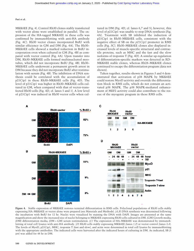
MKK6EE (Fig. 4). Control Rh30 clones stably transfectedwith vector alone were established in parallel. The ex-pression of the HA-tagged MKK6EE in these cells wasconfirmed by immunoblotting with anti-HA antibody(Fig. 4C). Rh30 vector clones incorporated BrdU withsimilar efficiency in GM and DM (Fig. 4A). The Rh30–MKK6EE cells showed a marked reduction in BrdU in-corporation even when cultured in GM (Fig. 4B) as com-pared with vector control (Fig. 4A). Upon transfer intoDM, Rh30–MKK6EE cells formed multinucleated myo-tubes, which did not incorporate BrdU (Fig. 4B). Rh30–MKK6EE cells underwent a permanent growth arrest inDM because they did not incorporate BrdU after restimu-lation with serum (Fig. 4B). The inhibition of DNA syn-thesis could be correlated with the accumulation ofp21Cip1 in these Rh30–MKK6EE cells (Fig. 4D). Thelevel of p21Cip1 was higher in Rh30–MKK6EE cells cul-tured in GM, when compared with that of vector-trans-fected Rh30 cells (Fig. 4D, cf. lanes 5 and 1). A low levelof p21Cip1 was induced in Rh30 vector cells when cul-
tured in DM (Fig. 4D, cf. lanes 6,7 and 5); however, thislevel of p21Cip1 was unable to stop DNA synthesis (Fig.4A). Treatment with SB inhibited the induction ofp21Cip1 in Rh30–MKK6EE cells, consistent with thenegative effect of SB on the p21Cip1 promoter in RMScells (Fig. 3C). Rh30–MKK6EE clones also displayed in-creased levels of muscle-specific structural and contrac-tile proteins, such as MHC and the fast and the slowisoforms of troponin T (Fig. 4D). A similar up-regulationof differentiation-specific markers was detected in RD–MKK6EE stable clones, whereas Rh28–MKK6EE clonescontinued to escape the differentiation program (data notshown).
Taken together, results shown in Figures 3 and 4 dem-onstrated that activation of p38 MAPK by MKK6EEcould restore MyoD activity and override the differentia-tion block in RMS cells, which do not contain an acti-vated p38 MAPK. The p38 MAPK-mediated enhance-ment of MEF2 activity could also contribute to the res-cue of the myogenic program in these RMS cells.
Figure 4. Stable expression of MKK6EE restores terminal differentiation in RMS cells. Polyclonal populations of Rh30 cells stablyexpressing HA–MKK6EE or vector alone were prepared (see Materials and Methods). (A,B) DNA synthesis was determined followingthe incubation with BrdU for 12 hr. Nuclei were visualized by staining the DNA with DAPI. Images are presented at the samemagnification and show the increased size of nuclei belonging to MKK6EE-expressing Rh30 cells cultured in DM. (GM) Growth media;(DM) differentiation media; (DM > GM) serum restimulation. (C) The expression of HA–MKK6EE was demonstrated by immunob-lotting of total cell lysates with anti-HA antibody. (D) Rh30 cells stably expressing MKK6EE (lanes 1,2) or vector control (lanes 3,4).The levels of MyoD, p21Cip1, MHC, troponin T (fast and slow), and actin were determined in total cell lysates by immunoblottingwith the appropriate antibodies. The indicated cells were harvested after the indicated hours of culturing in DM. As indicated, SB (5µM) was added for 48 hr in DM.
Puri et al.
580 GENES & DEVELOPMENT
Cold Spring Harbor Laboratory Press on January 2, 2020 - Published by genesdev.cshlp.orgDownloaded from
Differentiation-programmed and stress-induced p38MAPK activation are two distinct responses
We tested whether the activity of p38 MAPK could bestimulated in RMS cells by extracellular stress and pro-inflammatory cytokines, which are known to trigger thestress-activated JNK and p38 pathways (Minden and Ka-rin 1997; Robinson and Cobb 1997). Exposure to Sorbitol,UV, or TNFa could stimulate p38 MAPK (Fig. 5A, lanes2,3,4) to a similar extent as in normal fibroblasts (Fig. 5A,cf. lanes 2, 3, and 4 with 6, 7, and 8). However, stress orcytokine stimulation did not activate MyoD (Fig. 5B),nor did they relieve the differentiation block in RD cells(Fig. 5A,C). In other experiments, we have found that UVand TNFa actually repressed myogenic differentiation innormal myoblasts, despite a transient activation of p38MAPK (P.L. Puri, unpubl.). The inability of stress or cy-tokines to activate MyoD could be due to two reasons.First, these extracellular stimuli led only to a transientp38 MAPK activation (data not shown), which might notbe sufficient to activate MyoD and hence the myogenicprogram (Marshall 1995). Second, these extracellularstimuli also activated parallel pathways, which mightexert an anti-differentiation effect. Beside p38 MAPK,JNK is also activated by stress and cytokines (Mindenand Karin 1997). JNK stimulation leads to the activationof the Jun family of transcription factors, which have
been reported both to antagonize MyoD function and torepress myogenic differentiation (Bengal et al. 1992; Li etal. 1992). We found that TNFa and UV could indeedantagonize the positive effect of MKK6EE on myogenictranscription (Fig. 5D), supporting the notion that theycan activate an inhibitory pathway. Furthermore, the ec-topic expression of an activated form of JNKK2(JNKK2CA) (Wu et al. 1997), which induces JNK activity,partially abrogated the stimulatory effect of MKK6EE onMyoD function in RD cells (Fig. 5D). The anti-myogeniceffect of JNK can be also observed in muscle cell linesstably expressing JNKK2CA (Z. Wu. and P.L. Puri, un-publ.) We cannot be certain that JNK is the only inhibi-tory kinase responsible for the anti-myogenic effect ofTNFa and UV, because we have not been able to com-pletely inactivate the endogenous JNK in RMS cells byusing JNK and JNKK dominant negatives (data notshown). Nevertheless, our data demonstrate that theconstitutive activation of p38 MAPK by MKK6EE (orMKK3EE) was able to restore differentiation and that theactivation of JNK pathway by stress or an inflammatorycytokine is inhibitory to the positive effect of MKK6EE.
Discussion
We have shown that the ability of MyoD to stimulatemuscle-specific gene expression (Lassar et al. 1994) and
Figure 5. Activation of p38 MAPK in RD cells bystress and cytokines fails to induce myogenic differen-tiation. (A) The levels of phosphorylated (activated)p38 MAPK were determined by immunoblotting withthe appropriate antibodies (see Materials and Methods)in total lysate from RD and 10T1/2 cell lines afterexposure to several stress (0.4 M Sorbitol for 30 min; 10ng/ml TNFa for 15 min; 40 J/m2 UV, for 20 min). Thelevels of actin, MyoD, p21Cip1, and MHC were alsodetermined by immunoblotting with the appropriateantibodies (see Materials and Methods). The p38MAPK assay activity was measured by an immuno-complex kinase assay using GST–ATF2 fusion proteinas a substrate. (B) Activation of the MyoD-dependentpromoter 4RE luc in RD cells exposed or not to UV (40J/m2, 20 min) and TNFa (10 ng/ml for 15 min) wasdetermined by luciferase assay. Cells were transfectedin GM and, at 18 hr post-transfection, transferred intoDM, and then exposed to TNFa or UV. After an addi-tional 24 hr, cells were harvested and the luciferaseactivity calculated. The luciferase activity was nor-malized to the expression of cotransfected b-gal-encod-ing plasmid. The data presented is representative offour independent experiments. (C) RD cells were ex-posed or not to UV (40 J/m2, 20 min) and TNFa (10ng/ml for 15 min) and kept 36 hr in DM. After fixation,cells were stained for MHC expression (visualized byusing a fluorescein-conjugated secondary antibody).(D) Activation of the MyoD-dependent promoter 4REluc in RD cells exposed or not to UV (40 J/m2, 20 min)and TNFa (10 ng/ml for 15 min) was determined aftercotransfection with the indicated plasmids (0.2 µg
MKK6EE, 0.4 µg JNKK2CA). Cells were transfected in GM and, at 18 hr post-transfection, transferred into DM. After additional 24 hrcells were harvested and the luciferase activity calculated. The luciferase activity was normalized to the expression of cotransfectedb-gal-encoding plasmid. The data presented are representative of four independent experiments.
Deficient activation of p38 MAPK in rhabdomyosarcoma cells
GENES & DEVELOPMENT 581
Cold Spring Harbor Laboratory Press on January 2, 2020 - Published by genesdev.cshlp.orgDownloaded from
to suppress cell growth (Crescenzi et al. 1990; Sorrentinoet al. 1990), requires the activity of p38 MAPK. The dif-ferentiation-programmed pathway leading to p38 MAPKactivation is repressed in RMS cells, in which MyoD isinactive. Our results indicate that stimulation of the la-tent MyoD in RMS can be achieved by the enforced ac-tivation of p38 MAPK through the expression of an ac-tivated form of its upstream activator (either MKK6 orMKK3). The ectopic expression of MKK6EE in RMS celllines that do not contain activated p38 MAPK led to theinduction of morphological differentiation (formation ofmultinucleated myotubes), biochemical differentiation(increased activity of muscle creatine kinase), and in theup-regulation of contractile proteins, such as myosinheavy chain (MHC) and troponin T (fast and slow iso-forms). The activation of such a broad spectrum ofmuscle markers distinguished the effect of MKK6EEfrom the previously reported partial activation of myo-genic markers in some RMS cell lines by certain com-pounds, like TPA and INFa2a (Bouche et al. 1992; Thu-lasi et al. 1996). MKK6EE activates MyoD as well asMEF2 factors in RMS cells. Thus, the p38-dependent en-hancement of MEF2 activity could also contribute to therescue of the myogenic program in these RMS cells. No-tably, we noticed that, unlike MyoD, the basal activityof MEF2A and MEF2C in RD and Rh30 cells was notreduced as compared with that observed in normal fibro-blasts or myocytes. This implicates a p38-independentbasal activation of MEF2 factors in RMS that may ex-plain why these cells display normal levels of certainmyogenic markers, like myogenin, whose promoter ismainly regulated by MEF2-binding sites (Yee and Rigby1993).
In contrast to MKK6EE and MKK3EE, overexpressionof the p38 isoforms a, b, g, and d, either alone or incombination, failed to restore the differentiation pro-gram in RMS (data not shown), indicating that the path-way leading to MyoD activation is silenced upstream ofp38 MAPK. In addition, the ectopic expression of thewild-type MKK6 also failed to rescue the RMS defects(data not shown), indicating a block that is also upstreamof MKK6. However, because MKK6EE can override thedifferentiation block in RD and Rh30 RMS cells, thereare no defects downstream of MKK6 in these two RMScells. The differentiation defect with the Rh28 cells, onthe other hand, appears to lie either downstream of p38MAPK or in parallel to this pathway. Based on the failureof RD/Rh28 heterkaryons to rescue their latent MyoD,Tapscott et al. (1993) have proposed previously thatthese two tumor cell lines may share a common geneticdefect. Our data show that the defect in RD cells is re-cessive to the constitutive activation of p38 MAPK,whereas the genetic defect of Rh28 cells is dominantover the effect of MKK6EE. Because Rh28 cells, whichhave constitutively high p38 MAPK activity, could notcomplement the defect of RD cells in heterokaryon ex-periments, it is possible that additional anti-myogenicpathways, such as those activated by stress or cytokines(i.e., JNK pathway), might also be constitutively acti-vated in Rh28 cells. Alternatively, the p38 MAPK of
Rh28 cells might be activated by an MKK3/6-indepen-dent pathway, which does not exert the same myogeniceffect. Furthermore, inhibitory mechanism(s) down-stream to p38 MAPK could also operate in Rh28 cells toblock differentiation. The presence of any of these in-hibitory mechanisms, either alone or in combination, inRh28 cells might explain the failure to achieve comple-mentation between Rh28/RD heterokaryon fusions.
Interestingly, the activity of p38 MAPK can be stimu-lated in RMS cells by extracellular stress and cytokines(Sorbitol, TNFa, UV), which activate a number of MAPKpathways in addition to p38 MAPK. However, stress orcytokines did not induce RMS differentiation. This ob-servation implies the existence of two distinct programsleading to the activation of the MKK6–p38 MAPK path-way. The first is the prototypical stress-activated pro-gram, which is transient and is still functional in RMS.An alternative program that is triggered in myogeniccells by a differentiation cue yet to be identified, stimu-lates persistent p38 MAPK activation, and is blocked ina majority of RMS. The expression of MKK6EE wouldmimic the differentiation-programmed activation of p38MAPK. By contrast, activation of p38 MAPK by stressand proinflammatory cytokines is part of a stress-acti-vated response that does not lead to induction of differ-entiation. The failure of these extracellular stimuli torestore myogenic differentiation in RMS therefore couldbe due to two factors: the transient activation of p38MAPK, and the activation of a parallel pathway that ex-erts anti-myogenic activity. Thus, the sustained and se-lective activation of the MKK6–p38 pathway, either bypharmacological agents or gene therapy approach, mightbe useful to induce the differentiation and thus inhibitthe proliferation of RMS.
Although the induction of terminal differentiation byMKK6EE in RMS correlates with the MyoD-dependentincrease of p21Cip1 (Figs. 3C and 4D), the accumulationof p21Cip1 cannot entirely account for the effect inducedby MKK6EE in these cells. Overexpression of p21Cip1 inRMS cells placed in DM could only cause a serum-re-versible cell cycle arrest (P.L. Puri, unpubl.), without in-ducing the expression of differentiation (Knudsen et al.1998). Therefore, transient cell cycle arrest and acquisi-tion of a differentiated phenotype are two separable pro-cesses not only in normal myocytes (Puri et al. 1997a)but also in RMS cells. Activation of p38 MAPK byMKK6/3 might stimulate terminal differentiation inRMS cells by coupling these two processes. Consistentwith our finding, Ellinger-Ziegelbauer et al. (1999) havereported that a conditionally activated form of anotherupstream activator of p38 MAPK (MEKK3) can inducecell cycle arrest and reverse Ras-induced transformation,further implying p38 MAPK pathway as a negative regu-lator of cell growth in transformed cells. Although inRMS cells p38 MAPK is likely to exert its growth-sup-pressive effect by restoring MyoD activity, the molecularbasis underlying such a effect are still unknown. Ourdata (Fig. 1B) indicate that p38 MAPK stimulates MyoDactivity by a mechanism that does not involves the in-teraction with the p38 MAPK substrate MEF2. More-
Puri et al.
582 GENES & DEVELOPMENT
Cold Spring Harbor Laboratory Press on January 2, 2020 - Published by genesdev.cshlp.orgDownloaded from
over, we failed to detect a p38 MAPK-dependent activa-tion of MyoD through direct phosphorylation (data notshown). Thus, p38 MAPK could indirectly activateMyoD by targeting bHLH-interacting proteins or cofac-tors like p300 and PCAF (Puri et al. 1997b).
Our results may also have interesting implications indiagnostic typing of RMS. The conventional histologicaltyping of RMS distinguishes between embryonal, alveo-lar, and botryoid forms, based on morphological features(Arndt and Crist 1999). Consistent with the results ob-tained in RMS cell lines, we have found, by examiningthe p38 MAPK status in RMS tumor samples, that amajority of the RMS tumors did not contain the acti-vated form of p38 MAPK regardless their histotype (P.Zhang and P.L. Puri, unpubl.). We propose that analysisof p38 MAPK might be a tool to identify RMS that aresensitive to therapeutic strategy based on the enforcedactivation of p38 MAPK.
Materials and methods
Cell culture conditions, transfections, and luciferase assay
Mouse C2C12 and 10T1/2 cells were cultured in DMEMsupplemented with either 20% FBS (GM) or 2% horse serum(DM). Human RMS cells (RD, Rh30, and Rh28), kindly providedby K. Arden (Ludwig Cancer Research Institute, University ofCalifornia, San Diego), were cultured in RPMI 1640 DMEMsupplemented with either 20% FBS (GM) or 2% horse serum(DM). Polyclonal population of Rh30, RD, and Rh28 cells stablyexpressing MKK6EE was prepared by transfecting these cellswith pcDNA3–HA–MKK6EE, followed by selection in G418(0.5 mg/ml) for 3 weeks. Ten clones were expanded and eachtested for the expression of HA–MKK6EE and for p38 kinaseactivity and the positive clones were pooled. The experimentswere done at the first three passages of these clones (at laterpassages the expression of HA–MKK6EE and the p38 MAPKactivity decrease). RD-10T1/2 heterokaryons were prepared asdescribed (Tapscott et al. 1993). Transfections were performedusing lipofectamine reagent (GIBCO) and luciferase assays wereperformed as described previously (Puri et al. 1997b). The kinaseinhibitors SB203580 and PD98059 were purchased from Calbio-chem.
BrdU incorporation
BrdU incorporation was determined by indirect immunofluo-rescence using anti-BrdU antibodies, as described previously(Puri et al. 1997b).
Muscle creatine kinase assay
The activity of the enzyme creatine phosphokinase (CPK) wasmeasured following the procedures of the commercial kit fromSigma. Cells were washed in TBS and the cell extracts used forthe assay were prepared in TBS-containing buffer with 1%Tween, phosphatase, and protease inhibitors.
Immunoblot analysis
Endogenous myogenin, MHC, MyoD, and actin of C2C12 andRMS cell lines were detected by immunoblotting using mono-clonal anti-myogenin (F5D), anti-MyoD (5.8), anti-MHC(MF20), and anti-actin (Ab-1, Oncogene). Levels of either total or
phosphorylated p38 in the same cell lines were detected by im-munoblotting using anti-p38 antibodies from New England Bio-labs. Endogenous p21Cip1 levels in Rh30 cell clones were de-tected by using polyclonal anti-p21 (Ab-5, Oncogene). Anti- tro-ponin T fast (C-18) and slow (C-19) antibodies were purchasedfrom Santa Cruz.
Kinase assay
In vitro kinase assays were performed as described previously(Wu et al. 1997). Briefly, 50- to 100-µg cell extracts were incu-bated with antibodies against p38a in the presence of 30 µl ofprotein A–Sepharose beads for 2 hr at 4°C. After extensive wash-ing, the immunocomplexes were incubated with 1 µg of GST–ATF in the presence of 10 µCi of [g-32P] ATP in 25 µl of kinasebuffer for 20 min at 30°C, analyzed by SDS-PAGE, and visual-ized by autoradiography.
Immunofluorescence
Cells were fixed in 3.3% formaldehyde for 10 min and perme-abilized with 0.25% Triton X-100 for additional 10 min. Indirectimmunofluorescence was carried out using polyclonal rabbitanti-MHC as described previously (Puri et al. 1997b)
Adenoviral infections
Adenoviral infection was performed in confluent RMS cells asdescribed previously (Huang et al. 1997).
Acknowledgments
We thank Dr. Karen Arden, Dr. Vittorio Sartorelli, Dr. StefanoAlema, Dr. Peter Houghton and Dr. Woodring E. Wright forgenerously providing reagents. Dr. Vittorio Sartorelli and Dr.Gioacchino Natoli are also acknowledged for their commentsand helpful suggestions during the preparation of the manu-script. This work was supported by fellowships from MedicalResearch Council of Canada (Z.W.), from American Cancer So-ciety (L.D.W.), and by grants from Telethon (P.L.P.), from NIHState of California Cancer Research Program (M.K.), and fromNCI (J.Y.J.W.).
The publication costs of this article were defrayed in part bypayment of page charges. This article must therefore be herebymarked “advertisement” in accordance with 18 USC section1734 solely to indicate this fact.
References
Alema, S. and F. Tato. 1994. Oncogenes and muscle differentia-tion: Multiple mechanism of interference. Semin. CancerBiol. 5: 147–156.
Arndt, C.A.S. and W.M. Crist. 1999. Medical progress: Commonmusculoskeletal tumors of childhood and adolescence. N.Engl. J. Med. 341: 342–353.
Arnold, H.-H. and B. Winter. 1998. Muscle differentiation: Morecomplexity to the network of myogenic regulators. Curr.Opin. Genet. Dev. 8: 539–544.
Bengal, E., L. Ransone, R. Scharfmann, V.J. Dwarki, S.J. Tap-scott, H. Weintraub, and I.M. Verma. 1992. Functional an-tagonism between cJun and MyoD proteins: a direct physicalassociation. Cell 68: 507–519.
Black, B.L., J.D. Molkentin, and E. Olson. 1998. Multiples rolesfor the MyoD basic region in transmission of transcriptionalactivation signals and interaction with Mef2. Mol. Cell. Biol.18: 69–77.
Deficient activation of p38 MAPK in rhabdomyosarcoma cells
GENES & DEVELOPMENT 583
Cold Spring Harbor Laboratory Press on January 2, 2020 - Published by genesdev.cshlp.orgDownloaded from
Bouche, M., F. Zappelli, M. Polimenu, S. Adamo, W.C. Wetsel,M.I. Senni, and M. Molinaro. 1992. Rapid activation anddownregulation of protein kinase C in 12-O-tetradec-anoylphorbol-13-acetate-induced differentiation of humanrhabdomyosarcoma cells. Cell Growth Differ. 6: 845–852.
Crescenzi, M., T.P. Fleming, A.B. Lassar, H. Weintraub, and A.S.Aaronson. 1990. MyoD induces growth arrest independentof differentiation in normal and transformed cells. Proc.Natl. Acad. Sci. 87: 8442–8446.
Cuenda, A. and P. Cohen. 1999. Stress activated protein ki-nase-2 /p38 and a rapamycin-sensitive pathway are requiredfor C2C12 myogenesis. J. Biol. Chem. 274: 4341–4346.
Dias, P., D.M. Parham, D.N. Shapiro, S.J. Tapscot, and P.J.Houghton. 1992. Monoclonal antibodies to the myogenicregulatory protein MyoD1: Epitope mapping and diagnosticutility. Cancer Res. 52: 6431–6439.
Ellinger-Ziegelbauer, H., K. Kelly, and U. Siebenlist. 1999. Cellcycle arrest and reversion of Ras-induced transformation bya conditionaly activated form of mitogen-activated proteinkinase kinase kinase 3. Mol. Cell. Biol. 19: 3857–3868.
Engelman, J.A., M.P. Lisanti, and P.E. Scherer. 1998. Specificinhibitors of p38 mitogen-activated protein kinase block3T3-L1 adipogenesis. J. Biol. Chem. 27: 32111–32120.
Florini, J.R., D.Z. Ewton, and S.A. Coolican. 1996. Growth hor-mone and the insulin-like growth factor system in myogen-esis. Endocr. Rev. 17: 481–517.
Han, J., J.D. Lee, Y. Jiang, Z. Li, L. Feng, and R.J. Ulevitch. 1996.Characterization of the structure and function of a novelMAP kinase kinase (MKK6). J. Biol. Chem. 271: 2886–2891
Han, J., Y. Jiang, Z. Li, V.V. Kravchenko, and R.J. Ulevitch. 1997.Activation of the transcription factor Mef2c by the MAPkinase p38 in inflammation. Nature 368: 296–299.
Hill, C.S and R. Treisman. 1995. Transcriptional regulation byextracellular signals: Mechanism and specificity. Cell80: 199–211.
Huang, S., Y. Jang, Z. Li, E. Nishida, P. Mathias, S. Lin, R.J.Ulevitch, G.R. Nemerow, and J. Han. 1997. Apoptosis sig-nalling pathway in T cells is composed of ICE/CED-3 familyproteases and MAP kinase kinase 6b. Immunity 6: 739–749.
Knudsen, E.S., C. Pazzagli, T.L. Born, B.L. Bertolaet, K.E. Knud-sen, K.C. Arden, R.R. Henry, and J.R. Feramisco. 1998. El-evated cyclins and cyclin dependent kinase activity in rhab-domyosarcoma cell line RD. Cancer Res. 58: 2042–2049.
Lassar, A.B., S.X. Skapek, and B. Novich. 1994. Regulatorymechanisms that coordinate skeletal muscle differentiaionand cell cycle withdrawal. Curr. Opin. Cell. Biol. 6: 788–794.
Li, L., J.C. Chambard, M. Karin, and E. Olson. 1992. Fos and Junrepress transcriptional activation by myogenin and MyoD:the amino terminus of Jun can mediate repression. Genes &Dev. 6: 676–689.
Marshall, C.J. 1995. Specificity of receptor tyrosine kinase sig-nalling: Transient versus sustained extracellular signal-regu-lated kinase activation. Cell 80: 179–185.
Minden, A. and M. Karin. 1997. Regulation and function of theJNK subgroup of MAP kinases. Biochim. Biophys. Acta1333: F85–F104.
Molkentin, J.D., B.L. Black, J.F. Martin, and E.N. Olson. 1995.Cooperative activation of muscle gene expression by Mef2and myogenic bHLH proteins. Cell 83: 1125–1136.
Olson, E.N. 1992. Interplay between proliferation and differen-tiaiton within the myogenic lineage. Dev. Biol. 154: 261–272.
Ornatansky, O.I., D.M. Cox, P. Tangirala, J.J. Andreucci, Z.A.Quinn, J.L. Wrana, R. Prywes, Y.-T. Yu, and J. McDermott.1999. Posttranscriptional control of the MEF2A transcrip-
tional regulatory protein. Nucleic Acids Res. 21: 2646–2656.Otten, A.D., E.J. Firpo, A.N. Gerber, L.L. Brody, J.M. Roberts,
and S.J. Tapscott. 1997. Inactivation of MyoD mediated ex-pression of p21 in tumor cell lines. Cell Growth & Diff.8: 1151–1160.
Puri, P.L., C. Balsano, V.L. Burgio, P. Chirillo, G. Natoli, L.Ricci, E. Mattei, A. Graessmann, and M. Levrero. 1997a.MyoD prevents cyclinA/cdk2 containing E2F complex for-mation in terminally differentiated myocytes. Oncogene14: 1171–1184.
Puri, P.L., V. Sartorelli, X.-J. Yang, Y. Hamamori, V.V. Ogryzko,B.H. Howard, L. Kedes, J.Y.J. Wang, A. Graessmann, Y. Na-katani, and M. Levrero. 1997b. Differential roles of p300 andPCAF acetyltransferases in muscle differentiation. Mol. Cell1: 35–45,
Raingeaud, J., A.J. Whitmarsh, T. Barret, B. Derijard, and R.Davis. 1996. MKK3- and MKK6-regulated gene expression ismediated by the p38 mitogen activated protein kinase signaltransduction pathway. Mol. Cell. Biol. 16: 1247–1255.
Robinson, M.J. and M.H. Cobb. 1997. Mitogen-activated proteinkinase pathways. Curr. Opin. Cell Biol. 9: 180–186.
Sorrentino, V., R. Peppekok, R.L. Davis, W. Ansorge, and L.Philipson. 1990. Cell proliferation inhibited by MyoD inde-pendent of differentiation. Nature 345: 813–815.
Tapscott, S.J., M.J. Thayer, and H. Weintraub. 1993. Deficiencyin Rhabdomyosarcoma of a factor required for MyoD activ-ity and myogenesis. Science 259: 1450–1453.
Thulasi, R., P. Dias, P.J. Houghton, and J.A. Houghton. 1996.A2a-Interferon-induced differentiation of human alveolarrhabdomyosarcoma cells: Correlation with down-regulationof the Insulin-like growth factor type I receptor. Cell Growth& Differ. 7: 531–541.
Walsh, K. and H. Perlman. 1997. Cell cycle exit upon myogenicdifferentiation. Curr. Opin. Genet. Dev. 7: 597–602.
Wu, Z, J. Wu, E. Jacinto, and M. Karin. 1997. Molecular cloningand characterization of human JNKK2, a novel Jun NH2-terminal kinase-specific kinase. Mol. Cell. Biol. 17: 7407–7416.
Yang, S.H., A. Galanis, and A.D. Sharrocks. 1999. Targeting ofp38 mitogen activated protein kinases to MEF2 transcriptionfactors. Mol. Cell. Biol. 19: 4028–4038.
Yee, S.P. and P.W. Rigby. 1993. The regulation of the myogeningene expression during embryonic development of themouse. Genes & Dev. 7: 1277–1289.
Zhao, M., L. New, V.V. Kravchenko, Y. Kato, H. Gram, F. diPadova, E.N. Olson, R.J. Ulevitch, and J. Han. 1999. Regula-tion of the Mef2 family of transcription factors by p38. Mol.Cell. Biol. 19: 21–30.
Zetser, A., E. Gredinger, and E. Bengal. 1999. p38 mitogen-acti-vated protein kinase pathway promotes skeletal muscle dif-ferentiation. J. Biol. Chem. 274: 5193–5200.
Puri et al.
584 GENES & DEVELOPMENT
Cold Spring Harbor Laboratory Press on January 2, 2020 - Published by genesdev.cshlp.orgDownloaded from
10.1101/gad.14.5.574Access the most recent version at doi: 14:2000, Genes Dev.
Pier Lorenzo Puri, Zhenguo Wu, Peilin Zhang, et al. MAP kinase in human rhabdomyosarcoma cellsInduction of terminal differentiation by constitutive activation of p38
References
http://genesdev.cshlp.org/content/14/5/574.full.html#ref-list-1
This article cites 36 articles, 17 of which can be accessed free at:
License
ServiceEmail Alerting
click here.right corner of the article or
Receive free email alerts when new articles cite this article - sign up in the box at the top
Cold Spring Harbor Laboratory Press
Cold Spring Harbor Laboratory Press on January 2, 2020 - Published by genesdev.cshlp.orgDownloaded from


![Induction of Erythroid Differentiation in Human Leukemic K ......[CANCER RESEARCH 50, 1231-1236. February 15. 1990] Induction of Erythroid Differentiation in Human Leukemic K-562 Cells](https://img.dokumen.tips/doc/110x75/60b088961b1fcf1e2a746f9b/induction-of-erythroid-differentiation-in-human-leukemic-k-cancer-research.jpg)