Embed Size (px)
Citation preview

Top Heterocycl Chem (2010) 24: 177–204DOI: 10.1007/7081_2010_31# Springer-Verlag Berlin Heidelberg 2010Published online: 3 June 2010
Indoles and Related Heterocycles
Hemraj Juwarker, Jae-min Suk, and Kyu-Sung Jeong
Abstract Indole and the related heterocycles have emerged as efficient building
blocks for the creation of novel anion receptors. This chapter focuses on the
employment of indoles, carbazoles, and indolocarbazoles in the creation of molec-
ular clefts, macrocycles, oligomers, and sensors. The majority of these structures
utilize the heterocyclic NH as a hydrogen bond donor in binding to anions of
various sizes and geometries. These heterocycles are often connected by amides
and ureas which also function as hydrogen bond donors in anion binding. Finally,
molecular sensors based on indoles and carbazoles have been described, showing
color or fluorescence changes upon anion binding or deprotonation of the hetero-
cyclic NH by basic anions.
Keywords Anion recognition � Carbazole �Hydrogen bond � Indole � Indolocarbazole �Macrocycle � Molecular cleft � Molecular sensor � Oligomer
Contents
1 Introduction . . . . . . . . . . . . . . . . . . . . . . . . . . . . . . . . . . . . . . . . . . . . . . . . . . . . . . . . . . . . . . . . . . . . . . . . . . . . . . . 178
2 Molecular Clefts . . . . . . . . . . . . . . . . . . . . . . . . . . . . . . . . . . . . . . . . . . . . . . . . . . . . . . . . . . . . . . . . . . . . . . . . . . 180
2.1 Indole–Amide Hybrids . . . . . . . . . . . . . . . . . . . . . . . . . . . . . . . . . . . . . . . . . . . . . . . . . . . . . . . . . . . . . . 180
2.2 Indole–Urea Hybrids . . . . . . . . . . . . . . . . . . . . . . . . . . . . . . . . . . . . . . . . . . . . . . . . . . . . . . . . . . . . . . . . 183
2.3 Carbazoles . . . . . . . . . . . . . . . . . . . . . . . . . . . . . . . . . . . . . . . . . . . . . . . . . . . . . . . . . . . . . . . . . . . . . . . . . . 186
2.4 Indolocarbazoles . . . . . . . . . . . . . . . . . . . . . . . . . . . . . . . . . . . . . . . . . . . . . . . . . . . . . . . . . . . . . . . . . . . . 187
2.5 Others . . . . . . . . . . . . . . . . . . . . . . . . . . . . . . . . . . . . . . . . . . . . . . . . . . . . . . . . . . . . . . . . . . . . . . . . . . . . . . . 190
3 Macrocycles . . . . . . . . . . . . . . . . . . . . . . . . . . . . . . . . . . . . . . . . . . . . . . . . . . . . . . . . . . . . . . . . . . . . . . . . . . . . . . 191
3.1 Anion Receptors . . . . . . . . . . . . . . . . . . . . . . . . . . . . . . . . . . . . . . . . . . . . . . . . . . . . . . . . . . . . . . . . . . . . 192
3.2 Ion Pair Receptor . . . . . . . . . . . . . . . . . . . . . . . . . . . . . . . . . . . . . . . . . . . . . . . . . . . . . . . . . . . . . . . . . . . 193
4 Acyclic Oligomers . . . . . . . . . . . . . . . . . . . . . . . . . . . . . . . . . . . . . . . . . . . . . . . . . . . . . . . . . . . . . . . . . . . . . . . . 193
4.1 Oligoindoles . . . . . . . . . . . . . . . . . . . . . . . . . . . . . . . . . . . . . . . . . . . . . . . . . . . . . . . . . . . . . . . . . . . . . . . . 195
H. Juwarker, J.-M. Suk, and K.-S. Jeong (*)
Department of Chemistry, Yonsei University, 134, Sinchon-Dong, Seodaemun-Gu, Seoul, Korea
e-mail: [email protected]

4.2 Oligoindolocarbazoles . . . . . . . . . . . . . . . . . . . . . . . . . . . . . . . . . . . . . . . . . . . . . . . . . . . . . . . . . . . . . . 196
5 Molecular Sensors . . . . . . . . . . . . . . . . . . . . . . . . . . . . . . . . . . . . . . . . . . . . . . . . . . . . . . . . . . . . . . . . . . . . . . . . 198
5.1 Indole-Based Sensors . . . . . . . . . . . . . . . . . . . . . . . . . . . . . . . . . . . . . . . . . . . . . . . . . . . . . . . . . . . . . . . 198
5.2 Carbazole-Based Sensors . . . . . . . . . . . . . . . . . . . . . . . . . . . . . . . . . . . . . . . . . . . . . . . . . . . . . . . . . . . 200
6 Conclusions . . . . . . . . . . . . . . . . . . . . . . . . . . . . . . . . . . . . . . . . . . . . . . . . . . . . . . . . . . . . . . . . . . . . . . . . . . . . . . . 202
References . . . . . . . . . . . . . . . . . . . . . . . . . . . . . . . . . . . . . . . . . . . . . . . . . . . . . . . . . . . . . . . . . . . . . . . . . . . . . . . . . . . . 203
Abbreviations
AcO Acetate
Bz Benzyl
BzO Benzoate
CD Circular dichroism
CIC Chloride ion channel
CIS Complexation induced shift
DABCO 1,4-Diazabicyclo[2.2.2]octane
DMSO Dimethyl sulfoxide
Ka Association constant
Kd Dissociation constant
NMR Nuclear magnetic resonance
NrtA Nitrate-binding protein
phen Phenanthroline
SBP Sulfate-binding protein
Ser Serine
TBA Tetrabutylammonium
THF Tetrahydrofuran
Trp Tryptophan
UV/Vis Ultraviolet/visible
1 Introduction
Molecular or ionic recognition is a fundamental event in biochemical processes
which include transport, signaling, transcription, and catalysis. Over the last four
decades, chemists have devoted their efforts toward the creation of synthetic
molecules that can imitate and reproduce the molecular recognition phenomenon
observed in biological systems. In the design of synthetic receptors, the most
important task is choosing the noncovalent interactions to be employed in the
binding between a receptor and a substrate. This is controlled by the careful
selection of functional groups in the synthesis of the receptor. In particular,
hydrogen bonding has been proven as a prevalent and powerful noncovalent
interaction employed by synthetic receptors for anion binding, as anions serve as
good hydrogen bonding acceptors.
178 H. Juwarker et al.

The hydrogen bond has been known to be strongly electrostatic in nature [1]. It is
also viewed as an incipient proton transfer reaction, and more advanced proton
transfer gives rise to stronger hydrogen bonds [2]. In this context, hydrogen bonding
strength has often been correlated to the acidity and basicity of the interacting
partners; in general, the more acidic proton serves as the better hydrogen bonding
donor, and the more basic atom functions as the better acceptor. Figure 1 shows the
pKa values in DMSO of the representative molecules containing the functional
groups that serve as good hydrogen bond donors.
Indole is a heterocyclic aromatic compound with a good hydrogen bond donor
NH. Since indole is a key component of the amino acid tryptophan, it is not
surprising to find some examples of proteins that employ the indole NH for anion
binding. For example, sulfate-binding protein (SBP) of Salmonella typhimiriumsequesters sulfate dianion with a dissociation constant (Kd) of 0.17 mM by a total of
seven hydrogen bonds, formed with five amide NHs of the backbone, one indole
NH of Trp192 side chain, and one OH of Ser130 [5, 6]. The next example is nitrate-
binding protein, NrtA from Synechocystis sp. PCC 6803. The nitrate ion binds with
Kd = 0.3 mM by electrostatic interactions and six hydrogen bonds including one
hydrogen bond with the indole NH of Trp102 [7, 8] (Fig. 2).
It is evident that several hydrogen bond donors are present in the anion binding
sites of proteins, which operate together in a convergent and cooperative manner to
achieve strong and selective binding to a specific anionic substrate. In this regard,
chemists have designed and prepared anion receptors that contain multiple func-
tional groups capable of hydrogen bonding with anions. While indoles are prevalent
in the synthesis of natural products and pharmaceuticals, they have only recently
been recognized as useful building blocks for the construction of synthetic anion
receptors. Since 2004, anion receptors containing indole, carbazole and indolocar-
bazole moieties have been described, but this field is still in its infancy [9]. Details
studied to date will be described in this chapter by classifying the type of anion
receptors as molecular clefts, macrocycles, acyclic oligomers, and molecular
sensors.
H3C H3CN
O
N
S
N N
O
N N
S
25.5 18.5 26.9 21.0
N NN
23.0 21.0 19.9
H
H
H
H H
H
H
H
H
H
H
H
HHH
Fig. 1 pKa values in DMSO of representative molecules used as hydrogen bond donors in
synthetic anion receptors [3, 4]
Indoles and Related Heterocycles 179

2 Molecular Clefts
A general class of anion receptors is molecular clefts. These can be defined as
acyclic, concave molecules with cavities or indentations in the molecular surface,
into which a guest can fit but is not completely encapsulated [10, 11]. They are
often, if not always, characterized by convergent functional groups directed toward
each other, but separated by an appropriate linker, thus providing the space for
anion binding. Molecular clefts are more synthetically feasible than macrocycles,
and serve as platforms on which to modulate key structural elements before
synthetic expansion into more complex structures. In the following examples, it
can be noticed that indoles and related heterocycles are linked together by amides
and ureas that afford additional hydrogen bonding sites for anions, thus increasing
binding strength and selectivity.
2.1 Indole–Amide Hybrids
Amides have been the most popular functional group employed in the creation of
synthetic receptors for anions. They have found utility in linking together indole
units while simultaneously providing additional NHs for anion binding, thus max-
imizing interactions with anions.
Anion receptors 1 and 2 containing two indole groups attached to pyridine-2,
6-carboxamide and isophthalimide cores were synthesized by Gale and coworkers
in 2007 [12]. Receptor 1 possesses a binding cavity preorganized by intramolecular
N(pyridine)lllHN(amide) hydrogen bonds. 1 binds anions in the order of fluoride
(Ka > 104 M�1) � acetate (250 M�1) > dihydrogen phosphate (70 M�1) >chloride (<10M�1) with 1:1 stoichiometry in 0.5% water/DMSO. Crystal structures
NH
Trp192N
H
O
NH
O
HN
O
N O
O
H
Ser45
O
NH
Asp11
N
H
OAla173
Ser130
Ala133
S
O
O
NH
N+ NHH
H3N+
Lys269
NH2
O
Gln155
NH
O
Gly240N
O
O O
His196
Trp102Gly132
HO-
H
O-
Fig. 2 Hydrogen-bonding modes of two anion binding proteins utilizing the indole NH: sulfate-
binding protein (left) and nitrate-binding protein (right). Hydrogen bonds are marked as dotted line[5–8]
180 H. Juwarker et al.

of complex 1lTBA+F� show a “twisted” conformation where fluoride forms four
hydrogen bonds with all the receptor NHs. Relative to 1, receptor 2 showed slightly
higher binding affinities; the association constants (Ka, 0.5% water/DMSO) for
acetate, dihydrogen phosphate and benzoate were 880, 1,140, and 120 M�1, respec-
tively. 2 formed 1:1 complexes with these anions but formed 1:2 complexes with
fluoride (K1 ¼ 940 M�1, K2 ¼ 21 M�1 in 5% water/DMSO). The same group also
prepared anion receptors 3 and 4with flexible alkyl spacers that showed low binding
affinities for anions (fluoride, acetate, benzoate, chloride) in 0.5% water/DMSO
(Ka ¼ 10–150 M�1) with a slight selectivity towards dihydrogen phosphate
(260 M�1) [13] (Fig. 3).
In 2008, Jurczak and coworkers reported anion receptors 5–7which contain rigid
aromatic spacers, pyrrole, azulene and pyridine, respectively [14]. They examined
the binding properties of 5–7 with benzoate and dihydrogen phosphate in 0.5%
water/DMSO-d6. The relative binding affinities (Ka) with benzoate were in the
order of 6 (526 M�1) > 5 (33 M�1) > 7 (28 M�1) based on changes in the
chemical shifts of the indole or amide NHs, while those with dihydrogen phosphate
were in the order of 6 (2,400 M�1) > 7 (164 M�1) > 5 (35 M�1). Although 5
possesses an additional hydrogen bond donor of the pyrrole NH, 6 showed the
highest binding affinities. Authors rationalized that electrostatic repulsions between
the pyrrole and amide NH groups in 5 destabilize the syn–syn conformation
required for anion binding. Despite possessing a more preorganized binding cavity,
X
N
O
N
O
HH
N NH H
1 X = N2 X = CH
NNHH
OO
N NH H
R R
3 R = H4 R = NO2
2 . F– 2 . F–
Fig. 3 Structures of indole–amide hybrid clefts 1–4 and top and side views of X-ray crystal
structures of 2 with fluoride [12, 13]
Indoles and Related Heterocycles 181
7 showed lower binding affinities than 6, possibly due to electrostatic repulsions
between the bound anion and the pyridine nitrogen.
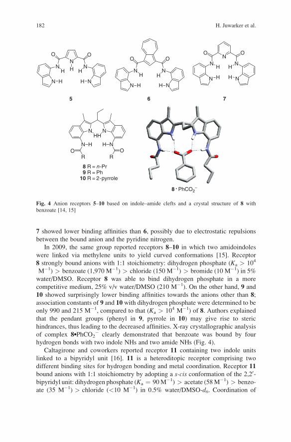
In 2009, the same group reported receptors 8–10 in which two amidoindoles
were linked via methylene units to yield curved conformations [15]. Receptor
8 strongly bound anions with 1:1 stoichiometry: dihydrogen phosphate (Ka > 104
M�1) > benzoate (1,970 M�1) > chloride (150 M�1) > bromide (10 M�1) in 5%
water/DMSO. Receptor 8 was able to bind dihydrogen phosphate in a more
competitive medium, 25% v/v water/DMSO (210 M�1). On the other hand, 9 and
10 showed surprisingly lower binding affinities towards the anions other than 8;
association constants of 9 and 10 with dihydrogen phosphate were determined to be
only 990 and 215 M�1, compared to that (Ka > 104 M�1) of 8. Authors explained
that the pendant groups (phenyl in 9, pyrrole in 10) may give rise to steric
hindrances, thus leading to the decreased affinities. X-ray crystallographic analysis
of complex 8lPhCO2� clearly demonstrated that benzoate was bound by four
hydrogen bonds with two indole NHs and two amide NHs (Fig. 4).
Caltagirone and coworkers reported receptor 11 containing two indole units
linked to a bipyridyl unit [16]. 11 is a heteroditopic receptor comprising two
different binding sites for hydrogen bonding and metal coordination. Receptor 11
bound anions with 1:1 stoichiometry by adopting a s-cis conformation of the 2,20-bipyridyl unit: dihydrogen phosphate (Ka ¼ 90M�1) > acetate (58M�1) > benzo-
ate (35 M�1) > chloride (<10 M�1) in 0.5% water/DMSO-d6. Coordination of
N NHH
N NH H
RO
RO
NO O
NNHH H
N NHH
O O
NNHH
N NHH
NN
O
N
O
HH
N NHH
8 R = n -Pr9 R = Ph
10 R = 2-pyrrole
5 6 7
8 . PhCO2–
Fig. 4 Anion receptors 5–10 based on indole–amide clefts and a crystal structure of 8 with
benzoate [14, 15]
182 H. Juwarker et al.

Pt (II) to the bipyridyl induced a conformational switch from an open ended structure
(s-trans) to a convergent cleft (s-cis). Relative to 11, the preorganized receptor 12
now displayed much increased affinities under identical conditions; dihydrogen
phosphate (Ka ¼ 3,644 M�1), benzoate (280 M�1), acetate (189 M�1), chloride
(37 M�1). The enhanced binding affinities of 12 are due to the higher degree of
preorganization induced by Pt (II) coordination which locks the free rotation of the
bipyridyl units. Unlike in solution, crystal structures of 11 showed 1:2 complexes
with acetate and chloride, adopting the s-trans conformation where each of the
bound anions was held by two hydrogen bonds to the indole and amide NHs (Fig. 5).
2.2 Indole–Urea Hybrids
The utility of urea in synthetic anion receptors stems directly from its ability to
simultaneously donate two hydrogen bonds, and offers a hydrogen-bonding motif
complementary to bidentate oxoanions (e.g., carboxylates) [17]. Combining urea
and thiourea groups with indoles is therefore, a feasible strategy to further increase
the number of NH donors in the binding cavity of an anion receptor.
N N
H HN NH
O
NH
O
Nn-Bu n-BuH H
13
In 2007, Jeong and coworkers reported receptor 13 containing a 2,20-biindolylscaffold and urea groups [18]. Receptor 13 has two indole NHs and four urea NHs,
N N
N
O
H N
O
H
N NH H
PtClCl
N
N
N–H
H–N
O
O
N
N
H
H
PtCl2(DMSO)2
1211
Fig. 5 Heteroditopic receptors 11 and 12 [16]
Indoles and Related Heterocycles 183

and displayed strong binding affinities towards oxoanions such as acetate, dihydro-
gen phosphate and pyrophosphate in DMSO (Ka ¼ �105 M�1). According to 1H
NMR spectra, the two indole NHs and two inner urea NHs participated in hydrogen
bonding with these oxoanions, but the two terminal urea NHs did not. On the other
hand, all six NH protons in 13 were simultaneously involved in hydrogen bonding
with alkyl dicarboxylates such as malonate, succinate, glutarate and adipate. 13
bound these dicarboxylates strongly in a highly polar medium 10% v/v MeOH/
DMSO with association constants ranging from 1.6 � 105 M�1 (malonate) to
8.1 � 105 M�1 (adipate).
A series of anion receptors based on an indole scaffold functionalized with
amide and urea in the 2- and 7-positions have been reported by the Gale group
[19–22]. As a representative example, amide–urea hybrid 14 bears four hydrogen
bond donors of two urea NHs, one indole NH and one amide NH. 14 binds
oxoanions strongly such as acetate (Ka ¼ 104 M�1), dihydrogen phosphate
(4,950 M�1) and benzoate (4,460 M�1) in 0.5% H2O/DMSO. 1H NMR titrations
caused large downfield shifts (>1 ppm, e.g., acetate) for all NHs of the indole, urea
and amides. Titration curves showed that complexation induced shifts (CIS) for
urea and indole NHs reached a plateau upon addition of 1 equivalent of an anion,
but the amide NH signal continued to shift downfield when excess of an anion was
added. Authors suggest that the amide NH points out of the binding cavity to
accommodate further binding in the presence of excess anion. A crystal structure
of complex 14lTBA+Cl� supports this claim by showing one chloride anion tightly
hydrogen bonded to the indole and urea NHs and a second bound to the amide NH
out of the cavity (Fig. 6).
Gale and coworkers reported 1,3-diindolylureas 15–18 containing four conver-
gent hydrogen bond donors; two indole NHs and two urea NHs [23]. According to1H NMR titrations, receptor 15 strongly bound oxoanions such as acetate, benzoate
and dihydrogen phosphate with Ka > 104 M�1, but weakly bound chloride
(Ka¼ 128 M�1) in 0.5% water/DMSO-d6. It was also observed that while receptors15 and 16 formed 1:1 complexes with anions in solution, diverse stoichiometries
were observed in the solid state. For example, crystallization of 15 in the presence
of excess bicarbonate (HCO3�) provided the crystal structure of complex
(15)2lCO32� in which carbonate, not bicarbonate, was held by two molecules of
N
N
H
N
O
ON
HH
H
14 14 . (Cl–)2
Fig. 6 Urea–amide hybrid 14 and its crystal structure with chloride [19–22]
184 H. Juwarker et al.
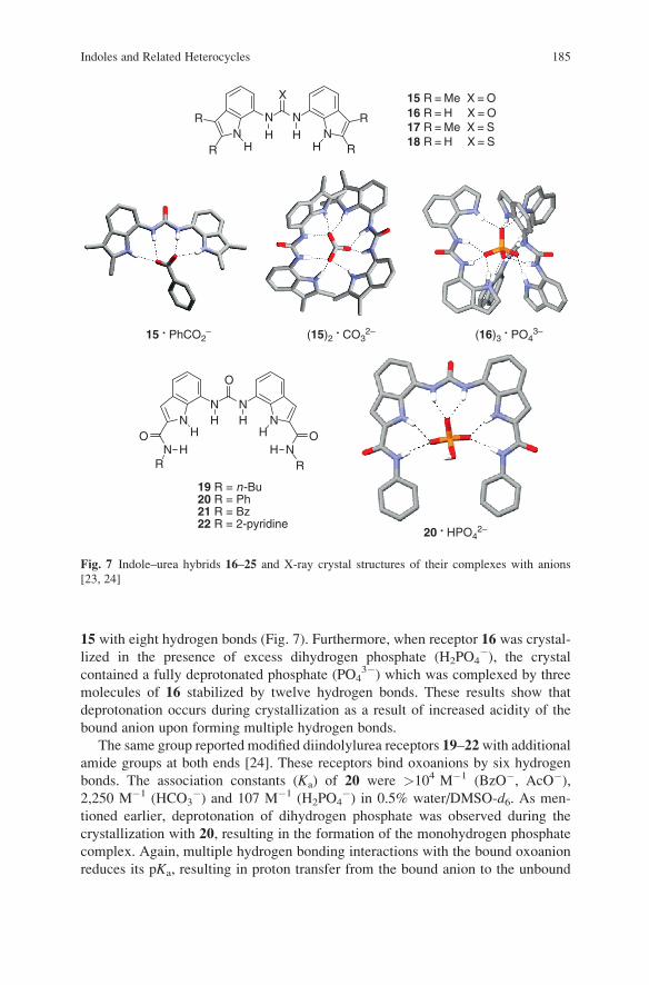
15 with eight hydrogen bonds (Fig. 7). Furthermore, when receptor 16 was crystal-
lized in the presence of excess dihydrogen phosphate (H2PO4�), the crystal
contained a fully deprotonated phosphate (PO43�) which was complexed by three
molecules of 16 stabilized by twelve hydrogen bonds. These results show that
deprotonation occurs during crystallization as a result of increased acidity of the
bound anion upon forming multiple hydrogen bonds.
The same group reported modified diindolylurea receptors 19–22with additional
amide groups at both ends [24]. These receptors bind oxoanions by six hydrogen
bonds. The association constants (Ka) of 20 were >104 M�1 (BzO�, AcO�),2,250 M�1 (HCO3
�) and 107 M�1 (H2PO4�) in 0.5% water/DMSO-d6. As men-
tioned earlier, deprotonation of dihydrogen phosphate was observed during the
crystallization with 20, resulting in the formation of the monohydrogen phosphate
complex. Again, multiple hydrogen bonding interactions with the bound oxoanion
reduces its pKa, resulting in proton transfer from the bound anion to the unbound
N
X
N NR
R R
RH H
HH
N
N N
O
HHN N
NO
NO
HHR R
H H
15 R = Me X = O16 R = H X = O17 R = Me X = S18 R = H X = S
19 R = n-Bu20 R = Ph21 R = Bz22 R = 2-pyridine
15 . PhCO2–
20 . HPO42–
(15)2 . CO3
2– (16)3 . PO4
3–
Fig. 7 Indole–urea hybrids 16–25 and X-ray crystal structures of their complexes with anions
[23, 24]
Indoles and Related Heterocycles 185
species in solution. These results parallel certain enzymatic behaviors, where
biocatalytic transformations involving oxoanionic substrates are initiated upon
significant increases in substrate acidity upon hydrogen bonding in the active
site [25].
2.3 Carbazoles
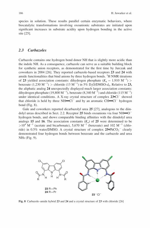
Carbazole contains one hydrogen bond donor NH that is slightly more acidic than
the indole NH. As a consequence, carbazole can serve as a suitable building block
for synthetic anion receptors, as demonstrated for the first time by Jurczak and
coworkers in 2004 [26]. They reported carbazole-based receptors 23 and 24 with
amide functionalities that bind anions by three hydrogen bonds. 1H NMR titrations
of 23 yielded association constants: dihydrogen phosphate (Ka ¼ 1,910 M�1) >benzoate (1,230 M�1) > chloride (13 M�1) in 5% D2O/DMSO-d6. Relative to 23,
the aliphatic analog 24 unexpectedly displayed much larger association constants:
dihydrogen phosphate (19,800M�1), benzoate (8,340 M�1) and chloride (115 M�1)
under identical conditions. A X-ray crystal structure of complex 23lCl� showed
that chloride is held by three NHlllCl� and by an aromatic CHlllCl� hydrogen
bond (Fig. 8).
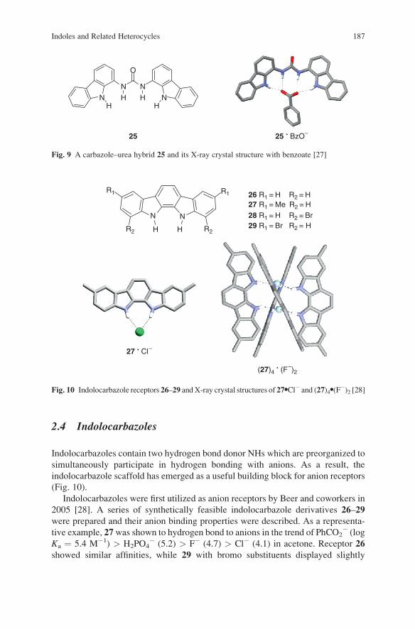
Gale and coworkers reported dicarbazolyl urea 25 [27], analogous to the diin-
dolyl ureas described in Sect. 2.2. Receptor 25 binds oxoanions via four NHlllO�
hydrogen bonds, and shows comparable binding affinities with the diindolyl urea
analogs 15 and 16. The association constants (Ka) of 25 were determined to be
>104 M�1 (acetate and bicarbonate), 5,670 M�1 (benzoate) and 102 M�1 (chlo-
ride) in 0.5% water/DMSO. A crystal structure of complex 25lPhCO2� clearly
demonstrated four hydrogen bonds between benzoate and the carbazole and urea
NHs (Fig. 9).
NN N
O O
RR
Cl Cl
H H H
23 R = Ph24 R = Pr
23 . Cl–
Fig. 8 Carbazole–amide hybrid 23 and 24 and a crystal structure of 23 with chloride [26]
186 H. Juwarker et al.
2.4 Indolocarbazoles
Indolocarbazoles contain two hydrogen bond donor NHs which are preorganized to
simultaneously participate in hydrogen bonding with anions. As a result, the
indolocarbazole scaffold has emerged as a useful building block for anion receptors
(Fig. 10).
Indolocarbazoles were first utilized as anion receptors by Beer and coworkers in
2005 [28]. A series of synthetically feasible indolocarbazole derivatives 26–29
were prepared and their anion binding properties were described. As a representa-
tive example, 27was shown to hydrogen bond to anions in the trend of PhCO2� (log
Ka ¼ 5.4 M�1) > H2PO4� (5.2) > F� (4.7) > Cl� (4.1) in acetone. Receptor 26
showed similar affinities, while 29 with bromo substituents displayed slightly
N N
O
N NH HHH
25 25 . BzO–
Fig. 9 A carbazole–urea hybrid 25 and its X-ray crystal structure with benzoate [27]
NN
R1 R1
R2 R2H H
26 R1 = H R2 = H27 R1 = Me R2 = H28 R1 = H R2 = Br29 R1 = Br R2 = H
27 . Cl–
(27)4 . (F–)2
Fig. 10 Indolocarbazole receptors 26–29 andX-ray crystal structures of 27lCl� and (27)4l(F�)2 [28]
Indoles and Related Heterocycles 187

higher binding affinities in the same trend of PhCO2� (log Ka ¼ 5.9 M�1) >
H2PO4� (5.3) > F� (5.0) > Cl� (4.9) due to the electron-withdrawing effect.
A crystal structure of 27lCl� demonstrates that the chloride ion is held by two
equal hydrogen bonds with NHs. Additionally, an interesting crystal structure of a
2:1 complex between 27 and TBA+F� was obtained in which four indolocarbazole
molecules self-assembled in a helical fashion around two fluoride anions by
NHlllF� hydrogen bonds.
Jeong et al. prepared receptors 30 and 31 to reveal the effect of preorganization
on anion binding affinities [29]. Owing to dipole–dipole repulsions, the biindole
unit in 30 exists in an s-trans conformation. In the presence of an anion, however,
the conformation switches to an s-cis enabling both NHs to simultaneously hydro-
gen bond to the same anion. On the contrary, two NHs in indolocarbazole receptor
31 are covalently locked to donate hydrogen bonds in the same direction. This
difference was proven to have a large effect on the magnitude of the binding affinity
(Fig. 11).
In detail, the binding constants Ka of receptor 30 were 1.4 � 105 M�1 (acetate),
5.1 � 103 M�1 (chloride), 2.1 � 102 M�1 (bromide), 77 M�1 (hydrogen sulfate)
and 6 M�1 (iodide) in acetonitrile. Meanwhile, 31 displayed much higher binding
affinities: >2 � 106 M�1 (acetate), 1.1 � 105 M�1 (chloride), 8.7 � 103 M�1
(bromide), 2.1 � 103 M�1 (hydrogen sulfate) and 1.8 � 102 M�1 (iodide). It is
evident that 31 binds anions 20–40 times more strongly than 30, corresponding to
the additional stabilization of DDG ¼ �2 kcal mol�1.
N
N
H
H
N N
H H
A–
s-trans s-cis
Receptor 32, containing both hydrogen bond donors (NHs) and acceptors (pyridyl
nitrogen), was prepared for selective binding of dihydrogen phosphate [30]. The
association constant (Ka) of 32 with H2PO4� was determined to be 1.1 � 105 M�1
in CH3CN which is higher than those obtained with other anions: 2.2 � 104 M�1
(AcO�), 5.0 � 103 M�1 (Cl�), 2.1 � 103 M�1 (CN�), 1.6 � 103 M�1 (HSO4� ),
5.6 � 102 M�1 (Br�) and 40 M�1 (I�). Authors explain that the high binding
affinity for dihydrogen phosphate stems from two additional hydrogen bonds
between the pyridyl nitrogens and the hydroxyl groups of dihydrogen phosphate.
Using reference molecules without pyridyl moieties, these hydrogen bonds were
proven to additionally stabilize the complex by DDG ¼ �3.2 kcal mol�1.
In addition to NHs, the hydroxyl groups of serine, tyrosine, and threonine have
also been found to participate in anion binding in biological systems. Examples
include phosphate-binding protein [31, 32] and a CIC chloride channel [33, 34].
188 H. Juwarker et al.
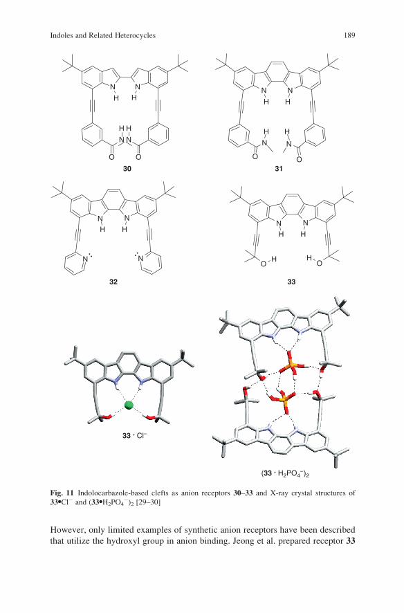
However, only limited examples of synthetic anion receptors have been described
that utilize the hydroxyl group in anion binding. Jeong et al. prepared receptor 33
N N
H H
N N
O
H
O
H
N N
H H
30
(33 . H2PO4–)2
N N
H H
N N
O
H
O
H
31
33 . Cl–
N N
H H
N NOO
HH
32 33
Fig. 11 Indolocarbazole-based clefts as anion receptors 30–33 and X-ray crystal structures of
33lCl� and (33lH2PO4�)2 [29–30]
Indoles and Related Heterocycles 189
containing two NH and two OH groups [35]. It was demonstrated in two crystal
structures that two hydroxyl groups were involved in hydrogen bonding with
chloride and hydrogen phosphate. In the solid state, 33 formed a 1:1 complex
with chloride, while a 2:2 complex with dihydrogen phosphate was observed.
Two hydroxyl groups in 33 were measured to greatly increase the binding affinities
towards anions by forming two additional hydrogen bonds, stabilizing the complex
up to DDG ¼ �5 kcal mol�1 in 1% H2O/CH3CN.
2.5 Others
Beer et al. employed indolocarbazole 26 in creating an anion-templated pseudor-
otaxane 34 [36]. The sulfate anion is dianionic and tetrahedral and has been known
to form multiple hydrogen bonds. For example, sulfate is effectively bound by two
molecules of 26, as demonstrated in the crystal structure of complex 34, which
shows each of the two indolocarbazoles binding one pair of oxygen atoms of
sulfate. By replacing one of the indolocarbazoles with isophthalamide-containing
crown ether, a pseudorotaxane type complex 34 self-assembled upon hydrogen
bonding to sulfate (Fig. 12).
Indole based tripodal receptors were reported by Ito and coworkers in 2007 [37].
Triindolylmethane 35 contains three indolyl NHs suitably positioned to collectively
hydrogen bond to anions. 35was shown to possess a binding selectivity for chloride
in CDCl3 (Ka ¼ 1,200 M�1) over other anions tested: Br� (105 M�1), I� (27 M�1),
HSO4� (34 M�1) and NO3
� (36 M�1). Authors claimed the selectivity for chloride
is due to a complementary size of the binding pocket for chloride over other larger
anions.
Browning et al. reported anion binding properties of 36, a C3-symmetric phos-
phine with three indolyl substituents [38]. Receptor 36 was designed to form three
hydrogen bonds with anions and the phosphine core was able to coordinate to
transitional metals. 1H NMR spectroscopy titrations in CD2Cl2 yielded association
constants (Ka): 3,920 M�1 (Cl�), 2,730 M�1 (AcO�), 320 M�1 (Br�) and 150 M�1
(BF4�). Crystal structures of complex 36lF� demonstrated a 1:1 binding stoichi-
ometry by three NHlllF� hydrogen bonds. Authors also reported a crystal structure
of a 36 (as a copper–phenanthroline complex) with BF4�. Despite the weak
hydrogen bonding nature of BF4� in solution [40], the crystal structure shows
that upon coordination to Cu(I), three fluorides of BF4� can form hydrogen bonds
with each of the three indolyl NHs in the receptor.
Jang and coworkers prepared a ditopic receptor 37 that possessed a biindolyl
core and Zn porphyrin arms. The biindolyl unit served as an anion binding site
while the porphyrin units allowed for the coordination of neutral ligands, e.g.,
1,4-diazabicyclo[2.2.2]octane (DABCO) [39]. Receptor 37 bound a chloride ion
with an association constant (Ka) of 4.93 � 104 M�1 in THF, which was enhanced
in the presence of DABCO (1 equiv) to 7.10 � 105 M�1. Likewise, the presence of
chloride (1 equiv) also increased the binding affinity of 37 with DABCO from
190 H. Juwarker et al.

Ka ¼ 2.02 � 106 M�1 to 2.48 � 107 M�1 in THF. The results clearly prove allo-
steric binding events with positive cooperativity.
3 Macrocycles
Macrocycles have been extensively studied as synthetic receptors for a variety of
molecular and ionic guests despite their synthetic challenge. In particular, macro-
cycles that function as anion receptors typically possess a well-defined cavity in
which multiple NH hydrogen bond donors are presented in a convergent manner.
N
N N
NZn
O
OMe
N NH H
NO
NO
HO
HO
O O
O
OO
NH
SO O
OO
NO
NO
HO
HO
O O
O
OO
NHSO42–
X
N
N
NH
H
H
34
35 X : CH36 X : P
36 . F–
26
N
N
N
NN
NZn
O
MeO
37
H
H
36 . [Cu(I)(phen)]BF4
Fig. 12 Sulfate-templated pseudorotaxane 34, tripodal indoles 35 and 36, and crystal structures of
36 complexed with anions, and a ditopic receptor 37 [36–39]
Indoles and Related Heterocycles 191
While a large number of cyclic oligopyrroles such as calix[n]pyrrole and sapphyr-
ins have been reported [41], indole based macrocycles are extremely rare. Although
not studied as anion receptors, Black and coworkers first reported the synthesis of
calix[n]indoles 38 and 39, macrocycles comprising three or four indole rings
connected by methylene groups [42, 43].
38 n = 139 n = 2
NMeO
MeO
OMe
NN
OMe
OMe
R
R
OMe
HH H
R
n
3.1 Anion Receptors
Jeong and coworkers reported macrocycles 40 and 41 in which two indolocarbazoles
were connected by linear ethynyl and butadiynyl bridges [44, 45]. Each macrocycle
possesses a rigid, planar cavity with four NHs that converge to form strong hydrogen
bonds with anions. Macrocycle 40 contains a small cavity with the dimension of
2.6 A � 2.6 A between NH protons, to which only a single atoms of small ionic
radius (e.g., F�, O�) can coordinate. This is evident in the crystal structure of complex
40lBu4N+Cl� where chloride is held by four NHlllCl� hydrogen bonds and sits
above the cavity, but is not inserted due to the limited space for the large chloride ion.
It is worthwhile noting that tetrabutylammonium is placed on the aromatic surface
near the bound chloride ion owing to cation-p and electrostatic interactions.
The association constants (Ka) of 40 with halides were determined to be in the order
of F� (5.6 � 108 M�1) > Cl� (2.1 � 106 M�1) � Br� (1.9 � 103 M�1) > I�
(3.0 � 102 M�1) in acetonitrile. 40 also showed strong binding to polyatomic anions:
AcO� (6.5 � 106 M�1) > H2PO4� (3.2 � 106 M�1) > N3
� (9.1 � 105 M�1) >HSO4
� (6.8 � 105 M�1) > NO3� (3.9 � 105 M�1) > CN� (7.5 � 104 M�1) in
same solvent. The 1H NMR spectra showed two interesting features; first, anion
induced changes (Dd) in chemical shifts of the NH signals correlated linearly to the
binding affinities. Secondly, two sets of the 1H NMR signals were observed when less
than 1 equivalent of an anion was added at room temperature due to slow exchange
between free macrocycle and its complex on the 1H NMR (500 MHz) time scale.
Compared to 40, macrocycle 41 contains a larger cavity (2.6 A � 5.1 A) and
can accommodate more than one atom simultaneously. The crystal structure of
complex 41lBu4N+N3
� clearly shows both end nitrogen atoms of the azide simul-
taneously bound within the cavity in a bridged manner, each forming two hydrogen
bonds. In contrast, the crystal structure of complex 40lBu4N+N3
� displays that only
192 H. Juwarker et al.
one nitrogen atom is coordinated to the cavity. This structural difference between
40 and 41 was manifested in their relative binding affinities with anions. According
to UV/Vis titration experiments, 41 formed more stable complexes with polyatomic
anions such as azide and oxoanions in 10% v/v methanol/acetone. For example, the
association constants (Ka) of 40 and 41 were determined to be 2,300 and
81,000 M�1 for azide, 4,300 and 2,20,000 M�1 for dihydrogen phosphate, 94 and
15,000 M�1 for nitrate, and 81 and 13,000 M�1 for hydrogen sulfate. These results
prove that for polyatomic anions, a bridged (end-to-end) hydrogen bonding mode
like in 41lN3� is more stable than an end-on mode as seen in 40lN3
�, despiteforming the same number of hydrogen bonds (Fig. 13).
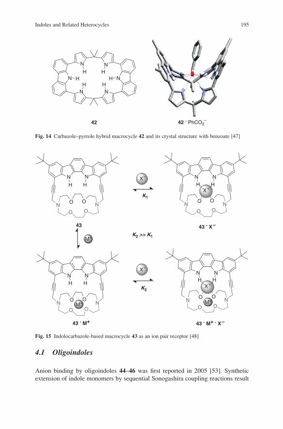
Sessler et al. reported a pyrrole–carbazole hybrid macrocycle 42 in which two
carbazole moieties were incorporated into a previously studied calix[4]pyrrole [46]
to create an extended binding cavity containing six hydrogen bond donor NHs [47].
A crystal structure of 42lPhCO2� revealed that binding of benzoate occurred
primarily from the four pyrrolic NH groups resulting in a folded, “wing-like”
conformation of the macrocycle. Association constants (Ka) in dichloromethane
were determined for acetate (2.3 � 105 M�1), benzoate (7.7 � 104 M�1),
oxalate (3.1 � 104 M�1), succinate (9.5 � 103 M�1), dihydrogen phosphate
(7.2 � 104 M�1) and chloride (3.5 � 104 M�1). Authors suggested that the contri-
bution from the carbazole NHs was minor as the groups are not oriented in plane
with the oxygens of benzoate (Fig. 14).
3.2 Ion Pair Receptor
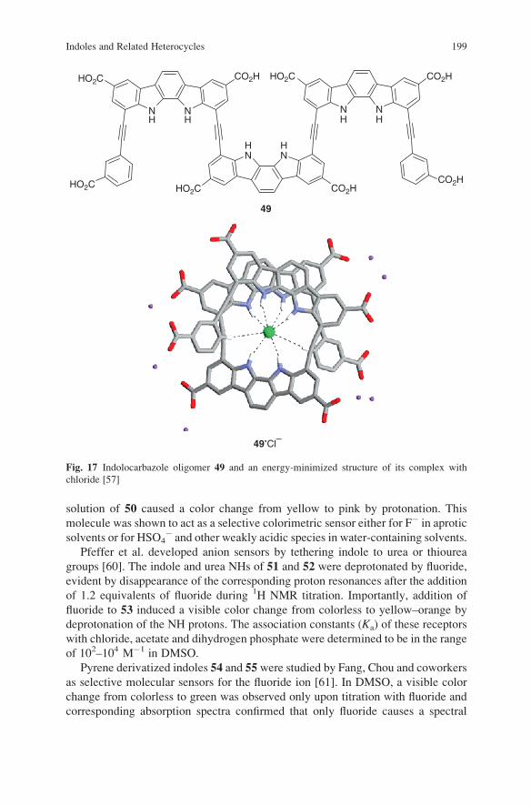
Jeong et al. also reported an ion pair receptor 43 containing both cation- and anion-
binding sites [48]. By attaching a diazacrown unit to an indolocarbazole scaffold,
two heterotopic binding sites are placed in close proximity, able to form a contact
ion pair which is crucial to minimize unfavorable energy required for charge
separation. In a polar medium (10% v/v DMSO/CD3CN), receptor 43 weakly
binds chloride with an association constant (Ka) of 7 M�1. However, the binding
affinity significantly increased in the presence of an alkali metal cation. For
example, addition of 1 equivalent of Li+, Na+ and K+ yielded association constants
of 120 M�1, 14,000 M�1 and 6,200 M�1, respectively. Sodium ion showed the
largest enhancement (2,000-fold), attributed to favorable electrostatic interactions
between the bound halide and alkali metal cation (Fig. 15).
4 Acyclic Oligomers
The use of oligomers in anion recognition is closely related to the field of foldamer
research. Foldamers are synthetic molecules in which noncovalent forces guide the
folding into ordered secondary structures such as a-helices and b-turns [49–51].
Foldamers have recently been developed as novel receptors for molecules and ions
Indoles and Related Heterocycles 193

as folding often results in a helical cavity which is isolated from the outer environ-
ment [52]. Anion binding foldamers are created by synthetic extension of monomer
units containing hydrogen bond donor groups into longer oligomeric strands.
Jeong’s group, in particular, has reported examples of indole based foldamers
used as anion receptors.
NH
NH
HN
HN
NH
NH
HN
HN
40 41
40 . Cl–
40 . N3– 41 . N3
–
40 . Cl–
Fig. 13 Indolocarbazole-based macrocycles 40, 41 and top and side views of X-ray crystal
structures of 40lCl�, and X-ray crystal structures of 40 and 41 with N3� [44, 45]
194 H. Juwarker et al.
4.1 Oligoindoles
Anion binding by oligoindoles 44–46 was first reported in 2005 [53]. Synthetic
extension of indole monomers by sequential Sonogashira coupling reactions result
N
N
N
HH
HN
N
N
HH
H
42 42 . PhCO2–
Fig. 14 Carbazole–pyrrole hybrid macrocycle 42 and its crystal structure with benzoate [47]
M+
NN
N N
H H
O O
O O
NN
N N
H H
O O
O O
NN
N N
H H
O O
O O
NN
N N
H H
O O
O O
K1
K2
K2 >> K1
43 43 . X–
43 . M+ 43 . M+ . X–
M+
X–
X–
X–
X–
M+
Fig. 15 Indolocarbazole-based macrocycle 43 as an ion pair receptor [48]
Indoles and Related Heterocycles 195

in oligomeric strands containing multiple indole donor NH groups. The oligoin-
doles exist in an extended stair-like conformation in the absence of anion where the
biindole unit maintains an s-trans conformation to minimize dipole–dipole repul-
sion as previously discussed in the Sect. 2.4. However, upon addition of tetrabuty-
lammonium chloride, the biindoles adopt an s-cis conformation enabling the indole
NH protons to simultaneously participate in hydrogen bonding with chloride. In
turn, the oligoindoles fold into helical structures with one turn comprising four
indole rings, proved by 1H NMR spectroscopy. The association constants between
oligoindoles and tetrabutylammonium chloride were 1.3 � 105 M�1 for 44,
1.2 � 106 M�1 for 45, and >107 M�1 for 46. In a more competitive medium for
hydrogen bonding (10% v/v H2O/CH3CN), the association constants were deter-
mined to be 210 M�1 and 23,000 M�1 for 45 and 46, respectively. The binding
affinities greatly increased with longer chain lengths of the oligomers, supporting
the formation of helical complexes (Fig. 16).
Authors found that the fluorescent properties of the oligoindoles were strongly
dependant on the nature of the terminal groups; an oligoindole 47 containing
benzoate termini was the most fluorescent [54]. Addition of an anion led to dramatic
hypochromic and bathochromic shifts (Dl up to 65 nm) of the emission band,
changing the solution color from bright blue to dim bluish green. The authors
rationalized this effect as excimer formation between the p-stacked aromatic planes
in a helical conformation. Fluorescence titrations afforded the association constants
with moderate selectivity in the range of 103–106 M�1 for anions (I� � NO3� <
Br� < AcO� < N3� � CN� < Cl� < F�) in 20% v/v MeOH/CH2Cl2.
Oligoindoles 44–47 possess no chiral component and therefore fold to give a
racemic mixture of right- and left-handed helices in the presence of an anion [55,
56]. In order to induce the preferential formation of one particular helix, a chiral
oligoindole 48 capped with 1-(S)-phenylethylamido units was synthesized. Circular
dichroism (CD) spectroscopy of 48 alone displayed no appreciable Cotton effect.
Addition of tetrabutylammonium chloride, however, led to strong induced CD
signals in CH2Cl2, which gradually intensified and saturated upon addition of
approximately 1 equivalent of chloride. The association constant between 48 and
tetrabutylammonium chloride ion was determined to be 2.9 � 105 M�1 in 1% v/v
MeOH/CH2Cl2. Finally, the enantiomer of 48 with 1-(R)-phenylethylamido units
showed the same CD behaviors but the opposite Cotton effect. These results
demonstrate that anion binding can induce the preferential formation of one race-
mic helix over another.
4.2 Oligoindolocarbazoles
Hydrogen bonding between synthetic receptors and molecular/ionic guests in water
is almost negligible owing to strong competition with solvent molecules. In general,
synthetic anion receptors employing hydrogen bonds bind anions strongly in
organic solvents but not in water. However, proteins utilize hydrogen bonds to
196 H. Juwarker et al.

N
N
N
RO
RO2C
RO2C
RO2C
CO2R CO2R
CO2R
N
OR
n
44 n = 145 n = 246 n = 3
N
N
3
R = (CH2CH2O)2CH3
N
N 2
N
O
*
CH3
H
N
O
*H
CH3
H
H
H
H
H
H
H
H
45 . Cl–
45 . Cl–
48
47
Fig. 16 Oligoindoles 44–48 and top and side views of an energy-minimized structure of complex
45lCl� (side chains of diethylene glycol esters and ethers are replaced with hydrogen atoms)
[53–56]
Indoles and Related Heterocycles 197

bind anions efficiently in water. A plausible explanation is that the binding site is
surrounded by hydrophobic organic fragments, and in part segregated from bulk
water, thus rendering an organic solvent-like microenvironment.
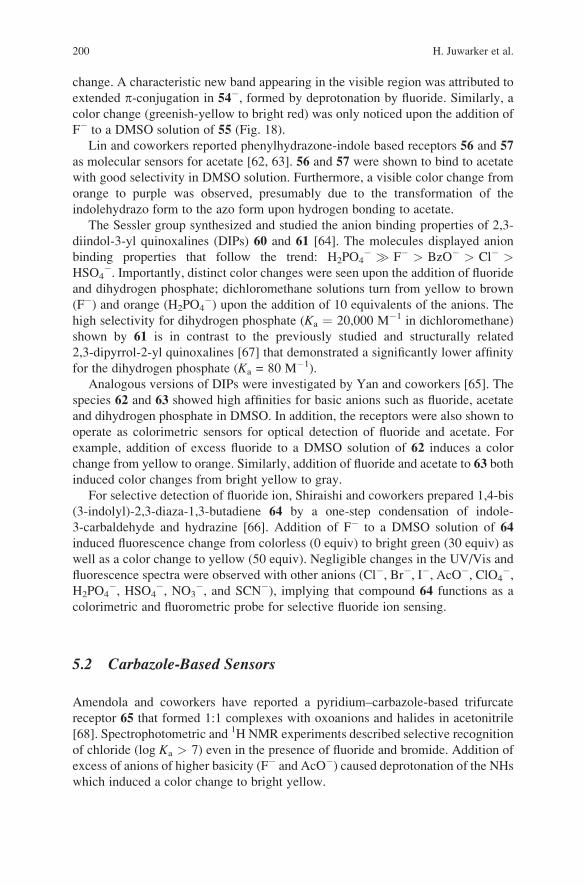
Oligo(indolocarbazole) 49 was designed to encapsulate anions in water by
hydrogen bonds, similar to proteins [57]. Oligomer 49 is soluble in water under
basic conditions and folds into a helical conformation to generate an internal cavity
surrounded by six NH protons. The 1H NMR spectrum of 49 changes noticeably
upon addition of sodium fluoride, chloride, and bromide, but no change was
observed with sodium iodide and perchlorate. The association constants of 49 with
sodium fluoride, chloride, and bromide were determined to be 46, 65, and 19 M�1,
respectively. Lithium, sodium and potassium chloride afforded an identical associa-
tion constant, implying no cation effect. Moreover, binding affinities between a
methyl ester derivative of 49 and halides decreased in the order of F� (1,83,000M�1)
> Cl� (36,800 M�1) > Br� (1,350 M�1) > I� (86 M�1) in an organic medium,
4:1 v/v DMSO/MeOH. As demonstrated here, fluoride binds more strongly than
chloride in an organic medium, but less strongly in water. Authors rationalized this
difference by the solvation energy; the fluoride ion solvates much more strongly in
water than the chloride ion does, which reduces net binding energy (Fig. 17).
5 Molecular Sensors
Molecular sensors that yield distinct color or fluorescence changes upon anion
binding are extremely useful in that they allow for naked-eye detection of analytes.
These features are advantageous for prompt, qualitative detection of anionic ana-
lytes that are toxic (e.g., pertechnetate, fluoride), or that may cause environmental
pollution (e.g., nitrate, phosphate). Molecular sensors in general consist of binding
and signaling sites [58]. Upon binding, chromophores or fluorophores in the
signaling units are electronically perturbed which typically results in a color or
fluorescence change. In this regard, a number of indole-based molecular sensors
have been prepared. Furthermore, the acidity of the indole NH can result in
deprotonation by basic anions (e.g., fluoride, carboxylate) that also induce distinct
color changes.
5.1 Indole-Based Sensors
Shao and coworkers have reported an oxidized bis(indolyl)methane 50; a simple
chromophore containing an acidic hydrogen bond donor moiety and a basic hydro-
gen bond acceptor moiety [59]. Deprotonation of the indole NH by basic anions
induced a color change. For example, deprotonation by fluoride in acetonitrile led to
a color change from yellow to red. Protonation of the basic indole component also
resulted in a color change. Addition of acidic HSO4� to a 4:1 v/v CH3CN/H2O
198 H. Juwarker et al.
solution of 50 caused a color change from yellow to pink by protonation. This
molecule was shown to act as a selective colorimetric sensor either for F� in aprotic
solvents or for HSO4� and other weakly acidic species in water-containing solvents.
Pfeffer et al. developed anion sensors by tethering indole to urea or thiourea
groups [60]. The indole and urea NHs of 51 and 52 were deprotonated by fluoride,
evident by disappearance of the corresponding proton resonances after the addition
of 1.2 equivalents of fluoride during 1H NMR titration. Importantly, addition of
fluoride to 53 induced a visible color change from colorless to yellow–orange by
deprotonation of the NH protons. The association constants (Ka) of these receptors
with chloride, acetate and dihydrogen phosphate were determined to be in the range
of 102–104 M�1 in DMSO.
Pyrene derivatized indoles 54 and 55 were studied by Fang, Chou and coworkers
as selective molecular sensors for the fluoride ion [61]. In DMSO, a visible color
change from colorless to green was observed only upon titration with fluoride and
corresponding absorption spectra confirmed that only fluoride causes a spectral
NH
NH
HO2C
HO2C
HO2C
HO2C
CO2H
CO2H
CO2H
CO2H
HN
HN
NH
NH
49
49.Cl–
Fig. 17 Indolocarbazole oligomer 49 and an energy-minimized structure of its complex with
chloride [57]
Indoles and Related Heterocycles 199
change. A characteristic new band appearing in the visible region was attributed to
extended p-conjugation in 54�, formed by deprotonation by fluoride. Similarly, a
color change (greenish-yellow to bright red) was only noticed upon the addition of
F� to a DMSO solution of 55 (Fig. 18).
Lin and coworkers reported phenylhydrazone-indole based receptors 56 and 57
as molecular sensors for acetate [62, 63]. 56 and 57 were shown to bind to acetate
with good selectivity in DMSO solution. Furthermore, a visible color change from
orange to purple was observed, presumably due to the transformation of the
indolehydrazo form to the azo form upon hydrogen bonding to acetate.
The Sessler group synthesized and studied the anion binding properties of 2,3-
diindol-3-yl quinoxalines (DIPs) 60 and 61 [64]. The molecules displayed anion
binding properties that follow the trend: H2PO4� � F� > BzO� > Cl� >
HSO4�. Importantly, distinct color changes were seen upon the addition of fluoride
and dihydrogen phosphate; dichloromethane solutions turn from yellow to brown
(F�) and orange (H2PO4�) upon the addition of 10 equivalents of the anions. The
high selectivity for dihydrogen phosphate (Ka ¼ 20,000 M�1 in dichloromethane)
shown by 61 is in contrast to the previously studied and structurally related
2,3-dipyrrol-2-yl quinoxalines [67] that demonstrated a significantly lower affinity
for the dihydrogen phosphate (Ka = 80 M�1).
Analogous versions of DIPs were investigated by Yan and coworkers [65]. The
species 62 and 63 showed high affinities for basic anions such as fluoride, acetate
and dihydrogen phosphate in DMSO. In addition, the receptors were also shown to
operate as colorimetric sensors for optical detection of fluoride and acetate. For
example, addition of excess fluoride to a DMSO solution of 62 induces a color
change from yellow to orange. Similarly, addition of fluoride and acetate to 63 both
induced color changes from bright yellow to gray.
For selective detection of fluoride ion, Shiraishi and coworkers prepared 1,4-bis
(3-indolyl)-2,3-diaza-1,3-butadiene 64 by a one-step condensation of indole-
3-carbaldehyde and hydrazine [66]. Addition of F� to a DMSO solution of 64
induced fluorescence change from colorless (0 equiv) to bright green (30 equiv) as
well as a color change to yellow (50 equiv). Negligible changes in the UV/Vis and
fluorescence spectra were observed with other anions (Cl�, Br�, I�, AcO�, ClO4�,
H2PO4�, HSO4
�, NO3�, and SCN�), implying that compound 64 functions as a
colorimetric and fluorometric probe for selective fluoride ion sensing.
5.2 Carbazole-Based Sensors
Amendola and coworkers have reported a pyridium–carbazole-based trifurcate
receptor 65 that formed 1:1 complexes with oxoanions and halides in acetonitrile
[68]. Spectrophotometric and 1H NMR experiments described selective recognition
of chloride (log Ka > 7) even in the presence of fluoride and bromide. Addition of
excess of anions of higher basicity (F� and AcO�) caused deprotonation of the NHswhich induced a color change to bright yellow.
200 H. Juwarker et al.

NN
N
N
H
H
NH
NH
NH
N NH
N
O
NH
NH
NR1
H
H
N
O
NHH
NH
NS
H
NO2
O2NO2NNO2
NO2
NO2
NO2
NO
N NH
NO
N N
OOH
H
NO
N NH
O
O
AcO–
AcO–
NO
N NH
HH
H
N N
N NH H
R
N N
N NH H
R
50
51 R1 = O, R2 = H52 R1 = S, R2 = CF3
R2
53
54 55
58
56
6460 R = H61 R = NO2
62 R = H63 R = NO2
59
57
Fig. 18 Indole- and related heterocycle-based molecular sensors 50–64 [59–66]
Indoles and Related Heterocycles 201
A carbazole–phenanthroline based anion receptor 66 was reported by Lin et al.
in which two carbazole groups were attached to a phenanthroline core by imine
linkages [69]. 1H NMR and UV/Vis titration experiments demonstrated a high
selectivity for iodide (Ka ¼ 5 � 104 M�1) while not having any affinity for other
halides. Authors attributed this selectivity to a complementary fit of the large anion.
Carbazole was also utilized by Kim and coworkers in developing fluorescent
receptors 67–69 [70]. These compounds were demonstrated to function as “dual-
channel” anion sensors, displaying both a colorimetric and fluorescent output upon
anion binding. By linking urea moieties to the carbazole scaffold, the molecular
sensors were synthesized having both chromogenic and fluorogenic signaling sub-
units. By plotting fluorescence intensity vs. absorption shift of the UV/Vis spectrum
in the presence of many anions, diverse sets of anions could be identified as each
signaling subunit demonstrated a different optical response upon binding to a
specific anion (Fig. 19).
6 Conclusions
In the last five years, the anion binding ability of indoles and related heterocycles
has been realized. Structurally diverse anion receptors based on indoles, carbazoles,
and indolocarbazoles have been reported, attributed to the hydrogen bonding donor
N
Cl Cl
N NH
O
NH
O
NR RH H
H
67 R =
69 R =
68 R =
NN NO2
N NNN
N NH H
NN N
N
H
H
H
65 66
N+
N+
+
Fig. 19 Carbazole-based molecular sensors 65–69 [68–70]
202 H. Juwarker et al.
ability of their NHs. Molecular clefts are a good starting point to validate the utility
of these heterocycles in anion binding. Often, linking multiple indoles by other NH
donor groups offers stronger binding to anions. Indoles have also been incorporated
into preorganized macrocycles containing cavities of fixed sizes where hydrogen
bonding from convergent NHs results in specificity towards complementary anions.
Furthermore, acyclic oligomers based on the indole scaffold were designed to fold
into discrete structures by encapsulating anions in helical cavities. Finally, the
anion binding properties of indoles and related heterocycles have been applied
toward the creation of molecular sensors that yield specific color and fluorescence
changes upon complexation. It is clear that indoles and related heterocycles are
suitable building blocks for the construction of novel receptors and functional
devices. As the molecular toolbox expands to generate more sophisticated struc-
tures and functions, we expect to see these heterocycles utilized further.
References
1. Morokuma K (1977) Acc Chem Res 10:294
2. Steiner T (2002) Angew Chem Int Ed 41:48
3. Bordwell FG, Druker GE, Fried HE (1981) J Org Chem 46:632
4. Bordwell FG, Ji G-Z (1991) J Am Chem Soc 113:8398
5. Pflugrath JW, Quiocho FA (1985) Nature 314:257
6. He JJ, Quiocho FA (1991) Science 251:1479
7. Koropatkin NM, Pakrasi HB, Smith TJ (2006) Proc Natl Acad Sci 103:9820
8. Okunola OA, Santacroce PV, Davis JT (2008) Supramol Chem 20:169
9. Gale PA (2008) Chem Commun:4525
10. Rebek J Jr (1987) Science 235:1478
11. Harmata M (2004) Acc Chem Res 37:862
12. Bates GW, Gale PA, Light ME (2007) Chem Commun:2121
13. Caltagirone C, Gale PA, Hiscock JR, Hursthouse MB, Light ME, Tizzard GJ (2009) Supramol
Chem 21:125
14. Zielinski T, Dydio P, Jurczak J (2008) Tetrahedron 64:568
15. Dydio P, Zielinski T, Jurczak J (2009) Chem Commun:4560
16. Caltagirone C, Mulas A, Isaia F, Lippolis V, Gale PA, Light ME (2009) Chem Commun:6279
17. Choi K, Hamilton AD (2003) Coord Chem Rev 240:101
18. Lee JY, Lee MH, Jeong K-S (2007) Supramol Chem 19:257
19. Bates GW, Triyanti, Light ME, Albrecht M, Gale PA (2007) J Org Chem 72:8921
20. Makuc D, Triyanti, Albrecht M, Plavec J, Rissanen K, Valkonen A, Schalley CA (2009) Eur J
Org Chem:4854
21. Caltagirone C, Hiscock JR, Hursthouse MB, Light ME, Gale PA (2008) Chem Commun:3007
22. Makuc D, Lenarcic M, Bates GW, Gale PA, Plavec J (2009) Org Biomol Chem 7:3505
23. Caltagirone C, Hiscock JR, Hursthouse MB, Light ME, Gale PA (2008) Chem Eur J 14:10236
24. Gale PA, Hiscock JR, Moore SJ, Caltagirone C, Hursthouse MB, Light ME (2009) Chem
Asian J 5:555
25. Taylor MS, Jacobsen EN (2006) Angew Chem Int Ed 45:1520
26. Chmielewski MJ, Charon M, Jurczak J (2004) Org Lett 6:3501
27. Hiscock JR, Caltagirone C, Light ME, Hursthouse MB, Gale PA (2009) Org Biomol Chem
7:1781
28. Curiel D, Cowley A, Beer PD (2005) Chem Commun:236
Indoles and Related Heterocycles 203
29. Chang K-J, Chae MK, Lee C, Lee J-Y, Jeong K-S (2006) Tetrahedron Lett 47:6385
30. Kwon TH, Jeong K-S (2006) Tetrahedron Lett 47:8539
31. Copley RR, Barton GJ (1994) J Mol Biol 242:321
32. Hirsch AKH, Fisher FR, Diederich F (2007) Angew Chem Int Ed 46:338
33. Dutzler R, Campbell EB, Cadene M, Chait BT, Mackinnon R (2002) Nature 415:287
34. Dutzler R, Campbell EB, Mackinnon R (2003) Science 300:108
35. Ju J, Park M, Suk J-M, Lah MS, Jeong K-S (2008) Chem Commun:3546
36. Chmielewski MJ, Zhao L, Brown A, Curiel D, Sambrook MR, Thompson AL, Santos SM,
Felix V, Davis JJ, Beer PD (2008) Chem Commun:3154
37. Oi W, Nishiki M, Ito K (2007) Lett Org Chem 4:112
38. Yu JO, Browning CS, Farrar DH (2008) Chem Commun:1020
39. Lee C-H, Yoon H, Jang W-D (2009) Chem Eur J 15:9972
40. Restorp P, Berryman OB, Sather AC, Ajami D, Rebek Jr, J (2009) Chem Commun:5692
41. Sessler JL, Gale PA, Cho W-S (2006) Anion receptor chemistry. RSC, Cambridge
42. Black DS, Bowyer MC, Kumar N, Mitchell PSR (1993) J Chem Soc Chem Commun:819
43. Black DS, Craig DC, Kumar N (1995) Tetrahedron Lett 36:8075
44. Chang K-J, Moon D, Lah MS, Jeong K-S (2005) Angew Chem Int Ed 44:7926
45. Kim NK, Chang K-J, Moon D, Lah MS, Jeong K-S (2007) Chem Commun:3401
46. Gale PA, Sessler JL, Kral V, Lynch VM (1996) J Am Chem Soc 118:5140
47. Piatek P, Lynch VM, Sessler JL (2004) J Am Chem Soc 126:16073
48. Chae MK, Lee J-I, Kim N-K, Jeong K-S (2007) Tetrahedron Lett 48:6624
49. Gellman SH (1998) Acc Chem Res 31:173
50. Hill DJ, Mio MJ, Prince RB, Jughes TS, Moore JS (2001) Chem Rev 101:3893
51. Hecht S, Huc I (eds) (2007) Foldamers: structure, properties, and applications. Wiley-VCH,
Weinheim
52. Juwarker H, Suk J, Jeong K-S (2009) Chem Soc Rev 38:3316
53. Chang K-J, Kang B-N, Lee M-H, Jeong K-S (2005) J Am Chem Soc 127:12214
54. Kim U-I, Suk J-M, Naidu VR, Jeong K-S (2008) Chem Eur J 14:11406
55. Naidu VR, Kim MC, Suk J-M, Kim H-J, Lee M, Sim E, Jeong K-S (2008) Org Lett 10:5373
56. Naidu VR, Suk J-M, Lee GW, Jeong K-S (2009) Bull Korean Chem Soc 30:482
57. Suk J-M, Jeong K-S (2008) J Am Chem Soc 130:11868
58. de Silva AP, Nimal Gunaratne HQ, Gunnlaugsson T, Huxley AJM, McCoy CP,
Rademacher JT, Rice TE (1997) Chem Rev 97:1515
59. He X, Hu S, Liu K, Guo Y, Xu J, Shao S (2006) Org Lett 8:333
60. Pfeffer FM, Lim KF, Sedgwick KJ (2007) Org Biomol Chem 5:1795
61. Lin C-I, Selvi S, Fang J-M, Chou P-T, Lai C-H, Cheng Y-M (2007) J Org Chem 72:3537
62. Wang Y, Lin H, Shao J, Cai Z-S, Lin H-K (2008) Talanta 74:1122
63. Shao J, Wang Y, Lin H, Li J, Lin H (2008) Sens Actuators B 134:849
64. Sessler JL, Cho D-G, Lynch VM (2006) J Am Chem Soc 128:16518
65. Wang T, Bai Y, Ma L, Yan X-P (2008) Org Biomol Chem 6:1751
66. Shiraishi Y, Maehara H, Hirai T (2009) Org Biomol Chem 7:2072
67. Sessler JL, Maeda H, Mizuno T, Lynch VM, Furuta H (2002) J Am Chem Soc 124:13474
68. Amendola V, Boiocchi M, Fabbrizzi L, Palchetti A (2005) Chem Eur J 11:5648
69. Yu M, Lin H, Lin H (2008) Supramol Chem 20:357
70. Thangadurai TD, Singh NJ, Hwang I-C, Lee JW, Chandran RP, Kim KS (2007) J Org Chem
72:5461
204 H. Juwarker et al.