Embed Size (px)
Citation preview

CELL JOURNAL(Yakhteh), Vol 18, No 4, Jan-Mar (Winter) 2017 597
Original Article
Indoleamine 2,3-Dioxygenase Is Dispensable for The Immunomodulatory Function of Stem Cells from
Human Exfoliated Deciduous Teeth
Razieh Alipour, M.Sc.1, Masoumeh Masoumi Karimi, M.Sc.2, Batool Hashemi-Beni, Ph.D.1, Minoo Adib, Ph.D.1*, Nasrin Sereshki, M.Sc.1, Farzaneh Sadeghi, M.Sc.1
1. Department of Immunology, Medical School, Isfahan University of Medical Sciences, Isfahan, Iran2. School of Medicine, Shahroud University of Medical Science, Shahroud, Iran
*Corresponding Address: P.O.Box: 81744-176, Department of Immunology, Medical School, Isfahan University of Medical Sciences, Hezar Jerib Street, Isfahan, Iran
Email: [email protected]
Received: 28/Nov/2015, Accepted: 6/Apr/2016AbstractObjective: In this study, we sought to better understand the immunoregulatory function of stem cells derived from human exfoliated deciduous teeth (SHED). We studied the role of the interferon gamma (IFN-γ)-indoleamine 2,3-dioxygenase (IDO)-axis in immunoregu-lation of SHED compared to bone marrow derived mesenchymal stem cells (BMMSCs) under the same conditions.
Materials and Methods: In this cross-sectional study, recently isolated human T cells were stimulated either by mitogen or inactivated allogeneic peripheral blood mononu-clear cells (PBMCs). These T cells were subsequently co-cultured with, either SHED or BMMSCs in the presence or absence of 1-methyl-tryptophan (1-MT) or neutralizing anti-human-IFN-γ antibodies. In all co-cultures we evaluated lymphocyte activation as well as IDO activity. Results: SHED, similar to conventional BMMSCs, had anti-proliferative effects on stimu-lated T cells and reduced their cytokine production. This property of SHED and BMMSCs was changed by IFN-γ neutralization. We detected IDO in the immunosuppressive super-natant of all co-cultures. Removal of IDO decreased the immunosuppression of BMMSCs. Conclusion: SHED, like BMMSCs, produced the IDO enzyme. Although IFN-γ is one of inducer of IDO production in SHED, these cells were not affected by IFN-γ in the same manner as BMMSCs. Unlike BMMSCs, the IDO enzyme did not contribute to their immu-nosuppression and might have other cell-type specific roles.
Keywords: Immunomodulation, IDO, Mesenchymal Stem/Stromal Cells, SHED, IFN-γ Cell Journal(Yakhteh), Vol 18, No 4, Jan-Mar (Winter) 2017, Pages: 597-608
Citation: Alipour R, Masoumi Karimi M, Hashemi-Beni B, Adib M, Sereshki N, Sadeghi F. Indoleamine 2,3-dioxygenase is dispensable for the immunomodulatory function of stem cells from human exfoliated deciduous teeth. Cell J. 2017; 18(4): 597-608.
IntroductionMesenchymal stem/stromal cells (MSCs) are a
rare stromal pluripotent, self-renewal cell popula-tion that belong to the adult stem cell subgroup. Initially, they have been introduced as non-hemat-opoietic bone marrow-derived cells (1, 2).
Diverse tissues and organs contain MSCs. The presence of MSCs in circulating blood, placenta, adipose tissues, dental pulp, and numerous other tissues have been shown (3, 4). The differentiation
abilities (5), angiogenesis, tissue remodeling/re-pair properties, growth factor release, and support of hematopoiesis are described as other character-istics of MSCs (3). The most intriguing features of MSCs considered superior to these attributes are their unique immune properties.
MSCs have the capacity to modulate functions of various immune cells, as demonstrated both in vitro and in vivo in animal models and humans (2, 6). MSCs can suppress human CD3+, CD4+, and

CELL JOURNAL(Yakhteh), Vol 18, No 4, Jan-Mar (winter) 2017 598
IDO Is Dispensable in Immunoregulation of SHED
CD8+ T lymphocytes (7, 8). They possess the abil-ity to suppress proliferation of activated B cells and can alter the natural killer (NK) cell phenotype as well as suppress their proliferation, cytokine se-cretion, and cytotoxicity. Mature dendritic cells (DCs) are less functional in the presence of MSCs (9, 10).
Despite extensive investigations the exact un-derlying mechanism of the immunomodulatory role of MSCs is unclear (11). Nevertheless, it can be concluded from studies that the immu-nosuppressive ability of MSCs is not innate but induced by pro-inflammatory cytokines, in par-ticular interferon gamma (IFN-γ) (12). A single factor alone does not appear to be responsible for immunosuppression; rather, multiple factors (although not equally) may be involved. Some of the suggested MSC-derived soluble factors involved in their immunosuppression include indoleamine 2,3-dioxygenase (IDO), transform-ing growth factor-β1 (TGF-β1), prostaglandin-E2 (PGE2), interleukin-10 (IL-10), and human leukocyte antigen G (HLA-G) (13-15).
Amongst these, there is more agreement on the role of the IFN-γ-IDO axis. Numerous studies have established that upon stimulation with IFN-γ, MSCs express IDO, an evolutionally conserved en-zyme that degrades tryptophan through the kynure-nine (KYN) pathway. IDO causes local deprivation of tryptophan and results in KYN metabolites that have immunosuppressive effects (16, 17).
Due to their remarkable capabilities, MSCs have tremendous potential in many modern therapeutic areas such as regenerative medicine, tissue engi-neering, and in novel immunosuppressant thera-pies. Thus, MSCs are the subject of numerous studies. In the majority of these studies, the classi-cal bone marrow derived MSCs (BMMSCs) have been used. However, MSCs are relatively rare in human bone marrow. Their numbers and differen-tiation capacity decline with age. In addition, bone marrow aspiration is an invasive procedure (18). Regarding these limitations, conversely the prom-ising results from clinical trials and consequently large numbers of MSCs required for future cell-based therapies, necessitates finding alternative sources for MSCs.
In recent years, stem cells from human exfoli-ated deciduous teeth (SHED) have emerged as
a new population of MSCs (19) which can be a preferable source for their ever-increasing applica-tions. exfoliated deciduous teeth are readily acces-sible and free from ethical concerns as the teeth are discarded. An advantage is the lack of pain and trauma to the patient when they are harvested. Ob-taining stem cells from exfoliated deciduous teeth requires a simple, convenient method (20, 21).
Approximately 15 years ago Miura et al. (22) isolated SHED. Therefore, there is limited data; particularly the published studies on the immune properties of SHED are few (23). We have previ-ously shown that SHED are comparable to con-ventional MSCs or BMMSCs. They suppress the proliferation and cytokine production of in vitro activated T cells (24). In the current study, we have examined the probable role of the IFN-γ-IDO axis in immunosuppression of SHED compared to BMMSCs.
Materials and MethodsApproximately 105 T cells stimulated by phy-
tohemagglutinin (PHA) in the lymphocyte trans-formation test (LTT) or allogeneic peripheral blood mononuclear cells (PBMCs) in MLC were placed adjacent to various numbers of SHED or BMMSCs, as separate co-cultures. MSCs were inactivated by mitomycin in order to prevent ex-pansion and consequently delete any further inter-ference with lymphocyte proliferation. Previous studies demonstrated that the dose of MSCs might affect their immunoregulation, therefore we used a different number of MSCs.
The Ethics Committee of Isfahan University of Medical Sciences approved this analytical-de-scriptive, cross-sectional study.
In vitro expansion of human stem cells One T25 cell culture flask of passage 2 SHED
was kindly provided by the Torabi Nejad Research Center, Dental School, Isfahan University of Med-ical Sciences, Isfahan, Iran for this research.
Passage 3 human BMMSCs were purchased as a 70% confluent T75 cell culture flask from Royan Institute, Isfahan, Iran. In order to obtain an ad-equate number of cells to culture with the lympho-cytes, we expanded both BMMSCs and SHED in vitro as previously described (24).

CELL JOURNAL(Yakhteh), Vol 18, No 4, Jan-Mar (Winter) 2017 599
Alipour et al.
Isolation of human blood cells T Lymphocytes were isolated from the buffy-
coat of a healthy volunteer using the RosetteSep® Human T Cell Enrichment Cocktail (StemCell Technologies, Vancouver, BC, Canada). The lym-phocytes were suspended in complete RPMI1640 that consisted of RPMI1640 with L-glutamine (Sigma, Germany) supplemented with 10% fetal calf serum (FCS, Gibco. Germany), and 1% peni-cillin/streptomycin (Roche, Germany).
PBMCs were obtained by Ficoll density gradi-ent from heparinized blood samples of a healthy volunteer donor who was allogeneic to the person who donated the T lymphocytes. The isolated cells were suspended in complete RPMI1640.
Simple co-culturesDirect co-cultures
Both types of human MSCs were mitomycin C inactivated by incubation for 3 hours in 7.5 µg/ml. Then, 100 µl aliquots of completed RPMI1640 plus decreasing numbers (104, 4×10³, 2×10³ and 103 cells) of viable MSCs either SHED or BMMSCs were plated in flat-bottomed 96-well plates, then allowed to adhere for 12 to 16 hours. Completed RPMI1640 without MSCs was used as the control.
Next, we performed the mitogen proliferation assay (lymphocyte transformation test, LTT) as follows: 105 T cells in 100 µl completed RPMI1640 stimulated by 4 μg/ml PHA (Roche, Germany) were added to the MSCs. Controls consisted of 105 PHA-stimulated T cells (positive control) or unstimulated 105 T cells (negative control) added to wells that contained only complete medium. Additionally, as background con-trols, we added 100 µl medium (without T cells) to wells that contained only the co-cultures of diminish-ing numbers of MSCs plus SHED or BMMSCs. The cultures were incubated for 3 days at 37˚C in a hu-midified atmosphere of 5% CO2.
In the mixed lymphocyte cultures (MLCs), once the MSCs adhered to the plates we added responder cells (105 T cells) in 50 µl of completed RPMI1640, fol-lowed by stimulator cells that consisted of an equal number of mitomycin inactivated PBMCs (25 µg/ml for 40 minutes) in another 50 µl of medium. The positive control consisted of responder plus stimulator cells, whereas the negative control consisted of only responder cells. Cultures were used as background
controls in the same manner as the LTTs. The cultures were incubated for 5 days at 37˚C in a humidified at-mosphere of 5% CO2.
Indirect co-cultures Transwell analysis was performed with a 24-well
transwell insert system (Costar) with a pore size of 0.4 µm. The lower chambers contained 2.5×10⁴, 104, 5×10³, and 2.5×10³ inactivated MSCs with ei-ther SHED or BMMSCs. For the LTTs, 2.5×10⁵
PHA-stimulated human T cells, and for MLCs, 2.5×10⁵ T cells (plus an equal number of stimula-tor cells) were cultured in the upper chambers of the wells. Appropriate controls included positive, negative and background controls were set. All transwell cultures were performed in a total vol-ume of 500 µl of completed RPMI 1640 medium.
All cultures were established in triplicate. After incubation, we assessed T lymphocyte prolifera-tion and cytokine secretion.
Extra co-cultures
In order to investigate the role of IFN-γ and IDO in suppression of MSCs, at the beginning of the culture periods we added either neutralizing mon-oclonal anti-human IFN-γ antibodies (10 mg/ml, R&D Systems, USA), as the Ab-co-cultures, or 1-methyl-tryptophan (1-MT, Sigma Aldrich, Ger-many) as the MT-co-cultures. The concentrations were selected from literature searches of previous studies followed by in-house optimization.
Proliferation assays and cytokine analysis
T cell proliferation was evaluated by measuring BrdU (a thymidine analogue) incorporation into the DNA of proliferating T cells by a Cell Prolif-eration ELISA, BrdU (colorimetric) kit (Roche, Germany) according to the manufacturer’s proto-col. The optical density (OD) of each co-culture/culture indicated lymphocyte proliferation.
To eliminate the effect of probable BrdU incorpo-ration in MSCs co-cultured with stimulated lympho-cytes, we used a set of cultures which contained only MSCs; the numbers of MSCs in these cultures were the same as the co-cultures (namely background con-trols). We subtracted the absorbance of these cultures from the absorbance of the corresponding co-cultures (numbers and MSC type) and this OD was used.
CELL JOURNAL(Yakhteh), Vol 18, No 4, Jan-Mar (winter) 2017 600
IDO Is Dispensable in Immunoregulation of SHED
The amount of interleukin-2 (IL-2) and IFN-γ were assayed in pooled supernatant of three re-peats of each culture with the Human IL-2 En-zyme-linked Immunosorbent Assay (ELISA) Kit and Human IFN-γ ELISA Kit (both from R&D Systems, USA).
Kynurenine assay to assess indoleamine 2,3-dioxygenase activity
We mixed 100 µl of each culture supernatant with 50 µl of 30% trichloroacetic acid (Merck, Germany). The solution was vortexed and sub-sequently incubated at a temperature 56˚C for 20 minutes, then centrifuged at 800 g for 5 min-utes. We transferred 75 µl of the supernatant to 96-well flat-bottom plates and added an equal volume of Ehrlich reagent (100 mg of p-dimeth-ylbenzaldehyde (Sigma Aldrich, Germany) in 5 ml of glacial acetic acid (Merck, Germany). The absorbance was read at 450 nm in a microplate reader. The concentration of KYN was calcu-lated using a standard curve of L-KYN (Sigma-Aldrich, Germany) concentration (0-100 µM).
Statistical analysis
Statistical analysis was done by the Statistical Package for the Social Sciences (SPSS) software version 16. Statistical significance was calculated using t test analyses and univariate analysis of var-iance. Significance was set at P<0.05.
Results
We examined the propagation of stimulated T lymphocytes as a marker of their activation. Ini-tially, the proliferation of each culture/co-culture was calculated by dividing the obtained OD by the OD of unstimulated T cells.
Proliferation (P)=OD of the stimulated T cells with or without MSCs (cultures or co-cultures)/OD of unstimulated T cells (negative controls) Then, to simplify the comparison of the inhibi-tory effect of MSCs on T cells, we calculated the proliferation inhibition (PI) by the following formula:
PI=1-[proliferation (P) of intended co-cultures/proliferation (P) of the corresponding positive control (LTT or MLC without MSCs)]×100.
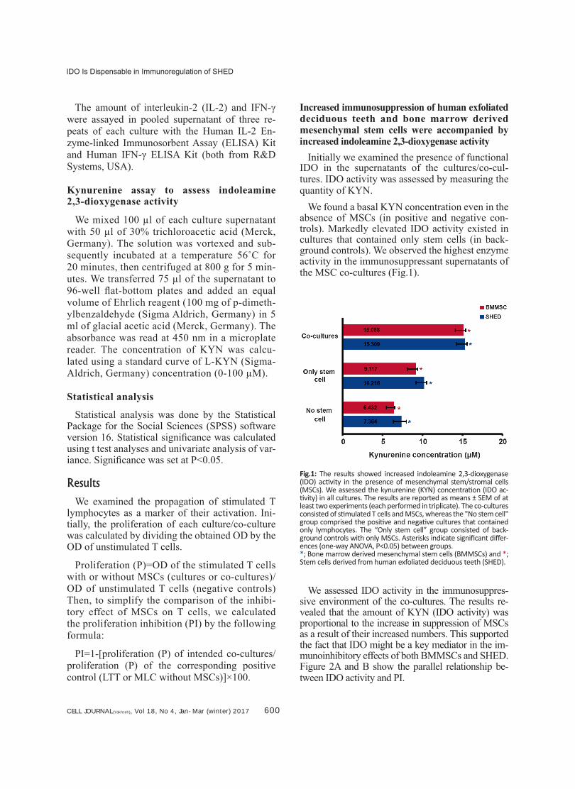
Increased immunosuppression of human exfoliated deciduous teeth and bone marrow derived mesenchymal stem cells were accompanied by increased indoleamine 2,3-dioxygenase activity
Initially we examined the presence of functional IDO in the supernatants of the cultures/co-cul-tures. IDO activity was assessed by measuring the quantity of KYN.
We found a basal KYN concentration even in the absence of MSCs (in positive and negative con-trols). Markedly elevated IDO activity existed in cultures that contained only stem cells (in back-ground controls). We observed the highest enzyme activity in the immunosuppressant supernatants of the MSC co-cultures (Fig.1).
Fig.1: The results showed increased indoleamine 2,3-dioxygenase (IDO) activity in the presence of mesenchymal stem/stromal cells (MSCs). We assessed the kynurenine (KYN) concentration (IDO ac-tivity) in all cultures. The results are reported as means ± SEM of at least two experiments (each performed in triplicate). The co-cultures consisted of stimulated T cells and MSCs, whereas the "No stem cell" group comprised the positive and negative cultures that contained only lymphocytes. The “Only stem cell” group consisted of back-ground controls with only MSCs. Asterisks indicate significant differ-ences (one-way ANOVA, P<0.05) between groups. *; Bone marrow derived mesenchymal stem cells (BMMSCs) and *; Stem cells derived from human exfoliated deciduous teeth (SHED).
We assessed IDO activity in the immunosuppres-sive environment of the co-cultures. The results re-vealed that the amount of KYN (IDO activity) was proportional to the increase in suppression of MSCs as a result of their increased numbers. This supported the fact that IDO might be a key mediator in the im-munoinhibitory effects of both BMMSCs and SHED. Figure 2A and B show the parallel relationship be-tween IDO activity and PI.

CELL JOURNAL(Yakhteh), Vol 18, No 4, Jan-Mar (Winter) 2017 601
Alipour et al.
Fig.2: Elevation in indoleamine 2,3-dioxygenase (IDO) activity ac-companied by inhibition of mesenchymal stem/stromal cells (MSCs). A. The kynurenine (KYN) concentrations were measured in the co-cultures that contained different numbers of MSCs. The horizontal axis showed the ratio of stimulated T cells to MSCs [stem cells de-rived from human exfoliated deciduous teeth (SHED) or bone mar-row mesenchymal stem cells (BMMSCs)] in the co-cultures (identical to the horizontal axis of graph B) and B. Proliferation of stimulated T cells in the presence of different number of MSCs. Proliferation inhibition (PI) calculation was calculated as detailed in the text. Re-sults are mean ± SEM of at least two experiments (each performed in triplicate). Asterisks indicate significant differences (one-way ANOVA, P<0.05) between groups.*; BMMSCs and *; SHED.
1-methyl-tryptophan and neutralizing anti-in-terferon gamma antibody decreased bone marrow derived mesenchymal stem cell immunosuppression
We used 1-MT, as an inhibitor of IDO, to further elucidate whether the suppressive effect of MSCs was attributed to IDO. We added 1 mM of 1-MT to the co-cultures, which were named MT-co-cultures. The ad-dition of 1-MT restored the activation of T cells in the BMMSC co-cultures. As the PI was markedly dimin-ished (Fig.3), the IL-2 and IFN-γ levels increased sig-nificantly (Table 1) compared to the simple co-cultures.
The level of KYN, as a marker of IDO activity, showed a remarkable drop in the MT-co-cultures (Table 1). We examined the role of IFN-γ, in extra immunosuppres-sant co-cultures, by adding neutralizing anti-human IFN-γ antibody at thte first of the cultures/co-cultures at a concentration of 4 μg/ml (Ab-co-cultures).
Results of the BMMSC co-cultures showed a no-table decrease in the suppression of lymphocyte pro-liferation (Fig.3). This reduction occurred in conjunc-tion with a remarkable diminish in IDO activity. In addition to the reduction in PI, we observed an eleva-tion in IL-2 to some extent in the Ab-co-cultures. This supported the fact that Ab could reverse the activation of stimulated T cells. There was a substantial drop in the IFN-γ level (Table 1), more likely because of the presence of neutralizing antibody.
In terms of the Ab- and MT-co-cultures, we observed that lymphocyte activation in the Ab-co-cultures re-versed more at all numbers of BMMSCs. However, PI (P=0.115), IL-2 quality (P=0.380), and IDO activity (P=0.134) did not show statistically significant chang-es compared to the MT-co-cultures (Table 1).
Effects of 1-methyl-tryptophan and neutralizing anti-interferon gamma (anti-IFN-γ) antibody on human exfoliated deciduous teeth (SHED) immunosuppression
We had co-cultures of SHED and T cells; the sim-ple co-cultures, the Ab-co-cultures with neutralizing anti-IFN-γ antibody (4 mg/ml), and the MT-co-cul-tures with 1-MT (1 mM) in the same manner as the BMMSC co-cultures.
We observed in the Ab-co-cultures that the antibody could not efficiently block the immunosuppressive ef-fect of SHED since the PI (Fig.3) and IL-2 production (Table 1) changed slightly compared to the simple co-cultures. However, we observed considerably re-duced IFN-γ levels and IDO activity (Table 1).
Surprisingly, the results of SHED and T lympho-cyte MT-co-cultures had tremendous growth in PI compared to the simple co-cultures (Fig.3). In con-junction with the PI results, we observed reduced cy-tokine levels which was meaningful for IL-2 but not for IFN-γ (Table 1). There was significantly decreased IDO activity in the MT-co-cultures compared to the simple co-cultures (Table 1).
A comparison between MT- and Ab-co-cultures showed remarkable differences in PI (P<0.05) and cytokine (for both cytokines, P<0.05) levels.
A
B

CELL JOURNAL(Yakhteh), Vol 18, No 4, Jan-Mar (winter) 2017 602
IDO Is Dispensable in Immunoregulation of SHED
Table 1: Concentrations of cytokines and indoleamine 2,3-dioxygenase (IDO) activity in supernatants of the Ab-, simple- and MT-co-cultures as determined by ELISA (cytokines) or colorimetric method (IDO). The corresponding P values (one-way ANOVA) are shown in the right
of its column (the upper and lower P values correspond to the differences between the simple co-cultures and either the Ab- co-cultures or MT-co-cultures)
Co-cultures BMMSCs SHED
IL-2 (pg/ml)
P value IFN-γ (pg/ml)
P value IDO activity (μM)
P value IL-2 (pg/ml)
P value IFN-γ (pg/ml)
P value IDO activity (μM)
P value
Ab 44.83±1.96
0.058 15.82±0.77
0.056 13.96±0.30
<0.05 67.95±4.08
0.593 28.69±1.08
<0.05 13.92±0.25
<0.05
Simple 37.02 ± 0.48
25.15±1.92
14.93±0.31
65.19±3.11
44.96±2.50
15.33±0.22
MT 48.44±1.96
<0.05 62.83±4.73
<0.05 13.15±0.27
<0.05 38.42±1.37
<0.05 38.15±1.77
0.540 12.46±0.30
<0.05
BMMSCs; Bone marrow mesenchymal stem cells, SHED; Stem cells derived from human exfoliated deciduous teeth, and IFN-γ; Interferon gamma.
Fig.3: Inhibition of stem cells derived from human exfoliated deciduous teeth (SHED) and bone marrow mesenchymal stem cells (BMMSCs) on stimulated T lymphocytes in three set of co-cultures: 1 mM 1-methyl tryptophan (1-MT), 4 μg/ml neutralizing anti-interferon gamma (anti-IFN-γ) antibody, and simple co-cultures (without any exogenous factor). The results are presented as the means and SEM from at least two experiments (each performed in triplicate). Total inhibition for three sets of co-cultures are shown by the solid line whereas the columns show inhibition of co-cultures with different numbers of mesenchymal stem/stromal cells (MSCs). Asterisks indicate significant differences (one-way ANOVA, P<0.05) between groups. *; BMMSCs and *; SHED.
CELL JOURNAL(Yakhteh), Vol 18, No 4, Jan-Mar (Winter) 2017 603
Alipour et al.
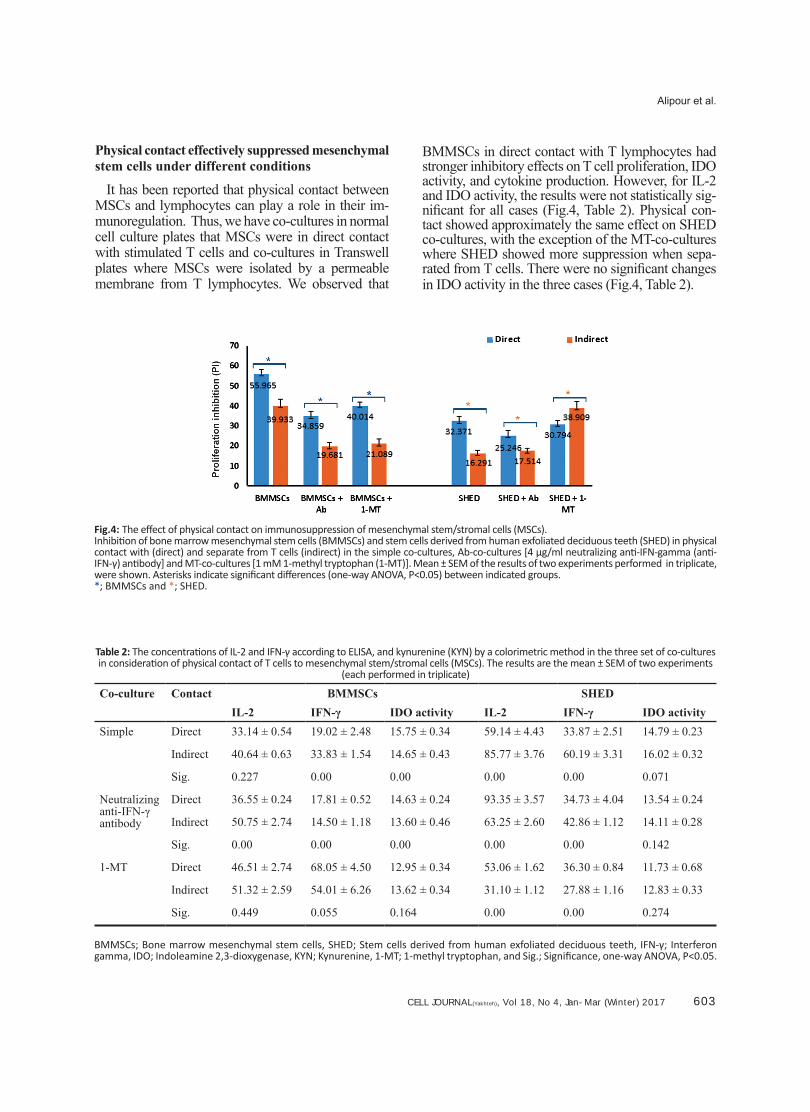
Physical contact effectively suppressed mesenchymal stem cells under different conditions
It has been reported that physical contact between MSCs and lymphocytes can play a role in their im-munoregulation. Thus, we have co-cultures in normal cell culture plates that MSCs were in direct contact with stimulated T cells and co-cultures in Transwell plates where MSCs were isolated by a permeable membrane from T lymphocytes. We observed that
BMMSCs in direct contact with T lymphocytes had stronger inhibitory effects on T cell proliferation, IDO activity, and cytokine production. However, for IL-2 and IDO activity, the results were not statistically sig-nificant for all cases (Fig.4, Table 2). Physical con-tact showed approximately the same effect on SHED co-cultures, with the exception of the MT-co-cultures where SHED showed more suppression when sepa-rated from T cells. There were no significant changes in IDO activity in the three cases (Fig.4, Table 2).
Fig.4: The effect of physical contact on immunosuppression of mesenchymal stem/stromal cells (MSCs).Inhibition of bone marrow mesenchymal stem cells (BMMSCs) and stem cells derived from human exfoliated deciduous teeth (SHED) in physical contact with (direct) and separate from T cells (indirect) in the simple co-cultures, Ab-co-cultures [4 μg/ml neutralizing anti-IFN-gamma (anti-IFN-γ) antibody] and MT-co-cultures [1 mM 1-methyl tryptophan (1-MT)]. Mean ± SEM of the results of two experiments performed in triplicate, were shown. Asterisks indicate significant differences (one-way ANOVA, P<0.05) between indicated groups.*; BMMSCs and *; SHED.
Table 2: The concentrations of IL-2 and IFN-γ according to ELISA, and kynurenine (KYN) by a colorimetric method in the three set of co-cultures in consideration of physical contact of T cells to mesenchymal stem/stromal cells (MSCs). The results are the mean ± SEM of two experiments
(each performed in triplicate)
Co-culture Contact BMMSCs SHEDIL-2 IFN-γ IDO activity IL-2 IFN-γ IDO activity
Simple Direct 33.14 ± 0.54 19.02 ± 2.48 15.75 ± 0.34 59.14 ± 4.43 33.87 ± 2.51 14.79 ± 0.23
Indirect 40.64 ± 0.63 33.83 ± 1.54 14.65 ± 0.43 85.77 ± 3.76 60.19 ± 3.31 16.02 ± 0.32
Sig. 0.227 0.00 0.00 0.00 0.00 0.071
Neutralizing anti-IFN-γ antibody
Direct 36.55 ± 0.24 17.81 ± 0.52 14.63 ± 0.24 93.35 ± 3.57 34.73 ± 4.04 13.54 ± 0.24
Indirect 50.75 ± 2.74 14.50 ± 1.18 13.60 ± 0.46 63.25 ± 2.60 42.86 ± 1.12 14.11 ± 0.28
Sig. 0.00 0.00 0.00 0.00 0.00 0.142
1-MT Direct 46.51 ± 2.74 68.05 ± 4.50 12.95 ± 0.34 53.06 ± 1.62 36.30 ± 0.84 11.73 ± 0.68
Indirect 51.32 ± 2.59 54.01 ± 6.26 13.62 ± 0.34 31.10 ± 1.12 27.88 ± 1.16 12.83 ± 0.33
Sig. 0.449 0.055 0.164 0.00 0.00 0.274
BMMSCs; Bone marrow mesenchymal stem cells, SHED; Stem cells derived from human exfoliated deciduous teeth, IFN-γ; Interferon gamma, IDO; Indoleamine 2,3-dioxygenase, KYN; Kynurenine, 1-MT; 1-methyl tryptophan, and Sig.; Significance, one-way ANOVA, P<0.05.

CELL JOURNAL(Yakhteh), Vol 18, No 4, Jan-Mar (winter) 2017 604
IDO Is Dispensable in Immunoregulation of SHED
DiscussionExperimental data indicate that IDO activity in-
duced preferentially by IFN-γ mainly contributes to the immunosuppressive effect of MSCs (25, 26). However IFN-γ is also considered to be one of the important factors that affect the immunoregu-latory properties of MSCs (12, 18).
SHED are recently discovered compared to other MSCs (19, 22). Despite numerous studies concerning the role of the IFN-γ-IDO axis in im-munosuppression of various MSCs, we have not found any publications that discuss the role of this axis in the immunoregulation of SHED. A better understanding of SHED immunomodulation will offer an insight into their use for clinical applica-tions. Therefore, in this study, we have explored the role of IDO and IFN-γ in immunoregulatory effects of SHED and compared them to BMMSCs as conventional MSCs.
IDO catalyzes tryptophan into KYN, which can either enter the blood or additionally metabolize to further KYN metabolites which, in turn, exert immunoregulatory properties (16). Measurement of KYN concentration by an indirect colorimetric method is frequently used as an easy, acceptable method that estimates IDO activity (27-29).
The results showed notable activity of IDO in both MSC cultures (background controls), com-pared to the positive and negative controls that contained no MSCs. This observation indicated that either SHED or BMMSCs might produce functional IDO under normal conditions. The IDO activity between the two MSC cultures (two back-ground controls) was similar.
Numerous studies have inconsistently reported that MSCs did not express IDO under basal cul-ture conditions. Ryan et al. (17) and DelaRosa et al. (30) detected neither the expression of IDO protein nor IDO activity in the supernatant of hu-man BMMSCs and human adipose-derived MSCs (hASCs) before IFN-γ treatment. However, other investigations implied that MSCs continuously expressed this enzyme in the absence of stimuli. According to results reported by Yoo et al. (31) the RNA of IDO was detectable in untreated hASCs. Additionally, Chang et al. (32) reported the IDO protein in two untreated human MSC populations, BMMSCs and placenta-derived multipotent cells
(PDMCs). Similarly, Djouad et al. (33) detected clearly IDO activity in the supernatants of primary human MSCs.
This discrepancy could be explained somewhat by the heterogeneous nature of MSCs (34). Distinct BMMSC subsets that differ in their immunophe-notype, morphology (35), and immunosuppressive action (36) have been reported. It has been well established that the clinical features of the subjects from whom the cells were isolated affect the char-acteristics of hMSCs (37). Evidences showed that either the culture or manipulation could impact the biological properties of MSCs (38, 39). Thus it is not unexpected that under the current study cul-ture conditions, the two MSC groups (SHED and BMMSCs) produced functional IDO that caused detectable increased KYN concentration in their supernatants.
There was significantly increased IDO activity in co-cultures of both SHED and BMMSCs. Ad-ditionally in simple co-cultures, along with the in-creased MSC numbers that resulted in increased suppression, we observed increased IDO activity. As a conclusion from these data, IDO might con-tribute to the immunosuppression of both MSC types used in this research.
Next, we sought to confirm IDO involvement in immunosuppression of MSCs by the addition of 1-MT, an IDO inhibitor to the co-cultures. In the MT-co-cultures of BMMSCs, 1-MT significantly restored the lymphocyte activation which agreed with our results from the simple co-cultures. In-creased immunosuppression was accompanied with augmented IDO activity in the simple co-cultures. The inhibition of IDO activity was fol-lowed by decreased immunosuppression in the MT-co-cultures. Thus, our findings represented a key role of IDO in BMMSCs-induced immu-nosuppression which has been demonstrated in previous studies (40-42).
Unpredictably, in the MT-co-cultures with SHED, blocked IDO activity by 1-MT did not reduce immunoinhibition; rather, there was a re-markable increase.
This unexpected outcome contrasted numerous reports on various MSCs (25, 31, 32). However we have been unable to find any study on SHED that evaluated IDO. These results, however, need
CELL JOURNAL(Yakhteh), Vol 18, No 4, Jan-Mar (Winter) 2017 605
Alipour et al.
additional investigations. The following points are noteworthy.
IDO is known as an immunoregulatory enzyme (43) that participates in immunosuppressive events such as the escape of tumor cells from host im-mune surveillance (44) and allogeneic fetal toler-ance (45).
However some reports (46) question the im-munosuppressive nature of IDO. For example, in patients with systemic lupus erythematosus (SLE) as the disease progresses there is increased IDO activity in the blood (47). SLE is an autoimmune disease alleviated by immunosuppressants, hence, there is no explanation as to why IDO activity par-allels disease exacerbation. Similar results have been reported in rheumatoid arthritis (RA) patients (48). Scott et al. (49) observed that when IDO ac-tivity was inhibited by subcutaneous administra-tion of 1-MT in a mice model of RA, the disease was alleviated. 1-MT is expected to aggravate the disease by blocking the immunosuppressant IDO enzyme. IDO has been shown to aggravate inflam-mation in airways in animal models of allergic in-flammation (50). These studies imply that IDO is not permanently immunosuppressive and it may have immune stimulatory effects under currently unknown conditions.
Recently, it has been revealed that beside the catalytic function of IDO as an enzyme, it also contains immunoreceptor tyrosine-based inhibitory motifs (ITIMs) which can bind to diverse molecular partners and affect intracellular signaling pathways (51, 52). Orabona et al. (53) have shown that under defined conditions IDO binds to SOCS3 (an intra-cellular signaling molecule) and the resultant IDO-SOCS3 complex is subsequently degraded.
In the SHED co-cultures, there might have been conditions under which IDO did not play an im-munosuppressive role. However, we do not know the exact conditions responsible for this role. How-ever, it is a weak possibility that 1-MT may play an unknown role in SHED. Some clinical researches suggest that the D isomer of 1-MT (D-1-MT) can play a role other than inhibition of IDO (54). This isomer (D-1-MT) does not participate in the inhi-bition of IDO activity, however, it enhances im-munity in cancer patients (55, 56). Of note, in the current study, we have used a mixture of both the D and L 1-MT isomers.
According to the results of this study, we cannot completely explain the increase in immunoinhibi-tory action of SHED in the MT-co-cultures. Ad-ditional research is necessary to confirm and elu-cidate this result.
It has been established that the immunomodu-latory capacity of MSCs is induced or at least augmented under inflammatory conditions (6-9). In this context, various methods have been used to examine the effect of IFN-γ as an inflamma-tory cytokine on the immunoregulatory action of MSCs. IFN-γ has a dual role. It is one of the first cytokines secreted from activated T cells and pro-motes their activation on one hand. However, on the other hand, IFN-γ induces MSC immunosup-pression and enables them to more efficiently in-hibit T lymphocyte activation (12). In this study, we have attempted to remove IFN-γ which was secreted by cells in the co-cultures (and is not ex-ogenous) to levels that enabled T cell activation, yet limited immunosuppression of MSCs as low as possible.
The results showed that Abs caused a meaningful decrease in both the proliferation and cytokine pro-duction of stimulated T cells in the Ab-co-cultures of BMMSCs. Thus IFN-γ must have an enhanc-ing effect on immunoregulation of BMMSCs. This finding was compatible with reported results from numerous researchers in terms of BMMSCs (57, 58) and other types of MSCs (12, 17). Although the results of IFN-γ SHED co-cultures showed a decrease in immunosuppression, we did not ob-serve any significant decrease in T cell prolifera-tion or cytokine production (IL-2). We found no study on SHED to compare our results. However, immunoregulatory actions of oral MSCs such as gingiva-derived MSCs (GMSCs) (59), periodon-tal ligament stem cells (PDLSCs), and dental pulp stem cells (DPSCs) (60) definitely increase in the presence of IFN-γ.
Our results did not completely agree with these studies. This might be due to inherent differences in various MSCs types and the individual charac-teristics of SHED.
Nevertheless, immunoinhibitory characteristics of SHED might be affected by IFN-γ. However be-cause of its differences from BMMSCs, the condi-tions (for example the amount of IFN-γ) in which they are affected vary from BMMSCs. It has been
CELL JOURNAL(Yakhteh), Vol 18, No 4, Jan-Mar (winter) 2017 606
IDO Is Dispensable in Immunoregulation of SHED
identified that different tissue-derived MSCs re-spond differently to cytokines (61-63). This pos-sibility was potentiated by our observation that with a SHED/T cell ratio of 1:10, the reduction in inhibition was more than expected and did not fol-low the trend shown in other ratios. It showed a decline compared to other ratios. Erkers et al. (64) showed that in contrast to BMMSCs, low levels of IFN-γ had no effect on the suppressive capacity of decidual stromal cells (DSCs).
As a whole, we cannot conclude with certainty that IFN-γ did not affect immunoregulation of SHED. We expected that if other concentrations of IFN-γ or other methods such as pretreatment of SHED by IFN-γ were applied, SHED might be af-fected by IFN-γ. Next, we evaluated the IDO ac-tivity in Ab-co-cultures to check the IFN-γ-IDO axis in immunoregulation of MSCs.
In the current study, for BMMSCs along with a significant reduction in IFN-γ by neutralizing Abs, we observed significant decrease in IDO activity in the co-cultures, which was followed by a sig-nificant diminish in immunoinhibition. A compari-son of Ab-co-cultures to 1-MT-co-cultures showed that although the Ab had a more reductive effect, however as a whole, the two co-cultures did not significantly differ in terms of lymphocyte pro-liferation and cytokine secretion. Similar results were previously reported in which IFN-γ induced IDO play a major role or at least one of the im-portant roles in immunosuppression of human BMMSCs (58, 63).
For SHED, we observed that the drop in IFN-γ in the Ab-co-cultures coincided with a decline in IDO activity. However, the reduction of IFN-γ in supernatants and the decrease in IDO activity did not accompany a significant decrease in immuno-suppression. Therefore, it seemed that IFN-γ could increase IDO activity in SHED as well as in nu-merous other MSC populations. However, this en-zyme does not mainly participate in immunosup-pression of SHED. Rossi et al. (65) observed that human amniotic tissue derived MSCs (hAMTC) produced IDO. Although 1-MT could prominently reduce IDO activity, hAMTC induced immuno-suppression did not change. Obviously this should be confirmed by similar experiments and further genetic studies.
Finally, the evaluation of direct and indirect
co-cultures (simple-co-cultures, Ab-co-cultures, MT-co-cultures) showed that in all cultures with physical contact between immune and stem cells, there was stronger immunosuppression. This was justified by the fact that some contact-dependent mechanisms also had a role in immunoregula-tion of MSCs (31, 64). However, the results of the SHED MT-co-cultures were inverse, which should be further investigated. According to Nasef et al. (66), when MSCs have direct or indirect contact with immune cells, they might apply separate soluble immunoregulatory factors that affect these cells. Thus, in the MT-co-cultures, SHED prob-ably produced factors with more immunomodula-tory effects when separated from T lymphocytes compared to when they were in physical contact. MT-co-cultures of SHED showed different indi-vidual results compared to BMMSCs.
ConclusionThis study demonstrated that SHED as a subset
of oral MSCs has immune properties that resem-ble other MSCs. SHED are similar to conventional MSCs or BMMSCs. They have anti-proliferative ef-fects on stimulated T cells and reduce cytokine pro-duction from them. These properties of SHED may be affected by inflammatory conditions that occur in the presence of IFN-γ. However, we observed a non-significant decrease in immunosuppression of SHED after the use of neutralizing anti-IFN-γ anti-bodies, which differed from BMMSCs. SHED pro-duced immunoregulatory factors such as IDO. Simi-lar to BMMSCs, at least one of the inducers of IDO in SHED is IFN-γ. However unlike BMMSCs, this molecule does not mainly contribute to their immu-nosuppression and can have other cell-type specific roles. The IFN-γ-IDO axis seems to exist in SHED but it has no remarkable role in immunoregulatory effects. Finding the molecules involved in immu-nomodulatory effects of SHED and their differences with other MSCs requires additional research.
AcknowledgmentsThis work was financially supported by the Re-
search and Technology Assistant of Isfahan Uni-versity of Medical Sciences. The authors would like to thank Torabi Nejad Research Center, Facul-ty of Dentistry, Isfahan University of Medical Sci-ences for generously providing SHED. We thank Nafiseh Esmaeili and Mrs. Aliakbari for practical

CELL JOURNAL(Yakhteh), Vol 18, No 4, Jan-Mar (Winter) 2017 607
Alipour et al.
help and scientific assistance. The authors declare that they have no competing interests.
References1. Hematti P. Role of mesenchymal stromal cells in solid
organ transplantation. Transplant Rev (Orlando). 2008; 22(4): 262-273.
2. Griffin MD, Ritter T, Mahon BP. Immunological aspects of allogeneic mesenchymal stem cell therapies. Hum Gene Ther. 2010; 21(12): 1641-1655.
3. Chen Y, Shao JZ, Xiang LX, Dong XJ, Zhang GR. Mesen-chymal stem cells: a promising candidate in regenerative medicine. Int J Biochem Cell Biol. 2008; 40(5): 815-820.
4. Charbord P. Bone marrow mesenchymal stem cells: his-torical overview and concepts. Hum Gene Ther. 2010; 21(9): 1045-1056.
5. Caplan AI. Why are MSCs therapeutic? New data: new insight. J Pathol. 2009; 217(2): 318-324.
6. Kode JA, Mukherjee S, Joglekar MV, Hardikar AA. Mes-enchymal stem cells: immunobiology and role in immu-nomodulation and tissue regeneration. Cytotherapy. 2009; 11(4): 377-391.
7. Le Blanc K, Ringden O. Immunomodulation by mesen-chymal stem cells and clinical experience. J Intern Med. 2007; 262(5): 509-525.
8. Knaan-Shanzer S. Concise review: the immune status of mesenchymal stem cells and its relevance for therapeutic application. Stem Cells. 2014; 32(3): 603-608.
9. Atoui R, Chiu RC. Concise review: immunomodulatory properties of mesenchymal stem cells in cellular trans-plantation: update, controversies, and unknowns. Stem Cells Transl Med. 2012; 1(3): 200-205.
10. Stagg J. Immune regulation by mesenchymal stem cells: two sides to the coin. Tissue Antigens. 2007; 69(1): 1-9.
11. Figueroa FE, Carrion F, Villanueva S, Khoury M. Mesen-chymal stem cell treatment for autoimmune diseases: a critical review. Biol Res. 2012; 45(3): 269-277.
12. Krampera M. Mesenchymal stromal cell 'licensing': a mul-tistep process. Leukemia. 2011; 25(9): 1408-1414.
13. Glenn JD, Whartenby KA. Mesenchymal stem cells: emerging mechanisms of immunomodulation and therapy. World J Stem Cells. 2014; 6(5): 526-539.
14. Kyurkchiev D, Bochev I, Ivanova-Todorova E, Mourdjeva M, Oreshkova T, Belemezova K, et al. Secretion of immu-noregulatory cytokines by mesenchymal stem cells. World J Stem Cells. 2014; 6(5): 552-570.
15. Reinders ME, Rabelink TJ, De Fijter JW. The role of mes-enchymal stromal cells in chronic transplant rejection after solid organ transplantation. Curr Opin Organ Transplant. 2013; 18(1): 44-50.
16. Orabona C, Grohmann U. Indoleamine 2,3-dioxygenase and regulatory function: tryptophan starvation and be-yond. Methods Mol Biol. 2011; 677: 269-280.
17. Ryan JM, Barry F, Murphy JM, Mahon BP. Interferon-gam-ma does not break, but promotes the immunosuppressive capacity of adult human mesenchymal stem cells. Clin Exp Immunol. 2007; 149(2): 353-363.
18. Stolzing A, Jones E, McGonagle D, Scutt A. Age-related changes in human bone marrow-derived mesenchymal stem cells: consequences for cell therapies. Mech Ageing Dev. 2008; 129(3): 163-173.
19. Suchanek J, Visek B, Soukup T, El-Din Mohamed SK, Iv-ancakova R, Mokry J, et al. Stem cells from human exfoli-ated deciduous teeth-isolation, long term cultivation and phenotypical analysis. Acta Medica (Hradec Kralove). 2010; 53(2): 93-99.
20. Koyama N, Okubo Y, Nakao K, Bessho K. Evaluation of pluripotency in human dental pulp cells. J Oral Maxillofac Surg. 2009; 67(3): 501-506.
21. Telles PD, Machado MA, Sakai VT, Nor JE. Pulp tissue from primary teeth: new source of stem cells. J Appl Oral Sci. 2011; 19(3): 189-194.
22. Miura M, Gronthos S, Zhao M, Lu B, Fisher LW, Robey PG, et al. SHED: stem cells from human exfoliated de-ciduous teeth. Proc Natl Acad Sci USA. 2003; 100(10): 5807-5812.
23. Yamaza T, Kentaro A, Chen C, Liu Y, Shi Y, Gronthos S, et al. Immunomodulatory properties of stem cells from human exfoliated deciduous teeth. Stem Cell Res Ther. 2010; 1(1): 5.
24. Alipour R, Adib M, Masoumi Karimi M, Hashemi-Beni B, Sereshki N. Comparing the immunoregulatory effects of stem cells from human exfoliated deciduous teeth and bone marrow-derived mesenchymal stem cells. Iran J Al-lergy Asthma Immunol. 2013; 12(4): 331-344.
25. Tipnis S, Viswanathan C, Majumdar AS. Immunosuppres-sive properties of human umbilical cord-derived mesen-chymal stem cells: role of B7-H1 and IDO. Immunol Cell Biol. 2010; 88(8): 795-806.
26. Francois M, Romieu-Mourez R, Li M, Galipeau J. Human MSC suppression correlates with cytokine induction of indoleamine 2,3-dioxygenase and bystander M2 mac-rophage differentiation. Mol Ther. 2012; 20(1): 187-195.
27. Roemeling-van Rhijn M, Mensah FK, Korevaar SS, Leijs MJ, van Osch GJ, Ijzermans, JN, et al. Effects of hypoxia on the immunomodulatory properties of adipose tissue-derived mesenchymal stem cells. Front Immunol. 2013; 4: 203.
28. Leijs, MJ, van Buul GM, Lubberts E, Bos PK, Verhaar JA, Hoogduijn MJ, et al. Effect of arthritic synovial fluids on the expression of immunomodulatory factors by mes-enchymal stem cells: an explorative in vitro Study. Front Immunol. 2012; 3: 231.
29. Schmidt SK, Ebel S, Keil E, Woite C, Ernst JF, Benzin AE, et al. Regulation of IDO activity by oxygen supply: inhibitory effects on antimicrobial and immunoregulatory functions. PLoS One. 2013; 8(5): e63301.
30. DelaRosa O, Lombardo E, Beraza A, Mancheno-Corvo P, Ramirez C, Menta R, et al. Requirement of IFN-gamma-mediated indoleamine 2,3-dioxygenase expression in the modulation of lymphocyte proliferation by human adipose-derived stem cells. Tissue Eng Part A. 2009; 15(10): 2795-2806.
31. Yoo KH, Jang IK, Lee MW, Kim HE, Yang MS, Eom Y, et al. Comparison of immunomodulatory properties of mes-enchymal stem cells derived from adult human tissues. Cell Immunol. 2009; 259(2): 150-156.
32. Chang CJ, Yen ML, Chen YC, Chien CC, Huang HI, Bai CH, et al. Placenta-derived multipotent cells exhibit im-munosuppressive properties that are enhanced in the presence of interferon-gamma. Stem Cells. 2006; 24(11): 2466-2477.
33. Djouad F, Charbonnier LM, Bouffi C, Louis-Plence P, Bony C, Apparailly F, et al. Mesenchymal stem cells inhibit the differentiation of dendritic cells through an interleukin-6-dependent mechanism. Stem Cells. 2007; 25(8): 2025-2032.
34. Bernardo ME, Locatelli F, Fibbe WE. Mesenchymal stro-mal cells. Ann N Y Acad Sci. 2009; 1176: 101-117.
35. Battula VL, Treml S, Bareiss PM, Gieseke F, Roelofs H, De Zwart P, et al. Isolation of functionally distinct mesen-chymal stem cell subsets using antibodies against CD56, CD271, and mesenchymal stem cell antigen-1. Haemato-logica. 2009; 94(2): 173-184.

CELL JOURNAL(Yakhteh), Vol 18, No 4, Jan-Mar (winter) 2017 608
IDO Is Dispensable in Immunoregulation of SHED
36. Nasef A, Zhang YZ, Mazurier C, Bouchet S, Bensidhoum M, Francois S, et al. Selected Stro-1-enriched bone mar-row stromal cells display a major suppressive effect on lymphocyte proliferation. Int J Lab Hematol. 2009; 31(1): 9-19.
37. Baer PC, Kuci S, Krause M, Kuci Z, Zielen S, Geiger H, et al. Comprehensive phenotypic characterization of human adipose-derived stromal/stem cells and their subsets by a high throughput technology. Stem Cells Dev. 2013; 22(2): 330-339.
38. Gao L, Liu F, Tan L, Liu T, Chen Z, Shi C. The immunosup-pressive properties of non-cultured dermal-derived mes-enchymal stromal cells and the control of graft-versus-host disease. Biomaterials. 2014; 35(11): 3582-3588.
39. Moll G, Alm JJ, Davies LC, von Bahr L, Heldring N, Sten-beck-Funke L, et al. Do cryopreserved mesenchymal stro-mal cells display impaired immunomodulatory and thera-peutic properties? Stem Cells. 2014; 32(9): 2430-2442.
40. Meisel R, Brockers S, Heseler K, Degistirici O, Bulle H, Woite C, et al. Human but not murine multipotent mesen-chymal stromal cells exhibit broad-spectrum antimicrobial effector function mediated by indoleamine 2,3-dioxyge-nase. Leukemia. 2011; 25(4): 648-654.
41. Su J, Chen X, Huang Y, Li W, Li J, Cao K, et al. Phylo-genetic distinction of iNOS and IDO function in mesen-chymal stem cell-mediated immunosuppression in mam-malian species. Cell Death Differ. 2014; 21(3): 388-396.
42. Ren G, Su J, Zhang L, Zhao X, Ling W, L'Huillie A, et al. Species variation in the mechanisms of mesenchymal stem cell-mediated immunosuppression. Stem Cells. 2009; 27(8): 1954-1962.
43. Andersen MH. The specific targeting of immune regulation: T-cell responses against Indoleamine 2,3-dioxygenase. Cancer Immunol Immunother. 2012; 61(8): 1289-1297.
44. Prendergast GC, Smith C, Thomas S, Mandik-Nayak L, Laury-Kleintop L, Metz R, et al. Indoleamine 2,3-dioxyge-nase pathways of pathogenic inflammation and immune escape in cancer. Cancer Immunol Immunother. 2014; 63(7): 721-735.
45. von Rango U. Fetal tolerance in human pregnancy--a cru-cial balance between acceptance and limitation of tropho-blast invasion. Immunol Lett. 2008; 115(1): 21-32.
46. Muller AJ, Mandik-Nayak L, Prendergast GC. Beyond im-munosuppression: reconsidering indoleamine 2,3-dioxy-genase as a pathogenic element of chronic inflammation. Immunotherapy. 2010; 2(3): 293-297.
47. Pertovaara M, Hasan T, Raitala A, Oja SS, Yli-Kerttula U, Korpela M, et al. Indoleamine 2,3-dioxygenase activity is increased in patients with systemic lupus erythematosus and predicts disease activation in the sunny season. Clin Exp Immunol. 2007; 150(2): 274-278.
48. Schroecksnadel K, Winkler C, Duftner C, Wirleitner B, Schirmer M, Fuchs D. Tryptophan degradation increases with stage in patients with rheumatoid arthritis. Clin Rheu-matol. 2006; 25(3): 334-337.
49. Scott GN, DuHadaway J, Pigott E, Ridge N, Prendergast GC, Muller AJ, et al. The immunoregulatory enzyme IDO paradoxically drives B cell-mediated autoimmunity. J Im-munol. 2009; 182(12): 7509-7517.
50. Xu H, Oriss TB, Fei M, Henry AC, Melgert BN, Chen L, et al. Indoleamine 2,3-dioxygenase in lung dendritic cells promotes Th2 responses and allergic inflammation. Proc Natl Acad Sci USA. 2008; 105(18): 6690-6695.
51. Fallarino F, Grohmann U, Puccetti P. Indoleamine 2,3-di-oxygenase: from catalyst to signaling function. Eur J Im-
munol. 2012; 42(8): 1932-1937.52. Orabona C, Pallotta MT, Grohmann U. Different partners,
opposite outcomes: a new perspective of the immunobiol-ogy of indoleamine 2,3-dioxygenase. Mol Med. 2012; 18: 834-842.
53. Orabona C, Pallotta MT, Volpi C, Fallarino F, Vacca C, Bianchi R, et al. SOCS3 drives proteasomal degradation of indoleamine 2,3-dioxygenase (IDO) and antagonizes IDO-dependent tolerogenesis. Proc Natl Acad Sci USA. 2008; 105(52): 20828-20833.
54. Lob S, Konigsrainer A, Schafer R, Rammensee HG, Op-elz G, Terness P. Levo- but not dextro-1-methyl tryptophan abrogates the IDO activity of human dendritic cells. Blood. 2008; 111(4): 2152-2154.
55. Lob S, Konigsrainer A. Is IDO a key enzyme bridging the gap between tumor escape and tolerance induction? Lan-genbecks Arch Surg. 2008; 393(6): 995-1003.
56. Soliman H, Mediavilla-Varela M, Antonia S. Indoleamine 2,3-dioxygenase: is it an immune suppressor? Cancer J. 2010; 16(4): 354-359.
57. Liang C, Chen SL, Wang M, Zhai WJ, Zhou Z, Pang AM, et al. Synergistic immunomodulatory effects of interferon-gamma and bone marrow mesenchymal stem cells. Zhon-ghua Xue Ye Xue Za Zhi. 2013; 34(3): 213-216.
58. Hemeda H, Jakob M, Ludwig AK, Giebel B, Lang S, Bran-dau S. Interferon-gamma and tumor necrosis factor-alpha differentially affect cytokine expression and migration properties of mesenchymal stem cells. Stem Cells Dev. 2010; 19(5): 693-706.
59. Zhang QZ, Su WR, Shi SH, Wilder-Smith P, Xiang AP, Wong A, et al. Human gingiva-derived mesenchymal stem cells elicit polarization of m2 macrophages and enhance cutaneous wound healing. Stem Cells. 2010; 28(10): 1856-1868.
60. Wada N, Menicanin D, Shi S, Bartold PM, Gronthos S. Immunomodulatory properties of human periodontal liga-ment stem cells. J Cell Physiol. 2009; 219(3): 667-676.
61. Yang J, Song T, Wu P, Chen Y, Fan X, Chen H, et al. Dif-ferentiation potential of human mesenchymal stem cells derived from adipose tissue and bone marrow to sinus node-like cells. Mol Med Rep. 2012; 5(1): 108-113.
62. Strioga M, Viswanathan S, Darinskas A, Slaby O, Michalek J. Same or not the same? Comparison of adipose tissue-derived versus bone marrow-derived mesenchymal stem and stromal cells. Stem Cells Dev. 2012; 21(14): 2724-2752.
63. Prasanna SJ, Gopalakrishnan D, Shankar SR, Vasandan AB. Pro-inflammatory cytokines, IFNgamma and TNFal-pha, influence immune properties of human bone marrow and Wharton jelly mesenchymal stem cells differentially. PLoS One. 2010; 5(2): e9016.
64. Erkers T, Nava S, Yosef J, Ringden O, Kaipe H. Decidual stromal cells promote regulatory T cells and suppress allo-reactivity in a cell contact-dependent manner. Stem Cells Dev. 2013; 22(19): 2596-2605.
65. Rossi D, Pianta S, Magatti M, Sedlmayr P, Parolini O. Characterization of the conditioned medium from amniotic membrane cells: prostaglandins as key effectors of its im-munomodulatory activity. PLoS One. 2012; 7(10): e46956.
66. Nasef A, Chapel A, Mazurier C, Bouchet S, Lopez M, Mathieu N, et al. Identification of IL-10 and TGF-beta tran-scripts involved in the inhibition of T-lymphocyte prolifera-tion during cell contact with human mesenchymal stem cells. Gene Expr. 2007; 13(4-5): 217-226.