Embed Size (px)
Citation preview
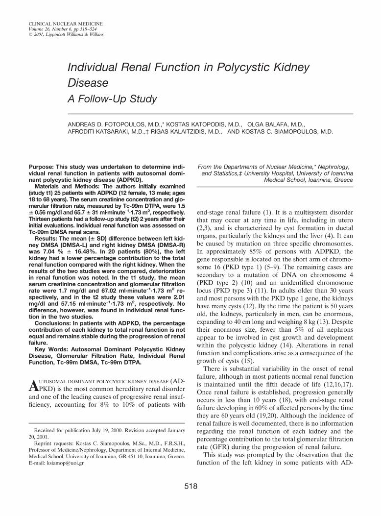
CLINICAL NUCLEAR MEDICINEVolume 26, Number 6, pp 518–524© 2001, Lippincott Williams & Wilkins
Individual Renal Function in Polycystic KidneyDiseaseA Follow-Up Study
ANDREAS D. FOTOPOULOS, M.D.,* KOSTAS KATOPODIS, M.D.,† OLGA BALAFA, M.D.,†AFRODITI KATSARAKI, M.D.,‡ RIGAS KALAITZIDIS, M.D.,† AND KOSTAS C. SIAMOPOULOS, M.D.†
Purpose: This study was undertaken to determine indi-vidual renal function in patients with autosomal domi-nant polycystic kidney disease (ADPKD).
Materials and Methods: The authors initially examined(study t1) 25 patients with ADPKD (12 female, 13 male; ages18 to 68 years). The serum creatinine concentration and glo-merular filtration rate, measured by Tc-99m DTPA, were 1.56 0.56 mg/dl and 65.7 6 31 ml·minute-1·1.73 m2, respectively.Thirteen patients had a follow-up study (t2) 2 years after theirinitial evaluations. Individual renal function was assessed onTc-99m DMSA renal scans.
Results: The mean (6 SD) difference between left kid-ney DMSA (DMSA-L) and right kidney DMSA (DMSA-R)was 7.04 % 6 16.48%. In 20 patients (80%), the leftkidney had a lower percentage contribution to the totalrenal function compared with the right kidney. When theresults of the two studies were compared, deteriorationin renal function was noted. In the t1 study, the meanserum creatinine concentration and glomerular filtrationrate were 1.7 mg/dl and 67.02 ml·minute-1·1.73 m2 re-spectively, and in the t2 study these values were 2.01mg/dl and 57.15 ml·minute-1·1.73 m2, respectively. Nodifference, however, was found in individual renal func-tion in the two studies.
Conclusions: In patients with ADPKD, the percentagecontribution of each kidney to total renal function is notequal and remains stable during the progression of renalfailure.
Key Words: Autosomal Dominant Polycystic KidneyDisease, Glomerular Filtration Rate, Individual RenalFunction, Tc-99m DMSA, Tc-99m DTPA.
AUTOSOMAL DOMINANT POLYCYSTIC KIDNEY DISEASE (AD-PKD) is the most common hereditary renal disorder
and one of the leading causes of progressive renal insuf-ficiency, accounting for 8% to 10% of patients with
end-stage renal failure (1). It is a multisystem disorderthat may occur at any time in life, including in utero(2,3), and is characterized by cyst formation in ductalorgans, particularly the kidneys and the liver (4). It canbe caused by mutation on three specific chromosomes.In approximately 85% of persons with ADPKD, thegene responsible is located on the short arm of chromo-some 16 (PKD type 1) (5–9). The remaining cases aresecondary to a mutation of DNA on chromosome 4(PKD type 2) (10) and an unidentified chromosomelocus (PKD type 3) (11). In adults older than 30 yearsand most persons with the PKD type 1 gene, the kidneyshave many cysts (12). By the time the patient is 50 yearsold, the kidneys, particularly in men, can be enormous,expanding to 40 cm long and weighing 8 kg (13). Despitetheir enormous size, fewer than 5% of all nephronsappear to be involved in cyst growth and developmentwithin the polycystic kidney (14). Alterations in renalfunction and complications arise as a consequence of thegrowth of cysts (15).
There is substantial variability in the onset of renalfailure, although in most patients normal renal functionis maintained until the fifth decade of life (12,16,17).Once renal failure is established, progression generallyoccurs in less than 10 years (18), with end-stage renalfailure developing in 60% of affected persons by the timethey are 60 years old (19,20). Although the incidence ofrenal failure is well documented, there is no informationregarding the renal function of each kidney and thepercentage contribution to the total glomerular filtrationrate (GFR) during the progression of renal failure.
This study was prompted by the observation that thefunction of the left kidney in some patients with AD-
Received for publication July 19, 2000. Revision accepted January20, 2001.
Reprint requests: Kostas C. Siamopoulos, M.Sc., M.D., F.R.S.H.,Professor of Medicine/Nephrology, Department of Internal Medicine,Medical School, University of Ioannina, GR 451 10, Ioannina, Greece.E-mail: [email protected]
From the Departments of Nuclear Medicine,* Nephrology,†and Statistics,‡ University Hospital, University of Ioannina
Medical School, Ioannina, Greece
518

PKD is more compromised compared with that of theright. Our aim in this study was to determine the renalfunction of each kidney in patients with ADPKD and toevaluate the rate of renal function loss from each kidneywith the progression of renal failure.
Materials and Methods
Patients
We examined 25 patients with ADPKD (13 male, 12 female;mean age, 42.8 years; age range, 18 to 68 years). A positivehereditary history was evident in all patients. In each case, thediagnosis of polycystic disease was established by the attendingnephrologist using standard investigations including renal ul-trasound or computed tomography.
In all patients the serum creatinine concentration was de-termined by the Jaffe kinetic method, adapted for an autoana-lyzer (Olympus AU 600). The GFR was measured using aTc-99m DTPA urinary clearance method. Finally, individualrenal function was assessed on Tc-99m DMSA scans and usingthe geometric mean method.
Thirteen patients had a follow-up study within 2 years(range, 23 to 25 months). These patients included 7 men, witha mean age 46.7 years (age range, 23 to 68 years). The serumcreatinine concentration in the first study was (mean 6 SD) 1.76 0.64 mg/dl (range, 1 to 3.5 mg/dl), and in the second study itwas 2.01 6 1.66 mg/dl (range, 1 to 7.2 mg/dl).
This study was approved by the local research ethics com-mittee of the University Hospital of Ioannina, and all patientsgave full informed consent.
Assessment of Renal Function
Each patient was brought to the nuclear medicine depart-ment at the University Hospital of Ioannina after an overnightfast on 2 consecutive days.
On day 1, the GFR was measured. On day 2, Tc-99m DMSAimaging was performed. On day 1, the patient was asked todrink 600 ml fluid 30 minutes before the test to initiate diure-sis. An intravenous line was placed in one arm with a dextrose-and-water (5%) infusion at 125 ml/hour. Immediately beforeTc-99m DTPA administration, the patient was asked to void.Two syringes containing 1 mCi (37 MBq) Tc-99m DTPA wereprepared. One was used as a standard and the other wasinjected intravenously. The syringes were weighed on an ana-lytical balance before and after injection or dilution. Bloodsamples from the opposite arm were drawn into heparin-pre-pared tubes for the renal Tc-99m DTPA clearance study 2, 3,and 4 hours after Tc-99m DTPA injection.
Urine output was also measured at 2, 3, and 4 hours. Du-plicate plasma and urine samples and a diluted Tc-99m DTPAstandard were counted in a gamma well counter (PackardCrystal II 5400 series).
Determination of the Glomerular Filtration Rate
The Tc-99m DTPA urinary clearance was calculated from amodification of the standard
U z V
P
photometry method. The final clearance (2 to 4 hours) wascalculated as the average of the clearance at 2 to 3 hours and(3 to 4 hours using the following formula:
GFR 5~U z V2-3h/P2.5h 1 U z V3-4h/P3.5h!
2(1)
Where U 5 urine (counts per minute/ml)
V 5 voided urine (ml)
P 5 midpoint plasma (counts per minute/ml)
Renal Function
On day 2, patients underwent Tc-99m DMSA imaging forinvestigation of renal disease and estimation of individual renalfunction. All patients were well hydrated before DMSA wasadministered. An intravenous injection of Tc-99m DMSA (3mCi; 111 MBq) was administered. Four hours later, the pa-tients were scanned in the supine position using a large-field-of-view gamma camera (Siemens SP 175, Erlangen, Germany;low-energy, parallel-hole collimator; 500 kcounts in a 256 3256 matrix). Paired anterior and posterior images were used tocalculate the geometric mean count for each kidney. The geo-metric mean value was calculated as the square root of theproduct of background-corrected anterior and posterior kid-ney counts.
Statistical Analysis
Statistical analyses were performed using the Student’spaired t test for each group, and probability values less than0.05 were considered significant. Regressions were calculatedusing one-way analysis of variance. The significance of corre-lations were determined from Pearson’s rank-order coeffi-cients.
Results
Overall Renal Function
At the start of the study (t1), in the 25 patients withADPKD (13 male, 12 female), the serum creatinineconcentration (mean 6 SD) was 1.5 6 0.56 mg/dl(range, 0.9 to 3.5 mg/dl), whereas their GFR, measuredwith Tc-99m DTPA urinary clearance, was 65.7 6 31ml·minute-1·1.73 m2 (range, 10 to 129).
Individual Renal Function
Of the population of 25 patients, the mean value ofleft kidney percentage contribution was 46.66% 617.9% (SD), the right kidney percentage contributionwas 53.7% 6 8.7% (SD) (P 5 0.043), and the meandifference between the left and right kidneys was 7.04%6 16.48% (SD).
In most patients (n 5 20; 80% of cases), the renalfunction of the right kidney was greater than that of leftkidney, which was 43.2% 6 3.7% (SD), with a meanvalue ranging from 36% to 50%. In the remaining fivepatients (20%), left kidney function was greater than
No. 6 519INDIVIDUAL RENAL FUNCTION IN ADPKD Y Fotopoulos et al

right kidney function, with a mean value of 59.4% 67.02% (SD) and range of 53% to 68%.
Long-Term Follow-Up
Thirteen of the patients with ADPKD had a follow-upstudy (t2) within 2 years of the first study (range, 23 to 25months). All patients (7 male, 6 female) were clinically
stable in the previous 6 months. Their serum creatinineconcentrations in the t1 study (mean 6 SD) was 1.7 60.64 mg/dl (range, 1 to 3.5), whereas their GFR was
Fig. 1. The first study in patient 1, a 59-year-old woman withADPKD. The GFR was 82 ml·minute-1·1.73 m2. A Tc-99m DMSAscan shows multiple focal defects in both kidneys. Differential renalfunction: left, 39%; right, 61%. Fig. 2. The second study in patient 1. Two years later, the GFR
was 73 ml·minute-1·1.73 m2. The Tc-99m DMSA scan shows gen-eralized decreased uptake and increased size of the multiple focaldefects in both kidneys. Differential renal function: left, 41%; right,59%.
520 Vol. 26CLINICAL NUCLEAR MEDICINE June 2001

Fig. 3. The first study in patient 2, a 48-year-old man with AD-PKD. The GFR was 129 ml·minute-1·1.73 m2. The Tc-99m DMSAscan shows multiple focal defects in both kidneys. Differential renalfunction: left, 45%; right, 55%.
Fig. 4. The second study in patient 2. Two years later, the GFRwas 108.5 ml·minute-1·1.73 m2. The Tc-99m DMSA scan showsmultiple focal defects and large hypoactive areas, especially at theexternal part of the right kidney and at the upper pole of the left, withdeformation of the contour. Differential renal function: left, 46%;right, 54%.
TABLE 1. Change of Renal Function Parameters
n513Scr mg/dl
(Mean 6 SD)GFR* ml z min21 z 1.73 m2
(Mean 6 SD)
DMSA
L (%)(Mean 6 SD)
R (%)(Mean 6 SD)
t1 1.7 6 0.64 67.02 6 35.26 46.83 6 7.16 53.15 6 7.18t2 2.01 6 1.86 57.15 6 32.12 45.69 6 6.68 54.3 6 6.68Alteration % 1 18.3 2 14.8 2 2.45 1 2.16P ,0.05 ,0.05 NS NS
* Rate of GFR loss (D t12t2/year) 5 4.93ml z min21 z 1.73 m2.GFR, glomerular filtration rate; Scr, serum creatinine.
No. 6 521INDIVIDUAL RENAL FUNCTION IN ADPKD Y Fotopoulos et al

67.02 6 35.26 ml·minute-1·1.73 m2 (range, 10 to 129ml·minute-1·1.73 m2).
Compared with the first study, the serum creatinineconcentration increased in the t2 study (mean 6SD) to2.01 6 1.66 mg/dl (range, 1.0 to 7.2) and GFR decreased
(mean 6SD) to 57.15 6 32 ml·minute-1·1.73 m2 (range,7.7 to 132 ml·minute-1·1.73 m2).
In most of the 13 patients (80%) in whom follow-upstudies were performed at 2 years left kidney functionwas less than that of the right, with a mean percentagecontribution of 42.2% 6 4.3% (SD) (range, 35% to50%). In the three remaining patients (20%), left kidneyfunction was greater than that of the right, with a mean
Fig. 5. The first study in patient 3, a 56-year-old man with AD-PKD. The GFR was 56 ml·minute-1·1.73 m2. The Tc-99m DMSAscan shows multiple focal defects, with contour deformity in bothkidneys. Differential renal function: left, 47%; right, 53%.
Fig. 6. The second study in patient 3. Two years later, the GFRwas 63 ml·minute-1·1.73 m2. The Tc-99m DMSA scan shows a slightdeterioration. Differential renal function: left, 45%; right, 55%.
522 Vol. 26CLINICAL NUCLEAR MEDICINE June 2001

percentage contribution of 57.0% 6 1.7% (SD) (range,55% to 58%).
By comparing the results of the two studies (t1 and t2) inthe 13 patients with ADPKD, we can see that renal func-tion deteriorates (Figs. 1 to 6). The mean plasma creatininevalue was 1.7 mg/dl in the first study and 2.0 mg/dl in thesecond, whereas GFR was 67 ml·minute-1·1.73 m2 in thefirst study and 57.1 ml·minute-1·1.73 m2 in the second study.
However, there was no difference in the percentagecontribution of each kidney in the two studies. The meanpercentage contribution value of the left kidney wassimilar in the two studies (46.8% in the first comparedwith 45.7% in the second study), and the mean differ-ences between the left and right kidneys in the first andsecond studies were not statistically significant (6.3%versus 8.6%; Table 1).
Discussion
Because cysts are present at birth in patients withADPKD (21,22), it might be thought that impairment ofrenal function should follow a progressive course toterminal renal failure, suggesting linearity of the de-crease in GFR from birth to end-stage renal failure.However, most patients with PKD type 1 maintain nor-mal renal function for a prolonged period. Although theage of onset of renal failure varies, it usually occurs atage 40 years and leads rapidly to end-stage renal failurewithin approximately 10 years (18). The rate of declinein GFR in patients with ADPKD who have progressiverenal failure was reported in one study (23) as a meanGFR loss of 5.8 ml·minute-1·year-1, which is similar toour findings of 4.93 ml·minute-1·year-1.
Accurate GFR measurement is essential in patientswith ADPKD, in whom small changes in renal functionare clinically important. In previous studies, we andother investigators compared the reproducibility of sev-eral methods of GFR measurement (24). The reproduc-ibility of creatinine clearance measurement is generallynot good compared with radioisotopic methods, presum-ably because the method depends closely on patientcompliance to provide complete urine collections. An-other drawback of the use of the creatinine clearancemethod is that GFR is overestimated, particularly inpatients with compromised renal function. This is due inpart to the variability in endogenous production andincreased tubular secretion of creatinine. For these rea-sons, the creatinine clearance method is not suitable foraccurate serial measurements of GFR.
In this study, we choose to use the Tc-99m DTPAurinary clearance method because of previously demon-strated good correlation with inulin clearance (the ac-cepted gold standard), especially in the lower range ofGFR values (25–27).
Individual renal function helps the physician to makea clinical assessment of therapy and will help determinewhether the right or the left kidney should be removed ifnephrectomy is considered. In the current study, thepercentage contribution of each kidney to renal functionwas assessed in all patients using a gamma camera andgeometric mean method, which is effectively indepen-dent of kidney depth and size. In this study, we foundthat despite a deterioration of renal function betweenthe two measurements (in studies t1 and t2), there wasno difference in individual renal function, suggestingthat the rate of loss of renal function is similar for bothkidneys.
We also found that in patients with ADPKD, thecontribution of each kidney to total renal function is notequal, and the right kidney proved to be better in mostpatients (in both studies, in 80% of cases). The reasonfor this is unclear. It may relate to differences in hemo-dynamics resulting from anatomic alterations within thekidneys as a consequence of cyst growth. Another theoryis that this difference is genetically determined. Furtherresearch is needed to clarify this observation.
We have shown that, in patients with ADPKD, thepercentage contribution of each kidney to the total renalfunction is unequal, with the right kidney proving to bemore ‘functional’. The rate of loss of renal function issimilar for both kidneys. This knowledge of the individ-ual function of each kidney in patients with ADPKDmay prove clinically useful when nephrectomy is consid-ered.
References
1. Gabow PA: Autosomal dominant polycystic kidney disease.N Engl J Med 29:332, 1993.
2. Pretorius DH, Lee ME, Manco-Johnson ML, et al: Diagnosis ofautosomal dominant polycystic kidney disease in utero and inthe young infant. J Ultrasound Med 6:249, 1987.
3. Fick GM, Johnson AM, Strain JD, et al: Characteristics of veryearly onset autosomal dominant polycystic kidney disease. J AmSoc Nephrol 3:1863, 1993.
4. Grantham JJ: Mechanisms of progression in autosomal dominantpolycystic kidney disease. Kidney Int 53(Suppl 63):93, 1997.
5. Hughes J, Ward CJ, Belen P, et al: The polycystic kidney disease(PKD1) gene encodes a novel protein with multiple cell recog-nition domains. Nature Genet 10:151, 1995.
6. The American PKD1 Consortium: analysis of the genomic se-quence for the autosomal dominant polycystic kidney disease(PKD1) gene predicts the presence of a leucine-rich repeat.Hum Mol Genet 4:575, 1995.
7. International Polycystic Kidney Disease Consortium: Polycystickidney disease: the complete structure of the PKD1 gene and itsprotein. Cell 81:289, 1995.
8. Ward CJ, Turley H, Ong ACM, et al: Polycystin, the polycystickidney disease 1 protein, is expressed by epithelial cells in fetal,adult and polycystic kidney. Proc Natl Acad Sci USA 93:1524,1996.
9. Griffin MD, Torres VE, Grande JP, et al: Immunolocalization ofpolycystin in human tissues and cultured cells. Proc Am AssocPhys 108:185, 1996.
10. Mochizuki T, Wu G, Hayashi T, et al: PKD2, gene for polycystic
No. 6 523INDIVIDUAL RENAL FUNCTION IN ADPKD Y Fotopoulos et al

kidney disease that encodes an integral membrane protein.Science 272:13330, 1996.
11. Daoust MC, Reynold BM, Bichet DT, et al: Evidence for a thirdgenetic locus for autosomal dominant polycystic disease. Na-ture Genet 5:359, 1995.
12. Parfey PS, Bear JC, Morgan J, et al: The diagnosis and prognosisof autosomal dominant polycystic kidney disease. N Engl J Med323:1085, 1990.
13. Levine E, Grantham JJ: Radiology of cystic kidneys. In: GardnerKD Jr, Bernstein J, eds. The Cystic Kidney. Boston: KluwerAcademic Publishers, 1990, pp 171–206.
14. Grantham JJ, Geiser JL, Evan AP: Cyst formation and growth inautosomal dominant polycystic kidney disease. Kidney Int 31:1145, 1987.
15. Gabow PA, Bennett W: Renal manifestations: complication man-agement and long term outcome of autosomal dominant poly-cystic kidney disease. Semin Nephrol 11:643, 1991.
16. Churchill DN, Bear JC, Morgan J, et al: Prognosis of adult onsetpolycystic kidney disease re-evaluated. Kidney Int 26:190, 1984.
17. Delaney VB, Adler S, Bruns FJ, et al: Autosomal dominant poly-cystic kidney disease: presentation, complications and progno-sis. Am J Kidney Dis 5:104, 1985.
18. Franz KA, Reubi FC: Rate of Functional deterioration in polycys-tic kidney disease. Kidney Int 23:526, 1983.
19. Dalgaard OZ: Bilateral polycystic disease of the kidneys: a fol-low-up of two hundred and eighty-four patients and their fam-ilies. Acta Med Scand 158(Suppl):326, 1957.
20. Pochet JM, Albouze G, Bobrie G, et al: The natural history ofinherited renal diseases. Contrib Nephrol 75:100, 1989.
21. Grantham JJ. The etiology, pathogenesis and treatment of auto-somal dominant polycystic kidney disease: recent advances.Am J Kidney Dis 28:788, 1996.
22. Welling LW, Grantham JJ: Cystic and developmental diseases ofthe kidney. In: Brenner BM, Rector FC, eds. The Kidney.Philadelphia: WB Saunders, 1991, p 1657.
23. Choukroun G, Itakura Y, Albouze G, et al: Factors influencingprogression of renal failure in autosomal dominant polycystickidney disease. J Am Soc Nephrol 6:1634, 1995.
24. Fotopoulos AD, Blaufox MD, Lee HB, et al: Effect of residualurine on apparent renal clearance in patients with reducedfunction. In: O’Reilly P, Taylor A, Nally J, eds. RadionuclidesNephro-Urology. Blue Bell, PA: Field and Wood Medical Pe-riodicals, 1994, pp 163–7.
25. Perrone RD, Steinman TI, Beck GJ, et al: The Modification ofDiet in Renal Disease Study: utility of radioisotopic filtrationmarkers in chronic renal insufficiency: simultaneous compari-son of 125I-iothalamate, 169Yb-DTPA, 99m Tc-DTPA, andinulin. Am J Kidney Dis 16:224, 1990.
26. Shemesh O, Golbetz H, Kriss JP, et al: Limitations of creatinine asa filtration marker in glomerulopathic patients. Kidney Int 28:830, 1985.
27. Blaufox MD, Aurell M, Budeck B, et al: Report of the radionu-clides in nephrourology. Committee on Renal Clearance.J Nucl Med 37:1883, 1996.
524 Vol. 26CLINICAL NUCLEAR MEDICINE June 2001