Embed Size (px)
Citation preview

INFECTION AND IMMUNITY,0019-9567/00/$04.0010
Jan. 2000, p. 30–37 Vol. 68, No. 1
Copyright © 2000, American Society for Microbiology. All Rights Reserved.
Increased Expression of Periplasmic Cu,Zn Superoxide DismutaseEnhances Survival of Escherichia coli Invasive Strains
within Nonphagocytic CellsANDREA BATTISTONI,1* FRANCESCA PACELLO,1 SILVIA FOLCARELLI,1 MARIA AJELLO,2
GIOVANNA DONNARUMMA,2 RITA GRECO,2 MARIA GRAZIA AMMENDOLIA,3
DANIELE TOUATI,4 GIUSEPPE ROTILIO,1 AND PIERA VALENTI2
Department of Biology, Universita di Roma “Tor Vergata,” 00133 Rome,1 Istituto Superiore di Sanita, 00161 Rome,3
and Istituto di Microbiologia, II Universita di Napoli, 80138 Naples,2 Italy, andInstitut Jacques Monod, CNRS, Universites Paris 6 and 7, Paris, France4
Received 2 August 1999/Returned for modification 10 September 1999/Accepted 21 October 1999
We have studied the influence of periplasmic Cu,Zn superoxide dismutase on the intracellular survival ofEscherichia coli strains able to invade epithelial cells by the expression of the inv gene from Yersinia pseudo-tuberculosis but unable to multiply intracellularly. Intracellular viability assays, confirmed by electron micros-copy observations, showed that invasive strains of E. coli engineered to increase Cu,Zn superoxide dismutaseproduction are much more resistant to intracellular killing than strains containing only the chromosomal sodCcopy. However, we have found only a slight difference in survival within HeLa cells between a sodC-null mutantand its isogenic wild-type strain. Such a small difference in survival correlates with the very low expression ofthis enzyme in the wild-type strain. We have also observed that acid- and oxidative stress-sensitive E. coliHB101(pRI203) is more rapidly killed in epithelial cells than E. coli GC4468(pRI203). The high mortality ofE. coli HB101(pRI203), independent of the acidification of the endosome, is abolished by the overexpression ofsodC. Our data suggest that oxyradicals are involved in the mechanisms of bacterial killing within epithelialcells and that high-level production of periplasmic Cu,Zn superoxide dismutase provides bacteria with an ef-fective protection against oxidative damage. We propose that Cu,Zn superoxide dismutase could offer an im-portant selective advantage in survival within host cells to bacteria expressing high levels of this enzyme.
Until a few years ago Cu,Zn superoxide dismutase (Cu,ZnSOD) was considered almost exclusively a eukaryotic en-zyme, protecting the cytosol and the extracellular environmentof higher organisms from damage by oxygen free radicals (1).Recently, Cu,ZnSOD has been identified in the periplasmicspace of a wide range of gram-negative bacteria, including Bru-cella abortus (6), Haemophilus spp., Actinobacillus spp., Pasteu-rella spp., Neisseria meningitidis (24–26), Escherichia coli K-12(7), Legionella pneumophila (40), Salmonella spp. (9), and My-cobacterium tuberculosis (45). This enzyme is thought to pro-tect bacteria from toxic oxygen-free radicals generated outsidethe cell or in the periplasm itself, since superoxide is unable tocross the cytoplasmic membrane (21). Therefore, Cu,ZnSODhas been proposed to be a determinant of virulence in bacteriapotentially exposed to toxic free radicals produced by the hostin response to bacterial infection. In vivo experiments havedemonstrated the role of bacterial Cu,ZnSOD in the virulenceand pathogenicity of infecting microorganisms (15, 18, 19, 36,42, 43), while in vitro models have provided conflicting dataconcerning Cu,ZnSOD involvement in bacterial resistance tomacrophage killing (19, 42) or survival within nonprofessionalphagocytes (42). However, more recent results have shownthat this enzyme protects Salmonella enterica serovar Typhi-murium (15) and an overproducing strain of E. coli (4) frommacrophage killing and that neutropenia restores virulence toan attenuated Cu,ZnSOD-deficient strain of Haemophilus du-creyi in a swine model of chancroid (36).
It is well known that some bacteria, defined as facultativeintracellular pathogens, are able to survive within host profes-sional or nonprofessional phagocytes and that this ability playsa pivotal role in infection and disease (20). While it has beenclearly demonstrated that the oxidative burst contributes to bac-terial killing in phagocytic cells, it is unknown whether non-phagocytic cells are able to kill bacteria by an oxidative pathway.
Considering the wide occurrence of Cu,ZnSOD in faculta-tive intracellular bacteria, we decided to investigate whetherthis enzyme could offer a selective advantage in survival withinepithelial cells. To test this hypothesis, we have used E. colistrains bearing the inv gene from Yersinia pseudotuberculosis inthe pRI203 plasmid (23). The expression of invasin, the prod-uct of the inv gene, renders noninvasive E. coli strains able toenter cultured mammalian cells but unable to replicate intra-cellularly. In fact, recombinant invasive E. coli HB101 residesin endocytic vesicles (23) and the number of intracellular via-ble bacteria significantly diminishes some hours after infection(13). However, this strain carries mutations in the recA andrpoS (39) genes, which are both expected to increase bacterialsensitivity towards oxidative stress. In particular, rpoS encodesa stationary-phase sigma factor, RpoS, which controls a regu-lon of over 30 genes required for survival in the stationaryphase, including several genes providing protection against oxi-dative stress and resistance to low pH (17, 27). As rpoS isbelieved to play an important role in bacterial survival withinphagocytes (12, 33, 41, 44) and has been shown to modulatesodC expression in E. coli (20a), in this work we have comparedthe intracellular survival of E. coli HB101(pRI203) with that ofa different E. coli strain, GC4468(pRI203), expressing func-tional rpoS and recA genes.
We have found significantly higher intracellular survival in
* Corresponding author. Mailing address: Department of Biology,Universita di Roma “Tor Vergata,” Via della Ricerca Scientifica, 00133Rome, Italy. Phone: 39-0672594372. Fax: 39-0672594311. E-mail:[email protected].
30
on August 31, 2018 by guest
http://iai.asm.org/
Dow
nloaded from
all invasive strains of E. coli bearing the sodC gene on a mul-ticopy plasmid than in those containing the chromosomal copyor an inactivated sodC gene. These results suggest that bacteriaencounter an oxidative stress upon invasion of epithelial cellsand show that overproduction of Cu,ZnSOD offers a selectiveadvantage.
MATERIALS AND METHODS
Reagents. Ampicillin, bafilomycin A1, bovine serum albumin, diethyldithiocar-bamate, cytochrome c, gentamicin, kanamycin, penicillin, pyrogallol, streptomy-cin, trypsin, xanthine, and xanthine oxidase were purchased from Sigma. Restric-tion endonucleases, DNA-modifying enzymes, and catalase were obtained fromBoehringer Mannheim. All other chemicals were purchased from BDH and wereof the highest grade available. Oligonucleotides were synthesized by Genset.Culture tissue media were obtained from Seromed.
Plasmids, bacterial strains, and culture conditions. The plasmids, E. colistrains, and oligonucleotides used in this work are listed in Tables 1 and 2.Plasmid pRI203 was kindly provided by S. Falkow. This plasmid, which carriesthe inv gene of Y. pseudotuberculosis, renders E. coli cells able to invade animalcells (23). A DNA fragment containing the whole E. coli sodC gene (22) wasobtained by PCR amplification of E. coli chromosomal DNA carried out with theoligonucleotides prom59 and prom39. The approximately 920-bp amplified DNAwas digested with EcoRI and HindIII and subcloned in the corresponding sitesof pRI203 to obtain pRIEcSOD. In order to study the transcriptional regulationof sodC, its promoter region (corresponding to the 223 bp before the translationstart site) was amplified with the oligonucleotides prom59 and lacZ. The ampli-fied DNA fragment was digested with EcoRI and BamHI and inserted intopromoter probe plasmid pMC1403 (11) to obtain pMCPromEcSOD. In thisvector, the sodC promoter is cloned upstream of the lacZ coding region, allowingthe possibility of analyzing sodC expression following the accumulation of b-ga-lactosidase (b-Gal).
E. coli HB101 (8) and E. coli GC4468 (10) were from our laboratory collection.The rpoS mutant E. coli QC1110 was obtained by P1 transduction of thekatF13::Tn10 allele (28) in the parental strain GC4468. Regions (about 1,150 bp)of DNA flanking and entering into the sodC structural gene were amplified byPCR and ligated in order to generate a 350-bp internal deletion of the sodC gene.The primers used to amplify the upstream fragment (ending at position 93 of thestructural sodC gene sequence) were sodCb2, generating an EcoRV restrictionsite at its 59 end, and sodCb1, generating a SalI restriction site at its 59 end. Theprimers used to amplify the downstream fragment (starting at position 438 of thestructural sodC gene) were sodCa1, generating a SalI site at its 59 end, andsodCa2, generating an EcoRV restriction site at its 59 end. The PCR productswere cloned into the pCR2-1-topo cloning vector (Invitrogen) to create pDT32and pDT31. After appropriate digestion, the fragments were successively trans-ferred into pUC19 and ligated at the SalI restriction site, generating a deletedsodC gene (pDT34). The deleted region was substituted by a kanamycin resis-tance gene block (Pharmacia) inserted at the SalI site. The construction wasverified by sequencing. The resulting plasmid (pDT35) was digested with EcoRVand the 3.6-kb linear fragment containing the sodC mutated region was used totransfer the mutation to the chromosome, transforming a wild-type strain bear-ing plasmid pTP223, which avoids nucleolytic digestion of linear DNA (32). AKanr Amps transformant was selected. The strain was further tested for correctintegration of the kanamycin-containing sodC gene by PCR with different prim-
ers, including oligonucleotides internal to both the kanamycin and sodC genes,and by bracketing the insertion fragment. Amplifications with sodC3-sodC4,sodC1-sodC4, sodC2-sodC3, sodC4-kan1, and sodC3-kan2 oligonucleotide pairs(see Table 2) were carried out. In all cases, a band of the expected molecularweight was obtained. The sodC mutation was P1, transduced into GC4468 andselected for Kanr (QC2725). The sodC mutation was 56% cotransducible withsodB, in good agreement with the distance between the two genes (;10 kb).Activity assays carried out on a periplasmic extract of QC2725 confirmed the lackof Cu,ZnSOD in this strain.
E. coli strains were grown on Luria-Bertani (LB) medium containing 100 mg ofampicillin per ml. E. coli cells bearing pRI203 and pRIEcSOD exhibited identicalgrowth rates and viability. Bacterial cells were grown overnight until the station-ary phase was reached; a bacterial suspension in exponential phase was obtainedby subculturing the overnight cultures in fresh medium at 37°C. The cultures, indifferent growth phases, were pelleted and washed with phosphate-bufferedsaline (PBS) without Ca21 and Mg21 and then resuspended in minimal essentialmedium (MEM) for the invasion assay at a concentration of about 107 cells/ml.The minimal bactericidal concentration of gentamicin for each strain was deter-mined by counting the number of CFU after incubation of 107 bacterial cells/mlin gentamicin-containing MEM for 2 h at 37°C.
Host cells. HeLa S3 cells (from an epithelioid carcinoma of the human cervix)and Caco-2 cells (from a human colonic carcinoma) were grown as monolayersat 37°C in MEM supplemented with 1.2 g of NaHCO3 per liter, 2 mM glutamine,100 U of penicillin per ml, 0.1 mg of streptomycin per ml, and 10% heat-inactivated fetal calf serum (FCS) in a 5% CO2 incubator. During the infectionexperiments, FCS was added at a concentration of 2%.
Invasion assay. Invasion of cultured cells was assayed by a modification of thetechnique of Isberg and Falkow (23). Briefly, semiconfluent monolayers of HeLaS3 or Caco-2 cells grown without antibiotics in 12-well plates (Costar) wereinfected with invasive E. coli strains, in either exponential or stationary phase, ata multiplicity of infection (MOI) of 100 (defined as 100 bacteria per cell). Theinfection was performed for 1 h at 37°C. Then, cells were washed extensively withPBS without Ca21 and Mg21, and 1 ml of fresh medium containing 200 or 100mg of gentamicin per ml was added to each well, which was infected with E. coliGC4468(pRI203) or GC4468(pRIEcSOD), QC2725(pRI203) or QC2725(pRIEcSOD), and HB101(pRI203) or HB101(pRIEcSOD). After a further 2-hincubation period at 37°C, infected cells were washed extensively and subse-quently treated with trypsin-EDTA (a mixture of 0.05% trypsin [1/250] and0.02% EDTA) for 5 min at 37°C and lysed by the addition of 1.0 ml of cold 0.1%Triton X-100. Cell lysates were diluted in PBS and plated on LB mediumcontaining 100 mg of ampicillin per ml to quantify the number of viable intra-cellular bacteria.
Intracellular survival assay. After bacterial infection, HeLa or Caco-2 cellswere washed and fresh medium containing 50 mg of gentamicin per ml, 2% FCS,1.2 g of NaHCO3 per liter, and 2 mM glutamine was added. The monolayerswere then incubated for 4, 6, 24, and 48 h at 37°C. At these times, cells werewashed and lysed and the number of viable intracellular bacteria was evaluatedby CFU counts.
Effect of ammonium chloride and bafilomycin on bacterial invasion and sur-vival. Prior to the invasion assays, HeLa cell monolayers were preincubated for30 min with or without 20 mM NH4Cl or 100 nM bafilomycin A1, either of whichis known to inhibit vacuolar acidification (34). After bacterial infection, genta-micin- and NH4Cl- or bafilomycin-containing medium was added to the cellmonolayers. At different times, cultured cells were washed with PBS and lysed,and viable intracellular bacteria were counted, as described above.
Electron microscopy. Twenty-four-well tissue culture plates were seeded with1 3 106 HeLa cells/well and pulsed with ferritin (0.2 mg/ml) for labeling oflysosomes as described by D’Arcy et al. (14). After 3 h of incubation at 37°C,monolayers were washed five times with MEM to remove free ferritin; then, they
TABLE 2. Oligonucleotides used
Oligo-nucleotide Sequence
prom59 ...........59-ATGAATTCGTTTACCATGGCAGCCGC-39prom39 ...........59-GCAAGCTTGGCGTTCAGCAAAAATCAC-39lacZ ................59-TTTGGATCCATAGGACCTCCGTTCAT-39sodCa1 ...........59-CGACGTCGACGTTGGCGGCGATAATATGTCCG-39sodCa2 ...........59-GCGATATCGCCTTCTGGCAGCGACTACG-39sodCb1...........59-CGAGTCGACGTGACGAGGTTCATCTCGAC-39sodCb2...........59-CGTCGATATCAGACGAACCGGAGCAACCATCACTC-39sodC1 .............59-CGCGTTCCGATCCGTTATCGC-39sodC2 .............59-GAACTGGCTGTGGTGGCAGAGGAG-39sodC3 .............59-CTACGCCCGACCAACGTCGCCGACTATC-39sodC4 .............59-GAGGTGCTGCGCCTTTGTCGGCAGC-39kan1 ...............59-GATTCAGGCCTGGTATGAGTCAGC-39kan2 ...............59-CCCGTTGAATATGGCTCATAACACC-39
TABLE 1. Bacterial strains and plasmids
Strain orplasmid Relevant characteristic(s) Reference
or source
E. coli K-12HB101 F2 leuB6 supE44 hsdS20(rB
2 mB2)
recA13 ara-14 proA2 galK2 lacY1rpsL20 xyl-5 mtl-1
8
GC4468 F2 D(lac-argF)U169 rpsL179 10QC2725 GC4468 sodC::Kanr This workQC1110 GC4468 katF::Tn10 Tetr This work
PlasmidsPCR 2-1-topo Cloning vector InvitrogenpTP223 gam bet exo-expressing plasmid
under Ptac control32
pRI203 Y. pseudotuberculosis invA cloned inpBR325
23
pRIEcSOD E. coli sodC cloned in pRI203 This workpMC1403 Promoter probe vector 11pMCPromEcSOD sodC promoter cloned upstream of
lacZ in pMC1403This work
VOL. 68, 2000 INVASIVE E. COLI SURVIVAL IN EPITHELIAL CELLS 31
on August 31, 2018 by guest
http://iai.asm.org/
Dow
nloaded from
were infected with E. coli strains bearing pRI203 and pRIEcSOD at an MOI of100 (see “Invasion assay,” above). At different time intervals (2, 24, and 48 h),cells were incubated with a 0.05 trypsin–0.02% EDTA solution, washed gentlywith PBS, and pelleted at 600 3 g for 10 min. Pellets were fixed with 2.5 mMglutaraldehyde in cacodylate buffer for 1 h at room temperature and postfixed in1% unbuffered OsO4. Cells were then dehydrated with increasing concentrationsof ethyl alcohol and embedded in Agar 100. Ultrathin sections were stained withuranyl acetate and lead citrate and examined with a Philips 208S transmissionelectron microscope.
Activity assays. Cu,ZnSOD activity was assayed by the pyrogallol method (30).The periplasmic fraction was obtained by a procedure described previously (3),with the only difference that cells were resuspended at an optical density at 600nm (OD600) of 100. The low expression level of Cu,ZnSOD and the presence ofsmall amounts of cytoplasmic FeSOD and MnSOD in the periplasmic extractsprevent accurate measurements of Cu,ZnSOD activity in E. coli. Therefore, tocharacterize our model we have determined the Cu,ZnSOD activity in periplas-mic extracts before and after a 15-min incubation with 2 mM diethyldithiocar-bamate, a copper chelator which inactivates the Cu,ZnSOD enzyme withoutaffecting the activity of MnSOD and FeSOD (7). Protein content was determinedby the method of Lowry et al. (29). b-Gal activity was measured by a previouslydescribed procedure (35).
Characterization of the sodC strain. Susceptibility to extracellular superoxidewas evaluated by monitoring bacterial survival upon exposure to superoxidegenerated by the action of xanthine oxidase on xanthine. Bacteria grown untilstationary phase were washed and suspended at a density of about 105 cells/ml inPBS containing 0.1 to 1 mM xanthine and 1 U of catalase (Boehringer Mann-heim) to remove hydrogen peroxide generated by xanthine oxidase and ensurethat bacterial death was due to superoxide and not to the hydrogen peroxideformed by the spontaneous dismutation of the superoxide anion under assayconditions (37). Superoxide generation was initiated by the addition of xanthineoxidase to a final concentration of 0.1 to 0.5 U/ml. Effective superoxide formationunder the above-mentioned conditions was checked by monitoring the rate ofreduction of cytochrome c (previously purified by gel filtration) at 550 nm (31).Aliquots of the reaction mixture were diluted and plated at different times todetermine the number of CFU per milliliter.
To detect the bacterial survival in stationary phase, E. coli GC4468 and E. coliQC2725 cells were grown in a 250-ml Erlenmeyer flask containing 50 ml of LBmedium at 37°C. Each day, the OD of the cultures was checked and cell survivalwas evaluated by plating serial dilutions of the bacterial suspensions.
RESULTS
Cu,ZnSOD expression in E. coli. Measurements of Cu,ZnSOD activity in periplasmic extracts of E. coli HB101 andGC4468 cells bearing pRI203 or pRIEcSOD are reported inTable 3. Cu,ZnSOD was detectable in all strains only in thestationary phase and the enzyme activity was two- to threefoldhigher in strains bearing the sodC gene on the multicopy plas-mid than in strains bearing the control vector. Strain GC4468showed higher Cu,ZnSOD activity than HB101. It is worthnoting that the Cu,ZnSOD activity values we have found inE. coli strains overexpressing sodC are well below those previ-ously measured in some bacterial pathogens (22, 40). We havealso studied sodC expression with a fusion between the sodCpromoter and the nucleotide sequence which codifies for b-Gal (Table 4). Our results confirm previously reported datashowing that rpoS controls sodC (20a, 22) but in addition in-dicate that a low level of transcription from the sodC promotermay also occur in the absence of this specific sigma factor.
Invasiveness and intracellular survival of different invasiveE. coli strains. The capacity of E. coli HB101 or E. coli GC4468harboring pRI203 and pRIEcSOD to invade epithelial HeLacells or enterocyte-like Caco-2 cells was assayed. Cell mono-layers were infected at 37°C for 1 h with an MOI of 100 atdifferent growth phases. Data of intracellular CFU counts,reported in Table 5, showed a different invasion efficiency(assayed at 2 h postinfection) of E. coli strains depending onthe bacterial growth phase: as previously reported (13, 38), thehighest level of invasiveness was obtained with logarithmicallygrown bacteria, which synthesized the greatest amount of in-vasin. Intracellular bacterial survival at 24 and 48 h after in-fection was also determined. The results showed that E. coliHB101(pRI203) and E. coli GC4468(pRI203) were susceptibleto intracellular killing, whereas E. coli HB101(pRIEcSOD) andE. coli GC4468(pRIEcSOD) were resistant. When cell mono-layers were infected with bacteria in the stationary growthphase, in addition to a significant decrease of invasive efficiencyfor all the strains tested, E. coli HB101(pRI203) displayed thelowest intracellular viability (no viable bacterial cell was recov-ered within HeLa cells after 24 h of infection). Gentamicinincubation times longer than 48 h of infected monolayers wereexcluded owing to antibiotic entry into cell monolayers and theconsequent killing of intracellular bacteria. Similar resultswere obtained from the infection of Caco-2 cell monolayers,although the resulting invasive efficiency was at least 50-foldlower (data not shown).
To further understand the role of sodC in determining theintracellular survival of E. coli, we also constructed a strainwith sodC-deleted. Wild-type and mutant E. coli strains exhib-ited identical morphology, growth rates, and survival in thestationary phase. In full agreement with the results recentlyreported by Gort and coworkers (20a), we were unable toobserve a significant mortality either in GC4468 (wild type) orin QC2725 (sodC-null mutant) E. coli cells exposed to anextracellular superoxide challenge obtained by incubation ofcells in the presence of xanthine and xanthine oxidase. Fur-thermore, the invasion efficiency and intracellular survival ofmutant E. coli QC2725 harboring pRI203 or pRIEcSOD weretested; the results are reported in Table 5. Twenty-four hoursafter invasion, the intracellular survival of logarithmicallygrown and stationary E. coli QC2725 (pRI203) was very closeto that of E. coli GC4468(pRI203). However, at 48 h afterinfection, the intracellular survival of E. coli QC2725(pRI203)was lower than that of E. coli GC4468(pRI203), thus suggest-ing that physiological levels of Cu,ZnSOD production by E.coli is functionally important for long-term survival within theendosome. E. coli QC2725(pRIEcSOD) resistance to intra-cellular killing was found to be similar to that of the parentalstrain, E. coli GC4468(pRIEcSOD).
TABLE 3. Cu,ZnSOD activity in E. coli periplasmic extractsa
E. coli strainActivity (U/mg) at:
24 h 48 h
HB101(pRI203) 6.1 6 2.7 14.5 6 4.5HB101(pRIEcSOD) 13.9 6 3.9 32.6 6 8.8GC4468(pRI203) 11.1 6 2.3 22.8 6 5.9GC4468(pRIEcSOD) 34.5 6 8.3 71.7 6 11.0
a Cu,ZnSOD activity was determined by subtracting diethyldithiocarbamate-resistant activity from the total SOD activity present in periplasmic extracts in theabsence of such a copper-chelating agent. Cu,ZnSOD activity was not detectablein mid-log-phase E. coli cells. Values represent means 6 standard deviations.
TABLE 4. rpoS-dependent transcriptional activityof E. coli sodC promotera
E. coli strainb-Gal activity (U) at:
3 h 24 h 48 h
GC4468(pMC1403) 0.40 6 0.27 0.2 6 0.17 0.1 6 0.2GC4468(pMCPromEcSOD) 58.75 6 18.50 258.7 6 63.3 420 6 32.6QC1110(pMC1403) 0.1 6 0.05 0.11 6 0.1 0.14 6 0.03QC1110(pMCPromEcSOD) 3.05 6 0.4 31.5 6 7.54 39.4 6 1.8
a Overnight E. coli cultures were diluted 1:100 in LB medium and grown up to2 days at 37°C. Samples were withdrawn at different times, and activity valueswere standardized to cell concentrations. Cells collected at 3 h after subculturingwere still in mid-log phase. The values reported here are the means 6 standarddeviations of four different experiments.
32 BATTISTONI ET AL. INFECT. IMMUN.
on August 31, 2018 by guest
http://iai.asm.org/
Dow
nloaded from

Influence of endosome pH on bacterial survival. Acidifica-tion is an important event in several endocytic pathways. E. coliHB101 is known to be highly sensitive to low pH, due to amutation in the rpoS gene (39). To verify if such a pH sensi-tivity was responsible for the higher mortality of E. coli HB101(pRI203) than of E. coli GC4468(pRI203), the effects of twodifferent inhibitors of vacuolar acidification, the lipophilicweak base ammonium chloride, and the antibiotic bafilomycinA1, were tested. The invasion efficiency and intracellular sur-vival of E. coli HB101 were measured in cell monolayerstreated with 20 mM NH4Cl or 100 nM bafilomycin for 30 minprior to infection and during the whole experiment. Cell mono-layers were infected with logarithmically grown or stationary E.coli HB101(pRI203). NH4Cl or bafilomycin pretreatment hadno effect on either the entry or the intracellular survival of E.coli strains (Table 6). These results rule out the possibility thatintracellular killing was due to a susceptibility to low pH me-diated by the altered rpoS gene in E. coli HB101(pRI203).Similar experiments were carried out with E. coli HB101(pRIEcSOD), and similar levels of bacterial survival withintreated and untreated cells were observed (data not shown).
Electron microscopy analysis. The fate of E. coli HB101(pRI203) and E. coli HB101(pRIEcSOD) during cell infectionwas visualized by transmission electron microscopy, and fer-
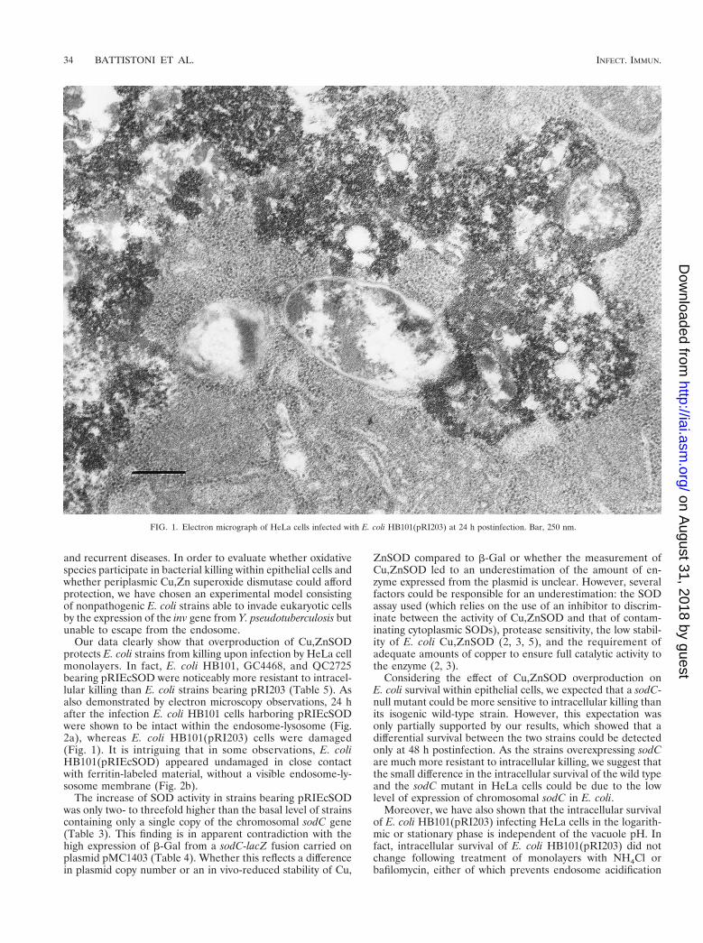
ritin was used as a lysosomal marker to demonstrate phago-some-lysosome fusion. After 2 h of infection, the strain ap-peared intact, while after 24 or 48 h E. coli HB101(pRI203)appeared damaged within the endosome-lysosome (Fig. 1).In contrast, cells infected with E. coli HB101(pRIEcSOD)showed that the endosome-lysosome still contained intact bac-teria (Fig. 2a); in some observations, it was not possible tovisualize a membrane surrounding E. coli pRIEcSOD cells thatappeared undamaged, even though they were in close contactwith ferritin-labeled material (Fig. 2b).
DISCUSSION
Several authors have investigated the possible involvementof periplasmic Cu,Zn superoxide dismutase in the mechanismsof bacterial protection against the respiratory burst elicited byphagocytes. It has been shown that Cu,ZnSOD-deficient mu-tants of several bacterial genera are less virulent in in vivomodels and that, at least in some cases, the enzyme plays aprotective role against superoxide generated by macrophages.Some bacteria producing Cu,ZnSOD are facultative intracel-lular microorganisms. This lifestyle, in a protected cellularniche, is thought to allow bacteria to escape the host defensemechanisms, thus contributing to the establishment of chronic
TABLE 5. Invasiveness and intracellular survival of invasive E. coli strainsa
E. coli strain Growth phase No. of intracellularbacteria at 2 h
Intracellular survival at:
24 h 48 h
HB101(pRI203) Logarithmic (1.8 6 1.3) 3 106 (5.9 6 3.3) 3 103 0HB101(pRIEcSOD) (2.3 6 1.5) 3 106 (4.0 6 1.7) 3 105 (1.5 6 0.6) 3 105
HB101(pRI203) Stationary (2.0 6 0.6) 3 105 0 0HB101(pRIEcSOD) (9.2 6 0.5) 3 104 (6.3 6 0.6) 3 104 (1.0 6 0.5) 3 104
GC4468(pRI203) Logarithmic (1.3 6 0.5) 3 106 (1.4 6 0.7) 3 104 (1.0 6 0.6) 3 103
GC4468(pRIEcSOD) (2.1 6 0.8) 3 106 (1.8 6 0.6) 3 106 (3.6 6 1.9) 3 105
GC4468(pRI203) Stationary (3.0 6 0.4) 3 105 (3.8 6 0.6) 3 104 (1.0 6 0.8) 3 103
GC4468(pRIEcSOD) (2.8 6 0.3) 3 105 (1.3 6 0.9) 3 105 (1.2 6 1.0) 3 105
QC2725(pRI203) Logarithmic (1.0 6 0.4) 3 106 (1.6 6 0.5) 3 104 (2.0 6 0.5) 3 102
QC2725(pRIEcSOD) (1.1 6 0.3) 3 106 (0.8 6 0.4) 3 106 (3.6 6 0.4) 3 105
QC2725(pRI203) Stationary (3.3 6 0.6) 3 105 (2.7 6 0.6) 3 104 (2.0 6 0.5) 3 102
QC2725(pRIEcSOD) (5.1 6 0.5) 3 105 (1.8 6 0.7) 3 105 (1.6 6 0.4) 3 105
a Invasiveness (measured as the number of viable intracellular bacteria 2 h postinfection) and survival of E. coli strains within HeLa cells at 24 and 48 h postinfectionwere determined by infecting cell monolayers with bacteria (MOI, 100) in different growth phases, as described in Materials and Methods. Values are the means 6standard deviations of at least four independent experiments.
TABLE 6. Effect of endosome pH-neutralizing reagents on E. coli HB101(pRI203) intracellular survivala
E. coli strain and additive Growth phase No. of intracellularbacteria at 2 h
Intracellular survival at:
24 h 48 h
HB101(pRI203) Logarithmic (9.3 6 0.9) 3 105 (4.6 6 0.5) 3 103 0HB101(pRI203) plus NH4Cl (3.3 6 0.7) 3 105 (5.9 6 3.3) 3 103 0HB101(pRI203) plus bafilomycin (8.4 6 0.7) 3 105 (2.8 6 0.8) 3 103 0
HB101(pRI203) Stationary (3.2 6 0.5) 3 105 0 0HB101(pRI203) plus NH4Cl (3.8 6 0.7) 3 105 0 0HB101(pRI203) plus bafilomycin (2.6 6 0.5) 3 105 0 0
a Effect of 20 mM NH4Cl or 100 nM bafilomycin A1 on invasion efficiency (number of intracellular bacteria at 2 h) and intracellular survival of E. coli HB101(pRI203)infecting HeLa cell monolayers at an MOI of 100. Values are the means 6 standard deviations of four independent experiments.
VOL. 68, 2000 INVASIVE E. COLI SURVIVAL IN EPITHELIAL CELLS 33
on August 31, 2018 by guest
http://iai.asm.org/
Dow
nloaded from
and recurrent diseases. In order to evaluate whether oxidativespecies participate in bacterial killing within epithelial cells andwhether periplasmic Cu,Zn superoxide dismutase could affordprotection, we have chosen an experimental model consistingof nonpathogenic E. coli strains able to invade eukaryotic cellsby the expression of the inv gene from Y. pseudotuberculosis butunable to escape from the endosome.
Our data clearly show that overproduction of Cu,ZnSODprotects E. coli strains from killing upon infection by HeLa cellmonolayers. In fact, E. coli HB101, GC4468, and QC2725bearing pRIEcSOD were noticeably more resistant to intracel-lular killing than E. coli strains bearing pRI203 (Table 5). Asalso demonstrated by electron microscopy observations, 24 hafter the infection E. coli HB101 cells harboring pRIEcSODwere shown to be intact within the endosome-lysosome (Fig.2a), whereas E. coli HB101(pRI203) cells were damaged(Fig. 1). It is intriguing that in some observations, E. coliHB101(pRIEcSOD) appeared undamaged in close contactwith ferritin-labeled material, without a visible endosome-ly-sosome membrane (Fig. 2b).
The increase of SOD activity in strains bearing pRIEcSODwas only two- to threefold higher than the basal level of strainscontaining only a single copy of the chromosomal sodC gene(Table 3). This finding is in apparent contradiction with thehigh expression of b-Gal from a sodC-lacZ fusion carried onplasmid pMC1403 (Table 4). Whether this reflects a differencein plasmid copy number or an in vivo-reduced stability of Cu,
ZnSOD compared to b-Gal or whether the measurement ofCu,ZnSOD led to an underestimation of the amount of en-zyme expressed from the plasmid is unclear. However, severalfactors could be responsible for an underestimation: the SODassay used (which relies on the use of an inhibitor to discrim-inate between the activity of Cu,ZnSOD and that of contam-inating cytoplasmic SODs), protease sensitivity, the low stabil-ity of E. coli Cu,ZnSOD (2, 3, 5), and the requirement ofadequate amounts of copper to ensure full catalytic activity tothe enzyme (2, 3).
Considering the effect of Cu,ZnSOD overproduction onE. coli survival within epithelial cells, we expected that a sodC-null mutant could be more sensitive to intracellular killing thanits isogenic wild-type strain. However, this expectation wasonly partially supported by our results, which showed that adifferential survival between the two strains could be detectedonly at 48 h postinfection. As the strains overexpressing sodCare much more resistant to intracellular killing, we suggest thatthe small difference in the intracellular survival of the wild typeand the sodC mutant in HeLa cells could be due to the lowlevel of expression of chromosomal sodC in E. coli.
Moreover, we have also shown that the intracellular survivalof E. coli HB101(pRI203) infecting HeLa cells in the logarith-mic or stationary phase is independent of the vacuole pH. Infact, intracellular survival of E. coli HB101(pRI203) did notchange following treatment of monolayers with NH4Cl orbafilomycin, either of which prevents endosome acidification
FIG. 1. Electron micrograph of HeLa cells infected with E. coli HB101(pRI203) at 24 h postinfection. Bar, 250 nm.
34 BATTISTONI ET AL. INFECT. IMMUN.
on August 31, 2018 by guest
http://iai.asm.org/
Dow
nloaded from
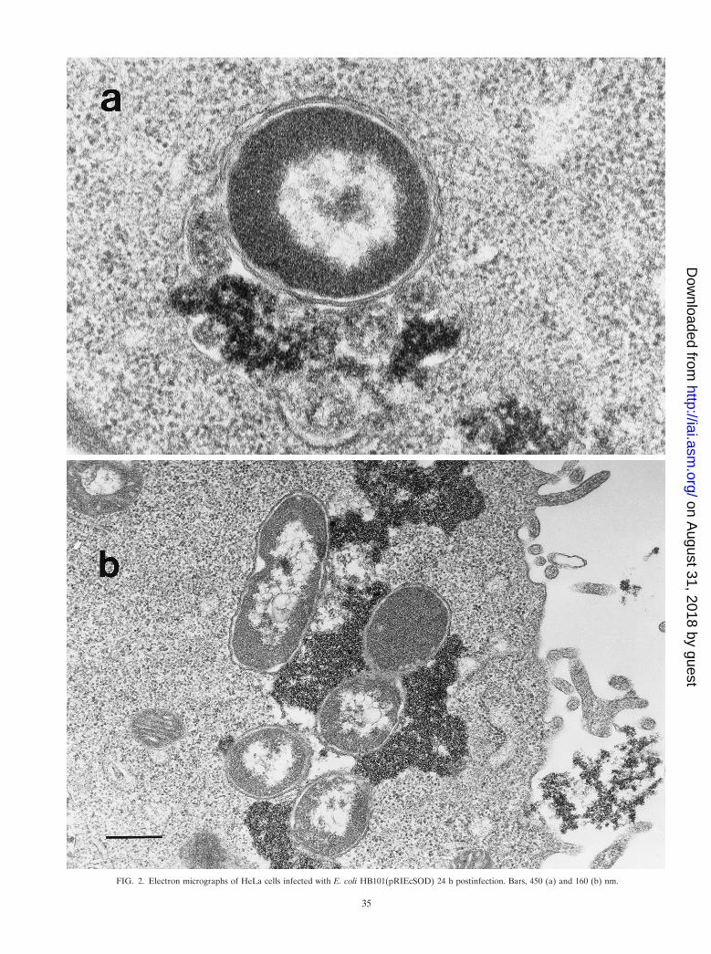
FIG. 2. Electron micrographs of HeLa cells infected with E. coli HB101(pRIEcSOD) 24 h postinfection. Bars, 450 (a) and 160 (b) nm.
35
on August 31, 2018 by guest
http://iai.asm.org/
Dow
nloaded from
(Table 6). These results indicate that the acid-sensitive pheno-type of E. coli HB101 (39) is not responsible for the low via-bility of this strain within epithelial cells. The characterizationof the peculiar features responsible for the different survival inepithelial cells of invasive E. coli HB101 and GC4468 is outsidethe aims of this work. However, it is interesting that overex-pression of sodC enhances survival of both of these strains andappears to be sufficient to increase the survival of invasive E.coli HB101 (which is known to be highly sensitive to oxidativestress due to mutations in the genes encoding RecA and RpoS)to a level close to that of invasive E. coli GC4468.
Taken together, our findings suggest that oxyradical damageis involved in the mechanisms of bacterial killing by epithelialcells. Such a proposal is in agreement with a previous studywhich showed that Caco-2 and IEC-18 intestinal epithelial cellsare able to kill bacteria by an oxidative pathway (16). While itis well established that superoxide production by an NADPHoxidase located on the cell membrane plays a pivotal role in theoxygen-dependent antimicrobial systems of phagocytic cells,no information is available about the presence of specific an-timicrobial mechanisms based on free radicals generatingenzymes in epithelial cells. The observation that Cu,ZnSODprotects E. coli from killing within epithelial cells, however,suggests that free radicals can also be produced on the endo-somal membrane of nonphagocytic cells.
Although obtained with a model which uses nonpathogenicrecombinant E. coli K-12 strains rendered invasive by the ex-pression of a Y. pseudotuberculosis gene, our data encourageinvestigations of whether sodC plays a role in the intracellularsurvival of those pathogens that are naturally able to invadeepithelial cells.
ACKNOWLEDGMENTS
This work was partially supported by the MURST PRIN grant “Roleof free or chelated metal ions in intracellular infections” and by theCNR target project on “Biotechnology.”
REFERENCES
1. Bannister, J. V., W. H. Bannister, and G. Rotilio. 1987. Aspects of thestructure, function, and applications of superoxide dismutase. Crit. Rev.Biochem. 22:111–180.
2. Battistoni, A., and G. Rotilio. 1995. Isolation of an active and heat-stablemonomeric form of Cu,Zn superoxide dismutase from the periplasmic spaceof Escherichia coli. FEBS Lett. 374:199–202.
3. Battistoni, A., S. Folcarelli, R. Gabbianelli, C. Capo, and G. Rotilio. 1996.The Cu,Zn superoxide dismutase from Escherichia coli retains monomericstructure at high protein concentration. Evidence for altered subunit inter-action in all the bacteriocupreins. Biochem. J. 320:713–716.
4. Battistoni, A., G. Donnarumma, R. Greco, P. Valenti, and G. Rotilio. 1998.Overexpression of a hydrogen peroxide-resistant periplasmic Cu,Zn super-oxide dismutase protects Escherichia coli from macrophage killing. Biochem.Biophys. Res. Commun. 243:804–807.
5. Battistoni, A., S. Folcarelli, L. Cervone, F. Polizio, A. Desideri, A. Giartosio,and G. Rotilio. 1998. Role of the dimeric structure in Cu,Zn superoxidedismutase. pH-dependent, reversible denaturation of the monomeric en-zyme from Escherichia coli. J. Biol. Chem. 273:5655–5661.
6. Beck, B. L., L. B. Tabatabai, and J. E. Mayfield. 1990. A protein isolatedfrom Brucella abortus is a Cu-Zn superoxide dismutase. Biochemistry 16:372–376.
7. Benov, L. T., and I. Fridovich. 1994. Escherichia coli expresses a copper- andzinc-containing superoxide dismutase. J. Biol. Chem. 269:25310–25314.
8. Boyer, H., and D. Roulland-Dussoix. 1969. A complementation analysis ofthe restriction and modification of DNA in Escherichia coli. J. Mol. Biol. 41:459–472.
9. Canvin, J., P. R. Langford, K. E. Wilks, and J. S. Kroll. 1996. Identificationof sodC encoding periplasmic [Cu,Zn]-superoxide dismutase in Salmonella.FEMS Microbiol. Lett. 136:215–220.
10. Carlioz, A., and D. Touati. 1986. Isolation of superoxide dismutase mutantsin Escherichia coli: is superoxide dismutase necessary for aerobic life?EMBO J. 5:623–630.
11. Casadaban, M. J., J. Chou, and S. N. Cohen. 1980. In vitro gene fusions thatjoin an enzymatically active b-galactosidase segment to amino-terminal frag-
ments of exogenous proteins: Escherichia coli plasmid vectors for the detec-tion and cloning of translational initiation signals. J. Bacteriol. 143:971–980.
12. Chen, C. Y., L. Eckmann, S. J. Libby, F. C. Fang, S. Okamoto, M. F. Kagnoff,J. Fierer, and D. G. Guiney. 1996. Expression of Salmonella typhimuriumrpoS and rpoS-dependent genes in the intracellular environment of eukary-otic cells. Infect. Immun. 64:4739–4743.
13. Conte, M. P., P. Mastromarino, M. Nicoletti, P. Visca, P. Valenti, and L.Seganti. 1990. Effect of polyelectrolytes on entry of Escherichia coli HB101(pRI203) into HeLa cells. Microb. Pathog. 9:191–198.
14. D’Arcy, P., J. Hart, and M. R. Young. 1975. Interference with normalphagosome-lysosome fusion in macrophage, using ingested yeast cells andsuramin. Nature 256:47–49.
15. De Groote, M. A., U. A. Ochsner, M. U. Shiloh, C. Nathan, J. M. McCord,M. C. Dinauer, S. J. Libby, A. Vazquez-Torres, Y. Xu, and F. C. Fang. 1997.Periplasmic superoxide dismutase protects Salmonella from products ofphagocyte NADPH-oxidase and nitric oxide synthase. Proc. Natl. Acad. Sci.USA 94:13997–14001.
16. Deitch, E. A., Y. Haskel, N. Cruz, D. Xu, and P. R. Kvietys. 1995. Caco-2 andIEC-18 intestinal epithelial cells exert bactericidal activity through an oxi-dant-dependent pathway. Shock 4:345–350.
17. Eisenstark, A., M. J. Calcutt, M. Becker-Hapak, and A. Ivanova. 1996. Roleof Escherichia coli rpoS and associated genes in defense against oxidativedamage. Free Radic. Biol. Med. 21:975–993.
18. Fang, F. C., M. A. DeGroote, J. W. Foster, A. J. Baumler, U. Ochsner, T.Testerman, S. Bearson, J. C. Giard, Y. Xu, G. Campbell, and T. Laessig.1999. Virulent Salmonella typhimurium has two periplasmic Cu,Zn-super-oxide dismutases. Proc. Natl. Acad. Sci. USA 96:7502–7507.
19. Farrant, J. L., A. Sansone, J. R. Canvin, M. J. Pallen, P. R. Langford, T. S.Wallis, G. Dougan, and J. S. Kroll. 1997. Bacterial copper- and zinc-cofac-tored superoxide dismutase contributes to the pathogenesis of systemic sal-monellosis. Mol. Microbiol. 25:785–796.
20. Finlay, B. B., and S. Falkow. 1997. Common themes in microbial pathoge-nicity revisited. Microbiol. Mol. Biol. Rev. 61:136–169.
20a.Gort, A. S., D. M. Ferber, and J. A. Imlay. 1999. The regulation and role ofthe periplasmic copper, zinc superoxide dismutase of Escherichia coli. Mol.Microbiol. 32:179–191.
21. Hassan, H. M., and I. Fridovich. 1979. Paraquat and Escherichia coli. Mech-anism of production of extracellular superoxide radical. J. Biol. Chem. 254:10846–10852.
22. Imlay, K. R., and J. Imlay. 1996. Cloning and analysis of sodC, encoding thecopper-zinc superoxide dismutase of Escherichia coli. J. Bacteriol. 178:2564–2571.
23. Isberg, R. R., and S. Falkow. 1985. A single genetic locus encoded by Yersiniapseudotuberculosis permits invasion of cultured animal cells by Escherichiacoli K-12. Nature 317:262–264.
24. Kroll, J. S., P. R. Langford, and B. M. Loynds. 1991. Copper-zinc superoxidedismutase of Haemophilus influenzae and H. parainfluenzae. J. Bacteriol. 173:7449–7457.
25. Kroll, J. S., P. R. Langford, K. E. Wilks, and A. D. Keil. 1995. Bacterial [Cu,Zn]superoxide dismutase: phylogenetically distinct from the eukaryotic en-zyme, and not so rare after all! Microbiology 141:2271–2279.
26. Langford, P. R., B. M. Loynds, and J. S. Kroll. 1996. Cloning and molecularcharacterization of Cu,Zn superoxide dismutase from Actinobacillus pleuro-pneumoniae. Infect. Immun. 64:5035–5041.
27. Loewen, P. C., and R. Hengge-Aronis. 1994. The role of the sigma factorsigma S (KatF) in bacterial global regulation. Annu. Rev. Microbiol. 48:53–80.
28. Loewen, P. C., and B. L. Triggs. 1985. Genetic mapping of KatF, a locus thataffect the synthesis of a second catalase species in Escherichia coli. J. Bac-teriol. 160:668–675.
29. Lowry, O. H., N. J. Rosebrough, A. L. Farr, and R. J. Randall. 1951. Proteinmeasurement with the Folin phenol reagent. J. Biol. Chem. 193:265–275.
30. Marklund, S., and G. Marklund. 1974. Involvement of the superoxide anionradical in the autoxidation of pyrogallol and a convenient assay for super-oxide dismutase. Eur. J. Biochem. 47:469–474.
31. McCord, J. M., and I. Fridovich. 1969. Superoxide dismutase. An enzymicfunction for erythrocuprein (hemocuprein). J. Biol. Chem. 244:6049–6055.
32. Murphy, K. C. 1998. Use of bacteriophage lambda recombination functionsto promote gene replacement in Escherichia coli. J. Bacteriol. 180:2063–2071.
33. Nickerson, C. A., and R. Curtiss III. 1997. Role of sigma factor RpoS ininitial stages of Salmonella typhimurium infection. Infect. Immun. 65:1814–1823.
34. Rathman, M., M. D. Sjaastad, and S. Falkow. 1996. Acidification of phago-somes containing Salmonella typhimurium in murine macrophages. Infect.Immun. 64:2765–2773.
35. Sambrook, J., E. F. Fritsch, and T. Maniatis. 1989. Molecular cloning: alaboratory manual, 2nd ed. Cold Spring Harbor Laboratory Press, ColdSpring Harbor, N.Y.
36. San Mateo, L. R., K. L. Toffer, P. E. Orndorff, and T. H. Kawula. 1999.Neutropenia restores virulence to an attenuated Cu,Zn superoxide dis-mutase-deficient Haemophilus ducreyi strain in the swine model of chancroid.Infect. Immun. 67:5345–5351.
36 BATTISTONI ET AL. INFECT. IMMUN.
on August 31, 2018 by guest
http://iai.asm.org/
Dow
nloaded from
37. Schnell, S., and H. M. Steinman. 1995. Function of stationary-phase induc-tion of periplasmic copper-zinc superoxide dismutase and catalase/peroxi-dase in Caulobacter crescentus. J. Bacteriol. 177:5924–5929.
38. Small, P. L., R. R. Isberg, and S. Falkow. 1987. Comparison of the ability ofenteroinvasive Escherichia coli, Salmonella typhimurium, Yersinia pseudotu-berculosis, and Yersinia enterocolitica to enter and replicate within HEp-2cells. Infect. Immun. 55:1674–1679.
39. Small, P., D. Blankenhorn, D. Welty, E. Zinser, and J. L. Slonczewski. 1994.Acid and base resistance in Escherichia coli and Shigella flexneri: role of rpoSand growth pH. J. Bacteriol. 176:1729–1737.
40. St. John, G., and H. M. Steinman. 1996. Periplasmic copper-zinc superoxidedismutase of Legionella pneumophila: role in stationary-phase survival.J. Bacteriol. 178:1578–1584.
41. Swords, W. E., B. M. Cannon, and W. H. Benjamin, Jr. 1997. Avirulence of
LT2 strains of Salmonella typhimurium results from a defective rpoS gene.Infect. Immun. 65:2451–2453.
42. Tatum, F. M., P. G. Detilleux, J. M. Sacks, and S. M. Hallings. 1992.Construction of Cu-Zn superoxide dismutase deletion mutants of Brucellaabortus: analysis of survival in vitro in epithelial and phagocytic cells and invivo in mice. Infect. Immun. 60:2863–2869.
43. Wilks, K. E., K. L. Dunn, J. L. Farrant, K. M. Reddin, A. R. Gorringe, P. R.Langford, and J. S. Kroll. 1998. Periplasmic superoxide dismutase in me-ningococcal pathogenicity. Infect. Immun. 66:213–217.
44. Wilmes-Riesenberg, M. R., J. W. Foster, and R. Curtiss III. 1997. An alteredrpoS allele contributes to the avirulence of Salmonella typhimurium LT2.Infect. Immun. 65:203–210.
45. Wu, C. H., J. J. Tsai-Wu, Y. T. Huang, C. Y. Lin, G. G. Lioua, and F. J. Lee.1998. Identification and subcellular localization of a novel Cu,Zn superoxidedismutase of Mycobacterium tuberculosis. FEBS Lett. 13:192–196.
Editor: P. E. Orndorff
VOL. 68, 2000 INVASIVE E. COLI SURVIVAL IN EPITHELIAL CELLS 37
on August 31, 2018 by guest
http://iai.asm.org/
Dow
nloaded from