Embed Size (px)
Citation preview

RESEARCH ARTICLE Open Access
Increased chromosomal radiosensitivity inasymptomatic carriers of a heterozygousBRCA1 mutationAnnelot Baert1, Julie Depuydt1, Tom Van Maerken2, Bruce Poppe2, Fransiska Malfait2, Katrien Storm3,Jenneke van den Ende3, Tim Van Damme2, Sylvia De Nobele2, Gianpaolo Perletti1,4, Kim De Leeneer2,Kathleen B. M. Claes2† and Anne Vral1*†
Abstract
Background: Breast cancer risk increases drastically in individuals carrying a germline BRCA1 mutation. Theexposure to ionizing radiation for diagnostic or therapeutic purposes of BRCA1 mutation carriers is counterintuitive,since BRCA1 is active in the DNA damage response pathway. The aim of this study was to investigate whetherhealthy BRCA1 mutations carriers demonstrate an increased radiosensitivity compared with healthy individuals.
Methods: We defined a novel radiosensitivity indicator (RIND) based on two endpoints measured by the G2
micronucleus assay, reflecting defects in DNA repair and G2 arrest capacity after exposure to doses of 2 or 4 Gy. Weinvestigated if a correlation between the RIND score and nonsense-mediated decay (NMD) could be established.
Results: We found significantly increased radiosensitivity in the cohort of healthy BRCA1 mutation carrierscompared with healthy controls. In addition, our analysis showed a significantly different distribution over the RINDscores (p = 0.034, Fisher’s exact test) for healthy BRCA1 mutation carriers compared with non-carriers: 72 % ofmutation carriers showed a radiosensitive phenotype (RIND score 1–4), whereas 72 % of the healthy volunteersshowed no radiosensitivity (RIND score 0). Furthermore, 28 % of BRCA1 mutation carriers had a RIND score of 3 or 4(not observed in control subjects). The radiosensitive phenotype was similar for relatives within several families, butnot for unrelated individuals carrying the same mutation. The median RIND score was higher in patients with amutation leading to a premature termination codon (PTC) located in the central part of the gene than in patientswith a germline mutation in the 5′ end of the gene.
Conclusions: We show that BRCA1 mutations are associated with a radiosensitive phenotype related to acompromised DNA repair and G2 arrest capacity after exposure to either 2 or 4 Gy. Our study confirms thathaploinsufficiency is the mechanism involved in radiosensitivity in patients with a PTC allele, but it suggests thatfurther research is needed to evaluate alternative mechanisms for mutations not subjected to NMD.
Keywords: BRCA1 mutations, DNA damage repair, Homologous recombination, G2/M cell-cycle checkpoint, Ionizingradiation, G2 micronucleus assay, Radiosensitivity indicator, Nonsense-mediated decay, Haploinsufficiency
* Correspondence: [email protected]†Equal contributors1Department of Basic Medical Sciences, Ghent University, Ghent, BelgiumFull list of author information is available at the end of the article
© 2016 Baert et al. Open Access This article is distributed under the terms of the Creative Commons Attribution 4.0International License (http://creativecommons.org/licenses/by/4.0/), which permits unrestricted use, distribution, andreproduction in any medium, provided you give appropriate credit to the original author(s) and the source, provide a link tothe Creative Commons license, and indicate if changes were made. The Creative Commons Public Domain Dedication waiver(http://creativecommons.org/publicdomain/zero/1.0/) applies to the data made available in this article, unless otherwise stated.
Baert et al. Breast Cancer Research (2016) 18:52 DOI 10.1186/s13058-016-0709-1

BackgroundBreast cancer (BC) is the most common malignancy inthe Western world (http://www.who.int/cancer/detection/breastcancer/en/). Approximately 15 % of all patients withBC have at least one relative affected by BC. About 15 %of all familial BCs can be attributed to a mutation in theBRCA1 or BRCA2 gene [1]. Since the discovery of BRCA1,many different functions have been attributed to this pro-tein. In its function as a tumour suppressor gene, BRCA1plays a crucial role in DNA double-strand break(DSB) repair pathways (reviewed in [2, 3]). BRCA1 is,for instance, important in homologous recombination(HR), a pathway for repair of DSB in late S and G2
phases of the cell cycle [4–6]. BRCA1 also plays animportant role in the G2/M checkpoint control, allow-ing the cell to repair DNA damage before proceedingto the next phase of the cell cycle [7].Carriers of a heterozygous BRCA1 mutation may show
enhanced radiosensitivity associated with an increasedcarcinogenic risk after exposure to diagnostic or thera-peutic ionizing radiation (IR). Several studies haveshown that exposure to diagnostic X-rays may causecancer in healthy BRCA1 mutation carriers [8–11],whereas researchers in other studies could not detect apositive association between exposure to IR and breastcancer risk in BRCA1/2 mutation carriers [12–15]. Also,researchers analysing the impact of (adjuvant) radiother-apy on breast cancer risk in BRCA1 and BRCA2 muta-tion carriers reported no univocal conclusion [16]. Thecontradictory data obtained in these studies are duemainly to the constraints in the design of the performedstudies.Since long-term studies of the effect of exposure to IR
in mutation carriers are difficult to set up and are re-puted to be unethical, it is clear that more empiric dataare needed to determine in vitro the radiosensitivity ofpatients carrying a germline mutation. To date, researchhas yielded contradictory results [17–26].The G0 micronucleus assay performed on peripheral
blood lymphocytes exposed to in vitro doses of 2 to4 Gy is frequently used to assess chromosomal radiosen-sitivity. However, this assay is not optimized to detectdefects in DSB repair activated during the G2 phase ofthe cell cycle or the G2/M checkpoint, two processes inwhich BRCA1 plays a major role because irradiationtakes place in the G0 phase of the cell cycle.We previously reported a modified micronucleus
(MN) assay optimized to detect defects in the S or G2 phaseof the cell cycle. This assay efficiently detected increased ra-diosensitivity in a patient with a mild form of ataxia-telangiectasia (A-T) and in heterozygous relatives [27].In the present study, we applied the G2 micronucleus
assay to further elucidate whether healthy BRCA1 muta-tion carriers are characterized by an increased in vitro
radiosensitivity. The endpoints of the study were (1)micronucleus yields and (2) G2/M checkpoint efficiencyratio. Both endpoints were assessed after irradiatingphytohaemagglutinin (PHA)-stimulated peripheral bloodlymphocytes with doses of 2 Gy and 4 Gy. With this assay,we assessed the mean differences in radiosensitivity forheterozygous BRCA1 mutation carriers compared withhealthy volunteers. We also scored the overall radiosensi-tivity in each mutation carrier using a radiosensitivity indi-cator (RIND) scoring system. In addition, as our BRCA1population consisted of individuals carrying differentBRCA1 mutations, we investigated if there was a link be-tween radiosensitivity and the degree of nonsense-mediated decay (NMD) of the specific mutant allele.
MethodsSample collectionBlood samples were collected from individuals consult-ing the clinic of the Centre for Medical Genetics, GhentUniversity Hospital, Belgium (CMGG). Both ethylenedi-aminetetraacetic acid (EDTA) and heparin blood sam-ples were collected. EDTA samples were used formutation analysis at CMGG, whereas the G2 MN assaywas performed on heparinized blood samples. Inaddition, we collected heparinized blood samples fromhealthy volunteers (n = 20) without a personal or familyhistory of BC to determine the normal distribution ofmicronucleus yields in controls. Lymphocytes were iso-lated from the blood samples using Lymphoprep™(STEMCELL Technologies, Vancouver, BC, Canada) andwere preserved in liquid nitrogen for analysis of NMD ofthe mutant allele. For a number of mutation carriers (n= 4) and healthy volunteers (n = 7), a second blood sam-ple was taken to determine the reproducibility of the re-sults obtained with the G2 MN assay at different timepoints.This study was approved by the ethics committee of the
Ghent University Hospital (B670201111641 d.d. 20/09/2011). All study participants (n = 18) were counselled byclinical geneticists in the context of a predictive (n = 16)or diagnostic (n = 2) test for hereditary BC and signed aninformed consent form. Two of the eighteen BRCA1 mu-tation carriers had developed BC, but their cancer treat-ment had finished more than 2 years ago; these womenare both carriers of a substitution affecting the start codon(M01 and M02). The mean ages of the mutation carriersand healthy volunteers were 40.9 and 35.4 years, respect-ively (p = 0.26, t test).
Molecular analysisAll patients selected for this study had a family history ofbreast and/or ovarian cancer and a BRCA1 germlinemutation. Targeted analysis for the familial mutation wasperformed by direct sequencing on two independently
Baert et al. Breast Cancer Research (2016) 18:52 Page 2 of 12

extracted DNA samples. No molecular analyses were per-formed in healthy volunteers, owing to absence of per-sonal or familial anamnesis for BC.
G2 micronucleus assayFor the G2 MN assay, a large blood culture was set up inthe presence of the mitogen PHA. PHA stimulates T-lymphocyte division, resulting in a population of cyclinglymphocytes (G1, S1, G2 and M phase) after 3 days of in-cubation, when the blood culture is irradiated. On thecontrary, in the G0 MN assay, blood is first irradiatedand then cultured in the presence of PHA, resulting inthe irradiation of T lymphocytes in G0 phase. Theaddition of cytochalasin B (cyto B), an agent that blockscytokinesis, to the blood cultures allows the identifica-tion of first-division cells as binucleated (BN) cells. Anon- or misrepaired DSB can result in an acentricchromosomal fragment, which is detected as a micronu-cleus in the cytoplasm of the BN cell [17, 28].More precisely, for the G2 MN assay, a 50-ml blood cul-
ture was set up using 5 ml of heparinized blood, 45 ml ofcomplete RPMI (RPMI with 1 % L-glutamine and 0.5 %penicillin/streptomycin) containing 10 % foetal calf serum(FCS) and 1 ml of PHA (all Gibco®; Thermo Fisher, Rock-ford, IL) in a T75 culture flask [27]. This large culture wasset up to avoid interculture variation during the first days ofincubation. After 3 days, the culture was split, and sham ir-radiations as well as irradiations with 2 and 4 Gy 60Cogamma rays were performed. An overview of the experi-ments is shown in Fig. 1a. Immediately after irradiation,cyto B (6 μg/ml; Sigma-Aldrich, St. Louis, MO, USA) wasadded. To half of the irradiated cultures, caffeine (CAF,4 mM; Sigma-Aldrich), an agent that abrogates the G2/Mcheckpoint, was added to determine the G2/M checkpointefficiency ratio [29, 30]. On the basis of 5-bromo-2′-deox-yuridine (BrdU) results obtained in our previous study onATM [27] and new experiments performed for this study,a post-irradiation incubation period of 8 h was selectedfor detecting DNA damage induced in cells in G2 phase(Fig. 1b). The cultures were then treated with 75 mM KCland fixed once using a combination of methanol, aceticacid and Ringer’s solution (9 g NaCl + 0.42 g KCl + 0.24 gCaCl2 for 1 L of dH2O) (ratio 4/1/5, 4 °C) and thrice withmethanol and acetic acid (ratio 4/1, 4 °C). Finally, the cellsuspension was concentrated and spread on slides.After 4′,6-diamidino-2-phenylindole staining, the
slides were scanned with a Metafer 4 (MN score soft-ware module; MetaSystems; Altlussheim, Germany).This automated image analysis system selects BN cellsand determines the number of MN per BN cell (seeFig. 1a). For each condition, two cultures were set upand two slides per culture were analysed. A minimum of600 BN cells were scored on each coded slide for thepresence of MN. BN and MN selected in the automated
setting were manually checked for false-positives andfalse-negatives. To determine the G2/M checkpoint effi-ciency ratio, the quotient was determined for the MNyield obtained in the presence versus the absence of caf-feine (MNCaf+/MNCaf−). The lower this ratio was, themore radiosensitive an individual was.To assess the overall radiosensitive phenotype of each
patient, a RIND score was calculated. The mean andstandard deviation (SD) of the micronucleus yield andthe G2/M checkpoint efficiency ratio assessed in the co-hort of healthy volunteers served to determine cut-offsto define the radiosensitivity of individual BRCA1 muta-tion carriers. An individual value equal to or greater thanthe mean MN yields in controls + 1 SD scored 1 point(colour-coded orange). A value equal to or greater than themean MN yields of controls + 2 SD scored 2 points(colour-coded red). An individual value less than the meanof controls + 1 SD was scored as naught (colour-codedwhite). For the G2/M checkpoint, a similar conversion ofthe data was performed, with a value less than the meancheckpoint ratio assessed in controls − 1 SD scored 1 point(colour-coded orange) and the mean− 2 SD scored 2 points(color-coded red). An individual value greater than themean of controls− 1 SD was scored as naught (colour-coded white). The combined scores from MN yields (0, 1or 2 points) and checkpoint ratios (0, 1 or 2 points) werethen added to form a single RIND score (range 0–4).
Analysis of the cell-cycle phase at time of irradiationBrdU was added to blood cultures of three individuals toanalyse the phase of the cell cycle at the time of irradi-ation. BrdU (0.01 mM; Sigma-Aldrich) was added to thecultures immediately after irradiation. Binucleated cellsthat incorporated BrdU during synthesis were in S or G1
phase of the cell cycle at the moment of irradiation,whereas BrdU-negative binucleated cells were irradiatedin G2 phase. We corrected for daughter nuclei incorporat-ing BrdU after binucleation by adding BrdU 2 h before fix-ation to another subset of cultures. BrdU was visualized byfluorescence immunostaining with a monoclonal BrdU-specific antibody (M0744; Dako, Carpinteria, CA, USA).
Analysis of the stability of the mutant alleleFor this experiment, 2 × 10⁶ frozen lymphocytes werethawed and cultured in a mix of 1 ml of cRPMI, FCS(10 %), 2-mercaptoethanol (0.1 %, Gibco®) and sodiumpyruvate (1 %; Life Technologies; Carlsbad, CA, USA).PHA (10 μl/ml) was added to stimulate cell division. Atday 7, whole RNA was extracted using the QIAamp®RNeasy Mini Kit (QIAGEN, Valencia, CA, USA) accord-ing to the manufacturer’s instructions. Four hours beforeextraction, all cultures were split in two, and puromycin(200 μg/ml; Sigma-Aldrich) was added to one part.
Baert et al. Breast Cancer Research (2016) 18:52 Page 3 of 12

Puromycin was added to avoid NMD, a pathway respon-sible for the degradation of aberrant mRNA [31].The total RNA and purity was measured using the
DropSense96 reader (TRINEAN, Gentbrugge, Belgium).The RNA was converted into complementary DNA(cDNA) using the iScript™ cDNA Synthesis Kit (Bio-RadLaboratories, Hercules, CA, USA). When RNA was ex-tracted from cultures with puromycin, the convertedcDNA is referred to as cDNAp. Before this cDNA syn-thesis, samples were treated with DNase (Heat&Run Kit;ArcticZymes, Tromsø, Norway) to remove any possibleremaining genomic DNA (gDNA).To determine the ratio of the mutant BRCA1 allele ver-
sus the wild-type (WT) allele at the mRNA level, we per-formed polymerase chain reaction (PCR) amplificationand Sanger sequencing of the amplicon harbouring thegermline mutation and a heterozygous single-nucleotidepolymorphism (SNP) (c.2311 T > C), if present. An over-view of the primers used can be found in Additional file 1.Sanger sequencing was performed using the BigDye® Ter-minator Cycle Sequencing Kit (Life Technologies). PCRwas followed by ethanol precipitation, and fragments weredissolved in a mix of Hi-Di™ formamide (10 μl; Life Tech-nologies) and GeneScan™ 500 LIZ™ Size Standard (0.5 μl;
Applied Biosystems, Foster City, CA, USA). The frag-ments were analysed on the ABI PRISM® 3730KL GeneticAnalyzer (Life Technologies). Results were evaluated usingGeneMapper® software (Applied Biosystems) for visual in-spection and determination of the ratio of the peakheights representing the WT and mutant alleles, respect-ively (Additional files 2 and 3). Data were normalized witha control peak in the near vicinity. To confirm the results,the PCR amplicons were also sequenced with the sequen-cing by synthesis technology on a MiSeq instrument(Illumina, San Diego, CA, USA). The library preparationwas performed using an adapted Nextera XT protocol(Illumina) as described by De Leeneer et al. [32]. Readswere mapped with CLC Genomics Workbench (CLC bio/QIAGEN, Aarhus, Denmark). The variant allele frequency(VAF) (which reflects the ratio of WT versus mutant al-lele) of both the mutation and a SNP (if available) was de-termined in the mutation carriers and in controls withouta germline BRCA1 mutation.
ResultsAnalysis of the cell-cycle phase at time of irradiationOn the basis of data reported in the literature, the cellcycle of proliferating lymphocytes takes between 18 and
Fig. 1 Design of the G2 micronucleus (MN) assay and overview of the cell cycle. a Left: Day 0: setup of one 50-ml large blood culture for everypatient. Day 3: division of the cultures and subsequent irradiation. Addition of cytochalasin B and caffeine (CAF; if needed) immediately afterirradiation, followed by incubation during 8 h before harvesting. Right: 4′,6-diamidino-2-phenylindole-stained binucleated cell with micronucleusgenerated with the Metafer4 system (MetaSystems). b Overview of the cell cycle for proliferating lymphocytes and approximate duration of thedifferent phases with indication of the applied post-irradiation incubation period (black). At the start of irradiation, all lymphocytes in the bloodculture were mononucleated. After a post-irradiation incubation of 8 h, the cultures were fixed and binucleated (BN) cells (cells that went throughone mitosis) were scored for the presence of MN
Baert et al. Breast Cancer Research (2016) 18:52 Page 4 of 12
22 h. The cell cycle is presented in Fig. 1b, and the meanlength for each phase is indicated [33, 34]. With theBrdU immunostaining experiments, we determined thata post-irradiation incubation time of 8 h after 2- or 4-Gyirradiation resulted in blood cultures in which 80 %(±13 %) and 87 % (±3 %) binucleated cells, respectively,were in G2 phase (negative at BrdU staining) at the timeof irradiation. In sham-irradiated samples, the percent-age of BrdU-negative binucleated cells was much lower(56 ± 8 %). These observations suggest that synchronizationof the cells in G2 phase took place because of radiation-induced G2 arrest. When caffeine, an agent that abrogatesthe G2/M checkpoint [29], was added to the cultures, thepercentage of cells in G2 phase decreased (2 Gy 73 %± 6 %;4 Gy 79 % ± 2 %). The high percentage of cells irradiated inG2 phase indicates that the harvest of BN cells 8 h post-irradiation is optimal.
Results of G2 MN assayThe MN yield obtained for the sham-irradiated samplesdid not show a significantly different mean result be-tween mutation carriers and healthy volunteers (Table 1and Fig. 2). The mean values obtained for the radiosensi-tivity analysis are shown in Table 1 and Fig. 2. In BRCA1mutation carriers compared with healthy volunteers, asignificantly increased radiosensitivity was observed forboth endpoints at both radiation doses of 2 Gy and 4 Gy.Our results also show that the 4 Gy dose point was themost discriminative (highest significance based on p value)for both endpoints. We thus selected this dose to deter-mine the individual overall radiosensitive phenotype. Re-sults of the mutation analysis and individual resultsobtained for the two radiosensitivity endpoints upon 4 Gyirradiation are shown in Table 2. Each value received acolour code corresponding to a radiosensitivity score,from which a RIND score was calculated.Significantly different RIND values were found for
BRCA1 mutation carriers (median 2) and healthy
volunteers (median 0) (p = 0.0076, Mann-Whitney Utest). Figure 3 shows the distribution of the BRCA1 mu-tation carriers and healthy control subjects for the fivedifferent RIND scores. The distribution amongst theRIND scores between the two groups is significantly dif-ferent (p = 0.034, Fisher’s exact test). The significantlydifferent median and the significantly different distribu-tion over the RIND scores obtained for both groupspoint towards a difference in response to radiation.Repeated assessments were performed on blood speci-
mens taken on two different occasions from a randomsample of seven healthy volunteers and four mutationcarriers to exclude intraindividual variations. Althoughminor variations were observed, no significant differ-ences were observed between repeated measurements(p > 0.05 for both MN yield and G2/M checkpoint effi-ciency; repeated-measures analysis of variance).We also investigated whether relatives of the subjects
enrolled in this study, carrying the same mutation, hadsimilar RIND scores. Individuals with the same familyID are known to be related (Table 2). For families BR-32-0196, BR-32-1028 and BR-32-2256, we had access todata and samples of several relatives. Both father anddaughter of family BR-32-1028 showed a RIND score of3. The siblings from family BR-32-2256 show RINDscores of 1 and 2, and we obtained scores of 0 and 1 forthe third-degree relatives in family BR-32-0196. We thusconclude that there were no major differences in RINDscores within families. However, different RIND scores(ranging from 0–4) were observed between four individ-uals carrying the same mutation (c.2359dup), but wewere unaware of a close relationship.
Analysis of the stability of the mutant allele andcorrelation with the RIND scoreThe results of fragment analysis and massive parallel se-quencing were comparable (Table 3). All results forcDNA, but not for gDNA, analyses are obtained by in
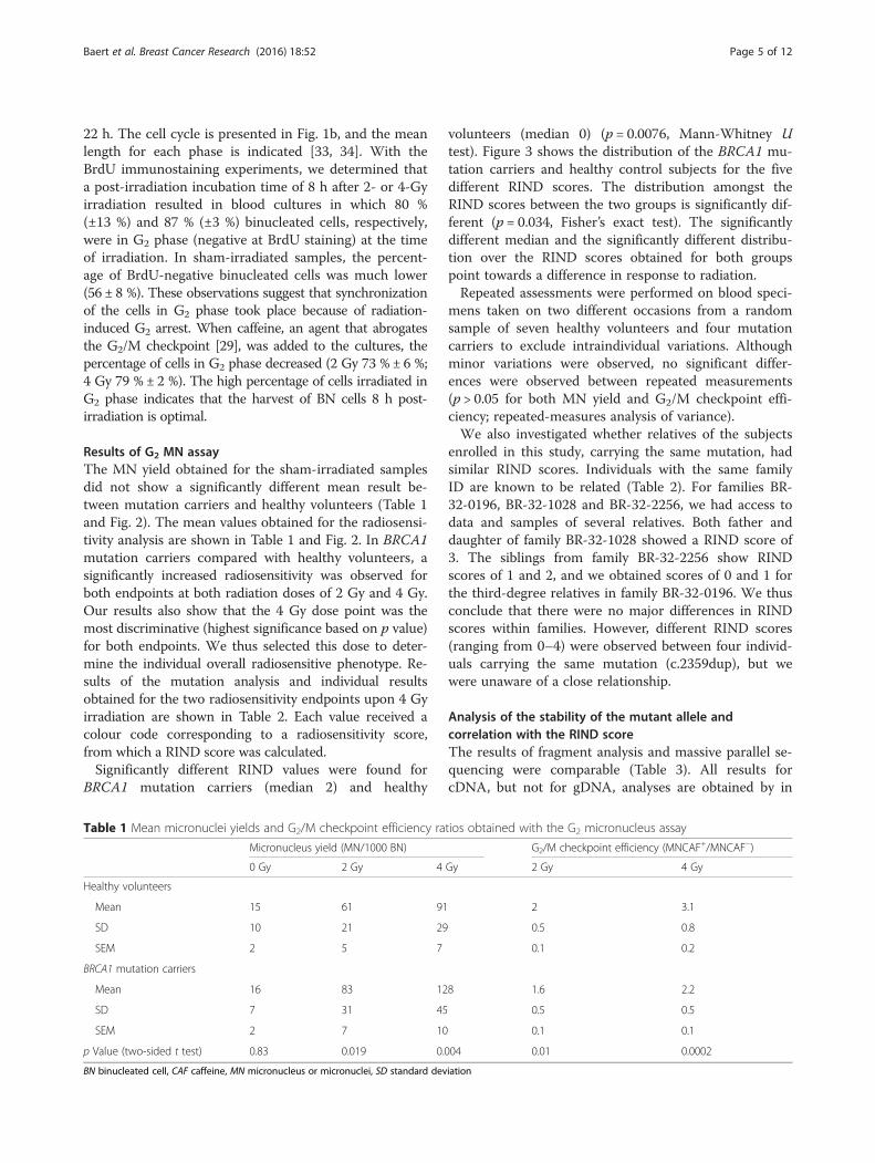
Table 1 Mean micronuclei yields and G2/M checkpoint efficiency ratios obtained with the G2 micronucleus assay
Micronucleus yield (MN/1000 BN) G2/M checkpoint efficiency (MNCAF+/MNCAF−)
0 Gy 2 Gy 4 Gy 2 Gy 4 Gy
Healthy volunteers
Mean 15 61 91 2 3.1
SD 10 21 29 0.5 0.8
SEM 2 5 7 0.1 0.2
BRCA1 mutation carriers
Mean 16 83 128 1.6 2.2
SD 7 31 45 0.5 0.5
SEM 2 7 10 0.1 0.1
p Value (two-sided t test) 0.83 0.019 0.004 0.01 0.0002
BN binucleated cell, CAF caffeine, MN micronucleus or micronuclei, SD standard deviation
Baert et al. Breast Cancer Research (2016) 18:52 Page 5 of 12

duplicate experiments. The results of the MiSeq analysisare expressed as the VAF in percent; 50 % means equalexpression of both alleles. The results of the fragmentanalysis are shown as a ratio of peak heights of mutantto WT allele. A value of 1 equals no loss of expression ofthe mutant allele. cDNA samples not treated with puro-mycin were scored as “evidence of NMD” (Table 3) whenan average VAF ≤31 % or a ratio of peak heights <0.7 wasobtained. We observed more variation for deletions/inser-tions than for substitutions. This can be explained by themore complex mapping of reads containing deletions/du-plications. A large deletion and software struggling to mapcorrectly to the reference sequence explains a VAF <50 %for gDNA in some patients (Table 3). Nevertheless, a dropin VAF for cDNA and not for cDNA with puromycin canstill be distinguished.For M02, M08 and M15, no lymphocytes were avail-
able to perform this assay. However, as we had access tocDNA from other individuals with the same mutation,we were able to gain insight into the stability of the mu-tant mRNA for these three mutation carriers. For M16,
no gDNA data could be obtained; information with re-gard to mutant mRNA stability for this mutation carrierwas obtained via cDNAp and M17.
Premature termination codon alleles in the central part ofthe geneFor all truncating mutations studied in the central partof the gene (7 unique mutations in 15 individuals), wefound evidence for NMD. In general, 25–30 % residualtruncated mRNA was detected in carriers of a prematuretermination codon (PTC) mutation. Ten of fifteen pa-tients with a truncating allele showed a radiosensitivephenotype (RIND score 1–4, median 1).
Mutations located in the 5′ part of the geneFor mutations in the 5′ part of the gene (n = 3), we haveno evidence for NMD. The effect at the mRNA level forthe c.212 + 3A > G splice-site mutation has previouslybeen studied with quantitative PCR by our group. Weshowed that no full-length transcript is formed, but thatit leads to a significantly increased expression of an
Fig. 2 Mean micronucleus (MN) yields and G2/M checkpoint efficiency ratios obtained with the G2 MN assay in healthy volunteers and BRCA1mutation carriers. Significance was determined with a two-sided t test. p Values for each of the endpoints and dose points are indicated in thegraph. Error bars represent the standard error of the mean. BN binucleated cell, Caf caffeine
Baert et al. Breast Cancer Research (2016) 18:52 Page 6 of 12

Table 2 Results of mutation screening and radiosensitivity assessment for each individual
Baert et al. Breast Cancer Research (2016) 18:52 Page 7 of 12

alternative transcript (out of frame skip of 22 last nucle-otides of exon 5; r.190_212del), which is not subjectedto NMD [35, 36]. As M03 is not heterozygous for any ofthe tested common SNPs, results could not be re-analysed with the approach described in this paper. Ourresults for M03 showed a RIND score of 2, which can beattributed to a decreased G2/M checkpoint efficiency.For M01, carrier of the c.1A > G start codon mutation,
no decline of the mutant allele could be detected whenanalysing the SNP data (Table 3). This suggests the con-servation of the start codon or an alternative one. Themutation itself could not be quantified, owing to thepresence of GC-rich areas near the start codon. Theintroduction of an alternative start codon could give riseto an aberrant protein with a dominant-negative effect[37]. Interestingly, both individuals carrying a mutation
Fig. 3 The distribution (%) of healthy volunteers and BRCA1 mutation carriers over the different radiosensitivity indicator (RIND) scores
Table 3 Stability of the mutant allele
Fragment analysis MiSeq (VAF)
Donor RIND score Ratio peak height (mutant/WT allele) cDNA cDNAp gDNA Evidence for NMD?
M01 c.1A > G 3 0.7 (0.6–0.8) 49 (42–55) 52 (51–52) 49 (49–49) No
M02 c.1A > C 2 / / / / No
M03 c.212 + 3A > G 2 / / / / No
M04 c.1961del 0 0.4 (0.4–0.4) 29 (25–33) 47 (43–51) 47 (47–47) Yes
M05 c.2359dup 3 0.6 (0.6–0.6) 28 (25–31) 42 (38–45) 48 (48–48) Yes
M06 c.2359dup 2 0.5 (0.5–0.5) 24 (23–25) 44 (41–47) 50 (50–50) Yes
M07 c.2359dup 0 0.4 (0.4–0.5) 29 (27–30) 48 (30–67) 57 (55–59) Yes
M08 c.2359dup 4 / / / / Yes
M09 c.3331_3334del 1 0.4 (0.4–0.4) 25 (18–31) 44 (34–48) 49 (49–49) Yes
M10 c.3481_3491del 2 0.4 (0.4–0.4) 23 (16–30) 42 (28–52) 39 (32–46)a Yes
M11 c.3481_3491del 1 0.4 (0.3–0.5) 23 (16–32) 41 (23–54) 39 (34–44)a Yes
M12 c.3481_3491del 2 0.4 (0.4–0.4) 26 (16–34) 39 (28–50) 42 (35–49)a Yes
M13 c.3661G > T 1 0.5 (0.3–0.6) 29 (28–30) 50 (41–57) 50 (46–53) Yes
M14 c.3661G > T 0 0.4 (0.3–0.4) 31 (29–32) 52 (52–52) 50 (50–50) Yes
M15 c.3661G > T 0 / / / / Yes
M16 c.4327C > T 3 0.5 (0.5–0.6) 24 (21–27) 51 (45–56) / Yes
M17 c.4327C > T 3 0.6 (0.4–0.7) 26 (21–30) 48 (44–53) 50 (50–50) Yes
M18 c.4931_4393delinsTT 0 0.4 (0.3–0.4) 30 (28–31) 37 (28–47) 52 (52–52) Yes
cDNA complementary DNA, cDNAp cDNA extracted in the presence of puromycin, gDNA genomic DNA, NMD nonsense mediated decay, RIND radiosensitivityindicator, VAF variant allele frequency, WT wild typeaVAF <50 % for gDNA
Baert et al. Breast Cancer Research (2016) 18:52 Page 8 of 12
affecting the start codon had, respectively, RIND scoresof 3 and 2. A median RIND score of 2 was observed forcarriers of a mutation in the 5′ part of the gene.
DiscussionOur study shows a significantly increased radiosensitivityin the group of healthy BRCA1 mutation carriers com-pared with healthy controls for the two endpoints mea-sured by the G2 MN assay for both doses of 2 and 4 Gy.Our results indicate that radiosensitivity in heterozygousBRCA1 mutation carriers is a complex phenotype linkedto defects in DNA damage repair, as well as to defectiveG2 arrest capacity. These results are in agreement withthe studies performed by Pantelias and Terzoudi [30].They applied the G2 chromatid break assay and reportedthat the G2/M checkpoint efficiency ratio is a good par-ameter for prediction of intrinsic radiosensitivity in A-Tand cancer patients.In vitro radiosensitivity has previously been investi-
gated in patients with BC and BRCA1 mutation carriers.Cardinale et al. recently published a meta-analysis com-bining all in vitro case-control studies in which the G0
MN assay on peripheral blood lymphocytes was used toanalyse in vitro radiosensitivity in women with BC orwith a known or putative genetic predisposition to BC[17]. Other cytogenetic assays, such as the chromosomeaberration and G2 chromatid break assay or survival as-says, have also been applied to determine radiosensitivityin a variety of cell types heterozygous for BRCA1 muta-tions, using different irradiation protocols [18–26]. It isdifficult, however, to correctly compare the results ofthese studies, as also concluded by Cardinale et al. [17],owing to different experimental set-ups to analyse invitro radiosensitivity. However, despite the heterogeneityof the studies, most of the data generated, includingours, are suggestive of a different radiosensitive pheno-type between BRCA1 heterozygous mutant cells andcontrol cells. Recent studies in which researchers investi-gated more specifically the functionality of the HR path-way in BRCA1 heterozygous cells by means of γ-H2AXand RAD51 foci assays point towards a less efficientDSB repair by HR. These findings further support theevidence of increased radiosensitivity observed inBRCA1 heterozygous cells when irradiated in the S orG2 phase of the cell cycle and are compatible with hap-loinsufficiency as the underlying mechanism [24, 25, 38].The strength of our study is that radiosensitivity was ana-lysed by means of two different endpoints obtained with aG2-specific MN assay developed by our group.The radiosensitivity results in this study were obtained
with doses of 2 and 4 Gy, which is considerably higherthan any lifetime cumulative dose received by mammog-raphy screening. The average dose delivered to the breastglandular tissue per mammographic screening session is
approximately 4 mGy [39]. Thus, direct extrapolation ofour radiosensitivity results to the risks of mammographyis not possible. Despite this limitation, our results maysuggest caution in the IR exposure of healthy tissues ofBRCA1 mutation carriers for diagnostic purposes. It is fur-thermore noteworthy that recent studies have shown that30-kV X-rays have a higher relative biological effect andare thus more harmful than conventional high-kilovoltageX-rays or 60Co gamma rays, on which current risk assess-ment is based. This implies that each mammogrammay induce more DNA damage than commonly estimated[40, 41]. The authors of several papers have suggested thatmammography screening might preferably be replaced bymagnetic resonance imaging to avoid IR before the age of30 years [16] or 40 years [42] in BRCA1 mutation carriers.In addition, and more appropriately, given the highdoses used in this assay, our results suggest cautionin the use of adjuvant radiotherapy following breast-conserving surgery. In this respect, there is a need forwell-designed studies to assess the incidence of secondipsi- or contralateral cancers upon adjuvant radiotherapyin mutation carriers [16].Since it was our aim to develop a radiosensitivity assay
applicable in a clinical setting, we performed the G2 MNassay on peripheral blood samples, which can easily beobtained during a genetic consult. Several studies havedemonstrated that radiosensitivity of an individual is alsodetectable in cells of a type different from cells in whichthe tumour develops [18, 43, 44]. The scoring systemwith RIND scores varying between 0 and 4 allowed us toassess overall radiosensitivity due to both DNA repairand G2 arrest capacity of each mutation carrier (Table 2and Fig. 3). With the help of this scoring system, we de-termined that 72 % of our healthy volunteers showed noradiosensitive phenotype. BRCA1 mutation carriers, onthe contrary, showed a distinct pattern towards higherradiosensitivity. Seventy-two percent of all mutation car-riers were found to be radiosensitive (RIND score 1–3).Moreover, 28 % of BRCA1 mutation carriers had RINDscores equal to 3 or 4, scores that were never observedin healthy volunteers. This simple scoring system can bevaluable in assisting physicians in their decision-makingin clinical follow-up and for refinement of radiotherapyat the individual level. Since our study is limited in sam-ple size, a larger prospective study with blood samples ofBRCA1 mutation carriers will be undertaken to confirmand prove the importance of an in vitro radiosensitivityscoring system to assist clinical management of BRCA1mutation carriers.Haploinsufficiency has been suggested as the main
mechanism for hereditary breast carcinogenesis [45].However, as NMD is not observed for mutations locatedin the 5′ and 3′ parts of the gene [46], a dominant-negative effect whereby the aberrant transcript abolishes
Baert et al. Breast Cancer Research (2016) 18:52 Page 9 of 12

the functionality of the WT allele cannot be excluded(for review, see [37, 47]). We wanted to evaluate if eitheror both mechanisms could influence the radiosensitivityscore. Information on the stability of the mutant allele atthe mRNA level was generated by both a fragment ana-lysis and a novel massive parallel sequencing approach.To our knowledge, this is the first time that next-generation sequencing is applied to evaluate the level ofNMD. The approach we describe here is straightforwardand cost-effective for study of the relative expression oftwo alleles in a single individual.The majority of patients included in this study are het-
erozygous for a mutation located in the central part ofthe gene leading to a PTC. These PTC-inducing muta-tions result in a truncated mRNA, which can be de-graded by NMD. Generally, we observed that themutated PTC allele was expressed at a ratio of 25–30 %of the WT allele at the mRNA level. Our results areconsistent with the data of Anczuców et al. and Perrin-Vidoz et al. [46, 47]. Both articles describe a similarreduction of mRNA expression from PTC alleles in thecentral part of the gene and demonstrate the involve-ment of NMD in this decrease of mutant mRNA. A ra-diosensitive phenotype in 10 of 15 mutation carrierswith a PTC allele undergoing NMD suggests haploinsuf-ficiency as a mechanism leading to this phenotype in thelarge majority of the individuals.Equal expression of the WT and mutant alleles at the
mRNA level was observed in the patients with the startcodon mutation (M01 and M02). For the patient withthe c.212 + 3A > G mutation (M03), equal expression ofa full-length transcript and a transcript lacking the last22 nucleotides of exon 5 was observed in previous re-search by our group and confirmed by others [35, 36]. Ahigher median RIND score in the individuals carrying amutation in the 5′ part of the gene, for which no NMDcould be detected, compared with individuals with aPTC allele may point towards another mechanism in-volved in the radiosensitive phenotype, such as adominant-negative effect. However, current knowledgeon translation of mutant alleles not subjected to NMDinto proteins is limited. Such detailed studies have not yetbeen undertaken; in most studies demonstrating a role forhaploinsufficiency, BRCA1 mutation carriers are com-pared as a group with non-carriers (e.g., [25, 38, 48]). Ourdata suggest the need for larger studies involving differenttypes of mutations.Unique to this radiosensitivity study compared with
others are that we had access to material from several in-dividuals from one family and that we could also evaluatethe radiosensitive phenotype from several unrelated indi-viduals carrying the same mutation. For four unrelatedcarriers of a Belgian founder mutation c.2359dup(p.Glu787fs*3), a large variation for the radiosensitive
phenotype based on the RIND score was observed (range0–4) (Table 2). Repeated assessments on blood specimens,taken on two different occasions from several individuals,ruled out that the different RIND scores were due toexperimental variation. Given the smaller variationbetween related individuals, and because of the pleio-tropic effect of IR on DNA (for example, strandbreaks, fork stalling, base damage, DNA adducts [49–51]), we are convinced that other genetic factors in-fluence the radiosensitivity. In previous research, forexample, researchers have demonstrated the effect ofSNPs on individual sensitivity to radiation therapy[52, 53]. In addition, our group has demonstrated theinfluence of RAD51, Ku70 and Ku80 SNPs as modu-lators of in vitro radiosensitivity in BRCA1 mutationcarriers and patients with BC [54, 55]. A larger studyand the inclusion of a control population with rela-tives not harbouring the familial germline mutationcould generate important insights.
ConclusionsIn our present study, using the G2 MN assay, we showthat healthy individuals carrying a germline mutation inBRCA1 are more radiosensitive than healthy controlsubjects after exposure to doses of 2 and 4 Gy. Seventy-two percent of the BRCA1 mutation carriers showed aradiosensitive phenotype, and 28 % of BRCA1 mutationcarriers had high RIND scores of 3 or 4. Analysis of themRNA stability of the mutant allele could not demon-strate a clear link between nonsense-mediated decay ofthe mutant allele and a radiosensitive phenotype.This, combined with the similar radiosensitive pheno-type observed for related individuals but not for unre-lated individuals carrying the same mutation, isindicative of the fact that additional genetic factorsbesides the BRCA1 mutation may play a role in theradiation response. Our study emphasizes the needfor large, prospective studies correlating the in vitrofindings and exposure to radiation with the risk ofdeveloping breast cancer.
Ethical approval and consent to participateThis study was approved by the ethics committee of theGhent University Hospital (B670201111641 d.d. 20/09/2011). All study participants signed an informed consentform.
Additional files
Additional file 1: Primers. The primer sequences (both forward andreverse). (PDF 29 kb)
Additional file 2: Results of the fragment analysis. Illustration of fragmentanalysis data of an SNP without loss of the mutant allele. (PDF 176 kb)
Baert et al. Breast Cancer Research (2016) 18:52 Page 10 of 12

Additional file 3: Results of the fragment analysis. Illustration of fragmentanalysis data of an SNP with loss of the mutant allele. (PDF 227 kb)
AbbreviationsA-T: ataxia-telangiectasia; BC: breast cancer; BN: binucleated cell; BrdU: 5-bromo-2′-deoxyuridine; CAF: caffeine; cDNA: complementary DNA;cDNAp: cDNA extracted in the presence of puromycin; CMGG: Centre forMedical Genetics, Ghent, Belgium; cRPMI: complete RPMI medium; cytoB: cytochalasin B; DSB: double-strand break; EDTA: ethylenediaminetetraaceticacid; FCS: foetal calf serum; gDNA: genomic DNA; HR: homologousrecombination; IR: ionizing radiation; MN: micronucleus or micronuclei;NMD: nonsense mediated decay; PCR: polymerase chain reaction;PHA: phytohaemagglutinin; PTC: premature termination codon;RIND: radiosensitivity indicator; SD: standard deviation; SNP: single-nucleotidepolymorphism; VAF: variant allele frequency; WT: wild type.
Competing interestsThe authors declare that they have no competing interests.
Authors’ contributionsAB and JD carried out the G2 MN assay and analysed the data. AB performedthe statistical analysis and drafted the manuscript. JD revised the manuscript.AV and KBMC are the senior researchers of this study, and they designed theG2 MN assay and the analysis of the stability of the mutant allele study,respectively. AV and KBMC supervised the principal young investigator ABand assisted in the interpretation of the data and the revision of themanuscript. KDL participated in the data acquisition regarding the stability ofthe mutant allele study and the interpretation of these data. KDL also revisedthe manuscript. GP participated in the revision of the manuscript and theinterpretation of the data by designing the radiosensitivity indicator (RIND)score. GP also assisted in the English language editing. BP, FM, TVM, KS, JvdE,TVD and SDN assisted in the sample and data acquisition and datainterpretation, and they also helped with the revision of the manuscript. Allagreed to be accountable for all aspects of the work. All authors read andapproved the final manuscript.
AcknowledgementsThe authors thank all individuals who contributed to this study by donatingblood samples. We thank Ilse Coene, Brecht Crombez, Céline Debrock,Mattias Van Heetvelde, Greet De Smet, Johanna Aernoudt, Leen Pieters andToke Thiron for their technical assistance and sharing of their knowledge. Wethank Prof. Dr. Thierens for the use of the irradiation facility. This study isfunded by the Belgian Foundation against Cancer (project 2012–216).
Author details1Department of Basic Medical Sciences, Ghent University, Ghent, Belgium.2Center for Medical Genetics, Ghent University Hospital, Ghent, Belgium.3Department of Medical Genetics, University of Antwerp/University Hospitalof Antwerp, Antwerp, Belgium. 4Biomedical Research Division, Department ofTheoretical and Applied Sciences, University of Insubria, Busto Arsizio, Italy.
Received: 29 January 2016 Accepted: 23 April 2016
References1. Couch FJ, Nathanson KL, Offit K. Two decades after BRCA: setting paradigms
in personalized cancer care and prevention. Science. 2014;343:1466–70.2. Caestecker KW, Van de Walle GR. The role of BRCA1 in DNA double-strand
repair: past and present. Exp Cell Res. 2013;319:575–87.3. Roy R, Chun J, Powell SN. BRCA1 and BRCA2: different roles in a common
pathway of genome protection. Nat Rev Cancer. 2012;12:68–78.4. Pfeiffer P, Goedecke W, Kuhfittig-Kulle S, Obe G. Pathways of DNA double-
strand break repair and their impact on the prevention and formation ofchromosomal aberrations. Cytogenet Genome Res. 2004;104:7–13.
5. Foulkes WD, Shuen AY. In brief: BRCA1and BRCA2. J Pathol. 2013;230:347–9.6. Cousineau I, Abaji C, Belmaaza A. BRCA1 regulates RAD51 function in
response to DNA damage and suppresses spontaneous sister chromatidreplication slippage: Implications for sister chromatid cohesion, genomestability, and carcinogenesis. Cancer Res. 2005;65:11384–91.
7. Yarden RI, Pardo-Reoyo S, Sgagias M, Cowan KH, Brody LC. BRCA1 regulatesthe G2/M checkpoint by activating Chk1 kinase upon DNA damage. NatGenet. 2002;30:285–9.
8. Pijpe A, Andrieu N, Easton DF, Kesminiene A, Cardis E, Noguès C, et al. Exposureto diagnostic radiation and risk of breast cancer among carriers of BRCA1/2mutations: retrospective cohort study (GENE-RAD-RISK). BMJ. 2012;345:e5660.
9. Lecarpentier J, Noguès C, Mouret-Fourme E, Stoppa-Lyonnet D, Lasset C,Caron O, et al. Variation in breast cancer risk with mutation position,smoking, alcohol, and chest X-ray history, in the French National BRCA1/2carrier cohort (GENEPSO). Breast Cancer Res Treat. 2011;130:927–38.
10. Andrieu N, Easton DF, Chang-Claude J, Rookus MA, Brohet R, Cardis E, et al.Effect of chest x-rays on the risk of breast cancer among BRCA1/2 MutationCarriers in the International BRCA1/2 Carrier Cohort Study: a report from theEMBRACE, GENEPSO, GEO-HEBON, and IBCCS Collaborators’ Group. J ClinOncol. 2006;24:3361–6.
11. Gronwald J, Pijpe A, Byrski T, Huzarski T, Stawicka M, Cybulski C, et al. Earlyradiation exposures and BRCA1-associated breast cancer in young womenfrom Poland. Breast Cancer Res Treat. 2008;112:581–4.
12. John EM, McGuire V, Thomas D, Haile R, Ozcelik H, Milne RL, et al.Diagnostic chest X-rays and breast cancer risk before age 50 years forBRCA1 and BRCA2 mutation carriers. Cancer Epidemiol Biomarkers Prev.2013;22:1547–56.
13. Narod SA, Lubinski J, Ghadirian P, Lynch HT, Moller P, Foulkes WD, et al.Screening mammography and risk of breast cancer in BRCA1 and BRCA2mutation carriers: a case-control study. Lancet Oncol. 2006;7:402–6.
14. Giannakeas V, Lubinski J, Gronwald J, Moller P, Armel S, Lynch HT, et al.Mammography screening and the risk of breast cancer in BRCA1 and BRCA2mutation carriers: a prospective study. Breast Cancer Res Treat. 2014;147:113–8.
15. Goldfrank D, Chuai S, Bernstein JL, Ramon y Cajal T, Lee JB, Alonso MC, et al.Effect of mammography on breast cancer risk in women with mutations inBRCA1 or BRCA2. Cancer Epidemiol Biomarkers Prev. 2006;15:2311–3.
16. Drooger JC, Hooning MJ, Seynaeve CM, Baaijens MH, Obdeijn IM, Sleijfer S, et al.Diagnostic and therapeutic ionizing radiation and the risk of a first and secondprimary breast cancer, with special attention for BRCA1 and BRCA2 mutationcarriers: a critical review of the literature. Cancer Treat Rev. 2015;41:187–96.
17. Cardinale F, Bruzzi P, Bolognesi C. Role of micronucleus test in predictingbreast cancer susceptibility: a systematic review and meta-analysis. Br JCancer. 2012;106:780–90.
18. Kote-Jarai Z, Salmon A, Mengitsu T, Copeland M, Ardern-Jones A, Locke I,et al. Increased level of chromosomal damage after irradiation oflymphocytes from BRCA1 mutation carriers. Br J Cancer. 2006;94:308–10.
19. Frankenberg-Schwager M, Gregus A. Chromosomal instability induced bymammography X-rays in primary human fibroblasts from BRCA1 and BRCA2mutation carriers. Int J Radiat Biol. 2012;88:846–57.
20. Ernestos B, Nikolaos P, Koulis G, Eleni R, Konstantinos B, Alexandra G, et al. Increasedchromosomal radiosensitivity in women carrying BRCA1/BRCA2mutations assessedwith the G2 assay. Int J Radiat Oncol Biol Phys. 2010;76:1199–205.
21. Barwell J, Pangon L, Georgiou A, Kesterton I, Langman C, Arden-Jones A,et al. Lymphocyte radiosensitivity in BRCA1 and BRCA2 mutation carriers andimplications for breast cancer susceptibility. Int J Cancer. 2007;121:1631–6.
22. Buchholz TA, Wu X, Hussain A, Tucker SL, Mills GB, Haffty B, et al. Evidenceof haplotype insufficiency in human cells containing a germline mutation inBRCA1 or BRCA2. Int J Cancer. 2002;561:557–61.
23. Hair JM, Terzoudi GI, Hatzi VI, Lehockey KA, Srivastava D, Wang W, et al.BRCA1 role in the mitigation of radiotoxicity and chromosomal instabilitythrough repair of clustered DNA lesions. Chem Biol Interact. 2010;188:350–8.
24. Sioftanos G, Ismail A, Föhse L, Shanley S, Worku M, Short SC. BRCA1 andBRCA2 heterozygosity in embryonic stem cells reduces radiation-inducedRad51 focus formation but is not associated with radiosensitivity. Int JRadiat Biol. 2010;86:1095–105.
25. Pathania S, Bade S, Le Guillou M, Burke K, Reed R, Bowman-Colin C, et al.BRCA1 haploinsufficiency for replication stress suppression in primary cells.Nat Commun. 2014;5:5496.
26. Febrer E, Mestres M, Caballín MR, Barrios L, Ribas M, Gutiérrez-Enríquez S,et al. Mitotic delay in lymphocytes from BRCA1 heterozygotes unable toreduce the radiation-induced chromosomal damage. DNA Repair (Amst).2008;7:1907–11.
27. Claes K, Depuydt J, Taylor AM, Last JI, Baert A, Schietecatte P, et al. Variantataxia telangiectasia: clinical and molecular findings and evaluation ofradiosensitive phenotypes in a patient and relatives. Neuromolecular Med.2013;15:447–57.
Baert et al. Breast Cancer Research (2016) 18:52 Page 11 of 12

28. Gutiérrez-Enríquez S, Ramón Y, Cajal T, Alonso C, Corral A, Carrasco P, et al.Ionizing radiation or mitomycin-induced micronuclei in lymphocytes ofBRCA1 or BRCA2 mutation carriers. Breast Cancer Res Treat. 2011;127:611–22.
29. Li N, Zhang H, Wang Y, Hao J. BRCA1 and its phosphorylation involved incaffeine-inhibitable event upstream of G2 checkpoint. Sci China Phys MechAstron. 2010;53:1281–5.
30. Pantelias GE, Terzoudi GI. A standardized G2-assay for the prediction ofindividual radiosensitivity. Radiother Oncol. 2011;101:28–34.
31. Claes K, Poppe B, Machackova E, Coene I, Foretova L, De Paepe A, et al.Differentiating pathogenic mutations from polymorphic alterations in thesplice sites of BRCA1 and BRCA2. Genes Chromosom Cancer. 2003;37:314–20.
32. De Leeneer K, Hellemans J, Steyaert W, Lefever S, Vereecke I, Debals E, et al.Flexible, scalable, and efficient targeted resequencing on a benchtop sequencerfor variant detection in clinical practice. Hum Mutat. 2015;36:379–87.
33. Sasaki MS, Norman A. Proliferation of human lymphocytes in culture.Nature. 1966;210:913–4.
34. Bernheim JL, Dorian RE, Mendelsohn J. DNA synthesis and proliferation ofhuman lymphocytes in vitro. I. Cell kinetics of response tophytohemagglutinin. J Immunol. 1978;120:955–62.
35. Claes K, Vandesompele J, Poppe B, Dahan K, Coene I, De Paepe A, et al.Pathological splice mutations outside the invariant AG/GT splice sites ofBRCA1 exon 5 increase alternative transcript levels in the 5′ end of theBRCA1 gene. Oncogene. 2002;21:4171–5.
36. Théry JC, Krieger S, Gaildrat P, Révillion F, Buisine MP, Killian A, et al.Contribution of bioinformatics predictions and functional splicing assays tothe interpretation of unclassified variants of the BRCA genes. Eur J HumGenet. 2011;19:1052–8.
37. Linger RJ, Kruk PA. BRCA1 16 years later: risk-associated BRCA1 mutationsand their functional implications. FEBS J. 2010;277:3086–96.
38. Vaclová T, Gómez-López G, Setién F, García Bueno JM, Macías JA, Barroso A,et al. DNA repair capacity is impaired in healthy BRCA1 heterozygousmutation carriers. Breast Cancer Res Treat. 2015;152:271–82. doi:10.1007/s10549-015-3459-3.
39. Pauwels EKJ, Foray N, Bourguignon MH. Breast cancer induced by X-raymammography screening? A review based on recent understanding of low-dose radiobiology. Med Princ Pract. 2016;25:101–9. doi:10.1159/000442442.
40. Heyes GJ, Mill AJ, Charles MW. Mammography-oncogenicity at low doses.J Radiol Prot. 2009;29:A123–32.
41. Depuydt J, Baert A, Vandersickel V, Thierens H, Vral A. Relative biologicaleffectiveness of mammography X-rays at the level of DNA andchromosomes in lymphocytes. Int J Radiat Biol. 2013;89:532–8.
42. Obdeijn IM, Winter-Warnars GA, Mann RM, Hooning MJ, Hunink MGM,Tilanus-Linthorst MM. Should we screen BRCA1 mutation carriers only withMRI? A multicenter study. Breast Cancer Res Treat. 2014;144:577–82.
43. Rieger KE, Hong WJ, Tusher VG, Tang J, Tibshirani R, Chu G. Toxicity fromradiation therapy associated with abnormal transcriptional responses toDNA damage. Proc Natl Acad Sci U S A. 2004;101:6635–40.
44. Foray N, Randrianarison V, Marot D, Perricaudet M, Lenoir G, Feunteun J.Gamma-rays-induced death of human cells carrying mutations of BRCA1 orBRCA2. Oncogene. 1999;18:7334–42.
45. Salmena L, Narod S. BRCA1 haploinsufficiency: consequences for breastcancer. Womens Health (Lond Engl). 2012;8:127–9.
46. Perrin-Vidoz L, Sinilnikova OM, Stoppa-Lyonnet D, Lenoir GM, Mazoyer S.The nonsense-mediated mRNA decay pathway triggers degradation ofmost BRCA1 mRNAs bearing premature termination codons. Hum MolGenet. 2002;11:2805–14.
47. Anczuków O, Ware MD, Buisson M, Zetoune AB, Stoppa-Lyonnet D,Sinilnikova OM, et al. Does the nonsense-mediated mRNA decaymechanism prevent the synthesis of truncated BRCA1, CHK2, and p53proteins? Hum Mutat. 2008;29:65–73.
48. Sedic M, Skibinski A, Brown N, Gallardo M, Mulligan P, Martinez P, et al.Haploinsufficiency for BRCA1 leads to cell-type-specific genomic instabilityand premature senescence. Nat Commun. 2015;6:7505.
49. Nikjoo H, O’Neill P, Wilson WE, Goodhead DT. Computational approach fordetermining the spectrum of DNA damage induced by ionizing radiation.Radiat Res. 2001;156:577–83.
50. Hagen U. Current aspects on the radiation induced base damage in DNA.Radiat Environ Biophys. 1986;25:261–71.
51. Dextraze ME, Gantchev T, Girouard S, Hunting D. DNA interstrand cross-linksinduced by ionizing radiation: an unsung lesion. Mutat Res. 2010;704:101–7.
52. Guo Z, Shu Y, Zhou H, Zhang W, Wang H. Radiogenomics helps to achievepersonalized therapy by evaluating patient responses to radiationtreatment. Carcinogenesis. 2015;36:307–17.
53. Popanda O, Marquardt JU, Chang-Claude J, Schmezer P. Genetic variation innormal tissue toxicity induced by ionizing radiation. Mutat Res. 2009;667:58–69.
54. Vral A, Willems P, Claes K, Poppe B, Perletti G, Thierens H. Combined effectof polymorphisms in Rad51 and Xrcc3 on breast cancer risk andchromosomal radiosensitivity. Mol Med Rep. 2011;4:901–12.
55. Willems P, Claes K, Baeyens A, Vandersickel V, Werbrouck J, De Ruyck K,et al. Polymorphisms in nonhomologous end-joining genes associated withbreast cancer risk and chromosomal radiosensitivity [published erratumappears in Genes Chromosomes Cancer. 2009;48:381]. Genes ChromosomesCancer. 2008;47:137–48.
• We accept pre-submission inquiries
• Our selector tool helps you to find the most relevant journal
• We provide round the clock customer support
• Convenient online submission
• Thorough peer review
• Inclusion in PubMed and all major indexing services
• Maximum visibility for your research
Submit your manuscript atwww.biomedcentral.com/submit
Submit your next manuscript to BioMed Central and we will help you at every step:
Baert et al. Breast Cancer Research (2016) 18:52 Page 12 of 12


![Intrinsic Radiosensitivity of Normal Human Fibroblasts and ... · (CANCER RESEARCH 52. 6348-6352. November 15. 1992] Intrinsic Radiosensitivity of Normal Human Fibroblasts and Lymphocytes](https://img.dokumen.tips/doc/110x75/60cc08f35a119f051502c1e0/intrinsic-radiosensitivity-of-normal-human-fibroblasts-and-cancer-research.jpg)