Embed Size (px)
Citation preview
For personal use. Only reproduce with permission from Elsevier Ltd
699
Newsdesk
Scientists in the USA report that theinclusion bodies typically found instriatal neurons in Huntington’s diseaseseem to delay rather than cause celldeath, at least in culture. Inclusionbodies may therefore protect against theneurodegeneration associated with thedisorder (Nature 2004; 431: 805–10).
In patients with Huntington’sdisease a mutant form of huntingtin(Htt) with repeated glutamine residuesis expressed. These mutant proteinsaggregate to form the inclusion bodies.
“Some researchers believe theseinclusions to be a major pathogenicfeature in the disease”, explains teamleader Steven Finkbeiner (Universityof California, San Francisco, USA),“while others suggest they may act as asink for the rogue protein, getting itout of the way. Others still think theseinclusion bodies may just beincidental.” Knowing for sure isimportant because any therapy aimedat preventing their formation could domore harm than good if their role isprotective.
The team developed a newtechnique that allowed the production
of Htt (tagged with fluorescent greenprotein)—both diffuse and in inclusionbodies—to be monitored over severaldays. This allowed them to compare thesurvival times of control neurons, thosewith diffuse Htt, and those withinclusion bodies.
“We found that the neuronsexpressing the mutant Htt in a diffuse
manner died sooner than thoseexpressing normal Htt, and that thelonger the polyglutamate extensions tothe protein, the worse they fared”,explains team member MontserratArrasate. “But the cells that formedinclusion bodies went on to live muchlonger. Also, as they formed, diffuse Httconcentrations fell. This suggests thatthe production of inclusion bodieseither helps sequester diffuse Htt or, insome way, promotes increased Httturnover, thus protecting the neuronsfrom its toxic effects.”
“Inclusion bodies certainlysequester a number of necessary cellproteins, an activity that may well beharmful”, comments José Lucas(Universidad Autonoma, Madrid,Spain). “But this research suggests thata therapy that removes inclusion bodiesmight have an even more undesirableeffect. One wonders whether we need toreassess the role of inclusion bodies inother neurodegenrative diseases such asAlzheimer’s and Parkinson’s diseases.”Adrian Burton
Inclusion bodies may be neuroprotective in Huntington’s disease
Low dietary intake of iron protectsdopaminergic neurons in a mousemodel of Parkinson’s disease, accordingto new research. But the role of dietaryiron is not straightforward and has beena contentious issue since the 1970swhen Moussa Youdim began investiga-ting the effects of iron deficiency onbrain function.
“Our study showed that reducedlevels of dietary iron may indeedprotect neurons that are exposedto factors that increase the riskof Parkinson’s disease”, explainslead author Cathy Levenson (FloridaState University, Tallahassee, USA).“However, we also saw very clearevidence that in the absence of risk,low dietary iron increased the riskof developing Parkinson’s likesymptoms”.
Levenson and colleagues used amouse model of Parkinson’s disease inwhich the neurotoxin 1-methyl-4-phenyl-1,2,3,6-tetrapyridine (MPTP) isused to selectively destroy dopaminer-
gic neurons in the striatum. Theresearchers fed mice with a diet thateither contained adequate iron (+Fe;48 mg/kg) or low iron (–Fe; 4 mg/kg)for 6 weeks before injecting the micewith MPTP. 1 week after MPTPinjection, Levenson and colleaguestested motor function of the mice andmeasured neurotransmitter concentra-tions and lipid peroxidation products(such as sphingolipids and ceramides)in the striatum (Exp Neurol 2004;published online Oct 2, DOI: 10.1016/j.expneurol.2004.08.014). Sphingolipidsare converted to ceramides by sphingo-myelinases, which are activated in thepresence of reactive oxygen species.
The researchers report that, beforeMPTP lesion, motor function wasimpaired in the –Fe group comparedwith the +Fe group. However, afterMPTP lesion, they found that motorfunction was impaired in the +Fe groupbut not in the –Fe group compared withcontrol mice who received a salineinjection. Concentrations of dopamine,
and its metabolites, were reduced in thestriatum of +Fe mice after MPTP lesionbut were unchanged in –FE mice. Thereduction in striatal dopamineconcentration correlated with thedegree of motor impairment. Inaddition, the concentration ofsphingomyelin in the striatum of –Femice was twice that in +Fe mice; theopposite was true for ceramideconcentrations. These results suggestthat decreased dietary intake of ironmay protect dopaminergic neurons byreducing oxidative stress.
“While we are encouraged by thedata on the use of these drugs forParkinson’s disease, our work showsthat the role of iron in the normalsynthesis of transmitters and striatalfunction should not be under-estimated”, concludes Levenson. “Weneed clear guidelines for clinicians, notjust for the use of iron chelating drugs,but also for dietary intake and the use ofiron supplements.”Rebecca Love
Dietary iron in Parkinson’s disease: a double edged sword
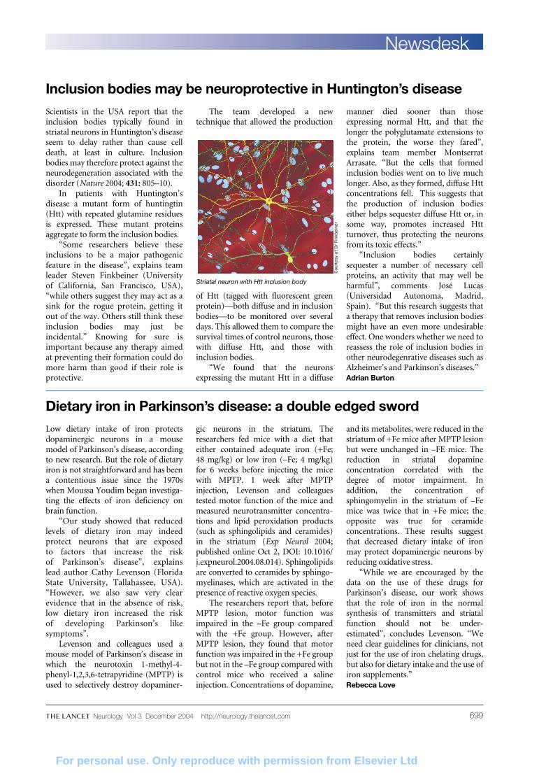
Striatal neuron with Htt inclusion body
Cou
rtes
y of
Dr
Fink
bein
er
Neurology Vol 3 December 2004 http://neurology.thelancet.com