Embed Size (px)
Citation preview

FEATURE ARTICLE
In vivo detection of cerebral cortical microinfarcts withhigh-resolution 7T MRISusanne J van Veluw1, Jaco JM Zwanenburg2, JooYeon Engelen-Lee3, Wim GM Spliet3, Jeroen Hendrikse2, Peter R Luijten2 andGeert Jan Biessels1
Cerebrovascular disease has an important role in cognitive decline and dementia. In this context, cerebral microinfarcts areattracting increasing attention, but these lesions could thus far not be detected in vivo. The aim of this study was to try to identifypossible cortical microinfarcts on high-resolution 7T in vivo magnetic resonance imaging (MRI) and to perform a histopathologicvalidation study on similar appearing lesions on 7T ex vivo MRI of postmortem brain tissue. The study population consisted of22 elderly subjects, who underwent 7T MRI. The fluid attenuated inversion recovery, T2, and T1 weighted scans of these subjectswere examined for possible cortical microinfarcts. In the ex vivo MRI study, 15 formalin-fixed coronal brain slices of 6 subjects withAlzheimer and vascular pathology were examined and subjected to histopathologic verification. On the in vivo scans, 15 corticallesions could be identified that were likely to be microinfarcts in 6 subjects. In the postmortem tissue, 6 similar appearing lesionswere identified of which 5 were verified as cortical microinfarcts on histopathology. This study provides strong evidence thatcortical microinfarcts can be detected in vivo, which will be of great value in further studies into the role of vascular disease incognitive decline and dementia.
Journal of Cerebral Blood Flow & Metabolism advance online publication, 19 December 2012; doi:10.1038/jcbfm.2012.196
Keywords: brain; histology; magnetic resonance imaging; microinfarcts; small vessel disease
INTRODUCTIONCognitive decline and dementia are major manifestations ofcerebrovascular disease. Approximately one in five patients withacute stroke develops dementia within a year after stroke.1 Evenmore often, cerebrovascular disease is involved in cognitivedecline or dementia in people without an obvious history ofstroke. Autopsy studies identify vascular pathology in the majorityof patients with dementia, also in those with a clinical diagnosis ofAlzheimer’s disease (AD).2,3 This vascular pathology frequentlyinvolves the small vessels of the brain, including small arteries andarterioles, but also capillaries and small veins, and is referred to ascerebral small vessel disease (SVD).4 Imaging correlates of SVD onconventional brain magnetic resonance imaging (MRI) includelacunar infarctions, white matter hyperintensities (WMHs), andmicrobleeds.4 Importantly, these MRI lesions do not fully capturethe burden of vascular damage in aging and dementia. In thiscontext, cerebral microinfarcts (CMIs) are attracting increasingattention. They are considered to be the single most widespreadform of brain infarction and thus a major component of the causalpathway between SVD and cognitive dysfunction.5,6 On autopsy,CMIs are observed in around 43% in patients with AD, 62% inpatients with vascular dementia, and 24% in nondemented elderlysubjects.6 Cerebral microinfarcts are typically defined as sharplydelineated microscopic ischemic lesions accompanied by cellulardeath or tissue necrosis, often associated with gliosis andcavitation.2,7 Cerebral microinfarcts can occur in both the white-matter and (sub)cortical regions of the brain, presumably more so
in watershed areas.6,8 Because of their small sizes (ranging from50mm to a few mm), CMIs escape detection by regular clinical MRIprotocols. With the introduction of high field strength (7.0 T) MRI,which allows high-resolution imaging with isotropic voxel sizes inthe submillimeter range, CMIs may be visualized in vivo. The aimof this study was to try to identify possible CMIs on 7.0 T magneticresonance (MR) images of elderly subjects without a knownhistory of dementia, and to perform a histopathologic validationstudy on similar appearing lesions on 7.0 T MR images ofpostmortem brain tissue that met the same criteria as in vivo.
MATERIALS AND METHODSStudy PopulationFunctionally independent elderly subjects, aged between 65 and 80, wererecruited through their general practitioners. Exclusion criteria werecontraindications for 7.0 T MRI (e.g., claustrophobia, metal objects suchas dental implants or prostheses in or on the body), or a known psychiatricor neurologic disorder that could influence cognitive functioning,including recent nondisabling stroke (o2 years) or any disabling stroke,major depression or a history of alcohol or substance abuse. Subjectsunderwent a Mini-Mental State Examination9 and had to have a score of 26or higher for inclusion in the present study. General practitioners made afirst selection against these criteria. In the period of October 2010 to 2011,54 eligible subjects were willing to participate in the study. Twenty-twowere excluded because of MR contraindications, and another eightbecause of other reasons. Hence, 24 individuals were included in thisstudy. They all underwent 7.0 T MRI scanning in the University Medical
1Department of Neurology, Rudolf Magnus Institute of Neuroscience, University Medical Center Utrecht, Utrecht, The Netherlands; 2Department of Radiology, University MedicalCenter Utrecht, Utrecht, The Netherlands and 3Department of Neuropathology, University Medical Center Utrecht, Utrecht, The Netherlands. Correspondence: SJ van Veluw,Department of Neurology, University Medical Center Utrecht G03.232, P.O. Box 85500, GA 3508 Utrecht, The Netherlands.E-mail: [email protected] work was supported by a VIDI grant from ZonMw, The Netherlands Organisation for Health Research and Development (grant number 91711384), and a grant from theNetherlands Heart Foundation (grant number 2010T073) to GJB.Received 3 September 2012; revised 31 October 2012; accepted 31 October 2012
Journal of Cerebral Blood Flow & Metabolism (2012), 1–8& 2012 ISCBFM All rights reserved 0271-678X/12 $32.00
www.jcbfm.com

Center Utrecht (UMCU, The Netherlands) with the same standardizedprotocol including fluid attenuated inversion recovery (FLAIR), a T2
weighted, and a T1 weighted sequence. The scans of two subjects couldnot be used because of severe motion artifacts. This resulted in a studypopulation of 22 subjects.
The study was approved by the medical ethics committee of the UMCUand all subjects gave written informed consent according to theDeclaration of Helsinki.
In Vivo Magnetic Resonance Imaging Scanning ProtocolScans were acquired on a whole-body 7.0 T MR system (Philips Healthcare,Cleveland, OH, USA) with a volume transmit and 16-channel receive headcoil (Nova Medical, Wilmington, MA, USA). Subjects included in the studylater than May 2011 (nine in total) were scanned with a volume transmitand 32-channel receive head coil (Nova Medical), which, as far as we couldjudge, did not cause any observable differences in lesion appearance. Thestandardized protocol included the following sequences:
(i) Volumetric (3D) magnetization prepared FLAIR with an acquired,isotropic resolution of 0.8� 0.8� 0.8 mm3, repetition time(TR)¼ 8,000 ms, nominal echo time (TE)¼ 300 ms using constantlow refocusing angles of 701, inversion time (TI)¼ 2,325 ms, matrixsize¼ 312� 304.10 Scan duration was 12 minutes 48 seconds.
(ii) 3D T2 weighted turbo-spin echo with an acquired, isotropicresolution of 0.7� 0.7� 0.7 mm3, TR¼ 3,158 ms, nominal TE¼ 301ms with a variable refocusing flip angle sweep,11,12 leading to anequivalent TE (for T2 contrast) of B57 ms for gray and white matter,matrix size¼ 356� 357. Scan duration was 10 minutes 15 seconds.
(iii) 3D T1 weighted sequence with an isotropic resolution of 1.0�1.0� 1.0 mm3, flip angle 81, TR¼ 4.8 ms, TE¼ 2.2 ms, TI¼ 1,240 ms, TRof the inversion pulses¼ 3,500 ms, matrix size¼ 200� 250. Scanduration was 1 minute 57 seconds.
All subjects also underwent a 3.0 T MRI (Intera, Philips, Best, TheNetherlands) on the same day, which included the following sequences:
(i) 2D multislice FLAIR with 48 slices, with an acquired resolution of1.0� 1.3� 3.0 mm3, TR¼ 11,000 ms, TE¼ 125 ms, TI¼ 2,800 ms,matrix size¼ 232� 148. Scan duration was 2 minutes 56 seconds.
(ii) 2D T2 weighted dual echo turbo-spin echo with 48 slices, with anacquired resolution of 1.0� 1.1� 3.0 mm3, TR¼ 3,198 ms, TE1¼ 19ms, TE2¼ 140 ms, matrix size¼ 232� 179. Scan duration was2 minutes 43 seconds.
(iii) 3D T1 weighted turbo-field echo with 192 slices, with an acquiredresolution of 1.0� 1.0� 1.0 mm3, flip angle 81, TR¼ 7.9, TE¼ 4.5,TI¼ 955 ms, TR of the inversion pulses¼ 3,000 ms, matrix size¼ 256� 232. Scan duration was 6 minutes 43 seconds.
Magnetic Resonance Imaging RatingHistopathologically, CMIs have been described as sharply delineatedmicroscopic ischemic lesions, often associated with gliosis and cavita-tion.2,7 Based on those features, we hypothesized that on ultra-highresolution MRI CMIs would appear as small hyperintense circumscript—with or without a hypointense center—lesions on FLAIR, hyperintense onT2 weighted, and hypointense or isointense on T1 weighted images. Wefocused on CMIs in the cerebral cortex, because autopsy studies indicatethat these lesions are particularly common in the cortex. Moreover, weexpected that in the subcortical gray and white matter it would be moredifficult to differentiate CMIs from other focal lesions including perivascularspaces and punctate WMHs (i.e., small focal hyperintensities on T2
13).In an initial evaluation the FLAIR scans (sagittal view) of all subjects were
thoroughly examined to identify possible cortical CMIs and the findingswere compared with T2 weighted and T1 weighted scans. During thisprocess, we did identify multiple hyperintense cortical lesions suspected tobe CMIs on FLAIR, but we did not see lesions with a hypointense center. Inall cases, these hyperintense FLAIR lesions were hyperintense on T2
weighted and hypointense on the corresponding T1 weighted scan. Basedon this initial evaluation, we operationally defined possible cortical CMIs on7.0 T MRI for further visual rating as hyperintense on FLAIR, hyperintenseon T2 weighted, and hypointense on T1 weighted scans. Each lesion had tobe detectable on all three views of the brain (sagittal, coronal, andtransversal), and restricted to the cortex. Moreover, they had to be distinctfrom perivascular spaces. We observed that some perivascular spacesextended from the white matter into the cortex, producing changes incortical signal intensities mimicking those of possible CMIs. Therefore, wediscarded all small cortical lesions that were in direct contact with aperivascular space in the underlying white matter as possible CMIs. Basedon those criteria, the MR images of all subjects were reevaluatedsystematically and possible cortical CMIs were scored. All identifiedpossible cortical CMIs were inspected by a second rater, experienced in thefield of neuroradiology. Both raters were blinded to clinical informationsuch as age. In the case of disagreement, for every location consensus wasreached.
When a possible cortical CMI was identified on 7.0 T, the corresponding3.0 T FLAIR, T2, and T1 weighted images were examined at the samelocation. This was performed by visual inspection using landmarks.
In addition, WMHs were assessed on the 3.0 T FLAIR images using theAge Related White Matter Changes scale,13 by two independent raters.Lacunar infarctions were also identified.
Postmortem TissueFor ex vivo MRI protocol optimization three formalin-fixed 10-mm thickcoronal brain slices were used. These slices, provided by the anatomydepartment of the UMCU, were from subjects who had donated theirbrains to science; therefore, detailed subject information was unavailable.
Table 1. Subject and tissue characteristics of the ex vivo study
Subject Neuropathologic diagnosisa SexF/M
Age(years)
Numberof slices
Fixationduration(months)
Numberof CMIsb
Ex vivoMRI
Numberof CMIsc
Histology
A AD (B&B VI) F 64 4 6 1 1B Lewy body dementia (B VI) M 83 3 3 1 0C AD (B&B VI) with hypertensive vasculopathy (with hemorrhagic
infarct)M 74 2 9 1 1
D Extensive hypoxic-ischemic brain damage (with early stage ofinfarction in the watershed areas)
M 71 2 15 1 1
E Alpha-synucleinopathy, classified as Lewy body dementia (B III-IV) M 87 2 14 0 0F AD (B&B II) with mild-to-severe hypoxic-ischemic brain damage
(with cystic degeneration of infarct in the left operculum as a resultof atherosclerotic vasculopathy)
M 79 2 6 4d 6e
CMI, cerebral microinfarct; F, female; M, male; AD, Alzheimer’s disease; B&B, Braak & Braak stage;29 B, Braak stage;30 MRI, magnetic resonance imaging.aNeuropathologic diagnosis was established on whole-brain and targeted histopathologic examination. bNumber of possible CMIs identified on ex vivo MRI.cNumber of definite CMIs verified on histopathologic examination. dTwo cortical CMIs in the brain tissue of this subject were solely found on ultra-highresolution ex vivo T2 (400� 400� 400 mm3) in retrospect, after the CMI was identified on histopathologic examination. eOne cortical CMI was found in thenegative control tissue taken from the brain tissue of this subject (and could not be found again on ex vivo MRI). One additional CMI was found in closeproximity to one of the CMIs that was identified on ex vivo MRI. These extra CMIs could in retrospect not be found on ex vivo MRI.
Cerebral microinfarcts on 7T MRISJ van Veluw et al
2
Journal of Cerebral Blood Flow & Metabolism (2012), 1 – 8 & 2012 ISCBFM

Initially, we scanned with the tissue submerged in Fomblin (Solvay Solexis,Bollate, Italy), a proton-free fluid without MR signal. Later, we scanned thetissue submerged in 4% formaldehyde solution (formalin). After carefulcomparison of MR image quality, it was decided to use formalin duringscanning, which proved to provide better tissue-fluid contrast.
After protocol optimization, 15 formalin-fixed 10-mm thick coronal brainslices from six subjects were submitted to ex vivo MRI. These subjectshad undergone autopsy in our hospital, after informed consent of thefamily was obtained. Patients were selected from the database of theneuropathology department based on reported cerebrovascular andAlzheimer pathology on autopsy. Details of subject and brain tissuecharacteristics are provided in (Table 1).
At least 2 hours in advance of scanning two coronal brain slices wereplaced in a plastic container, submerged in formalin. Care was taken toavoid air bubbles. Before scanning, the plastic container was sealed in aplastic bag, wrapped in a towel, and placed in the center of the MR headcoil. The use of the material of these patients for this study was inaccordance with local regulations and approved by the medical ethicscommittee of the UMCU.
Ex Vivo Magnetic Resonance Imaging Scanning ProtocolScans were acquired overnight on the same whole-body 7.0 T MR system asthe in vivo scans, with a volume transmit and 32-channel receive head coil(Nova Medical). We used two different protocols. The first ex vivo protocolincluded the same sequences as in vivo (FLAIR and T2 weighted, with exceptionof the T1 weighted) and is referred to as ‘clinical resolution protocol.’ The TI wasadjusted to obtain proper nulling of the formalin, in which the postmortemtissue was submerged during scanning. Also the refocusing angle wasreduced, to account for the fact that the relaxation times of formalin-fixedtissue are more reduced as compared with in vivo tissue.
The second protocol aimed to obtain an even higher resolution and isreferred to as ‘ultra-high resolution protocol.’ In this ultra-high resolutionprotocol, the postmortem tissue was scanned with the following parameters:
(i) 3D FLAIR with an acquired isotropic resolution of 400� 400� 400mm3, TR¼ 8,000 ms, nominal TE¼ 164 ms using constant lowrefocusing angles of 401, TI¼ 1,600 ms, matrix size¼ 500� 280. Scanduration was 4 hours 16 minutes 8 seconds.
(ii) T2 weighted turbo-spin echo with an acquired isotropic resolution of400� 400� 400mm3, TR¼ 3,500 ms, nominal TE¼ 164 ms using
constant low refocusing angles of 401, matrix size¼ 500� 280. Scanduration was 1 hour 52 minutes 3 seconds.
(iii) T2* weighted sequence with an isotropic acquired resolution of180� 180� 180mm3, flip angle 251, TR¼ 75 ms, TE¼ 20 ms, matrixsize¼ 832� 834. Scan duration was 4 hours 59 minutes 31 seconds.
Sampling Procedure and HistologyThe sampling procedure was established in three formalin-fixed 10-mmthick coronal brain slices, derived from subjects who had donated theirbrains to science (Figure 1). After this development stage, the followingprocedure was performed on the brain tissue of the patients used in thisstudy: after the ex vivo MRI scanning session, blocks of tissue (20�10� 0.5 mm3) were cut from the sites where possible cortical CMIs weredetected on the ex vivo MR images. All lesions that presented as corticalhyperintensities on both the clinical and ultra-high resolution ex vivo FLAIRas well as the T2 weighted images were sampled. The ultra-high resolutionT2* weighted sequence provided an extra level of detail and thereforeserved as a useful guide for sampling. As a negative control, two blocks oftissue in two different brain slices of two different subjects were cut from asite where no lesion was visible on any MR image. All blocks weredehydrated and preprocessed, after which they were embedded inparaffin. Further, 4-mm thick serial sections were cut using a microtome.Standard histologic staining was performed on these sections, whichincluded a hematoxylin/eosin (for cell bodies) stain, and a Luxol fast blue &Periodic acid-Schiff (for myelin) stain. Sections were studied under a CarlZeiss microscope, and photographs were obtained with a digital camera,mounted on top of the microscope. Sections were examined by theneuropathologist of our hospital. Cortical CMIs were determined based onthe presence of a sharply delineated area of ischemia, cellular death, andtissue necrosis. Furthermore, the presence of gliosis and cavitation wasnoted, and lesion diameter was measured on the obtained photograph.
RESULTSIn Vivo 7.0 T Magnetic Resonance ImagingThe 22 subjects included in this study had a mean age of68.7±3.2 years and a median Mini-Mental State Examinationscore of 29 (range 26 to 30). A total number of 15 lesions that met
Figure 1. Sampling procedure: correlation of ex vivo magnetic resonance imaging (MRI) and histology. The sampling procedure wasdeveloped in three brain slices from anonymous donors. In one of these brain slices a cortical lesion of interest, presumed to be ischemic, wasidentified on ex vivo MRI. This lesion appeared as a hyperintense cortical lesion with a hypointense center on a fluid attenuated inversionrecovery (FLAIR) (A; 400� 400� 400 mm3), and as a hyperintense cortical lesion on a T2 weighted (B; 400� 400� 400 mm3) magneticresonance (MR) image. After dissection of the tissue for sampling a cavity in the cortex was visible (C; tissue block is turned over). Onhistopathologic examination, this cortical lesion was identified as a cerebral microinfarct (CMI) with cavitation and gliosis in the surroundingtissue (D, and in greater detail E) (Luxol fast blue & Periodic acid-Schiff (L&P) stain). On higher magnification, gliosis is visible (F; arrow indicatesa reactive astrocyte) (hematoxylin/eosin (HE) stain). Tissue was submerged in Fomblin.
Cerebral microinfarcts on 7T MRISJ van Veluw et al
3
& 2012 ISCBFM Journal of Cerebral Blood Flow & Metabolism (2012), 1 – 8

our criteria for possible cortical CMIs were identified (see forexamples, Figures 2 and 3) in 6 of 22 subjects (27.3%). Foursubjects had one possible cortical CMI, one subject had twopossible cortical CMIs, and one subject had nine possible corticalCMIs. There were no clear differences in subject characteristics,WMH load and presence of lacunar infarctions between subjectswith or without CMIs (Table 2).
Possible cortical CMIs appeared as sharply delineated linearlesions, perpendicular to the cortical surface and extending intothe lower cortical layers. All identified possible cortical CMIs werep3 mm. They were primarily identified in the parietal and frontalcortical regions (one possible cortical CMI was located in the
occipital cortex). One subject had several small hyperintensecortical lesions on FLAIR and T2 presumed to be of ischemic originin the occipital cortex, but these were not classified as possibleCMIs, because they were related to a larger area of infarction. Thetemporal lobes proved difficult to assess, because of a low signal-to-noise ratio in these areas, which is a current known limitation ofthe applied 7.0 T FLAIR MR sequence.10 Of all possible corticalCMIs, the corresponding location on 3.0 T FLAIR, T2, and T1 wasexamined. Of the 15 possible cortical CMIs identified at 7.0 T, onewas visible on both the corresponding 3.0 T T2 and T1
(Supplementary Figure), another three were solely visible on thecorresponding 3.0 T T1, but none were visible at the corresponding3.0 T FLAIR.
Ex Vivo 7.0 T Magnetic Resonance ImagingCharacteristics and autopsy findings of the six subjects whosebrain slices were used for ex vivo MRI are provided in Table 1. In 15slices, a total of six hyperintense cortical lesions were identified onthe ultra-high as well as on the clinical resolution FLAIR and T2
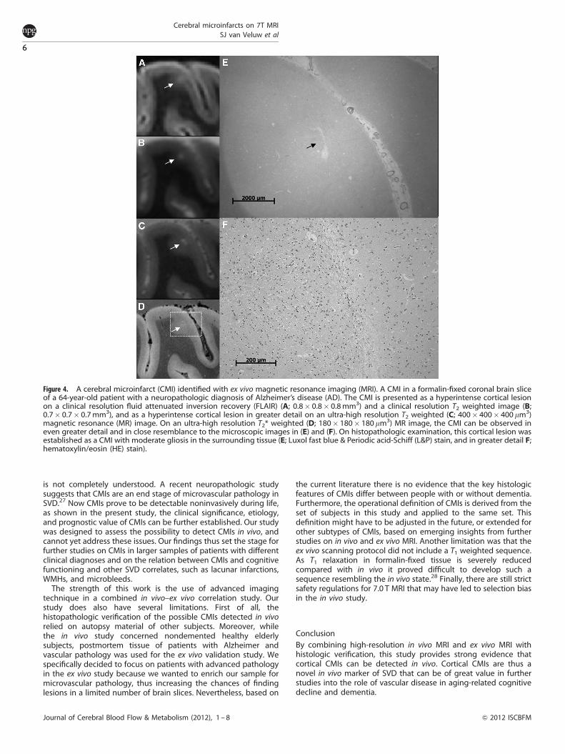
ex vivo MR images. Appearance of the lesions was similar to thepossible CMIs on in vivo MRI. The six lesions were sampled andfurther processed. At histopathologic examination, all but one ofthese lesions could be identified on the corresponding section. Onmicroscopy all five lesions that were identified appeared as focallesions of pallor with cellular death, attributed to ischemia, andmoderate gliosis in the surrounding tissue (Figure 4). Based onthese distinctive characteristics, these ischemic lesions wereclassified by the neuropathologist as definite CMIs. Their sizesvaried from B1,200 to 2,500 mm as measured on the obtainedmicroscopic photographs.
Microscopic examination identified three additional smallcortical CMIs that were not identified on the initial visualinspection of the ex vivo MRI. Two of these CMIs could beidentified after reinspection of the ex vivo MR images, on thecorresponding ultra-high resolution T2 weighted MR image (one isdepicted in Figure 5). These CMIs measured 470 and 1,300 mm,respectively. The third CMI (780 mm) was located close to a sulcuswhich limited its detection, on ex vivo MRI, possibly because ofpartial volume effects. In the two blocks of tissue that weresampled from regions where no lesion was visible on the MRimages (negative controls), one small cavitated cortical CMI(670 mm) was found on histopathologic examination. This corticalCMI could not be found on ex vivo MRI, after reinspection of thecorresponding images, possible also because of a partial volumeeffect.
DISCUSSIONThis study shows that CMIs can be detected noninvasively duringlife. We observed small cortical lesions on 7.0 T MR images ofnondemented elderly subjects that were compatible with ourpredefined features of cortical CMIs. Histopathologic validation oflesions with similar characteristics on ex vivo MRI showed thatthese lesions were indeed CMIs.
In the ex vivo part of this study, CMIs that appeared as focallesions of pallor with neuronal loss, attributed to ischemia, onhistology, gave a hyperintense signal on FLAIR and T2 weightedex vivo MR images.2,7 In the neuropathologic literature, there is noconsensus on the definition of the size of a CMI. Some studiesreport an upper limit for CMI size as ‘microscopic’ or ‘not visible tothe naked eye,’ but several studies also classify lesions up to a fewmm in size as CMIs.5,6 Our findings suggest that with ultra-highresolution ex vivo 7.0 T MRI CMIs larger than 0.5 mm can bedetected. With clinical resolution 7.0 T MRI (both ex vivo andin vivo) only the larger CMIs (Z1 mm) can be detected. Comparedwith neuropathology, in vivo MRI has the obvious advantage thatit allows (repeated) assessment of living subjects, thus allowing
Figure 2. Possible cerebral microinfarct (CMI) at 7.0 T in vivomagnetic resonance imaging (MRI). A possible cortical CMI in a76-year-old nondemented male is represented as a hyperintensecortical lesion on a fluid attenuated inversion recovery (FLAIR) (A;0.8� 0.8� 0.8mm3) and a T2 weighted (B; 0.7� 0.7� 0.7mm3), andas a hypointense lesion on a T1 weighted (C; 1.0� 1.0� 1.0mm3)magnetic resonance (MR) image on a sagittal view of the brain. Scalebar indicates 4mm.
Cerebral microinfarcts on 7T MRISJ van Veluw et al
4
Journal of Cerebral Blood Flow & Metabolism (2012), 1 – 8 & 2012 ISCBFM

longitudinal studies of CMIs in relation to other clinicalparameters. Another advantage is that MRI provides coverage ofalmost the complete brain, while histologic studies typically haveto rely on sampling of only a small proportion of the brain indifferent areas. At the 50mm to 0.5 mm range, however, ourcurrent MRI protocol will miss CMIs. In that perspective, the lesionsthat were identified with in vivo MRI in the current study are morelikely to be the ‘top of the iceberg’ of total CMI load. It is yetunclear if the etiology of these smallest CMIs is identical to that ofthe somewhat larger lesions. Nonetheless, the prevalence ofcortical CMIs in 6 of 22 (27.3%) elderly subjects in the in vivo partof the current study is in the same range (i.e., 24%), as theneuropathologic findings reported in nondemented elderlysubjects.6
Ex vivo MRI proves to be a valuable tool in determining MRmarkers in brain diseases with multiple small focal abnormalitiessuch as vascular lesions and demyelination, providing whole-braincoverage and targeted sampling for histopathologic examina-tion.14–16 A recent study suggested that prolonged storage (46years) of formalin-fixed tissue results in subtle histology artifacts,which sometimes on ex vivo MRI is indistinguishable from genuinebrain pathology.17 In our case, we have solely used postmortembrain tissue that has been stored for no longer than 15 months,
therefore we assume our samples are not susceptible formentioned alterations in the tissue and on the correspondingMR images.
Previously, CMIs have been identified on ex vivo (7.0 T) MRI inbrain tissue of a single patient with CADASIL (cerebral autosomal-dominant arteriopathy with subcortical infarcts and leukoence-phalopathy).18 The brain tissue of this patient presented withseveral CMIs on high-resolution T2* weighted scans, which werehistopathologically validated.18 Possible CMIs have recently beendescribed in an in vivo MRI study at 3.0 T.19 That study, which didnot verify the nature of the observed lesions histologically,included 70 subjects with AD or cerebrovascular disease andobserved small intracortical high signal lesions suspected to beCMIs on both advanced double inversion recovery and 3D FLAIRimages with high in-plane resolutions in only nine subjects.19 Thelesions depicted in that paper are of similar appearance as thelesions depicted in our study, albeit of larger size. Apparently, it isalso possible to detect CMIs at field strengths lower than 7.0 T, asour findings at 3.0 T also show. Probably, lesion detection at lowerfield strengths can be improved by further protocol optimization.We recommend that such protocols include at least heavily T2
weighted FLAIR, T2, and T1 weighted sequences. A separate studywould be needed to investigate to what extent our 7.0 T resultscan be translated to 3.0 T MRI, and how a 3.0 T protocol should beoptimized for this purpose. However, we expect that 7.0 Tdefinitely is more sensitive for CMI detection compared with3.0 T, either by an increased signal-to-noise ratio, improvedcontrast, a higher resolution, or by a combination of thesefactors.20
Cerebral microinfarcts have been studied extensively inneuropathologic studies. A systematic review of 32 of thesestudies, involving neuropathologic data of over 10,000 subjects,emphasized the potential clinical relevance of CMIs.6 Cerebralmicroinfarcts are an important correlate of aging-related cognitiveimpairment and dementia and vascular disease.7,21–24 Althoughthey are small, autopsy data suggest that CMIs might be the mostwidespread form of brain infarction and are thus conceivable tocause substantial clinical symptoms.5,25,26 In a clinicopathologicstudy examining the brains of 285 autopsied decedents as part ofthe HAAS (Honolulu-Asia Aging Study), it was found that neitherlarge infarcts, lacunar infarctions, nor hemorrhages addedsignificantly to the variance in the final antemortem cognitivetest score as established by the CASI (Cognitive Abilities ScreeningInstrument). Only CMIs were independently associated with poorCASI test scores.7 While the role of CMIs in aging and dementia isincreasingly recognized, the underlying pathophysiology of CMIs
Figure 3. Possible cerebral microinfarct (CMI) at 7.0 T in vivo magnetic resonance imaging (MRI). A possible cortical CMI in a 66-year-oldnondemented female is represented as a hyperintense cortical lesion on a fluid attenuated inversion recovery (FLAIR) (A; 0.8� 0.8� 0.8mm3)and a T2 weighted (B; 0.7� 0.7� 0.7mm3), and as a hypointense lesion on a T1 weighted (C; 1.0� 1.0� 1.0mm3) magnetic resonance (MR)image on a transversal view of the brain. Scale bar indicates 4mm.
Table 2. Subject characteristics of the in vivo study
With CMIs,n¼ 6
No CMIs,n¼ 16
Age (years) 70.5±4.3 68.2±2.4Male sex 2 (33) 7 (44)Hypertensiona 4 (67) 8 (50)Diabetes mellitusa 1 (17) 4 (25)Systolic blood pressure(mmHg)
136±14 145±14
Diastolic blood pressure(mmHg)
82±5 80±5
MMSE score 29 (27–30) 29 (26–30)White matter hyperintensities 3.5 (1–8.5) 2.5 (0.5–17)Lacunar infarctions 3 (50) 5 (31)
CMI, cerebral microinfarct; MMSE, Mini-Mental State Examination.Data are presented as mean±SD, n (%), or median (range).aSubjects were considered having arterial hypertension, or diabetesmellitus, if they had a known history of the disease or were receivingdrug treatment for these conditions.
Cerebral microinfarcts on 7T MRISJ van Veluw et al
5
& 2012 ISCBFM Journal of Cerebral Blood Flow & Metabolism (2012), 1 – 8
is not completely understood. A recent neuropathologic studysuggests that CMIs are an end stage of microvascular pathology inSVD.27 Now CMIs prove to be detectable noninvasively during life,as shown in the present study, the clinical significance, etiology,and prognostic value of CMIs can be further established. Our studywas designed to assess the possibility to detect CMIs in vivo, andcannot yet address these issues. Our findings thus set the stage forfurther studies on CMIs in larger samples of patients with differentclinical diagnoses and on the relation between CMIs and cognitivefunctioning and other SVD correlates, such as lacunar infarctions,WMHs, and microbleeds.
The strength of this work is the use of advanced imagingtechnique in a combined in vivo–ex vivo correlation study. Ourstudy does also have several limitations. First of all, thehistopathologic verification of the possible CMIs detected in vivorelied on autopsy material of other subjects. Moreover, whilethe in vivo study concerned nondemented healthy elderlysubjects, postmortem tissue of patients with Alzheimer andvascular pathology was used for the ex vivo validation study. Wespecifically decided to focus on patients with advanced pathologyin the ex vivo study because we wanted to enrich our sample formicrovascular pathology, thus increasing the chances of findinglesions in a limited number of brain slices. Nevertheless, based on
the current literature there is no evidence that the key histologicfeatures of CMIs differ between people with or without dementia.Furthermore, the operational definition of CMIs is derived from theset of subjects in this study and applied to the same set. Thisdefinition might have to be adjusted in the future, or extended forother subtypes of CMIs, based on emerging insights from furtherstudies on in vivo and ex vivo MRI. Another limitation was that theex vivo scanning protocol did not include a T1 weighted sequence.As T1 relaxation in formalin-fixed tissue is severely reducedcompared with in vivo it proved difficult to develop such asequence resembling the in vivo state.28 Finally, there are still strictsafety regulations for 7.0 T MRI that may have led to selection biasin the in vivo study.
ConclusionBy combining high-resolution in vivo MRI and ex vivo MRI withhistologic verification, this study provides strong evidence thatcortical CMIs can be detected in vivo. Cortical CMIs are thus anovel in vivo marker of SVD that can be of great value in furtherstudies into the role of vascular disease in aging-related cognitivedecline and dementia.
Figure 4. A cerebral microinfarct (CMI) identified with ex vivo magnetic resonance imaging (MRI). A CMI in a formalin-fixed coronal brain sliceof a 64-year-old patient with a neuropathologic diagnosis of Alzheimer’s disease (AD). The CMI is presented as a hyperintense cortical lesionon a clinical resolution fluid attenuated inversion recovery (FLAIR) (A; 0.8� 0.8� 0.8mm3) and a clinical resolution T2 weighted image (B;0.7� 0.7� 0.7mm3), and as a hyperintense cortical lesion in greater detail on an ultra-high resolution T2 weighted (C; 400� 400� 400 mm3)magnetic resonance (MR) image. On an ultra-high resolution T2* weighted (D; 180� 180� 180 mm3) MR image, the CMI can be observed ineven greater detail and in close resemblance to the microscopic images in (E) and (F). On histopathologic examination, this cortical lesion wasestablished as a CMI with moderate gliosis in the surrounding tissue (E; Luxol fast blue & Periodic acid-Schiff (L&P) stain, and in greater detail F;hematoxylin/eosin (HE) stain).
Cerebral microinfarcts on 7T MRISJ van Veluw et al
6
Journal of Cerebral Blood Flow & Metabolism (2012), 1 – 8 & 2012 ISCBFM

DISCLOSURE/CONFLICT OF INTERESTThe authors declare no conflict of interest.
ACKNOWLEDGEMENTSThe authors would like to acknowledge the Utrecht Vascular Cognitive Impairmentgroup. Members of the group involved in this project are M Brundel, LJ Kappelle,YD Reijmer (from the department of Neurology), J de Bresser, HJ Kuijf, WPThM Mali,MA Viergever, and KL Vincken (from the department of Radiology/Image SciencesInstitute). Furthermore, the authors thank RLAW Bleys (from the department ofAnatomy) for providing the brain slices for protocol optimization.
REFERENCES1 Pendlebury ST, Rothwell PM. Prevalence, incidence, and factors associated with
pre-stroke and post-stroke dementia: a systematic review and meta-analysis.Lancet Neurol 2009; 8: 1006–1018.
2 Kalaria RN, Kenny RA, Ballard CG, Perry R, Ince P, Polvikoski T. Towards defining theneuropathological substrates of vascular dementia. J Neurol Sci 2004; 226: 75–80.
3 Schneider JA, Arvanitakis Z, Bang W, Bennett DA. Mixed brain pathologiesaccount for most dementia cases in community-dwelling older persons. Neurol-ogy 2007; 69: 2197–2204.
4 Pantoni L. Cerebral small vessel disease: from pathogenesis and clinical char-acteristics to therapeutic challenges. Lancet Neurol 2010; 9: 689–701.
5 Smith EE, Schneider JA, Wardlaw JM, Greenberg SM. Cerebral microinfarcts: theinvisible lesions. Lancet Neurol 2012; 11: 272–282.
6 Brundel M, de Bresser J, Van Dillen JJ, Kappelle LJ, Biessels GJ. Cerebral micro-infarcts: a systematic review of neuropathological studies. J Cereb Blood FlowMetab 2012; 32: 425–436.
7 White L, Petrovitch H, Hardman J, Nelson J, Davis DG, Ross GW et al. Cere-brovascular pathology and dementia in autopsied Honolulu-Asia Aging Studyparticipants. Ann NY Acad Sci 2002; 977: 9–23.
8 Suter OC, Sunthorn T, Kraftsik R, Straubel J, Darekar P, Khalili K et al. Cerebralhypoperfusion generates cortical watershed microinfarcts in Alzheimer disease.Stroke 2002; 33: 1986–1992.
9 Tombaugh TN, McIntyre NJ. The mini-mental state examination: a comprehensive
review. J Am Geriatr Soc 1992; 40: 922–935.10 Visser F, Zwanenburg JJM, Hoogduin JM, Luijten PR. High-resolution magnetiza-
tion-prepared 3D-FLAIR imaging at 7.0 Tesla. Magn Reson Med 2010; 64: 194–202.11 Busse RF, Hariharan H, Vu A, Brittain JH. Fast spin echo sequences with very long
echo trains: design of variable refocusing flip angle schedules and generation of
clinical T2 contrast. Magn Reson Med 2006; 55: 1030–1037.12 Wisse LE, Gerritsen L, Zwanenburg JJ, Kuijf HJ, Luijten PR, Biessels GJ et al. Sub-
fields of the hippocampal formation at 7T MRI: In vivo volumetric assessment.
Neuroimage 2012; 61: 1043–1049.13 Wahlund LO, Barkhof F, Fazekas F, Bronge L, Augustin M, Sjogren M et al. A new
rating scale for age-related white matter changes applicable to MRI and CT. Stroke
2001; 32: 1318–1322.14 Gouw AA, Seewann A, Vrenken H, van der Flier WM, Rozemuller JM, Barkhof F
et al. Heterogeneity of white matter hyperintensities in Alzheimer’s disease: post-
mortem quantitative MRI and neuropathology. Brain 2008; 131: 3286–3298.15 Schmierer K, Parkes HG, So PW, An SF, Brandner S, Ordidge RJ et al. High field (9.4
Tesla) magnetic resonance imaging of cortical grey matter lesions in multiple
sclerosis. Brain 2010; 133: 858–867.16 De Reuck J, Auger F, Cordonnier C, Deramecourt V, Durieux N, Pasquier F et al.
Comparison of 7.0-T T2*-magnetic resonance imaging of cerebral bleeds in post-
mortem brain sections of Alzheimer patients with their neuropathological cor-
relates. Cerebrovasc Dis 2011; 31: 511–517.17 Van Duijn S, Nabuurs RJ, van Rooden S, Maat-Schieman ML, van Duinen SG, van
Buchem MA et al. MRI artifacts in human brain tissue after prolonged formalin
storage. Magn Reson Med 2011; 65: 1750–1758.18 Jouvent E, Poupon C, Gray F, Paquet C, Mangin JF, Le Bihan D et al.
Intracortical infarcts in small vessel disease: a combined 7-T postmortem
MRI and neuropathological case study in cerebral autosomal-dominant arterio-
pathy with subcortical infarcts and leukoencephalopathy. Stroke 2011; 42:
27–30.19 Li Y, Maeda M, Kida H, Matsuo K, Shindo A, Taniguchi A et al. In vivo detection of
cortical microinfarcts on ultrahigh-field MRI. J Neuroimaging 2012, e-pub ahead of
print 18 May 2012; doi:10.1111/j.1552-6569.2012.00722.x.
Figure 5. An additional cerebral microinfarct (CMI) identified with histology. A CMI in a formalin-fixed coronal brain slice of a 79-year-oldpatient with Alzheimer’s disease (AD) pathology. The CMI is presented as a hyperintense cortical lesion on an ultra-high resolution fluidattenuated inversion recovery (FLAIR) (A; arrow; 400� 400� 400 mm3) and an ultra-high resolution T2 weighted (B; arrow; 400� 400� 400mm3) magnetic resonance (MR) image. On histopathologic examination, this cortical lesion was established as a CMI (C, Luxol fast blue &Periodic acid-Schiff (L&P) stain, and in greater detail D, L&P stain). Note the smaller CMI in (C) and (D) (open arrow) which was initially solelyidentified on histopathologic examination, but at reinspection proved to be visible on the ultra-high resolution T2 weighted MR image as well(B; open arrow).
Cerebral microinfarcts on 7T MRISJ van Veluw et al
7
& 2012 ISCBFM Journal of Cerebral Blood Flow & Metabolism (2012), 1 – 8

20 Madai VI, von Samson-Himmelstjerna FC, Bauer M, Stengl KL, Mutke MA, Tovar-Martinez E et al. Ultrahigh-field MRI in human ischemic stroke - a 7 Tesla study.PLoS ONE 2012; 7: e37631.
21 Kovari E, Gold G, Herrmann FR, Canuto A, Hof PR, Bouras C et al. Cortical micro-infarcts and demyelination affect cognition in cases at high risk for dementia.Neurology 2007; 68: 927–931.
22 Sonnen JA, Santa Cruz K, Hemmy LS, Woltjer R, Leverenz JB, Montine KS et al.Ecology of the aging human brain. Arch Neurol 2011; 68: 1049–1056.
23 Richardson K, Stephan BC, Ince PG, Brayne C, Matthews FE, Esiri MM. Theneuropathology of vascular disease in the Medical Research CouncilCognitive Function and Ageing Study (MRC CFAS). Curr Alzheimer Res 2012; 9:687–696.
24 Kalaria RN. Cerebrovascular disease and mechanisms of CognitiveImpairment: evidence from clinicopathological studies in humans. Stroke 2012;43: 2526–2534.
25 Arvanitakis Z, Leurgans SE, Barnes LL, Bennett DA, Schneider JA. Microinfarct
pathology, dementia, and cognitive systems. Stroke 2011; 42: 722–727.26 Launer LJ, Hughes TM, White LR. Microinfarcts brain atrophy, and cognitive
function: the Honolulu Asia Aging Study Autopsy Study. Ann Neurol 2011; 70:
774–780.27 Deramecourt V, Slade JY, Oakley AE, Perry RH, Ince PG, Maurage CA et al. Staging
and natural history of cerebrovascular pathology in dementia. Neurology 2012; 78:
1043–1050.28 Pfefferbaum A, Sullivan EV, Adalsteinsson E, Garrick T, Harper C, Postmortem MR.
imaging of formalin-fixed human brain. Neuroimage 2004; 21: 1585–1595.29 Braak H, Braak E. Neuropathological stageing of Alzheimer-related changes. Acta
Neuropathol 1991; 82: 239–259.30 Braak H, Del Tredici K, Rub U, de Vos RA, Jansen Steur EN, Braak E. Staging of brain
pathology related to sporadic Parkinson’s disease. Neurobiol Aging 2003; 24: 197–211.
Supplementary Information accompanies the paper on the Journal of Cerebral Blood Flow & Metabolism website (http://www.nature.com/jcbfm)
Cerebral microinfarcts on 7T MRISJ van Veluw et al
8
Journal of Cerebral Blood Flow & Metabolism (2012), 1 – 8 & 2012 ISCBFM