Embed Size (px)
Citation preview

In Vitro Models for Biomechanical Stud-ies of Neural Tissues
Barclay Morrison III, D. Kacy Cullen and Michelle LaPlaca
Abstract In vitro models are invaluable tools for studying cell behavior in a highlycontrolled setting. Cell and tissue culture models of the nervous system can be utilizedto elucidate neurobiological phenomena that are difficult to observe, manipulate, ormeasure in vivo. In the context of biomechanics, culture models that accurately mimicspecific brain features can be used to determine tissue properties and tolerances tomechanical loading. There are several criteria that culture models must meet in orderto complement in vivo and macroscopic biomechanical studies. In addition to pro-viding an environment that is conducive to cell survival, cell type and source arecritical to the interpretation of results. In this review, we present design criteria forideal cultures, the current state of the art in neural cell and tissue culturing methods,and the advantages and limitations to using culture mimics. We will further presentwhat insights in vitro models can provide to complement in vivo and macroscopicbiomechanics in terms of meso- to microscale material properties and tissue-leveltolerance criteria. The discussion will focus primarily on central nervous system(CNS) tissue, which is inherently complex in cytoarchitecture and organization. Inaddition, the CNS is not typically exposed to mechanical loading beyond physio-logical motion; therefore, it is expected that cell death and functional failure may beparticularly prominent at large deformations and high loading rates. These and other
B. Morrison III (&)Biomedical Engineering, Columbia University, 351 Engineering Terrace,MC 8904, 1210 Amsterdam Avenue, New York, NY 10027, USAe-mail: [email protected]
D. K. CullenNeurosurgery, University of Pennsylvania, 105 Hayden Hall, 3320 Smith Walk,Philadelphia, PA 19104, USA
M. LaPlacaBiomedical Engineering, Georgia Institute of Technology, 313 Ferst Drive,Atlanta, GA 30332-0535, USA
Stud Mechanobiol Tissue Eng Biomater (2011) 3: 247–285 247DOI: 10.1007/8415_2011_79� Springer-Verlag Berlin Heidelberg 2011Published Online: 30 April 2011

factors must be considered when attempting to extract and culture CNS tissue or itscomponents for studying neurobiological or neuromechanical phenomena.
1 Neural Tissue Structure and Composition
1.1 Heterogeneity of Brain Tissue
Brain is a heterogeneous tissue that is divided into distinct anatomical and func-tional regions. The gray and white matter comprise the cellular constituents, andthe blood supply, cerebrospinal fluid/ventricular system, interstitial fluid, andextracellular matrix (ECM) make up the extracellular components. The intracel-lular space (ICS) and the extracellular space (ECS) can be considered as one scaleof heterogeneity. The ICS is a composite with neurons and their extensions, gliacells, vascular cells, and other support cells. The orientation is quite organized insome areas and apparently random in others. Most neuronal communication in thebrain exists on a local level (i.e. shorter interneurons, which comprise most neu-rons in the brain), with 1,000s of synapses on some neurons, creating an extremelycomplex network [1]. The brain has a very high cellularity compared to otherorgans and a very diverse cell population. Cell sizes range from\10 to[100 lmin cell soma diameter with axons ranging from microns to 100s of microns long.Dendrites are highly branched, permitting diversification and maximization ofinteractions between communicating neurons [1].
Brain tissue can be divided further by anatomical regions. Just rostral to the spinalcord is the brain stem, a deep, well-protected portion of the brain comprised of themedulla oblongata, pons, and midbrain, which houses the controls to manyhomeostatic functions such as respiratory and cardiovascular regulation. The cere-bellum, dorsal to the brainstem, helps control balance, posture, motor coordination,sensory and motor relaying. The diencephalon is the deep region of the brain thatcontains the thalamus (primary relay for sensory input and motor output) and thehypothalamus (neuroendocrine structure critical for maintaining homeostasis).Traveling toward the brain surface are the white matter tracts, the cerebral nuclei(basal ganglia, hippocampus, amygdala) and the cerebral cortex, comprised of sixdistinct, organized, interconnected layers. The isolation of any brain region (forprimary culture) should consider the connections with other brain areas not repre-sented in the culture and the possible effects (e.g., activation of compensatoryresponses) harvest and dissection may have. Interneuronal connections and inter-regional junctions are disrupted during tissue dissociation as are projection axons formost culture techniques, therefore the culturing procedures must consider thesemicroinjuries and provide conditions conducive for repair and healthy culture mat-uration and reconnection. Some regions, such as the hippocampus are particularlyvulnerable to excitotoxicity and the pH of the medium and constituents should allowfor normal receptor function and neurotransmitter reuptake. Yet, this region is idealfor studying phenomena related to excitotoxicity, such as ischemia and trauma.
248 B. Morrison III et al.

1.2 The Extracellular Space of Brain Tissue
In addition to the cell composition, the ECS is critical for communication,transport, and is essential for normal homeostatic function. It is approximately20% of the total intracranial volume with half of this volume attributed to theblood and half to the cerebrospinal fluid (CSF) [2]. Some macroscopic parametersused to characterize brain tissue structure are the volume fraction of ECS andeffective diffusivity or tortuosity. The ECS is composed of ECM proteins, as wellas ions, neurotransmitters, metabolites, and peptides among other molecules. Asmost cultures are derived from prenatal or perinatal tissue, one must consider thatthe ECS is larger in the developing brain (36–46% by volume) than the adult,which may play an important role in the diffusion of nutrients and waste. The sizeof the ECS is especially important, since in most tissue culture systems it isessentially infinite. From a functional point of view, the anisotropy and inhomo-geneity of brain tissue dictates the ECS shape, volume, and tortuosity, and henceintraneural (or interneuronal) diffusion parameters. For example, excess neuro-transmitters, in addition to cellular reuptake, may diffuse to nearby capillaries oralong elongated structures such as white matter tracts [3, 4]. Diffusion of extra-cellular substances will be discussed below in light of brain tissue dynamics andthe artificial culture environment.
2 Neural Culture Models
Cells and tissues of the body may be explanted and kept alive ex vivo for extendedperiods of time, a process termed cell or tissue culture. Keeping the explantedtissue alive (i.e. metabolically active and functional) and preserving its phenotypicstate similar to the in vivo state requires specialized culture conditions and nutrientmedium reproducing vital conditions found in vivo. Specialized equipmentrequired includes a tissue culture incubator which maintains physiological tem-perature (37�C) and gas tension (5% CO2), laminar flow cabinets to maintainsterility, and in some cases bioreactor perfusion systems as described below.Explanted tissues may be cultured as thin slices of tissue, called organotypic slicecultures, or dissociated into suspensions of single cells to be plated as dissociatedcultures. Cultures grown immediately after explantation are referred to as primarycultures.
2.1 Culture Model Utility
Cell and tissue culture models of the nervous system are desirable platforms forstudying cell responses to various stimuli, including biochemical and pharmaco-logical investigations, electrophysiological assessment, and mechanical stimulation
In Vitro Models for Biomechanical Studies of Neural Tissues 249

or injury. These types of manipulations are not mutually exclusive and therefore itis critical to take into account the multimodal reactions that may follow a seeminglysimple input. Well-characterized culture models, therefore, must mimic the cellstates of brain tissue, including cell phenotype(s), cell–cell connections, cell-extracellular communication, mechanical properties, chemical composition, andelectrochemical balance. The advantages of a well-defined culture preparation arethat the complexity of an intact organism is drastically reduced and the culture (bothextra- and intracellular environments) can be manipulated much more readily thanin an animal or human. Despite the inherent simplicity of culture systems, they canbe invaluable for use in understanding neural function as well as supra-physio-logical states. In order to assemble realistic brain tissue surrogates in a dish, onemust rigorously prioritize the above characteristics and properties in a non-biasedmanner. Simply put: do existing cell and tissue culture models mimic brain tissue?What key parameters are to be mimicked?
2.2 Design Criteria for Brain Mimetics
In order to recapitulate brain tissue in culture models, several criteria are criticaland are represented in various degrees of fidelity among culture models. Thesource of the tissue must be considered in terms of species, developmental age, andbrain regions. The type of culture is equally important: primary cells versus celllines, dissociated cells plated in a constraint-free culture vessel or in a reaggregateor constrained (e.g., scaffold) format, cultured explants or slices (see Table 1). Insome instances, a single cell type is desired, yet multi-typic cultures are becomingmore commonplace given the complex intercellular interactions that contribute tocellular response. In addition, as discussed above, the immediate extracellularenvironment must be defined in terms of medium, medium supplements (serum vs.defined supplements), substrate, dimension (2D or 3D), gas composition, pH, andtemperature (Table 1).
In addition to these basic design considerations, specialty applications, such asmechanical property testing, mechanical and electrical stimulation, and fluidiccontrol may require further manipulation. Geometric control will preserve thevolume fraction of cells to extracellular space and relies on representative cell–cellconnections and tissue-like cellular orientation. To this end, slice cultures meetthis criterion. Topological features, such as soma shape, neuritic branching andglial-neuronal interactions are often lost with cell dissociation, yet can be main-tained through culture manipulation (e.g., scaffolding).
While most brain regions can be cultured, we will focus on hippocampal andcortical dissociated and organotypic cultures in the subsequent sections. Theparticular purpose of a culture study should consider the cell type(s) to be cultured,as well as the source, age of animal, and other experimental variables. For an
250 B. Morrison III et al.
exhaustive methods-based description of culture methods across the regions of thenervous systems, the reader is referred to [5].
2.3 Dissociated Cell Cultures
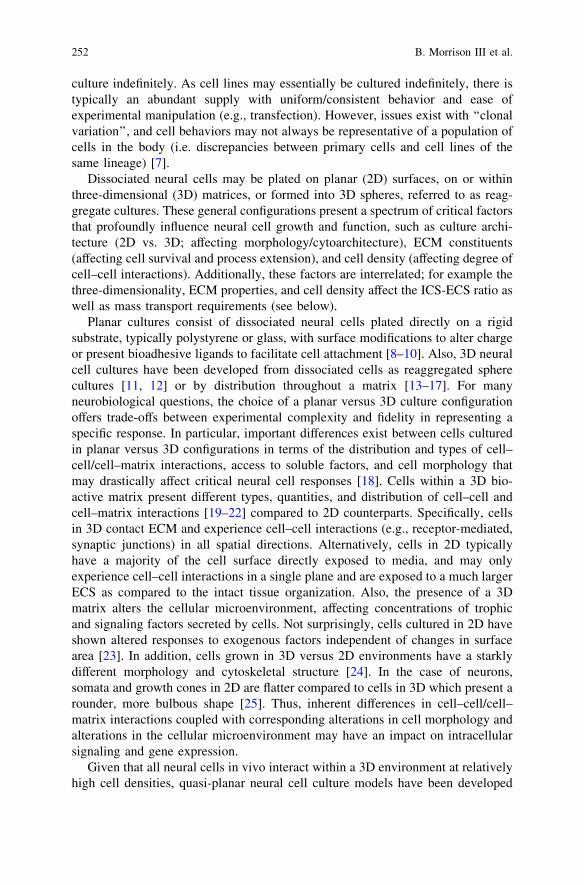
Specific cell populations may be isolated from animals or humans and dissociated,or separated from each other, to acquire a suspension consisting of numeroussingle cells. The classes of dissociated cell cultures consist of primary cells,secondary cells, and/or cell lines, which are then plated in some configuration (seeTable 1) based on experimental objectives. Primary cell cultures consist of cellsthat are isolated directly from tissues and plated for experimentation. Primary cellstherefore may closely represent the endogenous phenotype; however, many pri-mary neural cell types are difficult to keep alive for extended periods of time.Secondary cell cultures consist of cells that were permitted to proliferate forseveral cycles in vitro prior to experimental use. This process is referred to as‘‘passaging’’, and is typically done to amplify the cell number or purify a particularcell population; however, absent genetic manipulation passaging is limited by afinite number of cell divisions (Hayflick principle) [6]. Cell lines are primary cellsthat have been ‘‘immortalized’’, typically by genetic mutations or insertions, andmay represent a clone of a particular type of cell, which can, in theory, be kept in
Table 1 Neural culture variables and considerations. Adapted from Ref. [5]
Design Consideration Culture Options
Cell source/cell typeConvenienceAdapted developmental ageSimilarities with human nervous
system
Rodent fetal and neonatalCortex, hippocampus, cerebellum, spinal cord, and
sensory ganglia are common
Culture type/culture configurationPreservation of anatomy/histiotypic
organizationElectrophysiological integrityAbility to culture from developing
& adult animalsReplicate cultures, cell
accessibility, cell subset purification
Organ, slice, and explant (fragment) culturesDisaggregated or reaggregated cells and cell lines
Culture EnvironmentExtracellular fluid compositionTemperature, pH, gas phaseSubstrateDimension
37�C, 100% humidity, 5% CO2/95% airCulture medium such as DMEM, DMEM/F12,
or Neurobasal as neural cells require specificconcentrations of ions, amino acids, vitamins,cofactors, hormones, mitogens (for proliferatingcells), and other metabolites
Additional supplements include fetal bovine or horseserum, or chemically defined supplements
In Vitro Models for Biomechanical Studies of Neural Tissues 251
culture indefinitely. As cell lines may essentially be cultured indefinitely, there istypically an abundant supply with uniform/consistent behavior and ease ofexperimental manipulation (e.g., transfection). However, issues exist with ‘‘clonalvariation’’, and cell behaviors may not always be representative of a population ofcells in the body (i.e. discrepancies between primary cells and cell lines of thesame lineage) [7].
Dissociated neural cells may be plated on planar (2D) surfaces, on or withinthree-dimensional (3D) matrices, or formed into 3D spheres, referred to as reag-gregate cultures. These general configurations present a spectrum of critical factorsthat profoundly influence neural cell growth and function, such as culture archi-tecture (2D vs. 3D; affecting morphology/cytoarchitecture), ECM constituents(affecting cell survival and process extension), and cell density (affecting degree ofcell–cell interactions). Additionally, these factors are interrelated; for example thethree-dimensionality, ECM properties, and cell density affect the ICS-ECS ratio aswell as mass transport requirements (see below).
Planar cultures consist of dissociated neural cells plated directly on a rigidsubstrate, typically polystyrene or glass, with surface modifications to alter chargeor present bioadhesive ligands to facilitate cell attachment [8–10]. Also, 3D neuralcell cultures have been developed from dissociated cells as reaggregated spherecultures [11, 12] or by distribution throughout a matrix [13–17]. For manyneurobiological questions, the choice of a planar versus 3D culture configurationoffers trade-offs between experimental complexity and fidelity in representing aspecific response. In particular, important differences exist between cells culturedin planar versus 3D configurations in terms of the distribution and types of cell–cell/cell–matrix interactions, access to soluble factors, and cell morphology thatmay drastically affect critical neural cell responses [18]. Cells within a 3D bio-active matrix present different types, quantities, and distribution of cell–cell andcell–matrix interactions [19–22] compared to 2D counterparts. Specifically, cellsin 3D contact ECM and experience cell–cell interactions (e.g., receptor-mediated,synaptic junctions) in all spatial directions. Alternatively, cells in 2D typicallyhave a majority of the cell surface directly exposed to media, and may onlyexperience cell–cell interactions in a single plane and are exposed to a much largerECS as compared to the intact tissue organization. Also, the presence of a 3Dmatrix alters the cellular microenvironment, affecting concentrations of trophicand signaling factors secreted by cells. Not surprisingly, cells cultured in 2D haveshown altered responses to exogenous factors independent of changes in surfacearea [23]. In addition, cells grown in 3D versus 2D environments have a starklydifferent morphology and cytoskeletal structure [24]. In the case of neurons,somata and growth cones in 2D are flatter compared to cells in 3D which present arounder, more bulbous shape [25]. Thus, inherent differences in cell–cell/cell–matrix interactions coupled with corresponding alterations in cell morphology andalterations in the cellular microenvironment may have an impact on intracellularsignaling and gene expression.
Given that all neural cells in vivo interact within a 3D environment at relativelyhigh cell densities, quasi-planar neural cell culture models have been developed
252 B. Morrison III et al.

consisting of dissociated primary cells above a 3D matrix material [26–28].These systems effectively approximate a 3D morphology/orientation and maysupport high 2D cell densities. However, cell–cell and cell–matrix interactions arestill somewhat constrained, as cells are not distributed throughout the full thick-ness of the matrix. Conversely, complete three-dimensionality is achieved inreaggregate neural cultures that are developed by rotation-induced reassociation[11, 12, 29]. These systems produce spheres at high 3D cell densities wherediffusion-based mass transport is enhanced by convection due to circulating media.In reaggregate culture systems, the volume available for growth is inherentlylimited based on surface area to volume ratios that permit survival, thus limitingthe scope of 3D interactions. Moreover, the ECM components and cellular dis-tribution are difficult to control in reaggregate cultures; however, these models areextremely useful for studying cell–cell interactions, growth, and function at celldensities that closely match those found in vivo. Three-dimensional cell culturemodels consisting of neural cells distributed throughout a matrix have also beendeveloped [13, 15–17, 30–32]. In these systems, trade-offs exist between culturethickness (i.e., surface area to volume ratio), and hence the scope of 3D spatialinteractions, and cell density, necessitating that relatively thick ([500 lm) cul-tures use cell densities at least an order of magnitude lower than that found in braincortices [32, 33]. However, using these 3D scaffold-based neural cultures, keyparameters governing neuronal survival and neurite outgrowth based on scaffoldphysical/biological properties and mass transport phenomena have been uncov-ered. For example, primary dorsal root ganglion (DRG) neurons survive andextend neurites through hydrogel matrices dependent on the (1) physical properties(e.g., agarose pore size [34], stiffness [25]), (2) ligand concentration (e.g., collagen[35], RGD peptides in fibrin [36]), and (3) substrate geometry [37]. Also, in 3Dmatrices, DRG neurite growth was inhibited by both certain biochemical andmechanical transitions [38], whereas outgrowth was enhanced in engineeredmatrices by the presence of specific peptide sequences [39]. The survival of pri-mary neurons from the cerebral cortex has been demonstrated within 3D matricesof collagen and/or various hydrogels (e.g., poly[N-(2-hydroxypropyl)-meth-acrylamide] [16], poly(acrylate) [13], agarose mixed with collagen [15], collagencovalently linked to agarose [32], and Matrigel [31]), In general, primary corticalneuronal survival and the extent of neurite outgrowth are improved by the additionof specific bioactive cell–matrix interactions [13, 15, 16, 32]. However, neuronalsurvival and neurite outgrowth in 3D matrices are influenced by intrinsic (e.g.,neuronal maturation, receptor expression) as well as extrinsic (e.g., matrixmechanical properties, ligand concentration) signals. Complex, non-linear, andoften synergistic relationships exist between matrix mechanical properties and thepresence and density of a specific ligand. However, these culture systems present abroad scope of 3D cell–cell/cell–matrix interactions, over a length-scale of mil-limeters. Importantly, these systems may be engineered from the bottom-up,presenting exquisite control over neural cell populations and scaffold parameters toenable optimization based on the individual neural cell populations and celldensities.
In Vitro Models for Biomechanical Studies of Neural Tissues 253

Additionally, neural culture models consisting of multiple cell types closerapproximate the heterogeneity of in vivo neural tissue. This heterogeneity in cell-type composition may be particularly important to represent such interactions asphysical support and metabolic coupling between neurons and glial cells [40–42].Co-cultures consisting of neurons and glia are typically 2D models, althoughmulti-cellular 3D reaggregate cultures [43, 44] and 3D neuronal-astrocytic co-cultures [14, 45] have been developed. In 2D neuronal-astrocytic co-cultures, thecells typically self-organize into a base layer of astrocytes with neurons on top[46]. Although this distribution spatially constrains neuron-astrocyte and neuron-neuron interactions, planar co-cultures have established a pivotal role for astro-cytes in neuronal survival and synapse formation [47, 48]. These neural cellculture models consisting of multiple cell types are capable of maintaining manypositive aspects of in vitro modeling while closer approximating additional aspectsof neural cytoarchitecture.
2.4 The Effects of Cell Density on Neuronal Survival in 3DCulture
A direct relationship between neuronal plating density and neuronal survival hasbeen found [32, 49]. Primary cerebral cortical neurons were plated in thick(500–600 lm) bioactive matrices at cell densities varying over an order of mag-nitude (1,250–12,500 cells/mm3), and culture viability was assessed at 2 and7 days post-plating. At 2 days, neuronal survival was high in lower density cultures\5,000 cells/mm3, but poor in the higher density cultures. However, by 7 days,there was a parabolic relationship between cell plating density and cell viability, ascultures plated at either lower densities B2,500 cells/mm3 or higher densitiesC6,250 cells/mm3 exhibited extremely poor viability (\50% for each). However,neuronal cultures plated at 3,750–5,000 cells/mm3 produced an optimum viabilityof *90%. It was postulated that neuronal death in low cell density 3D culturereflected some minimum threshold for neuron-neuron interactions, both physicaland chemical, and was likely independent of mass transport limitations. However,mass transport limitations were likely the predominant reason that 3D neuronalcultures exhibited poor viability at high cell densities (C6,250 cells/mm3). Takentogether, these observations suggest an important balance between diffusionalrequirements (affecting the higher density cultures) and a potential threshold forcell–cell interactions (affecting the low density cultures). Increasing the celldensity in 3D effectively decreases the available space for diffusion, increasestortuosity factors, and increases the overall rates of nutrient consumption andwaste production, creating system-specific relationships between culture thickness,cross-sectional area for diffusion, cell type and cell density. Overall, viability in3D neuronal cultures was highly dependent on cell density, but an optimized celldensity range (3,750–5,000 cells/mm3 for 500–600 lm thick cultures) yielded
254 B. Morrison III et al.

cultures with extensive neurite arborization, robust neuronal survival and activeoutgrowth.
2.5 Theoretical Analysis of Diffusional Parameters
Cell–cell and cell–matrix interactions are important for the reconstitution of braintissue properties in a culture environment. The cell density is often an importantparameter considered for dissociated cultures, directly related to the intercellularspacing, and thus crucial to function. The brain has low porosity and diffusivitycompared to many other tissues. This is due, in part, to a high cell density and thetortuosity. Tortuosity is a measure of hindrance due to cellular obstructions tomolecular diffusion. The volume fraction of the tissue (a = volume of ECS/volume of tissue) and the tortuosity (k) have an inverse relationship. The volumefraction is therefore a function of the relative amounts of extracellular space, porespace, and void fractions [50], and is relatively low in brain tissue. For sufficientmass transport, the brain overcomes low diffusivity by maintaining tightly coupledneuronal-glial interactions, with astrocytes in particular having an extension indirect contact with the vasculature. In addition, as with most tissue in the body, adense vasculature network leaves brain cells in close proximity to a capillary, thusmaintaining relatively short distances (typically \100 lm) to the convectivenutrient source.
However, simple 3D neural cell cultures have no such vasculature, andtherefore typically rely on passive diffusion, which alters the cellular micro-environment by driving nutrients in and waste products out. Thus, masstransport phenomena become crucial in affecting the health and viability ofneural cells cultured in 3D. Fick’s 2nd Law of Diffusion provides a mathe-matical framework to describe passive diffusion in 3D neural constructs and, inparticular, assess the effects of increased cell density. For simplicity, thisanalysis was performed in one-dimension (e.g., 3D cell culture adhered to asubstrate), approximating the cell-containing matrix as a disc (with specifiedcross-sectional area and thickness in the z direction), and initially assuming aconstant diffusion coefficient, D.
oC
ot¼ D
o2C
oz2ð1Þ
This relationship accounts for passive diffusion into the matrix; however, thecells within the cultures are not passive, therefore the rate at which a particularnutrient is consumed must be considered. For this purpose, consumption includesa given nutrient being metabolized or immobilized on or within a cell, which ifthis process proceeds rapidly, local equilibrium may be assumed to exist betweenthe extracellular (free) and consumed components of the nutrient. In this case,the concentration S of consumed substance is assumed to be first order and will
In Vitro Models for Biomechanical Studies of Neural Tissues 255

be proportional to the concentration C of free (diffusing) nutrient by a con-sumption rate constant, k, described by the relationship S = kC. Thisconsumption rate constant will be specific to a particular compound, as somenutrients freely cross the cell membrane while others exhibit specific mecha-nisms of entry. Also, consumption will vary as a function of cell number, celltype and level of metabolic activity. Accordingly, Fick’s 2nd Law may bemodified to become
oC
ot¼ D
o2C
oz2� oS
ot¼ Dc
o2C
oz2; where Dc ¼
D
k þ 1ð2Þ
where a new diffusion coefficient, Dc, is the diffusion coefficient given nutrientconsumption, and is a synthesis of the free diffusion coefficient and the con-sumption rate constant. Assuming a large nutrient source (i.e. bulk mediumvolume above the matrix), the resulting concentration profile, C (z, t), within thecell culture may be calculated. The mass entering the matrix may then bedescribed by the integration of the concentration profile times the effective area fordiffusion (Aeff) over the thickness (w) through the matrix:
M ¼Z w
0Cðz; tÞAeffdz ð3Þ
In acellular constructs, Aeff would be equal to the overall cross-sectional area ofthe matrices; however, in cell-containing matrices, Aeff is the total cross-sectionalarea of the matrix minus the average area (per plane) occupied by cells:
Aeff ¼ Atotal � Acells ð4Þ
Thus, mass transport in this system is (1) governed by the diffusion/con-sumption characteristics of a particular compound, (2) proportional to theeffective cross-sectional area for diffusion, and (3) effectively delayed bythe culture thickness. For this analysis, the effective area for diffusion may beconsidered inversely proportional to cell density, as the open porosity of thematrix was assumed to be constant and thus not affected by cell density. Changesin diffusional area are demonstrated in confocal reconstructions of 3D neuronalcultures plated at various cell densities [49]. Increases in cell density willincrease the tortuosity of the system as an enhanced cell-neurite networknecessitates increasingly convoluted pathways for diffusion. Although thisanalysis addressed the rate of nutrient consumption, this relationship may bemodified to describe the rate of metabolic waste production and subsequentdiffusion out of the cellular microenvironment. This process will also beadversely affected by increases in cell density as this will increase the rate ofwaste production, possibly creating an unhealthy microenvironment for the cells.Here also, increases in culture thickness will serve to delay clearance of thewaste from deep within 3D constructs.
Overall, mass transport must surpass specific metabolic thresholds for 3Dculture systems to support viable cells, and given the inter-relationship between
256 B. Morrison III et al.

cell density (determining the effective area for diffusion) and culture thickness,changes in one parameter may compensate for another provided mass transportthresholds remain surpassed. In particular, the mathematical analysis was a usefultool to demonstrate that increasing the cell density in 3D effectively hinders masstransport by decreasing the available area for diffusion, increasing tortuosity, andincreasing the rates of nutrient consumption and waste production. This analysissupports our conclusion that mass transport limitations were the predominantreason that 3D neuronal cultures exhibited poor viability at high cell densities(C6,250 cells/mm3) [51]. Indeed, it has been demonstrated that enhanced masstransport through forced interstitial convection through the 3D neural cell culturesusing custom-built micro-bioreactors was sufficient to support neuronal and neu-ronal-astrocytic co-cultures at cell densities of 10,000–50,000 cells/mm3
(depending on total culture volume) [32, 33]. Thus, using interstitial convection toenhance mass transport enabled the construction of thick neural constructsapproaching the cell densities of 100,000–1,000,000 cells/mm3 reported in variouscortical regions in vivo [52, 53].
Thus, there are important design considerations and limitations that must beacknowledged for 3D in vitro systems. Although our analysis focused on theeffects of cell density and culture thickness in relatively large volume cultures(*200 lL), it is important to note that these factors alone will not dictate masstransport thresholds. To be complete, the overall construct dimensions, geometry,and physical properties need to be taken into account (e.g., surface area to volumeratio, thickness, pore size, tortuosity, etc.). For instance, reaggregate cultures(or neurospheres) may have a cell density of 1,000,000 cells/mm3, which at sev-eral hundred microns in diameter have a high surface area to volume ratiopotentially sufficient for support via passive diffusion (although there are necroticcores in many cases). In non-optimized 3D cultures, cells may experience diffu-sional transport limitations, causing essential nutrients to be absent from somecells and possibly leading to accumulation of toxic waste products. Suchphenomena may have devastating effects on cell survival, or insidiously mayconfound experimental results by altering gene expression, detrimentally effectingprotein production and fidelity, and leading to appreciable deviations from in vivobehavior [54]. However, properly designed and optimized 3D models, wherediffusional limits are not approached, may more faithfully recapitulate elements ofnative tissue than 2D models.
2.6 Application of 3D Neural Cell Cultures
Neural cell culture models have been reconstituted in 3D constructs to study neuralmechanobiological phenomena within 3D microenvironments [14, 17, 31, 32, 45].These models consisted of either predominantly primary cortical neurons or sep-arately isolated primary cortical neurons and cortical astrocytes, each harvestedfrom rodents, dissociated using standard techniques, and mixed in controlled ratios
In Vitro Models for Biomechanical Studies of Neural Tissues 257

[14, 31, 32]. These neural cells were homogeneously distributed throughout thefull thickness of bioactive scaffolds (typically 500–600 lm thick, up to 1 mm insome cases), and were maintained using a defined, serum-free, Neurobasal med-ium. Various ECM- or hydrogel-based scaffolds, including Matrigel (i.e., laminin,collagen IV, entactin, heparan sulfate proteoglycan [55, 56]), collagen IV, and/oragarose have been used. Notably, these systems may be engineered from thebottom-up, with precise control over cellular (e.g., phenotypes, ratios, densities)and scaffold parameters (controlling mechanical properties and extent of bioactivemotifs), to assess the influences of specific cellular and environmental factors onmechanobiological responses.
Neural cell survival, neurite growth, and functional maturation within theseconstructs were characterized. At the initial time of 3D plating, the dissociatedneural cells had a spherical morphology (absent neurites) and were entrappedthroughout the thickness of the matrix. Over days in culture, there was consid-erable process outgrowth resulting in the formation of 3D, interconnected neuralnetworks by one week [31, 32]. Plating density was a critical parameter forneurons in 3D, with an optimal cell viability obtained at 3,750 cells/mm3 (basedon the starting surface area to volume ratio) [49]. Astrocytic presence significantlyimproved long-term culture viability, as cell viability in neuronal cultures was*90% at 7 days, but\70% at 21 days, whereas viability in co-cultures was[95%up to 21 days. Notably, long-term survival of 3D neuronal-astrocytic co-cultureswas observed out to over 60 days in culture (unpublished observation).
Moreover, neuronal maturation was demonstrated through the expression ofmature isoforms of neuron-specific cytoskeletal proteins and proper electrophys-iological function over weeks in culture [45]. The presence of astrocytes alsoenhanced expression of neuronal functional markers via an increased rate ofsynapse formation and increased number of synapses per neuron. Neurons in 3Dco-culture were found to have normal resting membrane potentials (average -
56 mV), expressed voltage-sensitive ion channels (Na+ and K+ currents), displayedboth spontaneous and evoked action potentials (average spike height 70 mV), andexhibited functional synapse formation and network properties. These tissueengineered 3D neural cell cultures provide an innovative platform for neuro-physiological and mechanobiological investigations, and serve as an importantstep in the development of more physiologically-relevant neural tissue models.
2.7 Organotypic Slice Cultures
An alternative to artificial constructs, slices of brain tissue can be cultured forextended periods of time (i.e. in excess of 1 week) given the appropriate envi-ronment, thereby allowing the tissue to recover from the trauma of dissection.These cultures are surprisingly stable, maintaining the in vivo anatomy with highfidelity. Organotypic cultures are typically produced from young animals (\P11),and efforts have characterized the in vitro maturation process in detail, suggesting
258 B. Morrison III et al.
that they mature in vitro, albeit at a slower pace than in vivo. These culturesystems represent an in vitro substrate between the complexity of in vivo modelsand simplicity of dissociated cell culture, and have been used in models ofmechanical injury [57, 58]. Cultured structures include hippocampus [57–59],cortex [60], thalamo-cortical slices [61], whole coronal brain sections [62], andtransverse sections of the spinal cord [63, 64].
A critical determinant of organotypic culture health is gas transport which hasbeen addressed through a number of strategies. In one method, tissue was adheredwithin a plasma clot to a cover-slip placed in rotating tubes with medium [65].Cultures were sequentially submerged in medium and exposed to the atmosphereensuring adequate gas exchange and nutrient supply. One limitation was that thecultures thinned to one or two cell layers thick [65–67]. An alternative strategywhich produced much thicker cultures many cell layers thick maintained cultureson a silicone membrane with gentle rocking [57, 62]. A third method maintainedthick cultures atop a porous membrane with medium delivered through the pores[68–70].
Slice culturing has been observed to begin with an initial period of degenerationwhich was mostly cleared away by 5 days in vitro (DIV) [69, 71]. Over time, thetotal number of cells remained constant with only a 12% decrease at 21 DIV [69].Neuronal numbers remained constant over 21 DIV in the thicker interface cultures[69]. Since total protein content remained constant, the thinning of the interfacecultures may be explained by compaction of the ECS which is reminiscent ofdevelopmental compaction in vivo [69, 70].
At the single cell and ultrastructural levels, some initial modifications due to theharvesting procedure were noted, but after about a week, cells and synapsesdeveloped normally [69, 71–74]. As early as 4 DIV, synaptic density began torecover [65, 69, 71], and by 28 DIV synaptic density was similar to age-matched invivo levels [69, 72]. The neuronal projections of CA1 and CA3 were fairlyresistant to remodeling, although some occurred [67].
Microtubule associated protein-2 (MAP2), a cytoskeletal component, isoformexpression (protein and message) mirrored the in vivo developmental program[75, 76]. Tau, another cytoskeletal protein, was initially expressed as a singleisoform, but by 14 DIV, four isoforms were expressed, a process not replicated indissociated cultures [75, 76]. Myelin basic protein (86%) and NCAM (neural celladhesion molecule) isoforms (71–80%) were expressed at close to adult levels[76]. After an initial drop, MK-801 and AMPA binding, indicative of functionalglutamate receptor expression, recovered by 5–10 DIV to in vivo levels [76, 77].Glutamate receptor subtypes GluR1, Glur2/3, and NMDAR1 were expressed atcomparable levels to age-matched controls (50, 40, and 43%, respectively),although GluR4 was aberrantly expressed [76]. The anatomical pattern of exci-totoxic death was similar to the in vivo pattern suggesting an anatomically correctdistribution of NMDA, AMPA, and kainate receptors [78, 79].
Within the slices, the DG to CA3 to CA1 excitatory pathway was wellpreserved, although with some sprouting [80]. Over the first weeks in vitro,field population spike and excitatory post-synaptic potential (EPSP) amplitudes
In Vitro Models for Biomechanical Studies of Neural Tissues 259

increased until approximately 28 DIV and then stabilized [72, 81, 82]. Induction oflong term potentiation (LTP), a cellular correlate of learning, was possible after9–15 DIV, whereas in vivo, LTP can be induced after post-natal day 14 (P14)[72, 81]. Paired pulse facilitation (PPF), a measure of short-term synaptic poten-tiation, emerged in culture at approximately the same time as in vivo [72, 81].Cultures grown on cover-slips were more excitable than interface cultures, pos-sibly because of the substantial thinning and loss of cells, potentially includinginhibitory interneurons [83, 84]. In contrast, thicker cultures were less excitableand did not burst when followed out to 80 DIV [70, 85].
Progress has been made in the long-term culture of hippocampal tissue fromeither P20 [86] or as old as P30 [85] rat pups, although long-term culture of fullyadult hippocampal tissue is not currently possible [87]. Greater success has beenachieved with transverse spinal cord cultures which can be produced from mice asold as P21–42 [63].
3 Neural Mechanical Properties
Existing experimental data and scaling relationships have been used to empiricallyderive mechanical thresholds to predict physiological outcome in animal experi-ments [88]. In order to examine cell and tissue level responses, high resolutionfinite element models (FEMs) require material properties on a corresponding scale.It is imperative that these assigned material properties coincide with those of livingtissue to ensure accurate predictions of mechanical response to stress and strain invivo. In addition to providing increased fidelity to FEMs, the material properties ofliving tissue can be emulated in culture systems that study mechanical cellularresponse.
3.1 Mechanical Properties of Brain
Measuring material properties of the brain poses particular challenges, in part dueto its high cellularity as detailed above. Without the proper environmentalconditions, e.g. adequate gas exchange, nutrient medium, hydration, correctosmolarity, temperature, and pH, cells within the excised tissue will die rapidlyaltering mechanical properties of the tissue. Determining mechanical propertiesunder the same conditions required for culturing brain tissue avoids many of thesepitfalls which may have confounded earlier investigations. A large range of shearmoduli have been reported for different species, times postmortem, precondi-tioning, and frequencies/times, but a consensus is emerging that the shear modulusof brain is around 1 kPa, increasing with rate or frequency [89, 90].
Brain is one of the softest tissues in the body. Therefore to measure reac-tion forces during testing requires either very sensitive transducers or large
260 B. Morrison III et al.

samples. Previous studies using relatively large samples (10–30 mm) haveused shear [91–95], compression [96, 97], or tension [98, 99]. See in Chapter‘‘Brain Tissue Mechanical Properties’’ of this volume for detailed discussions ofbrain properties at macroscopic scales. The use of relatively large tissue samples(at least 10–30 mm) makes the production of a homogeneous tissue sample dif-ficult (depending on the structure of interest and species), thereby complicatinginterpretation of results. Brain is structurally heterogeneous with gray and whitematter at the grossest level of distinction with many finer features (on the order ofmicrons and millimeters) apparent upon histological examination. The use ofsmaller samples allows measurement of material properties of sub-regions andsmaller structures and is aided by the use of novel, highly sensitive microprobes.
One motivation to measure material properties of anatomical structures in thebrain is that inclusion of heterogeneous material properties in computationalmodels affects the induced strain fields and hence the pattern of tissue injury [100].Only by inclusion of particular anatomical structures within a model can thepredicted injury pattern be compared and validated against the in vivo histologicalinjury pattern. For example, the CA3 pyramidal layer of the hippocampus isparticularly vulnerable to fluid percussion injury (FPI) models of TBI [101].Predicting this preferential cell loss in CA3 with a computational model would bea substantial step toward its validation.
An alternate testing methodology that is less constrained by sample size isindentation [102–106]. Anatomical regions of interest can be tested in situ, limitedby the size of the indenter and the sensitivity of the force transducer. Recently, thisapproach was used to measure linear viscoelastic mechanical properties of ana-tomical structures within coronal sections of the rat brain through stress relaxationexperiments with a flat circular probe of radius 250 lm [106]. The CA1 subfield ofthe hippocampus was the stiffest brain region tested, whereas other hippocampalregions (CA3 and dentate gyrus) were significantly softer. The cerebellum was themost compliant region of the brain tested. On average, white matter regions weresofter than gray matter, and about a two-fold difference between the stiffest andmost compliant brain regions was observed (Fig. 1). Overall, the brain becamestiffer with animal age.
To capture the time-dependent relaxation behavior, multiple linear viscoelasticconstitutive models were fit to the data, including a Prony series approximation(Eq. 5), a continuous phase lag model (Eq. 6) [107, 108], and a power law model(Eq. 7) [109].
GðtÞ ¼ G1 þX
j
Gj � e� t
sj ð5Þ
GðtÞ ¼ Ginst
1þ cR s1
s2
e�tx
x dxh i
1þ c ln s2s1
h i24
35 ð6Þ
In Vitro Models for Biomechanical Studies of Neural Tissues 261

GðtÞ ¼ k
Cð1� bÞt�b; k ¼ A
s�bð7Þ
The Prony series fit the complete data set with the largest R2 value, although allmodel fits were greater than 0.92. Although the Prony series fit yields a non-uniqueset of parameters, its implementation into FEMs is straightforward, since it isusually an included constitutive description.
The main limitation of the indentation methodology is that the strain fieldbeneath the indenter is not uniform. For a flat circular indenter, strains are maximalat the indenter edge consisting of both compressive and tensile strains. An ana-lytical solution for calculating shear modulus exists:
GðtÞ ¼ 14� PðtÞð1� mÞ
Rdð8Þ
G100ms
G1s
G20s
Alv BS CbmG CbmW CC DG CA1 CA3 InnerCtx
MiddleCtx
OuterCtx
Th
Alv BS CbmG CbmW CC DG CA1 CA3 Ctx Th
400
800
1200
1600
400
800
1200
1600)a
P( suludoM raeh
S)a
P( suludoM raeh
Sa)
b)
P17
Adult
Fig. 1 Time dependent shear modulus determined from stress relaxation indentation experi-ments is presented for brain tissue of different aged rats: a P17 and b adult. Modulus is presentedat specific time points during the relaxation after indentations of approximately 10% strain (mean± standard error of the mean). Overall, adult brain was slightly stiffer. White matter was softerthan gray; the cerebellum was the most compliant structure tested. Alv alveus, BS brain stem,CbmG cerebellar gray matter, CbmW cerebellar white matter, CC corpus callosum, DG dentategyrus, CA1 CA1 region of the hippocampus, CA3 CA3 region of the hippocampus, Ctx cortex,Th thalamus. Reproduced with permission from Mary Ann Liebert [106]
262 B. Morrison III et al.
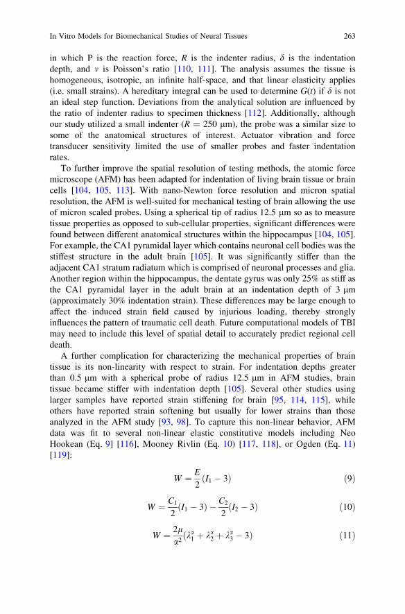
in which P is the reaction force, R is the indenter radius, d is the indentationdepth, and m is Poisson’s ratio [110, 111]. The analysis assumes the tissue ishomogeneous, isotropic, an infinite half-space, and that linear elasticity applies(i.e. small strains). A hereditary integral can be used to determine G(t) if d is notan ideal step function. Deviations from the analytical solution are influenced bythe ratio of indenter radius to specimen thickness [112]. Additionally, althoughour study utilized a small indenter (R = 250 lm), the probe was a similar size tosome of the anatomical structures of interest. Actuator vibration and forcetransducer sensitivity limited the use of smaller probes and faster indentationrates.
To further improve the spatial resolution of testing methods, the atomic forcemicroscope (AFM) has been adapted for indentation of living brain tissue or braincells [104, 105, 113]. With nano-Newton force resolution and micron spatialresolution, the AFM is well-suited for mechanical testing of brain allowing the useof micron scaled probes. Using a spherical tip of radius 12.5 lm so as to measuretissue properties as opposed to sub-cellular properties, significant differences werefound between different anatomical structures within the hippocampus [104, 105].For example, the CA1 pyramidal layer which contains neuronal cell bodies was thestiffest structure in the adult brain [105]. It was significantly stiffer than theadjacent CA1 stratum radiatum which is comprised of neuronal processes and glia.Another region within the hippocampus, the dentate gyrus was only 25% as stiff asthe CA1 pyramidal layer in the adult brain at an indentation depth of 3 lm(approximately 30% indentation strain). These differences may be large enough toaffect the induced strain field caused by injurious loading, thereby stronglyinfluences the pattern of traumatic cell death. Future computational models of TBImay need to include this level of spatial detail to accurately predict regional celldeath.
A further complication for characterizing the mechanical properties of braintissue is its non-linearity with respect to strain. For indentation depths greaterthan 0.5 lm with a spherical probe of radius 12.5 lm in AFM studies, braintissue became stiffer with indentation depth [105]. Several other studies usinglarger samples have reported strain stiffening for brain [95, 114, 115], whileothers have reported strain softening but usually for lower strains than thoseanalyzed in the AFM study [93, 98]. To capture this non-linear behavior, AFMdata was fit to several non-linear elastic constitutive models including NeoHookean (Eq. 9] [116], Mooney Rivlin (Eq. 10) [117, 118], or Ogden (Eq. 11)[119]:
W ¼ E
2ðI1 � 3Þ ð9Þ
W ¼ C1
2ðI1 � 3Þ � C2
2ðI2 � 3Þ ð10Þ
W ¼ 2la2ðka
1 þ ka2 þ ka
3 � 3Þ ð11Þ
In Vitro Models for Biomechanical Studies of Neural Tissues 263
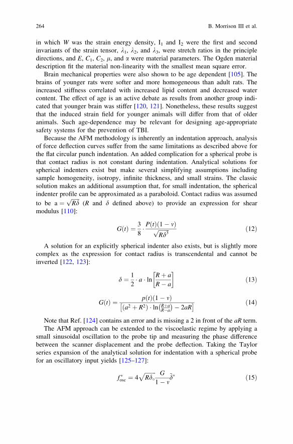
in which W was the strain energy density, I1 and I2 were the first and secondinvariants of the strain tensor, k1, k2, and k3, were stretch ratios in the principledirections, and E, C1, C2, l, and a were material parameters. The Ogden materialdescription fit the material non-linearity with the smallest mean square error.
Brain mechanical properties were also shown to be age dependent [105]. Thebrains of younger rats were softer and more homogeneous than adult rats. Theincreased stiffness correlated with increased lipid content and decreased watercontent. The effect of age is an active debate as results from another group indi-cated that younger brain was stiffer [120, 121]. Nonetheless, these results suggestthat the induced strain field for younger animals will differ from that of olderanimals. Such age-dependence may be relevant for designing age-appropriatesafety systems for the prevention of TBI.
Because the AFM methodology is inherently an indentation approach, analysisof force deflection curves suffer from the same limitations as described above forthe flat circular punch indentation. An added complication for a spherical probe isthat contact radius is not constant during indentation. Analytical solutions forspherical indenters exist but make several simplifying assumptions includingsample homogeneity, isotropy, infinite thickness, and small strains. The classicsolution makes an additional assumption that, for small indentation, the sphericalindenter profile can be approximated as a paraboloid. Contact radius was assumed
to be a ¼ffiffiffiffiffiffiRdp
(R and d defined above) to provide an expression for shearmodulus [110]:
GðtÞ ¼ 38� PðtÞð1� mÞffiffiffiffiffiffiffiffi
Rd3p ð12Þ
A solution for an explicitly spherical indenter also exists, but is slightly morecomplex as the expression for contact radius is transcendental and cannot beinverted [122, 123]:
d ¼ 12� a � ln Rþ a
R� a
� �ð13Þ
GðtÞ ¼ pðtÞð1� mÞa2 þ R2ð Þ � ln Rþa
R�a
� �� 2aR
� � ð14Þ
Note that Ref. [124] contains an error and is missing a 2 in front of the aR term.The AFM approach can be extended to the viscoelastic regime by applying a
small sinusoidal oscillation to the probe tip and measuring the phase differencebetween the scanner displacement and the probe deflection. Taking the Taylorseries expansion of the analytical solution for indentation with a spherical probefor an oscillatory input yields [125–127]:
f �osc ¼ 4ffiffiffiffiffiffiffiffiRd�
p G
1� m~d� ð15Þ
264 B. Morrison III et al.

in which fosc* , the AFM probe force, and , the tissue indentation, are complex
quantities with magnitude and phase. Splitting this into real and imaginary com-ponents yields the storage (G0) and loss (G00) modulus:
G0 ¼ ð1� mÞ
4ffiffiffiffiffiffiffiffiRd�p � adkar cos u� ka2
a2d � 2adar cos uþ a2
r
G00 ¼ �ð1� mÞ
4ffiffiffiffiffiffiffiffiRd�p � adkar sin u
a2d � 2adar cos uþ a2
r
� x � c� � ð16Þ
in which d0 is the static indentation depth, k is the cantilever stiffness, ad is themagnitude of the scanner displacement, ar is the magnitude of the cantileverdeflection, u is the phase lag between the two, c is the drag of the probe in thefluid, and x is the oscillation frequency. This methodology has been applied tomeasure viscoelastic properties of brain, producing both frequency andindentation-depth dependent storage and loss moduli (Fig. 2, unpublishedobservations). One of the recurring challenges is accurate contact pointidentification.
Mechanical properties of single brain cells have been measured with asimilar approach [113]. Hippocampal neurons and astrocytes were indentedwith a 3 lm sphere with superimposed oscillations of 30, 100, and 200 Hz,although no indentation depth was reported. Neurons were stiffer than astro-cytes. Shear modulus increased with frequency and ranged from about100–300 Pa.
Several sources of non linear behavior can complicate analysis of dynamicindentation data. The first is the effect of large deformation on material response. Ifdata from large-deformation indentation experiments is analyzed with linearelastic theory, a linearly elastic material will appear non-linear, exhibiting strainhardening behavior [128]. Simple shear excitation does not suffer the samecomplication. The second is due to the intrinsic non-linear material properties ofthe tissue which arises as the excitation strain is increased above the linear limit.A third source of non-linearity in response to a single frequency excitation is theexistence of higher order harmonics in the tissue response. These higher orderharmonics violate the basic assumptions of linear frequency analysis. Severalstrategies have been described to analyze this non-linear material behavior[129–131]. A Fourier transform of the tissue response will quickly identifywhether higher order harmonics are contaminating the data.
3.2 Mechanical Properties of 3D Neural Cultures
In light of what is known about brain tissue properties, rheological properties(specifically, complex modulus) of Matrigel (7.5 mg/mL), a commonly used 3Dcell scaffold, were measured over days in vitro as a function of frequency for the
In Vitro Models for Biomechanical Studies of Neural Tissues 265

0.51
1.52
2.5
5 10
50 100
200400
0
500
1000
1500
She
ar M
odul
us (
Pa)
a)
0.51
1.52
2.5
5 10
50 100
200400
0
500
1000
1500
2000
Frequency (Hz)Depth (microns)
She
ar M
odul
us (
Pa)
Frequency (Hz)Depth (microns)
b)
Fig. 2 Depth- and frequency-dependent a storage and b loss modulus determined from dynamicAFM indentation of the pig alveus with a spherical probe (mean + standard error of the mean).Modulus increased with excitation frequency, consistent with a viscoelastic material. Storagemodulus increased with indentation depth, suggesting non-linear behavior
266 B. Morrison III et al.

following conditions and time points: (1) Matrigel alone at 0 (immediately upongelation), 11, and 14 days following casting in serum free cell culture medium at37�C and 5% CO2; (2) Matrigel with neurons and astrocytes at 2,500 cells/mm3 at14 days in culture at 37�C and 5% CO2. All groups except the 0 days groupcontained 5% serum. All rheology experiments were performed in hydratedsamples on a Bohlin CVO Rheometer (East Brunswick, NJ) at 37�C under 0.005strain with a frequency sweep from 0.01 to 10 Hz (n = 2–4 samples per condi-tion). The trends among the conditions tested were similar across treatment groupswith respect to frequency and time under culture conditions, suggesting that 3Dcultures of cortical neurons/astrocytes in Matrigel retain the gross mechanicalproperties over 14 days and that this was independent of the presence ofcells (Fig. 3). Furthermore, the mechanical behavior was similar to that ofbrain [103–106, 121, 132, 133], further supporting the use of 3D cultures and
Co
mp
lex
Mo
du
lus
(Pa)
Frequency (Hz)
Matrigel only: 0 days incubation
Matrigel only: 11 days, media + 5% serum
Matrigel only: 14 days, media + 5% serum
Matrigel + cells: 14 days, media + 5% serum
0.01 0.03 0.1 0.3 1.0 3.2 101
100
10000
Fig. 3 The complex modulus of thick (500 lm) Matrigel constructs was examined as a functionof incubation days, the presence of serum, and the presence of cortical neurons/astrocytes. Alldimensions and incubation/rheology conditions were constant for the experimental groups(n = 2–4 samples per condition) for a frequency sweep of 0.01–10 Hz. As expected, complexmodulus increased with frequency. The presence of serum during gelation and incubation timeperiods did not affect the baseline properties (i.e. those at 0 days with no serum). Furthermore, thepresence of cells did not significantly affect the overall material properties of the 3D culture after14 days in culture. Data presented as mean ± standard error of the mean
In Vitro Models for Biomechanical Studies of Neural Tissues 267
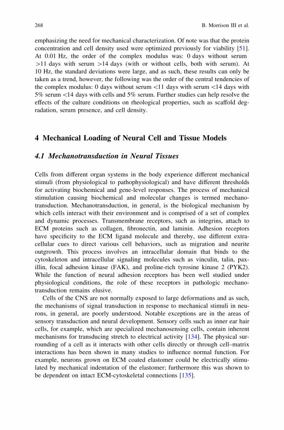
emphasizing the need for mechanical characterization. Of note was that the proteinconcentration and cell density used were optimized previously for viability [51].At 0.01 Hz, the order of the complex modulus was: 0 days without serum[11 days with serum [14 days (with or without cells, both with serum). At10 Hz, the standard deviations were large, and as such, these results can only betaken as a trend, however, the following was the order of the central tendencies ofthe complex modulus: 0 days without serum\11 days with serum\14 days with5% serum\14 days with cells and 5% serum. Further studies can help resolve theeffects of the culture conditions on rheological properties, such as scaffold deg-radation, serum presence, and cell density.
4 Mechanical Loading of Neural Cell and Tissue Models
4.1 Mechanotransduction in Neural Tissues
Cells from different organ systems in the body experience different mechanicalstimuli (from physiological to pathophysiological) and have different thresholdsfor activating biochemical and gene-level responses. The process of mechanicalstimulation causing biochemical and molecular changes is termed mechano-transduction. Mechanotransduction, in general, is the biological mechanism bywhich cells interact with their environment and is comprised of a set of complexand dynamic processes. Transmembrane receptors, such as integrins, attach toECM proteins such as collagen, fibronectin, and laminin. Adhesion receptorshave specificity to the ECM ligand molecule and thereby, use different extra-cellular cues to direct various cell behaviors, such as migration and neuriteoutgrowth. This process involves an intracellular domain that binds to thecytoskeleton and intracellular signaling molecules such as vinculin, talin, pax-illin, focal adhesion kinase (FAK), and proline-rich tyrosine kinase 2 (PYK2).While the function of neural adhesion receptors has been well studied underphysiological conditions, the role of these receptors in pathologic mechano-transduction remains elusive.
Cells of the CNS are not normally exposed to large deformations and as such,the mechanisms of signal transduction in response to mechanical stimuli in neu-rons, in general, are poorly understood. Notable exceptions are in the areas ofsensory transduction and neural development. Sensory cells such as inner ear haircells, for example, which are specialized mechanosensing cells, contain inherentmechanisms for transducing stretch to electrical activity [134]. The physical sur-rounding of a cell as it interacts with other cells directly or through cell–matrixinteractions has been shown in many studies to influence normal function. Forexample, neurons grown on ECM coated elastomer could be electrically stimu-lated by mechanical indentation of the elastomer; furthermore this was shown tobe dependent on intact ECM-cytoskeletal connections [135].
268 B. Morrison III et al.
4.2 Mechanical Response of Neural Cultures
Mechanotransduction has been studied in vitro by controlling the mechanicalproperties (i.e. rigidity, tortuosity, ligand density) of extracellular matrices andobserving cell behavior [30, 136–139]. It has been shown that neurons tend tosupport longer neuritic outgrowth on compliant substrates compared to rigidsupport scaffolds [25, 35, 140, 141], whereas fibroblasts, spread and migrate morereadily as rigidity was increased [142, 143]. When primary cortical neurons werecultured in a 3D environment with controlled mechanical properties and laminindensity, however, it was observed that the more rigid formulation (G0 = 565 Pa)supported more neurite extension than a softer (G0 = 175 Pa) gel, suggesting thatthe 3D environment may provide additional extracellular cues for supportingneurons (unpublished observation). Of note, the stiffer gel mechanical propertieswere closer to that of brain compared to the softer culture scaffold [103–106, 121,132, 133], suggesting that cell specific tissue properties should be emulated whencreating culture mimics. This concept of matching in vivo mechanical properties toculture conditions has been supported by other studies as well [140, 144, 145]. Theresponse to mechanical stimulation may also be affected by the degree of differ-entiation, type and level of stress, cytoarchitectural complexity, and physicalenvironment. Controlled biophysical cell and tissue culture conditions are espe-cially critical in which the mechanical environment can be independently variedand the neural response directly measured.
4.3 Mechanical Injury to Neural Tissue
By applying a range of mechanical stimuli to neural tissue models, one can betterunderstand specific cellular responses (e.g., neurotransmitter release, signalingpathways). Here, we focus on the mechanical responses to injurious physicalinsults observed in 2D and 3D neural cultures and the organotypic slice culturemodels. Ultimately, the identification of mechanotransduction mechanisms canbest be achieved through a multidisciplinary approach, with a variety of cellularand tissue models, animal and human studies, and computer simulations. Trau-matic insults to the brain and spinal cord deform cellular elements. Persistentinjury can occur from a single mechanical event and subsequent insults mayexacerbate the initial response, suggesting increased sensitivity due to an abnormalstate [146].
Cellular models of traumatic injury have been developed that mimic themechanical parameters associated with large, high rate deformation to cellularelements of the brain. These in vitro injury model paradigms include transection,compression, stretch, and shear. Two-dimensional in vitro cultures are valuabletools for studying penetrating and blunt impact injury [147, 148], as well asstretch injury [149–151]. Both 3D cultures derived from dissociated cells and
In Vitro Models for Biomechanical Studies of Neural Tissues 269

brain slices have been used for neural injury studies [17, 152]. The mostappropriate injury models, however, may be those that mimic the 3D in vivoconfiguration and that capture forces experienced during injury in vivo. In vitromodels of TBI allow for well-characterized and repeatable injuries, the param-eters of which can be controlled independently and precisely. While variousstudies have examined cellular and tissue levels thresholds to mechanical loading[31, 153–155], the input parameters inherently vary from system to system,pointing to the continuing need to systematically study a range of mechanicalstimuli to different culture systems in a comparative and parallel manner. Somegeneral observations have been made across some laboratories. For example, therate of application of mechanical loading dictates, to some degree, the severity ofthe injury [57, 60, 154, 156–160], likely due to the viscoelastic response. Strainlevel, independent of rate, has also been associated with injury severity [31, 57,60, 160–162]. It is likely that varying loading regimes, input parameters, cellpreparations, time points for outcome measures, and other experimental variablesplay a role in these different responses. In addition, one must consider the non-linearities that may be part of the injury response and the range of mechanicalparameters with respect to the analogous in vivo loading conditions, many ofwhich are difficult to link to specific pathologies.
Experimental models of superthreshold mechanical stimuli must be definedby: (1) the culture composition (including density, extracellular constituents,cell types, and developmental age) and mechanical properties; and (2) themechanical parameters (e.g., stress, strain, and modality) that mimic the phe-nomenon being studied. In vitro systems eliminate systemic responses, whilemaintaining the ability to manipulate and observe cells during and afterphysical stimulation [57, 163–166]. Cellular and tissue neural models preservethe ability to apply a more controlled mechanical input than in a correlate invivo environment. Because complexity, such as additional cell types orextracellular stimuli can be added in a controlled fashion, such models play animportant role in discerning the mechanical principles that govern physical andfunctional response and thresholds.
5 Defining Tolerance Criteria for Neural Tissue
5.1 Definition of Failure
Failure is a central theme of our studies on brain tissue and cultures, and ourstudies have benefited from a definition that is broader than the typical engineeringdefinition of ultimate failure. Mechanical failure may be an adequate definition forload-bearing tissues of the human body like bone and even for soft tissues in themost extreme cases. However, tearing (fracture) of tissue or cells is only one modeof failure, and many others may exist for biological tissues as discussed below.
270 B. Morrison III et al.

A fundamental difference between inert engineering materials and biologicaltissues is that the latter are alive and perform some form of active, physiologicalfunction. Therefore, the very concept of failure must be revised. The exact defi-nition may be tissue-specific such as fracture for bone or laceration for skin.However, these definitions may be too insensitive to identify the onset of injury forthe brain, i.e. tolerance criteria. For non load-bearing, highly cellularized, livingtissues, failure can be defined in myriad ways and may occur far below mechanicalfailure limits.
5.2 Tolerance Criteria for Living Brain Tissue
Quantification of tolerance criteria for brain has a number of requirementsincluding a living preparation, a means to apply precisely controlled mechanicalstimuli, and a mechanism to verify tissue biomechanics. For brain and other highlycellular tissues, cell death may be a widely applicable outcome. This workingdefinition of injury has been applied to determine tissue-level tolerance criteria forcell death in the rat hippocampus and cortex [60, 160]. In these studies, organo-typic slice cultures of brain were subjected to controlled mechanical deformation,and the resultant cell death quantified in a region-specific manner over time [57].An empirical relationship between input mechanical stimuli (tissue strain andstrain rate) and cell death was calculated. The goal was to provide these transferfunctions for incorporation into FEMs, providing them with the capability topredict biological responses in addition to typical mechanical parameters such asstress and strain.
It was found that cell death was not immediate in response to deformation thatwas below cellular ultimate failure limits [57, 60, 160]. Instead, cell deathincreased over 4 days after injury, highlighting a fundamental difference toengineering materials. Both CA3 and CA1 of the hippocampus were the mostvulnerable regions to mechanical injury. Dentate gyrus was less vulnerable thanother hippocampal regions. Cortex was much less susceptible than the hippo-campus as a whole; interestingly, cell death in the cortex increased with increasingstrain rate in contrast to hippocampal cell loss which was insensitive to rate.To illustrate, after 0.20 biaxial strain, approximately 35% of cells in CA1 and CA3died versus 20% in the dentate gyrus and less than 5% in the cortex.
By quantifying both the input mechanical stimulus and the output biologicalresponse, it was possible to determine numerical relationships for predicting the timecourse and magnitude of post-injury cell death (Fig. 4). These three empirical func-tions (Eq. 17) could be incorporated into FEMs, thereby equipping them with bio-logical predictions to supplement mechanical predictions of stress and strain.
In Vitro Models for Biomechanical Studies of Neural Tissues 271
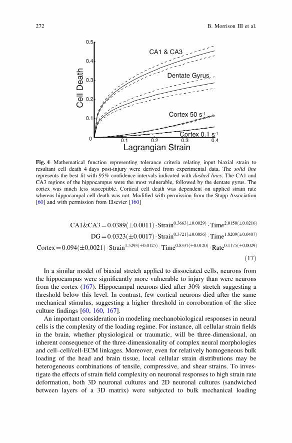
CA1&CA3¼0:0389ð�0:0011Þ �Strain0:3663ð�0:0029Þ �Time2:0150ð�0:0216Þ
DG¼0:0323ð�0:0017Þ �Strain0:3721ð�0:0056Þ �Time1:8209ð�0:0407Þ
Cortex¼0:094ð�0:0021Þ �Strain1:5293ð�0:0125Þ �Time0:8337ð�0:0120Þ �Rate0:1175ð�0:0029Þ
ð17Þ
In a similar model of biaxial stretch applied to dissociated cells, neurons fromthe hippocampus were significantly more vulnerable to injury than were neuronsfrom the cortex (167). Hippocampal neurons died after 30% stretch suggesting athreshold below this level. In contrast, few cortical neurons died after the samemechanical stimulus, suggesting a higher threshold in corroboration of the sliceculture findings [60, 160, 167].
An important consideration in modeling mechanobiological responses in neuralcells is the complexity of the loading regime. For instance, all cellular strain fieldsin the brain, whether physiological or traumatic, will be three-dimensional, aninherent consequence of the three-dimensionality of complex neural morphologiesand cell–cell/cell-ECM linkages. Moreover, even for relatively homogeneous bulkloading of the head and brain tissue, local cellular strain distributions may beheterogeneous combinations of tensile, compressive, and shear strains. To inves-tigate the effects of strain field complexity on neuronal responses to high strain ratedeformation, both 3D neuronal cultures and 2D neuronal cultures (sandwichedbetween layers of a 3D matrix) were subjected to bulk mechanical loading
0.1 0.2 0.3 0.40
0.1
0.2
0.3
0.4
0.5
Cel
l Dea
th
Lagrangian Strain
CA1 & CA3
Dentate Gyrus
Cortex 0.1 s-1
Cortex 50 s-1
Fig. 4 Mathematical function representing tolerance criteria relating input biaxial strain toresultant cell death 4 days post-injury were derived from experimental data. The solid linerepresents the best fit with 95% confidence intervals indicated with dashed lines. The CA1 andCA3 regions of the hippocampus were the most vulnerable, followed by the dentate gyrus. Thecortex was much less susceptible. Cortical cell death was dependent on applied strain ratewhereas hippocampal cell death was not. Modified with permission from the Stapp Association[60] and with permission from Elsevier [160]
272 B. Morrison III et al.

Fig. 5 Cell viability in networks of neurons cultured in 2D and 3D following shear strain. Cells weresubjected to high strain rate and magnitude shear deformation using a custom-built device. Viabilitywas assessed 24 h after the application of control or strain conditions (live cells stained in cytosol andappear as larger areas with branching neurite patterns (green), nuclei of dead cells stained as punctatesmaller areas (red)). Confocal reconstructions of neuronal cultures in 2D after a static control or b 50%strain, 30s-1 strain rate. Neuronal cultures in 3D after c static control or d 50% strain, 30s-1 strain rate(scale bar = 50lm). e Graphical representation of cell viability. There was not a significant differencein viability in naive controls compared to static controls for either 2D or 3D culture; however, therewas a significant decrease in both 2D and 3D viability after high rate deformation versus uninjuredcontrols (�p \ 0.05). Moreover, there was a significant decrease in viability in 3D versus 2D undermatched loading conditions (*p \ 0.05), implicating the complexity of the local cellular strain fieldsin more detrimental outcome following traumatic loading. Error bars represent standard deviation.Reprinted with permission from Mary Ann Liebert [31]
In Vitro Models for Biomechanical Studies of Neural Tissues 273

consistent with tissue-level strains in closed-head TBI [31]. At matched strainmagnitude (50% shear strain) and strain rates (20–30s-1) to the 3D cell-containingmatrices, neuronal death was greater when neurons were cultured in 3D versusmonolayer (Fig. 5). These data implicate the complexity of the local cellular strainfield, in addition to the magnitude and rate of individual strain components, in
Fig. 6 Neurite loss in 3D neuronal cultures following shear strain. Confocal reconstructions of3D neuronal cultures at 24 h following a sham conditions, b excitotoxicity via 100 lM glutamateexposure (positive control), or c shear deformation at 50% strain, 30s-1 strain rate (scalebar = 50 lm). Live neurons and remaining neurites stained with calcein can be seen as roundedstructures with typical branching patterns (green) and the nuclei of dead cells are seen as punctatebright areas (red). There was approximately a 50% reduction in the total number of neuritesfollowing either chemical injury (excitotoxicity) or mechanical injury. d The orientation ofremaining neurites was measured following these conditions. Following excitotoxic injury, thedistribution of remaining neurites was consistent across all angles of orientation, indicatingindiscriminate neurite loss. However, following high strain rate shear deformation, there waspreferential loss of neurites at angles close to the horizontal plane (�p \ 0.05), correlating withorientations predicted to experience maximum shear strain, suggesting heightened sensitivity toshear strain. Error bars represent standard deviation. Adapted with permission from Elsevier [17]
274 B. Morrison III et al.

thresholds for neuronal death, and also indicate that tolerances were lower inrealistic tissue constructs. Moreover, within these 3D constructs, the unique strainfields of individual neurons and neurites (micro-strains) may be calculated basedon the cell orientation with respect to the bulk loading (meso-strains) [17]. Thisanalysis revealed that the majority of neurite loss correlated with the direction ofmaximum shear strain (Fig. 6), demonstrating increased sensitivity to the strainmode in complex loading regimes.
In comparison to neurons, significant astrocyte cell death was observed after25% shear strain in the same constructs [17]. Although astrocyte death did notsignificantly increase after 50% shear compared to 25%, astrocyte death signifi-cantly increased as the strain rate was increased from 1 to 30s-1 [14, 17]. Inter-estingly, high shear strain (50%), quasi-static (1s-1) loading did not induce celldeath, although this caused significant astrocyte activation measured by prolifer-ation and hypertrophy, suggesting a direct mechanism of mechano-activation inastrocytes. However, at an intermediate strain rate of 10s-1 inducing significant celldeath, astrocyte activation was maximal [14], demonstrating that local cell deathmay amplify the astrocyte responses to trauma.
Multiple models injure cultured tissue with uniaxial as opposed to biaxialstrains, either by fluid shear [151] or substrate deformation [159]. After fluid shearstress of NT-2 cells, a neuronal cell line, significant lactate dehydrogenase (LDH)release was measured immediately after 16% cell strain and at 24 h after 12%strain at strain rates below 7s-1 [151, 156]. After uniaxial strain applied to hip-pocampal cultures by substrate deformation, cell death was significantly increasedafter 50% but not 17% strain, suggesting a threshold above 17%, although in afollow up study, significant cell death was reported after 6% strain [168, 169].When hippocampal slice cultures were subjected to uniaxial strain, no activatedcaspase 3 or calpain was detected after 50% strain. Caspase 3 is essential forexecution of the intrinsic apoptotic pathway whereas calpain is activated by ele-vated intracellular calcium and is thought to play a role in necrotic cell death.Calpain was consistently activated after 75 and 100% uniaxial strain, whereascaspase 3 was activated after 75% but not 100% strain [170]. Although cell deathwas not directly measured, these results suggest that injury may activate differentcell-death pathways depending on severity.
The reasons for the discrepancy in cell-death response after uniaxial loadingbetween the fluid shear stress and substrate strain models may be due to experi-mental considerations. Verification of cell/tissue deformation was absolutelyessential for each experiment. Although tissue may appear to be adhered to thesubstrate after injury, it may be anchored in only one region such that it does notstretch, but does not float away either. In the shear stress model, cell strain duringinjury was confirmed, whereas in the uniaxial slice model, it was not.
In a comparison of uniaxial and biaxial strain applied to cortical neurons, thepost-injury calcium response was significantly greater after 30 and 50% biaxialstrain with very few neurons responding to uniaxial strain [171]. Cell membranepermeability was greater after biaxial deformation, as well [171]. Although no cell
In Vitro Models for Biomechanical Studies of Neural Tissues 275

death was measured 24 h after any injury up to 50% strain, these results suggestedthat biaxial strain may be a more severe injury than uniaxial strain [171].
The brain carries out essential functions of information processing, motorcontrol, and consciousness which are dependent on neuronal electrophysiologicalfunction at the cellular level. One can image many injury mechanisms which mayalter neuronal function without requiring cell death. Therefore, an additional tol-erance criterion may be more appropriate for nervous system tissue: disruption ofelectrophysiological function. After either 5 or 10% biaxial strain applied to hip-pocampal slice cultures, evoked activity was significantly altered [172]. Themaximal evoked response (Rmax) was decreased by up to 75%, and the tissuebecame less excitable with up to 120% increase in the stimulus required to generatea half maximal response (I50) [172]. These studies were conducted with micro-electrode arrays (MEA) which have the advantage of sampling tissue activity fromup to 60 locations simultaneously, providing unbiased and comprehensive quan-tification of activity throughout the neuronal network. MEA recordings are alsocompatible with dissociated cultures. After uniaxial strain via fluid shear stress,acute changes in activity were noted including an increase in burst interval [173].
6 Conclusions and Future directions
Neural cell and tissue culture models offer a unique platform for studyingmechanical response to physiological forces as well as neural trauma, wherephysical forces may cause structural failure due to excessive stress and strain, orresult in sublethal dysfunction. Cells in culture, however, may differ in cell–cellconfiguration, material properties, cell signaling, and gene expression from in vivocounterparts. Recreating these structural components at the nano-, micro-, meso-and macro-scale, together with physiological and physical properties, allowsdevelopment of robust culture systems, which in turn, aids in developing bothconstitutive models and guides for computer simulations. The formulation ofanalytical models that consider multi-scale tissue characteristics then become verypowerful tools for assessment of the biomechanical response to various stimuli.
In vitro models of neural cells have proven to be effective in the systematicidentification of specific mechanisms of mechanotransduction and injury causa-tion. The primary attributes of in vitro models include control of cellular com-position, control of local environment, accessibility for imaging, experimentalmanipulability, and elimination of systemic effects. In vitro neural models mustmimic the in vivo environment in terms of composition, cell type, extracellularspace, cell volume fraction, tortuosity, cell cytoarchitecture, distribution of cell–cell/cell–matrix interactions, and the multicellular composition. Properly designedcellular models may serve as a more accurate guide to cellular growth, interaction,and responses to biochemical and/or mechanical stimuli.
In the continued drive to increase the biofidelity of ex vivo constructs to the invivo brain, new scaffold materials may provide improved substrates to support
276 B. Morrison III et al.
higher cell and process density to more closely approximate in vivo brain. Pat-terning cell types in 3D may allow for replication of the complex connectivity ofmultiple cell types that typifies the CNS. Our studies have demonstrated thebenefits of perfusion for supporting these high cell densities. Microfluidic perfu-sion systems may hold promise for making these types of cultures more com-monplace. Explant culture systems can reproduce a degree of cytoarchitecturalcomplexity in an effort to maintain in vivo characteristics. However, a majorlimitation is that only post-natal/juvenile tissue can be cultured successfully.Future efforts to culture adult brain tissue would fill a critical need for determiningwhether tissue-level tolerance criteria are affected by age.
The ex vivo systems provide unfettered access to the cultures at multiple spatialscales. At the tissue level, it has been possible to quantify tissue biomechanicsduring the applied dynamic event, whether it is injury or mechanical testing.However, dynamic measurement of cellular or subcellular deformation hasremained difficult, but could significantly add to our understanding of cell-levelbiomechanics which may be one source of heterogeneous cellular response. Suchinsight is essential for multi-scale modeling of TBI with strategies such as rep-resentative volume element modeling.
The biological response, as a consequence of mechanotransduction cascades,occurs on a slower time scale than an injurious stimulus. Through the use of fluo-rescent reporter systems such as optogenetic approaches, continuous monitoring ofphysiological parameters becomes possible. Correlating the activation of cell deathcascades with mechanical injury parameters will further define tolerance criteria andthe limits of safe exposures, which are absolutely critical for safety system design.
Analysis of indentation data for the determination of mechanical properties isinherently complex given the heterogeneous strain field beneath the indenter.However, sophisticated numerical analysis methods such as inverse FE approachescould be leveraged to more accurately extract constitutive properties. Suchnumerical methods explicitly include factors normally ignored in classicalindentation solutions. The accuracy of derived constitutive properties is increasedby explicitly modeling the complex strain field beneath the indenter, as well.Indentation methods such as these will provide material property data at highspatial resolution, which will be instrumental for the next generation of compu-tation models for simulating TBI and predicting anatomical patterns of cell death.These models, in turn, may be used to design the next generation of safety systemsto reduce the socioeconomic costs of TBI.
Acknowledgments The authors thank Varadraj Vernekar, Ph.D. for contributions to the 3Dculture mechanical testing study, and Benjamin S. Elkin, Ph.D. for the dynamic AFM indentationstudies.
In Vitro Models for Biomechanical Studies of Neural Tissues 277

References
1. Kindle, E., Schwartz, J., Jessell, T.: Principles of Neural Science. McGraw-Hill, New York(2000)
2. Sycoma, E., Nicholson, C.: Diffusion in brain extracellular space. Physiol. Rev. 88,1277–1340 (2008)
3. Sycoma, E.: Extra synaptic volume transmission and diffusion parameters of theextracellular space. Neuroscience 129, 861–876 (2004)
4. Sycoma, E.: Diffusion properties of the brain in health and disease. Neurochem. Int. 45,453–466 (2004)
5. Fedoroff, S., Richardson, A.: Protocols for Neural Cell Culture, 3rd edn. Humana Press,Totowa (2001)
6. Hayflick, L., Moorhead, P.: The serial cultivation of human diploid cell strains. Exp. CellRes. 25, 585–621 (1961)
7. Jacobson, M.: Clonal analysis and cell lineages of the vertebrate central nervous system.Annu. Rev. Neurosci. 8, 71–102 (1985)
8. Potter, S.M., DeMarse, T.B.: A new approach to neural cell culture for long-term studies.J. Neurosci. Methods 110, 17–24 (2001)
9. Dichter, M.A.: Rat cortical neurons in cell culture: culture methods, cell morphology,electrophysiology, and synapse formation. Brain. Res. 149, 279–293 (1978)
10. Corey, J.M., Wheeler, B.C., Brewer, G.J.: Compliance of hippocampal neurons to patternedsubstrate networks. J. Neurosci. Res. 30, 300–307 (1991)
11. Choi, H.K., Won, L., Heller, A.: Dopaminergic neurons grown in three-dimensionalreaggregate culture for periods of up to one year. J. Neurosci. Methods 46, 233–244 (1993)
12. Hsiang, J., Heller, A., Hoffmann, P.C., Mobley, W.C., Wainer, B.H.: The effects of nervegrowth factor on the development of septal cholinergic neurons in reaggregate cell cultures.Neuroscience 29, 209–223 (1989)
13. O’Connor, S.M., Andreadis, J.D., Shaffer, K.M., Ma, W., Pancrazio, J.J., Stenger, D.A.:Immobilization of neural cells in three-dimensional matrices for biosensor applications.Biosens. Bioelectron. 14, 871–881 (2000)
14. Cullen, D.K., Simon, C.M., LaPlaca, M.C.: Strain rate-dependent induction of reactiveastrogliosis and cell death in three-dimensional neuronal-astrocytic co-cultures. Brain Res.1158, 103–115 (2007)
15. O’Connor, S.M., Stenger, D.A., Shaffer, K.M., Ma, W.: Survival and neurite outgrowth ofrat cortical neurons in three-dimensional agarose and collagen gel matrices. Neurosci. Lett.304, 189–193 (2001)
16. Woerly, S., Plant, G.W., Harvey, A.R.: Cultured rat neuronal and glial cells entrappedwithin hydrogel polymer matrices: a potential tool for neural tissue replacement. Neurosci.Lett. 205, 197–201 (1996)
17. LaPlaca, M.C., Cullen, D.K., McLoughlin, J.J., Cargill 2nd, R.S.: High rate shear strain ofthree-dimensional neural cell cultures: a new in vitro traumatic brain injury model.J. Biomech. 38, 1093–1105 (2005)
18. Edelman, D.B., Keefer, E.W.: A cultural renaissance: in vitro cell biology embraces three-dimensional context. Exp. Neurol. 192, 1–6 (2005)
19. Cukierman, E., Pankov, R., Stevens, D.R., Yamada, K.M.: Taking cell-matrix adhesions tothe third dimension. Science 294, 1708–1712 (2001)
20. Cukierman, E., Pankov, R., Yamada, K.M.: Cell interactions with three-dimensionalmatrices. Curr. Opin. Cell. Biol. 14, 633–639 (2002)
21. Schmeichel, K.L., Bissell, M.J.: Modeling tissue-specific signaling and organ function inthree dimensions. J. Cell Sci. 116, 2377–2388 (2003)
22. Yamada, K.M., Pankov, R., Cukierman, E.: Dimensions and dynamics in integrin function.Braz. J. Med. Biol. Res. 36, 959–966 (2003)
278 B. Morrison III et al.

23. Miller, B.E., Miller, F.R., Heppner, G.H.: Factors affecting growth and drug sensitivity ofmouse mammary tumor lines in collagen gel cultures. Cancer Res. 45, 4200–4205 (1985)
24. Grinnell, F.: Fibroblast biology in three-dimensional collagen matrices. Trends Cell Biol.13, 264–269 (2003)
25. Balgude, A.P., Yu, X., Szymanski, A., Bellamkonda, R.V.: Agarose gel stiffness determinesrate of DRAG neurite extension in 3M cultures. Biomaterials 22, 1077–1084 (2001)
26. Coates, P.W., Fermini, B., Strahlendorf, J.C., Strahlendorf, H.K.: Utilization of three-dimensional culture for early morphometric and electrophysiological analyses of solitarycerebellar neurons. Dev. Neurosci. 14, 35–43 (1992)
27. Coates, P.W., Nathan, R.D.: Feasibility of electrical recordings from unconnected vertebrateCNS neurons cultured in a three-dimensional extracellular matrix. J. Neurosci. Methods20, 203–210 (1987)
28. O’Shaughnessy, T.J., Lin, H.J., Ma, W.: Functional synapse formation among rat corticalneurons grown on three-dimensional collagen gels. Neurosci. Lett. 340, 169–172 (2003)
29. Spector, D.H., Boss, B.D., Strecker, R.E.: A model three-dimensional culture system formammalian dopaminergic precursor cells: application for functional intracerebraltransplantation. Exp. Neurol. 124, 253–264 (1993)
30. Bellamkonda, R., Ranieri, J.P., Aebischer, P.: Laminin oligopeptide derivatized agarose gelsallow three-dimensional neurite extension in vitro. J. Neurosci. Res. 41, 501–509 (1995)
31. Cullen, D.K., LaPlaca, M.C.: Neuronal response to high rate shear deformation depends onheterogeneity of the local strain field. J. Neurotrauma 23, 1304–1319 (2006)
32. Cullen, D.K., Lessing, M.C., LaPlaca, M.C.: Collagen-dependent neurite outgrowth andresponse to dynamic deformation in three-dimensional neuronal cultures. Ann Biomed. Eng.35, 835–846 (2007)
33. Vukasinovic, J., Cullen, D.K., LaPlaca, M.C., Glezer, A.: A microperfused incubator fortissue mimetic 3D cultures. Biomed. Microdevices 11, 1155–1165 (2009)
34. Dillon, G.P., Yu, X., Sridharan, A., Ranieri, J.P., Bellamkonda, R.V.: The influence ofphysical structure and charge on neurite extension in a 3D hydrogel scaffold. J. Biomater.Sci. Polym. Ed. 9, 1049–1069 (1998)
35. Willits, R.K., Skornia, S.L.: Effect of collagen gel stiffness on neurite extension.J. Biomater. Sci. Polym. Ed. 15, 1521–1531 (2004)
36. Schense, J.C., Hubbell, J.A.: Three-dimensional migration of neurites is mediated byadhesion site density and affinity. J. Biol. Chem. 275, 6813–6818 (2000)
37. Yu, T.T., Shoichet, M.S.: Guided cell adhesion and outgrowth in peptide-modified channelsfor neural tissue engineering. Biomaterials 26, 1507–1514 (2005)
38. Yu, X., Bellamkonda, R.V.: Dorsal root ganglia neurite extension is inhibited by mechanicaland chondroitin sulfate-rich interfaces. J. Neurosci. Res. 66, 303–310 (2001)
39. Borkenhagen, M., Clemence, J.F., Sigrist, H., Aebischer, P.: Three-dimensional extracellularmatrix engineering in the nervous system. J. Biomed. Mater. Res. 40, 392–400 (1998)
40. Tsacopoulos, M.: Metabolic signaling between neurons and glial cells: a short review.J. Physiol. Paris 96, 283–288 (2002)
41. Tsacopoulos, M., Magistretti, P.J.: Metabolic coupling between glia and neurons.J. Neurosci. 16, 877–885 (1996)
42. Aschner, M.: Neuron-astrocyte interactions: implications for cellular energetics andantioxidant levels. Neurotoxicology 21, 1101–1107 (2000)
43. Pardo, B., Honegger, P.: Differentiation of rat striatal embryonic stem cells in vitro:monolayer culture vs three-dimensional coculture with differentiated brain cells.J. Neurosci. Res. 59, 504–512 (2000)
44. Pulliam, L., Stubblebine, M., Hyun, W.: Quantification of neurotoxicity and identification ofcellular subsets in a three-dimensional brain model. Cytometry 32, 66–69 (1998)
45. Irons, H.R., Cullen, D.K., Shapiro, N.P., Lambert, N.A., Lee, R.H., LaPlaca, M.C.: Three-dimensional neural constructs: a novel platform for neurophysiological investigation.J. Neural. Eng. 5, 333–341 (2008)
In Vitro Models for Biomechanical Studies of Neural Tissues 279
46. Ahmed, S.M., Rzigalinski, B.A., Willoughby, K.A., Sitterding, H.A., Ellis, E.F.: Stretch-induced injury alters mitochondrial membrane potential and cellular ATP in culturedastrocytes and neurons. J. Neurochem. 74, 1951–1960 (2000)
47. Steinschneider, R., Delmas, P., Nedelec, J., Gola, M., Bernard, D., Boucraut, J.: Appearanceof neurofilament subunit epitopes correlates with electrophysiological maturation in corticalembryonic neurons cocultured with mature astrocytes. Brain. Res. Dev. Brain. Res. 95,15–27 (1996)
48. Nakanishi, K., Nakanishi, M., Kukita, F.: Dual intracellular recording of neocortical neuronsin a neuron-glia co-culture system. Brain Res. Brain. Res. Protoc. 4, 105–114 (1999)
49. LaPlaca, M.C., Vernekar, V.N., Shoemaker, J.T., Cullen, D.K.: Three-dimensional neuronalcultures. In: Berthiaume, F., Morgan, J. (eds.) Methods in Bioengineering: 3D TissueEngineering. Artech House Publishers, London (2010)
50. Nicholson, C., Sykova, E.: Extracellular space structure revealed by diffusion analysis.Trends Neurosci. 21, 207–215 (1998)
51. Cullen, D.K., Vukasinovic, J., Glezer, A., Laplaca, M.C.: Microfluidic engineered high celldensity three-dimensional neural cultures. J. Neural Eng. 4, 159–172 (2007)
52. Braitenberg, V.: Brain size and number of neurons: an exercise in synthetic neuroanatomy.J. Comput. Neurosci. 10, 71–77 (2001)
53. Gabbott, P.L., Stewart, M.G.: Distribution of neurons and glia in the visual cortex (area 17)of the adult albino rat: a quantitative description. Neuroscience 21, 833–845 (1987)
54. Alves, P., Moreira, J., Rodrigues, J., Aunins, J., Carrondo, M.: Two-dimensional versusthree-dimensional culture systems: effects on growth and productivity of BHK cells.Biotechnol. Bioeng. 52, 429–432 (1996)
55. Kleinman, H.K., McGarvey, M.L., Hassell, J.R., Star, V.L., Cannon, F.B., et al.: Basementmembrane complexes with biological activity. Biochemistry 25, 312–318 (1986)
56. Vukicevic, S., Kleinman, H.K., Luyten, F.P., Roberts, A.B., Roche, N.S., Reddi, A.H.:Identification of multiple active growth factors in basement membrane Matrigel suggestscaution in interpretation of cellular activity related to extracellular matrix components. Exp.Cell Res. 202, 1–8 (1992)
57. Morrison III, B., Cater, H.L., Benham, C.D., Sundstrom, L.E.: An in vitro model oftraumatic brain injury utilising two-dimensional stretch of organotypic hippocampal slicecultures. J. Neurosci. Meth. 150, 192–201 (2006)
58. Stoppini, L., Buchs, P.A., Muller, D.: Lesion-induced neurite sprouting and synapseformation in hippocampal organotypic cultures. Neuroscience 57, 985–994 (1993)
59. Gahwiler, B.H.: Organotypic monolayer cultures of nervous tissue. J. Neurosci. Meth. 4,329–342 (1981)
60. Elkin, B.S., Morrison III, B.: Region-specific tolerance criteria for the living brain. StappCar Crash J. 51, 127–138 (2007)
61. Sieg, F., Wahle, P., Pape, H.C.: Cellular reactivity to mechanical axonal injury in anorganotypic in vitro model of neurotrauma. J. Neurotrauma 16, 1197–1213 (1999)
62. Morrison III, B., Eberwine, J.H., Meaney, D.F., McIntosh, T.K.: Traumatic injury inducesdifferential expression of cell death genes in organotypic brain slice cultures determined bycomplementary DNA array hybridization. Neuroscience 96, 131–139 (2000)
63. Krassioukov, A.V., Ackery, A., Schwartz, G., Adamchik, Y., Liu, Y., Fehlings, M.G.:An in vitro model of neurotrauma in organotypic spinal cord cultures from adult mice.Brain Res. Brain Res. Protoc. 10, 60–68 (2002)
64. Stavridis, S.I., Dehghani, F., Korf, H.W., Hailer, N.P.: Characterisation of transverse sliceculture preparations of postnatal rat spinal cord: preservation of defined neuronalpopulations. Histochem. Cell Biol. 123, 377–392 (2005)
65. Gahwiler, B.H.: Development of the hippocampus in vitro: cell types, synapses, andreceptors. Neuroscience 11, 751–760 (1984)
66. Del. Rio., J.A., Heimrich, B., Soriano, E., Schwegler, H., Frotscher, M.: Proliferation anddifferentiation of glial fibrillary acidic protein immunoreactive glial cells in organotypicslice cultures of rat hippocampus. Neuroscience 43, 335–347 (1991)
280 B. Morrison III et al.

67. Caeser, M., Aertsen, A.: Morphological organization of rat hippocampal slice cultures.J. Comp. Neurol. 307, 87–106 (1991)
68. Stoppini, L., Buchs, P.A., Muller, D.: A simple method for organotypic cultutres of nervoustissue. J. Neurosci. Meth. 37, 173–182 (1991)
69. Buchs, P.A., Stoppini, L., Muller, D.: Structural modifications associated with synapticdevelopment in area CA1 of rat hippocampal organotypic cultures. Brain Res. Dev. BrainRes. 71, 81–91 (1993)
70. Mielke, J.G., Comas, T., Woulfe, J., Monette, R., Chakravarthy, B., Mealing, G.A.:Cytoskeletal, synaptic, and nuclear protein changes associated with rat interface organotypichippocampal slice culture development. Brain Res. Dev. Brain Res. 160, 275–286 (2005)
71. De Simoni, A., Griesinger, C.B., Edwards, F.A.: Development of rat CA1 neurones in acuteversus organotypic slices: role of experience in synaptic morphology and activity.J. Physiol. 550, 135–147 (2003)
72. Collin, C., Miyaguchi, K., Segal, M.: Dendritic spine density and LTP induction in culturedhippocampal slices. J. Neurophysiol. 77, 1614–1623 (1997)
73. Gahwiler, B.H.: Slice cultures of cerebellar, hippocampal, and hypothalamic tissue.Experientia 40, 235–243 (1984)
74. Robain, O., Barbin, G., de Billette, V., Jardin, L., Jahchan, T., Ben-Ari, Y.: Developmentof mossy fiber synapses in hippocampal slice culture. Brain Res. Dev. Brain Res. 80,244–250 (1994)
75. Hartel, R., Matus, A.: Cytoskeletal maturation in cultured hippocampal slices. Neuroscience78, 1–5 (1997)
76. Bahr, B.A., Kessler, M., Rivera, S., Vanderklish, P.W., Hall, R.A., et al.: Stablemaintenance of glutamate receptors and other synaptic components in long-termhippocampal slices. Hippocampus 5, 425–439 (1995)
77. Martens, U., Wree, A.: Distribution of [3H]MK-801, [3H]AMPA and [3H]kainate bindingsites in rat hippocampal long-term slice cultures isolated from external afferents. Anat.Embryol. (Berl) 203, 491–500 (2001)
78. Morrison III, B., Pringle, A.K., McManus, T., Ellard, J., Bradley, M., et al.: L-Arginyl-3, 4-Spermidine is neuroprotective in several in vitro models of neurodegeneration and in vivoischaemia without suppressing synaptic transmission. Brit. J. Pharm. 137, 1255–1268(2002)
79. Vornov, J.J., Tasker, R.C., Lost, D.: Direct observation of the agonist-specific regionalvulnerability to glutamate, NMDA, and kainate neurotoxicity in organotypic hippocampalcultures. Exp. Neurol. 114, 11–22 (1991)
80. Gutierrez, R., Heinemann, U.: Synaptic reorganization in explanted cultures of rathippocampus. Brain Res. 815, 304–316 (1999)
81. Parent, J.M., Yu, T.W., Leibowitz, R.T., Geschwind, D.H., Sloviter, R.S.,Lowenstein, D.H.: Dentate granule cell neurogenesis is increased by seizures andcontributes to aberrant network reorganization in the adult rat hippocampus. J. Neurosc.Off. J. Soc. Neurosci. 17, 3727–3738 (1997)
82. Muller, D., Buchs, P.A., Stoppini, L.: Time course of synaptic development in hippocampalorganotypic cultures. Brain Res. Mol. Brain Res. 71, 93–100 (1993)
83. McBain, C.J., Boden, P., Hill, R.G.: Rat hippocampal slices ‘in vitro’ display spontaneousepileptiform activity following long-term organotypic culture. J. Neurosci. Meth. 27, 35–49(1989)
84. Fowler, J., Bornstein, M.B., Crain, S.M.: Sustained hyperexcitability elicited by repetitiveelectric stimulation of organotypic hippocampal explants. Brain Res. 378, 398–404 (1986)
85. Xiang, Z., Hrabetova, S., Moskowitz, S.I., Casaccia-Bonnefil, P., Young, S.R., et al.: Long-term maintenance of mature hippocampal slices in vitro. J. Neurosci. Meth. 98, 145–154(2000)
86. Finley, M., Fairman, D., Liu, D., Li, P., Wood, A., Cho, S.: Functional validation of adulthippocampal organotypic cultures as an in vitro model of brain injury. Brain Res. 1001,125–132 (2004)
In Vitro Models for Biomechanical Studies of Neural Tissues 281

87. Wilhelmi, E., Schoder, U.H., Benabdallah, A., Sieg, F., Breder, J., Reymann, K.G.:Organotypic brain-slice cultures from adult rats: approaches for a prolonged culture time.Altern. Lab Anim. 30, 275–283 (2002)
88. Takhounts, E.G., Eppinger, R.H., Campbell, J.Q., Tannous, R.E., Power, E.D.: On thedevelopment of the SIMon finite element head model. Stapp Car Crash J. 47, 107–133(2003)
89. Cheng, S., Clarke, E.C., Bilston, L.E.: Rheological properties of the tissues of the centralnervous system: a review. Med. Eng. Phys. 30, 1318–1337 (2008)
90. Hrapko, M., van Dommelen, J.A., Peters, G.W., Wismans, J.S.: The influence of testconditions on characterization of the mechanical properties of brain tissue. J. Biomech. Eng.130, 031003 (2008)
91. Fallenstein, G.T., Hulce, V.D.: Dynamic mechanical properties of human brain tissue.J. Biomech. 2, 217–226 (1969)
92. Arbogast, K.B., Margulies, S.S.: Material characterization of the brainstem from oscillatoryshear tests. J. Biomech. 31, 801–807 (1998)
93. Nicolle, S., Lounis, M., Willinger, R., Palierne, J.F.: Shear linear behavior of brain tissueover a large frequency range. Biorheology 42, 209–223 (2005)
94. Bilston, L.E., Liu, Z., Phan-Thien, N.: Large strain behaviour of brain tissue in shear: someexperimental data and differential constitutive model. Biorheology 38, 335–345 (2001)
95. Takhounts E., Crandall, J.R., Darvish, K.K.: On the importance of nonlinearity of braintissue under large deformations. Stapp Car Crash J. 47, 79–92 (2003)
96. Tamura, A., Hayashi, S., Watanabe, I., Nagayama, K., Matsumoto, T.: Mechanicalcharacterization of brain tissue in high-rate compression. J. Biomech. Sci. Eng. 2, 115–126(2007)
97. Cheng, S., Bilston, L.E.: Unconfined compression of white matter. J. Biomech. 40, 117–124(2007)
98. Miller, K., Chinzei, K.: Mechanical properties of brain tissue in tension. J. Biomech. 35,483–490 (2002)
99. Franceschini, G., Bigoni, D., Regitnig, P., Holzapfel, G.A.: Brain tissue deforms similarly tofilled elastomers and follows consolidation theory. J. Mech. Phys. Solids 54, 2592–2620(2006)
100. Mao, H., Jin, X., Zhang, L., Yang, K.H., Igarashi, T., et al.: Finite element analysis ofcontrolled cortical impact-induced cell loss. J. Neurotrauma 27, 877–888 (2010)
101. Hicks, R., Soares, H., Smith, D., McIntosh, T.: Temporal and spatial characterization ofneuronal injury following lateral fluid-percussion brain injury in the rat. Acta Neuropathol.91, 236–246 (1996)
102. van Dommelen, J.A., van der Sande, T.P., Hrapko, M., Peters, G.W.: Mechanical propertiesof brain tissue by indentation: interregional variation. J. Mech. Behav. Biomed. Mater.3, 158–166 (2010)
103. Gefen, A., Margulies, S.S.: Are in vivo and in situ brain tissues mechanically similar? J.Biomech. 37, 1339–1352 (2004)
104. Elkin, B.S., Azeloglu, E.U., Costa, K.D., Morrison III, B.: Mechanical heterogeneity of therat hippocampus measured by AFM indentation. J. Neurotrauma 24, 812–822 (2007)
105. Elkin, B.S., Ilankovan, A., Morrison, III B.: Age-dependent regional mechanical propertiesof the rat hippocampus and cortex. J. Biomech. Eng. 132, 011010 (2010)
106. Elkin, B.S., Ilankovan, A., Morrison, III B.: A detailed viscoelastic characterization of theP17 and adult rat brain. J. Neurotrauma (2011, in press)
107. Tripathy, S., Berger, E.J.: Measuring viscoelasticity of soft samples using atomic forcemicroscopy. J. Biomech. Eng. 131, 094507 (2009)
108. Neubert, H.K.P.: A simple model representing internal damping in solid materials.Aeronaut. Quart. 14, 187–210 (1963)
109. Schiessel, H., Metzler, R., Blumen, A., Nonnenmacher, T.F.: Generalized viscoelasticmodels: their fractional equations with solutions. J. Phys. A Math. Gen. 28, 6567–6584(1995)
282 B. Morrison III et al.

110. Harding, J.W., Sneddon, I.N.: The elastic stresses produced by the indentation of the planesurface of a semi-infinite elastic solid by a rigid punch. Proc. Camb. Philol. Soc. 41,16–26 (1945)
111. Cheng, L., Xia, X., Yu, W., Scriven, L.E., Gerberich, W.W.: Flat-punch indentaiton ofviscoelastic material. J. Polym. Sci. B 38, 10–22 (2000)
112. Hayes, W.C., Keer, L.M., Herrmann, G., Mockros, L.F.: A mathematical analysis forindentation tests of articular cartilage. J. Biomech. 5, 541–551 (1972)
113. Lu, Y.B., Franze, K., Seifert, G., Steinhauser, C., Kirchhoff, F., et al.: Viscoelasticproperties of individual glial cells and neurons in the CNS. Proc. Natl. Acad. Sci. U.S.A103, 17759–17764 (2006)
114. Darvish, K.K., Crandall, J.R.: Nonlinear viscoelastic effects in oscillatory shear deformationof brain tissue. Med. Eng. Phys. 23, 633–645 (2001)
115. Donnelly, B.R., Medige, J.: Shear properties of human brain tissue. J. Biomech. Eng. 119,423–432 (1997)
116. Rivlin, R.S.: Large elastic deformations of isotropic material I fundamental concepts. Philol.Trans. R. Soc. Lond. A 240, 459–490 (1948)
117. Mooney, M.: A theory of large elastic deformation. J. Appl. Phys. 11, 582–592 (1940)118. Adkins, J.E., Rivlin, R.S.: Large elastic deformations of isotropic materials IX. The
deformation of thin shells. Philol. Trans. R. Soc. Lond. A 244, 505–531 (1952)119. Ogden, R.W.: Large deformation isotropic elasticity—correlation of theory and experiment
for compressible rubberlike solids. Proc. R. Soc. Lond. A 328, 567–583 (1972)120. Gefen, A., Gefen, N., Zhu, Q., Raghupathi, R., Margulies, S.S.: Age-dependent changes in
material properties of the brain and braincase of the rat. J. Neurotrauma 20, 1163–1177(2003)
121. Prange, M.T., Margulies, S.S.: Regional, directional, and age-dependent properties of thebrain undergoing large deformation. J. Biomech. Eng. 124, 244–252 (2002)
122. Ting, T.C.T.: Contact stresses between a rigid indenter and a viscoelastic half-space.J. Appl. Mech. 33, 845–854 (1966)
123. Segedin, C.M.: The relationship between load and penetration for a spherical punch.Mathematika 4, 156–161 (1957)
124. Sneddon, I.N.: The relation between load and penetration in the axisymmetric Boussinesqproblem for a punch of arbitrary profile. Int. J. Eng. Sci. 3, 47–57 (1965)
125. Mahaffy, R.E., Park, S., Gerde, E., Kas, J., Shih, C.K.: Quantitative analysis of theviscoelastic properties of thin regions of fibroblasts using atomic force microscopy.Biophys. J. 86, 1777–1793 (2004)
126. Mahaffy, R.E., Shih, C.K., MacKintosh, F.C., Kas, J.: Scanning probe-based frequency-dependent microrheology of polymer gels and biological cells. Phys. Rev. Lett. 85, 880–883(2000)
127. Alcaraz, J., Buscemi, L., Puig-de-Morales, M., Colchero, J., Baro, A., Navajas, D.:Correction of microrheological measurements of soft samples with atomic force microscopyfor the hydrodynamic drag on the cantilever. Langmuir 18, 716–721 (2002)
128. Lodge AS. 1964. Elastic Liquids. London: Academic Press129. Wilhelm, M.: Fourier-transform rheology. Macromol. Mater. Eng. 287, 83–105 (2002)130. Cho, K.S., Hyun, K., Ahn, K.H., Lee, S.J.: A geometrical interpretation of large amplitude
oscillatory shear response. J. Rheol. 49, 747–758 (2005)131. Ewoldt, R.H., Hosoi, A.E., McKinley, G.H.: New measures for characterizing nonlinear
viscoelasticity in large amplitude oscillatory shear. J. Rheol. 52, 1427–1458 (2008)132. Coats, B., Margulies, S.S.: Material properties of porcine parietal cortex. J. Biomech. 39,
2521–2525 (2006)133. Thibault, K.L., Margulies, S.S.: Age-dependent material properties of the porcine cerebrum:
effect on pediatric inertial head injury criteria. J. Biomech. 31, 1119–1126 (1998)134. Gillespie, P.G., Muller, U.: Mechanotransduction by hair cells: models, molecules, and
mechanisms. Cell 139, 33–44 (2009)
In Vitro Models for Biomechanical Studies of Neural Tissues 283

135. Lin, Y.W., Cheng, C.M., Leduc, P.R., Chen, C.C.: Understanding sensory nervemechanotransduction through localized elastomeric matrix control. PLoS One 4,e4293 (2009)
136. Stabenfeldt, S.E., Garcia, A.J., LaPlaca, M.C.: Thermoreversible laminin-functionalizedhydrogel for neural tissue engineering. J. Biomed. Mater. Res. A 77A, 718–725 (2006)
137. Dai, W.G., Belt, J., Saltzman, W.M.: Cell-binding peptides conjugated to poly(ethyleneglycol) promote neural cell-aggregation. Bio-Technology 12, 797–801 (1994)
138. Hern, D.L., Hubbell, J.A.: Incorporation of adhesion peptides into nonadhesive hydrogelsuseful for tissue resurfacing. J. Biomed. Mater. Res. 39, 266–276 (1998)
139. Mahoney, M.J., Anseth, K.S.: Three-dimensional growth and function of neural tissue indegradable polyethylene glycol hydrogels. Biomaterials 27, 2265–2274 (2006)
140. Georges, P.C., Miller, W.J., Meaney, D.F., Sawyer, E.S., Janmey, P.A.: Matrices withcompliance comparable to that of brain tissue select neuronal over glial growth in mixedcortical cultures. Biophys. J. 90, 3012–3018 (2006)
141. Flanagan, L.A., Ju, Y.E., Marg, B., Osterfield, M., Janmey, P.A.: Neurite branching ondeformable substrates. Neuroreport 13, 2411–2415 (2002)
142. Guo, W.H., Frey, M.T., Burnham, N.A., Wang, Y.L.: Substrate rigidity regulates theformation and maintenance of tissues. Biophys. J. 90, 2213–2220 (2006)
143. Pelham Jr., R.J., Wang, Y.: Cell locomotion and focal adhesions are regulated by substrateflexibility. Proc. Natl. Acad. Sci. U.S.A. 94, 13661–13665 (1997)
144. Engler, A.J., Griffin, M.A., Sen, S., Bonnemann, C.G., Sweeney, H.L., Discher, D.E.:Myotubes differentiate optimally on substrates with tissue-like stiffness: pathologicalimplications for soft or stiff microenvironments. J. Cell. Biol. 166, 877–887 (2004)
145. Georges, P.C., Janmey, P.A.: Cell type-specific response to growth on soft materials.J. Appl. Physiol. 98, 1547–1553 (2005)
146. Slemmer, J.E., Matser, E.J., de Zeeuw, C.I., Weber, J.T.: Repeated mild injury causescumulative damage to hippocampal cells. Brain 125, 2699–2709 (2002)
147. Balentine, J.D., Greene, W.B., Bornstein, M.: In vitro spinal cord trauma. Lab. Invest. 58,93–99 (1988)
148. Regan, R.F., Choi, D.W.: The effect of NMDA, AMPA/kainate, and calcium channelantagonists on traumatic cortical neuronal injury in culture. Brain Res. 633, 236–242 (1994)
149. Cargill, R.S., Thibault, L.E.: Acute alterations in [Ca2 +]i in NG108–15 cells subjected tohigh strain rate deformation and chemical hypoxia: An in vitro model for neural trauma.J. Neurotrauma 13, 395–407 (1996)
150. Ellis, E.F., McKinney, J.S., Willoughby, K.A., Liang, S., Povlishock, J.T.: A new model forrapid stretch-induced injury of cells in culture: Characterization of the model usingastrocytes. J. Neurotrauma 12, 325–339 (1995)
151. LaPlaca, M.C., Thibault, L.E.: An in vitro traumatic injury model to examine the responseof neurons to a hydrodynamicaly induced deformation. Ann. Biomed. Eng. 25, 665–677(1997)
152. Morrison III, B., Meaney, D.F., McIntosh, T.K.: Mechanical characterization of an in vitrodevice designed to quantitatively injure living brain tissue. Ann. Biomed. Eng. 26, 381–390(1998)
153. Smith, D.H., Wolf, J.A., Lusardi, T.A., Lee, V.M., Meaney, D.F.: High tolerance anddelayed elastic response of cultured axons to dynamic stretch injury. J. Neurosci. 19, 4263–4269 (1999)
154. Morrison III, B., Cater, H.L., Wang, C.C., Thomas, F.C., Hung, C.T., et al.: A tissue leveltolerance criterion for living brain developed with an in vitro model of traumatic mechanicalloading. Stapp Car Crash J. 47, 93–105 (2003)
155. Elkin, B.S., Morrison 3rd, B.: Region-specific tolerance criteria for the living brain. StappCar Crash J. 51, 127–138 (2007)
156. LaPlaca, M.C., Lee, V.M., Thibault, L.E.: An in vitro model of traumatic neuronal injury:loading rate-dependent changes in acute cytosolic calcium and lactate dehydrogenaserelease. J. Neurotrauma 14, 355–368 (1997)
284 B. Morrison III et al.

157. Geddes, D.M., Cargill 2nd, R.S., LaPlaca, M.C.: Mechanical stretch to neurons results in astrain rate and magnitude-dependent increase in plasma membrane permeability.J. Neurotrauma 20, 1039–1049 (2003)
158. Pfister, B.J., Weihs, T.P., Betenbaugh, M., Bao, G.: An in vitro uniaxial stretch model foraxonal injury. Ann. Biomed. Eng. 31, 589–598 (2003)
159. Lusardi, T.A., Rangan, J., Sun, D., Smith, D.H., Meaney, D.F.: A device to study theinitiation and propagation of calcium transients in cultured neurons after mechanical stretch.Ann. Biomed. Eng. 32, 1546–1558 (2004)
160. Cater, H.L., Sundstrom, L.E., Morrison III, B.: Temporal development of hippocampal celldeath is dependent on tissue strain but not strain rate. J. Biomech. 39, 2810–2818 (2006)
161. Bain, A.C., Meaney, D.F.: Tissue-level thresholds for axonal damage in an experimentalmodel of central nervous system white matter injury. J. Biomech. Eng. 122, 615–622 (2000)
162. Galle, B., Ouyang, H., Shi, R., Nauman, E.: Correlations between tissue-level stresses andstrains and cellular damage within the guinea pig spinal cord white matter. J. Biomech. 40,3029–3033 (2007)
163. Barbee, K.A.: Mechanical cell injury. Ann. N. Y. Acad. Sci. 1066, 67–84 (2005)164. Kumaria, A., Tolias, C.M.: In vitro models of neurotrauma. Br. J. Neurosurg 22, 200–206
(2008)165. LaPlaca, M.C., Simon, C.M., Prado, G.R., Cullen, D.K.: CNS injury biomechanics and
experimental models. Prog. Brain Res. 161, 13–26 (2007)166. Morrison III, B., Saatman, K.E., Meaney, D.F., McIntosh, T.K.: In vitro central nervous
system models of mechanically induced trauma: a review. J. Neurotrauma 15, 911–928(1998)
167. Geddes, D.M., LaPlaca, M.C., Cargill, R.S.: Susceptibility of hippocampal neurons tomechanically induced injury. Exp. Neurol. 184, 420–427 (2003)
168. Lusardi, T.A., Wolf, J.A., Putt, M.E., Smith, D.H., Meaney, D.F.: Effect of acute calciuminflux after mechanical stretch injury in vitro on the viability of hippocampal neurons.J. Neurotrauma 21, 61–72 (2004)
169. Lusardi, T.A., Smith, D.H., Wolf, J.A., Meaney, D.F.: The separate roles of calcium andmechanical forces in mediating cell death in mechanically injured neurons. Biorheology 40,401–409 (2003)
170. DeRidder, M.N., Simon, M.J., Siman, R., Auberson, Y.P., Raghupathi, R., Meaney, D.F.:Traumatic mechanical injury to the hippocampus in vitro causes regional caspase-3 andcalpain activation that is influenced by NMDA receptor subunit composition. Neurobiol.Dis. 22, 165–176 (2006)
171. Geddes-Klein, D.M., Schiffman, K.B., Meaney, D.F.: Mechanisms and consequences ofneuronal stretch injury in vitro differ with the model of trauma. J. Neurotrauma 23, 193–204(2006)
172. Yu, Z., Morrison III, B.: Experimental mild traumatic brain injury induces functionalalteration of the developing hippocampus. J. Neurophysiol. 103, 499–510 (2010)
173. Prado, G.R., Ross, J.D., Deweerth, S.P., LaPlaca, M.C.: Mechanical trauma inducesimmediate changes in neuronal network activity. J. Neural. Eng. 2, 148–158 (2005)
In Vitro Models for Biomechanical Studies of Neural Tissues 285


![A 3-D Biomechanical Skeleton Parametric Fitting Model from ...isbweb.org/images/conf/2001/Longabstracts/PDF/0200_0299/0205.pdf · [1,2,3]. The following anatomical repere points are](https://img.dokumen.tips/doc/110x75/6003cf35a84ca5685e79a213/a-3-d-biomechanical-skeleton-parametric-fitting-model-from-123-the-following.jpg)