Embed Size (px)
Citation preview

Louisiana State UniversityLSU Digital Commons
LSU Master's Theses Graduate School
2004
In vitro evaluation of the Securos Cranial CruciateLigament Repair System and fluorocarbon leaderline for use as lateral fabella-tibial suturesMax Nielsen BanwellLouisiana State University and Agricultural and Mechanical College
Follow this and additional works at: https://digitalcommons.lsu.edu/gradschool_theses
Part of the Veterinary Medicine Commons
This Thesis is brought to you for free and open access by the Graduate School at LSU Digital Commons. It has been accepted for inclusion in LSUMaster's Theses by an authorized graduate school editor of LSU Digital Commons. For more information, please contact [email protected].
Recommended CitationBanwell, Max Nielsen, "In vitro evaluation of the Securos Cranial Cruciate Ligament Repair System and fluorocarbon leader line foruse as lateral fabella-tibial sutures" (2004). LSU Master's Theses. 3792.https://digitalcommons.lsu.edu/gradschool_theses/3792

IN VITRO EVALUATION OF THE SECUROS CRANIAL CRUCIATE LIGAMENT REPAIR SYSTEM AND FLUOROCARBON LEADER LINE
FOR USE AS LATERAL FABELLA-TIBIAL SUTURES
A Thesis
Submitted to the Graduate Faculty of the Louisiana State University and
Agricultural and Mechanical College in partial fulfillment of the
requirements for the degree of Master of Science
in
The Interdepartmental Program in Veterinary Medical Sciences through the Department of
Veterinary Clinical Sciences
by Max Nielsen Banwell
B.S., University of Nebraska – Lincoln, 1998 D.V.M., Kansas State University, 2000
May 2004

ii
To my wife, mother, and father

iii
ACKNOWLEDGEMENTS
I would like to acknowledge the members of my masters committee, Dr. Giselle
Hosgood, Dr. Cheryl Hedlund, Dr. Sharon Kerwin, and Dr. John Metcalf. All have been
instrumental in the completion of this work. I would also like to personally thank Dr.
Cheryl Hedlund for her mentorship and friendship. As my residency mentor she has been
unwavering in her support and guidance throughout my program.
I would also like to take this opportunity to thank Dr. Jacqueline Davidson, Dr.
Loretta Bubenik, and Dr. Susanne Lauer for providing me with the finest with surgical
training and helping to teach me not only how to be a surgeon but also a better
veterinarian.
There is no way to adequately thank Ms. Jackie Bourgeois for what she does for
this department. She is truly appreciated.
Thank you to the Louisiana State University School of Veterinary Medicine VCS
CORP Funds for financial support.

iv
TABLE OF CONTENTS
DEDICATION…………………………………………………………………………….ii
ACKNOWLEDGEMENTS………………………………………………………………iii
LIST OF TABLES………………………………………………………………………..vi
LIST OF FIGURES……………………………………………………………………...vii
ABSTRACT…………………………………………………………………………….viii
CHAPTER 1. CRANIAL CRUCIATE LIGAMENT INJURY IN THE DOG AND MECHANICAL EVALUATIONS OF EXTRACAPSULAR REPAIR TECHNIQUES: INTRODUCTION AND LITERATURE REVIEW ...................1
1.1 Introduction…………………………………………………...…….....2 1.2 Anatomy and Pathophysiology……………………………………......3 1.3 Surgical Techniques for Dogs with Cranial Cruciate Ligament
Rupture ………………………………………………………….…....9 1.4 Biomechanics for Testing Biomaterials……………………………..12 1.5 Mechanical Evaluations of Extracapsular Repairs Using Nylon Leader
Line…………………………………………………………………..16 1.6 Polyvinylidene Fluoride (Fluorocarbon) as an Alternative Material to
Nylon for Extracapsular Repair……………………………………..19 1.7 References………………………………………………………..….20
CHAPTER 2.
IN VITRO EVALUATION OF THE 18 KG AND 36 KG SECUROS CRANIAL CRUCIATE LIGAMENT REPAIR SYSTEMTM ……………………………….24
2.1 Introduction…………………………………………………………..25 2.2 Materials and Methods……………………………………………….27 2.3 Results………………………………………………………………..29 2.4 Discussion…………………………………………………………....33 2.5 End Notes…………………………………………………………….38 2.6 References…………………………………………………………....38 CHAPTER 3.
IN VITRO EVALUATION OF FLUOROCARBON LEADER LINE FOR USE AS A LATERAL FABELLA-TIBIAL SUTURE……………………………….40 3.1 Introduction…………………………………………………………..41 3.2 Materials and Methods……………………………………………….43 3.3 Results………………………………………………………………..46 3.4 Discussion……………………………………………………………52 3.5 End Notes…………………………………………………………….56 3.6 References……………………………………………………………56

v
CHAPTER 4.
SUMMARY………….…………………………………………………………59 4.1 Summary……………………………………………………………60
APPENDIX
LETTERS OF COPYRIGHT PERMISSION…………………..……………...63 VITA……………………………………………………………………………………71

vi
LIST OF TABLES
Table 2.1 – Mean (SEM) force at failure, elongation and stiffness for non-cycled testing.......................................................................................................30
Table 2.2 – Mean (SEM) force at failure, elongation and stiffness for cycled testing…………………………………………………………….……..32 Table 2.3 – Mean (SEM) force at failure for non-cycled and cycled testing…………..33 Table 3.1 – Mean (SEM) force at failure, elongation and stiffness for non-sterilized and sterilized PVDF…………………………….………………………47 Table 3.2 – Mean (SEM) force at failure, elongation and stiffness for non-sterilized PVDF and NLL…………………………..……………………………..50 Table 3.3 – Mean (SEM) length and diameter measurements of non-sterilized and
sterilized PVDF………………..………………………………………..51

vii
LIST OF FIGURES Figure 1.1 – Ligaments of the left stifle joint……………………………...…………..4 Figure 1.2 – Menisci and ligaments of the left stifle joint………………...………...…4 Figure 1.3 – Cruciate and meniscal ligaments of the left stifle joint.....……..………...5 Figure 1.4 – A theoretical load – deformation curve ………………………………...13 Figure 1.5 – A theoretical stress – strain curve……………………………………….14 Figure 3.1 – Load vs. elongation curves for 18, 27 and 36 kgt NLL and FCL….……49

viii
ABSTRACT
Cranial cruciate ligament (CCL) rupture is a common injury in the dog and major
cause of degenerative joint disease. The pathophysiology of CCL rupture in the dog is
well described. Osteoarthritis secondary to CCL rupture causes severe pain and
lameness. There are many surgical techniques accepted for dogs with CCL rupture. A
commonly performed technique is an extracapsular repair with a lateral fabella-tibial
suture (LFS) using large diameter nylon leader line (NLL).
Mechanical demands placed upon the LFS are high requiring the material used be
able to withstand a high amount of force, undergo minimal elongation, and have a high
stiffness. Studies evaluating materials for use for LFS have found NLL to have the most
appropriate mechanical profile for use. However, the large diameter, low coefficient of
friction, and memory of NLL make knot security a concern, as well, the surgical handling
of the material is not ideal. Our hypothesis stated that the Securos Cranial Cruciate
Ligament Repair SystemTM, a commercially available crimp-clamp system used to secure
two ends of NLL together for a LFS, would perform mechanically superior to a clamped
square knot using NLL. Furthermore, fluorocarbon (polyvinylidene fluoride; PVDF) a
novel biomaterial of reduced diameter for a given tensile strength, would mechanically
perform better than NLL using a clamped square knot.
The Securos Cranial Cruciate Ligament Repair SystemTM is an acceptable method
of fixation of NLL loops used for LFS. Loops formed with 27 and 36 kgt NLL using the
36 kg Securos® crimp-clamps performed as well or better than a clamped square knot.
However, loops secured with the 18 kg Securos® crimp-clamp system using 18 kgt NLL

ix
did not perform as well as a clamped square knot, and their use cannot be recommended
based on these results.
Fluorocarbon leader line (FCL) performed mechanically similar to NLL and
eliminated elongation under low load observed with NLL. Steam sterilization has
dramatic effects on FCL and is not recommended. Ethylene oxide sterilization showed
no significant mechanical or structural changes to FCL and is recommended.
Fluorocarbon leader line appears to be an acceptable alternative to NLL for use as a LFS.

1
CHAPTER 1. CRANIAL CRUCIATE LIGAMENT INJURY IN THE DOG AND MECHANICAL EVALUATIONS OF EXTRACAPSULAR REPAIR TECHNIQUES: INTRODUCTION AND LITERATURE REVIEW

2
1.1 INTRODUCTION
Cranial cruciate ligament (CCL) injury is one of the most common orthopedic
injuries in the dog and a major cause of degenerative joint disease. Untreated animals
show degenerative changes in the stifle joint within a few weeks, and will have severe
degenerative changes within a few months (Piermattei and Flo 1997). The severe
degenerative joint disease causes pain and decreased function of the affected limb.
Cranial cruciate ligament rupture in the dog was first described in 1926. Since 1952,
investigations on CCL rupture have been extensively published in the veterinary
literature. These reports describe the nature of the injury, pathophysiology, and outcome
in animals undergoing surgical stabilization (Vasseur 1993). Numerous surgical
techniques for stabilization of the stifle after CCL rupture have been described for the
dog (DeAngelis and Lau 1970; Flo 1975; Arnoczky et al. 1979; Hulse et al. 1980; Shires
et al. 1984; Smith and Torg 1985; Slocum and Slocum 1993; Vasseur 1993; Piermattei
and Flo 1997). But there remains a lack of objective results regarding clinical outcome in
dogs. The results published to date show that clinical outcome is similar regardless of the
procedure performed (Jevens et al. 1996; Moore and Read 1996). Hence, there is
considerable debate regarding which surgical procedure affords the animal the best
outcome. Popular techniques at present include extracapsular repair using a lateral
fabella-tibial suture (Flo 1975), or tibial plateau leveling osteotomy (Slocum and Slocum
1993). The lateral fabella-tibial suture is used commonly because of its ease of
application and good clinical outcome. This repair provides temporary stabilization of
the stifle joint while periarticular fibrosis develops which provides long term stability. If
the suture loosens or breaks before adequate fibrous tissue forms, the stifle becomes

3
unstable and degenerative joint disease will progress rapidly. High mechanical demands
are placed on the suture after repair, prompting numerous studies to identify the most
suitable suture material and method of fixation (Prostredny et al. 1991; Caporn and Roe
1996; Lewis et al. 1997; Anderson et al. 1998; Nwadike and Roe 1998; Huber et al. 1999;
McKee and Miller 1999; Sicard et al. 1999; Peycke et al. 2002; Sicard et al. 2002).
These studies have determined nylon leader line to have the most appropriate
characteristics for use as a lateral fabella-tibial suture. Appropriate characteristics for use
as a lateral fabella-tibial suture include a high force at failure, a small amount of
elongation, and high stiffness (Huber et al. 1999). However, because of the NLL
memory, low coefficient of friction, and large diameter, knot security may still be a
problem.
This study evaluated the mechanical properties of a commercially available
crimp-clamp system, used to tension and fasten the two ends of the nylon leader line.
Furthermore, the mechanical properties of a novel biomaterial was evaluated to ascertain
its suitability as a lateral fabella-tibial suture.
1.2 ANATOMY AND PATHOPHYSIOLOGY
1.2.1 ANATOMY – Briefly, the stifle joint is a complex condylar synovial joint
functioning between the femur and tibia. The main spheroidal part is formed by the
thick, roller-like condyles of the femur articulating with the flattened condyles of the
tibia. The incongruence present between the condyles of the femur and tibial plateau is
occupied by two fibrocartilages, or menisci. The stifle joint capsule in the dog is the
largest of the body. There are many ligaments that provide stability to the stifle joint.
Meniscal ligaments attach the menisci to the tibia and femur. The femorotibial ligaments

4
are the collateral ligaments and the cruciate ligaments. Lateral and medial collateral
ligaments function to provide a great deal of stability and work together with the cruciate
ligaments to provide a limitation of rotary movement and limit hyperextension (Evans
1993) (Figure 1.1 and 1.2).
Figure 1.1 – Ligaments of the left stifle joint (Evans HE. Miller’s Anatomy of the Dog (Evans 1993); copyright permission pending.)
Figure 1.2 – Menisci and ligaments of the left stifle joint, dorsal aspect (Evans HE. Miller’s Anatomy of the Dog (Evans 1993); copyright permission pending.)
The cruciate ligaments of the stifle joint are located within the joint cavity. The
cranial cruciate ligament runs from the caudomedial part of the lateral condyle of the
femur to the intercondyloid area of the tibia. The caudal cruciate ligament runs from the
lateral surface of the medial femoral condyle caudodistally to the lateral area of the
5
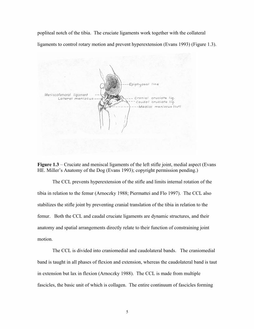
popliteal notch of the tibia. The cruciate ligaments work together with the collateral
ligaments to control rotary motion and prevent hyperextension (Evans 1993) (Figure 1.3).
Figure 1.3 – Cruciate and meniscal ligaments of the left stifle joint, medial aspect (Evans HE. Miller’s Anatomy of the Dog (Evans 1993); copyright permission pending.)
The CCL prevents hyperextension of the stifle and limits internal rotation of the
tibia in relation to the femur (Arnoczky 1988; Piermattei and Flo 1997). The CCL also
stabilizes the stifle joint by preventing cranial translation of the tibia in relation to the
femur. Both the CCL and caudal cruciate ligaments are dynamic structures, and their
anatomy and spatial arrangements directly relate to their function of constraining joint
motion.
The CCL is divided into craniomedial and caudolateral bands. The craniomedial
band is taught in all phases of flexion and extension, whereas the caudolateral band is taut
in extension but lax in flexion (Arnoczky 1988). The CCL is made from multiple
fascicles, the basic unit of which is collagen. The entire continuum of fascicles forming

6
the ligament is surrounded by paratenon, a connective tissue covering. The majority of
the blood supply to the CCL enters from the surrounding synovium (Arnoczky 1993).
Flexion and extension of the stifle occur about a transverse axis, whereas rotary
movements of the tibia about the femur occur around the longitudinal axis. Rotary
movement is controlled by the geometry of the condyles and ligamentous constraints
(Arnoczky et al. 1977; Arnoczky 1993). The cruciate ligaments twist upon each other
when the limb is flexed, thus limiting the amount of internal rotation. The cruciate
ligaments also provide for cranio-caudal stability of the stifle. In general the CCL
prevents cranial translation of the tibia, and the caudal cruciate ligament (CdCL) prevents
caudal translation of the tibia.
1.2.2 PATHOPHYSIOLOGY – Rupture of the CCL causes hindlimb lameness in the
dog. The CCL may rupture due to acute traumatic injury, but more commonly, as a non-
specific injury causing chronic lameness in middle-aged dogs. Physical and radiographic
evidence of degenerative joint disease is frequently present. Rupture of the CCL occurs
more commonly in large breed dogs than small breeds (Piermattei and Flo 1997). Acute
traumatic injury is more commonly seen in dogs less than 4 years of age, while the
syndrome of chronic lameness and degenerative joint disease is seen most frequently in
middle-age dogs (Piermattei and Flo 1997). CCL rupture has been shown to have a
higher prevalence in spayed female dogs. Ovariectomy in rats decreases elastin content
and may affect collagen metabolism. The effects of chronic hypoestrogenemia on the
metabolism and mechanical properties of the ligament in the dog is unknown (Vasseur
1993). The tensile strength of a dog’s cruciate ligament deteriorates with aging, and
these changes are more pronounced and occur at an earlier age in large breed dogs

7
compared to dogs weighing less than 15 kg (Vasseur et al. 1985). Cranial cruciate
ligament rupture may occur secondary to abnormal confirmation of the limb and
secondary to immune-mediated arthropathies affecting the stifle joint (Vasseur 1993).
Obesity has also been implicated (Vasseur 1993).
Partial ruptures of the CCL can also occur. Partial ruptures will progress to
complete ruptures, often in less than one year. Dogs commonly present with pain,
effusion, and degenerative joint disease similar to that observed in dogs with complete
ruptures (Vasseur 1993). Approximately 50% of dogs with CCL rupture will have
concurrent medial meniscal injury, and between 20-40 % of dogs will eventually rupture
the contralateral CCL (Moore and Read 1996).
Diagnosis of CCL rupture is based on a history of lameness and physical
examination findings. Presentation may vary between dogs presenting with acute
traumatic ruptures and those with chronic degenerative processes. Dogs with acute
traumatic rupture are severely lame and occasionally non-weight bearing on the affected
limb. Joint effusion may be present within several days of the injury. These dogs do not
exhibit obvious periarticular thickening. Dogs with chronic injuries have a variable
degree of lameness that may only be present only after exercise. Varying degrees of
muscle atrophy is evident and a prominent thickening of the medial aspect of the joint
(medial buttress) may be present. The patella tendon is often indistinguishable from the
surrounding tissues due to synovial effusion and periarticular thickening secondary to
degenerative joint disease. Crepitus may be palpated when the joint is put through a
range of motion (Vasseur 1993; Piermattei and Flo 1997).

8
The presence of cranial drawer motion or a cranial tibial thrust is pathognomonic
for CCL rupture (Slocum and Devine 1983; Vasseur 1993). Cranial drawer motion can
be detected with the dog standing or positioned in lateral recumbency. It is essential that
the examiner’s fingers be placed on the proper landmarks to elicit this motion. The
landmarks for palpation are the patella and lateral fabella on the femur, and the fibular
head and tibial tuberosity on the tibia. The test is performed repeatedly with the limb in
extension and in variable degrees of flexion. To elicit this motion the examiner places
cranial force upon tibia while holing the femur still with the other hand. If a CCL rupture
is present the tibia will shift forward in relation to the femur. With partial CCL ruptures
the craniomedial band is usually damaged leaving the caudolateral band intact. Since the
caudolateral band is taut in extension, cranial drawer motion may only be elicited with
the limb in variable degrees of flexion. In some dogs with partial ruptures or dogs with
chronic injuries, cranial drawer motion may not be detected due to stabilization of the
joint from fibrous tissue (Vasseur 1993). Cranial tibial thrust takes into account the
biomechanics of the stifle and includes the forces exerted by muscles during weight
bearing. Cranial tibial thrust is an active force created by weight bearing plus muscular
compression of the tibial plateau against the femoral condyles. Cranial tibial thrust is
dependent on the amount of compression as well as the slope of the tibial plateau
(Slocum and Devine 1983; Slocum and Slocum 1993). To evaluate cranial tibial thrust
the stifle is held in a slight degree of flexion, and the hock is flexed while placing axial
compression upon the tibia. The tibial tuberosity is palpated for cranial subluxation. The
cranial drawer test is thought to be more reliable and easier to perform (Vasseur 1993).

9
Radiographs of the affected stifle are of little diagnostic value but will document
the degree of osteoarthrosis (Piermattei and Flo 1997). Radiographic features consistent
with osteoarthrosis include the presence of osteophytes, and increase in synovial mass
and thinning of the infrapatellar fat pad. Radiographs of the affected stifle are important
to rule out the presence of concurrent diseases (neoplasia, erosive arthropathies), and to
measure the slope of the tibial plateau.
1.3 SURGICAL TECHNIQUES FOR DOGS WITH CRANIAL CRUCIATE LIGAMENT RUPTURE
Over the last 30 years, numerous techniques and modifications of them have been
described for stabilization of the stifle after CCL rupture. Techniques can be categorized
as; intracapsular, or extracapsular repairs, fibular head transposition, and corrective
osteotomies.
Currently the most popular techniques are extracapsular repairs and corrective
osteotomies. Although some surgeons continue to perform intracapsular repairs and
fibular head transposition, most have gone to a more simple extracapsular repair or
corrective osteotomies since reports show that the clinical outcome to be similar
regardless of the technique used (Jevens et al. 1996; Moore and Read 1996).
Biomechanical evaluation of the stability after several techniques showed fibular head
transposition was superior to various intracapsular and extracapsular repairs in regards to
immediate postoperative laxity and stiffness (Patterson et al. 1991). However, in follow-
up clinical examinations extracapsular techniques resulted in better joint stability and
limb function than fibular head transposition(Moore and Read 1996). Non-surgical
treatment consisting of 4-6 weeks of strict kennel confinement is a treatment option for
small dogs. Dogs weighing less than 15 kg had a satisfactory outcome after 3-6 weeks of

10
strict cage confinement, whereas dogs weighing greater than 15 kg were much less likely
to have a satisfactory outcome after a similar confinement period (Vasseur 1984).
Most authors’ site 85-90% clinical success after intracapsular and extracapsular
techniques (Shires et al. 1984; Moore and Read 1996; Piermattei and Flo 1997).
Similarly, anecdotal reports cite up to 95% of dogs undergoing corrective osteotomies
will have a good to excellent outcome (Slocum and Slocum 1993). It is difficult to find
comparisons of surgical techniques and clinical outcomes. Evaluations tend to rely
heavily upon client communications, and the techniques involved are not all inclusive
(Denny and Barr 1984; Shires et al. 1984; Metelman et al. 1995; Jevens et al. 1996;
Moore and Read 1996). Because there is no true “gold standard” surgical technique for
dogs after CCL rupture, studies tend to compare across technique categories rather than
between modifications within a technique. This being said, there is a consensus among
surgical specialists that dogs weighing greater than 15 kg will benefit from having
surgery, and it is estimated 85-95% of these dogs will have a good to excellent outcome
after surgery, with return to normal or near normal weight bearing function.
1.3.1 INTRACAPSULAR TECHNIQUES – A variety of intracapsular techniques and
modifications have been described. The principle of these techniques is to reconstruct the
CCL using an autograft, allograft, or synthetic material. Autografts harvested from the
hamstring, patellar tendon, fascia lata, or a combination of both are used most often
(Arnoczky et al. 1979; Hulse et al. 1980; Arnoczky et al. 1982; Thorson et al. 1989;
Lopez et al. 2003). Intracapsular techniques attempt to provide an anatomic or nearly
anatomic replacement for the damaged ligament. An in vitro examination several
intracapsular and extracapsular techniques indicated that intracapsular techniques resulted

11
closer to normal joint motion than extracapsular techniques (Arnoczky et al. 1977).
However, a later study showed that the instant center of motion in the stifle with
extracapsular suture placement techniques did not alter (Prostredny et al. 1991).
1.3.2 EXTRACAPSULAR TECHNIQUES – The first extracapsular techniques were
described by DeAngelis and Flo and variations of these are currently used (DeAngelis
and Lau 1970; Flo 1975). One or two strands of large monofilament nylon leader line is
passed around the lateral fabella, under the patellar tendon, through a hole drilled in the
tibial crest, and secured by a knot or crimp-clamp system. This technique is easy to apply
and provides consistent and satisfactory clinical results.
1.3.3 FIBULAR HEAD TRANSPOSITION – Fibular head transposition was
introduced as a surgical technique for the stifle after CCL rupture in 1985 (Smith and
Torg 1985). This technique moves the fibular head cranially with the lateral collateral
ligament attached, thus altering the orientation of the lateral collateral ligament and
preventing cranial drawer motion and internal rotation of the tibia. Clinical results after
this technique are similar to the aforementioned techniques, however, surgical expertise
is required. The mechanical stability of fibular head transposition immediately post-
operatively was shown to be superior to intracapsular and extracapsular techniques
(Patterson et al. 1991). This result prompted the recommendation to use this technique
for larger dogs that may place a high load on an intracapsular or extracapsular prosthesis.
1.3.4 CORRECTIVE OSTEOTOMIES – Two corrective osteotomies have been
described for the stifle after CCL rupture. Cranial tibial wedge osteotomies (TWO)
(Slocum and Devine 1984) and tibial plateau leveling osteotomy (TPLO) (Slocum and
Slocum 1993) have recently become popular for large dogs after anecdotal reports

12
suggested that large dogs did better after these techniques than others. Neither of these
techniques restore the stability of the stifle by mimicking the action of the CCL. Both
alter the joint biomechanics by changing the tibial plateau angle, thus eliminating cranial
tibial thrust. It has been proposed that cranial tibial thrust is the pathologic alteration of
motion that occurs after CCL rupture under normal weight bearing conditions. The
TPLO was shown to eliminate cranial tibial thrust in an in vitro study (Warzee et al.
2001). However, objective comparisons of the clinical outcomes after these techniques
and others is lacking. A great deal of controversy exists regarding TPLO since the
technique is patented. One must attend a course and become certified before performing
this technique.
1.4 BIOMECHANICS FOR TESTING BIOMATERIALS
Biomechanics is simply the application of mechanical engineering principles to
biologic systems to gain information on (1) the material and structural characteristics of
living tissues and biomaterials, (2) the impact of intrinsic and extrinsic physiologic and
nonphysiologic forces on a biological system, and (3) the influence of an object on a
biological system (Lucas et al. 1999).
Mechanics is the branch of physics and engineering that deals with forces and the
effects produced when forces are applied to objects. When a force acts on an object, that
object changes in some way. Force (force = mass X acceleration) is any agency acting
on an object that causes it to move, change its motion, or change its size or shape. Force
is commonly referred to as load, and when applied to a biologic system leads to
deformation or a change in shape (Radin et al. 1992; Burstein and Wright 1994; Lucas et
al. 1999). Isaac Newton is credited with the first correct statement of the laws governing

13
the events that occur when a force is applied to an object. Simply, these statements or
laws are the following: (1) A object at rest will stay at rest, (2) if a force is applied to an
object its velocity will change in proportion to that force, (3) if a force is applied to an
object and no acceleration occurs there must be an equal and opposite force acting on the
object. The basic unit of force is the Newton (N) where 1N = 0.225 X 1 lb (force) (Lucas et al. 1999).
To understand how a force affects a structure on which it acts one must have an
understanding of a basic load–deformation curve. The load–deformation curve evaluates
the behavior of a whole object and identifies several important structural characteristics
about an object. The most important characteristics that can be identified from a curve
are: (1) the load an object can withstand prior to failure, (2) the stiffness of the object or
its ability to resist a change in shape, (3) the amount of energy an object can absorb prior
to its failure (Figure 1.4).
Figure 1.4 – A theoretical load – deformation curve. Point (A) is the point when load is applied to an object, (B) is the yield point, (C) is the point where failure occurs.

14
If a load is applied in the elastic region (A-B) and then released, no permanent
deformation occurs. A load is applied past the yield point (B) into the non-elastic or
plastic region of the curve (B-C), permanent deformation will occur even if the load is
released. If loading is continued into the non-elastic region, an ultimate failure point is
reached (C). The area under the curve represents the amount of energy absorbed by the
object prior to failure. Stiffness (the ability of the object to resist a change in shape) is
calculated from the slope of the linear portion of the curve (Lucas et al. 1999).
To standardize representation of the mechanical behavior of a material (as
opposed to an object), it is necessary to normalize the load–deformation measurements
and eliminate the influence of geometry and dimensions. A stress–strain curve can be
use to evaluate a material and eliminates the influence of geometry and dimensions
(Figure 1.5).
Figure 1.5 – A theoretical stress – strain curve
For comparison of specimens of varying lengths deformation must be normalized
to the original specimen length. Strain is the normalization of deformation to the original

15
length. Thus, specimens of varying lengths can exhibit the same strain when a similar
load is applied. Strain had no units since it is normalized to length.
Stress is the normalization of applied force to the original cross-sectional area. It
can be thought of as the internal force intensity (force per unit area) at any given point or
plane. When evaluating the stress–strain curve the slope of the line relates the stress and
strain. The slope of the linear elastic region of the stress–strain curve is referred to as the
elastic modulus or modulus of elasticity. Modulus of elasticity is the material property
that relates stress to strain and is used to describe how stiff a material is in tension and
compression. The modulus of elasticity is known for a variety of biologic materials and
is used for comparisons. The unit for modulus of elasticity is the giga-pascal (GPa). For
example, the modulus of stainless steel is 200 GPa whereas, cortical bone is 20 GPa, thus
stainless steel is 10 times stiffer than cortical bone (Burstein and Wright 1994; Lucas et
al. 1999). Mechanical evaluation of materials can be complex and difficult to understand.
Most reports in the veterinary literature evaluate load–deformation or stress–strain.
Appropriate mechanical testing of biomaterials varies depending on the type of
material being tested. Some materials are affected more by their biologic environment.
And special considerations may apply when synthetic biomaterials are tested. For
example, implanted biomaterials are used at body temperature but testing is performed at
room temperature. Will the material behave in the same manner when exposed to body
temperature versus room temperature? One should attempt to mimic the environment the
material will be subjected to as close as possible. Although it would be ideal to test all
materials at body temperature while being exposed to body fluids, the chambers
necessary to complete testing of this nature can be prohibitively expensive.

16
The properties of metals are not severely affected by their environment.
However, the properties of polymeric materials may be. Polymers are sensitive to small
changes in their environment and are likely to interact with their surrounding
environment. The testing rate and frequency of polymeric materials is also important.
Time-dependent tests such as fatigue testing should be performed at a rate similar to what
the material will be subjected to in vivo (Lucas et al. 1999).
1.5 MECHANICAL EVALUATIONS OF EXTRACAPSULAR REPAIRS USING NYLON LEADER LINE
Most techniques used for CCL rupture attempt to eliminate cranial translation of
the tibia in relation to the femur, and prevent excessive internal rotation of the tibia.
Extracapsular techniques using a lateral fabella-tibial suture (LFS) of large diameter
nylon leader line (NLL) is well accepted and commonly performed (Caporn and Roe
1996). Studies have shown that NLL has the most appropriate characteristics (high force
at failure, minimal elongation, and high stiffness) for use in LFS stabilization of the
cruciate deficient stifle. However because of its memory, lower coefficient of friction,
and larger diameter compared to other materials commonly used, knot security may be a
concern (Caporn and Roe 1996; Lewis et al. 1997; Anderson et al. 1998; Nwadike and
Roe 1998; Huber et al. 1999). The LFS using NLL provides short term stabilization of
the stifle while periarticular fibrosis develops, resulting in long-lasting stifle stability
(Piermattei and Flo 1997). Before the use of NLL, surgeons employed large diameter,
commercially available suture material. The LFS, regardless of size or composition,
must withstand forces generated by cyclic loading during weight-bearing, tension, and
deformation generated by the formation of a knot, or produced by the application of a
crimp-clamp. The cyclic loading forces may cause the material may cause it to break or

17
loosen after surgery. If the LFS breaks or loosens before adequate periarticular fibrosis
has developed, stifle instability remains. Thus, material for use as a LFS is important
and has prompted many biomechanical evaluations in attempt to discern the best knot
method (Nwadike and Roe 1998; Huber et al. 1999), the most mechanically stable
material (Caporn and Roe 1996; Lewis et al. 1997; Huber et al. 1999; Sicard et al. 1999;
Sicard et al. 2002), alternatives to knot tying with the use of a crimp-clamp system
(Anderson et al. 1998; Peycke et al. 2002; Sicard et al. 2002), and the effects of
sterilization methods (Caporn and Roe 1996) on the biomechanical properties of NLL
and nylon fishing line (NFL) used in LFS stabilization.
A common problem during placement of the LFS technique is loss of tension in
the NLL during knot tying (Caporn and Roe 1996). Because of this problem variations of
knot types and fixation methods have been evaluated in the literature. Results vary, a
clamped square knot, square knot, slip knot, and crimp-clamp systems have all been
found to be acceptable methods of fixation of NLL (Anderson et al. 1998; Nwadike and
Roe 1998; Huber et al. 1999; Peycke et al. 2002; Sicard et al. 2002). It is known that the
structural properties of suture material are best preserved when a square knot is used
(Price 1948; Hermann 1971; Rosin and Robinson 1989). Tightening of the large
diameter NLL may result in stress concentration and weakening of the knot (Caporn and
Roe 1996).
Caporn and Roe (Caporn and Roe 1996) determined that the mechanical
properties of NLL were not significantly altered when steam or ethylene oxide
sterilization methods were used compared to unsterilized controls. This study also found
that the mechanical performance of NLL was superior to NFL for use as a LFS. Poorer

18
performance of 36 kg test NLL versus 27 kg test NLL during cycled testing was felt to be
the result of the inability to form a tight knot when using the larger material.
Huber and Egger (Huber et al. 1999) compared the mechanical characteristics of
several knot formations of large diameter monofilament materials. These authors
proposed that stiffness and yield are the most clinically relevant parameters for knot
assessment since the elongation knotted loops demonstrate prior to failure is to such a
degree that joint stability and or tissue coaptation in vivo would be compromised well
before this degree of elongation could actually occur. They concluded that clamping the
first throw of a square knot of NLL is preferable to maintain tension on the suture and
facilitate performing a tight knot. Clamping did not significantly alter the mechanical
properties and increased the structural stiffness of the material. They proposed that
clamping of the first throw creates a partial deformation under tension, thus allowing for
the formation of a more secure knot.
Knotting techniques using NLL require a minimum of 5 throws resulting in a
large, bulky knot that is irritating to the soft tissues (Caporn and Roe 1996). Some
knotting techniques also require an assistant for knot tensioning. The Securos clamping
system was designed to fasten two ends of the NLL together using a crimped metal tube
and can be performed without assistance. Loops secured with privately designed crimp-
clamps were evaluated using 27 kg test NLL and was shown to provide superior in vitro
mechanical performance compared to loops secured with clamped square knots
(Anderson et al. 1998). Two recent reports evaluated the 36 kg Securos system
(Peycke et al. 2002; Sicard et al. 2002). The first study evaluated the 36 kg Securos®
system and reported significantly less elongation occurred in crimped loops compared to

19
knotted samples, although these investigators found that the crimped loops had
significantly less tensile strength compared to knotted loops (Sicard et al. 2002).
However, the authors concluded that elongation may be more relevant than ultimate
tensile strength to the maintenance of joint stability, and should be the primary
consideration during suture material selection, and method of fixation for LFS security.
Another study evaluating the 36 kg Securos® system using both 27.3 kg test and 36.4 kg
test NLL against other knotted loops reported crimped loops performed as well or better
than traditional knotted loops (Peycke et al. 2002).
Despite limited mechanical data, the Securos system has achieved widespread
clinical use. There is no mechanical data reported for the 18 kg Securos system.
Biomechanical evaluation of this system should be tested against other accepted methods
before this system is used in vivo.
1.6 POLYVINYLIDENE FLUORIDE (FLUOROCARBON) AS AN ALTERNATIVE MATERIAL TO NYLON FOR EXTRACAPSULAR REPAIR
Recent reports have looked at the suitability of polyvinylidene fluoride (PVDF)
for use as a suture material (Urban et al. 1994; Laroche et al. 1995; Laroche et al. 1995;
Mary et al. 1998). PVDF has intrinsic elastic properties under tension which allow for
improved surgical handling and knot formation, and PVDF shows excellent
biocompatibility (Laroche et al. 1995). In a long-term in vivo implantation model in the
dog, PVDF was more stable and retained its surface properties better than polypropylene.
The PVDF is available as fluorocarbon fishing leader material and fishing line. It is
manufactured in tensile strengths up to 200 pounds. This material may be an alternative
to NLL for LFS stabilization because of its better surgical handling characteristics,
improved knot strength, smaller diameter for a given tensile strength, and decreased

20
absorption of water under physiologic conditions. A smaller diameter material will form
a smaller knot for a given tensile strength which would be less irritating to the soft
tissues.
1.7 REFERENCES
Anderson, C.C., Tomlinson, J.L., et al. (1998). "Biomechanical evaluation of a crimp clamp system for loop fixation of monofilament nylon leader material used for stabilization of the canine stifle joint." Vet Surg 27(6): 533-539.
Arnoczky, S.P. (1988). "The cruciate ligaments: The enigma of the canine stifle." J Small
Anim Pract 29: 71-90. Arnoczky, S.P. (1993). Pathomechanics of cruciate ligament and meniscal injuries.
Disease Mechanisms in Small Animal Surgery. M. J. Bojrab. Philadelphia, Lippincott Williams & Wilkins: 764-776.
Arnoczky, S.P., Tarvin, G.B., et al. (1982). "Anterior cruciate ligament replacement using
patellar tendon - an evaluation of graft revascularization in the dog." J Bone Joint Surg 64: 217-224.
Arnoczky, S.P., Tarvin, G.B., et al. (1979). "The over-the-top proceedure: A technique
for anterior cruciate ligament substitution in the dog." J Am Anim Hosp Assoc 15: 283-290.
Arnoczky, S.P., Torzilli, P.A., et al. (1977). "Biomechanical evaluation of anterior
cruciate ligament repair in the dog: An analysis of the instant center of motion." J Am Anim Hosp Assoc 13: 553-558.
Burstein, A.H. and Wright, T.M. (1994). Fundamentals of Orthopaedic Biomechanics.
Baltimore, Williams & Wilkins. Caporn, T.M. and Roe, S.C. (1996). "Biomechanical evaluation of the suitability of
monofilament nylon fishing and leader line for extra-articular stabilization of the canine cruciate ligament deficient stifle." Vet Comp Orthop Traumatol 9: 126-133.
DeAngelis, M. and Lau, R.E. (1970). "A lateral retinacular imbrication technique for the
surgical correction of anterior cruciate liament rupture in the dog." J Am Vet Med Assoc 157: 79-84.
Denny, H.R. and Barr, A.R. (1984). "An evaluation of two "over the top" techniques for
anterior cruciate ligament replacement in the dog." J Small Anim Pract 25: 759-769.

21
Evans, H.E. (1993). Miller's Anatomy of the Dog. Philidelphia, W.B. Saunders. Flo, G.C. (1975). "Modification of the lateral retinacular imbrication technique for
stabilizing cruciate ligament injuries." J Am Anim Hosp Assoc 11: 570-576. Hermann, J.B. (1971). "Tensile strength and knot security of surgical suture materials."
Am Surg 37: 209-217. Huber, D.J., Egger, E.L., et al. (1999). "The effect of knotting method on the structural
properties of large diameter nonabsorbable monofilament sutures." Vet Surg 28: 260-267.
Hulse, D.S., Michaelson, F., et al. (1980). "A technique for reconstruction of the anterior
cruciate ligament in the dog: Preliminary report." Vet Surg 9(135-140). Jevens, D.J., DeCamp, C.E., et al. (1996). "Use of force-plate analysis of gait to compare
two surgical techniques for treeatment of cranial cruciate ligament rupture in dogs." Am J Vet Res 57(3): 389-393.
Laroche, G., Marois, Y., et al. (1995). "Polyvinylidene fluoride (PVDF) as a biomaterial:
From polymeric raw material to monofilament vascular suture." J Biomed Mater Res 29(12): 1525-36.
Laroche, G., Marois, Y., et al. (1995). "Polyvinylidene fluoride monofilament sutures:
Can they be used safely for long-term anastomoses in the thoracic aorta?" Artif Organs 19(11): 1190-9.
Lewis, D.D., Milthorpe, B.K., et al. (1997). "Mechanical comparison of materials used
for extracapsular stabliization of the stifle joint in dogs." Aust Vet J 75: 890-896. Lopez, M.J., Markel, M.D., et al. (2003). "Hamstring graft technique for stabilization of
canine cranial cruciate ligament deficient stifles." Vet Surg 32: 390-401. Lucas, G.L., Cooke, F.W., et al. (1999). A Primer of Biomechanics. New York, Springer. Mary, C., Marois, Y., et al. (1998). "Comparison of the in vivo behavior of
polyvinylidene fluoride and polypropylene sutures used in vascular surgery." ASAIO J 44(3): 199-206.
McKee, W.M. and Miller, A. (1999). "A self-locking knot for lateral fabellotibial suture
stabilization of the cranial cruciate ligament deficient stifle in the dog." Vet Comp Orthop Traumatol 12: 78-80.
Metelman, L.A., Schwartz, P.D., et al. (1995). "An evaluation of three different cranial
cruciate ligament stabilization procedures as they relate to postoperative meniscal injuries." Vet Comp Orthop Traumatol 8: 118-123.

22
Moore, K.W. and Read, R.A. (1996). "Cranial cruciate ligament rupture in the dog - a retrospective study comparing surgical techniques." Aust Vet J 72: 281-285.
Nwadike, B.S. and Roe, S.C. (1998). "Mechanical comparison of suture material and
knot type used for fabello-tibial sutures." Vet Comp Orthop Traumatol 11: 47-52. Patterson, R.H., Smith, G.K., et al. (1991). "Biomechanical stability of four cranial
cruciate ligament repair techniques in the dog." Vet Surg 20(2): 85-90. Peycke, L.E., Kerwin, S.C., et al. (2002). "Mechanical comparison of six loop fixation
methods with monofilament nylon leader line." Vet Comp Orthop Traumatol 4: 210-214.
Piermattei, D.L. and Flo, G.L. (1997). Brinker, Piermattei, and Flo's Handbook of Small
Animal Orthopedics and Fracture Repair. Philadelphia, PA, W.B. Saunders: 516-580.
Price, P.B. (1948). "Stress, strain, and sutures." Ann Surg 128: 408-421. Prostredny, J.M., Bauer, M.S., et al. (1991). "Effects of suture type on stifle joint
biomechanics after extra-articular repair of cranial cruciate ligament transection in the dog." Vet Comp Orthop Traumatol 4: 144-149.
Radin, E.L., Rose, R.M., et al. (1992). Practical Biomechanics for the Orthopedic
Surgeon. New York, Churchill Livingstone Inc. Rosin, E. and Robinson, G.M. (1989). "Knot security of suture materials." Vet Surg
18(4): 269-273. Shires, P.K., Hulse, D.S., et al. (1984). "The under-and-over fascial replacement
technique for anterior cruciate ligament rupture in dogs: A retrospective study." J Am Anim Hosp Assoc 20: 69-77.
Sicard, G.K., Hayashi, K., et al. (2002). "Evaluation of 5 types of fishing material, 2
sterilization methods, and a crimp-clamp system for extra-articular stabilization of the canine stifle joint." Vet Surg 31: 78-94.
Sicard, G.K., Meinen, J., et al. (1999). "Comparison of fishing line for repair of the
cruciate deficient stifle." Vet Comp Orthop Traumatol 44: 138-141. Slocum, B. and Devine, T. (1983). "Cranial tibial thrust: A primary force in the canine
stifle." J Am Vet Med Assoc 183(4): 456-9. Slocum, B. and Devine, T. (1984). "Cranial tibial wedge osteotomy: A technique for
eliminating cranial tibial thrust in cranial cruciate ligament repair." J Am Vet Med Assoc 184: 564-569.

23
Slocum, B. and Slocum, T.D. (1993). "Tibial plateau leveling osteotomy for repair of cranial cruciate ligament rupture in the canine." Vet Clin North Am Small Anim Pract 23(4): 777-95.
Smith, G.K. and Torg, J.S. (1985). "Fibular head transposition for repair of cruciate-
deficient stifle in the dog." J Am Vet Med Assoc 187: 375-383. Thorson, E., Rodrigo, J.J., et al. (1989). "Replacement of the anterior cruciate ligament -
a comparison of autografts and allografts in dogs." Acta Orthop Scand 60: 555-560.
Urban, E., King, M.W., et al. (1994). "Why make monofilament sutures out of
polyvinylidene fluoride?" ASAIO J 40(2): 145-56. Vasseur, P.B. (1984). "Clinical results following nonoperative management for rupture of
the cranial cruciate ligament in dogs." Vet Surg 13: 243-246. Vasseur, P.B. (1993). Stifle joint. Textbook of Small Animal Surgery. D. Slatter.
Philidelphia, W.B. Saunders. 2: 1817-1865. Vasseur, P.B., Pool, R.R., et al. (1985). "Correlative biomechanical and histologic study
of the cranial cruciate ligament in dogs." Am J Vet Res 46: 1842-1854. Warzee, C.C., Dejardin, L.M., et al. (2001). "Effect of tibial plateau leveling on cranial
and caudal tibial thrusts in canine cranial cruciate-deficient stifles: An in vitro experimental study." Vet Surg 30(3): 278-86.

24
CHAPTER 2. IN VITRO EVALUATION OF THE 18 KG AND 36 KG SECUROS CRANIAL CRUCIATE LIGAMENT REPAIR SYSTEMTM

25
2.1 INTRODUCTION
Cranial cruciate ligament (CCL) rupture is a common injury causing stifle
instability and secondary degenerative joint disease. The CCL prevents hyperextension
of the stifle and limits internal rotation of the tibia in relation to the femur (Arnoczky
1988; Piermattei and Flo 1997). Surgical stabilization of the stifle after CCL rupture is
recommended for dogs weighing more than 15 kg (Vasseur 1984).
Many surgical techniques for stabilization of the stifle after CCL rupture have
been described (Flo 1975; Shires et al. 1984; Smith and Torg 1985; Slocum and Slocum
1993). Techniques can be classified as intracapsular, extracapsular, fibular head
transposition, and corrective osteotomies. Extracapsular repair with a lateral fabella-
tibial suture (LFS) of large diameter monofilament nylon leader line (NLL) is a well
accepted and commonly performed technique (Caporn and Roe 1996). The LFS provides
short-term stabilization of the stifle while periarticular fibrosis develops, which causes
long-term stifle stability (Piermattei and Flo 1997).
Extracapsular stabilization using a LFS requires a strong suture material that
minimizes bacterial adherence and has minimal plastic deformation (Nwadkie and Roe
1998). This material must withstand cyclic loading and tension. In addition, it must
withstand deformation during knot tying, or during application of a crimp-clamp.
Loading and tension on the material may lead to excessive elongation or disruption of the
LFS in vivo prior to the formation of adequate periarticular fibrosis. If this occurs, stifle
instability remains.
Evaluations of various suture materials, knot types, and other methods of fixation
of the suture have attempted to discern the best knot or fixation method (Nwadkie and

26
Roe 1998; Huber et al. 1999), the most mechanically suitable material (Caporn and Roe
1996; Lewis et al. 1997; Huber et al. 1999; Sicard et al. 1999; Sicard et al. 2002), and the
effect of sterilization methods (Caporn and Roe 1996; Sicard et al. 2002) on the
mechanical properties of NLL and nylon fishing line (NFL).
The Securos Cranial Cruciate Ligament Repair SystemTM was designed to fasten
two ends of NLL together using a crimped metal tube. A tensioning device provided by
the manufacturer is used to tension the loop and fasten the crimp-clamp without
assistance. This system eliminates the bulky knot formed with knotting techniques. Two
systems are available for 18 kg test (kgt) and 36 kgt NLL. A similar crimp-clamp system
for 27 kgt NLL demonstrated superior in vitro mechanical performance compared to
clamped square knots using 27 kgt NLL, resulting in loops that had a higher force at
failure and less elongation (Anderson et al. 1998). The 36 kg Securos crimp-clamp
system resulted in loops with significantly less strength and less elongation than knotted
samples for 36 kgt NLL (Sicard et al. 2002). In a second study, the 36 kg Securos
crimp-clamp system on both 27.3 kgt and 36.4 kgt NLL resulted in loops that performed
similar to or had superior mechanical performance than knotted loops (Peycke et al.
2002). Despite limited data on mechanical testing of the 36 kg system, and on the 18 kg
system, the Securos crimp-clamp system has achieved widespread clinical use.
This study evaluated the force at failure, loop elongation, and stiffness of
unsterilized NLL loops secured with the 18 kg and 36 kg Securos Cranial Cruciate
Ligament Repair SystemTM compared to a clamped square knot technique. Eighteen, 27
and 36 kgt NLL was used under non-cycled and cycled testing conditions.

27
2.2 MATERIALS AND METHODS
The materials tested were 18.2, 27.3, and 36.4 kgt monofilament NLLa,b. Eighty
strands of each material were cut to a length of 40 cm. Forty strands of each size were
secured with: 1) Square knot clamped after the first throw with a total of five throws
(CSK), and 2) Securos crimp-clamp system (SCC)c.
2.2.1 LOOP ASSEMBLY – Loops were fixed by the same surgeon (MNB) around a
customized 42.42 mm outer diameter polyvinyl-chloride pipe to ensure a consistent sized
loop. The pipe was customized for the application of the crimping device or needle
holders by cutting a 1.5 cm X 5 cm slot out of each end of the pipe. The edges of the slot
were covered with tape to prevent damage to the NLL during loop fixation. The ends of
all loops were cut 3 mm from the knot or clamp. All strands of NLL were passed through
room temperature 0.9% NaCl solutiond at room temperature prior to loop formation.
Clamped square knots were formed by clamping the first throw of the knot with modified
7 inch Mayo-Hegar needle holding forcepse (modified by filing the surface of the jaws so
they were completely smooth) closed to the third lock. The Securos® crimp-clamp
system was used according to the manufacturer’s recommendationsc. Both strands of the
NLL were passed through the primary crimp tube. Secondary crimp tubes were placed
10 mm from the primary tube on each tag end and crimped twice. The tensioning device
was then placed to distract the secondary clamps to a predetermined point on its locking
mechanism. The primary crimp tube was then clamped three times with the crimping
tool designed for this system. The 36 kg Securos system was used for both the 27.3
and 36.4 kgt SCC samples.

28
2.2.2 MECHANICAL TESTING – One group of 27 kgt NLL loops was tested at 40º C.
These samples were incubated in the mechanical testing machine chamber for 20 minutes
at 40° C prior to testing. For all other groups testing was performed at room temperature
(20° C). All loops were mounted onto an IPC Digital Servo Machinef between two
horizontally-oriented polished steel hooks set 56 mm apart.
For the non-cycled testing, tension was applied at a constant distraction rate of
500 mm/min until the loops failed by breaking or slipping. Force at failure, elongation,
and stiffness were recorded and compared across sizes of NLL and fixation method. The
stiffness measurement reported was the maximum recorded value obtained from the
linear portion of the load vs. elongation curve for each trial.
For the cycled testing, tension was applied at a constant distraction rate of 500
mm/min to a distraction limit of 7.5 mm for 49 cycles. The 18.2 kgt NLL samples were
not able to complete this cycled protocol. The protocol was then altered so the 18 kgt
loops were tested at a distraction limit of 6.0 mm. The loops were tensioned at a constant
distraction rate of 500 mm/min until the loops failed by breaking or slipping.
Calculations to determine force at failure, elongation, and stiffness were collected from
the final load to failure cycle for each trial. Force at failure, elongation, and stiffness
were recorded and compared across sizes of NLL and fixation method. The stiffness
measurement reported was the maximum recorded value obtained from the linear portion
of the load vs. elongation curve for each trial.
2.2.3 STATISTICAL METHODS – The force at failure, elongation, and stiffness were
considered continuous and evaluated for normality using the Shapiro-Wilk test with the
null hypothesis of normality rejected at p<0.05. Non-normal data was transformed. All

29
data was summarized as mean +/- SEM. The data for non-cycled and cycled loops was
analyzed separately using a two-way analysis of variance to evaluate the effect of loop
fixation method (Securos® vs. clamped square knot), and NLL size (18, 27 vs. 36 kgt)
on force to failure, elongation and stiffness. Where there were significant interaction
effects at p<0.05, pre-planned comparisons were made using the Scheffe’s procedure
maintaining type I error at 0.05. Thus, when specified, significant differences are
established at p < 0.05. For comparisons of the force at failure for noncycled vs. cycled
loops, the data was analyzed using a three-way analysis of variance to evaluate the effect
of loop fixation method (Securos® vs. clamped square knot), NLL size (18, 27 vs. 36
kgt) and cycling (non-cyled vs. cycled). Where there were significant interaction effects
at p<0.05, pre-planned comparisons were made using the Scheffe’s procedure
maintaining type I error at 0.05. Thus, when specified, significant differences are
established at p < 0.05. PROC UNIVARIATE and PROC ANOVAg (SAS v 8.2, SAS
Institute, Cary, NC) were used for all analyses.
2.3 RESULTS
2.3.1 NON-CYCLED TESTING – All of the loops failed at a portion originally
contained within the knot or crimp-clamp. All the 27.3 and 36.4 kgt SCC non-cycled
loops failed within the clamp. Conversely, all 18.2 kgt SCC non-cycled loops pulled
through the clamp rather that the NLL breaking.
The force at failure was significantly greater in all SCC loops than the CSK loops,
except for the 18.2 kgt group where the CSK was significantly greater (Table 1). The
force at failure was significantly greater in CSK loops than the CSK40°C loops for the
27.3 kgt NLL. The 36.4 kgt loops were significantly stronger (force at failure) than the

30
27.3 kgt loops, which were stronger than the 18.2 kgt loops. The CSK loops showed
greater elongation than the SCC loops within each size group of NLL. There was no
significant difference in elongation between CSK loops and CSK40°C loops for the 27.3
kgt NLL. The 36.4 kgt loops had significantly greater elongation than the 27.3 kgt loops.
Stiffness was not significantly different between SCC and CSK 18.2 kgt loops. The SCC
loops had significantly greater stiffness than CSK and CSK40°C loops for 27.3 kgt and
the CSK for 36.4 kgt NLL. Stiffness was not significantly different between CSK and
CSK40°C loops for 27.3 kgt or 36.4 kgt NLL groups or between SCC loops for 27.3 and
36.4 kgt NLL.
Table 2.1. Mean (SEM) force at failure, elongation, and stiffness for non-cycled loops formed with a clamped square knot (CSK) or the Securos® crimp clamp system (SCC) using monofilament nylon leader line. The coefficient of variation (CV = mean/SD X 100) of the force and elongation is also reported. Within each nylon leader line size, measurements with the same superscript are not significantly different. For comparisons between 27.3 kg and 36.4 kg nylon leader line, means with the same superscript are not significantly different. Note that type I error was maintained at 0.05 for all comparisons, hence where significant, p≤ 0.05. Knot Method Force CV Elongation CV Stiffness (N) (%) (mm) (%) (N/mm) 18.2 kg (40 lb) CSK a265.5 (3.6) 6.1 a11.5 (0.2) 9.7 a34.0 (1.7) SCC b132.9 (8.8) 29.6 b6.9 (0.3) 18.3 a32.0 (1.0)
27.3 kg (60 lb) CSK40°C o358.4 (6.0) 7.4 p14.0 (0.3) 10.9 p27.7 (0.7) CSK p387.7 (4.5) 5.2 p14.3 (0.4) 11.2 p28.6 (0.6) SCC q416.4 (8.8) 9.5 q12.3 (0.3) 11.5 q38.7 (0.4) 36.4 kg (80 lb) CSK r498.1 (3.6) 3.3 r18.7 (0.2) 4.6 p28.5 (0.6) SCC s522.0 (9.4) 8 s15.2 (0.2) 6.2 q38.6 (0.3)

31
2.3.2 CYCLED TESTING – All of the loops failed at a portion originally contained
within the knot or crimp-clamp. All of the 36.4 kgt SCC and CSK cycled samples
completed the cycled protocol. One 27.3 kgt CSK cycled sample failed by breaking prior
to completion of cycled protocol. Two 27.3 kgt SCC cycled samples failed by breaking
prior to completion of the cycled protocol.
The original protocol for cycled testing had to be altered due to the inability of the
18.2 kgt SCC samples to complete the protocol. The distraction limit for the cycles was
then reduced to 6 mm. Two 18.2 kgt CSK loops failed by breaking prior to completion
of the revised cycled protocol. Two 18.2 kgt SCC cycled samples failed by pulling
through the clamp prior to completion of the revised cycled protocol. All of the 18.2 kgt
SCC loops continued to pull through the clamp rather than the NLL breaking within the
clamp.
The SCC loops were significantly stronger (force at failure) than the CSK loops
for 36.4 kgt NLL (Table 2.2). The CSK loops were significantly stronger than the SCC
loops for 27.3 kgt NLL. Similarly, the 36.4 kgt NLL loops were significantly stronger
than the 27.3 kgt NLL loops. The strength of SCC loops and CSK loops were not
different for 18.2 kgt NLL.
The SCC loops showed significantly greater elongation than the CSK loops for
36.4 kgt NLL. The CSK loops showed significantly greater elongation than the SCC
loops for 27.3 kgt NLL. The 36.4 kgt SCC loops had significantly greater elongation
than all 36.4 kgt NLL and 27.3 kgt NLL loops. The CSK loops showed no difference in

32
elongation for 36.4 kgt and 27.3 kgt NLL. The SCC loops and CSK loops showed no
difference in elongation for 18.2 kgt NLL.
The SCC loops showed significantly greater stiffness than CSK loops for 36.4 kgt
NLL. The SCC loops showed significantly greater stiffness than CSK loops for 27.3 kgt
NLL. The 36.4 kgt SCC loops showed significantly greater stiffness than 27.3 kgt SCC
loops, which were significantly stiffer than 36.4 kgt CSK loops, which were significantly
stiffer than 27.3 kgt CSK loops. The SCC loops showed significantly greater stiffness
than CSK loops for 18.2 kgt NLL.
Table 2.2. Mean (SEM) force at failure, elongation, and stiffness for cycled loops formed with a clamped square knot (CSK) or the Securos® crimp clamp system (SCC) using monofilament nylon leader line. The coefficient of variation (CV = mean/SD X 100) of the force and elongation is also reported. Within each nylon leader line size, means with the same superscript are not significantly different. For comparisons between 27.3 kg and 36.4 kg nylon leader line, means with the same superscript are not significantly different. Note that type I error was maintained at 0.05 for all comparisons, hence where significant, p≤ 0.05. Knot Method Force CV Elongation CV Stiffness (N) (%) (mm) (%) (N/mm)
18.2 kg (40 lb) CSK-C a268.1 (3.2) 5.4 a8.2 (0.2) 13.2 a37.0 (0.6) SCC-C a286.9 (11.9) 17.6 a7.6 (0.3) 18.3 b45.7 (0.6) 27.3 kg (60 lb) CSK-C p386.8 (4.5) 5.0 p9.1 (0.2) 8.2 p48.3 (0.5) SCC-C q359.9 (17.3) 20.4 q6.7 (0.3) 18.6 q58.1 (1.3) 36.4 kg (80 lb) CSK-C r445.3 (5.2) 5.2 p9.5 (0.2) 11.1 r53.8 (0.6) SCC-C s564.5 (6.6) 5.2 r10.8 (0.4) 16.6 s68.8 (0.7)

33
A comparison between non-cycled loops vs. cycled loops showed SCC cycled
loops were significantly stronger than SCC non-cycled loops, which were significantly
stronger than CSK non-cycled loops, which were significantly stronger than the CSK
cycled loops for the 36.4 kgt NLL (Table 2.3). The SCC non-cycled loops were
significantly stronger than CSK non-cycled and cycled loops, which were significantly
stronger than the SCC cycled loops for the 27.3 kgt NLL. The SCC cycled loops and
CSK cycled and non-cycled loops were significantly stronger than the SCC non-cycled
loops for 18.2 kgt NLL.
Table 2.3. Mean (SEM) force at failure for non-cycled loops and cycled loops (+C) formed with a clamped square knot (CSK) or the Securos® crimp clamp system (SCC) using monofilament nylon leader line. Within each nylon leader line size, means with the same superscript are not significantly different. For comparisons between 27.3 kg and 36.4 kg nylon leader line, means with the same superscript are not significantly different. Note that type I error was maintained at 0.05 for all comparisons, hence where significant, p≤ 0.05. Knot Method Force
Knot Method Force
Knot Method Force
(N) (N) (N)
18.2 kg (40 lb) 27.3 kg (60 lb) 36.4 kg (80 lb)
CSK a265.5 (3.6) CSK p387.7 (4.5) CSK s498.1 (3.6)
CSK+C a268.1 (3.2) CSK+C p386.8 (4.5) CSK+C t445.3 (5.2)
SCC b132.9 (8.8) SCC q416.4 (8.8) SCC u522.0 (9.4)
SCC+C a286.9 (11.9) SCC+C r359.9 (17.3) SCC+C v564.5 (6.6)
2.4 DISCUSSION
The Securos Cranial Cruciate Ligament Repair SystemTM has advantages over
knotting methods for NLL loop fixation by eliminating the bulky knot, and not requiring
assistance. Crimp-clamp systems manufactured by Securos, Inc, are for use with 18.2 kgt

34
NLL and for use with 36.4 kgt NLL. We also evaluated 27.3 kgt NLL secured with the
36.4 kg Securos® crimp-clamp system for comparison to other studies (Anderson et al.
1998; Peycke et al. 2002). The results of our study support the use of the 36 kg Securos®
crimp-clamp system for both 27.3 and 36.4 kgt NLL. In both non-cycled and cycled
testing the SCC loops formed with 27.3 kgt or 36.4 kgt NLL performed as well or better
than a clamped square knot resulting in loops that were strong, undergoing minimal
elongation, and having high stiffness.
The 18.2 kg Securos® crimp-clamp system appears to be unsatisfactory. The
Securos® crimp-clamp system designed for use with 18.2 kgt NLL did not perform as
well mechanically in non-cycled testing as did clamped square knots for loops of the
same size NLL. All 18.2 kgt NLL loops secured with the Securos® crimp-clamps failed
by pulling through the crimp-clamps rather than the NLL breaking within the clamp.
Considerable variability was observed in the force at failure and elongation reflected by
coefficient of variations of 29.6% and 18.3%, respectfully. In cycled testing, the 18.2 kgt
SCC loops failed prior to completion of the original cycled protocol by pulling through
the clamp. Once the displacement was reduced, most 18.2 kgt SCC samples completed
the cycled protocol, but still ultimately failed by pulling through the clamp. These
findings raise concern about a design flaw with the original crimp tube diameter (pre-
clamping), or that the crimping instrument used with this system may not crimp the tube
to the appropriate diameter.
In contrast the 27.3 kgt and 36.4 kgt NLL secured with the 36 kg Securos®
crimp-clamp system had superior to or equivalent mechanical performance than CSK
loops of the same kilogram-test NLL resulting in loops that had a high force at failure,

35
minimal elongation and high stiffness. The 36 kg Securos® crimp-clamps are not
designed for use with the 27.3 kgt NLL. During cycled testing, two of the 27.3 kgt SCC
loops failed prior to completion of the protocol. However, these loops failed by the NLL
breaking within the clamp and not pulling through the clamp as was observed with the
18.2 kgt SCC loops.
Two available reports on the use of the 36 kg Securos® crimp-clamp system had
differing results (Peycke et al. 2002; Sicard et al. 2002). The first study (Sicard et al.
2002) reported SCC loops of 36.4 kgt NLL were significantly weaker than knotted
samples and exhibited significantly less elongation. This study differed from ours by
comparing to a control loop formed by a square knot with five throws (not clamped), and
a cycled testing protocol with a final loading rate of 1000 mm/min until failure. Stiffness
of the loops was not reported.
The second study (Peycke et al. 2002) used 36 kg Securos® crimp-clamp system
for both 27.3 kgt and 36.4 kgt NLL. This study utilized the same non-cycled testing
protocol as our study and reported that SCC loops of 27.3 kgt and 36.4 kgt NLL secured
with the Securos® crimp-clamps were as strong or stronger and showed similar to less
elongation than CSK using large diameter NLL.
The cycled testing protocol used in this study was derived from the results of non-
cycled testing. Load vs. elongation curves were evaluated to determine the distraction
limit that represented approximately 50% of the failure load for the loops. It was difficult
to determine the 50% failure load for the 18.2 kgt loops due to the variability of the 18.2
kgt SCC samples. A distraction limit of 7.5 mm was determined based upon the above
observations and used initially for all loops for consistency. The protocol was changed to

36
6.0 mm for the 18.2 kgt loops due to the inability of the 18.2 kgt SCC samples to
withstand the protocol. The results obtained for the cycled testing were taken from the
last load to failure cycle for each loop. The force at failure measurements should be an
accurate representation of the materials properties when subjected to repeated loads.
However, these measurements then do not account for material elongation that may have
occurred during cycling, thus the ability to draw conclusions from the elongation and
stiffness values obtained from the cycled testing may be limited.
Polymeric materials are sensitive to temperature, environment, and rate of testing.
These factors may have a significant effect upon the results of mechanical testing.
However, mechanical testing chambers that contain bodily fluids and can be temperature
regulated are very expensive. Polymers such as NLL are sensitive to small temperature
changes and are much more likely to interact with their environment than metals.
Polymers are more likely to absorb water and other molecules in the surrounding fluid
(Lucas et al. 1999). Previous studies evaluating NLL have performed all mechanical
testing at room temperature. One group of 27.3 kgt NLL (CSK40°C) was tested at 40° C
to determine the effect of temperature on the mechanical behavior of NLL. The force at
failure was significantly lower in the loops tested at 40° C compared to loops of the same
size NLL tested at room temperature. There were no significant differences in elongation
and stiffness between the two groups.
Crimp clamps allow fixation of a loop by generating friction between the surface
of the clamp and the material being fastened. Clamp placement causes a local
deformation of the material that occurs at the clamp-material interface. This induces a
bending moment in the NLL near the clamp. Local deformation and bending moments

37
also occur when the NLL is knotted making this portion of the loops the weakest
(Anderson et al. 1998). Crimp-clamp systems for fixation of NLL loops used for LFS
were proposed to eliminate elongation of the loop during knot slippage of large diameter
NLL. A study using a novel crimp-clamp system for 27.3 kgt NLL found significantly
less elongation and greater load to failure in crimped loops compared to knotted loops
(Anderson et al. 1998). These findings are similar to our results, however, the crimp-
clamp system used in that study was a novel design and not commercially available.
A clamped square knot with a total of five throws was used as a control in our
study similar to previous reports (Caporn and Roe 1996; Lewis et al. 1997; Huber et al.
1999). A load to failure rate of 500 mm/min was also based on previous work with NLL
(Caporn and Roe 1996; Anderson et al. 1998; Peycke et al. 2002). Unsterilized NLL was
used for all samples. The effects of sterilization on material properties of NLL have been
extensively examined (Caporn and Roe 1996; Lewis et al. 1997; Anderson et al. 1998;
Peycke et al. 2002; Sicard et al. 2002) and have found conflicting results for steam vs.
ethylene oxide sterilization protocols on the mechanical properties of NLL.
We conclude that the Securos Cranial Cruciate Ligament Repair System™ is an
acceptable method for fixation of NLL loops used for LFS. This study supports the use
of the 36 kg Securos® crimp-clamps with both 27.3 and 36.4 kgt NLL. However, use of
the 18.2 kg Securos® crimp-clamp system cannot be recommended. The significantly
less force required for failure in non-cycled testing, the high variability, and the tendency
to pull through the clamps rather than the NLL breaking suggest that the crimp tube
diameter or the crimping device may be inappropriate for use with 18.2 kgt NLL.

38
2.5 END NOTES
a Mason Hard Type Leader Material; Mason Tackle Company., Otisville, MI, USA
b Labeled as 40, 60, and 80 pound test, respectively
c Securos, Inc., E. Brookfield, MA 01515, USA
d Abbott Labs, N. Chicago, IL 60064, USA
e Weck Instruments, KMedic, 190 Veterans Drive, Northvale, NJ, USA
f Industrial Process Controls, Melbourne, Australia
g SAS v 8.2, SAS Institute, Cary, NC, USA
2.6 REFERENCES
Anderson, C.C., Tomlinson, J.L., et al. (1998). "Biomechanical evaluation of a crimp clamp system for loop fixation of monofilament nylon leader material used for stabilization of the canine stifle joint." Vet Surg 27(6): 533-539.
Arnoczky, S.P. (1988). "The cruciate ligaments: The enigma of the canine stifle." J Small
Anim Pract 29: 71-90. Caporn, T.M. and Roe, S.C. (1996). "Biomechanical evaluation of the suitability of
monofilament nylon fishing and leader line for extra-articular stabilization of the canine cruciate ligament deficient stifle." Vet Comp Orthop Traumatol 9: 126-133.
Flo, G.C. (1975). "Modification of the lateral retinacular imbrication technique for
stabilizing cruciate ligament injuries." J Am Anim Hosp Assoc 11: 570-576. Huber, D.J., Egger, E.L., et al. (1999). "The effect of knotting method on the structural
properties of large diameter nonabsorbable monofilament sutures." Vet Surg 28: 260-267.
Lewis, D.D., Milthorpe, B.K., et al. (1997). "Mechanical comparison of materials used
for extracapsular stabliization of the stifle joint in dogs." Aust Vet J 75: 890-896. Lucas, G.L., Cooke, F.W., et al. (1999). A Primer of Biomechanics. New York, Springer. Nwadike, B.S. and Roe, S.C. (1998). "Mechanical comparison of suture material and
knot type used for fabello-tibial sutures." Vet Comp Orthop Traumatol 11: 47-52.

39
Peycke, L.E., Kerwin, S.C., et al. (2002). "Mechanical comparison of six loop fixation methods with monofilament nylon leader line." Vet Comp Orthop Traumatol 4: 210-214.
Piermattei, D.L. and Flo, G.L. (1997). Brinker, Piermattei, and Flo's Handbook of Small
Animal Orthopedics and Fracture Repair. Philadelphia, PA, W.B. Saunders: 516-580.
Shires, P.K., Hulse, D.S., et al. (1984). "The under-and-over fascial replacement
technique for anterior cruciate ligament rupture in dogs: A retrospective study." J Am Anim Hosp Assoc 20: 69-77.
Sicard, G.K., Hayashi, K., et al. (2002). "Evaluation of 5 types of fishing material, 2
sterilization methods, and a crimp-clamp system for extra-articular stabilization of the canine stifle joint." Vet Surg 31: 78-94.
Sicard, G.K., Meinen, J., et al. (1999). "Comparison of fishing line for repair of the
cruciate deficient stifle." Vet Comp Orthop Traumatol 44: 138-141. Slocum, B. and Slocum, T.D. (1993). "Tibial plateau leveling osteotomy for repair of
cranial cruciate ligament rupture in the canine." Vet Clin North Am Small Anim Pract 23(4): 777-95.
Smith, G.K. and Torg, J.S. (1985). "Fibular head transposition for repair of cruciate-
deficient stifle in the dog." J Am Vet Med Assoc 187: 375-383. Vasseur, P.B. (1984). "Clinical results following nonoperative management for rupture of
the cranial cruciate ligament in dogs." Vet Surg 13: 243-246.

40
CHAPTER 3. IN VITRO EVALUATION OF FLUOROCARBON LEADER LINE FOR USE AS A LATERAL FABELLA-TIBIAL SUTURE*
* Reprinted by permission of “Veterinary and Comparative Orthopaedics and Traumatology” Banwell MN, Hosgood G, Kerwin SC, Hedlund J, Metcalf B. In vitro evaluation of fluorocarbon leader line for use as a fabella-tibial suture. Vet Comp Orthop Traumatol 2004; 17: 1-56

41
3.1 INTRODUCTION
Cranial cruciate ligament (CCL) rupture is a common injury causing stifle
instability in dogs, which is a major cause of degenerative joint disease (Piermattei and
Flo 1997). Surgical stabilization of the stifle after CCL rupture is recommended for dogs
weighing more than 15 kg (Vasseur 1984). A variety of surgical techniques for
stabilization of the stifle after CCL rupture have been described (Flo 1975; Shires et al.
1984; Smith and Torg 1985; Slocum and Slocum 1993). There remains considerable
debate regarding which surgical technique leads to the best clinical outcome. A
commonly performed technique is extracapsular stabilization with a lateral fabella-tibial
suture (LFS), using large diameter nylon leader line (NLL) (Caporn and Roe 1996). The
LFS provides short-term stabilization of the stifle while periarticular fibrosis develops,
causing permanent stifle stability (Piermattei and Flo 1997).
Extracapsular stabilization using a LFS requires a strong suture material that
minimizes bacterial adherence and has minimal plastic deformation. The implant
material must withstand cyclical loading and tension of approximately 120 N to 600 N,
during normal activity in the dog (Nwadike and Roe 1998). In addition, it must
withstand deformation during knot tying, or during the application of a crimp-clamp.
Loading and tension of some materials may lead to excessive elongation or failure of the
LFS prior to the formation of adequate periarticular fibrosis. If this occurs, the repair
fails.
Because of the mechanical demands placed on the LFS there have been many
studies evaluating the properties of NLL and other commercially available materials.
These studies have attempted to discern the: best knot or fixation method, the most

42
mechanically suitable material, and the effects of sterilization on NLL and nylon fishing
line (NFL) (Caporn and Roe 1996; Lewis et al. 1997; Anderson et al. 1998; Nwadike and
Roe 1998; Huber et al. 1999; Sicard et al. 1999; Peycke et al. 2002; Sicard et al. 2002).
These studies have shown that NLL appears to have suitable mechanical properties for
use as a LFS, however; inherent properties of NLL such as memory, low coefficient of
friction, and large diameter may compromise knot security.
The ideal suture material has long been recognized as one that handles easily,
holds securely when knotted, has sufficient strength for its intended purpose, and is non-
capillary, non-allergenic and non-carcinogenic. When considering the LFS, mechanical
forces on the suture necessitate a material that has a high tensile strength, minimal
elongation and high stiffness (Huber et al. 1999). Following the general principles of
suture selection in surgery, the smallest suture providing adequate strength should be
used. Smaller suture diameter results in the formation of a more secure knot. A knot
related effect has been demonstrated when using the larger 36 kgt NLL. Use of the larger
diameter and stiffer NLL results in a mechanically inferior loop, thus any benefit to be
gained from using a high tensile strength material may be lost (Caporn and Roe 1996).
Fluorocarbon (polyvinylidene fluoride; PVDF) has been investigated as a
biomaterial for a variety of surgical implants (Brosnahan et al. 1981; Urban et al. 1994;
Laroche et al. 1995; Klinge et al. 2002). Both PVDF and PVDF copolymers are available
as monofilament suture material worldwide. Studies have shown PVDF to have superior
handling characteristics, excellent biocompatibility, and less susceptibility to iatrogenic
damage from placement of needle holders than is polypropylene (Urban et al. 1994).
PVDF has a similar mechanical profile when compared to polypropylene for use as a

43
vascular suture material (Urban et al. 1994; Hong et al. 1998). When used for tendon
reconstruction, PVDF monofilament suture was found to be superior to polypropylene
suture, which resulted in a greater tendon gap and breaking strengths (Wada et al. 2001).
A PVDF monofilament is available as fluorocarbon leader line (FCL) from a
variety of companies that market fishing lines. These lines are marketed as being
superior to nylon because of superior knot strength, less water absorption, and greater
stiffness. For a given tensile strength, FCL is approximately one-half the diameter of
NLL of the same tensile strength. The smaller diameter FCL could eliminate some of the
problems encountered with knot security and handling of NLL. In addition, the smaller
diameter of FCL would result in less foreign material being implanted and a less bulky
knot, resulting in less tissue irritation in the region of implantation. If this is true, FCL
may prove to be a superior to NLL for use as a LFS.
Our study evaluated the effects of sterilization on length and diameter and the
mechanical performance of 18, 27, and 36 kg test (kgt) FCL constructed with a clamped
square knot. Mechanical performance was compared to non-sterilized clamped square
knots constructed with the same tensile strength NLL. Force at failure, loop elongation,
and stiffness were recorded under non-cycled testing conditions.
3.2 MATERIALS AND METHODS
The materials tested were 18.2, 27.3, and 36.4 kgt fluorocarbon leader line (FCL)a
and nylon leader line (NLL)b,c. The manufacturer reported diameters of the materials
were: 0.60, 0.73, 0.91 mm and 0.91, 1.07, 1.17 mm for the 18.2, 27.3, and 36.4 kgt FCL
and NLL, respectively. Sixty strands of each tensile strength FCL were cut to a length of
40 cm. Twenty strands of each tensile strength of FCL were sterilized by:

44
1) steam sterilization, 30 minutes at 270ºFd and by,
2) ethylene oxide sterilization, 100% ethylene oxide for 4 hours at 133ºFe.
Twenty strands of each tensile strength FCL were non-sterilized as controls. Twenty
strands of each tensile strength NLL were cut to a length of 40 cm and non-sterilized as
controls. All loops for each material tensile strength and type were fixed by one surgeon
around a customized 42.42 mm outer diameter polyvinyl chloride pipe to ensure a
consistent-size loop. The pipe was customized to allow for the application of needle
holders by cutting a 1.5 cm by 5 cm slot out of each end of the pipe using a small burr.
The edges of the slot were taped in order to prevent damage to the materials during loop
formation, prior to loop formation, each strand was passed through room temperature
0.9% NaCl solutionf and every strand of each material type and tensile strength was
secured by means of a clamped square knot. The clamped square knots were formed by
clamping the first throw of the knot with a pair of 17.7 cm Mayo-Hegar needle holding
forcepsg (modified by filing the surface of the jaws so they were completely smooth)
closed to the third ratchet of the locking mechanism. A total of five throws were placed
for each clamped square knot.
All of the mechanical testing was performed at room temperature (20ºC). All
loops were mounted onto an IPC Digital Servo Machineh between two horizontally-
oriented polished steel hooks set 56 mm apart and tension was applied to each loop at a
constant distraction rate of 500 mm/min until the loops failed by breaking or slipping.
The force at failure, elongation, and stiffness were recorded and compared between
groups. The stiffness measurement recorded was the maximum value obtained from the
linear portion of the load vs. elongation curve for each trial.

45
Twenty strands of FCL of each tensile strength were cut to a length of 30 cm.
The diameter measurements were taken from three points, randomly selected on each
strand, using a digital micrometeri calibrated to 10-2 mm. Ten strands from each tensile
strength FCL were then sterilized by steam and ten by ethylene oxide gas, as has been
previously described. The measurements were then repeated to determine changes in
post-sterilization length and diameter.
3.2.1 STATISTICAL METHODS – The force, elongation, stiffness, length and
diameter measurements were considered continuous and found to follow a normal
distribution using the Shapiro-Wilk test with failure to reject the null hypothesis of
normality at p<0.05. All of the data was summarized as mean +/- SEM.
For evaluation of force, elongation and stiffness of FCL, the data for each size
was analyzed using a one-way analysis of variance so as to evaluate the effect of
sterilization. Where there was a significant effect of sterilization at p<0.05, comparisons
between steam, ethylene oxide and non-sterilized samples were made using Scheffe’s
procedure maintaining type I error at 0.05.
For comparison of force, elongation and stiffness of non-sterilized FCL and non-
sterilized NLL, in order to evaluate the effect of material and size the data was analyzed
using a two-way analysis of variance. Where there was significant interaction of material
and size at p<0.05, comparisons across material type and sizes were made using
Scheffe’s procedure maintaining type I error at 0.05.
For evaluation of the length and diameter changes of FCL, the data for each size
was analyzed using a one-way analysis of variance to evaluate the effect of sterilization.
Where there was a significant effect due to sterilization at p<0.05, comparisons between

46
steam, ethylene oxide and non-sterilized samples were made using Scheffe’s procedure
maintaining type I error at 0.05.
Thus, where significance is noted in the results, unless specified, p<0.05. PROC
UNIVARIATE and PROC ANOVAj was used for the analysis.
3.3 RESULTS
All of the loops were successfully constructed around the customized polyvinyl
chloride pipe. Subjectively, handling and pliability was superior when using the FCL
when compared to the NLL. Handling and pliability of FCL did not appear to be affected
by ethylene oxide sterilization. Some difficulty in handling and reduced pliability of FCL
was experienced when steam sterilized, especially when constructing loops with 36.4 kgt
FCL.
For 18.2 and 27.3 kgt FCL, all of the loops failed at a portion originally contained
within the knot. The force at failure was significantly greater in steam sterilized loops
than non-sterilized and ethylene oxide sterilized loops (Table 3.1). For 36.4 kgt FCL,
loops that had been steam sterilized elongated to the limit of the machine without failure,
hence mechanical data was not collected. The force at failure for all sizes of FCL was
not significantly different between non-sterilized and ethylene oxide sterilized loops.
For 18.2 and 27.3 kgt FCL, elongation was significantly greater in steam
sterilized loops than non-sterilized and ethylene oxide sterilized loops. For all of the
sizes of FCL, the elongation was not significantly different between non-sterilized and
ethylene oxide sterilized loops. For 18.2 kgt FCL, stiffness was significantly greater in
ethylene oxide sterilized loops than in the steam sterilized FCL loops, but the stiffness
was not significantly different between the ethylene oxide sterilized loops and the

47
Table 3.1. Mean (SEM) ultimate force, elongation, and stiffness for non-sterilized and sterilized clamped square knot loops constructed with fluorocarbon leader line (n=20 for each group). The coefficient of variation (CV = mean/SD X 100) of the force and elongation is also reported. For force, elongation, and stiffness within each fluorocarbon leader line size, means with the same superscript are not significantly different. Note that type I error was maintained at 0.05 for all comparisons, hence where significant, p≤ 0.05. *Steam sterilized samples of 36.4 kg fluorocarbon leader line elongated under load to the limits of the testing machine without failure, hence no data is reported.
Size Force CV Elongation CV Stiffness Method of Sterilization (N) (%) (mm) (%) (N/mm) 18.2 kg (40 lb) Unsterilized b283.4 (4.5) 7.1 q13.3 (0.2) 6.5 x,y30.6 (3.4) Steam sterilized a316.8 (2.8) 3.9 p21.9 (0.2) 3.9 y24.2 (1.7) Ethylene oxide sterilized b285.7 (5.5) 8.6 q13.1 (0.2) 8.1 x38.3 (3.8)
27.3 kg (60 lb) Unsterilized b386.3 (5.8) 6.7 q14.2 (0.2) 7.8 x29.0 (1.0) Steam sterilized a411.0 (2.8) 3.1 p21.8 (0.1) 3.1 y23.2 (1.6) Ethylene oxide sterilized b371.4 (5.9) 7.1 q13.6 (0.2) 7 x32.4 (1.6)
36.4 kg (80 lb) Unsterilized a493.6 (10.4) 9.5 p21.1 (0.2) 4.2 x23.4 (0.3) Steam sterilized n/a* n/a* n/a* n/a* n/a* Ethylene oxide sterilized a513.1 (12.0) 10.5 p21.3 (0.3) 7.1 x25.1 (1.0)

48
non-sterilized loops, nor between the non-sterilized and steam sterilized loops. For 27.3
kgt FCL, stiffness was significantly greater in the non-sterilized and ethylene oxide
sterilized loops than the steam sterilized loops, but the stiffness was not significantly
different between non-sterilized and ethylene oxide sterilized loops. For 36.4 kgt FCL,
stiffness was not significantly different between non-sterilized and ethylene oxide
sterilized loops.
Load vs. elongation curves were plotted for each tensile strength and type of
material tested (Figure 3.1). When comparing non-sterilized FCL to equivalent tensile
strength non-sterilized NLL, the larger diameter (27.3 and 36.4 kgt) NLL undergoes a
noticeable amount of elongation under a fairly low load. This was not observed with any
tensile strength FCL and was less obvious for the 18.2 kgt NLL.
When comparing non-sterilized FCL to equivalent tensile strength non-sterilized
NLL, the force at failure was not significantly different between FCL loops and NLL
loops for any size tested (Table 3.2). For 18.2 kgt material, elongation was significantly
greater in FCL loops than NLL loops. For 27.3 kgt material, elongation was not
significantly different between FCL loops and NLL loops. For 36.4 kgt material,
elongation was significantly greater in FCL loops than NLL loops. The stiffness was not
significantly different between FCL loops and NLL loops for any size tested.
There was a consistent, significant effect of sterilization method on the diameter
and length of the FCL for each tensile strength tested. All of the steam sterilized samples
showed a significant reduction in length when compared to non-sterilized and ethylene
oxide sterilized samples (Table 3.3). All steam sterilized samples showed a significant
increase in diameter compared to non-sterilized and ethylene oxide sterilized samples.

49
Figure 3.1. Load vs. elongation curves for 18.2, 27.3, and 36.4 kgt non-sterilized
fluorocarbon leader line (FCL) and nylon leader line (NLL). Note the pronounced early elongation under minimal load for the NLL compared to the FCL. This phenomenon isparticularly noticeable for the larger diameter materials (27.3 and 36.4 kgt NLL). Load vs. elongation curves do not include all data points to failure.

50
Table 3.2. Mean (SEM) ultimate force, elongation, and stiffness for non-sterilized clamped square knot loops constructed with fluorocarbon leader line (FCL) and nylon leader line (NLL) (n=20 for each group). The coefficient of variation (CV = mean/SD X 100) of the force and elongation is also reported. For comparisons across size and type of material, down each column, means with the same superscript are not significantly different. Note that type I error was maintained at 0.05, hence where significant, p≤ 0.05.
Size Force CV Elongation CV Stiffness Method of Sterilization / Type (N) (%) (mm) (%) (N/mm)
18.2 kg (40 lb) Unsterilized / NLL a265.5 (3.6) 6.1 p11.5 (0.2) 9.7 x34.0 (1.7) Unsterilized / FCL a283.4 (4.5) 7.1 q13.3 (0.2) 6.5 x30.6 (3.4) 27.3 kg (60 lb) Unsterilized / NLL b387.7 (4.5) 5.2 q14.3 (0.4) 11.2 x,y28.6 (0.6) Unsterilized / FCL b386.3 (5.8) 6.7 q14.2 (0.2) 7.8 x,y29.0 (1.0) 36.4 kg (80 lb) Unsterilized / NLL c498.1 (3.6) 3.3 r18.7 (0.2) 4.6 x,y28.5 (0.6) Unsterilized / FCL c493.6 (10.4) 9.5 s21.1 (0.2) 4.2 y23.4 (0.3)

51
Table 3.3. Mean (SEM) length and diameter of non-sterilized, steam sterilized, and ethylene oxide sterilized fluorocarbon leader line. Within each size of fluorocarbon leader line means down each column with the same superscript are not significantly different. Note that type I error was maintained at 0.05, hence where significant, p≤ 0.05.
Method of Sterlization
Length (mm)
18.2 kg
Diameter (mm)
18.2 kg
Length (mm)
27.3 kg
Diameter (mm)
27.3 kg
Length (mm)
36.4 kg
Diameter (mm)
36.4 kg
Unsterilized b30.00 (0.00) b0.62 (0.00) q30.00 (0.00) q0.74 (0.00) y30.00 (0.0) y0.91 (0.00) Steam sterilized a26.26 (0.05) a0.67 (0.00) p26.53 (0.05) p0.78 (0.00) x23.24 (0.06) x1.03 (0.00) Ethylene oxide sterilized b29.99 (0.04) b0.63 (0.00) q30.00 (0.00) q0.74 (0.00) y29.95 (0.05) y0.91 (0.00)

52
There was not any significant difference in length or diameter between non-sterilized and
ethylene oxide sterilized samples.
3.4 DISCUSSION
For a new biomaterial to be considered for clinical application, it should not only
possess equivalent characteristics to existing materials in use, but also perform
significantly better than the currently used material. Evaluation of material for a LFS
should consider both the mechanical and handling characteristics (Peycke et al. 2002).
The results of this study suggest that non-sterilized FCL possesses equivalent mechanical
characteristics to that of non-sterilized NLL. The reduced diameter and pliability of FCL
make its handling characteristics superior to NLL. In addition, ethylene oxide
sterilization does not significantly affect the mechanical or handling characteristics of
FCL.
There was not any significant difference in force at failure and stiffness for any
tensile strength FCL and NLL tested. When choosing a material for use as a LFS
desirable mechanical properties include high tensile strength, minimal elongation, and
high stiffness. Although we were unable to show any significant differences in force at
failure and stiffness between FCL and NLL, elongation was greater in the 18.2 and 36.4
kgt FCL when compared to the same size NLL. However, the clinical importance of
evaluating elongation of materials for LFS has been questioned by previous investigators
(Huber et al. 1999). Ideally, a strong material with a high stiffness is desirable since it
would allow minimal elongation when subjected to loads less than that of failure. With
this consideration, FCL appears to be mechanically equivalent to NLL of the same tensile
strength.

53
Evaluating the load vs. elongation curves for each tensile strength and type of
material tested revealed an interesting trend (Figure 3.1). The larger diameter (27.3 and
36.4 kgt) NLL underwent a noticeable amount of elongation under a fairly low load.
This was not noticed with any tensile strength FCL and was less obvious for the 18.2 kgt
NLL. The diameters of the FCL tested were 0.60, 0.73, 0.91 mm for the 18.2, 27.3, and
36.4 kgt, respectively, compared to 0.91, 1.07, 1.17 mm for the 18.2, 27.3, and 36.4 kgt
NLL. The 36.4 kgt FCL then has the same diameter of the 18.2 kgt NLL. Since the
tendency for this elongation at a low load was less obvious in the 18.2 kgt NLL, we
speculate that this trend is related to the large diameter of the NLL. The large diameter
makes forming a tight knot difficult. This initial elongation could represent knot
tightening under low load or could be a result of a material property of the NLL.
Previous studies have demonstrated similar findings regarding 36.4 kgt NLL.
Unacceptable elongation with the 36.4 kgt NLL, using traditional knotting methods, is
proposed to be from the inability to form a tight knot (Caporn and Roe 1996). However,
this phenomenon was also apparent when looking at the 27.3 kgt NLL. Although we can
only speculate as to the mechanical behaviour of these materials in vivo, the resultant
stifle instability occurring due to elongation of the NLL at low loads may be eliminated
by the use of FCL.
In order to eliminate cranial drawer sign previous authors have recommended the
placement of a LFS with the tightest and most secure knot possible (Nwadike and Roe
1998). The only way to increase knot security when choosing a high tensile strength
material is to reduce the diameter of the material, or change the method of fixation.
Previous investigators have demonstrated equivalent to superior mechanical performance

54
of NLL when privately designed or commercially available crimp-clamp systems are
used (Anderson et al. 1998; Peycke et al. 2002; Sicard et al. 2002). Crimp-clamps are
used to secure FCL when making commercial fishing leaders, which would make it
possible to develop a crimp-clamp system for use with FCL thus eliminating the knot,
reducing the irritation to the soft tissues, and potentially improving its mechanical
performance.
Steam sterilization had profound effects on each tensile strength of FCL tested. A
significant reduction in length and significant increase in diameter were observed. These
effects were proportional to the size of the material, with the greatest effect seen with the
36.4 kgt FCL. Steam sterilization also changed the handling properties of the FCL during
loop formation. Significant increases in elongation, as well as reduction of stiffness
make the mechanical properties of steam sterilized FCL unacceptable for use as a LFS.
There were not any significant changes in mechanical properties when FCL was ethylene
oxide sterilized. Based on these findings, we recommend that FCL be ethylene oxide
sterilized when used for a LFS. There are environmental concerns in regards to ethylene
oxide gas sterilization because of the addition of diluent gases containing chlorinated
fluorocarbons. There are currently available means of reducing the environmental
contaminants by adding nitrogen as a diluent, or using 100 % ethylene oxide gas, which
was used in this study. Previous studies have also shown that PVDF monofilaments can
be sterilized by β and γ radiation without significant structural or mechanical change
(Urban et al. 1994).
Other studies have demonstrated a variety of potential benefits of PVDF suture
material compared to polypropylene. When compared to polypropylene, PVDF suture

55
induces a minimal inflammatory response, and has shown superior long-term stability by
retaining 93% of its initial tensile strength over a nine year period, when compared to
53% for polypropylene (Laroche et al. 1995). PVDF suture has demonstrated greater
knot pull strength and greater creep resistance when compared to polypropylene (Urban
et al. 1994; Wada et al. 2001). PVDF sutures have also been shown to be more resistant
to iatrogenic damage caused by needle holders (Urban et al. 1994).
There has been a variety of studies looking at various knot formations and
alternatives to knotting when using NLL (Anderson et al. 1998; Peycke et al. 2002). A
clamped square knot and sliding half-hitch have both been shown to perform well. A
sliding half-hitch has been recommended for use with 27 kgt NLL (Nwadike and Roe
1998). Clamping the first throw of a square knot was found to increase the structural
stiffness of the loop, also allowing for the formation of a tighter, more secure knot (Huber
et al. 1999). Based on these previous studies we chose to use a clamped square knot as
the method of loop fixation. It was outside the scope of this study to evaluate all the
potential knotting methods and their effects regarding the use of FCL. Further studies
looking at various knot formations and crimp-clamps would be indicated before their use
in a clinical setting.
In conclusion, FCL affords the veterinary surgeon another option when
considering a LFS. Polyvinylidene fluoride is a biologically inert, biocompatible
material. The reduced diameter and desirable material properties of FCL compared to
NLL allow for superior handling, formation of a less bulky and potentially more secure
knot, and less foreign material in the region of implantation. When a clamped square
knot is used FCL performs mechanically similar to NLL and eliminates elongation under

56
low load observed with NLL. Steam sterilization is not recommended when using FCL.
FCL has the potential to be used with a variety of knot configurations and crimp-clamp
systems, which could further improve its clinical performance.
3.5 END NOTES
a Seagaur Fluorocarbon Leader Line; GBS Distribution, Port St. Lucie, FL, USA
b Mason Hard Type Leader Material; Mason Tackle Co., Otisville, MI, USA
c Labelled as 40, 60, and 80 pound test, respectively
d Amsco (Prevac) Steam Sterilizer, Steris Co., Mentor, OH, USA
e H.W. Anderson Products, Chapel Hill, NC, USA
f Abbott Labs, N. Chicago, IL, USA
g Weck Instruments, K Medic, Northvale, NJ, USA
h Industrial Process Controls, Melbourne, Australia
i Preisser Digital Micrometer, Erich Preisser Co., West Germany
j SAS v 8.2, SAS Institute, Cary, NC, USA
3.6 REFERENCES
Anderson, C.C., Tomlinson, J.L., et al. (1998). "Biomechanical evaluation of a crimp clamp system for loop fixation of monofilament nylon leader material used for stabilization of the canine stifle joint." Vet Surg 27(6): 533-539.
Brosnahan, R.E., Kenner, G.H., et al. (1981). "Suitability of pigmented polyvinylide
fluoride encapsulated wire for implantation in the dog." J Biomed Mater Res 15(1): 111-3.
Caporn, T.M. and Roe, S.C. (1996). "Biomechanical evaluation of the suitability of
monofilament nylon fishing and leader line for extra-articular stabilization of the canine cruciate ligament deficient stifle." Vet Comp Orthop Traumatol 9: 126-133.
Flo, G.C. (1975). "Modification of the lateral retinacular imbrication technique for
stabilizing cruciate ligament injuries." J Am Anim Hosp Assoc 11: 570-576.

57
Hong, T., King, M.W., et al. (1998). "Development of in vitro performance tests and evaluation of nonabsorable monofilament sutures for cardiovascular surgery." ASAIO J 44: 776-785.
Huber, D.J., Egger, E.L., et al. (1999). "The effect of knotting method on the structural
properties of large diameter nonabsorbable monofilament sutures." Vet Surg 28: 260-267.
Klinge, U., Klosterhalfen, B., et al. (2002). "Pvdf as a new polymer for the construction
of surgical meshes." Biomaterials 23: 3487-3493. Laroche, G., Marois, Y., et al. (1995). "Polyvinylidene fluoride monofilament sutures:
Can they be used safely for long-term anastomoses in the thoracic aorta?" Artif Organs 19(11): 1190-9.
Lewis, D.D., Milthorpe, B.K., et al. (1997). "Mechanical comparison of materials used
for extracapsular stabliization of the stifle joint in dogs." Aust Vet J 75: 890-896. Nwadike, B.S. and Roe, S.C. (1998). "Mechanical comparison of suture material and
knot type used for fabello-tibial sutures." Vet Comp Orthop Traumatol 11: 47-52. Peycke, L.E., Kerwin, S.C., et al. (2002). "Mechanical comparison of six loop fixation
methods with monofilament nylon leader line." Vet Comp Orthop Traumatol 4: 210-214.
Piermattei, D.L. and Flo, G.L. (1997). Brinker, Piermattei, and Flo's Handbook of Small
Animal Orthopedics and Fracture Repair. Philadelphia, PA, W.B. Saunders: 516-580.
Shires, P.K., Hulse, D.S., et al. (1984). "The under-and-over fascial replacement
technique for anterior cruciate ligament rupture in dogs: A retrospective study." J Am Anim Hosp Assoc 20: 69-77.
Sicard, G.K., Hayashi, K., et al. (2002). "Evaluation of 5 types of fishing material, 2
sterilization methods, and a crimp-clamp system for extra-articular stabilization of the canine stifle joint." Vet Surg 31: 78-94.
Sicard, G.K., Meinen, J., et al. (1999). "Comparison of fishing line for repair of the
cruciate deficient stifle." Vet Comp Orthop Traumatol 44: 138-141. Slocum, B. and Slocum, T.D. (1993). "Tibial plateau leveling osteotomy for repair of
cranial cruciate ligament rupture in the canine." Vet Clin North Am Small Anim Pract 23(4): 777-95.
Smith, G.K. and Torg, J.S. (1985). "Fibular head transposition for repair of cruciate-
deficient stifle in the dog." J Am Vet Med Assoc 187: 375-383.

58
Urban, E., King, M.W., et al. (1994). "Why make monofilament sutures out of
polyvinylidene fluoride?" ASAIO J 40(2): 145-56. Vasseur, P.B. (1984). "Clinical results following nonoperative management for rupture of
the cranial cruciate ligament in dogs." Vet Surg 13: 243-246. Wada, A., Kubota, H., et al. (2001). "Comparison of mechanical properties of
polyvinylidene fluoride and polypropylene monofilament sutures used for flexor tendon repair." Journal of Hand Surgery 26B(3): 212-216.

59
CHAPTER 4. SUMMARY

60
4.1 SUMMARY
Cranial cruciate ligament (CCL) rupture continues to be a debilitating injury in
the dog. Untreated dogs show degenerative joint disease in the stifle within a few weeks,
and will have severe changes within a few months. Currently there are no means to
reverse the degenerative joint disease that develops. With current surgical techniques,
dogs will improve after surgery but will continue to have pain and lameness originating
from the stifle for the remainder of their life. This pain and lameness can dramatically
inhibit the performance of sporting breeds and disable the working dog, thus having a
dramatic impact on dogs and their owners. Surgeons continue to look for a better means
to stabilize the stifle joint in dogs with CCL rupture in order to reduce the degree of
degenerative joint disease that develops. Research in both humans and animals continue
to investigatemethods to regenerate damaged cartilage. At this time there are no feasible
clinical techniques for hyaline cartilage regeneration in the dog. Thus, treatment for CCL
rupture relies upon surgical stabilization of the stifle and medicinal therapies to alleviate
pain. Physical rehabilitation to improve muscle mass and joint range of motion can also
aid in recovery of dogs with CCL rupture.
Extracapsular surgical techniques using a lateral fabella-tibial suture (LFS) is a
commonly performed technique in dogs with CCL rupture. Compared to other
techniques, the LFS is less technically demanding and awards good to excellent clinical
results. Studies have demonstrated nylon leader line (NLL) to have appropriate material
properties for use, however, knot security may be compromised and NLL is difficult to
handle. To eliminate the bulky knot formed with NLL, crimp-clamp systems have been
used. The surface interactions between the clamp and the NLL generate friction to secure

61
the two ends of the NLL together. Having the appropriate crimp tube diameter pre- and
post-crimping is essential to generate enough friction to prevent pull-through yet not
extremely deform the material, and weaken the loop. The Securos Cranial Cruciate
Ligament Repair SystemTM is a crimp-clamp system that allows for tensioning and
securing the loop of NLL without assistance. The 36 kg Securos® system using both 27
and 36 kgt NLL performed well in this study, resulting in loops that had a high force at
failure, minimal elongation, and a high stiffness. We support the use of the 36 kg
Securos® system for use as a LFS using both 27 and 36 kgt NLL. The poor performance
of the 18 kg Securos® system when compared to a clamped square knot appears to be
due to inappropriate design of the crimp tube or an inadequate crimping device applied
for this system. The fact that the NLL pulled through the clamps and loops exhibited a
considerable variability in the force at failure and elongation suggests there is insufficient
friction generated between the clamp and material. We do not recommend use of the 18
kg Securos® system due to its unpredictable performance during in vitro testing.
Fluorocarbon (polyvinylidene fluoride; PVDF) leader line has many desirable
qualities for use as a LFS. Its small diameter to tensile strength ratio and pliable feel
facilitate the formation of a tight knot. As well, its increased abrasion resistance makes it
less likely to be damaged by the placement of needle holders during the formation of a
CSK. In this study, fluorocarbon leader line (FCL) had similar mechanical performance
to NLL when secured with a clamped square knot. As well, elongation under low load
observed with NLL was not observed with FCL. Fluorocarbon and fluorocarbon co-
polymers have become available world-wide as commercial suture materials.

62
Fluorocarbon suture is being marketed as a replacement to polypropylene due its
excellent biocompatibility, long-term structural integrity, and ease of handling.
Fluorocarbon leader line is an acceptable alternative to NLL for use as a LFS.

63
APPENDIX APPENDIX A – LETTERS OF COPYRIGHT PERMISSION

64

65

66

67
Dear applicant, We have received your permission request recently. In order to be able to answer this request without delay, we ask you to fill in the form below completely and return it to us. May we draw your attention to the fact, that we grant permissions only on a one-time and non-exclusive basis. Schattauer GmbH, Publishers for Medicine and Natural Sciences Rights and Permissions / Evelyn Guggemoos (Mrs.) Tel. 0711 / 22987-14, Fax 0711 / 22987-75, e-mail: [email protected] Permission request Date: 3-11-04 Name: Max N. Banwell, DVM Company/ Institution: Louisiana State University Tel.: 225-578-9600 Fax: 225-578-9559 e-mail: [email protected] Please put an “x“ in front of the appropriate answer. QUESTIONS AS TO THE APPLICANT Are you _X_ an author __ a commercial company/instutition (e.g. a publishing house, an agency / marketing com- pany, pharmaceutical company or else) or representing a commercial company/institution If yes, pls. specify the kind of company: __ an institution without commercial status (a society, self-help group or other)? If yes, pls. specify the kind of institution: QUESTIONS AS TO THE PUBLICATION SOURCE a. Where should the material for the reprint come from? Title of the book: Author of the book: Year of publication: or Title of the journal: Veterinary Comparative Ortopaedics and Traumatology

68
Issue no./publ. year / page … to page: 1/2004 – do not know page numbers, has not been distributed as of this date Title of the article: In Vitro Evaluation of Fluorocarbon Leader Line for use as a Fabella-tibial suture Author of the article: Max N. Banwell b. Which material exactly would you like to use for the reprint? _X_figure/s page/s 4 _X_table/s page/s 3,5 _X_text passage/s page no.1 to page no. 6 QUESTIONS AS TO THE TARGET PUBLICATION What will the material be used for? __a. for commercial publication*
__ in a book/ in a CD/ in a slide set or other
Title and kind of work: Author of the work: Publication date scheduled*: Print run/ number of copies*:
__ within an internet presentation*
Internet address: ------------------------------------------------------------------------------------------------------------------------- *For the attention of authors: In case the material is to be used for a new publication of a publishing house (except in scientific journals), we usally charge a fee. But we would like to discuss this directly with the publishing house, where the material is to be reprinted. Could you please be so kind to ask the publisher to contact us direclty and refer to this (your) permission request. ------------------------------------------------------------------------------------------------------------------------- __b. for scientific publication in a scientific journal Title of the journal: Issue of the journal: Title of the article: Author/s of the article: __c. for publication in a non-scientific journal Title of the journal: Issue of the journal: Title of the article: Author/s of the article: CIRCULATION:

69
__d. for non-commercial lectures/instructions (e.g. speeches) __e. for internal purposes or for a private archive _X_f. for other purposes (pls. explain): This article is part of my research that I completed for my Master’s in Science degree from the Louisiana State University. I need to include the material in my thesis document. Thank you In which language is the material to be published? English Thank you for completing this form and returning it to us. Copyright Permission:
To: Max N. Banwell, DVM, Louisiana State University
Dear Mr. Banwell,
Thank you for your permission request of March 11th
We grant you permission to reprint from
Author: Banwell / Hosgood / Kerwin / Hedlund / Metcall
Title: In vitro evaluation of fluorocarbon leader line for use as a fabella-tibial sutur
Publication: VCOT 1/2004, page 35 - 40
The permission is granted for reprint in:
Publication: Your Master's Thesis at Louisiana State University
The permission is granted on a non-exclusive, one-time only or life of the edition basis with distribution rights in the English language throughout the world.
We take it for granted that the following statement referring to the original publication will be included:
Banwell MN, Hosgood G, Kerwin SC, Hedlund J, Metcalf B. In vitro evaluation of fluorocarbon leader line for use as a fabella-tibial suture.
Vet Comp Orthop Traumatol 2004; 17: 1-56
The permission is contingent upon the following terms and conditions:
Permission fee: non
Voucher copy: 0 copy to Cornelia Pfeiffer
Cornelia Pfeiffer (Mrs.)

70
Rights and Permissions Schattauer GmbH Publishers for Medicine and Natural Sciences Hoelderlinstr. 3 70174 Stuttgart, GERMANY www.schattauer.de Phone 0049 (0) 711 / 22987- 14 Fax 0049 (0) 711 / 22987- 75 email: [email protected]

71
VITA
Max Nielsen Banwell was born June 28, 1974, and was raised in Lincoln,
Nebraska. He graduated from Lincoln High School in 1992. He attended the University
of Nebraska at Lincoln from 1992 to 1996. He was an active member of the Sigma Chi
fraternity at the University of Nebraska. He received his bachelor’s degree in Veterinary
Science in August of 1998 from the University of Nebraska at Lincoln. He was accepted
to Kansas State University College of Veterinary Medicine in Manhattan, Kansas in
1996, and graduated cum laude with a Doctor of Veterinary Medicine degree in 2000.
From June 2000 to June 2001, Dr. Banwell did an internship in small animal
medicine and surgery at the Virginia – Maryland Regional College of Veterinary
Medicine in Blacksburg, Virginia. Following the internship he was accepted into a small
animal surgery residency at Louisiana State University in Baton Rouge, Louisiana. Dr.
Banwell will be awarded the degree Master of Science in May 2004 and will complete
his residency training in small animal surgery in July of 2004.