Embed Size (px)
Citation preview
In Vitro Evaluation of Potential Bitterness-Masking Terpenoids fromthe Canada Goldenrod (Solidago canadensis)Jie Li,† Li Pan,† Joshua N. Fletcher,† Wei Lv,‡ Ye Deng,† Michael A. Vincent,§ Jay P. Slack,⊥
T. Scott McCluskey,⊥ Zhonghua Jia,⊥,∥ Mark Cushman,‡ and A. Douglas Kinghorn*,†
†Division of Medicinal Chemistry and Pharmacognosy, College of Pharmacy, The Ohio State University, 500 W. 12th Avenue,Columbus, Ohio 43210, United States‡Department of Medicinal Chemistry and Molecular Pharmacology, College of Pharmacy, and the Purdue University Center forCancer Research, Purdue University, West Lafayette, Indiana 47907, United States§Department of Biology, Miami University, Oxford, Ohio 45056, United States⊥Givaudan Flavors Corporation, Cincinnati, Ohio 45216, United States
*S Supporting Information
ABSTRACT: In a screening of extracts of selected plants native to Ohio against the human bitterness receptor hTAS2R31, achloroform-soluble extract of the aerial parts of Solidago canadensis (Canada goldenrod) was determined to have hTAS2R31antagonistic activity and, thus, was fractionated for isolation of potential bitterness-masking agents. One new labdane diterpenoid,solidagol (1), and six known terpenoids, including two labdane diterpenoids (2 and 3), three clerodane diterpenoids (6β-angeloyloxykolavenic acid, 6β-tigloyloxykolavenic acid, and crotonic acid), and a triterpenoid (longispinogenin), were isolated.Among these compounds, 3β-acetoxycopalic acid (2) was found to be the first member of the labdane diterpene class shown tohave inhibitory activity against hTAS2R31 activation (IC50 8 μM). A homology model of hTAS2R31 was constructed, and themolecular docking of 2 to this model indicated that this diterpenoid binds well to the active site of hTAS2R31, whereas this wasnot the case for the closely structurally related compound 3 (sempervirenic acid). The content of 2 in the chloroform-solubleportion of the methanolic extract of S. canadensis was up to 2.24 g/100 g dry weight, as determined by HPLC.
A bitter taste is generally considered to be undesirable inthe majority of foods, because many naturally occurring
toxic compounds induce a bitter-taste response.1,2 On the otherhand, bitterness may also serve as a marker for medicinalefficacy. Many potentially beneficial constituents in certainvegetables and herbal medicines, such as polyphenols,flavonoids, terpenoids, and glucosinolates, are described asbeing bitter.3 The aversion to bitter tastants is an inherentinstinct of humans that may lead to the avoidance of toxins, butit can also cause negative health effects. Perhaps the mostnotable of these negative health effects is poor patientcompliance due to the bitterness of some medicines.4
Additional aspects include the rejection of healthy foods dueto bitter-tasting constituents and avoidance of low-caloriefoodstuffs due to the inherent bitterness of some high-potencysweeteners.5 The use of fats, salt, and sugar to mask the
bitterness of foodstuffs further leads to health problemsassociated with the avoidance of bitter-tasting substances.5
One potential method of developing palatable foods withbeneficial secondary metabolites is to mask their bitter off-tasteswith bitterness-masking compounds at concentrations belowthe taste threshold of the masking compounds. Human bittertaste is mediated by the hTAS2R family of G protein-coupledreceptors. The discovery of human bitter taste receptors(hTAS2Rs) and recent development of high-throughputscreening methods for hTAS2R antagonists have enabled thedevelopment of potential bitterness-masking agents.6−9 In ascreening of selected plants native to Ohio against the humanbitterness receptor hTAS2R31 (formerly hTAS2R44), a
Received: February 14, 2014
Note
pubs.acs.org/jnp
© XXXX American Chemical Society andAmerican Society of Pharmacognosy A dx.doi.org/10.1021/np5001413 | J. Nat. Prod. XXXX, XXX, XXX−XXX
chloroform-soluble extract of Solidago canadensis L. (Aster-aceae) was determined to have hTAS2R31 antagonistic activity(IC50 <25 μg/mL) and, thus, was fractionated for isolation ofpotential bitterness-masking agents. The Canada goldenrod, S.canadensis, has had historical medicinal uses for the treatment offever, gastrointestinal ailments, inflammation, and symptomsassociated with diabetes.10,11 This plant produces secondarymetabolites representing a variety of structural classes, inclusiveof flavonoids, phenolics, and terpenoids.10
In the present investigation, a new labdane diterpenoid,solidagol (1), as well as two labdane diterpenoids (2 and 3),three clerodane diterpenoids (6β-angeloyloxykolavenic acid,6β-tigloyloxykolavenic acid, and crotonic acid), and atriterpenoid (longispinogenin) were isolated from the chloro-form-soluble extract of S. canadensis via bioactivity-guidedfractionation. These compounds were individually tested fortheir antagonistic activity against the hTAS2R31 humanbitterness receptor, using a standard protocol.
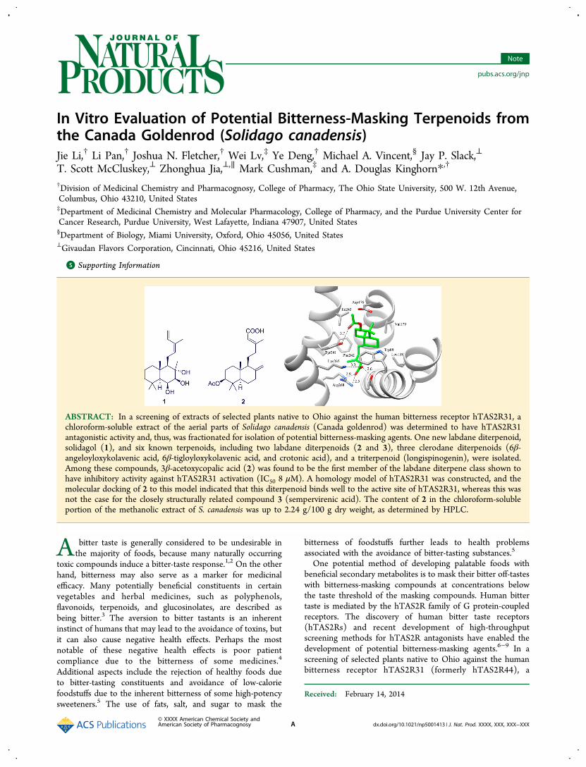
Solidagol (1) was determined to have a molecular formula ofC20H34O3 based on the 13C NMR spectroscopic data and the[M + Na]+ ion peak at m/z 345.2395 (calcd 345.2406) in theHRESIMS. The IR spectrum showed hydroxy group (3427cm−1) and double-bond (1710 cm−1) absorptions. The 1HNMR spectrum of compound 1 exhibited resonances for fourtertiary methyl groups (δH 0.99, H-18; 1.21, H-19; 1.21, H-20;1.36, H-17, each 3H, s), three aliphatic methylenes (δH 0.90and 1.63, each 1H, m, H-1; 1.45 and 1.70, each 1H, m, H-2;1.17 and 1.42, each 1H, m, H-3), one allylic methylene (δH2.27, 1H, ddd, J = 15.5, 6.9, 4.6 Hz, H-11a; 2.56, 1H, ddd, J =15.5, 7.5, 7.1 Hz, H-11b), two aliphatic methines (δH 0.97, 1H,brs, H-5; 1.32, 1H, dd, J = 7.1, 4.6 Hz, H-9), and twooxygenated methines (δH 3.42, 1H, d, J = 3.4 Hz, H-7; 4.41,1H, brd, J = 3.4 Hz, H-6), while resonances at δH 1.80 (3H, s,CH3, H-16), 5.14 (1H, d, J = 10.8 Hz, H-15a), 5.22 (1H, d, J =17.3 Hz, H-15b), 5.49 (1H, dd, J = 7.5, 6.9 Hz, H-12), and 6.89(1H, dd, J = 17.3, 10.8 Hz, H-14) were attributed to a 2-methylbuta-1,3-dien-1-yl side chain. The 13C NMR spectrum of1 showed 20 carbon resonances, which were classified from theDEPT and HSQC data as five methyl carbons, three quaternarycarbons, two tertiary sp3 carbons, two tertiary sp2 carbons, foursecondary sp3 carbons, one secondary sp2 carbon, and threeoxygen-bearing sp3 carbons (including two secondary and onetertiary). The characteristic NMR data of 1 were closelycomparable to those of crotomachlin, a labdane diterpenoidpreviously isolated from Koanophyllon conglobatum12 andCroton machrostachys.13 On comparison of the 1D- and 2D-NMR data (Figures 1 and 2) of these compounds, it wasrevealed that only one difference was evident in the double-bond configuration of the 2-methylbuta-1,3-dien-1-yl side chainlocated at C-11, with a (12E)-2-methylbuta-1,3-dien-1-yl groupin crotomachlin being replaced by a (12Z)-2-methylbuta-1,3-dien-1-yl moiety in 1. Thus, when compared to crotomachlin,H-12 in 1 showed a shielding of 0.1 ppm, while H-14 and H-15in 1 exhibited deshieldings of 0.55 and 0.22 ppm, respectively.
Correspondingly, C-12 and C-14 in 1 showed shieldings of 2.0and 8.0 ppm, respectively, while C-15 and C-16 in 1 showeddeshieldings of 3.6 and 8.1 ppm, respectively.12,13 The Zconfiguration of the double bond between C-12 (δC 133.6) andC-13 (δC 131.3) was confirmed by the NOESY correlation(Figure 2) between H-12 and H-16. The (12Z)-2-methylbuta-1,3-dien-1-yl side chain was placed at C-11 (δC 22.6) based onthe HMBC correlations of H-11 to C-12 and C-13. The Z-typeconjugated diene side chain has been observed occasionally forother labdane diterpenoids, e.g., austroilunin14 and (12Z)-abienol.15 The consistent chemical shifts and couplingconstants (Table 1) of the decalin resonances between 1 andcrotomachlin, along with the same positive sign of opticalrotation for both compounds as well as the NOESYexperiment, provided evidence that 1 shares the samestereochemistry at C-5 to C-10 as that of crotomachlin, theabsolute configuration of which was established using bothspectroscopic and synthetic methods.12,13 Biogenetically, Me-20is β-oriented and H-5 is α-oriented in labdane-typediterpenoids.16 The small proton coupling constant of H-6 (J= 3.4 Hz) indicated it is equatorial. Furthermore, the NOESYcorrelations between H-5/H-6, H-5/H-7, H-5/H-9, H-6/H-7,H-7/H-9, OH-6/Me-17, OH-6/Me-19, and OH-6/Me-20 wereused to establish the α-orientation of these protons and the β-orientation of OH-6, while the NOESY cross-peaks between H-11/Me-17 and H-11/Me-20 demonstrated that the C-11methylene and C-17 methyl groups are β-oriented. Therefore,compound 1 was established as (5S,6S,7S,8S,9R,10S)-labda-12Z,14-diene-6,7,8-triol and was accorded the trivial namesolidagol.On the basis of the physical and spectroscopic data
measurements ([α]D,1H NMR, 13C NMR, DEPT, 2D-NMR,
and MS) and the comparison of the data obtained withpublished values, the structures of the known compounds wereidentified as 3β-acetoxycopalic acid (2),17 sempervirenic acid
Figure 1. 1H−1H COSY (bold lines) and key HMBC (arrows)correlations of solidagol (1).
Figure 2. Key NOESY correlations of solidagol (1).
Journal of Natural Products Note
dx.doi.org/10.1021/np5001413 | J. Nat. Prod. XXXX, XXX, XXX−XXXB
(3),18 6β-angeloyloxykolavenic acid,19 6β-tigloyloxykolavenicacid,19 crotonic acid,20 and longispinogenin.21
In the present study, the bitterness-masking activity of all thecompounds isolated was evaluated in vitro by measuringreceptor-dependent calcium flux in human embryonic kidney(HEK-293T) host cells transfected with the human bitternessreceptor hTAS2R31 and stably expressing the chimeric G-protein α-subunit Gα16gust44. Among these isolates, compound 2was determined to be active with an IC50 value of 8 μM. Somestructurally similar terpenoids of 2 have been previouslyreported to possess binding affinity to hTAS2R46,6,8 andseveral clerodane and ent-kaurane diterpenes, e.g., hardwickiicacid, rebaudioside B, as well as their analogues, were recentlypatented for their effect in blocking the activation of thebitterness receptor by rebaudioside A.22,23 However, compound2 is the first member of the labdane diterpene class shown toexhibit inhibitory activity against hTAS2R31 activation. Incontrast, compound 3, a closely related labdane diterpene,showed no discernible hTAS2R31 antagonistic activity (IC50>25 μM).This interesting observation motivated an investigation of the
interaction modes of compounds 2 and 3 with hTAS2R31,using a molecular modeling and docking method. Since thestructural requirements of human bitter taste receptoractivation have been recently revealed7 and the sequences ofhTAS2R31 have been aligned with those of human β2-adrenergic receptor,9 a homology-based three-dimensionalmodel of hTAS2R31 was constructed based on the knowncrystal structure of human β2-adrenergic receptor. Thus,compound 2 was docked into the active site of hTAS2R31using the GOLD 3.01 docking program. The hypotheticalbinding mode of compound 2 in the active site of hTAS2R31 is
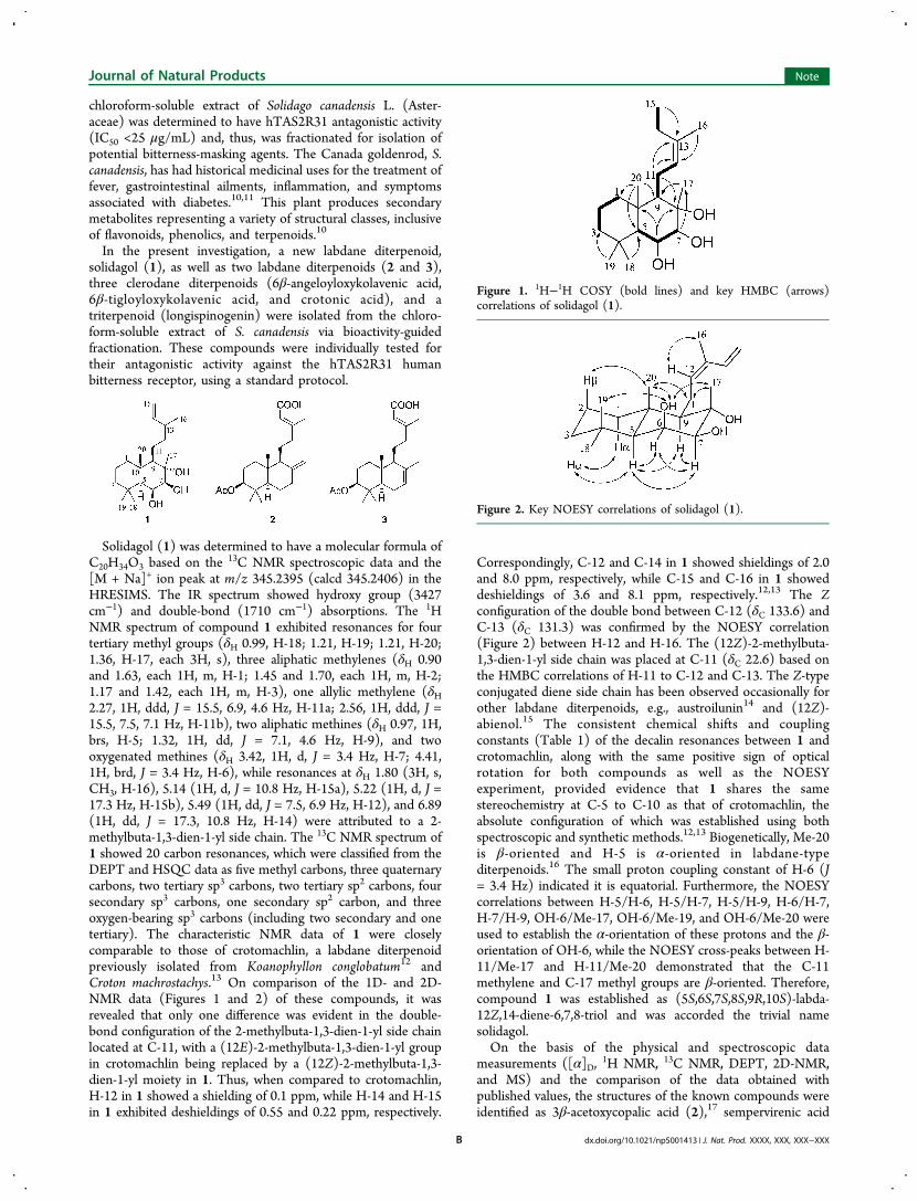
shown in Figure 3a. According to the docking results, thedecalin core of compound 2 is situated in a hydrophobic pocket
surrounded by Phe242, Ile245, Val179, Leu138, and Trp88amino acid residues. The acetyl group points toward thesolvent-accessible surface and hydrogen bonds with the sidechain of Tyr241. A previous study has already demonstrated theimportance of Lys265 and Arg268 in the interaction betweenaristolochic acid and hTAS2R31,7 and a mutagenesis assay wasused to prove the involvement of these two basic residues inthe antagonistic activity of 4-(2,2,3-trimethylcyclopentyl)-butanoic acid, a synthetic compound known as GIV3727.9 Inthe present study, the carboxy side chain of compound 2 wasshown to protrude deeply into the active site, forminghydrogen bonds with both Lys265 and Thr91. A salt bridgeinteraction was also observed between the carboxy group andArg268. Therefore, the docking experiment indicated thatcompound 2 binds well to hTAS2R31. In contrast, compound3 is unable to occupy the same binding pocket as doescompound 2, and it only binds to the outer loop region ofhTAS2R31, as shown in Figure 3b. This difference might be
Table 1. 1H (400 MHz) and 13C (100 MHz) NMR Data forSolidagol (1) in CDCl3
no. δC, type δH, mult. (J in Hz)
1α 42.3, CH2 0.90, m1β 1.63, m2α 18.8, CH2 1.45, m2β 1.70, m3α 43.7, CH2 1.17, m3β 1.42, m4 34.3, C5 55.7, CH 0.97, brs6 70.9, CH 4.41, brd (3.4)7 80.5, CH 3.42, d (3.4)8 77.4, C9 60.6, CH 1.32, dd (7.1, 4.6)10 39.6, C11a 22.6, CH2 2.27, ddd (15.5, 6.9, 4.6)11b 2.56, ddd (15.5, 7.5, 7.1)12 133.6, CH 5.49, dd (7.5, 6.9)13 131.3, C14 133.5, CH 6.89, dd (17.3, 10.8)15a 114.3, CH2 5.14, d (10.8)15b 5.22, d (17.3)16 20.1, CH3 1.80, s17 19.7, CH3 1.36, s18 (αCH3) 33.5, CH3 0.99, s19 (βCH3) 24.1, CH3 1.21, s20 16.9, CH3 1.21, sOH-6 2.02, s
Figure 3. (a) Hypothetical binding mode of compound 2 (green) inthe active site of hTAS2R31. The potential hydrogen bonding and saltbridge interactions are indicated with dashed lines. The distances arelabeled in angstroms. (b) Hypothetical binding mode of compound 2(green) in comparison with compound 3 (pink). The surface ofcompound 2 is displayed with mesh to show the shape of the ligandbinding pocket.
Journal of Natural Products Note
dx.doi.org/10.1021/np5001413 | J. Nat. Prod. XXXX, XXX, XXX−XXXC
due to the incorporation of the double bond in the decalin ringof compound 3, which would influence the conformation of thering and also change the orientation of the carboxy group.Among the terpenoids isolated in the present investigation,
the active compound 2 was obtained from its source plant inthe highest abundance. In order to determine the content ofthis compound in the S. canadensis sample used for isolation, anHPLC-DAD method with MS identification was developed andvalidated (Figure S2a and b, Supporting Information). Thecontent of 2 was determined to be 44.8 mg/100 g dry weight ofthe aerial parts of S. canadensis. In addition, the hexanes,chloroform, ethyl acetate, and 1-butanol partitions were alsoanalyzed using this method, and compound 2 was detected onlyin the chloroform partition, accounting for 2.24% of the dryweight of this partition, suggesting that S. canadensis might be apotential source for the development of bitterness-maskingdietary supplements or agents.
■ EXPERIMENTAL SECTIONGeneral Experimental Procedures. Optical rotations were
measured on a PerkinElmer 343 automatic polarimeter (PerkinElmer,Waltham, MA, USA). UV spectra were run on a Hitachi U-2910spectrophotometer (Hitachi, Tokyo, Japan). IR spectra were obtainedon a Nicolet 6700 FT-IR spectrometer (Thermo Scientific, Waltham,MA, USA). NMR spectroscopic data were recorded at roomtemperature on Bruker Avance DRX-300 and 400 MHz spectrometers(Bruker, Billerica, MA, USA). HRESI mass spectra were obtained onan LCT-TOF mass spectrometer (Waters Corp., Milford, MA, USA)operated in the positive-ion mode with NaI being used for masscalibration. Column chromatography was performed with SephadexLH-20 (Supelco, Bellefonte, PA, USA) and 65 × 250 and 230 × 400mesh silica gel (Sorbent Technologies, Atlanta, GA, USA). Analyticalthin-layer chromatography was conducted on precoated 250 μm thickPartisil Si gel 60F254 glass plates (Whatman, Clifton, NJ, USA). AnXBridge PrepC18 column (5 μm, 150 mm × 19 mm i.d., Waters) wasused for semipreparative HPLC, along with a Hitachi systemcomposed of an L-2130 pump and an L-2450 diode array detector.Solvents used for chromatographic separations were purchased fromFisher Scientific (Fair Lawn, NJ, USA). Other solvents were purchasedfrom Sigma-Aldrich (St. Louis, MO, USA) and were of chemical grade.Plant Material. The air-dried aerial parts of Solidago canadensis L.
(Asteraceae) were collected in Wayne Township, Butler County, Ohio(39.492061 N; −84.537746 W; 209 m alt.), by M.A.V., in October2010, who also identified this plant. A voucher specimen (MU271438) has been deposited in the W.S. Turrell Herbarium of MiamiUniversity, Oxford, Ohio.Extraction and Isolation. The air-dried aerial parts of S.
canadensis (1 kg) were ground and extracted exhaustively withMeOH. This dried MeOH extract (72 g) was then partitionedbetween 9:1 MeOH−H2O and hexanes. The MeOH−H2O layer wasevaporated to a thick tar and digested sequentially with CHCl3,EtOAc, and 1-butanol. The resulting CHCl3-soluble extract waspartially detannified with 1% NaCl in H2O to give 22 g of a finalCHCl3-soluble extract. This extract showed hTAS2R31 antagonisticactivity (IC50 <25 μg/mL) and thus was subjected to chromatographyover coarse silica gel, eluted with a CH2Cl2−acetone gradient (40:1,15:1, 10:1, 8:1, 6:1, 4:1, 3:1, 2:1, 1:1, 1:2, and pure acetone), to afford16 fractions (F01−F16), with the active fractions F02, F06, and F08(IC50 <25 μg/mL) being further purified. Fraction F02 was purifiedagain on a silica gel column, using a CHCl3−MeOH gradient solventsystem (50:1, 40:1, 30:1, 20:1, 15:1, 12:1, 10:1, 8:1, 6:1, and 5:1), toyield the triterpenoid longispinogenin (eluted with CHCl3−MeOH,30:1; 18.2 mg). Fraction F06 (4.6 g) was chromatographed furtherover a silica gel column with a CHCl3−MeOH solvent system (50:1,40:1, 30:1, 20:1, 15:1, 12:1, 10:1, 8:1, 6:1, 5:1, 3:1, 2:1, and 1:1) togive 12 subfractions (F0601−F0612). F0602 (eluted with CHCl3−MeOH, 30:1; 108 mg) was further purified by HPLC, using anXBridge PrepC18 column (5 μm, 150 mm × 19 mm i.d.) with
CH3CN−H2O [55:45, containing 0.05% trifluoroacetic acid (TFA) inH2O] at a flow rate of 10.0 mL/min, to yield 6β-angeloyloxykolavenicacid (tR = 122.5 min; 6.6 mg) and 6β-tigloyloxykolavenic acid (tR =92.0 min; 10.8 mg). F0604 (eluted with CHCl3−MeOH, 20:1; 980mg) was purified by Sephadex LH-20 column chromatography, withelution by CHCl3−MeOH (60:40 40:60, 20:80, and pure MeOH), toafford a further subfraction, F060408. This subfraction (157 mg) wasthen purified using the same HPLC column with 48% CH3CN−52%H2O containing 0.05% TFA, at a flow rate of 10.0 mL/min, to givecompounds 2 (tR = 106.5 min; 28.5 mg) and 3 (tR = 103.0 min; 3.4mg). Fraction F08 (898 mg) was applied to a silica gel column, elutedby a CHCl3−MeOH solvent system (30:1, 20:1, 15:1, 12:1, 10:1, 8:1,6:1, 5:1, 3:1, 2:1, and 1:1), to give six subfractions (F0801−F0806).F0804 (eluted with CHCl3−MeOH, 15:1; 96 mg) was further purifiedby HPLC on the same column, using an isocratic elution (CH3CN−H2O, 50:50, flow rate 8.0 mL/min), to afford 1 (tR = 25.8 min; 4.4mg) and crotonic acid (tR = 42.7 min; 2.4 mg).
Solidagol (1): colorless, amorphous solid; [α]20D +25 (c 0.1,CHCl3); UV (MeOH) λmax (log ε) 237 (3.74) nm; IR (film) νmax3427, 2925, 2863, 2848, 1710, 1559, 1459, 1439, 1386, 1363, 1216,1109, 1089, 1046, 909, 757 cm−1; 1H and 13C NMR data, see Table 1;HRESIMS m/z 345.2395 [M + Na]+ (calcd for C20H34O3Na,345.2406).
hTAS2R31 Antagonist Screening Assay. This bioassay wasconducted according to a literature procedure.6,9 In brief, the stablecell line for human bitterness receptor hTAS2R31 was generated bysubcloning the sst3:TAS2R:hsv cassette into the pcDNA3.1/Zeo(+)mammalian expression vector (Invitrogen, San Diego, CA, USA), andthe resulting construct was transfected into the HEK-293T host cellsthat stably express the chimeric G-protein subunit Gα16gust44. The stableGα16gust44/HEK-293T cells transfected with hTAS2R31 were selectedand preplated at a density of 15 000 cells per well in black 96-wellplates that had been precoated with 0.001% poly(ethyleneimine)(Acros Organics, Morris Plains, NJ, USA). After 48 h, the growthmedium was discarded and the cells were incubated in the dark for 1 hat 37 °C in 50 μL of loading buffer consisting of 1.5 μM Fluo-4 AM(Invitrogen) and 2.5 μM probenecid (Sigma-Aldrich) in Dulbecco’smodified Eagle medium (DMEM) without fetal bovine serum (FBS).After incubation, the plates were washed five times with 100 μL ofassay buffer each time and further incubated in the dark at roomtemperature for 30 min. The cells were then washed again five timesusing 100 μL of assay buffer each time, followed by the calciumresponses measurement in a fluorescence imaging plate reader(FLIPRTETRA) (Molecular Devices, Sunnydale, CA, USA). Saccharinwas employed as the cognate bitter receptor agonist for inhibitorscreening. Test compounds were prepared at a final concentration of25 μM in the presence of 1 mM sodium saccharin and assessed in aninitial screening to determine if they inhibit the hTAS2R31 responseto saccharin. Test compounds were also evaluated for inhibition of anon-taste GPCR pathway (isoproterenol, β1/β2-adrenergic receptoragonist) to ensure that any apparent inhibitory activity was bitternessreceptor-dependent and not simply due to nonspecific effects onintracellular calcium handling. To assess the potency of the selectivelyactive compounds (greater than 50% inhibition at 25 μM in the initialscreening), the half-maximal inhibitory concentration (IC50) wasdetermined via at least three separate experiments in duplicate. Forcalculation of IC50 values, the data were fitted in GraphPad Prismusing a four-parameter logistic fit equation.
Homology Modeling. The sequence of hTAS2R31 was obtainedfrom UniProtKB (entry P59538). The crystal structure of the β2-adrenergic receptor (PDB code: 2RH1) was selected as the template.The sequence alignment between hTAS2R31 and human β2-adrenergic receptor was directly taken from the literature.9 Based onthe alignment, 10 models were generated using the “automodel”protocol of Modeler 9v424 with the optimization level set to “high”.The best model according to the internal scoring function was selectedfor further model validation. A Ramachandran plot generated withProcheck indicated that most of the residues had Φ and Ψ angles inthe most favored (94.9%) or allowed regions (4%), and only a few
Journal of Natural Products Note
dx.doi.org/10.1021/np5001413 | J. Nat. Prod. XXXX, XXX, XXX−XXXD
residues were in the generously allowed (0.4%) or disallowed regions(0.7%).Molecular Docking. The structures of compounds 2 and 3 were
constructed and fully energy minimized with Sybyl 7.1 software. Theenergy-minimized structures were docked into the hTAS2R31homology model using the protein−ligand docking program GOLD3.01 with default parameters. The ligand binding pocket was definedby a sphere that centered at the amino nitrogen atom of Lys265 andhad a radius of 12 Å. The best docking poses were selected based onthe GOLD fitness score and the proximity of the ligand carboxy groupto Lys265 and Arg268.Analytical Method for the Content Determination of the
Active Compound 2 in S. canadensis Aerial Parts. The MeOHextract of the accurately weighed aerial parts of S. canadensis (air-dried,0.25 g) was subjected to passage over a Waters Sep-Pak Vac 20 cm3
C18 cartridge to remove the polar substances present (eluted by 10%MeOH−90% H2O). The pretreated extract (eluted by 95% MeOH−5% H2O) was dried and transferred into a 25 mL volumetric flask andmade up to the full volume with MeOH to prepare the extractsolution. The reference compound 2 was accurately weighed anddissolved in MeOH to produce the standard stock solution. This stocksolution was diluted to yield a series of standard solutions in theconcentration range 2−250 mg/L. The HPLC separation wasperformed on an XBridge C18 analytical column (5 μm, 150 mm ×4.6 mm i.d., Waters), along with a Hitachi system composed of an L-2130 pump and an L-2450 diode array detector. The mobile phaseconsisted of 0.05% TFA in H2O (A) and MeCN (B) using a gradientprogram of 48−50% B from 0 to 26 min, 50−60% B from 26 to 28min, 60% B from 28 to 50 min, and 60−100% B from 50 to 60 min.The mobile phase flow rate was 1 mL/min, and the columntemperature was set at 30 °C. After comparing the chromatogramsof the extract solution recorded at wavelengths within 200−365 nm, itwas found that 231 nm could best represent the profile of the analyte.Compound 2 in the extract was identified by comparing its retentiontime and UV spectrum with those of the standard and was confirmedby its molecular weight in the MS (Figure S2a and b, SupportingInformation). Method validation parameters included linearity,reproducibility, precision, and recovery. The five-point calibrationcurve of compound 2 was linear (y = 49277x + 9764; R2 = 0.9996)over the range 2−250 mg/L. The limit of detection (LOD) was 1.8 ng(signal-to-noise ratio = 3). Method reproducibility was evaluated byfive replicated analyses of the extract solution (n = 5), with a relativestandard deviation (RSD) value of 2.84% achieved. Method precisionwas investigated by repeatedly analyzing the same set of compound 2standard solution (n = 5), and the RSD of the calculated concentrationwas 1.88%. Recovery of compound 2 was determined using spikedsamples with 100% of the quantified level of compound 2 in fivereplicated samples. The recovery rate was 96.24% (RSD 3.88%).
■ ASSOCIATED CONTENT
*S Supporting Information1H NMR, 13C NMR, 13C DEPT 135 NMR, 1H−1H COSY,HSQC, HMBC, NOESY, HRESIMS, IR, and UV spectra forcompound 1. HPLC chromatograms of the MeOH extract of S.canadensis and compound 2 standard along with the relevantMS and UV spectra. This material is available free of charge viathe Internet at http://pubs.acs.org.
■ AUTHOR INFORMATION
Corresponding Author*Tel: 1-614-247-8094. Fax: 1-614-247-8119. E-mail: [email protected].
NotesThe authors declare no competing financial interest.∥Deceased October 5, 2013.
■ ACKNOWLEDGMENTSWe would like to thank Mr. J. W. Fowble and Dr. C. McElroy(College of Pharmacy, The Ohio State University) forfacilitating the acquisition of NMR data. Mr. M. Apsega fromthe Mass Spectrometry & Proteomics Facility, CampusChemical Instrument Center, The Ohio State University, isacknowledged for MS training.
■ DEDICATIONThis article is dedicated to the memory of our esteemedcolleague Dr. Zhonghua Jia.
■ REFERENCES(1) Roy, G. M. Crit. Rev. Food Sci. Nutr. 1990, 29, 59−71.(2) Lindemann, B. Nature 2001, 413, 219−225.(3) Drewnowski, A.; Gomez-Carneros, C. Am. J. Clin. Nutr. 2000, 72,1424−1435.(4) Binello, A.; Cravotto, G.; Nano, G. M.; Spagliardi, P. FlavourFrag. J. 2004, 19, 394−400.(5) Roy, G. M. Trends Food Sci. Technol. 1992, 3, 85−91.(6) Brockhoff, A.; Behrens, M.; Massarotti, A.; Appendino, G.;Meyerhof, W. J. Agric. Food Chem. 2007, 55, 6236−6243.(7) Brockhoff, A.; Behrens, M.; Niv, M. Y.; Meyerhof, W. Proc. Natl.Acad. Sci. U.S.A. 2010, 107, 11110−11115.(8) Brockhoff, A.; Behrens, M.; Roudnitzky, N.; Appendino, G.;Avonto, C.; Meyerhof, W. J. Neurosci. 2011, 31, 14775−14782.(9) Slack, J. P.; Brockhoff, A.; Batram, C.; Menzel, S.; Sonnabend, C.;Born, S.; Galindo, M. M.; Kohl, S.; Thalmann, S.; Ostopovici-Halip, L.;Simons, C. T.; Ungureanu, I.; Duineveld, K.; Bologa, C. G.; Behrens,M.; Furrer, S.; Oprea, T. I.; Meyerhof, W. Curr. Biol. 2010, 20, 1104−1109.(10) Bradette-Hebert, M. E.; Legault, J.; Lavoie, S.; Pichette, A. Chem.Pharm. Bull. 2008, 56, 82−84.(11) McCune, L.; Johns, T. J. Ethnopharmacol. 2002, 82, 197−205.(12) Bohlmann, F.; Scheidges, C.; King, R. M.; Robinson, H.Phytochemistry 1984, 23, 1190−1192.(13) Herlem, D.; Khuong-Huu, F.; Kende, A. S. Tetrahedron 1994,50, 2055−2070.(14) Ohtsuki, T.; Koyano, T.; Kowithayakorn, T.; Yamaguchi, N.;Ishibashi, M. Planta Med. 2004, 70, 1170−1173.(15) Zerbe, P.; Chiang, A.; Yuen, M.; Hamberger, B.; Hamberger, B.;Draper, J. A.; Britton, R.; Bohlmann, J. J. Biol. Chem. 2012, 287,12121−12131.(16) Dong, L. B.; He, J.; Wang, Y. Y.; Wu, X. D.; Deng, X.; Pan, Z.H.; Xu, G.; Peng, L. Y.; Zhao, Y.; Li, Y.; Gong, X.; Zhao, Q. S. J. Nat.Prod. 2011, 74, 234−239.(17) Braun, S.; Breitenbach, H. Tetrahedron 1977, 33, 145−150.(18) Purushothaman, K. K.; Sarada, A.; Saraswathy, A.; Connolly, J.D. Phytochemistry 1983, 22, 1042−1043.(19) Lu, T.; Menelaou, M. A.; Vargas, D.; Fronczek, F. R.; Fischer, N.H. Phytochemistry 1993, 32, 1483−1488.(20) Jakupovic, J.; Baruah, R. N.; Zdero, C.; Eid, F.; Pathak, V. P.;Chau-Thi, T. V.; Bohlmann, F.; King, R. M.; Robinson, H.Phytochemistry 1986, 25, 1873−1881.(21) Tori, K.; Yoshimura, Y.; Seo, S.; Sakurai, K.; Tomita, Y.; Ishii, H.Tetrahedron Lett. 1976, 17, 4163−4166.(22) Slack, J. P.; McCluskey, T. S.; Hartzel, C. A.; Odley, A. M.; Jia,Z. WO Patent 2013/072332 A1, May 23, 2013.(23) Bridges, J. R.; Carlson, A.; Patton, P. A. WO Patent 2012/102769 A1, August 2, 2012.(24) Sali, A.; Blundell, T. L. J. Mol. Biol. 1993, 234, 779−815.
Journal of Natural Products Note
dx.doi.org/10.1021/np5001413 | J. Nat. Prod. XXXX, XXX, XXX−XXXE