Embed Size (px)
Citation preview
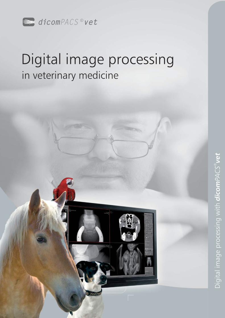
Digital image processingin veterinary medicine
dicom vetPACSR
Dig
ital im
ag
e p
roce
ssin
g w
ith
dic
om
vet
PAC
S®

Professionalimage processingin veterinarymedicine
dicom vetPACS®
will make your dream of a paperless veterinary practice
come true. All images as well as any type of document (e.g. diagnostic
reports, records of healing processes, faxes) are stored by dicom vetPACS®
in a digital patient file and can be accessed immediately with a simple
mouse click.
Well designed archiving and backup solutions guarantee fast access to
all data while observing the highest security standards in accordance with
the internationally recognised guidelines for human medicine. In addition,
dicom vetPACS®
can be integrated easily with all the popular practice
management systems.
The software includes acquisition, diagnosis, transferdicom vetPACS®
and archiving of image material. Since it has been designed and developed
in close cooperation with practising vets, you will find it easy to operate
and well suited to daily diagnosis.
Boasting several thousands installed workstations locally and abroad (as of
March 2013), the system has proven itself many times over. dicom vetPACS®
handles simple image processing requirements as brilliantly as complex
radiological networks.
Dig
ital im
ag
e p
roce
ssin
g w
ith
dic
om
vet
PAC
S®
dicom vetPACSR
of at one glancedicomPACS®
Benefits
2
Full diagnostic software for all workstations in your practice
(no 'light' versions)
User friendly and clearly arranged structure, minimal training
requirements and short familiarisation period
Individual adjustment of the user interface to your field of specialisation
and individual requirements
Flexible allocation of shortcut keys for many functions to allow fast
work without a mouse
Parallel processing (e.g. option to continue working during a CD
burning process)
Permanent online availability of all images and data in the network – no
need to store old images on CDs
“Perfect memory” – re-opening of images with all previous markings
and settings incl. zoom and orientation
Parallel diagnostic evaluation of several patients made possible by
opening any number of programme windows without loss of speed -
depending on the size of the working memory
Import of any external documents such as doctors' letters, faxes or X-ray
images – no additional module required
Installation with Windows, UNIX, LINUX or Apple Macintosh
Optimal data security, speed and compatibility by using standardised
SQL database technology
All images and documents are filed in the international DICOM
standard at all times
3
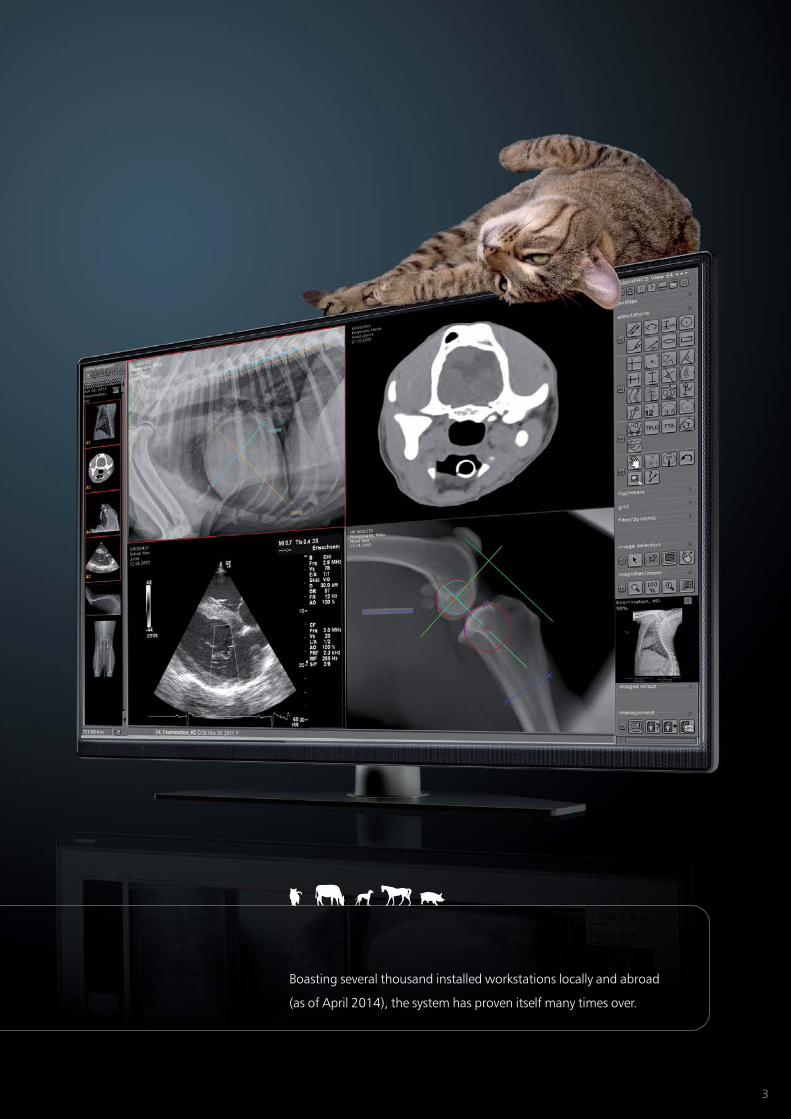
Boasting several thousand installed workstations locally and abroad
(as of April 2014), the system has proven itself many times over.
4
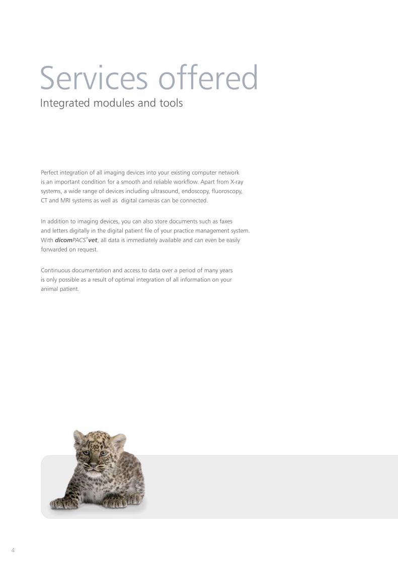
Perfect integration of all imaging devices into your existing computer network
is an important condition for a smooth and reliable workflow. Apart from X-ray
systems, a wide range of devices including ultrasound, endoscopy, fluoroscopy,
CT and MRI systems as well as digital cameras can be connected.
In addition to imaging devices, you can also store documents such as faxes
and letters digitally in the digital patient file of your practice management system.
With , all data is immediately available and can even be easilydicom vetPACS®
forwarded on request.
Continuous documentation and access to data over a period of many years
is only possible as a result of optimal integration of all information on your
animal patient.
Services offeredIntegrated modules and tools

ConnectivityThe diversity of dicomPACS
®vet
Image sources
Image output
Image viewing
Image processing
Image archiving
Multimonitorworkstation
Homeworkstation
ISDN
Telemedicine/Cloud
Interfaces to practicemanagement system
Archiv servere
DVD backupsystem
MRI/ CT/ NUKUltrasound
Diagnostic station
X-ray scanner
Laser printer
Laser imager
Viewing station
X-ray units
PatientCD burner
D sR ystem
CR ystems
Video projector
Operationdocumentation
Documentscanner
Amadeo completeDR system
MediciDR retrofits
Divario CR solutionwith cassettes
Solutionsof OR Technologyincl. acquisition anddiagnostic softwareNetwork
5
Leonardo DRsuitcase solution
Dental Vet CR/ DR
dicom vetPACSR
6
Prosthesis documentation - enables the user to plan operations
with digital prosthesis templates by one or more manufacturers
see page 18/ 19
Report module for X-ray services relating to equine prepurchase
examinations [currently only available for Germany] - enables the quick
compilation of reports by automatically assembling X-ray images. It follows the
“X-ray guideline” by the German organisations “Gesellschaft für Pferdemedizin
e.V.” (non-profit organisation for equine medicine) and “Bundestierärzte-
kammer e.V.” (Federal association of veterinarians).
see page 8 - 11
Special filter for the optimization of bones and soft parts -
details of interest may be made visible by means of special filter magnifiers
TPLO measuring function (Tibial Plateau Leveling Osteotomy) - it serves to
theoretically optimise the existing slope of the tibial plateau in domestic dogs
see page 18/ 19
TTA measuring tool (Tibial Tuberosity Advancement) - the TTA measuring
technique is used to apply the translated length measurements at the
tuberositas tibiae in dogs
see page 18/ 19
HD measuring technique for dogs - provides adicom vetPACS®
special tool to guarantee very fast and reliable determination of the Norberg
angle, including documentation
see page 18/ 19
Measuring the distraction index - This measuring tool serves to
determine the displacement of the femoral head from the joint socket
of the hip joint in dogs
see page 18/ 19
Buchanan‘s Vertebral Heart Score - This annotation is a simple and
reliable method to determine the size of the heart - it has been desiged
specifically for cats and dogs
see page 18/ 19
dicom vetPACS®
features
Value
7
Modified Maquet Procedure (MMP) - The MMP is a method of
measurement for dogs with a cruciate ligament disorder, in which the
distance for the placement of the MMP Wedge is determined.
see page 18/19
Statistics Module - enables freely configurable analysis of the
complete database
Video Modules - enable standard and non-standard video signals
to be recorded as single images and video sequences
Web Server - enables image distribution within the hospital or to
referring doctors via the internet and guarantees very fast image
accessibility in original quality (DICOM)
see page 14
Processing of CT and MRI series - includesdicom vetPACS®
professional tools such as MPR and MIP to evaluate cross
section series
see page 16/ 17
Telemedicine & Hanging protocols
Special solution for multiple archives
Cloud archiving
see page 15
for X-ray services relating to equine prepurchase examinations
Report module
Presale and prepurchase examinations for horses are always particularly
challenging for veterinarians. Such specialised examinations must be
carried out swiftly yet very meticulously
documented very well, in great detail and extremely consistently.
After all, the owner of the animal justifiably expects optimal service when it
comes to undertaking the examination and presenting the results in a professional
and comprehensible fashion. Since administrative work is bothersome yet vital for
veterinarians, too, we have developed a report module specifically for X-ray services
relating to prepurchase examinations in cooperation with renowned specialists.
This module enables the quick compilation of reports by automatically assembling
X-ray images. It follows the “X-ray guideline” by the German organisations
“Gesellschaft für Pferdemedizin e.V.” (non-profit organsation for equine medicine)
and “Bundestierärztekammer e.V.” (Federal association of veterinarians).
[currently only available for Germany]
Benefits:
8
Time-saving:
The prepurchasedicom vetPACS®
examination report module allows very
fast and professional preparation of
reports on the prepurchase examination,
including perfect layout and
documentation in .dicom vetPACS®
Easy to follow:
dicom vetPACS®
guarantees complete
implementation of the wording and
the structure of diagnostic reports in
accordance with the “Guideline for
pre-purchase X-ray examinations” by the
German “Gesellschaft für Pferdemedizin
e.V.“ and “Bundestierärztekammer e.V.”.
The texts can be easily edited and
included in the report to be compiled.
Reports with images:
The required X-ray images, including
all modifications such as zoom,
measurements and annotations,
and are automatically added to the
respective diagnosis (e.g. fetlock joint)
for documentation in the prepurchase
report. The layout is automatically
compiled (page breaks, image
positioning etc.)
Safe:
The complete report (WYSIWYG principle)
is automatically stored with the X-ray
images. Of course, these reports are also
available for patient CDs. This
guarantees that images and reports
are always kept together.
Presentation:
dicom vetPACS®
is a professional
marketing tool for referring doctors.
9
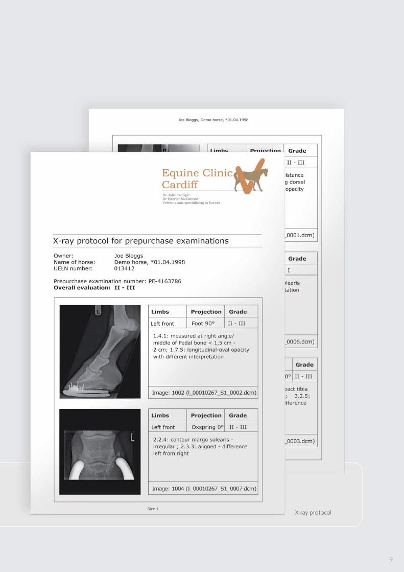
X-ray protocol

6. Report preparation
1. Call up the examination
Workflow for a prepurchase horse examination
10
4. Display diagnostic report options
2. Start report module
5. Diagnostic report/ evaluation
Create report Continue
3. Allocate the projectionCurrent image assign
left front
right front
left hind
left right
Foot
Foot
Foot
Knee
Knee
Foot
Knee
Knee
Back
back
Projection add or delete
Assignment cancel
Current image assign
left front
right front
left hind
left right
Foot
Foot
Foot
Knee
Knee
Foot
Knee
Knee
Back
back
Projection add or delete
Assignment cancel

By means of a single click, the completed prepurchase examination
report is stored together with the examination images in ,dicom vetPACS®
where it can be accessed again in the original version at any point in time.
11
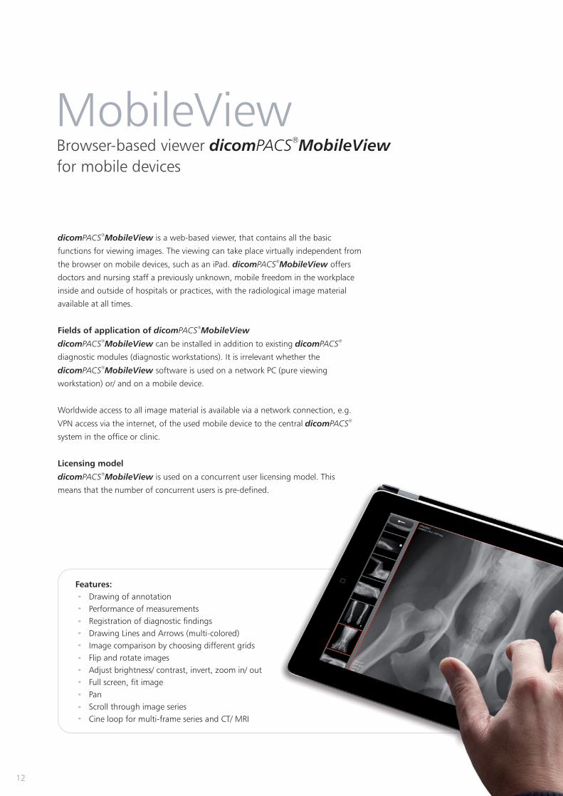
Browser-based viewer dicom MobileViewPACS®
for mobile devices
MobileView
dicom MobileViewPACS®
is a web-based viewer, that contains all the basic
functions for viewing images. The viewing can take place virtually independent from
the browser on mobile devices, such as an iPad. offersdicom MobileViewPACS®
doctors and nursing staff a previously unknown, mobile freedom in the workplace
inside and outside of hospitals or practices, with the radiological image material
available at all times.
Fields of application of dicom MobileViewPACS®
dicom MobileView dicomPACS PACS® ®
can be installed in addition to existing
diagnostic modules (diagnostic workstations). It is irrelevant whether the
dicom MobileViewPACS®
software is used on a network PC (pure viewing
workstation) or/ and on a mobile device.
Worldwide access to all image material is available via a network connection, e.g.
VPN access via the internet, of the used mobile device to the central dicomPACS®
system in the office or clinic.
Licensing model
dicom MobileViewPACS®
is used on a concurrent user licensing model. This
means that the number of concurrent users is pre-defined.
Features:
Drawing of annotation
Performance of measurements
Registration of diagnostic findings
Drawing Lines and Arrows (multi-colored)
Image comparison by choosing different grids
Flip and rotate images
Adjust brightness/ contrast, invert, zoom in/ out
Full screen, fit image
Pan
Scroll through image series
Cine loop for multi-frame series and CT/ MRI
12

The main advantages below at a glance:
High flexibility through the use within various internet browsers, including Microsoft Internet
Explorer, Mozilla Firefox, Google Chrome, Safari 5, Safari for iPad and Android browser
Intuitive operation
Supports the multi-touch operating technology (e.g. zoom in and out with two-fingers)
Supports full screen mode
Allows accessing the or database without anydicom DX-R dicomPACS PACS® ®
additional modules
Allows playing series (e.g. ultrasound)
High loading speed with modern streaming technology
Uses concurrent user licenses
13
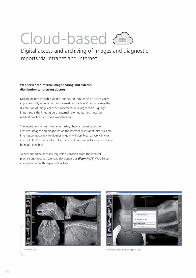
Web viewer Web viewer with image export tool
14
Web server for internal image sharing and external
distribution to referring doctors
Making images available via the internet (or intranet) is an increasingly
important daily requirement in the medical practice. One purpose is the
distribution of images or other documents in a larger clinic. Equally
important is the integration of external referring parties (hospitals,
medical practices) or home workstations.
The intention is always the same: faster, cheaper downloading of
archived images and diagnoses via the internet or intranet (also via slow
internet connections), in diagnostic quality if possible, to every clinic or
internet PC. The use of older PCs, thin clients or terminal servers must also
be made possible.
To accommodate as many requests as possible from the medical
practice and hospital, we have developed our Web ServerdicomPACS®
in cooperation with respected doctors.
Cloud-basedDigital access and archiving of images and diagnostic
reports via intranet and internet

15
ORCA - the Cloud-based archive solution by OR Technology
Even for state-of-the-art veterinary clinics and other veterinary facilities, the
rapidly rising data flood of digital images, diagnostic reports and other
documents is becoming increasingly challenging. Current legislation demands
safe and long-term storage of patient data which generally requires investing in
expensive hardware infrastructure as well as maintenance and corresponding
staff costs.
To this end, we developed the Cloud archiving solution, thus pavingORCA
the way for cost-effective and safe Cloud-based data archiving in big veterinary
practices and clinics. offers two application options:ORCA
Safe, long-term archiving of patient data with intelligent usage of
internal databases
Communication platform (exchange of images and diagnostic reports)
with colleagues and specialists or as an easy way to forward image data
to patients (an alternative to creating patient CDs)
Data is archived on European servers with the relevant safetyexclusively
certificates.
Benefits of Cloud archiving through ORCA
Minimal expenditure: does not require investing in expensive infrastructure such as server and data cables.ORCA
Scalability: The amount of memory required when using is determined by the demand.ORCA
Long-term security: archives data on many individual European servers in professional and air-conditionedORCA
data centres. Server technology is continuously updated.
Accessibility: stands out by being highly accessible. Since data is saved with multiple redundancy,ORCA
ORCA guarantees more continuity than a mere server solution.
Environmentally friendly: is sustainable – through the optimised use of resources and their distribution.ORCA
Location-independent: ORCA guarantees access to archived patient data - worldwide.
Simplicity: ORCA allows easy access to data from any computer – from your place of work, from the comfort
of your home or from any other computer or tablet PC.
Stress-free: deals with everything – no need to struggle with loose network cables, removed hardORCA
drives or software problems.
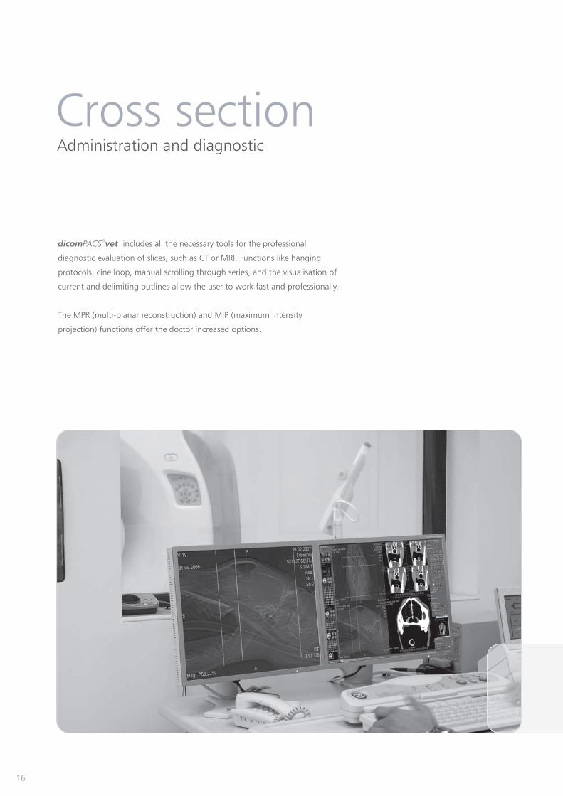
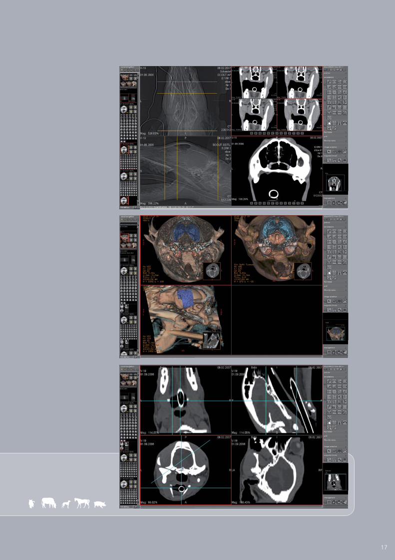
Administration and diagnostic
Cross section
dicom vetPACS®
includes all the necessary tools for the professional
diagnostic evaluation of slices, such as CT or MRI. Functions like hanging
protocols, cine loop, manual scrolling through series, and the visualisation of
current and delimiting outlines allow the user to work fast and professionally.
The MPR (multi-planar reconstruction) and MIP (maximum intensity
projection) functions offer the doctor increased options.
16
17
Röntgenprotokoll

Digital X-ray images have the advantage that exact measurements can be
taken at the monitor and the image quality can be improved by a number of
manipulations. now offers some special functions.dicom vetPACS®
Modified Maquet Procedure (MMP)
The MMP (Modified Maquet Procedure) is a method of measurement for dogs
with a cruciate ligament disorder, in which the distance for the placement of
the MMP Wedge is determined.
Pre-operative planning with the prosthesis documentation module
This module allows the user to plan and document an operation. After
activating this function, the active image is displayed in its original film-identical
size. The prosthesis template is displayed in the image as an annotation, or the
existing prosthesis template films are overlaid on the monitor.
TTA (Tibial Tuberosity Advancement) measuring tool
The TTA measuring technique is used to apply the translated length
measurements at the tuberositas tibiae in dogs.
HD measuring technique for dogs
dicom vetPACS®
provides a special tool to guarantee very fast and
reliable determination of the Norberg angle, including documentation.
One click suffices to insert all relevant lines and angles into the image,
where they can then be positioned as required.
TPLO (Tibial Plateau Leveling Osteotomy) measuring tool
It serves to theoretically optimise the existing slope of the tibial plateau
in domestic dogs.
Measuring the distraction index
This measuring tool serves to determine the displacement of the femoral
head from the joint socket of the hip joint in dogs.
Buchanan's Vertebral Heart Score
This annotation is a simple and reliable method to determine the size of the
heart. It has been designed specifically for cats and dogs. The height and width
of the heart are put into relation to the individual animal's vertebral body width.
Therefore, racial distinctions are brought to bear on the examinations results.
FeaturesSpecial functions for digital
X-ray imaging
18

TPLO/TTA measurement
TPLO/TTA measurement
Original image
Image copy with
magnifying glass filter
DI: 0,035
108.85°
105.07°
19
Integrated prosthesis documentation moduleBuchanan‘s Vertebral Heart Score
TTA measuring tool
HD measuring technique for dogs
TPLO measuring tool Measuring the distraction index
MMP (Modified Maquet Procedure)
Special filter for the optimization of bones and soft parts

Seamless integration with the administration software
Integration
Only an optimal interface guarantees perfect networking of all systems
such as the integration of the image archive with the specific administration
software. With a single mouse click you have immediate access to patient
data to prepare an imaging request or to load archive images.
dicom vetPACS®
is well designed, sophisticated and flexible. It can be
integrated easily with any veterinary administration programme.
20
Basic functions
The way we configure an interface
in detail so that everything works perfectly
depends on the existing administration
system. We would like to present three
examples of frequently used functions
below:
Example 1: Patient data is made
directly available from the index card for
the examination instruction for e.g. a
digital X-ray, MRI or similar.
Example 2: The examination instruction
is allocated to the digital patient register
of that particular patient, where it is
stored and archived.
Example 3: The archived data -
X-rays or documents - is called up directly
from the patient register. You can proceed
as you wish, directly choose a particular
image or document, or decide on a
specific selection, e.g. the last week's
exposures or just the ultrasound
exposures of a patient.
However you want to proceed, you
can be sure that it will work, because
we have already successfully
integrated intodicom vetPACS®
many administration systems.
Connection - example Vetera Connection - example AVImark

dicomPACS®vet in veterinary practice and hospital
Satisfied Customers
Equine Clinic Burg Müggenhausen, Germany, Dr Thomas Weinberger:
“The prepurchase examination module of proves to be adicomPACS®vet
great help with X-ray examinations and diagnostic ... This tool provides an
enormous reduction in work and liability risks. Moreover, it is great that the
current X-ray Code of Practice (German X-ray guide 07) is provided ... Overall,
we wish to repeat that we are very happy with the investment and the
changeover to ."dicomPACS®vet
Laboratory for Radiology and Ultrasonography of the University
of Life Sciences in Lublin, Poland, Dr Renata Komsta:
„... The image management software ensures that alldicomPACS®vet
images are automatically filed and can be found easily. Using analog technology,
archiving of images was not that easy and we often spent a long time looking
for a specific image in the archive. Special measuring functions of the software
(allowing length, angle and other measurements) as well as digital image quality
are an enormous improvement to our diagnostic options, which is extremely
beneficial to our research work. We are highly content with the DRMedici
system including the software and we can recommend itdicomPACS®vet
without any reservations to all veterinarians.”
Dierenkliniek Kerkelanden, Hilversum, Netherlands,
veterinarian Dr Erik Schurer:
“Due to high-speed image creation and improved image quality it is now much
easier for us to come to a diagnosis. ... We have the option to improve the image
quality and we can try out a variety of image processing tools such as contrast/
exposure and window leveling. In addition, a professional image quality also
contributes decisively to customer satisfaction and customer loyalty. It is now
possible to capture an image spontaneously during an appointment and prepare
a diagnostic report immediately thereafter. The new X-ray system has therefore
allowed us to increase our diagnostic options considerably”, says Dr. Schurer
expressing his satisfaction ...
Equine Center „Centro de Diagnóstico e Terapia Equina”
in Sao Paulo, Brazil, Dr. Fernanda Manzano de Campos:
“We are also very satisfied with the image managementdicomPACS®vet
system. The images are archived, we can take measurements and send images
via e-mail. It is also easy to operate. All in all, we are delighted with the
system by OR Technology“
Dr Manzano de Campos, Equine Center„Centro de Diagnóstico e Terapia Equina”
Dr Erik Schurer
AssistantAnna Lojszczyk-Szczepaniak
Dr Thomas Weinberger
You can find the extensive references here
www.or-technology.com --> Veterinary medicine --> References21
© Foto J. Piasecki

(Oehm und Rehbein GmbH)OR Technology
18057 Rostock, Germany, Neptunallee 7c
Tel. +49 381 36 600 500, Fax +49 381 36 600 555
www.or-technology.com, [email protected] [Stamp of distribiution partner]
Info hotline: +49 381 36 600 600
R TechnologyDigital X-ray and
Imaging Solutions
O
Ver
sion 0
04_04_2014
inklusi
veDiv
arioCR 36O
vet
CR-Syste
me mitZuku
nftDX-R Akquisit
ions-Sof
tware
- compact suitcase solutions forDR suitcasesmobile and portable X-ray incl. dicom DX-RPACS
®
acquisition software
DR retrofits - digital upgrade set forexisting X-ray systems incl. dicom DX-RPACS
®
acquisition software, available for stationaryand mobile X-ray machines
Accessories for X-raye.g. radiation protection walls, gloves
Image management (PACS) - comprises
acquisition, processing, diagnosis, transfer and
archiving of image material
Cloud-based archive solution - safe, long-term
archiving of patient data with intelligent usage of internal
databases communication platform with colleagues and
specialists and transfer of image data to patients
Complete digital X-ray systems (incl. stand, Bucky,generator, flat panel etc. and incl. dicom DX-RPACS
®
acquisition software), mobile X-ray solutionsas well as portable X-ray solutions
CR solutions - CR systems for digitalX-ray with cassettes incl. dicom DX-RPACS
®
acquisition software
X-ray acquisition software [only for OEMs] -
acquisition and diagnostic software for X-ray images
from flat panels or CR systems
Medici DR Systemsvet
DX-RdicomPACSR
X-ray Acquisition Software
dicom vetPACSR
Medici DR Systemsvet
Leonardo DR Systemsvet
Amadeo X-ray Systems vet
Divario CR Systemsvet
X-ray Accessories
ORCA
Overview - products of OR TechnologyVet portfolio


![GUÍA DEL IMPRESSIONHaga clic en [Inicio] → [Configuración] → [Impresoras y faxes] para abrir la ventana Impresoras y faxes. 2. En la ventana Impresoras y faxes, haga clic con](https://img.dokumen.tips/doc/110x75/5f9164828542ee7568163c4d/gua-del-impression-haga-clic-en-inicio-a-configuracin-a-impresoras.jpg)