Embed Size (px)
Citation preview
Fall 2009: Volume 9 Number 3
IN THIS ISSUE
3
5
6
7
National Cancer Institute Renews Mayo Clinic Cancer Center’s Support Grant
Navigator Notes:Lodging Assistance
Cancer Terms & Resources
Vickie Hallisy: A Passion forPerseverance
Use of Imaging Tests in Oncology: A Picture is Worth a Thousand WordsBy Robert McWilliams, M.D., medical oncologist
While medical oncologists often are viewed as ‘the cancer doctors,’ they are part of a patient care team that depends on others in surgery, pathology and increasingly, radiology. The accuracy and usefulness of different radiology or imaging tests has rapidly improved over the past several decades. These advances allow doctors unique insight into the appearance, behavior and degree of spreading of cancer cells. Highly accurate imaging provides more information for key decision-making and hopefully minimizes the need for invasive testing.
Radiology or imaging tests are used for cancer screening and diagnosis. Examples of commonly used screening tests include mammograms and, more recently, newer technologies such as virtual colonoscopy. Once cancer is diagnosed, images are then used to ‘stage,’ or determine how widespread a cancer is, and later evaluate a cancer’s response to therapy. These tests help determine whether the cancer grew, remained the same size or shrank. Radiologists use many imaging technologies. Common ones include X-ray, ultrasound, and MRI (magnetic resonance imaging), as well as CT (computed tomography), PET (positron emission tomography) and bone scans.
X-RayMost people are familiar with X-rays, which use a low dose of radiation to expose a film. These images are frequently used for a quick, relatively inexpensive look at specific areas of the body, especially the chest, abdomen, and bones. X-rays often can provide complete diagnostic information without the added expense or complexity of other tests.
UltrasoundUltrasound uses sound waves to create images of muscles, tendons, tissues and organs. It can be used with specific treatments such as radiofrequency ablation to locate a lesion and provide an accurate picture for radiologists to correctly position the treatment device in the tumor.
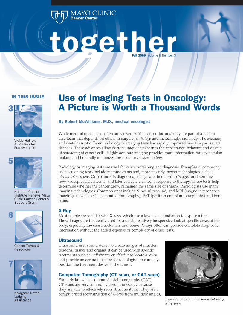
Computed Tomography (CT scan, or CAT scan)Formerly known as computed axial tomography (CAT), CT scans are very commonly used in oncology because they are able to effectively reconstruct anatomy. They are a computerized reconstruction of X-rays from multiple angles,
Example of tumor measurement using a CT scan.
2
and require only a low level of radiation exposure to produce three-dimensional images of bones and soft tissues. One significant benefit to oncology is the ability of CT scans to provide images accurately showing tumor growth or shrinkage.
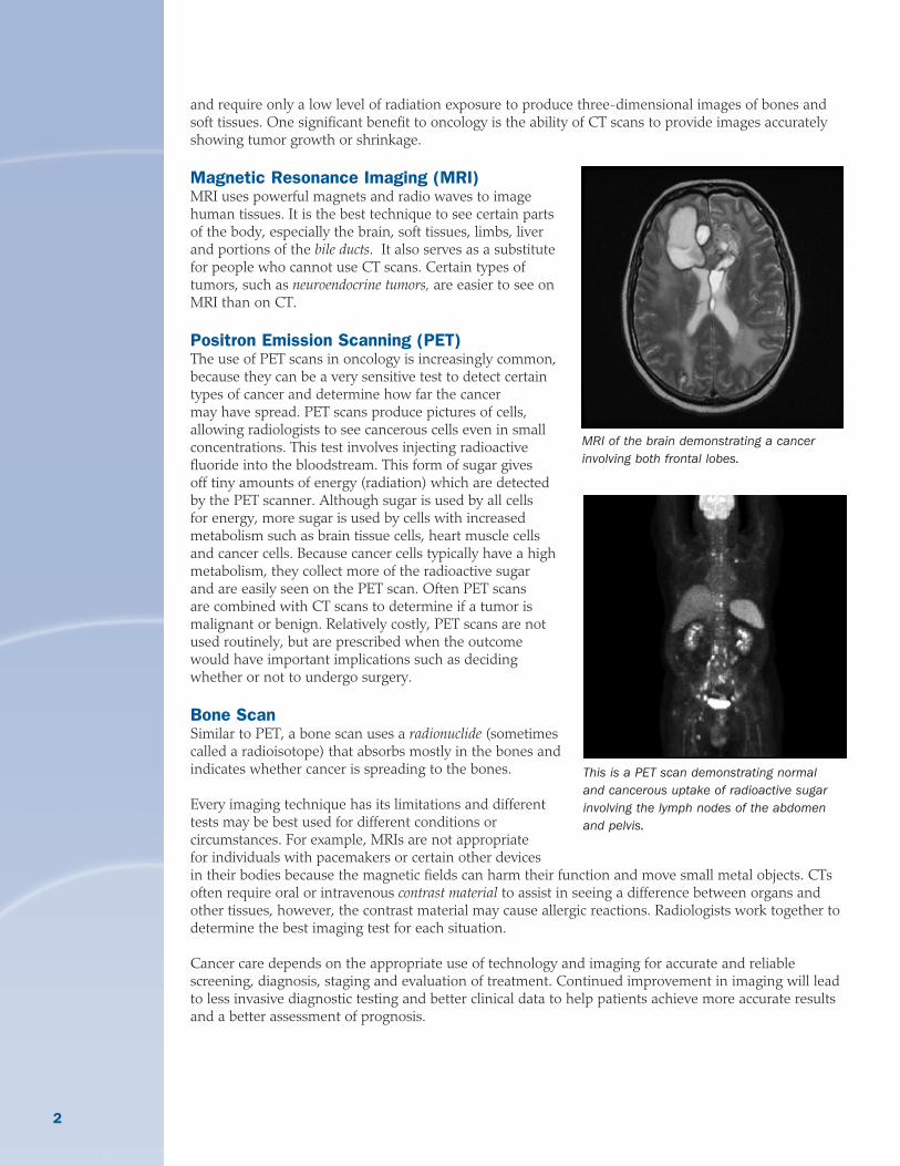
Magnetic Resonance Imaging (MRI)MRI uses powerful magnets and radio waves to image human tissues. It is the best technique to see certain parts of the body, especially the brain, soft tissues, limbs, liver and portions of the bile ducts. It also serves as a substitute for people who cannot use CT scans. Certain types of tumors, such as neuroendocrine tumors, are easier to see on MRI than on CT.
Positron Emission Scanning (PET)The use of PET scans in oncology is increasingly common, because they can be a very sensitive test to detect certain types of cancer and determine how far the cancer may have spread. PET scans produce pictures of cells, allowing radiologists to see cancerous cells even in small concentrations. This test involves injecting radioactive fluoride into the bloodstream. This form of sugar gives off tiny amounts of energy (radiation) which are detected by the PET scanner. Although sugar is used by all cells for energy, more sugar is used by cells with increased metabolism such as brain tissue cells, heart muscle cells and cancer cells. Because cancer cells typically have a high metabolism, they collect more of the radioactive sugar and are easily seen on the PET scan. Often PET scans are combined with CT scans to determine if a tumor is malignant or benign. Relatively costly, PET scans are not used routinely, but are prescribed when the outcome would have important implications such as deciding whether or not to undergo surgery.
Bone ScanSimilar to PET, a bone scan uses a radionuclide (sometimes called a radioisotope) that absorbs mostly in the bones and indicates whether cancer is spreading to the bones.
Every imaging technique has its limitations and different tests may be best used for different conditions or circumstances. For example, MRIs are not appropriate for individuals with pacemakers or certain other devices in their bodies because the magnetic fields can harm their function and move small metal objects. CTs often require oral or intravenous contrast material to assist in seeing a difference between organs and other tissues, however, the contrast material may cause allergic reactions. Radiologists work together to determine the best imaging test for each situation.
Cancer care depends on the appropriate use of technology and imaging for accurate and reliable screening, diagnosis, staging and evaluation of treatment. Continued improvement in imaging will lead to less invasive diagnostic testing and better clinical data to help patients achieve more accurate results and a better assessment of prognosis.
This is a PET scan demonstrating normal and cancerous uptake of radioactive sugar involving the lymph nodes of the abdomen and pelvis.
MRI of the brain demonstrating a cancer involving both frontal lobes.
3
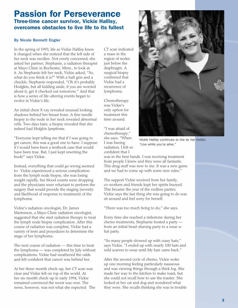
Passion for PerseveranceThree-time cancer survivor, Vickie Hallisy, overcomes obstacles to live life to its fullest
By Nicole Bennett Engler
In the spring of 1993, life as Vickie Hallisy knew it changed when she noticed that the left side of her neck was swollen. Not overly concerned, she asked her partner, Stephanie, a radiation therapist at Mayo Clinic in Rochester, Minn., to look at it. As Stephanie felt her neck, Vickie asked, “So, what do you think it is?” With a half grin and a chuckle, Stephanie responded, “Oh it’s probably Hodgkin, but all kidding aside, if you are worried about it, get it checked out tomorrow.” And that is how a series of life-altering events began to evolve in Vickie’s life. An initial chest X-ray revealed unusual looking shadows behind her breast bone. A fine needle biopsy to the node in her neck revealed abnormal cells. Two days later, a biopsy revealed that she indeed had Hodgkin lymphoma.
“Everyone kept telling me that if I was going to get cancer, this was a good one to have. I suppose if I would have been a textbook case that would have been true. But, I just kept rewriting the book!” says Vickie.
Instead, everything that could go wrong seemed to: Vickie experienced a serious complication from the lymph node biopsy, she was losing weight rapidly, her blood counts were dropping and the physicians were reluctant to perform the surgery that would provide the staging (severity and likelihood of response to treatment) of the lymphoma.
Vickie’s radiation oncologist, Dr. James Martenson, a Mayo Clinic radiation oncologist, suggested that she start radiation therapy to treat the lymph node biopsy complication. After this course of radiation was complete, Vickie had a variety of tests and procedures to determine the stage of her lymphoma.
The next course of radiation — this time to treat the lymphoma — was completed by July without complications. Vickie had weathered the odds and felt confident that cancer was behind her.
At her three-month check-up, her CT scan was clear and Vickie felt on top of the world. At her six-month check-up in early 1994, Vickie remained convinced the worst was over. The news, however, was not what she expected: The
CT scan indicated a mass in the region of nodes just below the diaphragm. A surgical biopsy confirmed that Vickie had a recurrence of lymphoma.
Chemotherapy was Vickie’s only option for treatment this time around.
“I was afraid of chemotherapy,” she says. “When I was having radiation, I felt so confident that I was in the best hands. I was receiving treatment from people I knew and they were all fantastic. This drug stuff was new to me. It was a new game and we had to come up with some new rules.”
The support Vickie received from her family, co-workers and friends kept her spirits buoyed. This became the year of the endless parties. Vickie says the last thing she was going to do was sit around and feel sorry for herself.
“There was too much living to do,” she says.
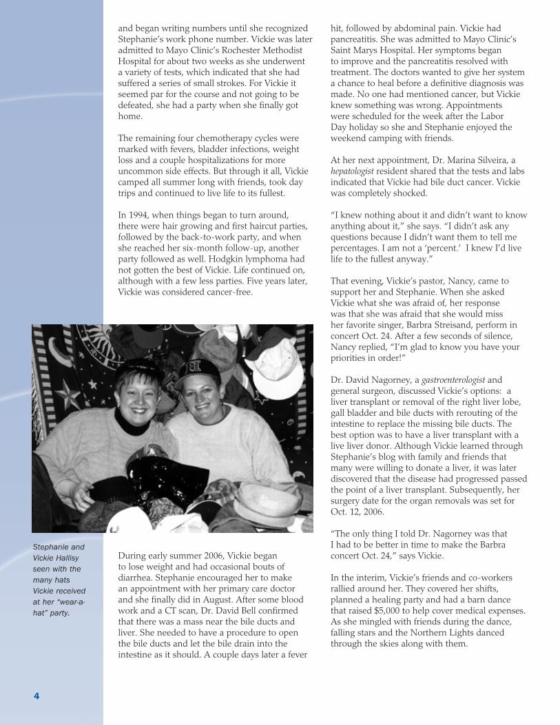
Every time she reached a milestone during her chemo treatments, Stephanie hosted a party — from an initial head-shaving party to a wear-a-hat party.
“So many people showed up with crazy hats,” says Vickie. “I ended up with nearly 100 hats and wild scarves to wear until My hair came back.”
After the second cycle of chemo, Vickie woke up one morning feeling particularly nauseous and was viewing things through a thick fog. She made her way to the kitchen to make toast, but she could not recall how to use the toaster. She looked at her cat and dog and wondered what they were. She recalls thinking she was in trouble
Vickie Hallisy continues to live by her motto: “Live while you’re alive.”
4
and began writing numbers until she recognized Stephanie’s work phone number. Vickie was later admitted to Mayo Clinic’s Rochester Methodist Hospital for about two weeks as she underwent a variety of tests, which indicated that she had suffered a series of small strokes. For Vickie it seemed par for the course and not going to be defeated, she had a party when she finally got home.
The remaining four chemotherapy cycles were marked with fevers, bladder infections, weight loss and a couple hospitalizations for more uncommon side effects. But through it all, Vickie camped all summer long with friends, took day trips and continued to live life to its fullest.
In 1994, when things began to turn around, there were hair growing and first haircut parties, followed by the back-to-work party, and when she reached her six-month follow-up, another party followed as well. Hodgkin lymphoma had not gotten the best of Vickie. Life continued on, although with a few less parties. Five years later, Vickie was considered cancer-free.
During early summer 2006, Vickie began to lose weight and had occasional bouts of diarrhea. Stephanie encouraged her to make an appointment with her primary care doctor and she finally did in August. After some blood work and a CT scan, Dr. David Bell confirmed that there was a mass near the bile ducts and liver. She needed to have a procedure to open the bile ducts and let the bile drain into the intestine as it should. A couple days later a fever
hit, followed by abdominal pain. Vickie had pancreatitis. She was admitted to Mayo Clinic’s Saint Marys Hospital. Her symptoms began to improve and the pancreatitis resolved with treatment. The doctors wanted to give her system a chance to heal before a definitive diagnosis was made. No one had mentioned cancer, but Vickie knew something was wrong. Appointments were scheduled for the week after the Labor Day holiday so she and Stephanie enjoyed the weekend camping with friends. At her next appointment, Dr. Marina Silveira, a hepatologist resident shared that the tests and labs indicated that Vickie had bile duct cancer. Vickie was completely shocked.
“I knew nothing about it and didn’t want to know anything about it,” she says. “I didn’t ask any questions because I didn’t want them to tell me percentages. I am not a ‘percent.’ I knew I’d live life to the fullest anyway.”
That evening, Vickie’s pastor, Nancy, came to support her and Stephanie. When she asked Vickie what she was afraid of, her response was that she was afraid that she would miss her favorite singer, Barbra Streisand, perform in concert Oct. 24. After a few seconds of silence, Nancy replied, “I’m glad to know you have your priorities in order!”
Dr. David Nagorney, a gastroenterologist and general surgeon, discussed Vickie’s options: a liver transplant or removal of the right liver lobe, gall bladder and bile ducts with rerouting of the intestine to replace the missing bile ducts. The best option was to have a liver transplant with a live liver donor. Although Vickie learned through Stephanie’s blog with family and friends that many were willing to donate a liver, it was later discovered that the disease had progressed passed the point of a liver transplant. Subsequently, her surgery date for the organ removals was set for Oct. 12, 2006.
“The only thing I told Dr. Nagorney was that I had to be better in time to make the Barbra concert Oct. 24,” says Vickie.
In the interim, Vickie’s friends and co-workers rallied around her. They covered her shifts, planned a healing party and had a barn dance that raised $5,000 to help cover medical expenses. As she mingled with friends during the dance, falling stars and the Northern Lights danced through the skies along with them.
Stephanie and Vickie Hallisy seen with the many hats Vickie received at her “wear-a-hat” party.
5
“I knew that night that there was so much positive energy surrounding me, and the universe seemed to be confirming what I believed to be true,” says Vickie. “I was going to live.”
The surgery on Oct. 12 was a success. There was Barbra paraphernalia hanging all over her room and a concert countdown on her white board. Every doctor, nurse, aide or housekeeper that stepped foot in her room knew the goal of Oct. 24 and they all worked to make it happen. Vickie was dismissed from the hospital the morning of the concert and she and Stephanie were chauffered to the concert in style in a Cadillac.
“I cried through the entire concert,” says Vickie. “Listening to Barbra’s beautiful voice was a dream of my lifetime. That was healing and such great therapy.”
Approaching three years cancer-free, Vickie credits her faith, family and friends — especially Stephanie — for helping her through such life-changing events. She feels blessed to be feeling healthy and enjoys her work as a patient care
assistant at Mayo’s Sick Child Care Center (Children’s R & R). Outside of work, Vickie enjoys traveling, cooking, gardening and spending time with her dog, Jonah.
Like she did during her cancer journey, Vickie continues to live by her motto: “Live while you’re alive.”
National Cancer Institute Renews Mayo Clinic Cancer Center Support Grant, Extends Comprehensive StatusMayo Clinic Cancer Center (MCCC) has received an additional five years of National Cancer Institute (NCI) funding as well as re-designation as a comprehensive cancer center.
“The NCI renewal of Mayo’s Cancer Center Support Grant ensures the continuity of research programs that contribute to improvements in medical options for each cancer patient who comes to Mayo Clinic,” says MCCC Director Robert Diasio, M.D. “This NCI grant is key in Mayo Clinic’s role to provide the best care for cancer patients.”
The NCI Cancer Center Support Grant (CCSG) award to MCCC totals more than $28 million over five years to provide support for MCCC researchers across the three sites. This renewal is the seventh consecutive five-year CCSG awarded to Mayo.
A unique and significant characteristic of the MCCC is that it is the only NCI-designated comprehensive cancer center that conducts research at distinct locations across the United States. MCCC is headquartered in Rochester with research campuses in Scottsdale, Ariz., and Jacksonville, Fla. With the approval of the NCI in 2003, MCCC incorporated its cancer research activities at its Minnesota, Arizona and Florida sites into a single, integrated institution.
“The NCI designation of ‘comprehensive cancer center’ implies a robust research institution that approaches cancer with a full spectrum of basic, translational and clinical studies leading to improved choices and opportunities for patients and their physicians to address each individual’s cancer,” Dr. Diasio says.
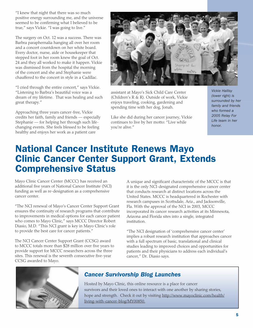
Vickie Hallisy (lower right) is surrounded by her family and friends who formed a 2005 Relay For Life team in her honor.
Cancer Survivorship Blog Launches Hosted by Mayo Clinic, this online resource is a place for cancer survivors and their loved ones to interact with one another by sharing stories, hope and strength. Check it out by visiting http://www.mayoclinic.com/health/living-with-cancer-blog/MY00850.
6
Cancer Terms
Book Review
Bile duct A tube that carries bile (fluid made by the liver that helps digest fat) between the liver and gallbladder and the intestine.
Contrast material A dye or other substance that helps show abnormal areas inside the body. Contrast material may be used with X-rays, CT scans, MRI or other imaging tests.
Gastroenterologist A doctor who specializes in diagnosing and treating disorders of the digestive system.
Hepatologist A physician who specializes in diseases of the liver.
Hodgkin lymphoma A cancer of the immune system. See Together issue, Fall 2008: vol. 8, no. 3, available at http://www.mayoclinic.org/cancer-education-rst/newsletters.html
Invasive testing A medical procedure that enters the body, usually by cutting or puncturing the skin or by inserting instruments into the body.
Neuroendocrine A tumor that forms from cells that release hormones in response to a signal from the tumor nervous system. Examples include carcinoid tumors, islet cell tumors, medullary thyroid carcinomas, pheochromocytomas and neuroendocrine carcinomas of the skin (Merkel cell cancer).
Pathology The description of cells and tissues based on microscopic evidence, and sometimes used to make a diagnosis of a disease.
Radiofrequency A procedure that uses radio waves to heat and destroy abnormal cells. ablation
Radionuclide An unstable form of a chemical element that releases radiation as it breaks down and becomes more stable. In medicine, it is used in imaging tests and in treatment. Also called radioisotope.
Virtual colonoscopy A method to examine the colon by taking a series of X-rays (called a CT scan) and using a high-powered computer to reconstruct 2-D and 3-D pictures of the interior surfaces of the colon.
Definitions obtained from www.cancer.gov. Defined terms are in italics in newsletter.
Writing for Wellness: A Prescription for Healing by Julie Davey, Idyll Arbor, Incorporated, 2007
Written by a breast cancer survivor and college writing professor, this book offers a blueprint of how to express the feelings that are experienced by patients facing cancer. The author uses writing as a means to promote spiritual and psychological healing, and encourages sharing of words to express the feelings and fears that are encountered.
Examples of prose and poetry of her “Writing for Wellness” students are included, as well as a section on how to translate feelings into words to begin the process of healing. This book serves as a resource for cancer patients, family members and caregivers, and the medical professionals who care for them.
A cancer diagnosis presents many challenging decisions, from treatment plans to costs associated with transportation and lodging. These decisions are best made with a full set of facts, and may be influenced by the distance involved in traveling to treatment or the availability of free or low-cost lodging while away from home.
American Cancer Society patient navigators are available to provide information, day-to-day help, and emotional support to cancer patients and their caregivers. Patient navigators are familiar with local resources available near treatment centers, including transportation and lodging, and have access to a national network of cancer resources.
Lodging options for individuals undergoing cancer treatment may include reduced-cost hotels, agency-regulated lodging programs or guest homes operated by non-profit and faith organizations.
The American Cancer Society’s Hope Lodge is a free home-away-from-home that offers a sense of community and comfort for cancer patients and
Navigator Notes: Lodging Assistance By Angela Young, American Cancer Society Patient Navigator
their caregiver while receiving cancer treatment.
The Society operates Hope Lodge facilities near its Mayo Clinic Minnesota and Arizona campuses. Across the nation, 27 Hope Lodges are spread across 18 states. Eligibility criteria varies per facility.
For those who prefer to stay at home and travel to their treatment center, a patient navigator can help research air and ground transportation options. Options may include the Society’s Road to Recovery Program, bus vouchers or tokens, local transport services and air transportation. In addition, some non-profit organizations offer funds through an application process for assistance with treatment-related travel costs.
A simple visit with a patient navigator can help individuals and family members create transportation and lodging plans.
While at Mayo Clinic in Minnesota, stop by the Cancer Education Center in the Gonda Building, street level, or the 10th floor resource area to meet the ACS patient navigators Jeri and Angela. You may also contact them by calling 507-266-9288.
Visit Celeste “CC” Chervenka at Mayo Clinic Arizona’s Patient Health and Education Library on the concourse level in the Clinic or call her at 480-301-5990.
For more information or to connect with a patient navigator in your area, call 1-800-227-2345 or visit www.cancer.org.
7Celeste “CC” Chervenka
Jeri Lensing Angela Young
American Cancer Society Navigator Notes
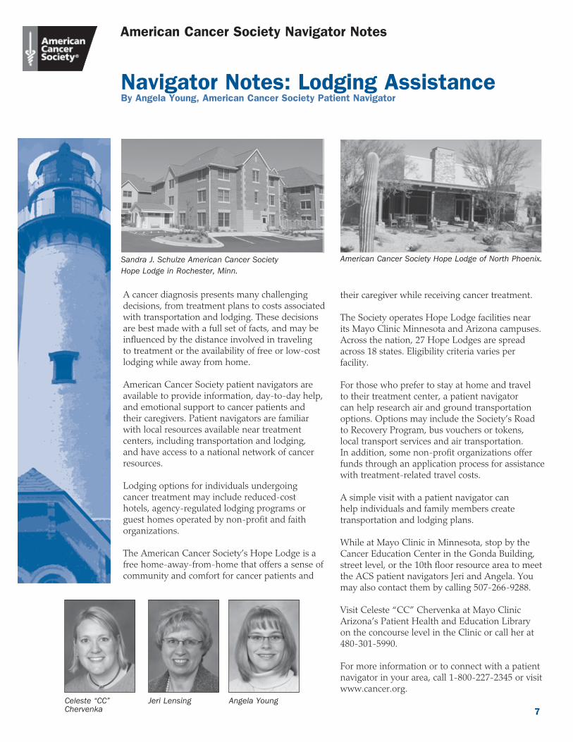
Sandra J. Schulze American Cancer Society Hope Lodge in Rochester, Minn.
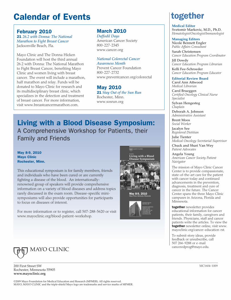
American Cancer Society Hope Lodge of North Phoenix.
together Medical Editor Svetomir Markovic, M.D., Ph.D. Hematologist/Oncologist/Immunologist
Managing Editors Nicole Bennett Engler Public Affairs Consultant Sarah Christensen Cancer Education Program Coordinator Jill Dowdy Cancer Education Program LibrarianKelli Fee-Schroeder Cancer Education Program Educator
Editorial Review Board Carol Ann Attwood Medical Librarian Carol Brueggen Certified Oncology Clinical Nurse Specialist Sylvan Hengesteg Chaplain Deborah A. Johnson Administrative Assistant Brent Moos Social Worker Jacalyn See Registered DietitianJulie Tienter Medical Oncology Secretarial Supervisor Chuck and Shari Van Wey Patient Advocates Angela Young American Cancer Society Patient Navigator
The mission of Mayo Clinic Cancer Center is to provide compassionate, state-of-the-art care for the patient with cancer today and continued advancements in the prevention, diagnosis, treatment and cure of cancer in the future. The Cancer Center spans the three Mayo Clinic campuses in Arizona, Florida and Minnesota.
together newsletter provides educational information for cancer patients, their family, caregivers and friends. Physicians, staff and cancer patients write the articles. To view the together newsletter online, visit www.mayoclinic.org/cancer-education-rst.
To submit story ideas, provide feedback or unsubscribe, call 507 266-9288 or e-mail [email protected].
MC1604-1009
Calendar of Events
February 201021 26.2 with Donna: The National Marathon to Fight Breast CancerJacksonville Beach, Fla.
Mayo Clinic and The Donna Hicken Foundation will host the third annual 26.2 with Donna: The National Marathon to Fight Breast Cancer, benefiting Mayo Clinic and women living with breast cancer. The event will include a marathon, half marathon and relay. Funds will be donated to Mayo Clinic for research and its multidisciplinary breast clinic, which specializes in the detection and treatment of breast cancer. For more information, visit www.breastcancermarathon.com.
March 2010Daffodil Days American Cancer Society800-227-2345www.cancer.org
National Colorectal Cancer Awareness MonthPrevent Cancer Foundation800-227-2732www.preventcancer.org/colorectal
May 201021 Stay Out of the Sun RunRochester, Minn.www.sosrun.org
Living with a Blood Disease Symposium: A Comprehensive Workshop for Patients, their Family and Friends
May 8-9, 2010Mayo ClinicRochester, Minn.
This educational symposium is for family members, friends and individuals who have been cured or are currently fighting a disease of the blood. An internationally-renowned group of speakers will provide comprehensive information on a variety of blood diseases and address topics rarely discussed in the exam room. Disease-specific mini-symposiums will also provide opportunities for participants to focus on diseases of interest.
For more information or to register, call 507-288-5620 or visit www.mayoclinic.org/blood-patient-workshop.