Embed Size (px)
Citation preview

IN THE NAME OF GOD

Case Presentation
Supervisor :
Dr. M. Derakhshan
Presenter :
Dr. S. Nikpour
Special Thanks to :
Dr. F. Mohammadizadeh

Patient is a 33 years old woman, G2L2 , last delivery 33
month ago
chief complaint : spoting
Sono Finding : A mass measuring


What’s your plan?

ß- HCG


HPL


Differential Diagnosis
Choriocarsinoma
Placental Site Trophoblastic Tumor (PSTT)
Epithelioid trophoblastic tumor


Choriocarcinoma:The characteristic biphasic pattern of cytotrophoblast and syncytiotrophoblast is seen.

Choriocarcinoma:Syncytiotrophoblastic component is often identified in the middle of cytotrophoblast instead of encasing the cytotrophoblastic component.

Choriocarcinoma:Choriocarcinoma predominantly composed of a monotonous sheet of cytotrophoblast with an indistinct syncytiotrophoblastic component may mimic poorly differentiated carcinoma. Immunostaining for β-hCG can be helpful in demonstrating attenuated syncytiotrophoblast.

Placental site trophoblastic tumor (PSTT):clinical presentation
It can occur in any reproductive age group, but has beenreported in postmenopausal women.
The majority follow a normal full-term or terminated pregnancy, suggesting that it may arise from a malignant transformation of previously healthy trophoblastic cells.
Ectopic or molar antecedent pregnancy is also seen.
The interval from antecedent pregnancy ranges from 5 to 131 months (mean, 34).
Patients present with only mildly elevated levels of serum β-hCG.
The most common presenting symptom is vaginal bleeding and/or amenorrhea, occasionally with symptoms related to the site of tumor metastasis.
Proteinuria, galactorrhea, virilization, and polycythemia have been described.

Placental site trophoblastic tumor (PSTT):gross findings
The tumor usually forms an ill-defined intramural or protruding mass in the uterine corpus, sometimes extending to the cervix.
Usually it is deeply invasive into the myometrium.
The cut surface is yellow or tan, and solid with frequent hemorrhage and necrosis.

Placental site trophoblastic tumor (PSTT):microscopic findings
Microscopically, tumors are highly infiltrative, separating myometrial fibers with confluent sheets of epithelioid cells that may become spindle shaped as they infiltrate between myometrium.
Tumor cells are predominantly mononucleated large polygonal cells with abundant amphophilic, eosinophilic, or clear cytoplasm.
Syncytiotrophoblast-like multinucleated cells can be identified, but are individually scattered among the mononucleated cells without forming a typical biphasic pattern of choriocarcinoma.

Placental site trophoblastic tumor (PSTT):microscopic findings
Nuclei have variable degrees of atypia, frequent nuclear grooves, and intranuclear cytoplasmic pseudoinclusions.
The mitotic count is variable between cases, and from field to field even in the same case, ranging from 0 to 20 (average 5.0) per 10 HPF.
Atypical mitotic figures are frequent.
Coagulative necrosis and deposition of extracellular eosinophilic fibrinoid material around the tumor cells is a frequent feature, often leaving islands of tumor cells only around blood vessels.
Chorionic villi are absent in almost all cases, except in a rare case associated with molar pregnancy.
Epithelioid trophoblastic tumor (ETT):clinical presentation
The tumor mostly occurs in women of reproductive age, but very rarely it can occur in postmenopausal women.
The interval between the preceding gestational event and the diagnosis of epithelioid trophoblastic tumor is variable, ranging from 1 month to 18 years.
The preceding gestational event may be full-term delivery, spontaneous abortion, hydatidiform mole, or invasive mole.

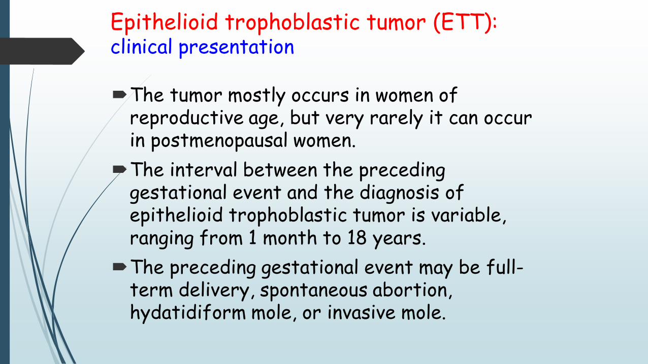
Epithelioid trophoblastic tumor (ETT):The tumor has generally well-circumscribed margins, frequently surrounded by an abundant lymphocytic infiltration.
Epithelioid trophoblastic tumor (ETT):The tumor cells have relatively smaller, uniform, and less pleomorphic nuclei than PSTT and abundant clear or eosinophilic cytoplasm growing in sheets. Frequently, eosinophilic hyaline material is present within the tumor.