Embed Size (px)
Citation preview
In the Name of Allah the Most Beneficent and Merciful
CardiomyopathiesCardiomyopathiesProf. Dr. Muhammad Akbar Chaudhry
M.R.C.P.( UK ) , F.R.C.P.( E )
F.R.C.P. ( LONDON ), F.A.C.C.
Designed at :Audiovisual Department. , Designed by: Masooma ZaidiDesigned at :Audiovisual Department. , Designed by: Masooma Zaidi

Cardiomyopathies
• Primary
“Heart muscle disease of unknown cause”
• Secondary
Myocardial involvement in systemic
disease “ (rare specific heart muscle disease)”
• Primary
“Heart muscle disease of unknown cause”
• Secondary
Myocardial involvement in systemic
disease “ (rare specific heart muscle disease)”
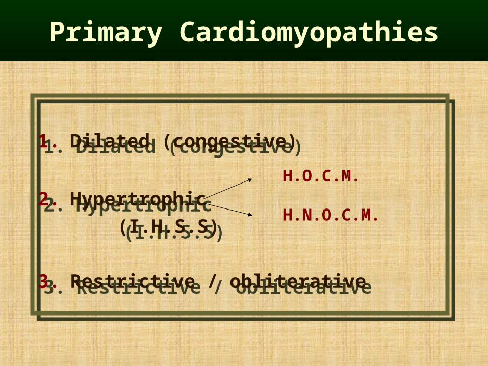
Primary Cardiomyopathies
1. Dilated (congestive)
2. Hypertrophic
(I.H.S.S)
3. Restrictive / obliterative
1. Dilated (congestive)
2. Hypertrophic
(I.H.S.S)
3. Restrictive / obliterative
H.O.C.M.
H.N.O.C.M.

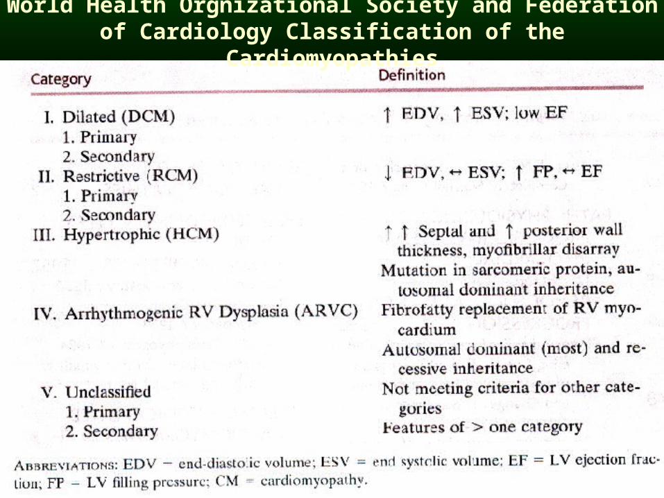
World Health Organization Classifications of Cardiomyopathies
World Health Organization Classifications of Cardiomyopathies
( 1980 )

Cont.
( 1995 )
Cont.

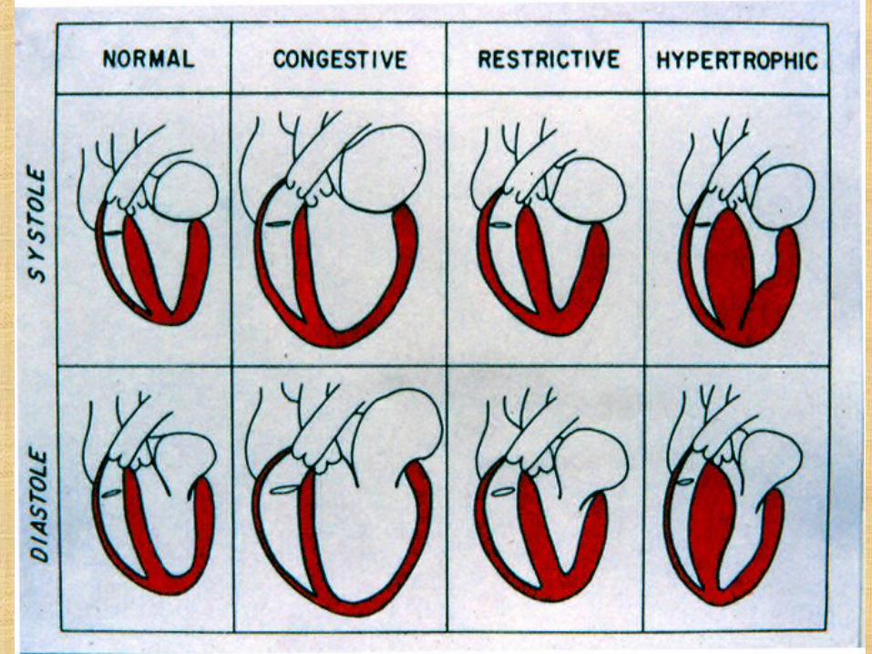
Functional Classification of Cardiomyopathies

Etiological Classification of
Cardiomyopathies
Etiological Classification of
Cardiomyopathies

Etiological Classification of
Cardiomyopathies
Cont.

Etiological Classification of
Cardiomyopathies
Cont.

Etiological Classification of
Cardiomyopathies
Cont.

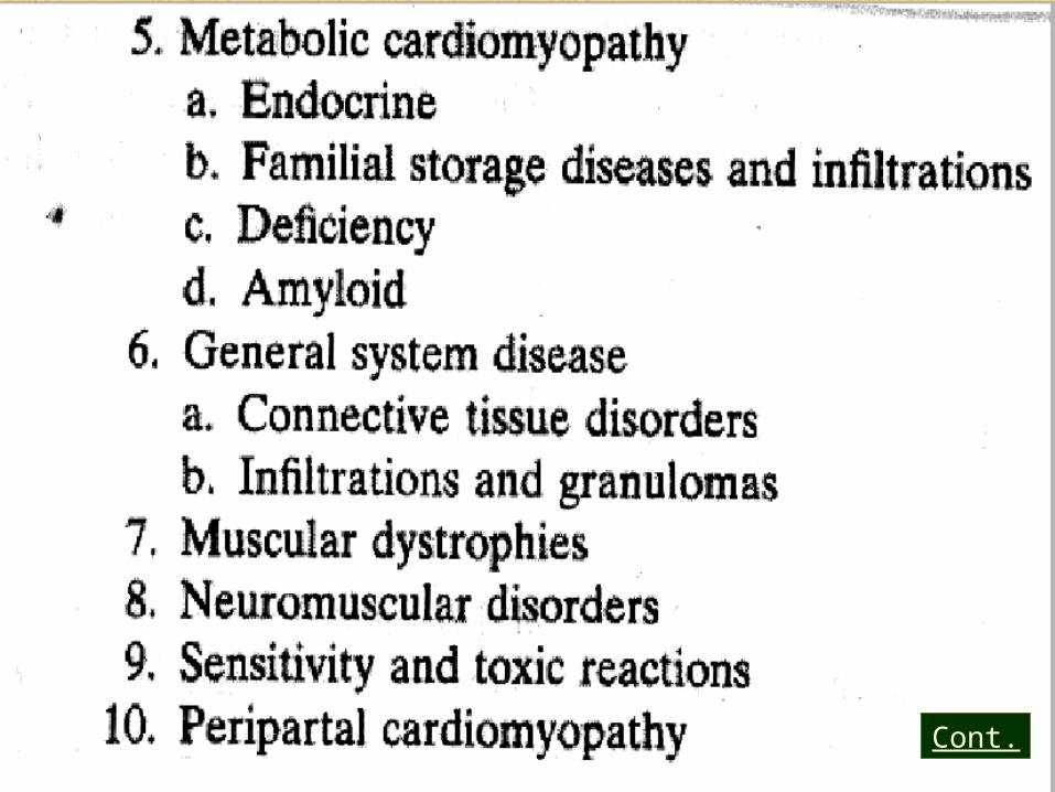
Etiological Classification of
Cardiomyopathies
Cont.

Etiological Classification of
Cardiomyopathies
Cont.
World Health Orgnizational Society and Federation of Cardiology Classification of the Cardiomyopathies

Various Etiologies That Can Lead to Cardiomyopathy
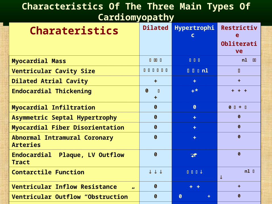
Characteristics Of The Three Main Types Of CardiomyopathyCharacteristics Of The Three Main Types Of Cardiomyopathy
Charateristics Dilated Hypertrophic Restrictive
Obliterative
Myocardial Mass nl
Ventricular Cavity Size nl
Dilated Atrial Cavity + + +
Endocardial Thickening 0 + +* + + +
Myocardial Infiltration 0 0 0 +
Asymmetric Septal Hypertrophy 0 + 0
Myocardial Fiber Disorientation 0 + 0
Abnormal Intramural Coronary Arteries 0 + 0
Endocardial Plaque, LV Outflow Tract 0 + 0
Contarctile Function nl
Ventricular Inflow Resistance 0 + + +
Ventricular Outflow “Obstruction” 0 0 + 0
LV Filling Pressure nl
Mitral Regurgitation + + +
Thickened Mitral Valve 0 + ±
Intracardiac thrombi + 0 1

Dilated Cardiomyopathy (D.C)Dilated Cardiomyopathy (D.C)
Cardiomegaly with dilatation of both ventricles.Impairment of systolic function.Increased myocardial mass.(dilatation more impressive than the hypertrophy)
FactorsAlcohol, hypertension, pregnancy, immunological disorders, viral infections, chemical agents.
Cardiomegaly with dilatation of both ventricles.Impairment of systolic function.Increased myocardial mass.(dilatation more impressive than the hypertrophy)
FactorsAlcohol, hypertension, pregnancy, immunological disorders, viral infections, chemical agents.

Three General Mechanism by Which Alterations in Gene Expression Can Influence the Development or Progression of
a Dilated Cardiomyopathy
Types of Process Examples

Types of Dilated Cardiomyopathies
Cont.

Cont.
Dilated Cardiomyopathy Dilated Cardiomyopathy
Epidemiology:• World wide• All ages and races• More common in men than in women.• ?genetic predisposition.Pathology• Dilatation of both ventricles (Lt . more than Rt.)• Poor vent .contraction and reduced ejection
fraction.• Relative stasis of blood and clot formation.• Focal endocardial thickening• Leaflets of cardiac valves usually normal
(occasionally margins show focal thickening) .• Valve ring dilation.

Dilated CardiomyopathyClinical Manifestations
HistoryInsidious onset of left vent. Failure
Followed by symptoms of Rt. Sided congestive failure
Chest pain may occur specially with exertion
HistoryInsidious onset of left vent. Failure
Followed by symptoms of Rt. Sided congestive failure
Chest pain may occur specially with exertion

PresentationPresentation
Progressive congestive cardiac failure.
Arrhythmias
Atypical chest pain
Physical examinationCardiomegaly
Gallop rhythm
C.C.F.
Varying degree of mitral regurgitation.
Atrial fibrillation 10-20%.
Progressive congestive cardiac failure.
Arrhythmias
Atypical chest pain
Physical examinationCardiomegaly
Gallop rhythm
C.C.F.
Varying degree of mitral regurgitation.
Atrial fibrillation 10-20%.

Physical Examination
Signs of C.C.F.Skin cold, pale , cyanosedPeripheral veins constrictedArterial pulse of small volumePulsus alternans Tachycardia at restJ.V.P. is raisedTricuspid regurgitationApex beat displaced out-side mid.C.lineRt.V & L.V. pulsation due to dilatations, systolic murmurs of mitral & tricuspid regurgitationsGallop rhythm –III H.S.& 4th H.S. P2loud.Reduced pulse pressurePericardial effusion may be presentBasal crepts in lungs with pleural effusionHepatomegoly & liver may be pulsatilePeriphral oedema & ascites
Signs of C.C.F.Skin cold, pale , cyanosedPeripheral veins constrictedArterial pulse of small volumePulsus alternans Tachycardia at restJ.V.P. is raisedTricuspid regurgitationApex beat displaced out-side mid.C.lineRt.V & L.V. pulsation due to dilatations, systolic murmurs of mitral & tricuspid regurgitationsGallop rhythm –III H.S.& 4th H.S. P2loud.Reduced pulse pressurePericardial effusion may be presentBasal crepts in lungs with pleural effusionHepatomegoly & liver may be pulsatilePeriphral oedema & ascites

InvestigationsInvestigations
1. X-ray chestDilated and enlarged heartPul. Venous hypertensionEnlarged main Pul. ArteriesPleural effusionWhen pericardial effusionwater bottle shape heart
2. ECGSinus tachycardiaNon specific changes flat & inverted T wavesAtrial fibrillation commonQRS low voltage &wideL.V.H.+Conductive disturbances


3. EchocardiographyAll chambers dilated &poorly contracting
Pericardial effusion may be present
Ventricular & atrial thrombi
Regional wall motion abnormalities
4. Ambulatory E.C.G monitoring
5. Angiocardiography
6. Endo-myocardial biopsy
3. EchocardiographyAll chambers dilated &poorly contracting
Pericardial effusion may be present
Ventricular & atrial thrombi
Regional wall motion abnormalities
4. Ambulatory E.C.G monitoring
5. Angiocardiography
6. Endo-myocardial biopsy
InvestigationsInvestigations