Embed Size (px)
Citation preview
Research ArticleIn Situ Cytokine Expression and Morphometric Evaluation ofTotal Collagen and Collagens Type I and Type III in Keloid Scars
Isabela Rios da Silva,1 Luciana Colombo Rodrigues da Cunha Tiveron,1
Marcos Vinicius da Silva,1 Alberto Borges Peixoto,2 Carla Aparecida Xavier Carneiro,2
M. A. dos Reis,1 Pedro Carvalho Furtado,1 Bárbara Rocha Rodrigues,2
Virmondes Rodrigues Jr.,1 and Denise Bertulucci Rocha Rodrigues1,2
1Federal University of Triângulo Mineiro (UFTM) ICBN and CEFORES, Uberaba, MG, Brazil2Laboratory of Biopathology and Molecular Biology, University of Uberaba (UNIUBE), Uberaba, MG, Brazil
Correspondence should be addressed to Virmondes Rodrigues Jr.; [email protected]
Received 26 December 2016; Revised 14 March 2017; Accepted 27 March 2017; Published 30 May 2017
Academic Editor: Raffaele Strippoli
Copyright © 2017 Isabela Rios da Silva et al. This is an open access article distributed under the Creative Commons AttributionLicense, which permits unrestricted use, distribution, and reproduction in anymedium, provided the original work is properly cited.
Keloids are characterized by excessive collagen deposition and growth beyond the edges of the initial injury, and cytokines may berelated to their formation. The objective of this study was to evaluate the collagen fibers, analyze in situ expression of cytokines inkeloid lesions, and compare to the control group. Results showed that there was a predominance of women and nonwhite and directblack ancestry. Keloid showed a significant increase in total and type III collagen. Significantly, the expression of mRNA for TGF-βin keloid was increased, the expressions of IFN-γ, IFN-γR1, and IL-10 were lower, and IFN-γR1 and TNF-α had no statisticaldifference. Correlations between collagen type III and TGF-β mRNA expression were positive and significant, IFN-γ, IFN-γR1,and IL-10 were negative and significant, and TNF-α showed no statistical difference. We conclude that there was a significantincrease of total collagen in keloid and predominance of collagen type III compared to the controls, showing keloid as animmature lesion. There is a significant increase in TGF-β mRNA in keloid lesions, and a significant decrease in IFN-γ andIL-10, suggesting that these cytokines are related to keloid lesions.
1. Introduction
The healing process is performed by a cascade of complex,dynamic, and overlapping events, followed by an inflamma-tory, proliferative, and remodeling reaction [1]. Changes inthese normal processes result in the formation of an exagger-ated scar called keloid, characterized by growth of the lesionbeyond the initial edges [2] and the nonspontaneous regres-sion over the years [3].
The pathophysiology of keloid is still not fully elucidatedyet, although changes in the expression of cytokines [4, 5],increase in fibroblast proliferation [6], and exacerbated colla-gen synthesis [7] have been described in the literature.
The most frequent types of collagen present in the der-mis are collagen type I with 80% and type III with 20%[8]. In the skin, collagen synthesis is performed by
fibroblasts, and the genes responsible for the productionof collagen type I are COL1A1, located on chromosome17, which encodes the α1(I) chain and COL1A2, locatedon chromosome 7, which encodes the α2(I) chain [9]. Col-lagen type III is produced by the COL3A1 gene, locatedon chromosome 2 [10]. Several signaling pathways, suchas MAP kinase and NF-kB, induce collagen synthesis bytranscription of mRNA, being translated in the RER,hydroxylated, and glycosylated into procollagen [11, 12].Later, there is excretion into the extracellular medium byexocytosis, where proteolytic enzymes cleave their C andN terminal propeptides, turning it into a tropocollagen.Then, tropocollagens bind together to form collagen fibrils,which give rise to collagen fibers [13]. Type I collagen isconsidered the mature collagen [14] for being a heterotri-mer composed of two identical chains α1(I) and one α2(I)
HindawiMediators of InflammationVolume 2017, Article ID 6573802, 11 pageshttps://doi.org/10.1155/2017/6573802

[15], responsible for the strength and tension of tissues[14]. Type III collagen is an immature collagen; it is ahomotrimer, consisting of three α1(III) chains [16], syn-thesized during the early stages of healing [14].
Collagen synthesis can be induced by TGF-β, whichbinds to serine tyrosine kinase ubiquitous receptor (TβRII),and then the receptor TGF-β I (TβRI) is recruited and phos-phorylated by TβRII. The signal propagates through Smads,a family of intracellular proteins, which, in turn, transportinformation to the nucleus, stimulating transcription ofgenes (COL1A1, COL1A2, and COL3A1) and inducing theproduction of collagens type I and type III [17]. Overexpres-sion of TβRI and TβRII and increased phosphorylation ofSmad proteins were found in keloid fibroblasts, whichappear to induce excessive production of collagen [18]. Stud-ies indicate that a failure in eliminating the overexpression ofthese receptors during the remodeling phase can lead to per-sistent autocrine effect of TGF-β on keloid fibroblasts, caus-ing increased collagen synthesis [19].
However, to maintain a normal healing process, proin-flammatory cytokines are required to maintain a balancein this healing process. TNF-α, which is important in theearly process of skin healing [20], appears to be associatedwith the suppression of collagen synthesis by fibroblasts[21] and the induction of enzymes that degrade collagen[22]. Similarly, IFN-γ appears to inhibit both the prolifer-ation of fibroblasts [23] and the synthesis of extracellularmatrix components (MEC) [24]. It has been shown thatIFN-γ induces activation of Jak/STAT1 [25] pathwaysand acts at the transcriptional level by inhibiting collagenmRNA synthesis and consequently the formation of colla-gen fibers [26].
An increase in IFN-γR1 expression seems to be influ-enced by the interaction between fibroblasts and keratino-cytes [27]. The absence of IFN-γ receptors has been foundin patients with Schistosoma mansoni infection and isassociated with formation of fibrosis [28]. Polymorphismin the gene of this receptor may be associated with severeliver fibrosis, and it is believed that this receptor may berelevant to the control of fibrosis formation in other dis-eases [29].
Likewise, IL-10 also appears to have an important rolein modulating the healing process, since in vitro studieshave demonstrated its role in the induction of collagensynthesis and action of enzymes that degrade collagen,such as MMP1 and MMP8 [30]. Furthermore, IL-10 actson phosphorylation, via STAT3/AKT signaling, thus inhi-biting the collagen synthesis [31]. Thus, this cytokine hasbeen investigated for the treatment of keloids, becauseintralesional injections with IL-10 in patients with keloidshave been performed and reduced the inflammatory pro-cess, with a decrease in symptoms and consequentlyimprovement in scar appearance without causing signifi-cant side effects [32].
In this context, in the present study, we evaluated thecollagen fibers and the in situ expression signature of pro-inflammatory and anti-inflammatory cytokines in keloidlesions compared to the control biopsies obtained fromnormal scar samples.
2. Material and Methods
2.1. Casuistry. We analyzed 73 biopsies, 33 from patientswith keloid and 40 from normal scars. The biopsies were per-formed by the team of plastic surgeons at the OutpatientClinic Maria da Glória of UFTM. The study included patientsdiagnosed with keloid, who had abnormal scarring, withgrowth of the lesion edges beyond the margins of the originalscar, and signs and symptoms such as pain, itching, redness,and induration. Patients received corticosteroid (triamcino-lone at 20mg/mL) once a month, and after improvement,patients underwent reconstructive surgery and a fragmentof the tissue to be discarded was collected for this study. Aftersurgery, the patients continued with corticoid applications toprevent further recurrences.
Fragments of the normal scar taken from secundiparousor multiparous patients during cesarean section were usedas the controls.
We excluded patients who had hypertrophic scars andsecond intention scars, patients using systemic immuno-suppressants, patients with autoimmune diseases, patientswith immunodeficiencies of any etiology, or patients whoare malnourished. This project was approved by theResearch Ethics Committee (CEP) of the Federal Univer-sity of Triângulo Mineiro (UFTM) under protocol number45647315.4.0000.5154.
2.2. Collection of Material. The material was collected at theoutpatient clinic for keloid treatment, in rooms for small sur-geries, by the doctors responsible for the sector. The lesionwas marked with a surgical pen, and after anesthesia with2% lidocaine, a cut was made in the lesion with a scalpel,excising the skin with keloid and posterior suture. A part ofthe fragment was stored in RNAlater (Ambion®) for RT-PCR, and the other part was fixed in buffered formalin forhistopathological analysis.
2.3. Preparation of Material for Histochemical Analysis. Thefragments fixed in 10% formaldehyde were dehydrated inincreasing concentrations of alcohol (70 to 100%), diapha-nized in xylene and embedded in paraffin. Slides were pre-pared in 4μm thick serial sections. Serial sections wereperformed so as to the slide number 1 was stained withhematoxylin and eosin, the slide number 2 was stained withpicrosirius (PS), and the other slides were stored for furtheranalysis. After staining, the slide was mounted with a coverslip and Entellan.
2.4. Morphometric Analysis. To quantify the percentage ofcollagen fibers, the slides stained with PS were analyzedunder polarized light at 40x objective (1600x final magnifica-tion). To quantify the percentage of collagen fibers, the slidesstained by PS were divided into four quadrants and the rep-resentative number of measures calculated through the accu-mulated mean [33]. From this calculation, 10 images perquadrant were analyzed, totaling 40 images per slide, oneslide for each case.
The digitized image showed the area consisting of colla-gen, with birefringence appearance. The analysis of collagentype I was performed by visualization of the red-yellow
2 Mediators of Inflammation
birefringence; collagen type III, by observing the green bire-fringence; and total collagen, by marking the two birefrin-gence colors, following protocols established in theliterature [34, 35].
For quantification of total collagen and capture of imagesfor differentiation of collagens type I and type III, we used avideo camera coupled to a common light microscope withLeica QWin Plus® system (Leica Microsystems Inc., Wetzlar,Germany) installed on a personal computer. The analysis ofthe percentage of collagen I and III was made through aninteractive image analysis system ImageJ® (NIH, Bethesda,Maryland, US).
The maturation index was calculated from the ratiobetween percentages of collagens type I and type III; valuesabove 1 (one) show the predominance of collagen I, that is,mature, and values below 1 indicate the predominance of col-lagen type III, with the collagen considered as immature [36].
2.5. RNA Extraction. Fragments of the skin with keloid andnormal scars stored in RNAlater were ground with a tissuetear and then RNA was extracted using a RNA extractionkit (RNA SV Total RNA Isolation System, Promega, USA)according to the manufacturer’s recommendations. Afterthese procedures, the obtained RNA was eluted in 30μLdeionized and RNase-free water for quantification and prep-aration of complementary DNA (cDNA).
The cDNA was prepared from 1μg RNA, 0.5μg Oligo dT(Promega, USA) and autoclaved ultrapure water (Milli-Q).This material was taken to the thermal cycler for a cycle of5 minutes at 70°C. After immediate cooling, the materialwas added to dNTP (2.5mM), M-MLV RT reverse transcrip-tase (ImProm-II, Promega, USA), and M-MLV-5X reactionbuffer (Promega, USA). This reaction was taken to the ther-mocycler for another 1-hour cycle at 42°C followed by 3minutes at 10°C. At the end, the material was added to theprepared cDNA 75μL autoclaved ultrapure water, and sam-ples were then frozen at −20°C until analysis.
2.6. Quantitative PCR Reactions (qPCR). The quantitativemRNA expression of the genes IL-10, TNF-α, IFN-γ, TGF-β, and IFN-γR1 were analyzed by PCR reactions in real time,in cDNA samples of fragment of the skin with keloid and thecontrol scar. The TaqMan system was used in the equipmentof PCR in real time (Applied Biosystems, USA) with appro-priate primers for such reactions and using β-actin as thecontrol. The cDNA synthesized from messenger RNA wasused according to the manufacturer’s instructions. Theresults were analyzed based on the value of CT (cycle thresh-old), and the arithmetic formula to achieve the relative quan-tification was ΔΔCt=ΔCt (treated)−Ct (control) [37].
2.7. Immunohistochemistry. Indirect immunohistochemistrywas performed in order to evaluate the expression of colla-gens type I and type III. Deparaffinized sections were treatedwith 3% hydrogen peroxide in methanol for 10min forendogenous peroxidase inhibition, incubated for 30min at90°C for antigen retrieval, and then incubated with PBS 2%BSA to reduce nonspecific binding. Next, the sections wereincubated with monoclonal antibody specific for human
anticollagen I (1 : 50; NOVUS, USA; cod-NB600-450) andanticollagen III (1 : 50; Abcam, UK; cod-ab7778). In the sec-ond step, a biotinylated Link System (LSAB-K0690, Dako,Carpinteria, CA, USA) was used according to manufacturerinstructions. The reaction was visualized by incubating thesections with diaminobenzidine (Sigma, USA) and counter-staining with hematoxylin.
2.8. Statistical Analysis. Statistical analysis was performedusing the software StatView (Abacus, USA). Assumption ofnormality of quantitative variables was checked by theKolmogorov-Smirnov. The variables showed nonhomoge-neous distribution or variance and were expressed as medianwith minimum and maximum values and percentiles andanalyzed by the nonparametric Mann–Whitney test. Thecorrelation between two continuous variables with nonnor-mal distribution was analyzed by the Spearman test (rS).To compare two continuous variables in the same patients,the Wilcoxon test (U) was applied. Results were consideredstatistically significant when the probability was less than5% (p < 0 05).
3. Results
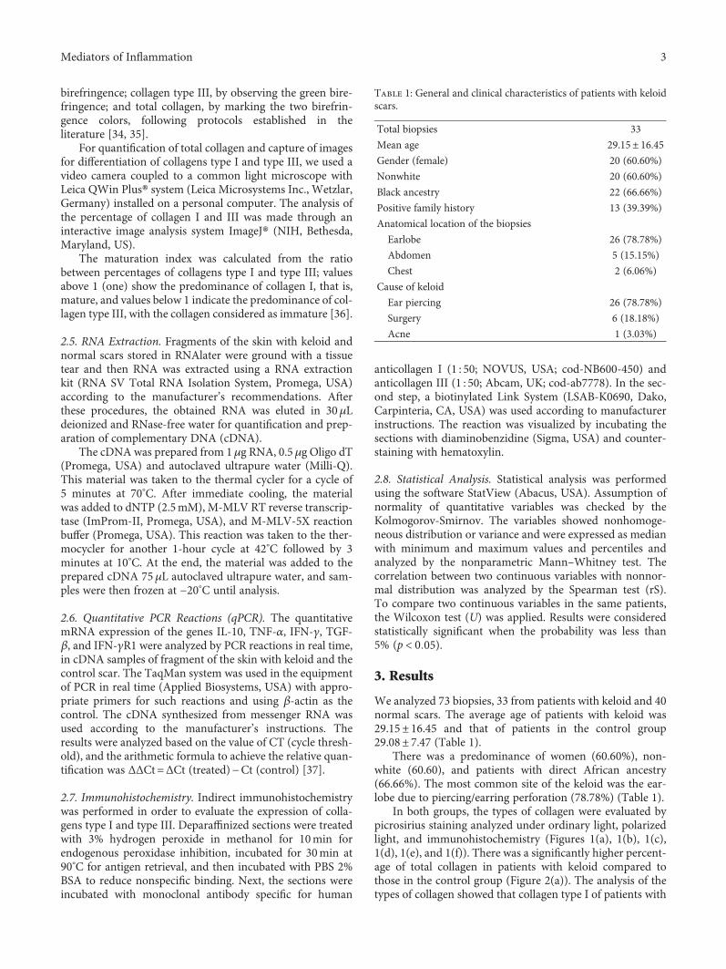
We analyzed 73 biopsies, 33 from patients with keloid and 40normal scars. The average age of patients with keloid was29.15± 16.45 and that of patients in the control group29.08± 7.47 (Table 1).
There was a predominance of women (60.60%), non-white (60.60), and patients with direct African ancestry(66.66%). The most common site of the keloid was the ear-lobe due to piercing/earring perforation (78.78%) (Table 1).
In both groups, the types of collagen were evaluated bypicrosirius staining analyzed under ordinary light, polarizedlight, and immunohistochemistry (Figures 1(a), 1(b), 1(c),1(d), 1(e), and 1(f)). There was a significantly higher percent-age of total collagen in patients with keloid compared tothose in the control group (Figure 2(a)). The analysis of thetypes of collagen showed that collagen type I of patients with
Table 1: General and clinical characteristics of patients with keloidscars.
Total biopsies 33
Mean age 29.15± 16.45Gender (female) 20 (60.60%)
Nonwhite 20 (60.60%)
Black ancestry 22 (66.66%)
Positive family history 13 (39.39%)
Anatomical location of the biopsies
Earlobe 26 (78.78%)
Abdomen 5 (15.15%)
Chest 2 (6.06%)
Cause of keloid
Ear piercing 26 (78.78%)
Surgery 6 (18.18%)
Acne 1 (3.03%)
3Mediators of Inflammation

keloids showed no significant difference compared to that ofpatients in the control group (Figure 2(b)). In turn, collagentype III was significantly higher in patients with keloid com-pared to those in the control group (Figure 2(b)). The matu-ration index indicated that biopsies of patients with keloidshowed collagen significantly more immature than the con-trol group (Figure 2(c)). The comparison between the typesof collagens, in both groups, showed that the percentage ofcollagen type I was significantly higher compared to that ofcollagen type III in the control group (Figure 2(b)). And inpatients with keloid, the percentages of collagens I and IIIwere similar, with no statistical difference (Figure 2(b)).There was also a significant positive correlation between the
percentages of collagens type I and type III in the groupsstudied (Figure 2(d)).
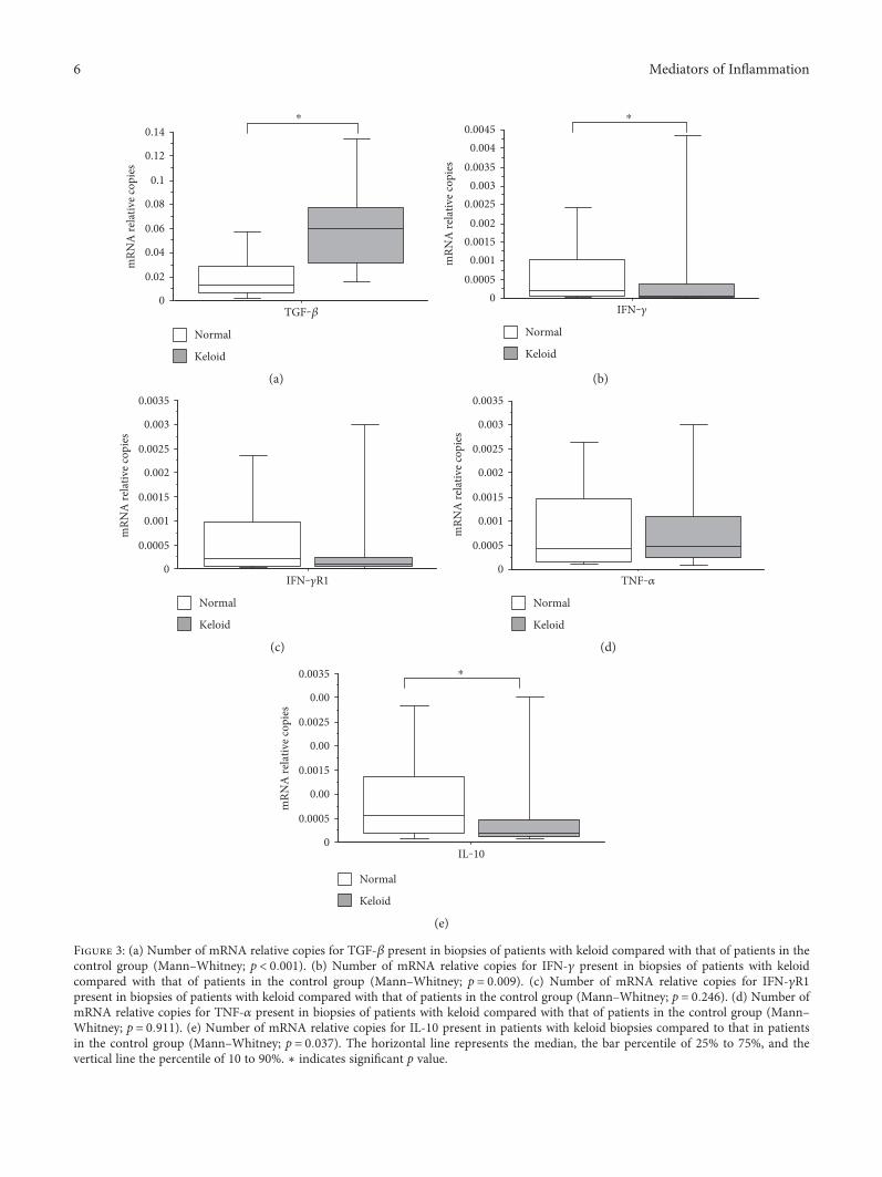
Gene expression analysis of the lesions studied showedthat the mRNA expression of TGF-β was significantly higherin patients with keloid compared to that in the control group(Figure 3(a)). The expressions of mRNA for IFN-γ and IL-10were significantly lower in patients with keloid compared tothe controls (Figures 3(b) and 3(e)).
Regarding the analysis on the relative number of mRNAcopies of IFN-γR1, patients with keloid had a lower relativenumber of copies of mRNA when compared to the controls,but without significant differences (Figure 3(c)). When ana-lyzing the relative number of copies of mRNA for TNF-α, it
(a) (b)
(c) (d)
(e) (f)
Figure 1: (a) Histological section of keloid stained with picrosirius seen in ordinary light (20x). (b) Histological section of keloid stained withpicrosirius seen in polarized light (20x). (c) Immunohistochemistry for type I collagen in keloid (20x). (d) Histological section of keloidstained with picrosirius seen in ordinary light (20x). (e) Histological section of keloid stained with picrosirius seen in polarized light (20x).(f) Immunohistochemistry for type III collagen in keloid (20x).
4 Mediators of Inflammation

shows no significant difference between patients with keloidcompared with those in the control group (Figure 3(d)).
When comparing the percentage of type I collagen andthe number of mRNA copies of TGF-β, IFN-γ, IFN-γR1,TNF-α, and IL-10 between the groups, there was no signifi-cant correlation (data not shown).
However, when analyzing the ratio between the percent-age of collagen type III and TGF-β mRNA expressionbetween the groups, there was a significant positive correla-tion (Figure 4(a)). In contrast, a significant negative correla-tion was found between the percentage of collagen type IIIand mRNA expression of IFN-γ, IFN-γR1, and IL-10between the groups (Figures 4(b), 4(c), and 4(d)).
On the other hand, there was no significant correlationwhen comparing the percentage of collagen type III and the
number of mRNA copies for TNF-α between the groups(data not shown).
4. Discussion
In the present study, we evaluated the percentages of totalcollagen and type I and type III collagens and the relativenumber of mRNA copies for TGF-β, IFN-γ, IFN-γR1,TNF-α, and IL-10 in keloid fragments compared to normalscars.
The average age of patients with keloid, in this study, was29.15± 16.45 years, predominantly women (60.60%) andnonwhite (60.60%). Similar to our results, the literatureshows that patients with keloid present an average agebetween 24 and 35.7 years [3, 38] and the appearance of these
012345678
(%)
Total collagen
⁎A
KeloidNormal
⁎⁎
02
468
1012
1416
(%)
Collagen type I Collagen type III
KeloidNormal
(a) (b)
⁎
0.25.5
.751
1.251.5
1.752
2.25
(%)
Maturation index
KeloidNormal
02468
101214161820
0 5 10 15 20 25 30Collagen type I
Colla
gen
type
III
KeloidNormal
(c) (d)
Figure 2: (a) Total collagen percentage present in biopsies of patients with keloid compared with that of patients in the control group(Mann–Whitney; p < 0 0001). (b) Percentage of collagens types I and III present in biopsies of patients with keloid compared with thatof patients in the control group. Analysis of collagen type I in patients with keloid compared with that in patients in the control group(Mann–Whitney; p = 0 653). Analysis of collagen type III in patients with keloid compared with that in patients in the control group(Mann–Whitney; p = 0 0001). Analysis of collagen types I and III in the control group (Wilcoxon; p < 0 0001). Analysis of collagentypes I and III in patients with keloid (Wilcoxon; p = 0 126). (c) Maturation index calculated from the percentages of collagen I by IIIpresent in biopsies of patients with keloid compared with that of the control group (Mann–Whitney; p < 0 0001). The horizontal linerepresents the median, the bar percentile of 25% to 75% and the vertical line percentile of 10 to 90. (d) Correlation between the percentageof collagens type I and type III in patients with keloid compared with patients in the control group (Spearman; p < 0 0001, z = 4 293).∗ indicates significant p value.
5Mediators of Inflammation
⁎
0
0.02
0.04
0.06
0.08
0.1
0.12
0.14m
RNA
rela
tive c
opie
s
TGF‒�훽
Keloid
Normal
⁎
00.0005
0.0010.0015
0.0020.0025
0.0030.0035
0.0040.0045
mRN
A re
lativ
e cop
ies
IFN‒�훾
Keloid
Normal
(a) (b)
0
0.0005
0.001
0.0015
0.002
0.0025
0.003
0.0035
mRN
A re
lativ
e cop
ies
IFN‒�훾R1
Keloid
Normal
0
0.0005
0.001
0.0015
0.002
0.0025
0.003
0.0035
mRN
A re
lativ
e cop
ies
TNF‒�훼
Keloid
Normal
(c) (d)
⁎
0
0.0005
0.00
0.0015
0.00
0.0025
0.00
0.0035
mRN
A re
lativ
e cop
ies
IL‒10
Keloid
Normal
(e)
Figure 3: (a) Number of mRNA relative copies for TGF-β present in biopsies of patients with keloid compared with that of patients in thecontrol group (Mann–Whitney; p < 0 001). (b) Number of mRNA relative copies for IFN-γ present in biopsies of patients with keloidcompared with that of patients in the control group (Mann–Whitney; p = 0 009). (c) Number of mRNA relative copies for IFN-γR1present in biopsies of patients with keloid compared with that of patients in the control group (Mann–Whitney; p = 0 246). (d) Number ofmRNA relative copies for TNF-α present in biopsies of patients with keloid compared with that of patients in the control group (Mann–Whitney; p = 0 911). (e) Number of mRNA relative copies for IL-10 present in patients with keloid biopsies compared to that in patientsin the control group (Mann–Whitney; p = 0 037). The horizontal line represents the median, the bar percentile of 25% to 75%, and thevertical line the percentile of 10 to 90%. ∗ indicates significant p value.
6 Mediators of Inflammation

lesions occurs between 11 and 40 years [39]. It has been sug-gested that this age group is associated with hormonalchanges, surgery, trauma [40], increased exposure to lesionsby perforations, such as earrings/piercings [41], and also ahigher recurrence during pregnancy [42]. In our study, therewas a prevalence of nonwhite, which has also been reportedin the literature [41, 43], and studies have been conductedto prove the presence of genetic factors from family heredityand the frequency in specific ethnic populations [43, 44].
Herein, total collagen was significantly higher in patientswith keloid compared to those in the control group. Studiesshow that the keloid fibroblasts produce more collagen thannormal skin fibroblasts [45] and keloid fragments have agreater volume of total collagen density when compared tothe control group [46]. Our results are consistent with the lit-erature [47, 48], in which the increase of collagen may berelated to several factors. It is believed that, in keloid lesions,
there is a decreased production of metalloproteinases(MMPs) [49] and an increased synthesis of inhibitors ofmetalloproteinases (TIMPs), thus deregulating the degrada-tion process of excessive collagen [50]. In addition, the excessin collagen synthesis is associated with changes in the expres-sions of some genes [51] and reduction in apoptotic activitycaused by mutation in p53 when compared to normal skinfibroblasts [52, 53]. In this sense, several factors may beinvolved in the excessive deposition of collagen formed inkeloid.
In our study, performed on biopsies from keloid lesionsand normal scars, we detected a significant increase in typeIII collagen (immature collagen) in patients with keloid com-pared those in the control group. A reduction in the cross-links of type III collagen fibers in keloids appears to interferewith the composition of MEC, hindering maturation and thereestablishment of scar stability, and leading to an increase in
TGF‒�훽
02468
101214161820
Colla
gen
type
III
0 0.05 0.1 0.15 0.2 0.25
KeloidNormal
IFN‒�훾
02468
101214161820
Colla
gen
type
III
0 0.001 0.002 0.003 0.004 0.005
KeloidNormal
(a) (b)
IFN‒�훾R1
02468
101214161820
Colla
gen
type
III
0 0.001 0.002 0.003 0.004 0.005
KeloidNormal
IL‒10
KeloidNormal
02468
101214161820
Colla
gen
type
III
0 0.001 0.001 0.002 0.002 0.003 0.003 0.004 0.004
(c) (d)
Figure 4: (a) Correlation between the percentage of type III collagen and the number of mRNA relative copies for TGF-β in patients withkeloid compared with that in the control group (Spearman; p = 0 001, z = 3 210). (b) Correlation between the percentage of type IIIcollagen and the number of mRNA relative copies for IFN-γ in patients with keloid compared with that in patients in the control group(Spearman; p = 0 015, z = −2 425). (c) Correlation between the percentage of type III collagen and the number of mRNA relative copiesfor IFN-γR1 in patients with keloid compared with that in patients in the control group (Spearman; p = 0 021, z = −2 303). (d) Correlationbetween the percentage of type III collagen and the number of mRNA relative copies for IL-10 in patients with keloid compared with thatin patients in the control group (Spearman; p = 0 014, z = −2 445).
7Mediators of Inflammation
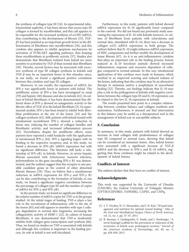
the synthesis of collagen type III [54]. In experimental tubu-lointerstitial nephritis, it has been shown that excess type IIIcollagen is formed by myofibroblast, and this cell appears tobe responsible for the increased synthesis of α1(III) mRNA,thus contributing to the development of fibrosis [55]. TGF-β1 has been pointed as an important growth factor in the dif-ferentiation of fibroblasts into myofibroblasts [56], and thiscytokine also appears to inhibit apoptosis mechanisms byactivation of P13K/AKT signaling pathways, thus keepingthese myofibroblasts at profibrotic activity [57]. Studies alsodemonstrate that fibroblasts isolated from keloid are moresensitive to activation by TGF-β than normal skin fibroblasts[58]. Thereby, several factors may be associated with exces-sive synthesis of type III collagen synthesis in keloids andTGF-β may be an important factor in this stimulus, since,in our study, we found a significant positive correlationbetween this cytokine and type III collagen.
Moreover, in our results, the expression of mRNA forIFN-γ was significantly lower in patients with keloid. Theantifibrotic action of IFN-γ has been investigated in renal[59] and hepatic [60] diseases and also in keloids [61]. In cul-ture of keloid fibroblasts, it has been demonstrated that dif-ferent doses of IFN-γ showed no antagonistic activity to thefibrotic effect of TGF-β in the keloid fibroblasts [5]. In exper-imental models, IFN-γ has been found to inhibit both fibro-blast proliferation at the site of the lesion [24] and thecollagen synthesis [62]. Still, patients with keloid treated withintralesional recombinant IFN-γ showed a reduction inlesion size, reducing the number of collagen bundles andfibroblastic activity, and increased inflammatory process[63]. Nevertheless, despite the antifibrotic effects, somepatients have reported a mild headache with the applicationof intralesional IFN-γ [64]. IFN-γ signaling begins with itsbinding to the respective receptors, and, in this study, wefound a decrease in IFN-γR1 mRNA expression but withno significant difference. The literature still lacks a rela-tionship of IFN-γR1 in keloids. However, in severe hepaticfibrosis associated with Schistosoma mansoni infection,polymorphism in the gene encoding IFN-γ R1 was demon-strated, and the authors suggest that this receptor is one pos-sible candidate genes in the control of other abnormalfibrotic diseases [29]. Thus, we believe that a simultaneousreduction in mRNA expression for IFN-γ and IFN-γ R1can be also contributing to the formation of keloids; in ourstudy, we found a significant negative correlation betweenthe percentage of collagen type III and the number of copiesof mRNA for IFN-γ and IFN-γR1.
In the present study, we found no significant difference inthe relative number of mRNA copies for TNF-α in the groupsstudied. At the initial stages of healing, TNF-α plays a keyrole in the recruitment of inflammatory cells to the site ofthe lesion [65] and still appears to interfere with type I colla-gen degradation in normal skin fragments, increasing thecollagenolytic activity of MMP-1 [22]. In culture of humanfibroblasts, it was demonstrated that TNF-α moderatelyinhibits both collagen gene transcription and synthesis [66].Thus, we found no studies on TNF-α associated with keloidsand although this cytokine is important in the healing pro-cess, its role in keloid is not well elucidated.
Furthermore, in this study, patients with keloid showedmRNA expression for IL-10 significantly lower comparedto the controls. We did not found any previously study asso-ciating the expression of IL-10 with keloids; however, in cul-ture of fibroblasts from patients with scleroderma andnormal skin, stimulated with IL-10, there was a decrease incollagen α1(I) mRNA expression in both groups. Theauthors believe that IL-10 might influence mRNA expressionof MEC components and further modify the development oftissue fibrosis [67]. As it is an anti-inflammatory cytokinethat plays an important role in the healing process, lesionsinduced in IL-10 knockout animals showed increasedinflammatory response and excessive deposition of colla-gen compared to normal animals. In this way, intralesionalapplications of this cytokine were made in humans, whichresulted in an improved scarring and reduced redness ofthe lesion, indicating that this cytokine may be an alternativetherapy to minimize and/or a prophylaxis of exacerbatedhealing [32]. Thereby, our findings indicate that IL-10 mayplay a role in the pathogenesis of keloids with negative corre-lation between the collagen type III and IL-10 expression canbe related to this low IL-10 expression in keloids.
The results presented here point to a complex relation-ship between cytokine balance and collagen synthesis andmaturation. Furthermore, it stressed out that interventionin this balance may be useful as a therapeutical tool in themanagement of keloid scar in susceptible subjects.
5. Conclusion
In summary, in this study, patients with keloid showed anincrease in total collagen with predominance of collagentype III compared to normal scars, showing that keloidcan be considered an immature lesion. Also, keloid lesionswere associated with a significant increase of TGF-βmRNA and the decrease in IFN-γ and IL-10 mRNA, sug-gesting that these cytokines might be related to the devel-opment of keloid lesions.
Conflicts of Interest
The authors declare that they have no conflict of interest.
Acknowledgments
This work was supported by the University of Uberaba(UNIUBE), the Federal University of Triângulo Mineiro(UFTM)/CEFORES, FAPEMIG, CAPES, and CNPq.
References
[1] S. V. Dryden, W. G. Shoemaker, and J. H. Kim, “Wound man-agement and nutrition for optimal wound healing,” Atlas ofthe Oral and Maxillofacial Surgery Clinics of North America,vol. 21, no. 1, pp. 37–47, 2013.
[2] B. Berman, S. Garikaparthi, E. Smith, and J. Newburger, “Anovel hydrogel scaffold for the prevention or reduction of therecurrence of keloid scars postsurgical excision,” Journal ofthe American Academy of Dermatology, vol. 69, no. 5,pp. 828–830, 2013.
8 Mediators of Inflammation

[3] B. Medhi, R. K. Sewal, L. Kaman, G. Kadhe, and A. Mane,“Efficacy and safety of an advanced formula silicone gel forprevention of post-operative scars,”Dermatology and Therapy,vol. 3, no. 2, pp. 157–167, 2013.
[4] J. Peltonen, L. L. Hsiao, S. Jaakkola et al., “Activation of colla-gen gene expression in keloids: co-localization of type I and VIcollagen and transforming growth factor-beta 1 mRNA,” TheJournal of Investigative Dermatology, vol. 97, no. 2, pp. 240–248, 1991.
[5] T. Hasegawa, A. Nakao, K. Sumiyoshi, R. Tsuboi, and H.Ogawa, “IFN-gamma fails to antagonize fibrotic effect ofTGF-beta on keloid-derived dermal fibroblasts,” Journal ofDermatological Science, vol. 32, no. 1, pp. 19–24, 2003.
[6] E. Suarez, F. Syed, T. A. Rasgado, A. Walmsley, P. Mandal, andA. Bayat, “Skin equivalent tensional force alters keloid fibro-blast behavior and phenotype,” Wound Repair and Regenera-tion, vol. 22, no. 5, pp. 557–568, 2014.
[7] S. Younai, L. S. Nichter, T. Wellisz, J. Reinisch, M. E. Nimni,and T. L. Tuan, “Modulation of collagen synthesis by trans-forming growth factor-beta in keloid and hypertrophic scarfibroblasts,” Annals of Plastic Surgery, vol. 33, no. 2, pp. 148–151, 1994.
[8] E. H. Epstein Jr. and N. H. Munderloh, “Isolation and charac-terization of CNBr peptides of human (alpha 1 (III) )3 collagenand tissue distribution of (alpha 1 (I) )2 alpha 2 and (alpha 1(III) )3 collagens,” The Journal of Biological Chemistry,vol. 250, no. 24, pp. 9304–9312, 1975.
[9] C. Huerre, C. Junien, D. Weil et al., “Human type I procol-lagen genes are located on different chromosomes,” Proceed-ings of the National Academy of Sciences of the United Statesof America, vol. 79, no. 21, pp. 6627–6630, 1982.
[10] E. Solomon, L. R. Hiorns, N. Spurr et al., “Chromosomalassignments of the genes coding for human types II, III, andIV collagen: a dispersed gene family,” Proceedings of theNational Academy of Sciences of the United States of America,vol. 82, no. 10, pp. 3330–3334, 1985.
[11] C. Schmidt, H. Pommerenke, F. Dürr, B. Nebe, and J. Rychly,“Mechanical stressing of integrin receptors induces enhancedtyrosine phosphorylation of cytoskeletally anchored proteins,”The Journal of Biological Chemistry, vol. 273, no. 9, pp. 5081–5085, 1998.
[12] J. Xu, M. M. Zutter, S. A. Santoro, and R. A. Clark, “A three-dimensional collagen lattice activates NF-kappaB in humanfibroblasts: role in integrin alpha2 gene expression and tissueremodeling,” The Journal of Cell Biology, vol. 140, no. 3,pp. 709–719, 1998.
[13] B. A. Booth, K. L. Polak, and J. Uitto, “Collagen biosynthe-sis by human skin fibroblasts. I. Optimization of the cultureconditions for synthesis of type I and type III procollagens,”Biochimica et Biophysica Acta, vol. 607, no. 1, pp. 145–160,1980.
[14] W. K. Stadelmann, A. G. Digenis, and G. R. Tobin, “Physiologyand healing dynamics of chronic cutaneous wounds,” Ameri-can Journal of Surgery, vol. 176, no. 2A Supplement,pp. 26S–38S, 1998.
[15] P. Bornstein and H. Sage, “Structurally distinct collagen types,”Annual Review of Biochemistry, vol. 49, no. 1, pp. 957–1003,1980.
[16] K. Gelse, E. Poschl, and T. Aigner, “Collagens—structure,function, and biosynthesis,” Advanced Drug Delivery Reviews,vol. 55, no. 12, pp. 1531–1546, 2003.
[17] R. Derynck and Y. E. Zhang, “Smad-dependent and Smad-independent pathways in TGF-beta family signalling,” Nature,vol. 425, no. 6958, pp. 577–584, 2003.
[18] G. S. Chin, W. Liu, Z. Peled et al., “Differential expression oftransforming growth factor-beta receptors I and II and activa-tion of Smad 3 in keloid fibroblasts,” Plastic and ReconstructiveSurgery, vol. 108, no. 2, pp. 423–429, 2001.
[19] P. Schmid, P. Itin, G. Cherry, C. Bi, and D. A. Cox, “Enhancedexpression of transforming growth factor-beta type I and typeII receptors in wound granulation tissue and hypertrophicscar,” The American Journal of Pathology, vol. 152, no. 2,pp. 485–493, 1998.
[20] W. L. Garner, S. Karmiol, J. L. Rodriguez, D. J. Smith Jr,and S. H. Phan, “Phenotypic differences in cytokine respon-siveness of hypertrophic scar versus normal dermal fibro-blasts,” The Journal of Investigative Dermatology, vol. 101,no. 6, pp. 875–879, 1993.
[21] K. Rapala, “The effect of tumor necrosis factor-alpha onwound healing. An experimental study,” Annales Chirurgiaeet Gynaecologiae. Supplementum, vol. 211, pp. 1–53, 1996.
[22] M. S. Agren, R. Schnabel, L. H. Christensen, and U.Mirastschijski, “Tumor necrosis factor-alpha-accelerated deg-radation of type I collagen in human skin is associated withelevated matrix metalloproteinase (MMP)-1 and MMP-3ex vivo,” European Journal of Cell Biology, vol. 94, no. 1,pp. 12–21, 2015.
[23] M. R. Duncan and B. Berman, “Gamma interferon is the lym-phokine and beta interferon the monokine responsible forinhibition of fibroblast collagen production and late but notearly fibroblast proliferation,” The Journal of ExperimentalMedicine, vol. 162, no. 2, pp. 516–527, 1985.
[24] A. J. Stout, I. Gresser, and W. D. Thompson, “Inhibition ofwound healing in mice by local interferon alpha/beta injec-tion,” International Journal of Experimental Pathology,vol. 74, no. 1, pp. 79–85, 1993.
[25] J. E. Darnell Jr., I. M. Kerr, and G. R. Stark, “Jak-STAT path-ways and transcriptional activation in response to IFNs andother extracellular signaling proteins,” Science, vol. 264,no. 5164, pp. 1415–1421, 1994.
[26] J. Rosenbloom, G. Feldman, B. Freundlich, and S. A. Jimenez,“Transcriptional control of human diploid fibroblast collagensynthesis by gamma-interferon,” Biochemical and BiophysicalResearch Communications, vol. 123, no. 1, pp. 365–372, 1984.
[27] J. Fransson, A. Emilson, A. Scheynius, and H. Hammar, “Pro-liferation and interferon-gamma receptor expression in psori-atic and healthy keratinocytes are influenced by interactionsbetween keratinocytes and fibroblasts in a skin equivalentmodel,” Archives of Dermatological Research, vol. 287, no. 6,pp. 517–523, 1995.
[28] S. A. Rezende, V. R. Oliveira, A. M. Silva, J. B. Alves, A. M.Goes, and L. F. Reis, “Mice lacking the gamma interferonreceptor have an impaired granulomatous reaction to Schisto-soma mansoni infection,” Infection and Immunity, vol. 65,no. 8, pp. 3457–3461, 1997.
[29] A. J. Dessein, D. Hillaire, N. E. Elwali et al., “Severe hepaticfibrosis in Schistosoma mansoni infection is controlled by amajor locus that is closely linked to the interferon-gammareceptor gene,” American Journal of Human Genetics, vol. 65,no. 3, pp. 709–721, 1999.
[30] J. H. Shi, H. Guan, S. Shi et al., “Protection against TGF-beta1-induced fibrosis effects of IL-10 on dermal fibroblasts and itspotential therapeutics for the reduction of skin scarring,”
9Mediators of Inflammation
Archives of Dermatological Research, vol. 305, no. 4, pp. 341–352, 2013.
[31] J. Shi, J. Li, H. Guan et al., “Anti-fibrotic actions of interleukin-10 against hypertrophic scarring by activation of PI3K/AKTand STAT3 signaling pathways in scar-forming fibroblasts,”PloS One, vol. 9, no. 5, Article ID e98228, 2014.
[32] I. Kieran, A. Knock, J. Bush et al., “Interleukin-10 reduces scarformation in both animal and human cutaneous wounds:results of two preclinical and phase II randomized controlstudies,” Wound Repair and Regeneration, vol. 21, no. 3,pp. 428–436, 2013.
[33] M. A. Williams, Autoradiography and Immunocytochemistry.Quantitative Methods in Biology, Elservier Medical Press,Amsterdam, The Netherlands, 1977.
[34] L. C. Junqueira, G. Bignolas, and R. R. Brentani, “Picrosiriusstaining plus polarization microscopy, a specific method forcollagen detection in tissue sections,” The Histochemical Jour-nal, vol. 11, no. 4, pp. 447–455, 1979.
[35] R. Lattouf, R. Younes, D. Lutomski et al., “Picrosirius red stain-ing: a useful tool to appraise collagen networks in normal andpathological tissues,” The Journal of Histochemistry and Cyto-chemistry, vol. 62, no. 10, pp. 751–758, 2014.
[36] I. C. Coelho-Lemos, A. C. Campos, M. de Almeida et al., “Inutero malnutrition influences wound healing of newborn ratsas measured by tensile strength and collagen deposition,”JPEN Journal of Parenteral and Enteral Nutrition, vol. 28,no. 4, pp. 241–244, 2004, discussion 245.
[37] K. J. Livak and T. D. Schmittgen, “Analysis of relative geneexpression data using real-time quantitative PCR and the2(-Delta Delta C(T)) Method,” Methods, vol. 25, no. 4,pp. 402–408, 2001.
[38] V. Tanaydin, J. Beugels, A. Piatkowski et al., “Efficacy ofcustom-made pressure clips for ear keloid treatment after sur-gical excision,” Journal of Plastic, Reconstructive & AestheticSurgery, vol. 69, no. 1, pp. 115–121, 2016.
[39] F. Furtado, B. Hochman, and L. M. Ferreira, “Evaluating keloidrecurrence after surgical excision with prospective longitudinalscar assessment scales,” Journal of Plastic, Reconstructive &Aesthetic Surgery, vol. 65, no. 7, pp. e175–e181, 2012.
[40] S. Shamsi Meymandi, A. Rezazadeh, and A. Ekhlasi, “Studyingintense pulsed light method along with corticosteroid injectionin treating keloid scars,” Iranian Red Crescent Medical Journal,vol. 16, no. 2, Article ID e12464, 2014.
[41] B. Berman and F. Flores, “Recurrence rates of excised keloidstreated with postoperative triamcinolone acetonide injectionsor interferon alfa-2b injections,” Journal of the American Acad-emy of Dermatology, vol. 37, no. 5 Part 1, pp. 755–757, 1997.
[42] H. D. Kim, S. M. Hwang, K. R. Lim, Y. H. Jung, S. M. Ahn, andJ. Kim Song, “Recurrent auricular keloids during pregnancy,”Archives of Plastic Surgery, vol. 40, no. 1, pp. 70–72, 2013.
[43] J. E. Lane, J. L. Waller, and L. S. Davis, “Relationship betweenage of ear piercing and keloid formation,” Pediatrics, vol. 115,no. 5, pp. 1312–1314, 2005.
[44] J. J. Brown, W. Ollier, G. Arscott et al., “Genetic susceptibilityto keloid scarring: SMAD gene SNP frequencies in Afro-Caribbeans,” Experimental Dermatology, vol. 17, no. 7,pp. 610–613, 2008.
[45] R. P. Abergel, D. Pizzurro, C. A. Meeker et al., “Biochemicalcomposition of the connective tissue in keloids and analysis ofcollagen metabolism in keloid fibroblast cultures,” The Journalof Investigative Dermatology, vol. 84, no. 5, pp. 384–390, 1985.
[46] J. Chen, S. Zhuo, X. Jiang et al., “Multiphoton microscopystudy of the morphological and quantity changes of collagenand elastic fiber components in keloid disease,” Journal ofBiomedical Optics, vol. 16, no. 5, p. 051305, 2011.
[47] E. Wulandari, S. W. Jusman, Y. Moenadjat, A. A. Jusuf, andM.Sadikin, “Expressions of collagen I and III in hypoxic keloidtissue,” The Kobe Journal of Medical Sciences, vol. 62, no. 3,pp. E58–E69, 2016.
[48] M. Naitoh, N. Hosokawa, H. Kubota et al., “Upregulation ofHSP47 and collagen type III in the dermal fibrotic disease,keloid,” Biochemical and Biophysical Research Communica-tions, vol. 280, no. 5, pp. 1316–1322, 2001.
[49] F. L. Yeh, H. D. Shen, and H. Y. Tai, “Decreased production ofMCP-1 and MMP-2 by keloid-derived fibroblasts,” Burns,vol. 35, no. 3, pp. 348–351, 2009.
[50] D. Ulrich, F. Ulrich, F. Unglaub, A. Piatkowski, and N. Pallua,“Matrix metalloproteinases and tissue inhibitors of metallo-proteinases in patients with different types of scars andkeloids,” Journal of Plastic, Reconstructive & Aesthetic Surgery,vol. 63, no. 6, pp. 1015–1021, 2010.
[51] L. Satish, J. Lyons-Weiler, P. A. Hebda, and A. Wells, “Geneexpression patterns in isolated keloid fibroblasts,” WoundRepair and Regeneration, vol. 14, no. 4, pp. 463–470, 2006.
[52] B. De Felice, C. Garbi, M. Santoriello, A. Santillo, and R. R.Wilson, “Differential apoptosis markers in human keloidsand hypertrophic scars fibroblasts,” Molecular and CellularBiochemistry, vol. 327, no. 1-2, pp. 191–201, 2009.
[53] G. M. Saed, D. Ladin, J. Olson, X. Han, Z. Hou, and D.Fivenson, “Analysis of p53 gene mutations in keloids usingpolymerase chain reaction-based single-strand conforma-tional polymorphism and DNA sequencing,” Archives ofDermatology, vol. 134, no. 8, pp. 963–967, 1998.
[54] P. E. Di Cesare, D. T. Cheung, N. Perelman, E. Libaw, L. Peng,and M. E. Nimni, “Alteration of collagen composition andcross-linking in keloid tissues,” Matrix, vol. 10, no. 3,pp. 172–178, 1990.
[55] W.W. Tang, G. Y. Van, andM. Qi, “Myofibroblast and alpha 1(III) collagen expression in experimental tubulointerstitialnephritis,” Kidney International, vol. 51, no. 3, pp. 926–931,1997.
[56] G. Gabbiani, B. J. Hirschel, G. B. Ryan, P. R. Statkov, and G.Majno, “Granulation tissue as a contractile organ. A study ofstructure and function,” The Journal of Experimental Medi-cine, vol. 135, no. 4, pp. 719–734, 1972.
[57] P. Kulasekaran, C. A. Scavone, D. S. Rogers, D. A. Arenberg, V.J. Thannickal, and J. C. Horowitz, “Endothelin-1 and trans-forming growth factor-beta1 independently induce fibroblastresistance to apoptosis via AKT activation,” American Journalof Respiratory Cell and Molecular Biology, vol. 41, no. 4,pp. 484–493, 2009.
[58] D. A. Bettinger, D. R. Yager, R. F. Diegelmann, and I. K.Cohen, “The effect of TGF-beta on keloid fibroblast prolifer-ation and collagen synthesis,” Plastic and ReconstructiveSurgery, vol. 98, no. 5, pp. 827–833, 1996.
[59] S. D. Oldroyd, G. L. Thomas, G. Gabbiani, and A. M. ElNahas, “Interferon-gamma inhibits experimental renal fibro-sis,” Kidney International, vol. 56, no. 6, pp. 2116–2127,1999.
[60] B. Knight, R. Lim, G. C. Yeoh, and J. K. Olynyk, “Inter-feron-gamma exacerbates liver damage, the hepatic progen-itor cell response and fibrosis in a mouse model of chronic
10 Mediators of Inflammation

liver injury,” Journal of Hepatology, vol. 47, no. 6, pp. 826–833, 2007.
[61] B. Berman and M. R. Duncan, “Short-term keloid treatmentin vivo with human interferon alfa-2b results in a selectiveand persistent normalization of keloidal fibroblast collagen,glycosaminoglycan, and collagenase production in vitro,”Journal of the American Academy of Dermatology, vol. 21,no. 4 Part 1, pp. 694–702, 1989.
[62] R. H. Miles, T. P. Paxton, D. Zacheis, D. J. Dries, and R. L.Gamelli, “Systemic administration of interferon-gammaimpairs wound healing,” The Journal of Surgical Research,vol. 56, no. 3, pp. 288–294, 1994.
[63] R. D. Granstein, A. Rook, T. J. Flotte et al., “A controlled trialof intralesional recombinant interferon-gamma in the treat-ment of keloidal scarring. Clinical and histologic findings,”Archives of Dermatology, vol. 126, no. 10, pp. 1295–1302, 1990.
[64] W. F. Larrabee JrC. A. East, H. S. Jaffe, C. Stephenson, and K.E. Peterson, “Intralesional interferon gamma treatment forkeloids and hypertrophic scars,” Archives of Otolaryngology –Head & Neck Surgery, vol. 116, no. 10, pp. 1159–1162, 1990.
[65] I. Birincioglu, M. Akbaba, A. Alver et al., “Determination ofskin wound age by using cytokines as potential markers,” Jour-nal of Forensic and Legal Medicine, vol. 44, pp. 14–19, 2016.
[66] J. A. Solis-Herruzo, D. A. Brenner, and M. Chojkier, “Tumornecrosis factor alpha inhibits collagen gene transcription andcollagen synthesis in cultured human fibroblasts,” The Journalof Biological Chemistry, vol. 263, no. 12, pp. 5841–5845, 1988.
[67] T. Yamamoto, B. Eckes, and T. Krieg, “Effect of interleukin-10on the gene expression of type I collagen, fibronectin, and dec-orin in human skin fibroblasts: differential regulation by trans-forming growth factor-beta and monocyte chemoattractantprotein-1,” Biochemical and Biophysical Research Communi-cations, vol. 281, no. 1, pp. 200–205, 2001.
11Mediators of Inflammation

Submit your manuscripts athttps://www.hindawi.com
Stem CellsInternational
Hindawi Publishing Corporationhttp://www.hindawi.com Volume 2014
Hindawi Publishing Corporationhttp://www.hindawi.com Volume 2014
MEDIATORSINFLAMMATION
of
Hindawi Publishing Corporationhttp://www.hindawi.com Volume 2014
Behavioural Neurology
EndocrinologyInternational Journal of
Hindawi Publishing Corporationhttp://www.hindawi.com Volume 2014
Hindawi Publishing Corporationhttp://www.hindawi.com Volume 2014
Disease Markers
Hindawi Publishing Corporationhttp://www.hindawi.com Volume 2014
BioMed Research International
OncologyJournal of
Hindawi Publishing Corporationhttp://www.hindawi.com Volume 2014
Hindawi Publishing Corporationhttp://www.hindawi.com Volume 2014
Oxidative Medicine and Cellular Longevity
Hindawi Publishing Corporationhttp://www.hindawi.com Volume 2014
PPAR Research
The Scientific World JournalHindawi Publishing Corporation http://www.hindawi.com Volume 2014
Immunology ResearchHindawi Publishing Corporationhttp://www.hindawi.com Volume 2014
Journal of
ObesityJournal of
Hindawi Publishing Corporationhttp://www.hindawi.com Volume 2014
Hindawi Publishing Corporationhttp://www.hindawi.com Volume 2014
Computational and Mathematical Methods in Medicine
OphthalmologyJournal of
Hindawi Publishing Corporationhttp://www.hindawi.com Volume 2014
Diabetes ResearchJournal of
Hindawi Publishing Corporationhttp://www.hindawi.com Volume 2014
Hindawi Publishing Corporationhttp://www.hindawi.com Volume 2014
Research and TreatmentAIDS
Hindawi Publishing Corporationhttp://www.hindawi.com Volume 2014
Gastroenterology Research and Practice
Hindawi Publishing Corporationhttp://www.hindawi.com Volume 2014
Parkinson’s Disease
Evidence-Based Complementary and Alternative Medicine
Volume 2014Hindawi Publishing Corporationhttp://www.hindawi.com