Embed Size (px)
Citation preview

In Planta Protein Sialylation through Overexpression of theRespective Mammalian Pathway*□S
Received for publication, November 25, 2009, and in revised form, February 3, 2010 Published, JBC Papers in Press, March 20, 2010, DOI 10.1074/jbc.M109.088401
Alexandra Castilho‡1, Richard Strasser‡1, Johannes Stadlmann§, Josephine Grass§, Jakub Jez‡, Pia Gattinger‡,Renate Kunert¶, Heribert Quendler�2, Martin Pabst§, Renaud Leonard§, Friedrich Altmann§,and Herta Steinkellner‡3
From the ‡Department of Applied Genetics and Cell Biology, §Department of Chemistry, and ¶Institute of Applied Microbiology,University of Natural Resources and Applied Life Sciences, Muthgasse 18, 1190 Vienna and �Polymun Scientific GmbH,Muthgasse 18, 1190 Vienna, Austria
Many therapeutic proteins are glycosylated and require ter-minal sialylation to attain full biological activity. Current man-ufacturingmethods based onmammalian cell culture allowonlylimited control of this important posttranslational modifica-tion, which may lead to the generation of products with lowefficacy. Here we report in vivo protein sialylation in plants,which have been shown to be well suited for the efficient gener-ation of complex mammalian glycoproteins. This was achievedby the introduction of an entire mammalian biosynthetic path-way in Nicotiana benthamiana, comprising the coordinatedexpression of the genes for (i) biosynthesis, (ii) activation, (iii)transport, and (iv) transfer of Neu5Ac to terminal galactose.Weshow the transient overexpression and functional integrity of sixmammalian proteins that act at various stages of the biosyn-thetic pathway and demonstrate their correct subcellular local-ization. Co-expression of these genes with a therapeutic glyco-protein, a humanmonoclonal antibody, resulted in quantitativesialylation of the Fc domain. Sialylation was at great uniformitywhen glycosylation mutants that lack plant-specific N-glycanresidues were used as expression hosts. Finally, we demonstrateefficient neutralization activity of the sialylated monoclonalantibody, indicating full functional integrity of the reporter pro-tein. We report for the first time the incorporation of the entirebiosynthetic pathway for protein sialylation in a multicellularorganism naturally lacking sialylated glycoconjugates. Besidesthe biotechnological impact of the achievement, this work mayserve as a general model for the manipulation of complex traitsinto plants.
The outstanding specificity of therapeutic glycoproteinsplaces them among the fastest growing class of pharmaceuticalproducts. Many of these drugs need terminal sialylation, thefinal and most complex step of human N-glycosylation, for
optimal therapeutic potency. Therefore manufacturing is cur-rently restricted tomammalian cell-based systems that are ableto perform this important posttranslational modification,althoughwithmajor limitations. The naturally present glycosy-lation repertoire of mammalian host cells promotes the gener-ation of a number of different terminal structures and thusleads to heterogeneous glycosylation. Moreover, these modifi-cationsmay differ from the authentic human glycosylation. Forexample, CHO4 cells, widely used for the expression of recom-binant glycoproteins, do not naturally produce human type�2,6-sialylation. These shortcomings in many cases lead todrugs with reduced biological potency, of which recombinanthormones such as human erythropoietin and interferons areprominent examples (1). In addition, about 10% of the N-gly-cans in the Fc region of human serum IgG is sialylated (2).Although the anti-inflammatory effects of sialylated IgG haverecently been demonstrated (3), very little information on theimpact of this common modification at the molecular level isavailable (4). This explains the general interest in novel expres-sion hosts and strategies to engineer glycosylation not only toincrease the value of therapeutic proteins but also to betterunderstand the role of glycosylation in general and sialylation inparticular in fundamental biological processes.Plants are considered an attractive alternative expression
platform for therapeutic human glycoproteins because they arecost-effective, highly scalable, free fromhumanpathogens, and,importantly, can carry out complex N-glycosylation (5, 6). Inaddition, plants glycosylate proteins with considerable uni-formity, which may provide an advantage over mammalian-based expression systems. However, plants lack sialylatedN-glycans, which restrict their utility as a versatile productionplatform. Recent studies have shown that the plant N-glycosy-lation pathway can be modified toward human-type glycosyla-tion (7–9). Elimination of plant-specific glycosylation in ourlaboratory and by others has resulted in the generation of plant
* This work was supported by grants from the Wiener Wissenschafts-, Fors-chungs- und Technologiefonds (WWTF) (Grant LS 154) and Austrian Sci-ence Fund (FWF) (Grants P18-314, L575-B13, and P19092 to R. S.).Author’s Choice—Final version full access.
□S The on-line version of this article (available at http://www.jbc.org) containssupplemental Figs. 1 and 2 and Table 1.
1 Both authors contributed equally to this work.2 Present address: Department of Immunology and Microbial Science, The
Scripps Research Institute, La Jolla, CA 92037.3 To whom correspondence should be addressed. Tel.: 43-1-47654-6700; Fax:
43-1-47654-6392; E-mail: [email protected].
4 The abbreviations used are: CHO, Chinese hamster ovary; GalT, �1,4-galac-tosyltransferase; ST-GalT, modified �1,4-galactosyltransferase; ST, �2,6-si-alyltransferase; CMAS, CMP-N-acetylneuraminic acid synthetase; NANS,N-acetylneuraminic acid phosphate synthase; GNE, UDP-N-acetylglu-cosamine 2-epimerase/N-acetylmannosamine kinase; CST, CMP-Neu5Actransporter; mAb, monoclonal antibody; HIV, human immunodeficiencyvirus; GFP, green fluorescent protein; Rubisco, ribulose-bisphosphate car-boxylase/oxygenase; WT, wild type; MES, 4-morpholineethanesulfonicacid; MS, mass spectrometry; MS/MS, tandem MS; LC, liquid chromatogra-phy; ESI, electrospray mass ionization; TOF, time-of-flight.
THE JOURNAL OF BIOLOGICAL CHEMISTRY VOL. 285, NO. 21, pp. 15923–15930, May 21, 2010Author’s Choice © 2010 by The American Society for Biochemistry and Molecular Biology, Inc. Printed in the U.S.A.
MAY 21, 2010 • VOLUME 285 • NUMBER 21 JOURNAL OF BIOLOGICAL CHEMISTRY 15923
by guest on March 22, 2018
http://ww
w.jbc.org/
Dow
nloaded from

lines (e.g. �XT/FT) that produce therapeutically relevant pro-teins carrying a human-like glycosylation profile (GnGn struc-tures). Importantly, monoclonal antibodies produced in suchlines exhibited enhanced effector functions (7–9). In addition,we demonstrated recently that by overexpressing a modifiedversion of the human �1,4-galactosyltransferase (GalT), plantN-glycans can be extended with �1,4-linked galactose at greatuniformity (10). Furthermore, these structures serve as accep-tor substrates for subsequent terminal sialylation. However,sialylation is particularly difficult to accomplish even in thepresence of �1,4-galactosylated structures because plants lacksome further essential prerequisites: (i) the biosyntheticcapability to produce the sugar nucleotide precursor CMP-sialic acid, specifically CMP-N-acetylneuraminic acid (CMP-Neu5Ac); (ii) a transporter that delivers CMP-sialic acid intothe Golgi in sufficient amounts; and (iii) a sialyltransferase (ST)to transfer sialic acid from CMP-Neu5Ac to terminal galactoseon the nascent glycoprotein.Moreover, these proteins and theirsubstrates must work in a highly coordinated fashion at differ-ent stages of the pathway, and thus organelle-specific targetingof several components is required to enable proper protein sia-lylation (see Fig. 1). Initial attempts to introduce Neu5Ac resi-dues into plant N-glycans have resulted in the expression ofsome of these proteins in plants (11, 12), and recent progress inour laboratory allowed the synthesis of the sugar nucleotideCMP-Neu5Ac from endogenous metabolites by the simultane-ous overexpression of three mammalian enzymes in Arabidop-sis thaliana (13). However, in planta protein sialylation has notyet been achieved.In this study, we aimed to achieve in planta sialylation in
Nicotiana benthamiana, a tobacco-related plant species partic-ularly well suited for recombinant protein expression (14). Sixmammalian proteins comprising the sialic acid pathway wereco-expressed together with a monoclonal antibody (mAb) inN. benthamiana wild type (WT) and glycosylation mutantplants thereof. Purified mAbs were subjected to glycosylationanalyses and tested for functional integrity.
EXPERIMENTAL PROCEDURES
Construction of Plant Expression Vectors—The binary vec-tors used for the expression of mammalian UDP-N-acetyl-glucosamine 2-epimerase/N-acetylmannosamine kinase(p19GNE), N-acetylneuraminic acid phosphate synthase(p19NANS), modified �1,4-galactosyltransferase (ST-GalT),�2,6-sialyltransferase (ST), as well as 2G12 (pTRAp-Ds; anti-HIV antibody) have been described previously (10, 11, 13, 15).For the expression of other mammalian proteins, we used
three binary vectors (p18, p19, and p20), derivatives of pPT2(16) only differing in their inserted tags (hemagglutinin, myc,and GFP, respectively, see Fig. 2). A truncated form of thehuman CMP-N-acetylneuraminic acid synthetase (CMAS)lacking 40 N-terminal amino acids (13) was amplified by PCRwith the primers CMAS11F/CMAS5R (Table 1) and clonedinto the XbaI/BamHI sites of p18, resulting in N-terminalhemagglutinin-tagged CMAS binary vector (p18CMAS).cDNA of the mouse CMP-Neu5Ac transporter (CST, GermanResource Center for Genome Research accession numberIRAVp968C0627D) was amplified by PCR with primers
CST2F/CST3R (Table 1) and cloned into the XbaI/BamHI siteof p19, resulting in a C-terminal Myc-tagged CST vector(p19CST). For subcellular localization experiments, the cDNAsfrom GNE, NANS, CMAS, and CST were cloned into the p20vector XbaI/BamHI site (17), which resulted in C-terminalGFP fusion vectors (p20 series, see Fig. 2). For PCR amplifica-tion of cDNAs, gene-specific primers were used (GNE3F/4R,NANS3F/4R, CMAS12F/6R, and CST2F/3R, Table 1).All binary vectors were transformed into the Agrobacterium
tumefaciens strain UIA 143. The origin of the mammalian pro-tein used in this study is summarized in supplemental Table 1.Transient Protein Expression in N. benthamiana—5–6-week
old N. benthamiana plants (4–6 leaf stage) were used for thetransient expression of heterologous proteins by agroinfiltra-tion as described previously (9, 17). For confocal laser scanningmicroscopy studies, agrobacteria carrying the respective binaryvector (p20GNE, p20NANS, p20CMAS, and p20CST) weregrown overnight in LBmedium supplemented with kanamycin(50�g/ml) and gentamycin (25�g/ml) at 29 °C. 1ml of bacteriaculture was washed twice in infiltration buffer (50mMMES, pH5.6, 2 mM sodium phosphate, 0.5% w/v D-glucose, and 300 �M
acetosyringone) and resuspended to a final A600 of 0.3 (�2,5 �108 cells/ml). For the in vivo synthesis of CMP-Neu5Ac,bacterial suspensions containing p19GNE, p19NANS, andp18CMAS were diluted to A600 of 0.3, mixed in a 1:1 ratio, andco-infiltrated into epidermal cells of N. benthamiana leaves.In co-expression experiments of ST-GalT and 2G12, the
respective bacterial suspensions were diluted to anA600 of 0.3–0.5 and mixed 1:1 prior to infiltration. For co-expression of allseven binary vectors, bacterial suspensions of p19GNE,p19NANS, p18CMAS, p19CST, ST-GalT, ST, and 2G12 werediluted to A600 of 0.3 and mixed in a 1:1 ratio.Confocal Laser Scanning Microscopy—3 days after infiltra-
tion of agrobacteria containing the p20GNE, p20NANS,p20CMAS and p20CST, the GFP expression was monitored inN. benthamiana leaves using a Leica TCS SP2 confocal laserscanning microscope as described before (17).In Vivo CMP-Neu5Ac Analysis—N. benthamiana leaves (0.1
g) co-infiltrated with p19GNE, p19NANS, and p18CMASwereused to analyze the in vivo synthesis of CMP-Neu5Ac asdescribed previously (13). Briefly, the supernatant from thehomogenized samples was passed through a C18-RP SPE car-tridge, and the flow-through was applied to a 10-mg HyperSepHypercarb SPE cartridge (Thermo Scientific). This was washedwith 1 ml of H2O, and CMP-Neu5Ac was eluted with 0.3 ml of60%AcCN in 65mMammonium formate buffer. The eluatewas
TABLE 1Primers used in this studyUnderlined letters indicate restriction sites.
Primer Restriction enzyme Sequence (5�–3�)
GNE3F XbaI tataTCTAGAatggagaagaacgggaacaacGNE4R BglII tataAGATCTgtggatcctgcgcgttgtgtaNANS3F XbaI tataTCTAGAatgccgctggagctggagctgNANS4R BamHI tataGGATCCagacttgatttttttgccatgCMAS11F XbaI tataTCTAGAaagcccccgcacctggcagccctaCMAS12F XbaI tataTCTAGAatgaagcccccgcacctggcagccctaCMAS5R BamHI tataGGATCCctatttttggcatgaattattaacCMAS6R BamHI tataGGATCCtttttggcatgaattattaacCST2F XbaI tataTCTAGAatggctccggcgagagaaaatgCST3R BamHI tataGGATCCcacaccaatgattctctcttt
In Planta Protein Sialylation
15924 JOURNAL OF BIOLOGICAL CHEMISTRY VOLUME 285 • NUMBER 21 • MAY 21, 2010
by guest on March 22, 2018
http://ww
w.jbc.org/
Dow
nloaded from

freeze-dried. The samples were analyzed on a Hypercarb col-umn (0.32 � 50 mm, Thermo Scientific) using a 65 mM ammo-nium formate buffer of pH 3.0 as the aqueous solvent. Analyteswere detected with an ESI-Q-TOF Ultima Global (Waters) inthe MS/MS mode with MS1 set on m/z � 613.1 Da and themass of the [M-H]� ion of CMP-Neu5Ac, and simulatedselected ion monitoring of m/z � 322.0 Da ([M-H]� of CMP)was performed with MS2.A. thaliana Transformation and in Vitro Activity Assay of the
CMP-Neu5Ac Transporter—A. thaliana wild-type plants weretransformed with p19CST (see Fig. 2) by floral dipping (16).Kanamycin-resistant plants were screened by PCR with gene-specific primers to confirm the presence of the codingsequences. Microsomal fractions were prepared according toFleischer and Kervina (18) in the extraction buffer containing 1mM EDTA and protease inhibitors (Complete Mini, EDTA-free, Sigma). Microsomes were suspended in 100 �l of SolutionA (10 mM Tris-HCl, pH 7.0) containing 250 mM sucrose, 1 mM
MgCl2, 0.5 mM �-mercaptopropanol, and protease inhibitorsper gram of initial material. The CMP-Neu5Ac transporterassay was started by the addition of 50 �l of solution A contain-ing 0.05 �Ci of 14C-labeled CMP-NeuAc to 50 �l of micro-somes. The reaction was performed in a 30 °C water bath for 5and 10 min and stopped by the addition of 1 ml of ice-coldSolution A supplemented with 1 �M non-radioactive CMP-NeuAc (stop reaction mix). The reaction mixture was pouredon an nitrocellulose filter (Advantec Toyo, A045A025A) andthereafter washed three times with 1 ml of ice-cold stop reac-tionmix. The radioactivity remaining on the filterswas countedusing a scintillation counter.IgG Purification—2G12 infiltrated leaves were ground in liq-
uid nitrogen, resuspended in ice-cold extraction buffer (100mM
Tris-HCl pH 6.8, 40 mM ascorbic acid, 500 mM NaCl, 1 mM
EDTA), and centrifuged (35,000� g/30min/4 °C). Supernatantwas vacuum-filtrated (Macherey-Nagel filter disks MN619eh,2–4 �m), and the filtrate was further clarified by isoelectricprecipitation at pH 4.8 (precipitation of �25% of total solubleprotein including Rubisco) and centrifuged. Clarified superna-tant was adjusted to pH 6.8 and incubated on ice for 20 min.After an additional centrifugation step, the extract was passedthrough a 0.45-�m filter (Millipore Stericup Durapore, lowbinding polyvinylidene difluoride membrane, 73mm/0.45 �m)before loading on the chromatography column. ProteinA affin-ity chromatographywas performed using the AKTAPurifier-10fast protein liquid chromatography system (GE Healthcare)and a prepacked protein A-Sepharose high performance col-umn (HiTrap ProtA HP 5 � 5 ml; GE Healthcare) according tothe manufacturer’s instructions.Analysis of Glycopeptides—N-Glycan analysis of 2G12 was
carried out by liquid chromatography-electrospray ionization-mass spectrometry (LC-ESI-MS) of tryptic glycopeptides (2, 9).Briefly, the heavy chain of SDS-PAGE separated IgGs wasexcised from the gel, S-alkylated, digested with trypsin, andsubsequently analyzed by LC-ESI-MS.Note: During this proce-dure, two glycopeptides are generated that differ in 482 Da (2).Structural Identification of Sialylated N-Glycans—The veri-
fication of the linkages of Neu5Ac and galactose as �2,6- and�1,4-, respectively, on diantennary N-glycans was carried out
by chromatography on porous graphitic carbon with detectionby ESI-MS (19). The retention times of free, reducedN-glycanswere compared with that of a panel of all possible diantennarymono- and disialylated N-glycans of the compositionNeu5Ac1–2Gal2GlcNAc4Man5 (19). Structural assignment ofthe glycan with a mass corresponding to the compositionNeu5Ac1Gal1GlcNAc3Man3, which could be either theMNa6-4or theNa6-4M isomer,was accomplished as follows. The anchorpoint of all isomer assignments is the synthesis of the MGnglycan using GlcNAc transferase I (19) and the generation ofthe pair MGn/GnM by enzymatic degradation of GnGn. Theisomer pair A4Na6-4/Na6-4A4 was originally identified by sepa-ration and digestion down toMGnorGnM.Here again, the twopeaks were separated and individually digested with �-galacto-sidase and �-N-acetylhexosaminidase. The retention times ofthe resulting isomers MNa6-4 or Na6-4M were thus determinedto be 35.9 and 44.7 min, respectively.2G12 Antigen Binding and HIV Neutralization Assay—2G12
antigen binding specificity was carried out by an enzyme-linkedimmunosorbent assay-based assay using recombinant gp160 asantigen (9, 15). Neutralization assays were performed using anAA-2 cell-based syncytium inhibition assay as described previ-ously (9, 20). Briefly, 2-fold dilution series of antibodies werepreincubated with virus at 102-103 50% tissue culture infectivedose (TCID50)/ml for 1 h at 37 °C. CD4-positive human AA-2cells were then added at a cell count of 4 � 105 cells/ml andfurther incubated for 5 days. The read-out was performedaccording to the method of Reed and Muench (21), and thepresence of at least one syncytium was scored positive. Allassays were performed as duplicates.
RESULTS
In Planta Synthesis of CMP-Neu5Ac—The initial step in themammalian sialylation pathway is the biosynthesis of the acti-vated sugar nucleotide precursor cytidine monophosphateN-acetylneuraminic acid (CMP-Neu5Ac). To achieve the gen-eration of this precursor from the endogenously presentmetab-oliteUDP-GlcNAc, the action of at least four enzymes is needed(Fig. 1): (i) GNE, a bifunctional enzyme, which catalyzes theconversion of UDP-GlcNAc to ManNAc and the phosphoryla-
FIGURE 1. Schematic representation of the mammalian pathway for sia-lylation of glycoconjugates. The enzymes involved in the process are: GNE,NANS, Neu5Ac-9-phosphate phosphatase (NANP), CMAS, CST, GalT, and ST.ManNAc-6-P, ManNAc-6-phosphate; NeuAc-9-P, NeuAc-9-phosphate; PEP,phosphoenolpyruvate.
In Planta Protein Sialylation
MAY 21, 2010 • VOLUME 285 • NUMBER 21 JOURNAL OF BIOLOGICAL CHEMISTRY 15925
by guest on March 22, 2018
http://ww
w.jbc.org/
Dow
nloaded from

tion of ManNAc to ManNAc-6-phosphate; (ii) NANS, whichcondenses ManNAc-6-phosphate and phosphoenolpyruvate,resulting in Neu5Ac-9-phosphate; (iii) a specific phosphataseacting onNeu5Ac-9-phosphate; and (iv) a CMAS that activatesthe resulting primary sialic acid (N-acetylneuraminic acid) inthe nucleus to CMP-Neu5Ac. Here we set out to simulta-neously express cDNAs from the respective mammalianenzymes. In previous experiments, we showed that A. thalianatransformed with mammalian GNE and NANS accumulatedNeu5Ac rather than Neu5Ac-9-phosphate (13), indicating thepresence of a Neu5Ac-9-phosphate homologue that catalyzesthe dephosphorylation step in this plant species (22). Thus thisprotein was not further considered in the present study. For abetter understanding, all constructs used in this study are illus-trated in Fig. 2. As the correct subcellular targeting of theseproteins is an essential prerequisite, live cell imaging was usedto determine the localization of transiently expressed GFPfusion proteins. Confocal laser scanning microscopy studiesexhibited that GNE and NANS were located in the cytoplasmand that CMAS was found exclusively in the nucleus (Fig. 3).These findings are consistent with the intracellular localiza-tions of these proteins in mammalian cells. Subsequently, themammalian cDNAs of these three enzymes were simulta-neously expressed in N. benthamiana leaves, and it was foundthat these plants contained substantial amounts of CMP-Neu5Ac (5.7 nmol g�1 fresh weight) as determined by ESI-TOF-MS/MS analysis (Fig. 4). These results are in good agree-ment with our data derived from A. thaliana transformed withthe three mammalian genes (13).Transfer of CMP-Neu5Ac into the Golgi—The transfer of the
activated donor substrate into the Golgi is accomplished byCST. Thus we overexpressed the respective mammalian cDNAin N. benthamiana. In a first step, correct localization of CSTwas determined by CSLM, and the transiently expressed GFPfusion exhibited Golgi labeling, the predicted compartment forthis protein (Fig. 3). Transgenic A. thaliana plants stably
expressing CST were used to carry out in vitro transporterassays. An accumulation of CMP-Neu5Ac in microsomes wasobserved in transformed plants (Fig. 5), demonstrating theexpression of a functional protein.In Planta Protein Sialylation—Finally, specific sialyltrans-
ferases (ST) transfer sialic acid to the acceptor substrate A4A4,a �1,4-galactosylated, diantennary N-glycan. In humans,Neu5Ac transfer is preferentially in �2,6- but also �2,3-linkage.
FIGURE 2. Schematic representation of the different plant expression cas-settes of the binary vectors generated in this study. Pnos, nopaline syn-thase gene promoter; Tnos, nopaline synthase gene terminator; KanR, neomy-cin phosphotransferase II gene; P35S, cauliflower mosaic virus promoter; ha,hemagglutinin epitope tag; g7T, Agrobacterium gene 7 terminator; mc, c-Mycepitope tag; LB, left border; RB, right border.
FIGURE 3. Subcellular localization of fluorescently tagged GNE, NANS,CMAS, and CST proteins. The corresponding constructs were transientlyexpressed in N. benthamiana leaf epidermal cells and analyzed by confocallaser scanning microscopy 3 days after inoculation. GNE- and NANS-GFPexhibited a typical cytoplasmic labeling, whereas CMAS-GFP showed exclu-sive staining of the nucleus. CST-GFP displayed a punctuate labeling through-out the cell, which indicates Golgi localization. Scale bar for p20CST � 5 �M;for others, scale bar � 20 �M.
FIGURE 4. In planta synthesis of CMP-Neu5Ac. LC-ESI-MS/MS analysis ofmolecular mass 613.1 Da from extracts of N. benthamiana WT, from CMP-Neu5Ac standard (S), and from extracts of N. benthamiana co-expressing theGNE, NANS, and CMAS mammalian proteins (GNE NANS CMAS) was per-formed. The peak at 22.7 min in the standard and the GNE�NANS�CMASsample corresponds to the CMP fragment obtained by fragmentation ofCMP-Neu5Ac ([M-H]� � 613.1 Da).
In Planta Protein Sialylation
15926 JOURNAL OF BIOLOGICAL CHEMISTRY VOLUME 285 • NUMBER 21 • MAY 21, 2010
by guest on March 22, 2018
http://ww
w.jbc.org/
Dow
nloaded from

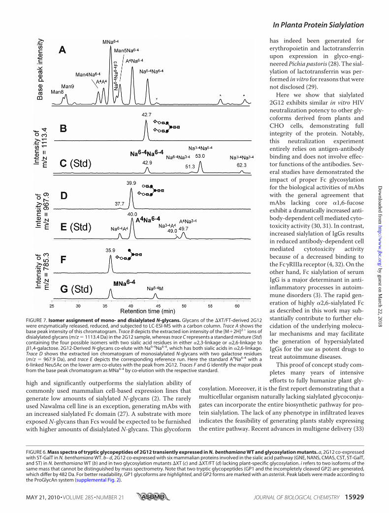
Herewe set out to generateNeu5Ac�2,6-Gal linkage, themajorglycoform of important therapeutic proteins, such as erythro-poietin, interferons, or IgG. The mammalian �2,6-sialyltrans-ferase has been expressed in plants previously (11). As �1,4-galactosylated N-glycans are not present in plants, we firstconverted endogenous structures to highly galactosylatedforms by overexpressing amodified version of the humanGalTthat acts in a late stage of the pathway (ST-GalT (10) inN. benthamianaWT). The enzyme was co-expressed togetherwith a therapeutically relevant glycoprotein, i.e. 2G12, a humananti-HIV mAb (23). Subsequently, the N-glycosylation profileof purified 2G12 was determined by ESI-TOF/MS analyses asdescribed previously (2). The glycosylation profile exhibited thegeneration of quantitative 2G12 galactosylation with threemajor glycoforms, namely MAXi, MAXFi and AAXF (Fig. 6a).(The subscript “i” indicates that other isoforms of the samemass, e.g.AMX andMAX, may theoretically exist). In addition,smaller peaks representing galactosylated and non-galactosy-lated glycoforms were detected (e.g. MGnX, MAi, MGnXFi,Man5Gn, GnGnXF, and Man8).To finally accomplish the transfer of CMP-Neu5Ac to galac-
tosylated N-glycans, all six proteins (GNE, NANS, CMAS,CST, ST-GalT, and ST) were co-expressed with 2G12 inN. benthamianaWT.TheN-glycans of purified 2G12 exhibitedefficient sialylation. The three major galactosylated variantssynthesized in the presence of ST-GalT were quantitativelyconverted to MNaXi, MNaXFi, and NaNaXF (Fig. 6b). To fur-ther fathom the potential of this engineering approach, glyco-sylation mutants lacking plant-specific glycan residues, i.e.lacking only the �1,2-xylose (�XT) and also lacking the core�1,3-fucose (�XT/FT) (9), were used as expression hosts.N-Glycans of 2G12 produced in �XT and �XT/FT were alsoefficiently sialylated and exhibited a highly uniform glycosyla-tion pattern with two dominant glycan species in �XT, namelyMNaFi and NaNaF (Fig. 6c), and three major sialylated glycanspecies in �XT/FT, namelyMNai, ANai, and NaNa (Fig. 6d). Inthe two glycosylationmutants, over 80% of the glycoformswere
sialylated, which demonstrates the coordinated expression andfunctional integrity of themammalian proteins in plants. As thepresence of plant homologues for enzymes of the sialylationpathway has been suggested (24, 25), we wanted to determinewhether the recombinant expression of all six proteins isrequired to accomplish in vivo sialylation.A series of expressionstudies where single proteins were omitted was carried out;however, no sialylated glycoforms were detected in any of theseexperiments (data not shown).Identification of �-2,6-Linked Sialic Acid—The transfer of
Neu5Ac to 2G12 galactosylated structures causes amass shift of291.1 Da of the respective peaks inMS spectra. To confirm thatthis shift actually corresponds to a �2,6-linkage of Neu5Ac to�1,4-linked galactose, LC-ESI-MS analysis was carried out asdescribed previously (19) using �XT/FT-derived 2G12 (Fig.7A). Thismethod allows identification of individual glycoformsby the comparison with elution positions of well defined stand-ards. Notably,�2,3- and�2,6-sialylation lead to different reten-tion times (2, 19). The peaks with retention times of 39.9 and42.7 min co-eluted with the standards A4Na6-4 and Na4-6Na4-6,respectively (Fig. 7, B–E), confirming the presence of mono-and disialylated glycans with Neu5Ac in �2,6-linkage onplant-produced 2G12. To further identify the isoform of theincompletely processed structure assigned as MNa, the peakwas analyzed against the corresponding standards and identi-fied as MNa6-4 (Fig. 7, F and G).Functional Integrity of 2G12 Glycoforms—To test the func-
tional properties of sialylated mAb, antigen binding and HIVneutralization assays were carried out as described recently (9,10) using 2G12 derided from �XT/FT. No differences in bind-ing capacities were found between CHO cell-derived 2G12,N. benthamiana WT 2G12 with mainly GnGnXF glycans, andsialylated 2G12 (Table 2). Finally, the ability of highly sialylated2G12 to neutralize HIV was examined using a syncytium inhi-bition assay (Tab. 2). As for antigen binding, no significant dif-ferences between the different glycoforms were observed,which demonstrates the full integrity of plant-derived sialylated2G12 glycoforms.
DISCUSSION
Here we report the efficient in planta sialylation of a thera-peutic protein as exemplified by a recombinant mAb. This wasachieved by the coordinated overexpression of six mammalianproteins that act at various stages of the biosynthetic pathway indifferent subcellular compartments. Quantitative sialylationwas facilitated by the specific modification of some of theseproteins, i.e. elimination of activity-destroying amino acidsequences (in the case of CMAS) (13) and by enabling propersubcellular localization (in the case of ST-GalT) (10). Notably,nearly all of the available acceptor substrates (i.e. galactosylatedglycans) present in WT and in the glycosylation mutants weresialylated. The synthesis of incompletely processed structuressuch as MNai, MNaFi, and MNaXi is most likely due to inap-propriate overexpression of ST-GalT interfering with theaction of GlcNAc-T II. Incompletely processed glycans withone antenna terminating in mannose (MAXFi andMAi, Fig. 6aand data not shown, respectively) were present in WT and�XT/FT plants expressing only ST-GalT, an observation
FIGURE 5. In vitro CMP-Neu5Ac transporter activity. Microsomal mem-brane vesicles derived from A. thaliana WT and transgenic CST-expressingline (CST) were incubated with [14C]CMP-Neu5Ac for 0, 5, and 10 min. Thevesicles were bound to nitrocellulose filter, and their incorporated radioactiv-ity was measured using a scintillation counter. The activity of CMP-Neu5Actransporter is assessed by the incorporation of more than 2000 dpm after 10min in CST plants.
In Planta Protein Sialylation
MAY 21, 2010 • VOLUME 285 • NUMBER 21 JOURNAL OF BIOLOGICAL CHEMISTRY 15927
by guest on March 22, 2018
http://ww
w.jbc.org/
Dow
nloaded from

already made previously (10). Thisshortcoming may be overcome byoptimizing the expression system asdemonstrated for stably trans-formedGalT�plants (10).Note thatGalT� plants were not consideredfor sialylation experiments becausethey are not yet in a homozygousstage.The high amount of sialylated
glycoforms of 2G12 upon express-ing proteins of the mammalian bio-synthetic pathway points at a con-siderable degree of functionalconservation between plant andmammalian cells. Moreover, theentire procedure did not interferewith IgG expression, which isremarkable in the light of the com-plexity of the process.As reported forA. thaliana trans-
formed with GNE, NANS, andCMAS (13), transient expression ofthese genes in N. benthamiana effi-ciently convertedManNAc-6-phos-phate to Neu5Ac rather than toNeu5Ac-9-phosphate, suggestingthe presence of a so far uncharacter-ized endogenous phosphatase act-ing on sialic acid-9-phosphate. Onthe other hand, although the pres-ence of plant homologues for theCMP-Neu5Ac transporter and sia-lyltransferases has been suggested(24, 25), our experiments indicatethat all required mammalian genesneed to be expressed to achieve inplanta protein sialylation. Notably,transient sialylation did not result inany obvious phenotype in the infil-trated leaves, indicating that at leastcertain plant tissues/organs can tol-erate the incorporation of highlycharged sialic acid into proteins.This is not surprising becauseN-glycans of total plant proteinsderived from plants expressing allsix genes of the sialylation pathwayexhibited a relatively low amount ofsialic acid (supplemental Fig. 1), incontrast to the efficient sialylationof the secreted mAb. The formationof disialylated mAb is remarkablebecause in human B-lymphocytes,�2,6-ST preferentially generatesmonosialylated Fc glycans (26). Inthis light, the in planta sialylation ofa recombinant protein is extremely
In Planta Protein Sialylation
15928 JOURNAL OF BIOLOGICAL CHEMISTRY VOLUME 285 • NUMBER 21 • MAY 21, 2010
by guest on March 22, 2018
http://ww
w.jbc.org/
Dow
nloaded from
high and significantly outperforms the sialylation ability ofcommonly used mammalian cell-based expression lines thatgenerate low amounts of sialylated N-glycans (2). The rarelyused Nawalma cell line is an exception, generating mAbs withan increased sialylated Fc domain (27). A substrate with moreexposedN-glycans than Fcs would be expected to be furnishedwith higher amounts of disialylated N-glycans. This glycoform
has indeed been generated forerythropoietin and lactotransferrinupon expression in glyco-engi-neeredPichia pastoris (28). The sial-ylation of lactotransferrin was per-formed in vitro for reasons thatwerenot disclosed (29).Here we show that sialylated
2G12 exhibits similar in vitro HIVneutralization potency to other gly-coforms derived from plants andCHO cells, demonstrating fullintegrity of the protein. Notably,this neutralization experimententirely relies on antigen-antibodybinding and does not involve effec-tor functions of the antibodies. Sev-eral studies have demonstrated theimpact of proper Fc glycosylationfor the biological activities of mAbswith the general agreement thatmAbs lacking core �1,6-fucoseexhibit a dramatically increased anti-body-dependent cellmediated cyto-toxicity activity (30, 31). In contrast,increased sialylation of IgGs resultsin reduced antibody-dependent cellmediated cytotoxicity activitybecause of a decreased binding tothe Fc�RIIIa receptor (4, 32). On theother hand, Fc sialylation of serumIgG is a major determinant in anti-inflammatory processes in autoim-mune disorders (3). The rapid gen-eration of highly �2,6-sialylated Fcas described in this work may sub-stantially contribute to further elu-cidation of the underlying molecu-lar mechanisms and may facilitatethe generation of hypersialylatedIgGs for the use as potent drugs totreat autoimmune diseases.This proof of concept study com-
pletes many years of intensiveefforts to fully humanize plant gly-
cosylation. Moreover, it is the first report demonstrating that amulticellular organism naturally lacking sialylated glycoconju-gates can incorporate the entire biosynthetic pathway for pro-tein sialylation. The lack of any phenotype in infiltrated leavesindicates the feasibility of generating plants stably expressingthe entire pathway. Recent advances in multigene delivery (33)
FIGURE 6. Mass spectra of tryptic glycopeptides of 2G12 transiently expressed in N. benthamiana WT and glycosylation mutants. a, 2G12 co-expressedwith ST-GalT in N. benthamiana WT. b– d, 2G12 co-expressed with six mammalian proteins involved in the sialic acid pathway (GNE, NANS, CMAS, CST, ST-GalT,and ST) in N. benthamiana WT (b) and in two glycosylation mutants �XT (c) and �XT/FT (d) lacking plant-specific glycosylation. i refers to two isoforms of thesame mass that cannot be distinguished by mass spectrometry. Note that two tryptic glycopeptides (GP1 and the incompletely cleaved GP2) are generated,which differ by 482 Da. For better readability, GP1 glycoforms are highlighted, and GP2 forms are marked with an asterisk. Peak labels were made according tothe ProGlycAn system (supplemental Fig. 2).
FIGURE 7. Isomer assignment of mono- and disialylated N-glycans. Glycans of the �XT/FT-derived 2G12were enzymatically released, reduced, and subjected to LC-ESI-MS with a carbon column. Trace A shows thebase peak intensity of this chromatogram. Trace B depicts the extracted ion intensity of the [M�2H]2� ions ofdisialylated glycans (m/z � 1113.4 Da) in the 2G12 sample, whereas trace C represents a standard mixture (Std)containing the four possible isomers with two sialic acid residues in either �2,3-linkage or �2,6-linkage to�1,4-galactose. 2G12-Derived N-glycans co-elute with Na6-4Na6-4, which has both sialic acids in �2,6-linkage.Trace D shows the extracted ion chromatogram of monosialylated N-glycans with two galactose residues(m/z � 967.9 Da), and trace E depicts the corresponding reference run. Here the standard A4Na6-4 with a6-linked Neu5Ac on the lower arm co-elutes with the peak from 2G12. Traces F and G identify the major peakfrom the base peak chromatogram as MNa6-4 by co-elution with the respective standard.
In Planta Protein Sialylation
MAY 21, 2010 • VOLUME 285 • NUMBER 21 JOURNAL OF BIOLOGICAL CHEMISTRY 15929
by guest on March 22, 2018
http://ww
w.jbc.org/
Dow
nloaded from

should allow the efficient generation of such plants to accom-plish in planta sialylation in the future. The availability of suchlines, together with recently generated glyco-engineeredmutants displaying human-type glycosylation of great uniform-ity (9, 10), pave the way not only for generating therapeuticglycoproteins with optimized biological activities obtained by acustomized N-glycosylation profile but also for studying theimpact of different glycoforms in biological processes. In casewhole plants would not tolerate protein sialylation during theircomplete developmental process, one of the six genes could betransiently expressed together with the protein of interest.Another alternative to this hurdle would be the use of tissue-specific or inducible promoters to specifically turn on proteinsialylation in leaves.Glycoengineered plants in combination with newly devel-
oped plant virus-based transient expression systems, allowingthe generation of a virtually unlimited number of different pro-teins at high amounts within 1 week after DNA construct deliv-ery (6, 34), provide a considerable advantage over existingglyco-modified expression platforms, including yeast andmammalian cells. Besides the importance of this work in bio-technological applications, it may serve as a model for themanipulation of complex metabolic pathways into plants forthe generation of varieties with new traits.
Acknowledgments—cDNA of �1,4-GalT and a binary vector express-ing ST were kindly provided by Eric G. Berger, University of Zurich,Switzerland, and Paul Dupree, University of Cambridge, UK,respectively.
REFERENCES1. Bork, K., Horstkorte, R., and Weidemann, W. (2009) J. Pharm. Sci. 98,
3499–35082. Stadlmann, J., Pabst, M., Kolarich, D., Kunert, R., and Altmann, F. (2008)
Proteomics 8, 2858–28713. Anthony, R. M., Nimmerjahn, F., Ashline, D. J., Reinhold, V. N., Paulson,
J. C., and Ravetch, J. V. (2008) Science 320, 373–3764. Scallon, B. J., Tam, S. H., McCarthy, S. G., Cai, A. N., and Raju, T. S. (2007)
Mol. Immunol. 44, 1524–15345. Stoger, E., Ma, J. K., Fischer, R., and Christou, P. (2005) Curr. Opin. Bio-
technol. 16, 167–1736. Gleba, Y., Klimyuk, V., and Marillonnet, S. (2007) Curr. Opin. Biotechnol.
18, 134–1417. Cox, K. M., Sterling, J. D., Regan, J. T., Gasdaska, J. R., Frantz, K. K., Peele,
C. G., Black, A., Passmore, D., Moldovan-Loomis, C., Srinivasan, M., Cui-
son, S., Cardarelli, P. M., and Dickey, L. F. (2006) Nat. Biotechnol. 24,1591–1597
8. Schuster,M., Jost,W.,Mudde,G. C.,Wiederkum, S., Schwager, C., Janzek,E., Altmann, F., Stadlmann, J., Stemmer, C., and Gorr, G. (2007) Biotech-nol. J. 2, 700–708
9. Strasser, R., Stadlmann, J., Schahs,M., Stiegler, G., Quendler, H.,Mach, L.,Glossl, J., Weterings, K., Pabst, M., and Steinkellner, H. (2008) Plant Bio-technol. J. 6, 392–402
10. Strasser, R., Castilho, A., Stadlmann, J., Kunert, R., Quendler, H., Gat-tinger, P., Jez, J., Rademacher, T., Altmann, F., Mach, L., and Steinkellner,H. (2009) J. Biol. Chem. 284, 20479–20485
11. Wee, E.G., Sherrier, D. J., Prime, T.A., andDupree, P. (1998)PlantCell10,1759–1768
12. Misaki, R., Fujiyama, K., and Seki, T. (2006) Biochem. Biophys. Res. Com-mun. 339, 1184–1189
13. Castilho, A., Pabst,M., Leonard, R., Veit, C., Altmann, F., Mach, L., Glossl,J., Strasser, R., and Steinkellner, H. (2008) Plant Physiol. 147, 331–339
14. Giritch, A., Marillonnet, S., Engler, C., van Eldik, G., Botterman, J.,Klimyuk, V., and Gleba, Y. (2006) Proc. Natl. Acad. Sci. U.S.A. 103,14701–14706
15. Schahs, M., Strasser, R., Stadlmann, J., Kunert, R., Rademacher, T., andSteinkellner, H. (2007) Plant Biotechnol. J. 5, 657–663
16. Strasser, R., Stadlmann, J., Svoboda, B., Altmann, F., Glossl, J., and Mach,L. (2005) Biochem. J. 387, 385–391
17. Strasser, R., Bondili, J. S., Vavra, U., Schoberer, J., Svoboda, B., Glossl, J.,Leonard, R., Stadlmann, J., Altmann, F., Steinkellner, H., and Mach, L.(2007) Plant Cell 19, 2278–2292
18. Fleischer, S., and Kervina, M. (1974)Methods in Enzymology 31, 6–4119. Pabst, M., Bondili, J. S., Stadlmann, J., Mach, L., and Altmann, F. (2007)
Anal. Chem. 79, 5051–505720. Trkola, A., Kuster, H., Rusert, P., Joos, B., Fischer, M., Leemann, C., Man-
rique, A., Huber,M., Rehr,M., Oxenius, A.,Weber, R., Stiegler, G., Vcelar,B., Katinger, H., Aceto, L., and Gunthard, H. F. (2005) Nat. Med. 11,615–622
21. Reed, L. J., and Muench, H. (1938) Am. J. Hyg. 27, 493–49722. Maliekal, P., Vertommen,D., Delpierre, G., andVan Schaftingen, E. (2006)
Glycobiology 16, 165–17223. Trkola, A., Purtscher, M., Muster, T., Ballaun, C., Buchacher, A., Sullivan,
N., Srinivasan, K., Sodroski, J.,Moore, J. P., andKatinger, H. (1996) J. Virol.70, 1100–1108
24. Bakker, H., Routier, F., Ashikov, A., Neumann, D., Bosch, D., andGerardy-Schahn, R. (2008) Carbohydr. Res. 343, 2148–2152
25. Takashima, S., Abe, T., Yoshida, S., Kawahigashi, H., Saito, T., Tsuji, S.,and Tsujimoto, M. (2006) J. Biochem. 139, 279–287
26. Barb, A. W., Brady, E. K., and Prestegard, J. H. (2009) Biochemistry 48,9705–9707
27. Stadlmann, J., Weber, A., Pabst, M., Anderle, H., Kunert, R., Ehrlich, H. J.,Schwarz, P. H., and Altmann, F. (2009) Proteomics 9, 4143–4153
28. Hamilton, S. R., Davidson, R. C., Sethuraman,N., Nett, J. H., Jiang, Y., Rios,S., Bobrowicz, P., Stadheim, T. A., Li, H., Choi, B. K., Hopkins, D., Wis-chnewski, H., Roser, J., Mitchell, T., Strawbridge, R. R., Hoopes, J., Wildt,S., and Gerngross, T. U. (2006) Science 313, 1441–1443
29. Choi, B. K., Actor, J. K., Rios, S., d’Anjou, M., Stadheim, T. A.,Warburton,S., Giaccone, E., Cukan, M., Li, H., Kull, A., Sharkey, N., Gollnick, P.,Kocieba, M., Artym, J., Zimecki, M., Kruzel, M. L., and Wildt, S. (2008)Glycoconj. J. 25, 581–593
30. Jefferis, R. (2009) Nat. Rev. Drug Discov. 8, 226–23431. Raju, T. S. (2008) Curr. Opin. Immunol. 20, 471–47832. Kaneko, Y., Nimmerjahn, F., and Ravetch, J. V. (2006) Science 313,
670–67333. Dafny-Yelin, M., and Tzfira, T. (2007) Plant Physiol. 145, 1118–112834. Sainsbury, F., and Lomonossoff, G. P. (2008) Plant Physiol. 148,
1212–1218
TABLE 2Functional properties of 2G12 glycoformsIn vitro HIV-1 neutralization activity was determined by a syncytium inhibitionassay. 2G12 antigen-binding tests were carried out by enzyme-linked immunosor-bent assay using recombinant gp160 as antigen. GnGnXF, major glycoformobtained from 2G12 produced in N. benthamianaWT; Neu5Ac glycoforms, sialy-lated 2G12 glycoforms as shown in Fig. 6d;CHO,CHO-cell derived 2G12, generatesa mixture of glycoforms (9,10).
2G12 Antigen binding IC50
% �g/mlGnGnXF 117 3.71Neu5Ac glycoforms 121 2.09CHO 100 7.40
In Planta Protein Sialylation
15930 JOURNAL OF BIOLOGICAL CHEMISTRY VOLUME 285 • NUMBER 21 • MAY 21, 2010
by guest on March 22, 2018
http://ww
w.jbc.org/
Dow
nloaded from

Friedrich Altmann and Herta SteinkellnerPia Gattinger, Renate Kunert, Heribert Quendler, Martin Pabst, Renaud Leonard,
Alexandra Castilho, Richard Strasser, Johannes Stadlmann, Josephine Grass, Jakub Jez,Mammalian Pathway
Protein Sialylation through Overexpression of the RespectiveIn Planta
doi: 10.1074/jbc.M109.088401 originally published online March 20, 20102010, 285:15923-15930.J. Biol. Chem.
10.1074/jbc.M109.088401Access the most updated version of this article at doi:
Alerts:
When a correction for this article is posted•
When this article is cited•
to choose from all of JBC's e-mail alertsClick here
Supplemental material:
http://www.jbc.org/content/suppl/2010/03/20/M109.088401.DC1
http://www.jbc.org/content/285/21/15923.full.html#ref-list-1
This article cites 34 references, 11 of which can be accessed free at
by guest on March 22, 2018
http://ww
w.jbc.org/
Dow
nloaded from