Embed Size (px)
Citation preview
N-terminomics identifies Prli42 as a membrane miniprotein conserved in Firmicutes and critical for stressosome activation in Listeria monocytogenes
Francis Impens#1,2,3,†, Nathalie Rolhion#1,2,3, Lilliana Radoshevich#1,2,3, Christophe Bécavin1,2,3,4, Mélodie Duval1,2,3, Jeffrey Mellin1,2,3, Francisco García del Portillo5, M. Graciela Pucciarelli5,6, Allison H. Williams7,8, and Pascale Cossart1,2,3,*
1Département de Biologie Cellulaire et Infection, Institut Pasteur, Unité des Interactions Bactéries-Cellules, F-75015 Paris, France
2Inserm, U604, F-75015 Paris, France
3INRA, Unité sous-contrat 2020, F-75015 Paris, France
4Institut Pasteur, Bioinformatics and Biostatistics Hub, C3BI, USR 3756 IP CNRS, Paris, France
5Centro Nacional de Biotecnología–Consejo Superior de Investigaciones Científicas (CNB-CSIC), Madrid, Spain
6Departamento de Biología Molecular, Universidad Autónoma de Madrid, Centro de Biología Molecular ‘Severo Ochoa’ (CBMSO-CSIC), Madrid, Spain
7Département de Microbiologie, Institut Pasteur, Unité des Biologie et génétique de la paroi bactérienne, F-75015 Paris, France
8INSERM, Groupe Avenir, F-75015 Paris, France
# These authors contributed equally to this work.
Abstract
*Correspondence and requests for materials should be addressed to P.C. [email protected].†Present address: Medical Biotechnology Center, VIB, Ghent University, 9000 Ghent, Belgium.
Data availability. The mass spectrometry data for the mapped peptides have been deposited at the ProteomeXchange Consortium (http://proteomecentral.proteomexchange.org) with data set identifier PXD000890 and DOI 10.6019/PXD000890, and mapped peptides are visualized in our homemade genome browser at http://nterm.listeriomics.pasteur.fr. All other data that support the findings of this study are available from the corresponding author upon request.
Author contributions P.C. initiated, conceived and supervised the project. F.I. initiated the project and performed the proteomics analysis and validation of the proteomics work and docking model. N.R. identified the oxidative stress phenotype, constructed nearly all the bacterial strains and performed the analysis of sigma B signalling. L.R. performed the macrophage experiments, the fractionation experiments and the virulence experiments. C.B. made the proteogenomics pipeline and is responsible for the bioinformatic analysis of the paper. M.D. performed the northern blots of Sigma B signalling. J.M. constructed the initial bacterial strains for validation. F.G.d.P. and M.G.P. contributed essential reagents. A.H.W. reconstituted the stressosome and imaged it using EM, and performed the docking model and all of the structural biology. L.R. and P.C. wrote the paper, with editing help and discussions from N.R., M.D., F.I. and A.H.W.
Reprints and permissions information is available at www.nature.com/reprints.
Competing interests The authors declare no competing financial interests.
Europe PMC Funders GroupAuthor ManuscriptNat Microbiol. Author manuscript; available in PMC 2018 February 07.
Published in final edited form as:Nat Microbiol. ; 2: 17005. doi:10.1038/nmicrobiol.2017.5.
Europe PM
C Funders A
uthor Manuscripts
Europe PM
C Funders A
uthor Manuscripts

To adapt to changing environments, bacteria have evolved numerous pathways that activate stress
response genes. In Gram-positive bacteria, the stressosome, a cytoplasmic complex, relays
external cues and activates the sigma B regulon. The stressosome is structurally well-characterized
in Bacillus, but how it senses stress remains elusive. Here, we report a genome-wide N-
terminomic approach in Listeria that strikingly led to the discovery of 19 internal translation
initiation sites and 6 miniproteins, among which one, Prli42, is conserved in Firmicutes. Prli42 is
membrane-anchored and interacts with orthologues of Bacillus stressosome components. We
reconstituted the Listeria stressosome in vitro and visualized its supramolecular structure by
electron microscopy. Analysis of a series of Prli42 mutants demonstrated that Prli42 is important
for sigma B activation, bacterial growth following oxidative stress and for survival in
macrophages. Taken together, our N-terminonic approach unveiled Prli42 as a long-sought link
between stress and the stressosome.
Listeria monocytogenes is a human pathogen that has emerged as a model organism in
infection biology1. The genome of L. monocytogenes strain EGD-e was sequenced in 2001
and predicted to encode 2846 open reading frames (ORFs)2. Tiling array-based
transcriptomic analyses then explored bacterial transcription3,4, and RNA-sequencing was
used to generate genome-wide transcription start (TSS) and termination site (TTS) maps5,6,
making this model bacterium ideally suited for further exploration of the regulation of gene
expression, at the level of translation. Ribosome profiling in Escherichia coli and Bacillus subtilis recently revealed a wealth of information about translational regulation, including
translational pausing at Shine–Dalgarno (SD)-like sequences7. While pause sites can be
differentiated from internal translation initiation sites (TISs) in mammalian cells using
pulse-chase experiments8, it has been challenging to do so with precision in bacteria9,10. In
contrast, N-terminal proteomics-based approaches can unambiguously detect bacterial TISs
on a genome-wide scale. N-terminal COFRADIC (combined fractional diagonal
chromatography) is one such method that isolates N-terminal peptides via two sequential
chromatographic separations11–14. Interestingly, in prokaryotes, N-terminal peptides are
formylated and this modification can be exploited to identify TISs (ref. 15).
Here, we report the first global TIS map of L. monocytogenes using an N-terminomics-
based proteogenomic approach that combines COFRADIC and treatment with a peptide
deformylase (PDF) inhibitor, actinonin16 (Fig. 1a). This genome-wide map yielded 62%
genome coverage of predicted TIS and led to unexpected discoveries, such as previously
unidentified TIS including 19 internal TIS and 6 miniproteins. We characterized one of these
miniproteins, Prli42, in detail, as a tail-anchored membrane protein that relays oxidative
stress signals to the stressosome to activate the general stress-sensing pathway, the sigma B
regulon.
Results
A proteogenomic approach to map TISs in Listeria
To map the translational landscape of L. monocytogenes strain EGD-e, we used N-terminal
COFRADIC followed by mapping of the identified peptide sequences onto the Listeria genome to locate the corresponding TISs (Fig. 1). To this end, we first created a protein
Impens et al. Page 2
Nat Microbiol. Author manuscript; available in PMC 2018 February 07.
Europe PM
C Funders A
uthor Manuscripts
Europe PM
C Funders A
uthor Manuscripts
sequence database by translating the L. monocytogenes EGD-e genome in all six reading
frames. This allowed us to work independently of the original genome annotation, a
requirement for the identification of novel ORFs. We grew Listeria in exponential phase at
20 °C and 37 °C and in stationary phase at 37 °C. We added the PDF inhibitor actinonin to
all three conditions at sub-inhibitory concentrations to increase TIS identification (Fig. 1b),
and compared identified TISs with an untreated control (37 °C, exponential phase). Equal
quantities of bacteria from the three growth conditions were mixed, proteins were extracted
and, after blocking free NH2, digested into peptides with either trypsin or endoproteinase
GluC (endoGluC). Due to their different cleavage specificities (trypsin after K/R or
endoGluC after E/D), these enzymes generate complementary N-terminal peptides and thus
increased coverage of our N-terminome (Supplementary Fig. 1a). We then isolated
formylated peptides using COFRADIC and performed liquid chromatography–tandem mass
spectrometry (LC-MS/MS) analysis. We identified the spectra using two different algorithms
(Mascot, from Matrix Science, and MaxQuant17) (Fig. 1c). Based on published data3,18,
we predicted that more than 90% of all previously annotated Listeria proteins should be
expressed in the growth conditions used, while 231 proteins would not be expressed
(Supplementary Table 1). We estimated that 314 of the 2,846 predicted Listeria proteins
would not be detected due to peptides that are too short or too long for mass-spectrometry
identification (Supplementary Fig. 1a and Supplementary Table 1). Furthermore, protein
secretion leads to removal of the N-terminal signal sequence, thereby preventing the
detection of N-terminal peptides for 309 predicted secreted proteins (Supplementary Table 1
and ref. 19). We therefore estimated that 2,109 out of 2,846 annotated Listeria TISs (74%)
could theoretically be detected by our approach.
Over 1,000,000 MS/MS spectra were recorded in over 200 LC-MS/MS runs, leading to the
identification of 12,000 unique peptides that were mapped onto the genome (Fig. 1c).
Actinonin treatment resulted in a sevenfold increase (20.5%) in peptides starting with a
formyl-Meti, compared to control (2.8% formyl-Meti peptides). We identified 1,401 TISs,
which we represented visually on a publically available whole-genome browser (http://
nterm.listeriomics.pasteur.fr) that integrates our proteogenomic data with all previous
transcriptomic data from tiling arrays and RNA-Seq3,5,6 (Fig. 2a).
Divergence from previously annotated TISs
The majority of detected N-terminal peptides correspond to previously annotated TISs
(aTISs)2. Our data validated predicted start codons for 1,322 L. monocytogenes ORFs,
which are evenly spread across the genome and not enriched for particular start codons or
Shine–Dalgarno (SD) sequences (Supplementary Fig. 1b,c and Supplementary Table 2).
Furthermore, TIS abundance correlated well with gene transcription data, and we identified
primarily cytoplasmic and membrane proteins (Fig. 2b, Supplementary Fig. 1d and
Supplementary Table 1)19. We identified 72 genes with potential leaderless transcripts (TSS
located fewer than ten nucleotides upstream from the observed TIS5, Supplementary Table
3), 13 of which lacked a second TIS. Leaderless transcripts can mediate a rapid stress
response20 and, interestingly, two of the genes we found with putative leaderless transcripts
play a role in sigma B signalling (for example, regulators of sigma B RsbU and RsbV). Our
Impens et al. Page 3
Nat Microbiol. Author manuscript; available in PMC 2018 February 07.
Europe PM
C Funders A
uthor Manuscripts
Europe PM
C Funders A
uthor Manuscripts
approach also identified small deviations from and several errors in previous annotation
(detailed in Supplementary Fig. 2 and Supplementary Tables 4–6).
Detection of internal TISs (iTISs)
We identified 19 N-terminal peptides that fell within annotated ORFs revealing in-frame
translation of shorter protein forms (Supplementary Table 7)5. We identified iTISs in
aspartokinase II (Lmo1235)21 and the heat shock protein ClpB (LmO2206)22, proteins
previously known to have two forms in other bacterial species. In E. coli, the long and short
isoforms of ClpB cooperate for efficient protein disaggregation23. To confirm the iTIS in
Listeria ClpB, we complemented a ΔclpB strain with a ClpB-flag. Immunoblotting validated
the presence of two isoforms, one long and one short. We then mutated the annotated AUG
or the internal GUG start codons of ClpB. In these mutants, only the short- or long-form
protein is produced, respectively, demonstrating that internal translation of ClpB occurs in
Listeria (Fig. 2c).
Another iTIS occurs in the bifunctional protein Lmo0219, which seems to be a Listeria-
specific gene fusion. The N-terminal portion encodes a putative tRNAIle lysidine synthetase
(TilS), while the C-terminal portion may function as a hypoxanthine-guanine
phosphoribosyltransferase (HGPRT)24. In Bacillus, these two enzymes form a complex and
are produced from distinct genes with overlapping stop and start codons (Fig. 2d). In
Listeria, however, the ORFs have fused in-frame with an iTIS to produce HGPRT.
Interestingly, when we mutated the internal start codon, we could not isolate clones that
expressed the full-length protein. Thus, loss of HGPRT could destabilize the TilS-HGPRT
fusion protein. More experiments are required to determine if this is the case (Fig. 2d).
Taken together, our data suggest that alternative translation initiation events may be more
common in bacteria than previously accepted.
Discovery of Listeria miniproteins
The initial annotation of the Listeria genome excluded ORFs of fewer than 40 amino acids2.
This strategy reduced the number of incorrectly annotated proteins, but probably excluded
several important small protein-coding genes. Here, we identified six miniproteins that were
previously annotated either as small RNAs (rli24, rli41 and rli42) or 5′ untranslated regions
(5′ UTRs of lmo0669 and lmo1064) (Supplementary Table 8). Their predicted three-
dimensional structure and topology were determined (Supplementary Fig. 3). Two of the
small ORFs were predicted to be encoded by the small RNA rli41, which probably functions
as a small bicistronic mRNA (ref. 3). Prli42 is encoded by the previously annotated small
RNA rli42 and is described below. Prli24, another miniprotein, is encoded by rli24, a small
RNA upstream of a hypothetical RNA ligase gene (lmo0257). We identified a 35-amino-acid
miniprotein in the 5′ UTR of Listeria corA (lmo1064). Interestingly, ribosome profiling
revealed a similar ORF in the 5′ UTR of corA in E. coli25. Finally, we detected a small
ORF in the 5′ UTR of a hypothetical oxidoreductase (Lmo0669).
Impens et al. Page 4
Nat Microbiol. Author manuscript; available in PMC 2018 February 07.
Europe PM
C Funders A
uthor Manuscripts
Europe PM
C Funders A
uthor Manuscripts
Miniprotein Prli42 is conserved and interacts with Listeria orthologues of components from the stressosome
Prli42 is a 31-amino-acid protein, and computational analysis using the TOPCONS
software26 suggests that it is a tail-anchored membrane protein (Supplementary Fig. 3).
When we compiled a database of all potential small bacterial ORFs between 20 and 60
amino acids and performed a blastP search on L. monocytogenes EGD-e Prli42, we
identified orthologous sequences of Prli42 in many other Firmicutes, including Bacilli and
Staphylococci species (Fig. 3a and Supplementary Table 9). To characterize Prli42 we first
sought interaction partners of the flag-tagged protein by co-immunoprecipitation followed
by LC-MS/MS analysis. Interestingly, we identified RsbR (Lmo0889), RsbS (Lmo0890) and
three RsbR-like proteins (Lmo0799, Lmo0161 and Lmo1642) as interaction partners of
Prli42 (Fig. 3b and Supplementary Table 10) in two independent analyses. We identified
other Prli42 partners as well, namely t-RNA synthetases and hypothetical proteins (Fig. 3b
and Supplementary Table 10). RsbR (regulator of sigma B protein) and RsbS are
components of the stressosome, a supramolecular complex involved in bacterial stress
signalling and activation of the general stress factor, sigma B, in Bacillus. Interestingly,
nearly three-quarters of species expressing Prli42 also displayed RsbR, RsbS and RsbT
homologues (Supplementary Table 9).
Listeria RsbR, RsbS and RsbT form a supramolecular complex in vitro
The formation of the stressosome in Listeria has not yet been reported. The cryo-electron
microscopy (cryo-EM) structure of the Bacillus stressosome complex has been solved and
consists of a core complex composed of the C-terminal STAS (sulfate transporter and anti-
sigma factor antagonist) domains of RsbR and RsbS with N-terminal RsbR homodimeric
protrusions that are proposed to act as sensors27. The core domain sequesters the kinase
RsbT, and releases it upon detection of environmental stresses. This initiates a signalling
cascade that culminates in sigma B-mediated induction of factors that help the bacteria to
counteract and resist stress28. To determine if a similar complex is formed in Listeria, we
individually purified Listeria RsbR, RsbS and RsbT (Lmo0891). We then reassembled the
structure by first combining RsbR and RsbS, similar to the purification of the Bacillus stressosome (Fig. 3c)27. RsbR and RsbS formed a core stressosome complex of a higher
molecular weight than each individual protein. The addition of RsbT (Supplementary Fig. 4)
led to a larger complex (Fig. 3c), which we then visualized using EM (Fig. 3d). When
imaged, the supramolecular complex was reminiscent of the Bacillus stressosome, revealing
that the stressosome assembles in Listeria.
Prli42 interacts with RsbR via its conserved N-terminal Lys5 and Arg8
To gain deeper insight into the molecular interaction between Prli42 and RsbR, their
association was simulated through computational docking. We took advantage of the crystal
structure of the homodimeric N-terminal domain of Bacillus RsbR to build a structural
model for Listeria RsbR (ref. 29; Supplementary Fig. 4d,e). We made the assumption that
the N-terminal domain would interact with Prli42 because the N-terminal dimers of RsbR lie
on the accessible exterior of the stressosome and are thought to be involved in signal
transduction27. We then simulated docking of the predicted structure of Prli42 with that of
Impens et al. Page 5
Nat Microbiol. Author manuscript; available in PMC 2018 February 07.
Europe PM
C Funders A
uthor Manuscripts
Europe PM
C Funders A
uthor Manuscripts
Listeria RsbR. The optimal model showed an interaction of the basic N-terminal tail of
Prli42, including conserved residues Lys4, Lys5 and Arg8, with an acidic patch formed
between two RsbR subunits, on the periphery of the stressosome (Fig. 3e).
To test this interaction model, we constructed Listeria strains that expressed Prli42-flag, in
which Lys5 and Arg8 residues were replaced by an alanine residue. Because K5A reduces
Prli42 stability, we mutated Lys5 to leucine or phenylalanine (Supplementary Fig. 4f) and
tested wild type (WT), K5L, K5F and R8A for their interaction with RsbR. We were able to
validate our proteomics results by immunoprecipitation and immunoblot, showing that WT
Prli42 can interact with RsbR while the Prli42–RsbR interaction was reduced in K5L and
K5F mutants and completely abrogated by the R8A mutation (Fig. 3f). In addition, mass-
spectrometry following immunoprecipitation of Prli42 R8A-flag demonstrated that RsbR
and its paralogues no longer co-immunoprecipitated with Prli42 (Supplementary Fig. 4g and
Supplementary Table 10). Together, these data establish the crucial role of the Lys5 and
Arg8 residues of Prli42 in binding RsbR, in support of our interaction model.
Prli42 can anchor RsbR to the bacterial membrane
To test whether Prli42 was membrane-anchored, we performed cellular fractionation. This
experiment indicated that Prli42 localized strictly to the membrane fraction (Fig. 4a). We
then visualized RsbR and estimated that RsbR is 60% membrane-associated and 40%
cytoplasmic (Fig. 4a and Supplementary Fig. 5b). Because Prli42 is membrane-localized and
the interaction with RsbR probably occurs at the interface of the membrane and the
cytoplasm, we hypothesized that Prli42 was involved in anchoring RsbR to the bacterial
membrane. We fractionated WT, Δprli42 and Δprli42+flag-Prli42 and observed that, upon
deletion of Prli42, ~40% of RsbR shifts from the bacterial membrane fraction to the
bacterial cytoplasm (Fig. 4b,c and Supplementary Fig. 5b). This shift is nearly but not
completely restored by complementation with Prli42 (45% RsbR membrane) (Fig. 4b,c and
Supplementary Fig. 5b). Taken together, our data show that Prli42 is needed, directly or
indirectly, for close association of RsbR with the membrane.
Prli42 is essential for the sigma B-mediated stress response to oxidative stress
Because the stressosome mediates the stress response in Bacillus and previous work from
our laboratory has shown that Prli42 transcription is upregulated during bacterial growth in
blood3, we hypothesized that Prli42 could affect sigma B signalling in Listeria. We thus
monitored sigma B signalling following hydrogen peroxide treatment during bacterial
growth, conditions similar to the oxidative stress encountered by bacteria during growth in
blood. Following hydrogen peroxide treatment, Δprli42 induced far less expression of
lmo2230, a sigma B-dependent gene, than WT or complemented bacteria (Fig. 4d and
Supplementary Fig. 5d). To determine whether Prli42 also had a functional effect on
bacterial growth following oxidative stress, we exposed bacteria to hydrogen peroxide and
hydrogen peroxide combined with iron to generate reactive oxygen species (ROS) via the
Fenton reaction, and assessed their survival. Listeria Δprli42 was significantly more sensitive
to oxidative stress than the WT and complemented strains (Fig. 4e). Interestingly, a mutant
of Prli42 R8A, for which the interaction between Prli42 and RsbR is impaired, is also more
sensitive to oxidative stress than WT (Fig. 4e), revealing the functional relevance of the
Impens et al. Page 6
Nat Microbiol. Author manuscript; available in PMC 2018 February 07.
Europe PM
C Funders A
uthor Manuscripts
Europe PM
C Funders A
uthor Manuscripts
Prli42–RsbR complex to the stress response. Futhermore, mutations in Prli42 do not affect
bacterial growth following several other stresses, suggesting that Prli42 is particularly
important for oxidative stress (Supplementary Fig. 5a,c). Taken together, the Prli42–RsbR
interaction partitions a subset of RsbR to the membrane, and absence of this interaction
sensitizes bacteria to oxidative stress.
Prli42 is essential for survival in macrophages
Because Prli42 is transcriptionally upregulated during bacterial growth in blood and Prli42
interacts with components of the stressosome, we investigated whether the Listeria stressosome is involved in virulence. Survival in bone-marrow-derived macrophages
(BMDMs) constitutes an oxidative stress as macrophages can produce ROS to target
intracellular bacteria. We thus monitored the survival of bacteria in BMDMs. The survival of
the Listeria Δprli42 strain was significantly attenuated in macrophages relative to WT
Listeria. This phenotype was complemented when Prli42 expression was restored and the
Prli42-R8A mutant phenocopied Prli42 deletion (Fig. 4f).
Sigma B controls virulence factor expression in Listeria in concert with the master regulator
of virulence, PrfA (ref. 30). We therefore assayed virulence factor expression following
oxidative stress to assess whether Prli42 affects virulence factor production. Following
oxidative stress, WT bacteria displayed an increased level of both ActA and listeriolysin O,
two essential virulence factors (Fig. 4g). In contrast, Δprli42 bacteria expressed lower levels
of virulence factors, a phenotype that was rescued by complementation. In summary, the
Listeria stressosome plays an important role in pathogenesis, and Prli42 mediates this effect
through increased expression of virulence factors in response to oxidative stress.
Discussion
N-terminal proteogenomics: alternative bacterial TIS and miniproteins
Using N-terminal proteogenomics we mapped the translational landscape of L. monocytogenes and obtained an overall coverage of 62%, which is similar to the number of
ORFs detected in ribosome profiling of E. coli and B. subtilis7,25,31. Our data revealed
additional start codons in proximity to aTISs, suggesting that ribosomes initiate translation
with a certain flexibility. Although slight differences in TIS may not result in functionally
different protein isoforms, protein stability could be affected according to the bacterial N-
end rule32. We also detected 19 TISs located within ORFs. Only a handful of such
alternative in-frame TISs have been described in bacteria, for example, ClpB (ref. 22) and
aspartokinase II (ref. 21). Interestingly, short- and long-protein isoforms generated by
internal translation initiation are often part of the same protein complex. In Salmonella SsaQ, a component of the Salmonella type III secretion system (T3SS) is translated in two
isoforms; the short form stabilizes the long form and the complex is required for efficient
type III secretion33. This is also the case for the TilS-HGPRT fusion protein24, suggesting
that use of internal TISs might be a common mechanism in bacteria to regulate protein
complex formation. Here, we identified six miniproteins in Listeria. In other bacteria, a few
miniproteins have already been discovered, often serendipitously34. Many miniproteins act
at the bacterial membrane and modulate the activity of larger membrane proteins or
Impens et al. Page 7
Nat Microbiol. Author manuscript; available in PMC 2018 February 07.
Europe PM
C Funders A
uthor Manuscripts
Europe PM
C Funders A
uthor Manuscripts
complexes. Several alter the transport of small molecules; for example, during the oxidative
stress response in E. coli, a miniprotein (MntS) blocks a manganese exporter (MntP) during
manganese limitation35. Another miniprotein in E. coli, MgrB, has been implicated in stress
signalling, but to our knowledge, a stress-signalling miniprotein has yet to be identified in
Gram-positive bacteria36,37.
Discovery of Prli42, a protein involved in the bacterial stress response
We have shown that Prli42 is a membrane miniprotein that tethers a component of the
stressosome, RsbR, to the membrane. The EM structure of the Bacillus stressosome
demonstrates that the N-terminal domain of RsbR forms homodimeric protrusions that are
proposed to sense stress, but how this occurs mechanistically has remained elusive. Here, we
have shown that Prli42 binding to RsbR requires conserved basic residues in its N-terminal
cytoplasmic tail. We have also shown that Prli42 itself is involved in the stress response, and
that its role is to facilitate the transmission of stress signals through the bacterial membrane.
The current hypothesis in the field is that the RsbR N-terminal protrusions are thought to act
as sensors and are accessible for binding to small molecules or other proteins to relay stress
signals to the stressosome27. As it is unclear how the stressosome might detect externally
applied stresses from its cytoplasmic location, we hypothesize that Prli42 could itself act as
a channel or interact with a membrane channel, thereby positioning RsbR to sense a signal.
We also propose that Prli42 could be the first member of a class of long-sought functional
membrane miniproteins, each specialized to respond to different types of stress or small
molecules. Additionally, there are five RsbR homologues in Listeria, among which one
(Lmo0799) has been characterized in detail as a blue-light sensor and is thought to sense
ROS and activate sigma B (ref. 38). Deletion of this sensor has specific downstream effects
on stress signalling, suggesting that different paralogues of RsbR could play both unique and
complementary roles in response to various stresses. An important future direction will be to
determine the molecular composition of individual stressosomes to understand how these
isoforms work together within the bacterial cell.
We have shown that Prli42 and the Listeria stressosome are important for virulence factor
expression and consequently survival in macrophages. These data are in line with previous
reports showing that sigma B regulates virulence factor expression3,30,39,40. A recent
paper has characterized a putative stressosome in the Gram-negative bacterium Vibrio brasiliensis41. The RsbR of this stressosome binds haem and effectively acts as an oxygen
sensor. In Bacillus, the haem-binding domain of RsbR is occluded and RsbR proteins cluster
into two phylogenetically separate clades in which Vibrio species are distant from Listeria and Bacillus species. Prli42 is conserved in Bacillus and Listeria, but is absent in species
related to V. brasiliensis (Supplementary Table 11). Because Prli42 levels increase during
infection in the blood, a hypoxic condition, and the mutant is less able to survive oxidative
stress, it is tempting to speculate that Listeria and Bacillus RsbR would require Prli42 to
bind haem, perhaps after inducing a conformational change, whereas V. brasiliensis RsbR
can bind haem by itself. Gram-positive and Gram-negative stressosomes thus could have
distinct strategies for stress sensing. Future structure–function comparisons of all three
stressosomes will undoubtedly shed light on these hypotheses. Taken together, our study
reveals a link between a previously uncharacterized tail-anchored membrane miniprotein,
Impens et al. Page 8
Nat Microbiol. Author manuscript; available in PMC 2018 February 07.
Europe PM
C Funders A
uthor Manuscripts
Europe PM
C Funders A
uthor Manuscripts
the Gram-positive bacterial stress response, and the role of the stressosome in virulence in
an important human pathogen.
Methods
Bacterial strains, plasmids, primers and growth conditions
For standard experiments, L. monocytogenes was grown overnight in brain heart infusion
(BHI) medium (Difco) at 37 °C while shaking at 200 r.p.m. Overnight cultures were
collected or diluted 1:50 in fresh BHI and grown at 37 °C until exponential phase (optical
density at 600 nm (OD600) of 1.0). When required, 7 μg ml−1 chloramphenicol or 5 μg ml−1
erythromycin was added to the culture medium.
For the control COFRADIC analysis, overnight cultures were diluted 1:75 into 50 ml BHI
and grown at 37 °C until exponential phase (OD600 = 1.2). For protein extraction, 6 × 10¹⁰
bacteria were pelleted by centrifugation for 20 min at 3,400g at 4 °C, washed with 50 ml of
cold PBS, and stored at −80 °C. For the main COFRADIC analysis, similarly diluted
cultures were grown until exponential phase at 20 °C (OD600 = 0.3) or 37 °C (OD600 = 1.0)
or early stationary phase at 37 °C (OD600 = 3.0), after which actinonin (Sigma) was added to
all cultures to a concentration of 50 μg ml−1. Following further incubation for 90 min,
bacteria were collected by centrifugation, and aliquots with 2.5 × 10¹⁰ bacteria were stored
at −80 °C.
To generate Listeria mutants, standard techniques for DNA manipulation were used. A
Prli42 deletion mutant was constructed using the pMAD shuttle plasmid42 as described
previously43, and confirmed by DNA sequencing. Sequencing also indicated an additional
point mutation in lmo1026 (C196→T). For the construction of pAD-based plasmids,
fragments obtained by PCR with EGD-e genomic DNA or synthetic DNA (Gblocks from
Integrated DNA Technologies) as template were cloned into the XmaI/SalI sites of the pAD
vector44 derived previously from the pPL2 vector45. To obtain start codon mutants of CplB
or LmO219, site-directed mutagenesis was performed by inverse PCR using pAD-CplB WT-
FLAG or pAD-LmO219 WT-FLAG as DNA template. Plasmids were verified by sequencing
with primers pPL2-Rv and pPL2 Fw and were transformed into L. monocytogenes by
electroporation. Integration in the chromosome was verified by PCR using primers NC16
and PL95 (ref. 46). To obtain pP1-HA-RsbR, DNA of the HA-tagged rsbR was synthetized
(Gblock from Integrated DNA Technologies), then amplified by PCR and cloned into the
SalI/SmaI sites of pP1. Strains and primers used in this study are listed in Supplementary
Table 12.
Protein extraction and isolation of N-terminal peptides by COFRADIC
Bacterial pellets were re-dissolved in 1 ml lysis buffer containing 50 mM sodium phosphate
pH 7.4, 100 mM NaCl, 1% 3-[(3-cholamidopropyl)dimethylammonio]-1-propanesulfonate
and complete protease inhibitor (Roche Diagnostics, 1 tablet per 100 ml) and lysed by
sonication (10 cycles of 10 s, 20% amplitude). For the actinonin COFRADIC analysis,
pellets containing equal amounts of bacteria from all three growth conditions were mixed
and lysed together. Extracts were cleared by centrifugation for 20 min at 16,000g at 4 °C,
Impens et al. Page 9
Nat Microbiol. Author manuscript; available in PMC 2018 February 07.
Europe PM
C Funders A
uthor Manuscripts
Europe PM
C Funders A
uthor Manuscripts
and the supernatant (soluble fraction) contained 2.6 and 4.2 mg of total protein for the
preliminary and main analysis, respectively, as measured by the Biorad protein assay. The
pellet, with primarily insoluble membrane and cell wall components, was also retained, and
during the downstream processing steps, proteins in this fraction were treated similarly to
the proteins in the soluble fraction. Only desalting steps (buffer swaps) were conducted
differently: proteins in the soluble fraction were desalted on disposable NAP-10 columns
(GE Healthcare), and proteins in the insoluble fraction were desalted by a combination of
centrifugation and re-dissolution steps.
The next steps of the N-terminal COFRADIC procedure were essentially performed as
described before12. Briefly, guanidinium hydrochloride (Gu.HCl) was added dry to the
soluble fraction to a final concentration of 4 M while the insoluble fraction was re-dissolved
in 1 ml of lysis buffer. Proteins were reduced and alkylated by incubation with 15 mM tris(2-
carboxyethyl)phosphine (TCEP) and 30 mM iodoacetamide for 20 min at 37 °C. After
desalting in 50 mM sodium phosphate buffer at pH 8 with or without 2 M Gu.HCl (for the
soluble and insoluble fraction, respectively), free amino groups were acetylated by
incubation with 20 mM of an N-hydroxysuccinimide (NHS) ester of trideutero-acetate
(provided by K. Gevaert and B. Ruttens, VIB/Ghent University) for 1 h at 30 °C, followed
by addition of 10 mM glycine to quench residual NHS esters and 120 mM hydroxylamine to
reverse potential O-acetylation and incubation for 20 min at room temperature. The protein
samples were desalted in 20 mM ammonium bicarbonate, boiled for 5 min and put on ice for
5 min. During the control COFRADIC analysis, proteins in both fractions were digested
overnight with sequencing-grade trypsin (Promega) (enzyme:substrate of 1:100 (wt/wt) for
the soluble fraction; 20 μg for the insoluble fraction) at 37 °C. For the actinonin analysis,
half of both fractions was digested with trypsin, while the other half was digested with
similar amounts of sequencing-grade Glu-C (Promega).
The resulting peptide mixtures were incubated with 1,250 mU Q-cyclase (Qiagen,) to drive
the formation of N-terminal pyroglutamate to completion and with 625 mU activated
pGAPase (Qiagen) to proteolytically remove this residue. N-terminal peptides in the trypsin-
digested samples were then pre-enriched by strong cation exchange (SCX) chromatography
on disposable SCX cartridges (Agilent Technologies) in a buffer containing 50% acetonitrile
in 10 mM sodium phosphate, pH 3.0. N-terminal peptides, which were not retained on the
SCX resin under these conditions, were collected in the run-through fraction, concentrated
by vacuum drying, and re-suspended in 10 mM ammonium acetate pH 5.5 in 2%
acetonitrile. Because Glu-C generated peptides do not end on a positively charged arginine
residue, N-terminal peptides cannot be pre-enriched by SCX under these conditions and
therefore the SCX step was omitted for the Glu-C digested samples. Methionine residues
were then oxidized by incubating the peptides with 0.5% H2O2 for 30 min at 30 °C,
followed by immediate injection of the samples on a capillary reversed-phase–high-
performance liquid chromatography (RP-HPLC) column (Zorbax 300SB-C18, 2.1 mm
internal diameter, 150 mm length, Agilent Technologies) for the first COFRADIC
separation. Peptides were separated by a linear gradient of acetonitrile (from 2 to 70% in
100 min) and peptides that eluted between 20 and 92 min were collected in 18 primary
COFRADIC fractions of 4 min each. Each primary fraction was incubated four times with
15 nmol 2,4,6-trinitrobenzenesulfonic acid (TNBS), and each fraction was re-separated by
Impens et al. Page 10
Nat Microbiol. Author manuscript; available in PMC 2018 February 07.
Europe PM
C Funders A
uthor Manuscripts
Europe PM
C Funders A
uthor Manuscripts
RP-HPLC under the same conditions as during the primary separation. N-terminal peptides,
which cannot be modified by TNBS, eluted from the column during the same time interval
and were collected in 16 fractions of 0.5 min. Internal and C-terminal peptides, which were
modified by TNBS, underwent a hydrophobic shift and were not collected. Secondary
fractions with 12 min difference in retention time were pooled to a total of 33 samples for
LC-MS/MS per analysis.
LC-MS/MS and data analysis
Secondary fractions were dried completely in a vacuum concentrator and re-dissolved in 50
μl solvent A (0.1% formic acid in water/acetonitrile (98:2, vol/vol)) of which 2–4 μl was
used for LC-MS/MS analysis on an Ultimate 3000 HPLC system (Dionex) in line connected
to an LTQ Orbitrap Velos mass spectrometer (Thermo Electron). Trapping was performed at
10 μl min−1 for 4 min in solvent A on a PepMapTM C18 column (0.3 mm inner diameter
(ID) × 5 mm (Dionex)) and following back-flushing from the trapping column, the sample
was loaded on a reverse-phase column (made in-house, 75 μm ID ×150 mm, 3 μm beads
C18 Reprosil-HD, Dr Maisch). Peptides were eluted by a linear increase from 2 to 55%
solvent B (0.08% formic acid in water/acetonitrile (2:8, vol/vol)) over 30 min at a constant
flow rate of 300 nl min−1. The mass spectrometer was operated in data-dependent mode as
described previously47, automatically switching between MS and MS/MS acquisition for
the ten most abundant ion peaks per MS spectrum.
From the MS/MS data in each LC run, Mascot generic files (mgf) were created using the
Mascot Distiller software (version 2.5.0.0, Matrix Science) and generated peak lists were
searched with Mascot using the Mascot Daemon interface (version 2.4.0, Matrix Science)
against two databases with L. monocytogenes EGD-e protein sequences generated in house.
The first database contained 4,414 sequences with a high potential to be translated. This
database was created starting from 2,846 L. monocytogenes EGD-e (ORFs predicted in 2001
upon sequencing of the Listeria genome2,18 (downloaded from http://
www.ncbi.nlm.nih.gov/, RefSeq ID NC_003210). For 588 of these ORFs, the annotated
sequence was replaced by a potential longer sequence generated by in-frame back-
translation from the annotated translation initiation codon to the first transcription start sites
of the mRNA reported in ref. 5. Also, 157 potential ORFs present in 5′ UTR regions of
annotated genes were added this way. Finally, a total of 676 and 735 potential ORFs were
added by three-frame translation of small RNA (sRNA) and antisense RNA (asRNA)
molecules, respectively (as described in ref. 18) from any of the known start codons (ATG,
GTG, TTG, CTG, ATT, ATA, ATC). Note that, for all potential ORFs, a minimal length of
six amino acids was required to be included. The second database contained 589,911
sequences with a minimum length of six amino acids, generated by six-frame translation of
the EGD-e genome from any of the known start codons (ATG, GTG, TTG, CTG, ATT, ATA,
ATC). Spectra from trypsin-digested samples were searched in both databases in
combination with Arg-C/P and semi-Arg-C/P enzyme settings (with one missed cleavage
allowed), leading to a total of four different searches for these samples. In all searches,
oxidation of methionine (+15.994915 Da), trideuteron-acetylation of lysine (+45.029395
Da) and carbamidomethylation of cysteine (+57.021464 Da) were set as fixed modifications,
while variable modifications included acetylation (+42.010565 Da), trideuteron-acetylation
Impens et al. Page 11
Nat Microbiol. Author manuscript; available in PMC 2018 February 07.
Europe PM
C Funders A
uthor Manuscripts
Europe PM
C Funders A
uthor Manuscripts

(+45.029395 Da) and formylation (+27.994915 Da) of N termini and pyroglutamate
formation of N-terminal glutamine residues (−17.026549 Da). Mass tolerance of the
precursor ions was set to 10 ppm, and mass tolerance of the fragment ions was set to 0.5 Da.
The peptide charge was set to 1+, 2+ or 3+, and the C13 setting of Mascot was set to 1.
Spectra from Glu-C generated samples were searched in the same two databases, but in
combination with Glu-C, semi-Glu-C and ‘none’ as enzyme settings (with one missed
cleavage allowed), leading to a total of six different searches. Other search parameters were
identical to the trypsin searches except for the additional variable modification of N-termini
by trinitrobenzene (+210.986535 Da). Only peptides that were ranked first and scored above
the threshold score set at 99% confidence were withheld. The estimated false discovery rate
was calculated as described previously48 by searching decoy databases (reversed versions of
both databases made with DBToolkit49) and was found to be a maximum of 0.68% for the
trypsin searches and 2.93% for the Glu-C searches. For processing of all MS data, the
ms_lims software platform was used50. Potential false positive peptide identifications were
selected and automatically removed by the Peptizer software application51 if more than one
of the following four rules were fulfilled: the MASCOT homology threshold was higher than
the MASCOT identity threshold or the peptide ion score, multiple confident hits were
identified, b-ion coverage was lower than 10%, and y-ion coverage was lower than 20%.
Furthermore, peptides shorter than six amino acids were removed in any case. In this way,
6,583 (3%) of the identified MS/MS spectra were not used for further analysis. The overall
efficiency of the COFRADIC procedure is reflected by the fraction of Peptizer validated
peptides with blocked alpha-amines. This fraction ranged from 53% for the endoGluC
digested samples to 85% for the trypsin-digested samples. The higher enrichment in the
latter samples is the result of the SCX pre-enrichment step that was performed for the tryptic
peptides, described above. The effect of actinonin treatment was evaluated by comparing the
average fraction of For-Met starting peptides in the (trypsin-digested) treated samples
(20.5%) versus the control (2.8%). The number of recorded, identified and removed spectra
as well as the number of N-terminally blocked and N-terminally formylated peptides are
indicated for each data set in Supplementary Table 13. Identified peptides from the control
and actinonin analyses were combined, and 13,582 N-terminally formylated, acetylated or
trideutero-acetylated peptides were mapped on the L. monocytogenes EGD-e genome, using
an in-house script. A total of 121 N-terminally modified peptides were mapped onto
multiple loci on the EGD-e genome, while the large majority were mapped onto a single
locus. Of these, mapping of 9,895 non-methionine starting peptides was straightforward,
while for 557 (out of 3,685) peptides starting with methionine, successful mapping was only
achieved when non-AUG start codons were considered. The MS data of mapped peptides
have been deposited to the ProteomeXchange Consortium (http://
proteomecentral.proteomexchange.org) via the PRIDE partner repository52 with the data set
identifier PXD000890 and DOI 10.6019/PXD000890, and mapped peptides are visualized in
our home-made genome browser on http://nterm.listeriomics.pasteur.fr. For the detection of
previously annotated TISs, the identification of a methionine-starting peptide or non-
methionine-starting peptide that mapped exactly to the corresponding start codon or
downstream codon, respectively, was considered sufficient, independent of the N-terminal
modification status of the peptide. For any other TIS reported here for the first time,
detection was strictly based on the identification of a Meti-starting peptide for non-AUG
Impens et al. Page 12
Nat Microbiol. Author manuscript; available in PMC 2018 February 07.
Europe PM
C Funders A
uthor Manuscripts
Europe PM
C Funders A
uthor Manuscripts
start codons and a formyl-Meti-starting peptide for AUG start codons. Furthermore,
identification of these peptides by a second search algorithm was required to increase the
confidence of our proteogenomic workflow. Therefore, in parallel to the above Mascot-
based workflow, all searches were repeated using the MaxQuant software (version 1.5.3.8),
with default search settings including a false discovery rate set at 1% on both the peptide and
protein level with a mass tolerance for precursor and fragment ions of 4.5 and 20 ppm,
respectively, during the main search. Fixed and variable modifications were identical to the
above Mascot parameters, and searches in both databases were combined with identical
enzyme settings, leading to two different searches for the trypsin-digested samples and three
different searches for the Glu-C-treated samples. Novel TISs were only withheld if the
corresponding N-terminal peptide was also identified by MaxQuant as indicated by its
presence in one of the ‘evidence’ output files.
Shine–Dalgarno profiling
For each mapped peptide, the first codon as well as the genome sequence 20 bp upstream
were extracted and used to calculate the hybridization free energy against the consensus anti-
Shine–Dalgarno sequence ‘AUCACCUCCUUU’53 using the UNAFold software (v3.8. with
default parameters, 37 °C)54.
RAST re-annotation of the Listeria genome
Annotation of the L. monocytogenes EGD-e genome sequence was performed using the
RAST annotation software55. A total of 2,903 protein-coding ORFs were detected, that is,
57 more than the 2,846 ORFs that were annotated by the Listeria consortium in 2001 (ref.
2); 2,591 ORFs were found to have the same start and stop positions in both annotations,
244 ORFs had a different start position in each annotation, and three ORFs had a different
stop position in the RAST annotation because they were described as pseudogenes in the
original genome annotation. Twenty-five ORFs were unique to the latter annotation, whereas
66 ORFs were only found in the RAST annotation. Thus, in addition to the original genome
annotation2, RAST annotation provided us with a list of predicted TISs to compare to our
N-terminome analysis. It also helped us to reduce from 737 to 376 the number of ORFs
described as hypothetical or unknown proteins. The RAST annotation data are included in
Supplementary Table 9 and RAST-predicted ORFs are shown in the genome browser when
they deviate from the original genome annotation (http://nterm.listeriomics.pasteur.fr).
Gene expression analysis
L. monocytogenes EGD-e gene expression data from ref. 3 (three data sets from
ArrayExpress E-MEXP-2138: BHI exponential phase at 30 °C, BHI exponential phase at
37 °C and BHI stationary phase at 37 °C) and ref. 18 (one data set from ArrayExpress E-
MTAB-1676: BHI exponential phase at 37 °C) were analysed together. Normalized
expression values from both ‘BHI exponential phase at 37 °C’ data sets were averaged and
summed with the values from the other two data sets to mimic mixing of the samples in our
N-terminome analysis (Supplementary Table 1). The histogram of the summed expression
values is shown in Fig. 2b and for each bin the per cent of proteins with detected aTISs was
calculated and plotted over the histogram. A clear correlation between the transcriptomics
and N-terminomics data was observed, except for the lowest expressed genes (up to bin −13)
Impens et al. Page 13
Nat Microbiol. Author manuscript; available in PMC 2018 February 07.
Europe PM
C Funders A
uthor Manuscripts
Europe PM
C Funders A
uthor Manuscripts
for which the per cent remained lower than 10%. Therefore, when the cumulative per cent of
detected aTISs was calculated for each protein, the first 230 genes for which this value
remained lower than 10% were considered as non-expressed (Supplementary Table 1).
Phylogenetic analysis of Prli42
To detect potential orthologues of Prli42 in other bacterial genomes, all complete bacterial
genomes were downloaded from the RefSeq database (5,000 genomes, version from
February 2016)56. For each genome, a list of possible ORFs was generated by six-frame
translation from any of the known start codons (ATG, GTG, TTG, CTG, ATT, ATA, ATC) to
the first stop codon. Lists of potential small ORFs with a size between 20 and 60 amino
acids were then extracted. From these lists, BlastP databases with small ORFs for each
genome were created. Prli42 was searched against our in-house databases using BlastP with
default parameters57. Each blast hit with an e-value below 0.1 and similarities above 25%
was retained. The list of potential Prli42 orthologues was manually curated for each species
before using MAFFT for multiple sequence alignment. The search for RsbR, RsbS and RsbT
homologues was carried out using the lists of annotated ORFs for all 5,000 genomes
provided on the RefSeq database. From these lists, we created BlastP databases and
performed a BlastP search with default parameters and no additional cutoffs.
Prli42-flag co-immunoprecipitation and LC-MS/MS analysis
Exponential cultures (20 ml) of L. monocytogenes EGD-e Δprli42, L. monocytogenes EGD-
e Δprli42 Prli42-flag and L. monocytogenes EGD-e Δprli42 Prli42-R8A-flag were washed
once with 50 ml PBS and re-dissolved in 13.5 ml lysis buffer (50 mM Tris HCl pH 7.5, 150
mM NaCl, 1 mM MgCl2, 0.5 mM 4-(2-aminoethyl) benzenesulfonyl fluoride hydrochloride
(AEBSF)). Nutanolysin (410 μg) (Sigma) was added to each sample and samples were
incubated for 1 h at 37 °C to digest the cell wall. Triton X-100 was added to both samples to
a concentration of 1% as well as 1 ml of a protease inhibitor stock solution (1 complete
protease inhibitor tablet (Roche) in 10 ml of water). Samples were sonicated by four pulses
of 15 s at an amplitude of 20% and centrifuged at 4 °C for 15 min at 16,000g. The
supernatant of both samples (containing 2 mg of total protein) was recovered and 125 μl of
settled M2 anti-flag beads (Sigma, washed three times with 1 ml of lysis buffer) were added
to both lysates. Each lysate was split into three aliquots and immunoprecipitation was
performed in triplicate overnight on a rotating wheel at 4 °C. Beads were collected by
centrifugation at 4 °C for 2 min at 2,000g, washed once with 1 ml lysis buffer and three
times with 1 ml digestion buffer (20 mM Tris HCl pH 8.0, 2 mM CaCl2). Beads were
resuspended in 100 μl digestion buffer and incubated with 1 μg sequencing-grade trypsin
(Promega) at 37 °C to release bound proteins. After 4 h of incubation, beads were removed
and another 1 μg of trypsin was added to the supernatant for further overnight protein
digestion. Peptides in every replicate were purified on Omix C18 tips (Agilent), dried and
re-dissolved in 15 μl 0.1% formic acid in water/acetonitrile (98:2, vol/vol), of which 6 μl was
injected for LC-MS/MS analysis using the same instrument and settings as described above,
except that peptides were eluted from the LC column by a linear increase from 2 to 55%
solvent B over 120 min and that a ‘top 15’ method was used. Data analysis was performed
with MaxQuant (version 1.5.3.8)17 with default search settings including a false discovery
rate set at 1% on both the peptide and protein level. Spectra were searched against a database
Impens et al. Page 14
Nat Microbiol. Author manuscript; available in PMC 2018 February 07.
Europe PM
C Funders A
uthor Manuscripts
Europe PM
C Funders A
uthor Manuscripts
of 2,846 proteins from L. monocytogenes strain EGD-e (RefSeq ID NC_003210,
downloaded from http://www.ncbi.nlm.nih.gov/). The mass tolerance for precursor and
fragment ions were set to 4.5 and 20 ppm, respectively, during the main search. Enzyme
specificity was set as C terminal to arginine and lysine, also allowing cleavage at proline
bonds with a maximum of two missed cleavages. Oxidation of methionine residues was set
as variable modification. Matching between runs was enabled with an alignment time
window of 20 min and a match time window of 1 min. Only proteins with at least one
unique or razor peptide were retained, leading to the identification of 771 Listeria proteins.
Proteins were quantified by the MaxLFQ algorithm integrated in the MaxQuant software58.
A minimum ratio count of two unique or razor peptides was required for quantification.
Further data analysis was performed with the Perseus software (version 1.5.2.6) after
loading the protein groups file from MaxQuant. Proteins only identified by site, reverse
database hits and contaminants were removed, and replicates of both samples were grouped.
Proteins with fewer than three valid values in at least one group were removed, and missing
values were imputed from a normal distribution around the detection limit. Then, t-tests
were performed (false discovery rate, FDR = 0.05, and exchangeability factor, S0 = 1) for
pairwise comparison and to generate the volcano plots depicted in Fig. 3b and
Supplementary Fig. 4g (Supplementary Table 10).
Prli42-flag co-immunoprecipitation and western blot against HA-RsbR
Exponential cultures (15 ml) of L. monocytogenes EGD-e Δprli42 and EGD-e Δprli42 Prli42-flag WT, K5L, K5F or R8A, all co-expressing HA-RsbR, were washed once with 15
ml PBS. One-third of the bacteria in each culture were lysed in 150 μl 2× diluted Tricine
sample buffer (Biorad) supplemented with 20 mM dithiothreitol (DTT), of which 30 μl
(corresponding to 10% of the pull-down input) was used for SDS–PAGE and
immunoblotting of the input fraction. Two-thirds of the bacteria in each culture were re-
dissolved in 10 ml lysis buffer (50 mM Tris HCl pH 7.5, 150 mM NaCl, 1 mM MgCl2, 0.5
mM AEBSF). Mutanolysin (300 μg) (Sigma) was added to each sample and samples were
incubated for 1 h at 37 °C to digest the cell wall. Triton X-100 was added to both samples to
a concentration of 1%, as well as 1 ml of a protease inhibitor stock solution (one complete
protease inhibitor tablet (Roche) in 10 ml of water). Samples were sonicated by three pulses
of 15 s at an amplitude of 20% and centrifuged at 4 °C for 15 min at 16,000g. The
supernatant of both samples was recovered, and 35 μl of settled M2 anti-flag beads (Sigma,
washed three times with 1 ml lysis buffer) was added to each lysate. Immunoprecipitation
was performed overnight on a rotating wheel at 4 °C. Beads were collected by centrifugation
at 4 °C for 3 min at 3,500g, washed twice with 1 ml lysis buffer, and proteins were eluted in
70 μl 2× diluted Tricine sample buffer (Biorad) and incubated for 5 min at room
temperature. Beads were removed, DTT was added to a concentration of 20 mM, and
samples were used for analysis by SDS–PAGE and immunoblotting against Prli42-flag and
HA-RsbR using mouse anti-flag (M2, Sigma) and rabbit anti-HA (Y11, Santa Cruz
Biotechnology) as primary antibodies, respectively.
Cellular fractionation
Listeria proteins were fractionated into three compartments (cell wall, membrane and
cytoplasm) as described previously59. A 2 ml volume of an exponential culture was
Impens et al. Page 15
Nat Microbiol. Author manuscript; available in PMC 2018 February 07.
Europe PM
C Funders A
uthor Manuscripts
Europe PM
C Funders A
uthor Manuscripts

pelleted, and the supernatant (SN) was precipitated on ice with 16% trichloracetic acid.
Bacteria were washed once in 2 ml PBS and once in 2 ml TMS (Tris, Mg, sucrose) buffer
(10 mM Tris HCl pH 6.9, 10 mM MgCl2 and 0.5 M sucrose) and incubated in TMS buffer
(TMS buffer containing 60 μg mutanolysin (Sigma) and 2 mM AEBSF) for 1 h at 37 °C to
digest the cell wall. Protoplasts were pelleted by centrifugation for 5 min at 15,000g, and the
supernatant containing cell wall proteins (CN) was TCA precipitated. Protoplasts were lysed
by four freeze–thaw cycles in 200 μl of 100 mM Tris pH 7.5, 100 mM NaCl and 10 mM
MgCl2 and the membrane (MB) and cytoplasmic (CY) fractions were separated by
centrifugation at 4 °C for 15 min at 16,000g. The membrane fraction was subsequently
resuspended in 200 μl radioimmunoprecipitation assay (RIPA) buffer. Equal percentages of
each fraction were analysed by SDS–PAGE and western blotting.
SDS–PAGE and western blotting
Bacteria from 2 ml exponential (for ClpB) or overnight (for Lmo0219) cultures were
collected by centrifugation and lysed in 150 μl or 200 μl 2× diluted Tricine sample buffer
(Biorad) supplemented with 20 mM DTT, respectively, by sonication (two cycles of 15 s,
20% amplitude). Samples were boiled for 5 min, centrifuged for 5 min at 16,000g and 20 μl
(for ClpB) or 30 μl (for Lmo0219) was loaded onto a 4–15% Mini-PROTEAN TGX Stain-
Free gel (Biorad). Proteins were separated in Tris/Tricine/SDS buffer (Biorad) at 120 V and
transferred onto a polyvinylidene fluoride (PVDF) membrane using the Pierce G2 Fast
Blotter (Thermo) with standard settings. Samples after cellular fractionation or co-
immunoprecipitation were analysed similarly, except that for the detection of Prli42, for
which 16.5% Mini-PROTEAN Tris-Tricine gels (Biorad) were used. Protein detection was
performed using standard protocols for membrane blocking and antibody incubation, and
proteins were revealed using Pierce ECL 2 Western Blotting Substrate (Fisher Scientific).
The following primary antibodies were used: mouse monoclonal anti-flag (M2, Sigma),
mouse monoclonal anti-HA (6E2, Cell Signaling Technology) and rabbit polyclonal anti-HA
(Y11, Santa Cruz Biotechnology); home-made mouse monoclonal antibody raised against
Internalin A L7.7 (ref. 60), rabbit polyclonal antibody raised against EF-Tu (ref. 61) and
rabbit polyclonal antibody raised against Internalin B R25 or RsbR. Anti-mouse and anti-
rabbit HRP-conjugated antibodies (AbCys) were used as secondary antibodies.
Three-dimensional modelling of Listeria miniproteins
Three-dimensional (3D) modelling of the identified Listeria miniproteins (Supplementary
Table 8) involved a template search, selection and alignment, followed by model building
and model evaluation using PHYRE Intensive62, MODELLER63 and LOMETS64
programs. Models were cross-checked using the ccp4 (ref. 65) suite MOLREP66 to thread
peptide sequences onto selected templates and produce a final model. MOLPROBITY was
used to analyse the geometry and stereochemical properties of all 3D models. PyMOL (The
PyMOL Molecular Graphics System, Version 1.7.4 Schrödinger) and COOT67 were used
for the visualization and analysis of all protein structures. All final structural figures were
generated with PyMOL.
Impens et al. Page 16
Nat Microbiol. Author manuscript; available in PMC 2018 February 07.
Europe PM
C Funders A
uthor Manuscripts
Europe PM
C Funders A
uthor Manuscripts
Structural modelling of Listeria RsbR and docking with Prli42
The 3D model of L. monocytogenes EGD-e RsbR was constructed from the X-ray structure
of the N-terminal domain of B. subtilis RsbR using residues 1–136 (PDB ID: 2BNL) as a
template. The final model of Listeria RsbR was generated by MOLREP. Initial docking of
Prli42 and RsbR was accomplished using AutoDock Vina68 and GRAMM-X69 to predict
potential binding modes. The final model of RsbR-Prli42 was aligned to the structure of B. subtilis RsbR in the cryo-EM reconstruction of the stressosome at medium resolution
(EMD-1558, EMD-1552 and EMD-1556) to assist in fine-tuning of the docking model. The
electrostatic surface prediction of RsbR and Prli42 was calculated using the Adaptive
Poisson–Boltzman Software program70 to demonstrate surface complementarity between
RsbR and Prli42. This was used as a guide to adjust the final model for chemical accuracy.
Iterative adjustment of the final docking model was made in COOT.
Protein purification
GST-RsbR, RsbS and RsbT proteins were expressed in BL21 (DE3) Gold competent cells
(Novagen) grown at 37 °C. All contructs were induced with 0.6 mM IPTG at an OD of 0.7–
0.8 and collected after 12 h of induction at 16 °C. Cells were resuspended in 20 mM Tris pH
8.5, 150 mM NaCl, 1 mM DTT and lysed by sonication at 4 °C. Soluble fractions were
recovered from cell debris by ultracentrifugation at 40,000g. After glutathione affinity
chromatography and thrombin cleavage, proteins were purified to homogeneity by size
exclusion chromatography (Superdex-200, GE) in 20 mM Tris pH 8.5, 1 mM DTT. After gel
filtration columns, proteins were immediately used for crystallization. Alternatively, proteins
were flash-frozen in liquid nitrogen and stored at −80 °C.
Proteins complex
The purified RsbR and RsbS were combined in a 1:2 ratio, that is, 2.5 mg of RsbR and 5 mg
of RsbS, and incubated at 4 °C for 2 h before being subjected to a gel filtration
(Superdex-200, GE) column pre-equilibrated in 20 mM Tris pH 8.5, 1 mM DTT. Only those
fractions containing RsbR-RsbS proteins in the void volume fractions were pooled and
concentrated by centrifugation using a 30 kDa molecular weight cutoff centrifugal filter
(Vivaspin). Purified RsbR:RsbS complex was combined with an excess of RsbT and
incubated at 4 °C for 2 h followed by gel filtration using a Superdex S200 10/300 column
(GE Healthcare) pre-equilibrated in 20 mM Tris HCl pH 8.5, 150 mM NaCl, 1 mM
adenosine diphosphate, 1 mM DTT. Only those fractions containing RsbR-RsbS-RsbT
proteins in the void volume were pooled and concentrated by centrifugation using a 30 kDa
molecular weight cutoff centrifugal filter (Vivaspin).
Electron microscopy
Visualization of the stressosome suprastructure was achieved by negative-stain EM. Briefly,
10 μl of a 0.01 mg ml−1 RsbR-RsbT-RsbS sample were absorbed on glow discharged-treated
carbon-coated copper grids and air-dried for 1 min. After absorption, excess samples were
blotted and washed away with three drops of water followed by negative staining with 2%
uranyl acetate, pH 4.5. Samples were imaged using an FEI Tecan T12 Biotwin electron
microscope operating at 100 keV at the Ultrapole of the Institut Pasteur.
Impens et al. Page 17
Nat Microbiol. Author manuscript; available in PMC 2018 February 07.
Europe PM
C Funders A
uthor Manuscripts
Europe PM
C Funders A
uthor Manuscripts
H2O2 and FeC6H5O7 treatment
Overnight cultures were diluted 1:100 into 25 ml BHI and grown at 37 °C until exponential
phase (OD600 = 1), after which 0.05% H2O2 (Sigma-Aldrich) or 2.5 μg ml−1 ferric citrate
(FeC6H5O7, Sigma) were added to all cultures. At various time intervals, aliquots were
collected, diluted and plated onto BHI plates to determine the number of colony-forming
units (c.f.u.).
Bacterial growth following stresses
Overnight cultures were diluted 1:25 into BHI +0.05% H2O2 or 4% NaCl or 200 μM CoSO4
or 400 μM CuSO4 or 0.025% Triton and 2% ethanol. OD600 values was measured using a
TECAN sunrise microplate reader.
Preparation of total RNA
The bacteria were grown in BHI at 37 °C under constant agitation until OD600 = 1. H2O2
was added at a concentration of 0.05%, and bacteria were collected at different time points,
pelleted and frozen at −80 °C. RNAs were extracted according to the FastRNA Pro protocol
(Qbiogene), with slight modifications: three phenol/chloroform extractions in the presence
of 300 μl Tris HCl pH 7.5, 100 mM were performed rather than using the Fast Pro Solution
and chloroform extraction. Fast prep was used twice for 45 s at speed 6.0, and sodium
acetate 0.3 M was added for RNA precipitation.
qRT–PCR
For each sample, 10 μg of RNA was treated with DNaseI (Turbo DNA-free kit, Ambion).
RNAs (500 ng) were reverse-transcribed with Quantiscript Reverse Transcriptase
(QuantiTect Reverse Transcription kit, Qiagen). Quantitative reverse-transcription–PCR
(qRT–PCR) reactions were carried out and quantified with SYBR Green master mix on a
C1000 Touch CFX384 machine (Biorad). Expression levels of lmo2230 were normalized to
the L. monocytogenes rpoB gene, and the fold change was calculated using the delta-delta
CT method. All samples were evaluated in triplicate and in at least three independent
experiments.
Northern blot
After separation on agarose gels 1.5% containing 20 mM guanidine thiocyanate, 5 μg of
total RNA was transferred onto Nytran N membranes (GE Healthcare) and crosslinked with
ultraviolet at 245 nm. For detection of lmo2230, γP32-ATP labelled lmo2230-REV
oligodeoxyribonucleotides was used in Ultrahyb-Oligo Hybridization buffer (Ambion). For
detection of the 5S, α-ATP labelled RNA probe was transcribed from PCR product
generated using T7-anti-5S and anti-5S-rev oligonucleotides and used in ULTRAhyb
Ultrasensitive Hybridization Buffer (Ambion). The experiment was reproduced three times.
Bacterial infection of macrophages
Primary BMDMs were obtained from female C57BL/6J WT mice. BMDMs were grown in
Roswell Park Memorial Institute medium (Gibco) supplemented with 10% fetal calf serum
(FCS, BioSera), 2 mM glutamine (Gibco), 1 mM sodium pyruvate (Gibco), 10 mM HEPES
Impens et al. Page 18
Nat Microbiol. Author manuscript; available in PMC 2018 February 07.
Europe PM
C Funders A
uthor Manuscripts
Europe PM
C Funders A
uthor Manuscripts
(Sigma-Aldrich), 50 μM β-mercaptoethanol, 100 U ml−1 penicillin/streptomycin (Gibco)
and homemade L929 cell-conditioned medium 20% (vol/vol). After 3 days of culture, fresh
complete medium containing L929-cell conditioned medium was added to the growing
macrophages. On day 7, cells were washed and seeded in complete medium without
antibiotic and incubated for 24 h before bacterial challenge. For infection of BMDMs,
bacteria were grown in BHI overnight then regrown to exponential phase and added to the
cells at a multiplicity of infection (MOI) of one, centrifuged at 900g for 1 min and incubated
at 37 °C for 1 h. To follow a synchronized population of bacteria, extracellular bacteria were
killed through incubation of the host cells with Dulbecco’s modified eagle medium
containing 20 μg ml−1 gentamicin (Sigma-Aldrich) for the duration of infection. For
enumeration of intracellular bacteria, infected cells were lysed with 0.1% Triton X-100 for 5
min and dilution series were plated onto BHI agar.
Animal ethics statement
This study was carried out in strict accordance with French national and European laws and
conformed to the Council Directive on the approximation of laws, regulations and
administrative provisions of the Member States regarding the protection of animals used for
experimental and other scientific purposes (86/609/Eec). Experiments that relied on
laboratory animals were performed in strict accordance with the Institut Pasteur’s
regulations for animal care and use protocol, which was approved by the Animal Experiment
Committee of the Institut Pasteur (approval no. 03–49).
Supplementary Material
Refer to Web version on PubMed Central for supplementary material.
Acknowledgements
This work was supported by grants to P.C. (European Research Council (ERC) Advanced Grant BacCellEpi (670823), ANR BACNET (BACNET 10-BINF-02-01), ANR Investissement d’Avenir Programme (10-LABX-62-IBEID), Human Frontier Science Program (HFSP; RGP001/2013), ERANET Infect-ERA PROANTILIS (ANR-13-IFEC-0004-02) and the Fondation le Roch les Mousquetaires) and by a grant to M.G.P from the Spanish Ministry of Economy and Competitiveness (BIO2014-55238-R). The authors thank E. Gouin and L. Maranghi for essential technical support, C. O’Byrne and J. Johansson for discussions, C. Thireau for technical support, the Pasteur Ultrapole and C. Rapisarda for help with the EM. The authors thank T. Msadek for providing the L. monocytogenes LO28 ΔclpB strain and the Pasteur Proteomics platform, in particular M. Matondo-Bouzanda and T. Chaze. F.I. received financial support from a Pasteur-Roux Fellowship. L.R. was supported by an HFSP long-term fellowship. A.H.W. was supported by an EMBO long-term fellowship (ALTF 732-2010) and an Institut Carnot–Pasteur Maladies Infectieuses fellowship. P.C. is a Senior International Research Scholar of the Howard Hughes Medical Institute.
References
1. Cossart P. Illuminating the landscape of host–pathogen interactions with the bacterium Listeria monocytogenes. Proc Natl Acad Sci USA. 2011; 108:19484–19491. [PubMed: 22114192]
2. Glaser P, et al. Comparative genomics of Listeria species. Science. 2001; 294:849–852. [PubMed: 11679669]
3. Toledo-Arana A, et al. The Listeria transcriptional landscape from saprophytism to virulence. Nature. 2009; 459:950–956. [PubMed: 19448609]
4. Archambaud C, et al. Impact of lactobacilli on orally acquired listeriosis. Proc Natl Acad Sci USA. 2012; 109:16684–16689. [PubMed: 23012479]
Impens et al. Page 19
Nat Microbiol. Author manuscript; available in PMC 2018 February 07.
Europe PM
C Funders A
uthor Manuscripts
Europe PM
C Funders A
uthor Manuscripts

5. Wurtzel O, et al. Comparative transcriptomics of pathogenic and non-pathogenic Listeria species. Mol Syst Biol. 2012; 8:583. [PubMed: 22617957]
6. Dar D, et al. Term-Seq reveals abundant ribo-regulation of antibiotics resistance in bacteria. Science. 2016; 352:aad9822. [PubMed: 27120414]
7. Li GW, Oh E, Weissman JS. The anti-Shine-Dalgarno sequence drives translational pausing and codon choice in bacteria. Nature. 2012; 484:538–541. [PubMed: 22456704]
8. Ingolia NT, Lareau LF, Weissman JS. Ribosome profiling of mouse embryonic stem cells reveals the complexity and dynamics of mammalian proteomes. Cell. 2011; 147:789–802. [PubMed: 22056041]
9. Mohammad F, Woolstenhulme CJ, Green R, Buskirk AR. Clarifying the translational pausing landscape in bacteria by ribosome profiling. Cell Rep. 2016; 14:686–694. [PubMed: 26776510]
10. Woolstenhulme CJ, Guydosh NR, Green R, Buskirk AR. High-precision analysis of translational pausing by ribosome profiling in bacteria lacking EFP. Cell Rep. 2015; 11:13–21. [PubMed: 25843707]
11. Gevaert K, et al. Exploring proteomes and analyzing protein processing by mass spectrometric identification of sorted N-terminal peptides. Nat Biotechnol. 2003; 21:566–569. [PubMed: 12665801]
12. Staes A, et al. Selecting protein N-terminal peptides by combined fractional diagonal chromatography. Nat Protoc. 2011; 6:1130–1141. [PubMed: 21799483]
13. Bland C, Hartmann EM, Christie-Oleza JA, Fernandez B, Armengaud J. N-terminal-oriented proteogenomics of the marine bacterium Roseobacter denitrificans Och114 using N-succinimidyloxycarbonylmethyl)tris(2,4,6-trimethoxyphenyl)phosphonium bromide (TMPP) labeling and diagonal chromatography. Mol Cell Proteomics. 2014; 13:1369–1381. [PubMed: 24536027]
14. Nakahigashi K, et al. Comprehensive identification of translation start sites by tetracycline-inhibited ribosome profiling. DNA Res. 2016; 23:193–201. [PubMed: 27013550]
15. Bienvenut WV, Giglione C, Meinnel T. Proteome-wide analysis of the amino terminal status of Escherichia coli proteins at the steady-state and upon deformylation inhibition. Proteomics. 2015; 15:2503–2518. [PubMed: 26017780]
16. Chen DZ, et al. Actinonin, a naturally occurring antibacterial agent, is a potent deformylase inhibitor. Biochemistry. 2000; 39:1256–1262. [PubMed: 10684604]
17. Cox J, Mann M. MaxQuant enables high peptide identification rates, individualized p.p.b.-range mass accuracies and proteome-wide protein quantification. Nat Biotechnol. 2008; 26:1367–1372. [PubMed: 19029910]
18. Becavin C, et al. Comparison of widely used Listeria monocytogenes strains EGD, 10403S, and EGD-e highlights genomic variations underlying differences in pathogenicity. mBio. 2014; 5:e00969–14. [PubMed: 24667708]
19. Renier S, Micheau P, Talon R, Hebraud M, Desvaux M. Subcellular localization of extracytoplasmic proteins in monoderm bacteria: rational secretomics-based strategy for genomic and proteomic analyses. PLoS ONE. 2012; 7:e42982. [PubMed: 22912771]
20. Malys N, McCarthy JE. Translation initiation: variations in the mechanism can be anticipated. Cell Mol Life Sci. 2011; 68:991–1003. [PubMed: 21076851]
21. Chen NY, Paulus H. Mechanism of expression of the overlapping genes of Bacillus subtilis aspartokinase II. J Biol Chem. 1988; 263:9526–9532. [PubMed: 2837491]
22. Park SK, et al. Site-directed mutagenesis of the dual translational initiation sites of the clpB gene of Escherichia coli and characterization of its gene products. J Biol Chem. 1993; 268:20170–20174. [PubMed: 8376377]
23. Nagy M, et al. Synergistic cooperation between two ClpB isoforms in aggregate reactivation. J Mol Biol. 2010; 396:697–707. [PubMed: 19961856]
24. Lin TH, Hu YN, Shaw GC. Two enzymes, TilS and HprT, can form a complex to function as a transcriptional activator for the cell division protease gene ftsH in Bacillus subtilis. J Biochem. 2014; 155:5–16. [PubMed: 24001521]
25. Oh E, et al. Selective ribosome profiling reveals the cotranslational chaperone action of trigger factor in vivo. Cell. 2011; 147:1295–1308. [PubMed: 22153074]
Impens et al. Page 20
Nat Microbiol. Author manuscript; available in PMC 2018 February 07.
Europe PM
C Funders A
uthor Manuscripts
Europe PM
C Funders A
uthor Manuscripts

26. Tsirigos KD, Peters C, Shu N, Kall L, Elofsson A. The TOPCONS web server for consensus prediction of membrane protein topology and signal peptides. Nucleic Acids Res. 2015; 43:W401–W407. [PubMed: 25969446]
27. Marles-Wright J, et al. Molecular architecture of the ‘stressosome,’ a signal integration and transduction hub. Science. 2008; 322:92–96. [PubMed: 18832644]
28. Marles-Wright J, Lewis RJ. The stressosome: molecular architecture of a signalling hub. Biochem Soc Trans. 2010; 38:928–933. [PubMed: 20658979]
29. Murray JW, Delumeau O, Lewis RJ. Structure of a nonheme globin in environmental stress signaling. Proc Natl Acad Sci USA. 2005; 102:17320–17325. [PubMed: 16301540]
30. Milohanic E, et al. Transcriptome analysis of Listeria monocytogenes identifies three groups of genes differently regulated by PrfA. Mol Microbiol. 2003; 47:1613–1625. [PubMed: 12622816]
31. Balakrishnan R, Oman K, Shoji S, Bundschuh R, Fredrick K. The conserved GTPase LepA contributes mainly to translation initiation in Escherichia coli. Nucleic Acids Res. 2014; 42:13370–13383. [PubMed: 25378333]
32. Dougan DA, Truscott KN, Zeth K. The bacterial N-end rule pathway: expect the unexpected. Mol Microbiol. 2010; 76:545–558. [PubMed: 20374493]
33. Yu XJ, Liu M, Matthews S, Holden DW. Tandem translation generates a chaperone for the Salmonella type III secretion system protein SsaQ. J Biol Chem. 2011; 286:36098–36107. [PubMed: 21878641]
34. Storz G, Wolf YI, Ramamurthi KS. Small proteins can no longer be ignored. Annu Rev Biochem. 2014; 83:753–777. [PubMed: 24606146]
35. Martin JE, Waters LS, Storz G, Imlay JA. The Escherichia coli small protein MntS and exporter MntP optimize the intracellular concentration of manganese. PLoS Genet. 2015; 11:e1004977. [PubMed: 25774656]
36. Lippa AM, Goulian M. Feedback inhibition in the PhoQ/PhoP signaling system by a membrane peptide. PLoS Genet. 2009; 5:e1000788. [PubMed: 20041203]
37. Lippa AM, Goulian M. Perturbation of the oxidizing environment of the periplasm stimulates the PhoQ/PhoP system in Escherichia coli. J Bacteriol. 2012; 194:1457–1463. [PubMed: 22267510]
38. Tiensuu T, Andersson C, Ryden P, Johansson J. Cycles of light and dark co-ordinate reversible colony differentiation in Listeria monocytogenes. Mol Microbiol. 2013; 87:909–924. [PubMed: 23331346]
39. Zhang Z, et al. Rsbv of Listeria monocytogenes contributes to regulation of environmental stress and virulence. Arch Microbiol. 2013; 195:113–120. [PubMed: 23192174]
40. Kazmierczak MJ, Mithoe SC, Boor KJ, Wiedmann M. Listeria monocytogenes sigma B regulates stress response and virulence functions. J Bacteriol. 2003; 185:5722–5734. [PubMed: 13129943]
41. Jia X, Wang JB, Rivera S, Duong D, Weinert EE. An O2-sensing stressosome from a Gram-negative bacterium. Nat Commun. 2016; 7:12381. [PubMed: 27488264]
42. Arnaud M, Chastanet A, Debarbouille M. New vector for efficient allelic replacement in naturally nontransformable, low-GC-content, Gram-positive bacteria. Appl Environ Microbiol. 2004; 70:6887–6891. [PubMed: 15528558]
43. Mellin JR, et al. A riboswitch-regulated antisense RNA in Listeria monocytogenes. Proc Natl Acad Sci USA. 2013; 110:13132–13137. [PubMed: 23878253]
44. Balestrino D, et al. Single-cell techniques using chromosomally tagged fluorescent bacteria to study Listeria monocytogenes infection processes. Appl Environ Microbiol. 2010; 76:3625–3636. [PubMed: 20363781]
45. Lauer P, Chow MY, Loessner MJ, Portnoy DA, Calendar R. Construction, characterization, and use of two Listeria monocytogenes site-specific phage integration vectors. J Bacteriol. 2002; 184:4177–4186. [PubMed: 12107135]
46. Hastings JW, Morin JG. Calcium-triggered light emission in Renilla. A unitary biochemical scheme for coelenterate bioluminescence. Biochem Biophys Res Commun. 1969; 37:493–498. [PubMed: 4390730]
47. Eskandarian HA, et al. A role for SIRT2-dependent histone H3K18 deacetylation in bacterial infection. Science. 2013; 341:1238858. [PubMed: 23908241]
Impens et al. Page 21
Nat Microbiol. Author manuscript; available in PMC 2018 February 07.
Europe PM
C Funders A
uthor Manuscripts
Europe PM
C Funders A
uthor Manuscripts

48. Kall L, Storey JD, MacCoss MJ, Noble WS. Assigning significance to peptides identified by tandem mass spectrometry using decoy databases. J Proteome Res. 2008; 7:29–34. [PubMed: 18067246]
49. Martens L, Vandekerckhove J, Gevaert K. DBToolkit: processing protein databases for peptide-centric proteomics. Bioinformatics. 2005; 21:3584–3585. [PubMed: 16030071]
50. Helsens K, et al. Ms_lims, a simple yet powerful open source laboratory information management system for MS-driven proteomics. Proteomics. 2010; 10:1261–1264. [PubMed: 20058248]
51. Helsens K, Timmerman E, Vandekerckhove J, Gevaert K, Martens L. Peptizer, a tool for assessing false positive peptide identifications and manually validating selected results. Mol Cell Proteomics. 2008; 7:2364–2372. [PubMed: 18667410]
52. Vizcaino JA, et al. The PRoteomics IDEntifications (PRIDE) database and associated tools: status in 2013. Nucleic Acids Res. 2013; 41:D1063–D1069. [PubMed: 23203882]
53. Ma J, Campbell A, Karlin S. Correlations between Shine–Dalgarno sequences and gene features such as predicted expression levels and operon structures. J Bacteriol. 2002; 184:5733–5745. [PubMed: 12270832]
54. Markham NR, Zuker M. UNAFold: software for nucleic acid folding and hybridization. Methods Mol Biol. 2008; 453:3–31. [PubMed: 18712296]
55. Aziz RK, et al. The RAST server: rapid annotations using subsystems technology. BMC Genomics. 2008; 9:75. [PubMed: 18261238]
56. Tatusova T, Ciufo S, Fedorov B, O’Neill K, Tolstoy I. Refseq microbial genomes database: new representation and annotation strategy. Nucleic Acids Res. 2014; 42:D553–D559. [PubMed: 24316578]
57. Camacho C, et al. BLAST+: architecture and applications. BMC Bioinformatics. 2009; 10:421. [PubMed: 20003500]
58. Cox J, et al. Accurate proteome-wide label-free quantification by delayed normalization and maximal peptide ratio extraction, termed MaxLFQ. Mol Cell Proteomics. 2014; 13:2513–2526. [PubMed: 24942700]
59. Jonquieres R, Bierne H, Fiedler F, Gounon P, Cossart P. Interaction between the protein InlB of Listeria monocytogenes and lipoteichoic acid: a novel mechanism of protein association at the surface of Gram-positive bacteria. Mol Microbiol. 1999; 34:902–914. [PubMed: 10594817]
60. Mengaud J, et al. Antibodies to the leucine-rich repeat region of internalin block entry of Listeria monocytogenes into cells expressing E-cadherin. Infect Immun. 1996; 64:5430–5433. [PubMed: 8945603]
61. Archambaud C, Gouin E, Pizarro-Cerda J, Cossart P, Dussurget O. Translation elongation factor EF-Tu is a target for Stp, a serine-threonine phosphatase involved in virulence of Listeria monocytogenes. Mol Microbiol. 2005; 56:383–396. [PubMed: 15813732]
62. Kelley LA, Sternberg MJ. Protein structure prediction on the Web: a case study using the Phyre server. Nat Protoc. 2009; 4:363–371. [PubMed: 19247286]
63. Webb B, Sali A. Protein structure modeling with MODELLER. Methods Mol Biol. 2014; 1137:1–15. [PubMed: 24573470]
64. Wu S, Zhang Y. LOMETS: a local meta-threading-server for protein structure prediction. Nucleic Acids Res. 2007; 35:3375–3382. [PubMed: 17478507]
65. Potterton E, Briggs P, Turkenburg M, Dodson E. A graphical user interface to the CCP4 program suite. Acta Crystallogr D. 2003; 59:1131–1137. [PubMed: 12832755]
66. Murshudov GN, Vagin AA, Dodson EJ. Refinement of macromolecular structures by the maximum-likelihood method. Acta Crystallogr D. 1997; 53:240–255. [PubMed: 15299926]
67. Emsley P, Cowtan K. Coot: model-building tools for molecular graphics. Acta Crystallogr D. 2004; 60:2126–2132. [PubMed: 15572765]
68. Trott O, Olson AJ. Autodock Vina: improving the speed and accuracy of docking with a new scoring function, efficient optimization, and multithreading. J Comput Chem. 2010; 31:455–461. [PubMed: 19499576]
69. Tovchigrechko A, Vakser IA. GRAMM-X public web server for protein–protein docking. Nucleic Acids Res. 2006; 34:W310–W314. [PubMed: 16845016]
Impens et al. Page 22
Nat Microbiol. Author manuscript; available in PMC 2018 February 07.
Europe PM
C Funders A
uthor Manuscripts
Europe PM
C Funders A
uthor Manuscripts

70. Dolinsky TJ, Nielsen JE, McCammon JA, Baker NA. PDB2PQR: an automated pipeline for the setup of Poisson–Boltzmann electrostatics calculations. Nucleic Acids Res. 2004; 32:W665–W667. [PubMed: 15215472]
Impens et al. Page 23
Nat Microbiol. Author manuscript; available in PMC 2018 February 07.
Europe PM
C Funders A
uthor Manuscripts
Europe PM
C Funders A
uthor Manuscripts
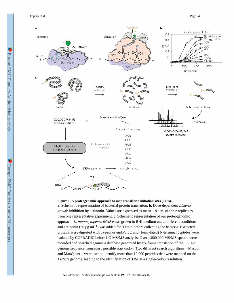
Figure 1. A proteogenomic approach to map translation initiation sites (TISs).a, Schematic representation of bacterial protein translation. b, Dose-dependent Listeria growth inhibition by actinonin. Values are expressed as mean ± s.e.m. of three replicates
from one representative experiment. c, Schematic representation of our proteogenomic
approach. L. monocytogenes EGD-e was grown in BHI medium under different conditions
and actinonin (50 μg ml−1) was added for 90 min before collecting the bacteria. Extracted
proteins were digested with trypsin or endoGluC and (formylated) N-terminal peptides were
isolated by COFRADIC before LC-MS/MS analysis. Over 1,000,000 MS/MS spectra were
recorded and searched against a database generated by six-frame translation of the EGD-e
genome sequence from every possible start codon. Two different search algorithms—Mascot
and MaxQuant—were used to identify more than 12,000 peptides that were mapped on the
Listeria genome, leading to the identification of TISs at a single-codon resolution.
Impens et al. Page 24
Nat Microbiol. Author manuscript; available in PMC 2018 February 07.
Europe PM
C Funders A
uthor Manuscripts
Europe PM
C Funders A
uthor Manuscripts
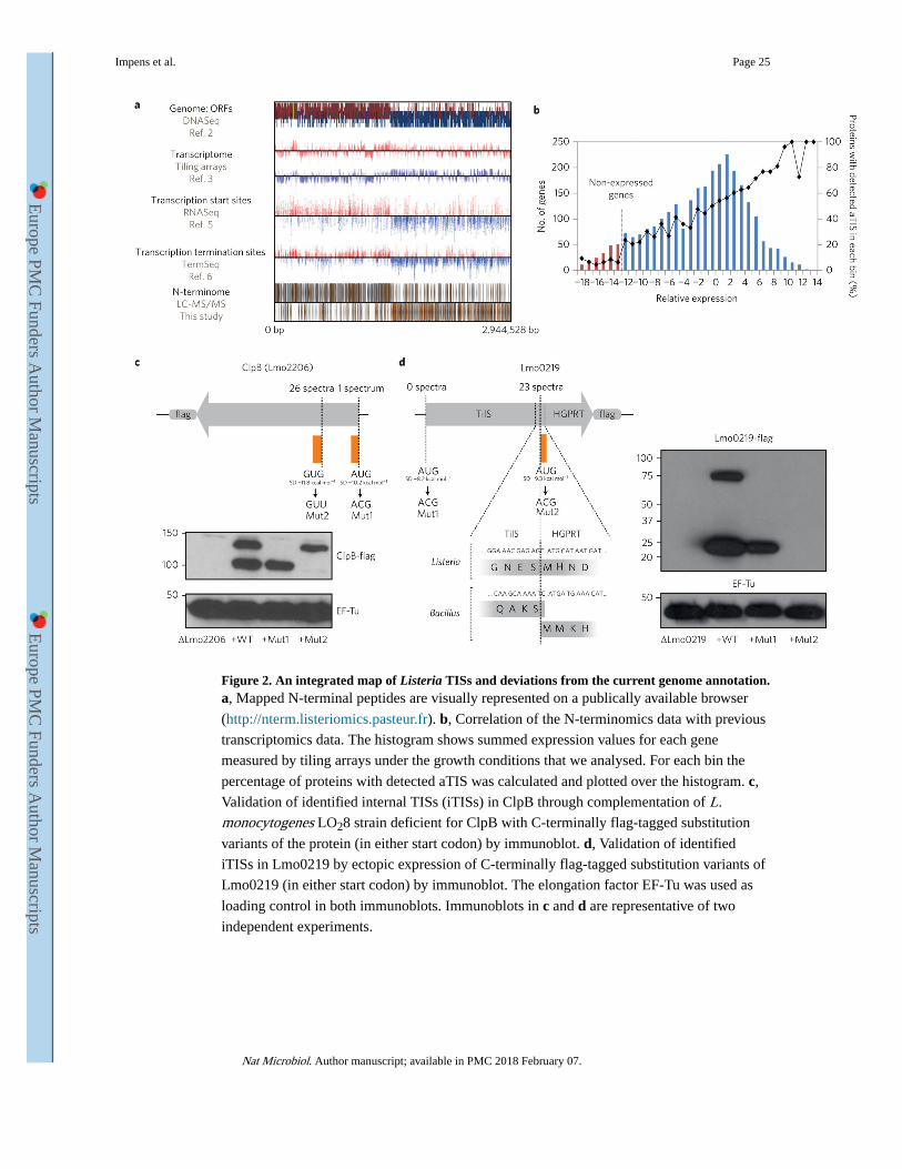
Figure 2. An integrated map of Listeria TISs and deviations from the current genome annotation.a, Mapped N-terminal peptides are visually represented on a publically available browser
(http://nterm.listeriomics.pasteur.fr). b, Correlation of the N-terminomics data with previous
transcriptomics data. The histogram shows summed expression values for each gene
measured by tiling arrays under the growth conditions that we analysed. For each bin the
percentage of proteins with detected aTIS was calculated and plotted over the histogram. c,
Validation of identified internal TISs (iTISs) in ClpB through complementation of L. monocytogenes LO28 strain deficient for ClpB with C-terminally flag-tagged substitution
variants of the protein (in either start codon) by immunoblot. d, Validation of identified
iTISs in Lmo0219 by ectopic expression of C-terminally flag-tagged substitution variants of
Lmo0219 (in either start codon) by immunoblot. The elongation factor EF-Tu was used as
loading control in both immunoblots. Immunoblots in c and d are representative of two
independent experiments.
Impens et al. Page 25
Nat Microbiol. Author manuscript; available in PMC 2018 February 07.
Europe PM
C Funders A
uthor Manuscripts
Europe PM
C Funders A
uthor Manuscripts

Figure 3. Miniprotein Prli42 is highly conserved and interacts with Listeria orthologues of the stressosome.a, Multiple sequence alignment of orthologous sequences of Prli42. Amino acids are
highlighted when conserved in more than 55% of the sequence. Stars indicate the conserved
basic residues (Lys4, Lys5 and Arg8). Colour code for amino acids is as follows: grey,
hydrophobic; blue, positively charged; red, negatively charged; orange, neutral; purple,
glycine. b, Identification of Prli42 protein interaction partners. The volcano plot shows the
intensity fold change (in log2) in the Prli42-expressing strain compared to the control
deletion strain on the x axis. The pull-down was performed in triplicate and a t-test was
Impens et al. Page 26
Nat Microbiol. Author manuscript; available in PMC 2018 February 07.
Europe PM
C Funders A
uthor Manuscripts
Europe PM
C Funders A
uthor Manuscripts
performed to calculate −log P values for each protein, indicated on the y axis. Black lines
indicate the boundary of significance as set by Perseus software (FDR = 0.05 and S0 = 1) c,
Assembly of the stressosome. The panels show elution from a Superdex-200 sizing column.
The RsbR:RsbS complex has an apparent mass of 1.5 MDa (top). The RsbR:RsbS: RsbT
complex has an apparent mass of 1.8 MDa (bottom). mAU, milli arbitrary units. d, Electron
micrographs of the RsbR:RsbS:RsbT complex, revealing a fourfold symmetry in the central
cavity of the supramolecular assembly. e, Model of the Prli42–RsbR interaction from
docking of Prli42 to the homodimeric N-terminal domain of Listeria RsbR, obtained through
homology modelling from the known N-terminal RsbR structure of B. subtilis (PDB ID:
2BNL). f, WT, K5L, K5F and R8A variants of Prli42-flag were expressed in the Prli42
deletion strain together with HA-RsbR, flag pull-down followed by immunoblotting against
the HA-tag. The stressosome was assembled at least three different times, and the elution
profiles shown are representative. The experiment in c was subsequently imaged by EM.
Immunoblots in f are representative of three independent experiments.
Impens et al. Page 27
Nat Microbiol. Author manuscript; available in PMC 2018 February 07.
Europe PM
C Funders A
uthor Manuscripts
Europe PM
C Funders A
uthor Manuscripts

Figure 4. Membrane miniprotein Prli42 partially tethers RsbR to the bacterial membrane and is essential for the sigma B signalling following oxidative stress and survival in macrophages.a, Prli42-flag and HA-RsbR were expressed in Δprli42 bacteria. Cellular fractions were
analysed by immunoblot. InlA, InlB and EF-Tu were used as controls for fractionation. b,
Localization of endogenous RsbR was analysed by immunoblot after cellular fractionation.
EF-Tu was used as a loading control. c, Quantification of RsbR levels in membrane or
cytosolic fractions relative to EF-Tu and normalized to levels in WT bacteria using ImageJ.
Values are expressed as mean ± s.e.m. of three independent experiments. d, 0.05% H2O2
Impens et al. Page 28
Nat Microbiol. Author manuscript; available in PMC 2018 February 07.
Europe PM
C Funders A
uthor Manuscripts
Europe PM
C Funders A
uthor Manuscripts

was added to exponential cultures and sigma B pathway activation was assessed by northern
blot using the lmo2230 gene and 5S as a loading control. e, Bacteria were grown in BHI
medium and 0.05% H2O2 ± iron (2.5 μg ml–1 FeC6H5O7) was added to cultures in
exponential growth. The number of living bacteria was monitored by counting c.f.u. For
each time point and for each strain, the number of c.f.u. is shown relative to the number of
c.f.u. at time zero. Results are expressed as mean ± s.e.m. of four independent experiments.
f, BMDMs were infected and cells were lysed at 24 h post-infection, and the released
bacteria were counted. WT c.f.u. average was normalized to 100 to cross-compare
experiments between macrophages from different mice. Results are expressed as mean ±
s.e.m. of five independent experiments. g, Expression of ActA and listeriolysin O (LLO)
following H2O2 treatment (as in d) was analysed by immunoblot and is representative of
three experiments. Lysates were from total bacterial pellet. Flag, RsbR and EF-Tu were used
as controls. Immunoblots and northern blot shown in a, b, d and g are representative of three
independent experiments. For all graphs, significance was determined by ordinary one-way
analysis of variance followed by Dunnett’s multiple comparison test, and significance is
shown relative to WT in each condition (***P < 0.001; *P < 0.05; NS, not significant).
Impens et al. Page 29
Nat Microbiol. Author manuscript; available in PMC 2018 February 07.
Europe PM
C Funders A
uthor Manuscripts
Europe PM
C Funders A
uthor Manuscripts