Embed Size (px)
Citation preview
Takara Bio USA, Inc.
1290 Terra Bella Avenue, Mountain View, CA 94043, USA
U.S. Technical Support: [email protected]
United States/Canada
800.662.2566
Asia Pacific
+1.650.919.7300
Europe
+33.(0)1.3904.6880
Japan
+81.(0)77.565.6999
Page 1 of 15
Takara Bio USA, Inc.
In-Fusion® HD Cloning Kit User Manual
Cat. Nos. Many
(121416)
In-Fusion® HD Cloning Kit User Manual
(121416) takarabio.com Takara Bio USA, Inc.
Page 2 of 15
Table of Contents I. Introduction ..................................................................................................................................................................... 3
II. List of Components ......................................................................................................................................................... 5
III. Additional Materials Required .................................................................................................................................... 5
IV. PCR and Experimental Preparation ............................................................................................................................ 6
A. Preparation of a Linearized Vector by Restriction Digestion ..................................................................................... 6
B. PCR Primer Design ..................................................................................................................................................... 6
C. PCR Amplification of Target Fragment ...................................................................................................................... 8
D. Control Reactions ........................................................................................................................................................ 9
V. Which Protocol Should You Follow? ............................................................................................................................. 9
VI. Protocol I: In-Fusion Cloning Procedure w/Spin-Column Purification ...................................................................... 9
A. Procedure for Spin-Column Purification of PCR Fragments ...................................................................................... 9
B. In-Fusion Cloning Procedure for Spin-Column Purified PCR Fragments ................................................................ 10
VII. Protocol II: In-Fusion Cloning Procedure w/Cloning Enhancer Treatment ............................................................. 11
A. Procedure for Treating Unpurified PCR Fragments with Cloning Enhancer ........................................................... 11
B. In-Fusion Cloning Procedure for Cloning Enhancer-Treated PCR Fragments ......................................................... 11
VIII. Transformation Procedure ......................................................................................................................................... 12
Procedure for Transformation Using Stellar Competent Cells ......................................................................................... 12
IX. Expected Results ....................................................................................................................................................... 12
X. Troubleshooting Guide ................................................................................................................................................. 13
Appendix A. Quick In-Fusion Cloning Protocol .................................................................................................................. 14
In-Fusion Cloning Procedure for a PCR-Amplified Vector & Fragment ......................................................................... 14
Appendix B. pUC19 Linearized Vector Information ............................................................................................................ 15
Table of Figures Figure 1. In-Fusion HD protocol overview. ............................................................................................................................ 4
Figure 2. Universal primer design for In-Fusion technology. ................................................................................................. 7
Figure 3. Examples of primers designed for In-Fusion cloning. ............................................................................................. 8
Figure 4. pUC19 Linearized Vector map and multiple cloning sites (MCS). ....................................................................... 15
Table of Tables Table 1. In-Fusion HD Protocol Outline ................................................................................................................................. 3
Table 2. Liquid Format ........................................................................................................................................................... 5
Table 3. Lyophilized Format ................................................................................................................................................... 5
Table 4. Recommended In-Fusion Reactions for Purified Fragments .................................................................................. 10
Table 5. Troubleshooting Guide for In-Fusion Experiments ................................................................................................ 13
In-Fusion® HD Cloning Kit User Manual
(121416) takarabio.com Takara Bio USA, Inc.
Page 3 of 15
I. Introduction In-Fusion HD Cloning Kits are designed for fast, directional cloning of one or more fragments of DNA into any
vector. The cornerstone of In-Fusion cloning technology is our proprietary In-Fusion Enzyme, which fuses DNA
fragments (e.g., PCR-generated inserts and linearized vectors) efficiently and precisely by recognizing 15-bp
overlaps at their ends. These 15-bp overlaps can be engineered by designing primers for amplification of the
desired sequences. In-Fusion HD Kits offer increased cloning efficiency over previous generations of In-Fusion
Kits, especially for long fragments, short oligonucleotides, and multiple fragments.
• Clone any insert, into any location, within any vector you choose
• Efficiently clone a broad range of fragment sizes
• Clone multiple DNA fragments simultaneously into any vector in a single reaction
• No restriction digestion, phosphatase treatment, or ligation required
• Final constructs are seamless with no extra or unwanted base pairs
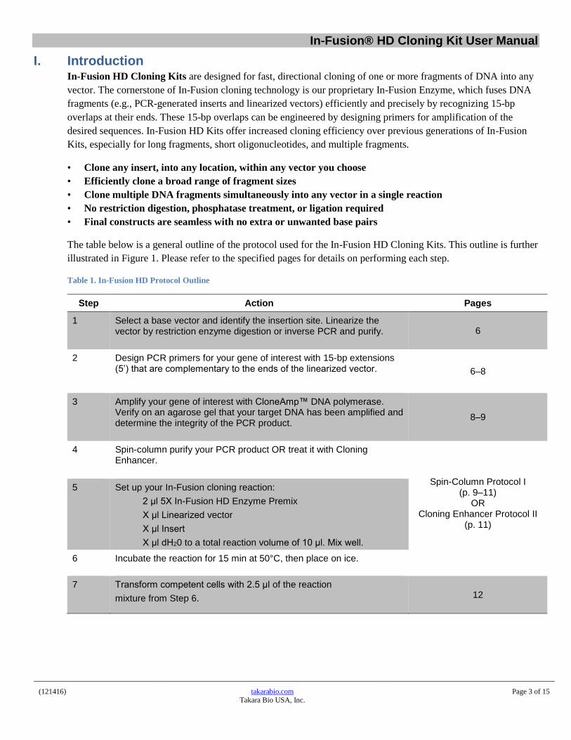
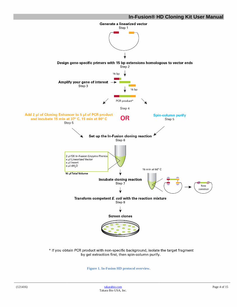
The table below is a general outline of the protocol used for the In-Fusion HD Cloning Kits. This outline is further
illustrated in Figure 1. Please refer to the specified pages for details on performing each step.
Table 1. In-Fusion HD Protocol Outline
Step Action Pages
1 Select a base vector and identify the insertion site. Linearize the vector by restriction enzyme digestion or inverse PCR and purify. 6
2 Design PCR primers for your gene of interest with 15-bp extensions (5’) that are complementary to the ends of the linearized vector. 6–8
3 Amplify your gene of interest with CloneAmp™ DNA polymerase. Verify on an agarose gel that your target DNA has been amplified and determine the integrity of the PCR product.
8–9
4 Spin-column purify your PCR product OR treat it with Cloning Enhancer.
Spin-Column Protocol I (p. 9–11)
OR Cloning Enhancer Protocol II
(p. 11)
5 Set up your In-Fusion cloning reaction:
2 μl 5X In-Fusion HD Enzyme Premix
X μl Linearized vector
X μl Insert
X μl dH20 to a total reaction volume of 10 μl. Mix well.
6 Incubate the reaction for 15 min at 50°C, then place on ice.
7 Transform competent cells with 2.5 μl of the reaction
mixture from Step 6. 12
In-Fusion® HD Cloning Kit User Manual
(121416) takarabio.com Takara Bio USA, Inc.
Page 4 of 15
Figure 1. In-Fusion HD protocol overview.
In-Fusion® HD Cloning Kit User Manual
(121416) takarabio.com Takara Bio USA, Inc.
Page 5 of 15
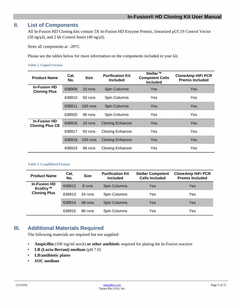
II. List of Components All In-Fusion HD Cloning kits contain 5X In-Fusion HD Enzyme Premix, linearized pUC19 Control Vector
(50 ng/μl), and 2 kb Control Insert (40 ng/μl).
Store all components at –20°C.
Please see the tables below for more information on the components included in your kit.
Table 2. Liquid Format
Product Name Cat. No.
Size Purification Kit
Included
Stellar™ Competent Cells
Included
CloneAmp HiFi PCR Premix Included
In-Fusion HD Cloning Plus
638909 10 rxns Spin Columns Yes Yes
638910 50 rxns Spin Columns Yes Yes
638911 100 rxns Spin Columns Yes Yes
638920 96 rxns Spin Columns Yes Yes
In-Fusion HD Cloning Plus CE
638916 10 rxns Cloning Enhancer Yes Yes
638917 50 rxns Cloning Enhancer Yes Yes
638918 100 rxns Cloning Enhancer Yes Yes
638919 96 rxns Cloning Enhancer Yes Yes
Table 3. Lyophilized Format
Product Name Cat. No.
Size Purification Kit
Included Stellar Competent
Cells Included CloneAmp HiFi PCR
Premix Included
In-Fusion HD EcoDry™
Cloning Plus
638912 8 rxns Spin Columns Yes Yes
638913 24 rxns Spin Columns Yes Yes
638914 48 rxns Spin Columns Yes Yes
638915 96 rxns Spin Columns Yes Yes
III. Additional Materials Required The following materials are required but not supplied:
• Ampicillin (100 mg/ml stock) or other antibiotic required for plating the In-Fusion reaction
• LB (Luria-Bertani) medium (pH 7.0)
• LB/antibiotic plates
• SOC medium
In-Fusion® HD Cloning Kit User Manual
(121416) takarabio.com Takara Bio USA, Inc.
Page 6 of 15
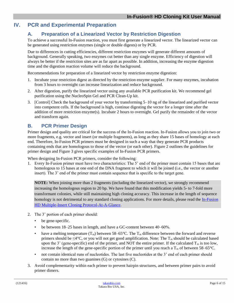
IV. PCR and Experimental Preparation
A. Preparation of a Linearized Vector by Restriction Digestion To achieve a successful In-Fusion reaction, you must first generate a linearized vector. The linearized vector can
be generated using restriction enzymes (single or double digests) or by PCR.
Due to differences in cutting efficiencies, different restriction enzymes will generate different amounts of
background. Generally speaking, two enzymes cut better than any single enzyme. Efficiency of digestion will
always be better if the restriction sites are as far apart as possible. In addition, increasing the enzyme digestion
time and the digestion reaction volume will reduce the background.
Recommendations for preparation of a linearized vector by restriction enzyme digestion:
1. Incubate your restriction digest as directed by the restriction enzyme supplier. For many enzymes, incubation
from 3 hours to overnight can increase linearization and reduce background.
2. After digestion, purify the linearized vector using any available PCR purification kit. We recommend gel
purification using the NucleoSpin Gel and PCR Clean-Up kit.
3. [Control] Check the background of your vector by transforming 5–10 ng of the linearized and purified vector
into competent cells. If the background is high, continue digesting the vector for a longer time after the
addition of more restriction enzyme(s). Incubate 2 hours to overnight. Gel purify the remainder of the vector
and transform again.
B. PCR Primer Design Primer design and quality are critical for the success of the In-Fusion reaction. In-Fusion allows you to join two or
more fragments, e.g. vector and insert (or multiple fragments), as long as they share 15 bases of homology at each
end. Therefore, In-Fusion PCR primers must be designed in such a way that they generate PCR products
containing ends that are homologous to those of the vector (or each other). Figure 2 outlines the guidelines for
primer design and Figure 3 gives specific examples of In-Fusion PCR primers.
When designing In-Fusion PCR primers, consider the following:
1. Every In-Fusion primer must have two characteristics: The 5’ end of the primer must contain 15 bases that are
homologous to 15 bases at one end of the DNA fragment to which it will be joined (i.e., the vector or another
insert). The 3’ end of the primer must contain sequence that is specific to the target gene.
NOTE: When joining more than 2 fragments (including the linearized vector), we strongly recommend
increasing the homologous region to 20 bp. We have found that this modification yields 5- to 7-fold more
transformant colonies, while still maintaining high cloning accuracy. This increase in the length of sequence
homology is not detrimental to any standard cloning applications. For more details, please read the In-Fusion
HD Multiple-Insert Cloning Protocol-At-A-Glance.
2. The 3’ portion of each primer should:
• be gene-specific.
• be between 18–25 bases in length, and have a GC-content between 40–60%.
• have a melting temperature (Tm) between 58–65°C. The Tm difference between the forward and reverse
primers should be ≤4°C, or you will not get good amplification. Note: The Tm should be calculated based
upon the 3’ (gene-specific) end of the primer, and NOT the entire primer. If the calculated Tm is too low,
increase the length of the gene-specific portion of the primer until you reach a Tm of between 58–65°C.
• not contain identical runs of nucleotides. The last five nucleotides at the 3’ end of each primer should
contain no more than two guanines (G) or cytosines (C).
3. Avoid complementarity within each primer to prevent hairpin structures, and between primer pairs to avoid
primer dimers.
In-Fusion® HD Cloning Kit User Manual
(121416) takarabio.com Takara Bio USA, Inc.
Page 7 of 15
4. You can perform a BLAST search to determine if the 3’ portion of each primer is unique and specific (at
www.ncbi.nlm.nih.gov/BLAST/).
5. We provide an online tool that simplifies In-Fusion PCR primer design for standard cloning reactions. Simply
provide your vector sequence, the restriction enzyme(s) used to linearize the vector (if that is the chosen
method for linearization), and the primer sequence required to amplify your region of interest. (Go to
http://www.takarabio.com/US/Products/Cloning_and_Competent_Cells/Selection_Guides/Online_In-
Fusion_Tools)
6. We generally use desalted oligonucleotide primers in PCR reactions. However, primer quality can depend on
the vendor and varies from lot to lot. If your primer quality is particularly poor (i.e., has many premature
termination products), or your primers are longer than 45 nucleotides, they may need to be PAGE purified;
however, we usually find this is unnecessary.
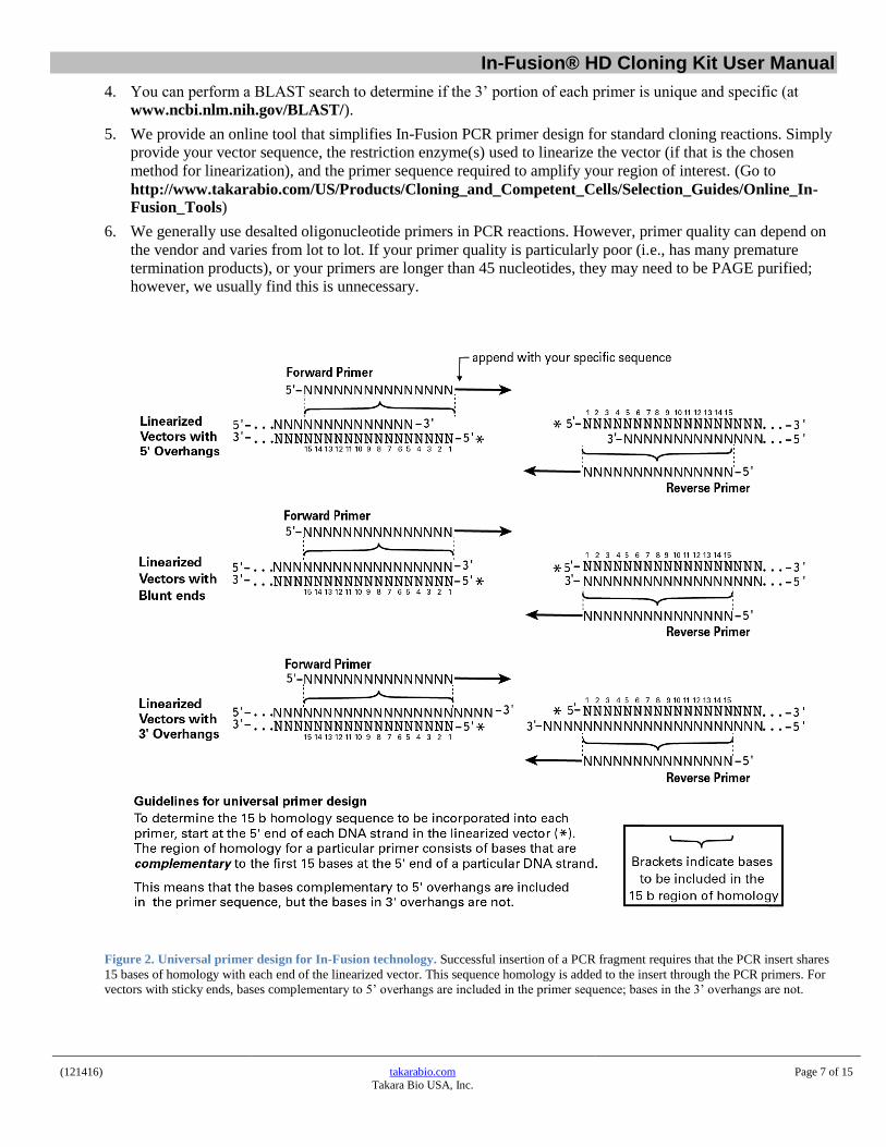
Figure 2. Universal primer design for In-Fusion technology. Successful insertion of a PCR fragment requires that the PCR insert shares
15 bases of homology with each end of the linearized vector. This sequence homology is added to the insert through the PCR primers. For
vectors with sticky ends, bases complementary to 5’ overhangs are included in the primer sequence; bases in the 3’ overhangs are not.
In-Fusion® HD Cloning Kit User Manual
(121416) takarabio.com Takara Bio USA, Inc.
Page 8 of 15
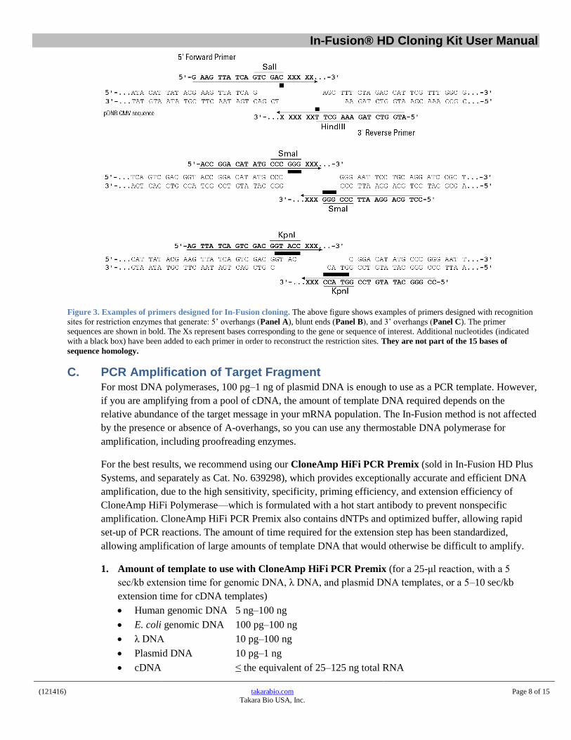
Figure 3. Examples of primers designed for In-Fusion cloning. The above figure shows examples of primers designed with recognition
sites for restriction enzymes that generate: 5’ overhangs (Panel A), blunt ends (Panel B), and 3’ overhangs (Panel C). The primer
sequences are shown in bold. The Xs represent bases corresponding to the gene or sequence of interest. Additional nucleotides (indicated
with a black box) have been added to each primer in order to reconstruct the restriction sites. They are not part of the 15 bases of
sequence homology.
C. PCR Amplification of Target Fragment For most DNA polymerases, 100 pg–1 ng of plasmid DNA is enough to use as a PCR template. However,
if you are amplifying from a pool of cDNA, the amount of template DNA required depends on the
relative abundance of the target message in your mRNA population. The In-Fusion method is not affected
by the presence or absence of A-overhangs, so you can use any thermostable DNA polymerase for
amplification, including proofreading enzymes.
For the best results, we recommend using our CloneAmp HiFi PCR Premix (sold in In-Fusion HD Plus
Systems, and separately as Cat. No. 639298), which provides exceptionally accurate and efficient DNA
amplification, due to the high sensitivity, specificity, priming efficiency, and extension efficiency of
CloneAmp HiFi Polymerase—which is formulated with a hot start antibody to prevent nonspecific
amplification. CloneAmp HiFi PCR Premix also contains dNTPs and optimized buffer, allowing rapid
set-up of PCR reactions. The amount of time required for the extension step has been standardized,
allowing amplification of large amounts of template DNA that would otherwise be difficult to amplify.
1. Amount of template to use with CloneAmp HiFi PCR Premix (for a 25-μl reaction, with a 5
sec/kb extension time for genomic DNA, λ DNA, and plasmid DNA templates, or a 5–10 sec/kb
extension time for cDNA templates)
Human genomic DNA 5 ng–100 ng
E. coli genomic DNA 100 pg–100 ng
λ DNA 10 pg–100 ng
Plasmid DNA 10 pg–1 ng
cDNA ≤ the equivalent of 25–125 ng total RNA
In-Fusion® HD Cloning Kit User Manual
(121416) takarabio.com Takara Bio USA, Inc.
Page 9 of 15
2. PCR product sizes that can be obtained with CloneAmp HiFi PCR Premix (with a 5 sec/kb
extension time for genomic DNA and λ DNA templates, or a 5–10 sec/kb extension time for cDNA
templates)
Human genomic DNA up to 6 kb
E. coli genomic DNA up to 10 kb
cDNA up to 6 kb
λ DNA up to 15 kb
3. Analysis of PCR products: When PCR cycling is complete, use an agarose gel to confirm that you
have obtained a single DNA fragment and to estimate the concentration of your PCR product.
Quantify the amount of DNA by measuring against a known standard or DNA mass ladder run on the
same gel.
D. Control Reactions When using the In-Fusion kit for the first time, we strongly recommend that you perform the positive and
negative control reactions in parallel with your In-Fusion cloning reaction. The positive control should
consist of a circular vector of known concentration (competent cells should give >2 x 10⁸ cfu/μg), and the
negative control should consist of a known amount of your linearized vector (see Section IX for Expected
Results). Performing the control reactions will verify that the system is working properly. The 2-kb
Control Insert included in the In-Fusion HD Cloning Kits has already been purified, so there is no need
for further treatment prior to the cloning reaction.
V. Which Protocol Should You Follow? Following PCR, verify by agarose gel electrophoresis that your target fragment has been amplified. If a single
band of the desired size is obtained, you can EITHER spin-column purify (follow Protocol I), OR treat your
PCR product with Cloning Enhancer (follow Protocol II). However, if non-specific background or multiple
bands are visible on your gel, isolate your target fragment by gel extraction, then spin-column purify (follow
Protocol I). If you use PCR to amplify your vector and insert and you obtain both a PCR-amplified vector AND
PCR-amplified fragment(s) without non-specific background, you can use the Quick In-Fusion Cloning Protocol
provided in Appendix A.
VI. Protocol I: In-Fusion Cloning Procedure w/Spin-Column Purification
A. Procedure for Spin-Column Purification of PCR Fragments 1. If non-specific background bands are observed on an agarose gel, isolate your target fragment by gel
extraction, then spin-column purify.
2. Spin-column purify your PCR product (e.g., insert) by using a silica-based purification system, such as
the NucleoSpin Gel and PCR Clean-Up kit. During purification, avoid nuclease contamination and
exposure of the DNA to UV light for long periods of time.
3. After purification, proceed with the In-Fusion Cloning Procedure for Spin Column-Purified PCR
Fragments (Section VI.B).
In-Fusion® HD Cloning Kit User Manual
(121416) takarabio.com Takara Bio USA, Inc.
Page 10 of 15
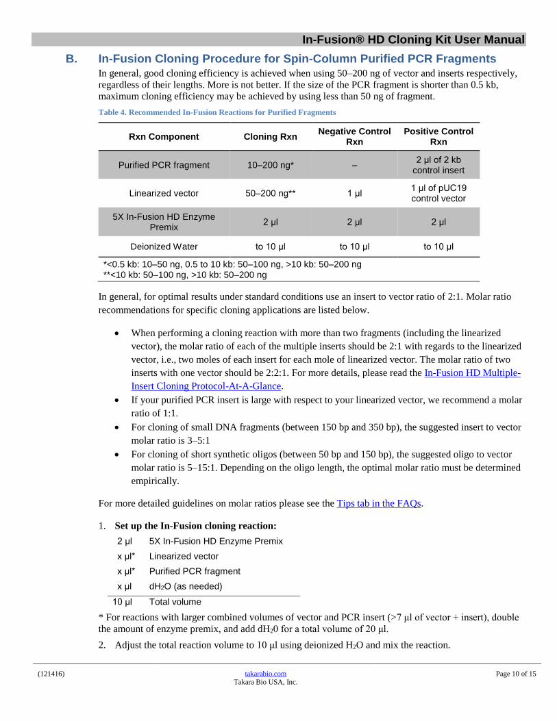
B. In-Fusion Cloning Procedure for Spin-Column Purified PCR Fragments In general, good cloning efficiency is achieved when using 50–200 ng of vector and inserts respectively,
regardless of their lengths. More is not better. If the size of the PCR fragment is shorter than 0.5 kb,
maximum cloning efficiency may be achieved by using less than 50 ng of fragment.
Table 4. Recommended In-Fusion Reactions for Purified Fragments
Rxn Component Cloning Rxn Negative Control
Rxn Positive Control
Rxn
Purified PCR fragment 10–200 ng* – 2 μl of 2 kb
control insert
Linearized vector 50–200 ng** 1 μl 1 μl of pUC19 control vector
5X In-Fusion HD Enzyme Premix
2 μl 2 μl 2 μl
Deionized Water to 10 μl to 10 μl to 10 μl
*<0.5 kb: 10–50 ng, 0.5 to 10 kb: 50–100 ng, >10 kb: 50–200 ng **<10 kb: 50–100 ng, >10 kb: 50–200 ng
In general, for optimal results under standard conditions use an insert to vector ratio of 2:1. Molar ratio
recommendations for specific cloning applications are listed below.
When performing a cloning reaction with more than two fragments (including the linearized
vector), the molar ratio of each of the multiple inserts should be 2:1 with regards to the linearized
vector, i.e., two moles of each insert for each mole of linearized vector. The molar ratio of two
inserts with one vector should be 2:2:1. For more details, please read the In-Fusion HD Multiple-
Insert Cloning Protocol-At-A-Glance.
If your purified PCR insert is large with respect to your linearized vector, we recommend a molar
ratio of 1:1.
For cloning of small DNA fragments (between 150 bp and 350 bp), the suggested insert to vector
molar ratio is 3–5:1
For cloning of short synthetic oligos (between 50 bp and 150 bp), the suggested oligo to vector
molar ratio is 5–15:1. Depending on the oligo length, the optimal molar ratio must be determined
empirically.
For more detailed guidelines on molar ratios please see the Tips tab in the FAQs.
1. Set up the In-Fusion cloning reaction:
2 μl 5X In-Fusion HD Enzyme Premix
x μl* Linearized vector
x μl* Purified PCR fragment
x μl dH2O (as needed)
10 μl Total volume
* For reactions with larger combined volumes of vector and PCR insert (>7 μl of vector + insert), double
the amount of enzyme premix, and add dH20 for a total volume of 20 μl.
2. Adjust the total reaction volume to 10 μl using deionized H2O and mix the reaction.
In-Fusion® HD Cloning Kit User Manual
(121416) takarabio.com Takara Bio USA, Inc.
Page 11 of 15
3. Incubate the reaction for 15 min at 50 °C, then place on ice.
NOTE: The In-Fusion reaction is completed within the required 15-min incubation. Longer
incubation times do NOT increase cloning efficiency, even with multiple-insert cloning reactions.
4. Continue to the Transformation Procedure (Section VIII). You can store the cloning reactions at
–20°C until you are ready.
VII. Protocol II: In-Fusion Cloning Procedure w/Cloning Enhancer Treatment
A. Procedure for Treating Unpurified PCR Fragments with Cloning Enhancer
IMPORTANT: DO NOT treat purified PCR products with the Cloning Enhancer.
Before setting up the In-Fusion cloning reaction, treat unpurified PCR products (e.g. fragments) as follows:
1. Add 2 μl of Cloning Enhancer to 5 μl of the PCR reaction.
2. Incubate at 37°C for 15 minutes, then at 80°C for 15 minutes in a PCR thermal cycler. If you used
more than 100 ng of DNA as a template in the PCR reaction, extend the 37°C incubation step to 20
minutes. If you are using a water bath or heat block rather than a thermal cycler, extend each of the
incubation steps to 20–25 minutes.
3. Proceed with the In-Fusion Cloning Procedure for Cloning Enhancer-Treated PCR Fragments
(Section VII.B). If you cannot proceed immediately, store treated PCR reactions at –20°C until you
are ready.
B. In-Fusion Cloning Procedure for Cloning Enhancer-Treated PCR Fragments
NOTE: If you use PCR to amplify your vector and insert and you obtain both a PCR-amplified
vector and PCR-amplified fragment without non-specific background you may use the Quick In-
Fusion Cloning Protocol provided in Appendix A.
1. Set up the In-Fusion cloning reaction:
2 μl 5X In-Fusion HD Enzyme Premix
x μl* Linearized vector
x μl** Purified PCR fragment
x μl dH2O (as needed)
10 μl Total volume
* Use 50–200 ng of linearized vector.
** Use 1–2 μl of Cloning Enhancer-treated fragments, regardless of their length. The total volume of
Cloning Enhancer-treated PCR fragments should be up to 4 μl per 10-μl reaction. If you obtain a low
product yield from your PCR reaction, we recommend purification of PCR fragments instead of Cloning
Enhancer treatment.
2. Adjust the total reaction volume to 10 μl using deionized H2O and mix the reaction.
3. Incubate the reaction for 15 min at 50°C, then place on ice.
NOTE: The In-Fusion reaction is completed within the required 15-min incubation. Longer
incubation times do NOT increase cloning efficiency, even with multiple-insert cloning reactions.
4. Continue to the Transformation Procedure (Section VIII). If you cannot transform cells immediately,
store the cloning reactions at –20°C until you are ready.
In-Fusion® HD Cloning Kit User Manual
(121416) takarabio.com Takara Bio USA, Inc.
Page 12 of 15
VIII. Transformation Procedure
Procedure for Transformation Using Stellar Competent Cells The following protocol has been optimized for transformation using Stellar Competent Cells, sold in In-Fusion HD
Plus kits and separately in several formats. If you are not using Stellar Competent Cells, follow the transformation
protocol provided with your cells, but you may need to dilute the In-Fusion reaction mixture prior to transformation
to increase cloning efficiency (See Table 5, Troubleshooting Guide). We strongly recommend the use of competent
cells with a transformation efficiency ≥1 x 10⁸ cfu/ug.
For complete information on the handling of Stellar Competent Cells, please see the Protocol.
1. Thaw Stellar Competent Cells on ice just before use. After thawing, mix gently to ensure even distribution,
and then move 50 µl of competent cells into a 14-ml round-bottom tube (Falcon tube). Do not vortex.
2. Add 2.5 µl of the In-Fusion reaction mixture to the competent cells.
IMPORTANT: DO NOT add more than 5 μl of the reaction to 50 μl of competent cells. MORE IS NOT
BETTER. Using too much of the reaction mixture inhibits the transformation.
3. Place the tubes on ice for 30 min.
4. Heat shock the cells for exactly 45 sec at 42°C.
5. Place tubes on ice for 1–2 min.
6. Add SOC medium to bring the final volume to 500 µl. SOC medium should be warmed to 37°C before using.
7. Incubate by shaking (160–225 rpm) for 1 hr at 37°C.
8. Place 1/100–1/5 of each transformation reaction into separate tubes and bring the volume to 100 μl with SOC
medium. Spread each diluted transformation reaction on a separate LB plate containing an antibiotic
appropriate for the cloning vector (i.e., the control vector included with the kit requires 100 μg/ml of
ampicillin).
NOTE: For cloning reactions with more than two fragments, we recommend plating a larger volume (1/5–1/3
of each transformation reaction).
9. Centrifuge the remainder of each transformation reaction at 6,000 rpm for 5 min. Discard the supernatant and
resuspend each pellet in 100 μl fresh SOC medium. Spread each sample on a separate LB plate containing the
appropriate antibiotic. Incubate all of the plates overnight at 37°C.
10. The next day, pick individual isolated colonies from each experimental plate. Isolate plasmid DNA using a
standard method of your choice (e.g. miniprep). To determine the presence of an insert, analyze the DNA by
restriction digestion or PCR screening.
IX. Expected Results The positive control plates typically develop several hundred white colonies when using cells with a minimum
transformation efficiency of 1 x 10⁸ cfu/μg. The negative control plates should have few colonies.
The number of colonies on your experimental plates will depend on the amount and purity of the PCR product
and linearized vector used for the In-Fusion cloning reaction.
The presence of a low number of colonies on both plates—typically, a few dozen colonies—
is indicative of either transformation with too much of the reaction, or poor DNA/primer quality.
The presence of many (hundreds) of colonies on the negative control is indicative of incomplete vector
linearization.
In-Fusion® HD Cloning Kit User Manual
(121416) takarabio.com Takara Bio USA, Inc.
Page 13 of 15
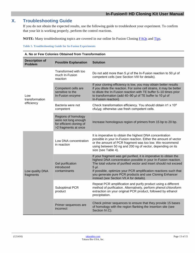
X. Troubleshooting Guide If you do not obtain the expected results, use the following guide to troubleshoot your experiment. To confirm
that your kit is working properly, perform the control reactions.
NOTE: Many troubleshooting topics are covered in our online In-Fusion Cloning FAQs and Tips.
Table 5. Troubleshooting Guide for In-Fusion Experiments
A. No or Few Colonies Obtained from Transformation
Description of Problem
Possible Explanation Solution
Low transformation efficiency
Transformed with too much In-Fusion reaction
Do not add more than 5 μl of the In-Fusion reaction to 50 μl of competent cells (see Section VIII for details).
Competent cells are sensitive to the In-Fusion enzyme
If your cloning efficiency is low, you may obtain better results if you dilute the reaction. For some cell strains, it may be better to dilute the In-Fusion reaction with TE buffer 5–10 times prior to transformation (add 40–90 μl of TE buffer to 10 μl of In-Fusion reaction).
Bacteria were not competent
Check transformation efficiency. You should obtain ≥1 x 10⁸ cfu/μg; otherwise use fresh competent cells.
Regions of homology were not long enough for efficient cloning of >2 fragments at once
Increase homologous region of primers from 15 bp to 20 bp.
Low quality DNA fragments
Low DNA concentration in reaction
It is imperative to obtain the highest DNA concentration possible in your In-Fusion reaction. Either the amount of vector or the amount of PCR fragment was too low. We recommend using between 50 ng and 200 ng of vector, depending on its size (see Table 4).
Gel purification introduced contaminants
If your fragment was gel purified, it is imperative to obtain the highest DNA concentration possible in your In-Fusion reaction. The total volume of purified vector and insert should not exceed 5 μl. If possible, optimize your PCR amplification reactions such that you generate pure PCR products and use Cloning Enhancer instead (see Section VII.A for details).
Suboptimal PCR product
Repeat PCR amplification and purify product using a different method of purification. Alternatively, perform phenol:chloroform extraction on your original PCR product, followed by ethanol precipitation.
Primer sequences are incorrect
Check primer sequences to ensure that they provide 15 bases of homology with the region flanking the insertion site (see Section IV.C).
In-Fusion® HD Cloning Kit User Manual
(121416) takarabio.com Takara Bio USA, Inc.
Page 14 of 15
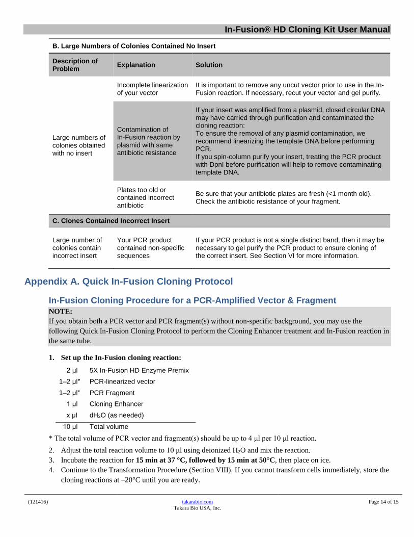
B. Large Numbers of Colonies Contained No Insert
Description of Problem
Explanation Solution
Large numbers of colonies obtained with no insert
Incomplete linearization of your vector
It is important to remove any uncut vector prior to use in the In-Fusion reaction. If necessary, recut your vector and gel purify.
Contamination of In-Fusion reaction by plasmid with same antibiotic resistance
If your insert was amplified from a plasmid, closed circular DNA may have carried through purification and contaminated the cloning reaction: To ensure the removal of any plasmid contamination, we recommend linearizing the template DNA before performing PCR. If you spin-column purify your insert, treating the PCR product with DpnI before purification will help to remove contaminating template DNA.
Plates too old or contained incorrect antibiotic
Be sure that your antibiotic plates are fresh (<1 month old). Check the antibiotic resistance of your fragment.
C. Clones Contained Incorrect Insert
Large number of colonies contain incorrect insert
Your PCR product contained non-specific sequences
If your PCR product is not a single distinct band, then it may be necessary to gel purify the PCR product to ensure cloning of the correct insert. See Section VI for more information.
Appendix A. Quick In-Fusion Cloning Protocol
In-Fusion Cloning Procedure for a PCR-Amplified Vector & Fragment
NOTE:
If you obtain both a PCR vector and PCR fragment(s) without non-specific background, you may use the
following Quick In-Fusion Cloning Protocol to perform the Cloning Enhancer treatment and In-Fusion reaction in
the same tube.
1. Set up the In-Fusion cloning reaction:
2 μl 5X In-Fusion HD Enzyme Premix
1–2 μl* PCR-linearized vector
1–2 μl* PCR Fragment
1 μl Cloning Enhancer
x μl dH2O (as needed)
10 μl Total volume
* The total volume of PCR vector and fragment(s) should be up to 4 μl per 10 μl reaction.
2. Adjust the total reaction volume to 10 μl using deionized H2O and mix the reaction.
3. Incubate the reaction for 15 min at 37 °C, followed by 15 min at 50°C, then place on ice.
4. Continue to the Transformation Procedure (Section VIII). If you cannot transform cells immediately, store the
cloning reactions at –20°C until you are ready.
In-Fusion® HD Cloning Kit User Manual
(121416) takarabio.com Takara Bio USA, Inc.
Page 15 of 15
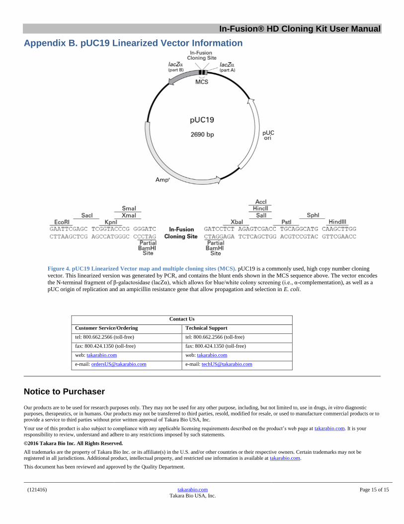
Appendix B. pUC19 Linearized Vector Information
Figure 4. pUC19 Linearized Vector map and multiple cloning sites (MCS). pUC19 is a commonly used, high copy number cloning
vector. This linearized version was generated by PCR, and contains the blunt ends shown in the MCS sequence above. The vector encodes
the N-terminal fragment of β-galactosidase (lacZα), which allows for blue/white colony screening (i.e., α-complementation), as well as a
pUC origin of replication and an ampicillin resistance gene that allow propagation and selection in E. coli.
Contact Us
Customer Service/Ordering Technical Support
tel: 800.662.2566 (toll-free) tel: 800.662.2566 (toll-free)
fax: 800.424.1350 (toll-free) fax: 800.424.1350 (toll-free)
web: takarabio.com web: takarabio.com
e-mail: [email protected] e-mail: [email protected]
Notice to Purchaser
Our products are to be used for research purposes only. They may not be used for any other purpose, including, but not limited to, use in drugs, in vitro diagnostic purposes, therapeutics, or in humans. Our products may not be transferred to third parties, resold, modified for resale, or used to manufacture commercial products or to
provide a service to third parties without prior written approval of Takara Bio USA, Inc.
Your use of this product is also subject to compliance with any applicable licensing requirements described on the product’s web page at takarabio.com. It is your
responsibility to review, understand and adhere to any restrictions imposed by such statements.
©2016 Takara Bio Inc. All Rights Reserved.
All trademarks are the property of Takara Bio Inc. or its affiliate(s) in the U.S. and/or other countries or their respective owners. Certain trademarks may not be registered in all jurisdictions. Additional product, intellectual property, and restricted use information is available at takarabio.com.
This document has been reviewed and approved by the Quality Department.


![kuKeywords: USERTM cloning, Cloning of synthesized nonclonal DNA fragments, Fusion of DNA - fragments, Uracil excision based cloning, uNCDFs, Geneart [Background] For synthesized DNA,](https://img.dokumen.tips/doc/110x75/5fd4e2ff1db7b3255b1a15b8/ku-keywords-usertm-cloning-cloning-of-synthesized-nonclonal-dna-fragments-fusion.jpg)


![Rapid Construction of Stable Infectious Full-Length cDNA ...€¦ · commercial kits, such as the In-Fusion® HD Cloning kit (Clontech) [38,39], GeneArt Seamless Cloning and Assembly](https://img.dokumen.tips/doc/110x75/5fd4e256255e8c2ddc28e4e9/rapid-construction-of-stable-infectious-full-length-cdna-commercial-kits-such.jpg)